Page 1 of 66 An Evidence Based Occupational Therapy Toolkit for Assessment and Treatment of the Upper Extremity Post Stroke Brenda Semenko, Leyda Thalman, Emily Ewert, Renee Delorme, Suzanne Hui, Heather Flett, Nicole Lavoie (Winnipeg Health Region Occupational Therapy Upper Extremity Working Group) ([email protected]) April 2015 Updated February 2017

Welcome message from author
This document is posted to help you gain knowledge. Please leave a comment to let me know what you think about it! Share it to your friends and learn new things together.
Transcript

Page 1 of 66
An Evidence Based Occupational Therapy Toolkit
for Assessment and Treatment of the Upper Extremity Post Stroke
Brenda Semenko, Leyda Thalman,
Emily Ewert, Renee Delorme, Suzanne Hui, Heather Flett, Nicole Lavoie
(Winnipeg Health Region Occupational Therapy Upper Extremity Working Group)
April 2015
Updated February 2017

Page 2 of 66
Table of Contents:
Section
Number Section Name
Page
Number
1.0 Acknowledgements 4
2.0 Introduction 5
3.0 A Model for Upper Extremity Assessment and Treatment Post Stroke 6
4.0
4.1
Screening Guidelines
Screening Questions
7
8
5.0 Determining Upper Extremity Level Guidelines 9
6.0
6.1
6.1.1
6.1.2
6.1.3
6.1.4
6.1.5
6.1.6
6.1.7
6.1.8
Assessment Guidelines
Assessment Matrix
Motor Function
Coordination
Strength
Range of Motion
Tone
Pain
Sensation
Edema
10
11
12
12
12
13
13
13
14
14
7.0 Goal Setting Guidelines 15
8.0
8.1
8.1.1
8.1.2
8.1.3
8.1.4
8.1.5
8.1.6
8.1.6a
8.1.6b
8.1.6c
8.1.6d
8.1.7
Treatment Guidelines
Treatment Matrix
Task Specific Training Guidelines
Arm Activity List A
Arm Activity List B
Homework A
Homework B
Homework C
Treatment Contract
Constraint Induced Movement Therapy
Functional Dynamic Orthoses
Functional Electrical Stimulation
Mental Imagery
Mental Imagery Sample Script
Joint Protection and Supports
Positioning and Supporting the Arm in Lying and in Sitting
Bed & Chair Positioning Following a Stroke – Right
Bed & Chair Positioning Following a Stroke – Left
Positioning and Supporting the Arm during Transfers and Mobility
Sling Me?
Positioning Devices
Positioning and Supporting the Hand
Splint Instructions
Shoulder Girdle Taping
Spasticity Management
16
17
20
21
22
23
24
25
26
27
28
29
30
31
32
32
33
34
35
36
37
38
40
41
42

Page 3 of 66
Section
Number Section Name
Page
Number
8.1.8
8.1.9
8.1.10
8.1.11
8.1.12
8.1.13
Supplementary Training Programs
Mirror Therapy
Mirror Therapy Sample Script
Sensory Stimulation and Re-training
Sensory Re-training Practical Examples
Safety Tips for Decreased Sensation
Range of Motion and Strength Training
Self-Range of Motion Exercises for the Arm
Edema Management
Virtual Reality
43
44
45
46
47
48
49
50
61
62
9.0 Reassessment Guidelines 63
10.0 References 64

Page 4 of 66
1.0 Acknowledgements:
The Winnipeg Health Region Occupational Therapy Upper Extremity Working Group would like to
acknowledge and thank the following individuals for their contributions to this document:
Daniel Doerksen
Denali Enns
Laura Foth
Glen Gray
Sherie Gray
Danielle Harling
Shayna Hjartarson
Michelle Horkoff
Sue Lotocki
Mona Maida
Linda Merry Lambert
Sharon Mohr
Cristabel Nett
Louise Nichol
Teresa Ouellette
Meghan Scarff
Kristel Smith
Marlene Stern
Ted Stevenson
Kaleigh Sullivan
Laura Wisener

Page 5 of 66
2.0 Introduction:
Stroke is a common neurological medical condition. Every year 62,000 Canadians experience a stroke
or transient ischemic attack (Hebert et al., 2016) and 405,000 Canadians live with the effects of stroke,
with that number projected to increase to between 654,000 and 726,000 by 2038 (Krueger et al., 2015).
Stroke impacts an individual’s ability to participate in former activities and life roles. Occupational
therapists provide assessment and treatment to increase independence in self-care, productivity, and
leisure activities, and frequently work with clients recovering from stroke. The literature on stroke
rehabilitation is continually evolving; therefore, occupational therapists must be knowledgeable about
evidence based practice and apply it within their practice settings.
The Canadian Stroke Best Practice Recommendations (Stroke Rehabilitation Module) were updated in
2015 and published in the International Journal of Stroke in April 2016. The Recommendations were
developed to guide rehabilitation in an effort to “increase clinician knowledge, streamline care, reduce
practice variations, optimize efficiency and ultimately improve patient outcomes after stroke” (Hebert et
al., 2016, p. 3). The upper extremity sections of the Recommendations are of significant value to
occupational therapists who frequently work with clients to maximize upper extremity function post
stroke. Occupational therapists have noted variations in upper extremity rehabilitation practice between
sites and programs in Winnipeg, Manitoba, and have identified the need for increased knowledge to
improve the consistency of practice across the stroke rehabilitation continuum of care.
A working group was created in an attempt to consistently implement the upper extremity sections of the
Canadian Stroke Best Practice Recommendations into daily clinical practice. A group of occupational
therapists from the Winnipeg Health Region collaborated to create a practical Toolkit for occupational
therapists working in acute, rehabilitation, outpatient, and community settings. Although this Toolkit
was developed specifically for occupational therapists, it is hoped that it will also be of benefit to
physiotherapists, rehabilitation assistants, and other healthcare professionals working on upper extremity
recovery post stroke. Several occupational therapists and physiotherapists provided feedback
throughout various stages of the Toolkit development.
The Toolkit includes: a model for upper extremity management, a list of upper extremity assessment
considerations and tools, and a list of specific upper extremity treatments, including practical resources.
The Toolkit was informed by the 2013 Canadian Stroke Best Practice Recommendations and the 2013
Evidence Based Review of Stroke Rehabilitation, as well as expertise from Winnipeg occupational
therapists across practice settings. The Toolkit was updated after the release of the 2015 Canadian
Stroke Best Practice Recommendations (Stroke Rehabilitation Module) and the 2016 update of the
Evidence Based Review of Stroke Rehabilitation. The purpose of this Toolkit is to improve the
consistency of implementing best practice management of the upper extremity following stroke. It
provides information to assist occupational therapists with clinical decision making as they assess, treat
and educate clients recovering from stroke. The affected upper extremity has been categorized into low,
intermediate or high levels to guide occupational therapists with selecting appropriate assessment tools
and treatments. Occupational therapists still need to consider their client’s physical status, cognition,
perception, affect, and motivation, as well as their physical and social environments when implementing
the resources in this Toolkit.
The evidence for upper extremity rehabilitation post stroke continues to emerge. It is critical that
occupational therapists are knowledgeable about the most recent evidence as well as the
recommendations and resources available to promote optimal upper extremity function throughout the
stroke rehabilitation continuum of care.
Page 6 of 66
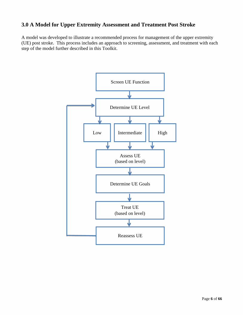
3.0 A Model for Upper Extremity Assessment and Treatment Post Stroke
A model was developed to illustrate a recommended process for management of the upper extremity
(UE) post stroke. This process includes an approach to screening, assessment, and treatment with each
step of the model further described in this Toolkit.
Intermediate
Assess UE
(based on level)
Determine UE Goals
Treat UE
(based on level)
Reassess UE
Screen UE Function
Determine UE Level
Low High

Page 7 of 66
4.0 Screening Guidelines:
The Canadian Stroke Best Practice Recommendations 1.ii states: “Initial screening and assessment
should be commenced within 48 h of admission by rehabilitation professionals in direct contact with the
patient (Evidence Level C)” (Hebert et al., 2016, p. 5).
An initial screen of upper extremity function is crucial at all points of the rehabilitation continuum of
care. The screen will determine further assessments required, assist with goal setting, and assist with the
choice of specific upper extremity treatments to best promote recovery and prevent complications (e.g.
pain, contractures, and edema). The following page is an example of some initial screening questions.
Questions should be modified based on the individual client’s presentation.
Page 8 of 66
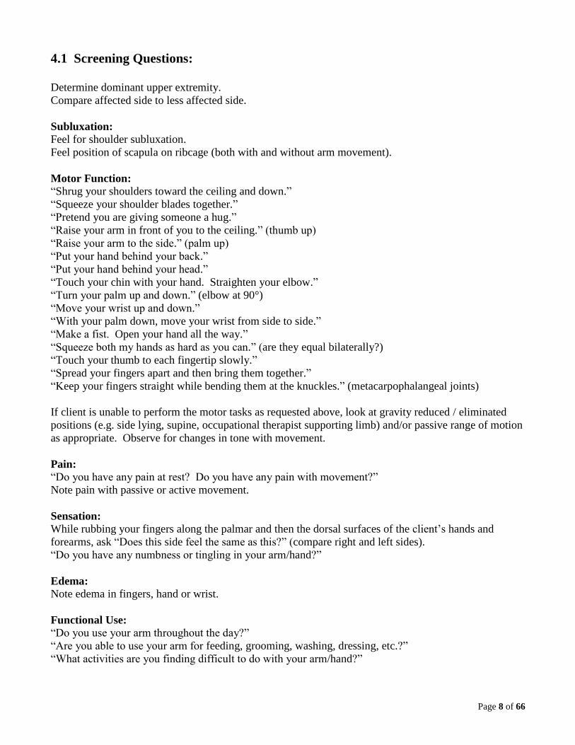
4.1 Screening Questions:
Determine dominant upper extremity.
Compare affected side to less affected side.
Subluxation:
Feel for shoulder subluxation.
Feel position of scapula on ribcage (both with and without arm movement).
Motor Function:
“Shrug your shoulders toward the ceiling and down.”
“Squeeze your shoulder blades together.”
“Pretend you are giving someone a hug.”
“Raise your arm in front of you to the ceiling.” (thumb up)
“Raise your arm to the side.” (palm up)
“Put your hand behind your back.”
“Put your hand behind your head.”
“Touch your chin with your hand. Straighten your elbow.”
“Turn your palm up and down.” (elbow at 90°)
“Move your wrist up and down.”
“With your palm down, move your wrist from side to side.”
“Make a fist. Open your hand all the way.”
“Squeeze both my hands as hard as you can.” (are they equal bilaterally?)
“Touch your thumb to each fingertip slowly.”
“Spread your fingers apart and then bring them together.”
“Keep your fingers straight while bending them at the knuckles.” (metacarpophalangeal joints)
If client is unable to perform the motor tasks as requested above, look at gravity reduced / eliminated
positions (e.g. side lying, supine, occupational therapist supporting limb) and/or passive range of motion
as appropriate. Observe for changes in tone with movement.
Pain:
“Do you have any pain at rest? Do you have any pain with movement?”
Note pain with passive or active movement.
Sensation:
While rubbing your fingers along the palmar and then the dorsal surfaces of the client’s hands and
forearms, ask “Does this side feel the same as this?” (compare right and left sides).
“Do you have any numbness or tingling in your arm/hand?”
Edema:
Note edema in fingers, hand or wrist.
Functional Use:
“Do you use your arm throughout the day?”
“Are you able to use your arm for feeding, grooming, washing, dressing, etc.?”
“What activities are you finding difficult to do with your arm/hand?”
Page 9 of 66
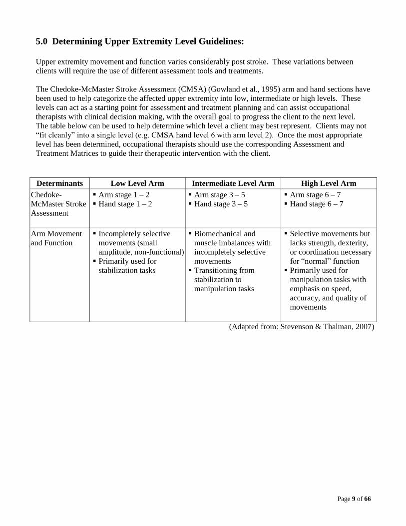
5.0 Determining Upper Extremity Level Guidelines:
Upper extremity movement and function varies considerably post stroke. These variations between
clients will require the use of different assessment tools and treatments.
The Chedoke-McMaster Stroke Assessment (CMSA) (Gowland et al., 1995) arm and hand sections have
been used to help categorize the affected upper extremity into low, intermediate or high levels. These
levels can act as a starting point for assessment and treatment planning and can assist occupational
therapists with clinical decision making, with the overall goal to progress the client to the next level.
The table below can be used to help determine which level a client may best represent. Clients may not
“fit cleanly” into a single level (e.g. CMSA hand level 6 with arm level 2). Once the most appropriate
level has been determined, occupational therapists should use the corresponding Assessment and
Treatment Matrices to guide their therapeutic intervention with the client.
Determinants Low Level Arm Intermediate Level Arm High Level Arm
Chedoke-
McMaster Stroke
Assessment
Arm stage 1 – 2
Hand stage 1 – 2
Arm stage 3 – 5
Hand stage 3 – 5
Arm stage 6 – 7
Hand stage 6 – 7
Arm Movement
and Function
Incompletely selective
movements (small
amplitude, non-functional)
Primarily used for
stabilization tasks
Biomechanical and
muscle imbalances with
incompletely selective
movements
Transitioning from
stabilization to
manipulation tasks
Selective movements but
lacks strength, dexterity,
or coordination necessary
for “normal” function
Primarily used for
manipulation tasks with
emphasis on speed,
accuracy, and quality of
movements
(Adapted from: Stevenson & Thalman, 2007)

Page 10 of 66
6.0 Assessment Guidelines:
The Canadian Stroke Best Practice Recommendations 2.2.iii states: “Clinicians should use standardized,
valid assessment tools to evaluate the patient’s stroke-related impairments, functional activity
limitations, and role participation restrictions and environment [Evidence Level C]. Tools should be
adapted for use in patients with communication differences or limitations due to aphasia.” (Hebert et al.,
2016, p. 9).
There are many upper extremity assessment tools available for use with clients post stroke. After the
screening is completed and the upper extremity level has been determined, the following Assessment
Matrix can then be used to help occupational therapists determine appropriate assessment tools for their
clients.
The intent is not to use all the assessment tools with each client but to choose assessments that will be
the most valuable in measuring change in that individual. Assessment tools may vary depending on the
availability and relevance to the practice setting.
The assessments listed in the Assessment Matrix are categorized according to their use with low,
intermediate and high level upper extremities post stroke. The list is not all-inclusive.
Page 11 of 66
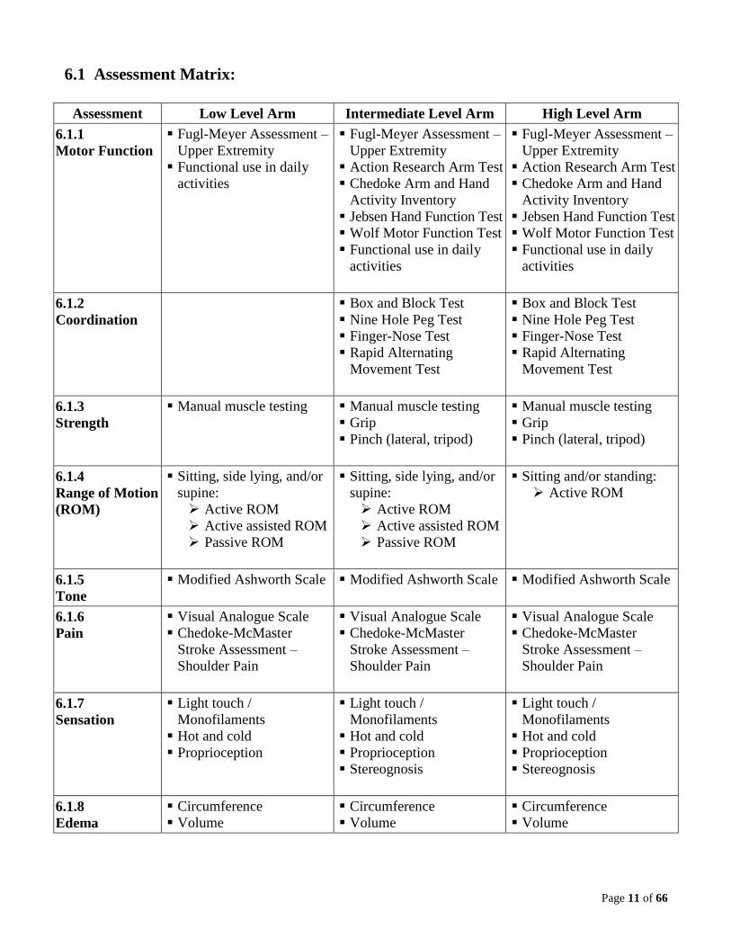
6.1 Assessment Matrix:
Assessment Low Level Arm Intermediate Level Arm High Level Arm
6.1.1
Motor Function
Fugl-Meyer Assessment –
Upper Extremity
Functional use in daily
activities
Fugl-Meyer Assessment –
Upper Extremity
Action Research Arm Test
Chedoke Arm and Hand
Activity Inventory
Jebsen Hand Function Test
Wolf Motor Function Test
Functional use in daily
activities
Fugl-Meyer Assessment –
Upper Extremity
Action Research Arm Test
Chedoke Arm and Hand
Activity Inventory
Jebsen Hand Function Test
Wolf Motor Function Test
Functional use in daily
activities
6.1.2
Coordination
Box and Block Test
Nine Hole Peg Test
Finger-Nose Test
Rapid Alternating
Movement Test
Box and Block Test
Nine Hole Peg Test
Finger-Nose Test
Rapid Alternating
Movement Test
6.1.3
Strength
Manual muscle testing Manual muscle testing
Grip
Pinch (lateral, tripod)
Manual muscle testing
Grip
Pinch (lateral, tripod)
6.1.4
Range of Motion
(ROM)
Sitting, side lying, and/or
supine:
Active ROM
Active assisted ROM
Passive ROM
Sitting, side lying, and/or
supine:
Active ROM
Active assisted ROM
Passive ROM
Sitting and/or standing:
Active ROM
6.1.5
Tone
Modified Ashworth Scale
Modified Ashworth Scale Modified Ashworth Scale
6.1.6
Pain
Visual Analogue Scale
Chedoke-McMaster
Stroke Assessment –
Shoulder Pain
Visual Analogue Scale
Chedoke-McMaster
Stroke Assessment –
Shoulder Pain
Visual Analogue Scale
Chedoke-McMaster
Stroke Assessment –
Shoulder Pain
6.1.7
Sensation
Light touch /
Monofilaments
Hot and cold
Proprioception
Light touch /
Monofilaments
Hot and cold
Proprioception
Stereognosis
Light touch /
Monofilaments
Hot and cold
Proprioception
Stereognosis
6.1.8
Edema
Circumference
Volume
Circumference
Volume
Circumference
Volume
Page 12 of 66
6.1.1 Motor Function
Fugl-Meyer Assessment – Upper Extremity (FMA-UE):
http://strokengine.ca/assess/module_fma_intro-en.html
Action Research Arm Test (ARAT):
http://strokengine.ca/assess/module_arat_intro-en.html
Chedoke Arm and Hand Activity Inventory (CAHAI):
http://strokengine.ca/assess/module_cahai_intro-en.html
There are four different versions of this assessment tool. Select the version that would be best suited for
the client’s upper extremity level.
Jebsen Hand Function Test:
http://strokengine.ca/assess/module_jhft_intro-en.html
Wolf Motor Function Test:
http://strokengine.ca/assess/module_wmft_intro-en.html
Functional use in daily activities:
Assess client’s ability to spontaneously incorporate their upper extremity into their self-care,
productivity and leisure activities.
6.1.2 Coordination
Box and Block Test (BBT):
http://strokengine.ca/assess/module_bbt_intro-en.html
Nine Hole Peg Test (NHPT):
http://strokengine.ca/assess/module_nhpt_intro-en.html
Finger-Nose Test (test for dysmetria):
In sitting, have client move his index finger from his nose to the occupational therapist’s index finger
(which is placed an arm’s length away from client). Record number of repetitions in 10 seconds.
Observe quality of movement and compare to less affected side.
Rapid Alternating Movement Test (test for dysdiadochokinesis):
In sitting, have client alternate between supination and pronation arm movements, while his hand is
supported on his thigh or on his other hand. Record number of repetitions in 10 seconds. Observe
quality of movement and compare to less affected side.
6.1.3 Strength
Manual Muscle Testing:
For manual muscle testing protocols, please see:
Clarkson, H. (2012). Musculoskeletal assessment: Joint range of motion and manual muscle testing (3rd
ed.). Philadelphia: Lippincott Williams & Wilkins.

Page 13 of 66
Grip Strength:
To reference the manual and standard testing procedure for using a Jamar dynamometer, please see:
https://www.chponline.com/store/pdfs/j-20.pdf.
For further information regarding grip strength assessment, please see:
Fess, E. (2011). Functional tests. In T. M. Skirven, A. L. Osterman, J. Fedorczyk, & P. C. Amadio
(Eds.), Rehabilitation of the hand and upper extremity (6th ed., Vol 1, pp. 152–162). Philadelphia:
Elsevier Mosby.
Pinch Strength:
For further information regarding pinch strength assessment, please see:
Fess, E. (2011). Functional tests. In T. M. Skirven, A. L. Osterman, J. Fedorczyk, & P. C. Amadio
(Eds.), Rehabilitation of the hand and upper extremity (6th ed., Vol 1, pp. 152–162). Philadelphia:
Elsevier Mosby.
6.1.4 Range of Motion
For passive and active range of motion measurement protocols, please see:
Clarkson, H. (2012). Musculoskeletal assessment: Joint range of motion and manual muscle testing (3rd
ed.). Philadelphia: Lippincott Williams & Wilkins.
Goniometry is the preferred method to measure range of motion and should be used to evaluate goals
that are targeted towards an increase in range of motion. Range of motion via goniometry must also be
used to determine appropriateness for splinting and to measure outcomes of splinting.
6.1.5 Tone
Modified Ashworth Scale:
http://strokengine.ca/assess/module_mashs_intro-en.html
A client’s positioning (sitting versus supine) should be consistent over time when measuring tone. It is
important to determine and document tonal differences with changes in position and activity. Clinical
observations of changes in tone are important.
6.1.6 Pain
“Causes of shoulder pain may be due to the hemiplegia itself, injury or acquired orthopedic conditions
due to compromised joint and soft tissue integrity. Shoulder pain may inhibit patient participation in
rehabilitation activities, contribute to poor functional recovery and can also mask improvement of
movement and function. Hemiplegic shoulder pain may contribute to depression and sleeplessness and
reduce quality of life” (Hebert et al., 2016, p. 13).
“The assessment of the painful hemiplegic shoulder should include evaluation of tone, strength, changes
in length of soft tissues, alignment of joints of the shoulder girdle, levels of pain and orthopedic changes
in the shoulder [Evidence Level C]” (Hebert et al., 2016, p. 13).

Page 14 of 66
It is important to consider the following when assessing pain: a) present at rest and/or with activity, b)
specific location, c) quality (e.g. sharp, burning, radiating, etc.), and d) position of the upper extremity.
Be sure to differentiate pain from “stretch” and “stiffness”. This information will help determine the
cause of pain and guide treatment.
Visual Analogue Scale:
There are a variety of visual analogue scales for pain. Ensure you use a consistent scale over time when
measuring pain. The following link has several examples of scales:
http://www.painedu.org/Downloads/NIPC/Pain%20Assessment%20Scales.pdf
Chedoke McMaster Stroke Assessment – Shoulder Pain:
http://strokengine.ca/assess/module_cmmsa_intro-en.html
6.1.7 Sensation:
For sensation testing protocols please see:
Cooper, C., & Canyock, J. D. (2013). Evaluation of sensation and intervention for sensory dysfunction.
In H. M. Pendleton, & W. Schultz-Krohn (Eds.), Pedretti’s occupational therapy: Practice skills for
physical dysfunction (7th ed., pp. 575-589). St. Louis, MS: Mosby, Inc.
Occupational therapists can consider more in depth sensory assessments, such as:
Nottingham Sensory Assessment Revised
http://www.nottingham.ac.uk/medicine/about/rehabilitationageing/publishedassessments.aspx
Fugl-Meyer Assessment – Upper Extremity (FMA-UE)
http://strokengine.ca/assess/module_fma_intro-en.html
Monofilaments are the preferred method to objectively measure light touch. For monofilament
protocols, please see: http://www.htherapy.co.za/user_images/splinting/Monofilaments.pdf.
Proprioception should also be assessed. For a demonstration of the Thumb Localization Test, a test of
proprioception, please see: https://vimeo.com/138227545.
6.1.8 Edema
For descriptions of edema assessment methods, please see:
Kasch, M. C., & Walsh, J. M. (2013). Hand and upper extremity injuries. In H. M. Pendleton, & W.
Schultz-Krohn (Eds.), Pedretti’s occupational therapy: Practice skills for physical dysfunction (7th ed.,
pp. 1037-1073). St. Louis, MS: Mosby, Inc.
Page 15 of 66
7.0 Goal Setting Guidelines:
It is important to identify goals to assist with planning upper extremity treatment and to determine a
client’s progress. Goals should be made in collaboration with the client to ensure tasks chosen are
meaningful and that the client and the occupational therapist are working toward the same outcomes.
“Patients and families should be involved in their management, goal setting and transition planning
(Evidence Level A)” (Hebert et al., 2016, p. 11).
The Canadian Occupational Performance Measure (COPM) can be used to help a client identify
occupational performance issues, which can then be translated into functional goals. The COPM is a
client centered outcome measure that determines change over time in a client’s self-perception of their
occupational performance issues (Law, Baptiste, Carswell, McColl, Polatajko & Pollock, 2014).
SMART goal setting is a method of setting goals which are: Specific, Measureable, Attainable, Realistic
and Time-Based. It clearly identifies a client’s goals and clarifies when goal attainment has been
achieved. SMART goal setting can be combined with the COPM. A copy of the SMART goals can be
provided to the client. Some examples of SMART goals include:
Client will zip up winter jacket independently with right hand in 2 weeks.
Client will eat all meals independently with left hand using built up utensils in 4 weeks.
Client will increase Box and Block Test score to 21 (25%) in 4 weeks.
The following resources may assist with goal setting:
Canadian Occupational Performance Measure
http://www.thecopm.ca
SMART Goals
https://ehealth.heartandstroke.ca/HeartStroke/HWAP2/Goals.aspx
“Goal Setting 101”
http://canadianstrokenetwork.ca/en/wp-content/uploads/2014/08/GettingOn-EN.pdf

Page 16 of 66
8.0 Treatment Guidelines:
The Canadian Stroke Best Practice Recommendations 5.1.A states: “Patients should engage in training
that is meaningful, engaging, repetitive, progressively adapted, task-specific and goal-oriented in an
effort to enhance motor control and restore sensorimotor function [Evidence Level: Early-Level A; Late-
Level A]. Training should encourage the use of patients’ affected limb during functional tasks and be
designed to simulate partial or whole skills required in activities of daily living . . . [Evidence Level:
Early-Level A; Late-Level A]” (Hebert et al., 2016, p. 11).
“All patients with stroke should receive rehabilitation therapy as early as possible once they are
determined to be rehabilitation ready and they are medically able to participate in active rehabilitation
(Evidence Level A), within an active and complex stimulating environment (Evidence Level C)”
(Hebert et al., 2016, p. 9).
There are many options available for upper extremity treatment post stroke. Based on the upper
extremity screening and assessment results as well as the client’s goals, specific treatments should be
chosen that best suit the client’s upper extremity level. Treatment activities should be task specific,
meaningful to the client, and easily graded so optimal challenge can be maintained. Specific treatments
may vary depending on availability and relevance to the practice setting. In all practice settings, the
client’s body position and trunk stability as well as the environmental set-up need to be considered to
maximize upper extremity function. It is also important to educate the client regarding the purpose of
the specific treatments being used. Education may enhance client engagement in the treatment process
which may then contribute to improved outcomes.
Although the optimal goal of upper extremity rehabilitation is to promote motor recovery and function
of the affected upper extremity, at times assistive devices and compensatory strategies may need to be
incorporated temporarily to enable participation. It is important to note that compensatory behavioral
changes “can also be maladaptive and interfere with improvements in function that could be obtained
using rehabilitative training” (Kleim & Jones, 2008, p. S226); therefore, early instruction in
compensatory strategies may be detrimental to learning new skills with the affected arm and interfere
with improvements in function that could be obtained through upper extremity rehabilitation. The
Canadian Stroke Best Practice Recommendations 5.1.C.i states: “Adaptive devices designed to improve
safety and function may be considered if other methods of performing specific functional tasks are not
available or tasks cannot be learned [Evidence Level C]” (Hebert et al., 2016, p. 12). Compensatory
strategies and the use of equipment should be frequently re-evaluated and weaned as appropriate.
The specific treatments listed in the Treatment Matrix are categorized according to their use with low,
intermediate and high level upper extremities post stroke. The list is not all-inclusive. Practical tools
are included for several treatments identified in the Treatment Matrix.

Page 17 of 66
8.1 Treatment Matrix:
8.1.1 Task specific training, “the repeated, challenging practice of functional, goal-oriented activities”
(Lang & Birkenmeier, 2014, p. xi), should be utilized with all treatment modalities. Occupational therapists
should strive for increased intensity and number of repetitions of upper extremity use. The optimal number
of repetitions is unknown; however, studies suggest that “hundreds of repetitions of task-specific practice
may be required to optimize function post stroke” (Birkenmeier, Prager, & Lang, 2010, p. 620).
Specific
Treatments Low Level Arm Intermediate Level Arm High Level Arm
8.1.2
Constraint
Induced
Movement
Therapy
(CIMT)
Work toward minimal
active movement
requirements for CIMT
program
Work toward minimal
active movement
requirements for CIMT
program
Refer to traditional or
modified CIMT program
as available
Provide individual
program based on CIMT
principles
8.1.3
Functional
Dynamic
Orthoses
(e.g. SaeboFlex,
SaeboReach,
SaeboGlove)
Work toward minimal
active and passive
movement requirements
for functional dynamic
orthosis
Use functional dynamic
orthosis (SaeboFlex or
SaeboReach) for daily
sessions, followed by
functional activities
without orthosis
Use functional dynamic
orthosis (SaeboGlove)
during daily activities
Wean from functional
dynamic orthosis
8.1.4
Functional
Electrical
Stimulation
(FES)
Target wrist extensor and
forearm muscles while
engaged in task specific
activities
Consider using to reduce
or prevent shoulder
subluxation
Target wrist extensor and
forearm muscles while
engaged in task specific
activities
8.1.5
Mental
Imagery
Use as an adjunct to other
treatments
Use as homework
Use as an adjunct to other
treatments
Use as homework
Use as an adjunct to other
treatments
Use as homework
Page 18 of 66
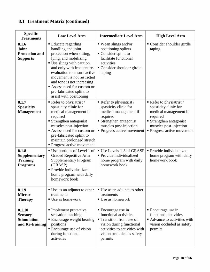
8.1 Treatment Matrix (continued)
Specific
Treatments Low Level Arm Intermediate Level Arm High Level Arm
8.1.6
Joint
Protection and
Supports
Educate regarding
handling and joint
protection when sitting,
lying, and mobilizing
Use slings with caution
and only with frequent re-
evaluation to ensure active
movement is not restricted
and tone is not increasing
Assess need for custom or
pre-fabricated splint to
assist with positioning
Wean slings and/or
positioning splints
Consider splint to
facilitate functional
activities
Consider shoulder girdle
taping
Consider shoulder girdle
taping
8.1.7
Spasticity
Management
Refer to physiatrist /
spasticity clinic for
medical management if
required
Strengthen antagonist
muscles post-injection
Assess need for custom or
pre-fabricated splint to
maintain prolonged stretch
Progress active movement
Refer to physiatrist /
spasticity clinic for
medical management if
required
Strengthen antagonist
muscles post-injection
Progress active movement
Refer to physiatrist /
spasticity clinic for
medical management if
required
Strengthen antagonist
muscles post-injection
Progress active movement
8.1.8
Supplementary
Training
Programs
Use portions of Level 1 of
Graded Repetitive Arm
Supplementary Program
(GRASP)
Provide individualized
home program with daily
homework book
Use Levels 1-3 of GRASP
Provide individualized
home program with daily
homework book
Provide individualized
home program with daily
homework book
8.1.9
Mirror
Therapy
Use as an adjunct to other
treatments
Use as homework
Use as an adjunct to other
treatments
Use as homework
8.1.10
Sensory
Stimulation
and Re-training
Implement protective
sensation teaching
Encourage weight bearing
positions
Encourage use of vision
during functional
activities
Encourage use in
functional activities
Transition from use of
vision during functional
activities to activities with
vision occluded as safety
permits
Encourage use in
functional activities
Advance to activities with
vision occluded as safety
permits

Page 19 of 66
8.1 Treatment Matrix (continued)
Specific
Treatments Low Level Arm Intermediate Level Arm High Level Arm
8.1.11
Range of Motion
(ROM)
and Strength
Training
Maintain / increase ROM
through:
Facilitation of active
movement by
therapist
Progression from
bilateral to unilateral
activities
Active assisted ROM
in sitting, supine, or
gravity reduced
positions
Passive ROM
Self-ROM
Use strength training
through available ROM
including use of mobile
arm support as indicated
Do not use pulleys
Maintain / increase ROM
through:
Active ROM while
providing verbal
and/or tactile cueing
Progression from
bilateral to unilateral
activities
Active assisted ROM
in sitting, supine, or
gravity reduced
positions
Passive ROM
Self-ROM
Use strength training
through available ROM
Do not use pulleys
Maintain / increase ROM
through:
Active ROM while
providing verbal
and/or tactile cueing
Use strength training
through available ROM
Monitor carefully if using
pulleys
8.1.12
Edema
Management
Encourage active, active-
assisted and passive
movement
Consider retrograde
massage
Educate regarding
positioning and elevation
Use compression
techniques
Assess need for custom or
pre-fabricated splint
Encourage active
movement
Consider retrograde
massage
Educate regarding
positioning and elevation
Use compression
techniques
Encourage active
movement
Consider retrograde
massage
Educate regarding
positioning and elevation
Use compression
techniques
8.1.13
Virtual Reality
Use as an adjunct to other
treatments
Use as homework
Use as an adjunct to other
treatments
Use as homework
Use as an adjunct to other
treatments
Use as homework
Page 20 of 66
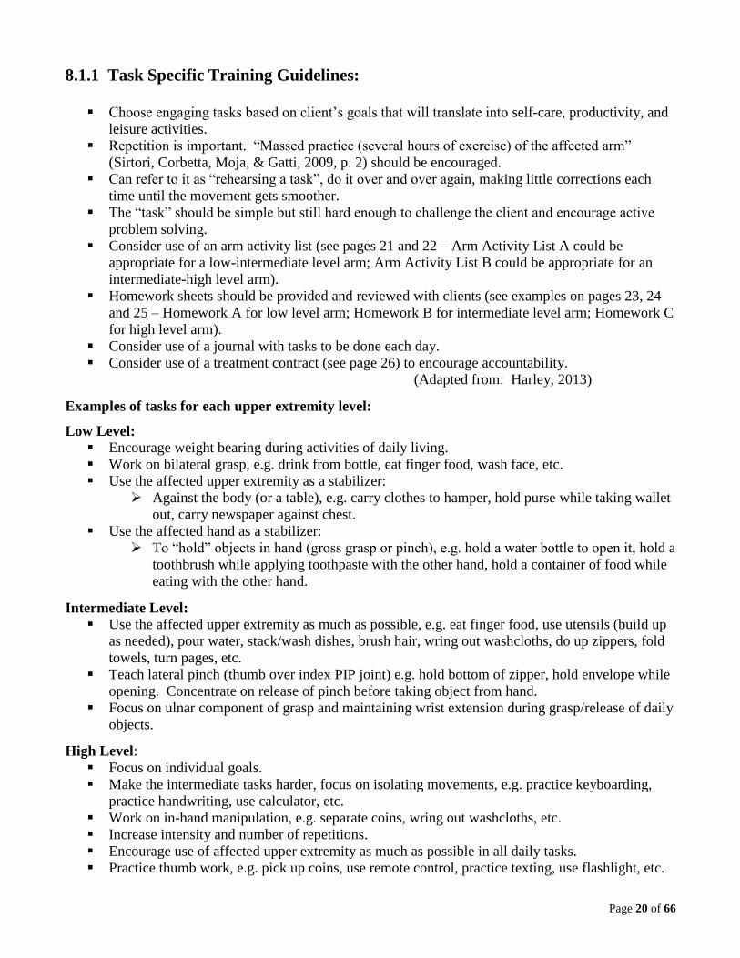
8.1.1 Task Specific Training Guidelines:
Choose engaging tasks based on client’s goals that will translate into self-care, productivity, and
leisure activities.
Repetition is important. “Massed practice (several hours of exercise) of the affected arm”
(Sirtori, Corbetta, Moja, & Gatti, 2009, p. 2) should be encouraged.
Can refer to it as “rehearsing a task”, do it over and over again, making little corrections each
time until the movement gets smoother.
The “task” should be simple but still hard enough to challenge the client and encourage active
problem solving.
Consider use of an arm activity list (see pages 21 and 22 – Arm Activity List A could be
appropriate for a low-intermediate level arm; Arm Activity List B could be appropriate for an
intermediate-high level arm).
Homework sheets should be provided and reviewed with clients (see examples on pages 23, 24
and 25 – Homework A for low level arm; Homework B for intermediate level arm; Homework C
for high level arm).
Consider use of a journal with tasks to be done each day.
Consider use of a treatment contract (see page 26) to encourage accountability.
(Adapted from: Harley, 2013)
Examples of tasks for each upper extremity level:
Low Level:
Encourage weight bearing during activities of daily living.
Work on bilateral grasp, e.g. drink from bottle, eat finger food, wash face, etc.
Use the affected upper extremity as a stabilizer:
Against the body (or a table), e.g. carry clothes to hamper, hold purse while taking wallet
out, carry newspaper against chest.
Use the affected hand as a stabilizer:
To “hold” objects in hand (gross grasp or pinch), e.g. hold a water bottle to open it, hold a
toothbrush while applying toothpaste with the other hand, hold a container of food while
eating with the other hand.
Intermediate Level:
Use the affected upper extremity as much as possible, e.g. eat finger food, use utensils (build up
as needed), pour water, stack/wash dishes, brush hair, wring out washcloths, do up zippers, fold
towels, turn pages, etc.
Teach lateral pinch (thumb over index PIP joint) e.g. hold bottom of zipper, hold envelope while
opening. Concentrate on release of pinch before taking object from hand.
Focus on ulnar component of grasp and maintaining wrist extension during grasp/release of daily
objects.
High Level:
Focus on individual goals.
Make the intermediate tasks harder, focus on isolating movements, e.g. practice keyboarding,
practice handwriting, use calculator, etc.
Work on in-hand manipulation, e.g. separate coins, wring out washcloths, etc.
Increase intensity and number of repetitions.
Encourage use of affected upper extremity as much as possible in all daily tasks.
Practice thumb work, e.g. pick up coins, use remote control, practice texting, use flashlight, etc.

Page 21 of 66
ARM ACTIVITY LIST A
Name: ___________________________________________
Add a new activity every day / week.
“2 hands” refers to interlocking grip as needed.
“Under arm” refers to holding item between upper arm and side of body.
Position hand on table in view
_____
Hold food with fork when cutting
_____
Hold toothpaste
_____
Carry a newspaper (under arm)
_____
Hold deodorant
_____
Carry a towel (under arm)
_____
Pull up blankets (2 hands)
_____
Carry a purse / wallet (under arm)
_____
Use call bell
_____
_______________________
_____
Pick up water bottle (2 hands)
_____
_______________________
_____
Eat finger food (2 hands)
_____
_______________________
_____
Hold washcloth
_____
_______________________
_____
Wash face (2 hands)
_____
_______________________
_____
Brush teeth (2 hands)
_____
_______________________
_____
Hold towel with hand
_____
_______________________
_____
Dry self (2 hands)
_____
_______________________
_____
Wipe table
_____
_______________________
_____
Hold paper down when writing
_____
_______________________
_____
Hold bowl/plate when eating
_____
_______________________
_____
Apply wheelchair brakes
_____
_______________________
_____
Use a fork / spoon to eat
_____
_______________________
_____
Occupational Therapist: _________________________ Phone: _________________________
(Adapted from: Thalman, 2002)
WRHA Occupational Therapy Upper Extremity Working Group 2015

Page 22 of 66
ARM ACTIVITY LIST B
Name: ___________________________________________
Add a new activity every day / week.
Fill out menu
_____
Put on shoes
_____
Use call bell
_____
Put on socks
_____
Pull up covers
_____
Pour liquids
_____
Turn on light switches
_____
Use fork
_____
Drink from a cup
_____
Use spoon
_____
Eat finger food
_____
Use knife
_____
Turn pages in a book / magazine
_____
Hold phone while talking
_____
Brush teeth
_____
Dial phone
_____
Brush hair
_____
Open fridge
_____
Turn on / off faucets
_____
Use computer mouse / keyboard
_____
Wash self with washcloth
_____
Practice handwriting
_____
Flush toilet
Wipe self
_____
_____
Open doors
Unload dishwasher
_____
_____
Pull pants up and down
_____
Put away groceries
_____
Do up zippers / buttons
_____
_______________________
_____
Wipe table
_____
_______________________
_____
Take clothes out of closet / drawer
_____
_______________________
_____
Hang up clothes
_____
_______________________
_____
Occupational Therapist: _________________________ Phone: _________________________
(Adapted from: Thalman, 2002)
WRHA Occupational Therapy Upper Extremity Working Group 2015

Page 23 of 66
Name: ___________________________________________
HOMEWORK A
Try to include your arm in as many tasks as possible to give the muscles an opportunity to “turn on”.
Please do these exercises 2-3 times a day. If something hurts, STOP what you are doing and discuss
with your therapist.
Lying in bed (on back):
1. “Push” both your shoulder blades and elbows down into the bed. Relax. Repeat 10 times.
2. “Push” your hand down into the bed. Relax. Repeat 10 times.
3. Interlock the fingers of both your hands. Raise your hands to the side of your head and make
a “chopping” motion across your body. Repeat 10 times each direction.
Sitting up:
1. Place your hand flat on a pillow (or arm board if you have). “Push” down onto your forearm
and hand. Relax. Repeat 10 times.
2. “Wash” the table top using a washcloth, back and forth and side to side. Use your other hand
to help if needed. Repeat for 2 minutes.
3. Interlock the fingers of both your hands
a) Reach for and grasp a plastic bottle or other container. Bring it to your chin, then return
to the table and let go each time. Repeat 8 times.
b) Eat finger foods with your fingers interlocked.
c) Take a washcloth in both hands. Rub over your entire face (you can do it with a wet or
dry cloth). Place on your lap and LET IT GO. Pick it up again and repeat 5 times.
4. Place a towel in your armpit. Try to press your arm to your body to keep it there, while your
other hand tries to pull it out. Repeat 10 times.
5. Use your hand to hold a plastic bottle. Open the bottle with your stronger hand, then attempt
to let go of the bottle with your weaker hand. Relax. Repeat 8 times.
6. Bring both your shoulders to your ears. Relax. Repeat 10 times.
Occupational Therapist: _________________________ Phone: _________________________
WRHA Occupational Therapy Upper Extremity Working Group 2017
Page 24 of 66
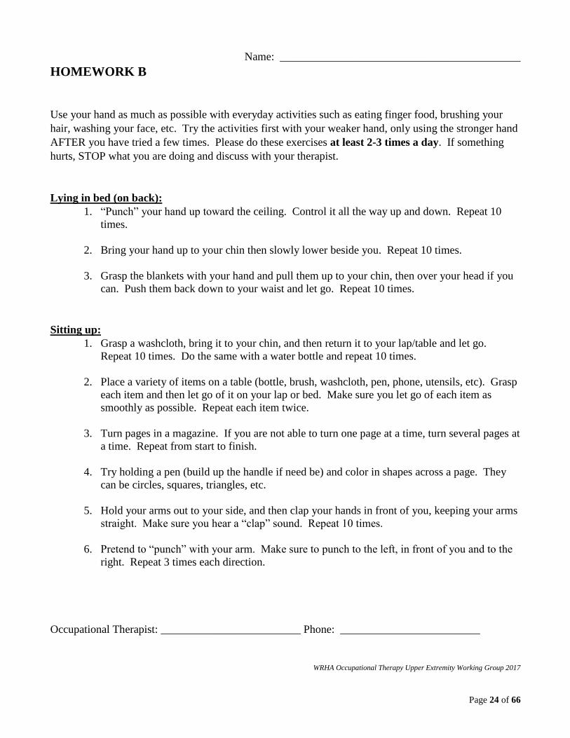
Name: ___________________________________________
HOMEWORK B
Use your hand as much as possible with everyday activities such as eating finger food, brushing your
hair, washing your face, etc. Try the activities first with your weaker hand, only using the stronger hand
AFTER you have tried a few times. Please do these exercises at least 2-3 times a day. If something
hurts, STOP what you are doing and discuss with your therapist.
Lying in bed (on back):
1. “Punch” your hand up toward the ceiling. Control it all the way up and down. Repeat 10
times.
2. Bring your hand up to your chin then slowly lower beside you. Repeat 10 times.
3. Grasp the blankets with your hand and pull them up to your chin, then over your head if you
can. Push them back down to your waist and let go. Repeat 10 times.
Sitting up:
1. Grasp a washcloth, bring it to your chin, and then return it to your lap/table and let go.
Repeat 10 times. Do the same with a water bottle and repeat 10 times.
2. Place a variety of items on a table (bottle, brush, washcloth, pen, phone, utensils, etc). Grasp
each item and then let go of it on your lap or bed. Make sure you let go of each item as
smoothly as possible. Repeat each item twice.
3. Turn pages in a magazine. If you are not able to turn one page at a time, turn several pages at
a time. Repeat from start to finish.
4. Try holding a pen (build up the handle if need be) and color in shapes across a page. They
can be circles, squares, triangles, etc.
5. Hold your arms out to your side, and then clap your hands in front of you, keeping your arms
straight. Make sure you hear a “clap” sound. Repeat 10 times.
6. Pretend to “punch” with your arm. Make sure to punch to the left, in front of you and to the
right. Repeat 3 times each direction.
Occupational Therapist: _________________________ Phone: _________________________
WRHA Occupational Therapy Upper Extremity Working Group 2017

Page 25 of 66
Name: ___________________________________________
HOMEWORK C
Use your hand for EVERYTHING! Repeat these exercises at least 4-5 times a day. If something
hurts, STOP what you are doing and discuss with your therapist.
1. Hold a pen at the bottom. Work your fingertips up the pen to the top, and then back down
slowly. Repeat 10 times.
2. While holding a remote or phone in your hand, take your thumb and touch each outside button
once, slowly. Make sure you are moving your weaker hand without help from your stronger
hand. Repeat 2 times.
3. Place 5 different coins on a table. Pick them up one at a time and place them into your palm.
Slowly take them out in order of amount, one at a time, using your thumb and index finger.
Repeat 3 times.
4. Handwriting (as appropriate) - do one paragraph a day in the same notebook to compare your
progress.
5. Place 3 washcloths in a basin or sink filled with water. Take one washcloth out at a time,
squeezing as much water out as possible, using only your weaker hand to turn the cloth in your
hand to change the grip. Repeat 2 times.
6. Tap a balloon in the air for 3 minutes keeping track of how many taps you are able to get in a
row. Try to increase the height of the balloon to make it harder. (You can do this one with a
partner too.)
Occupational Therapist: _________________________ Phone: _________________________
WRHA Occupational Therapy Upper Extremity Working Group 2017
Page 26 of 66
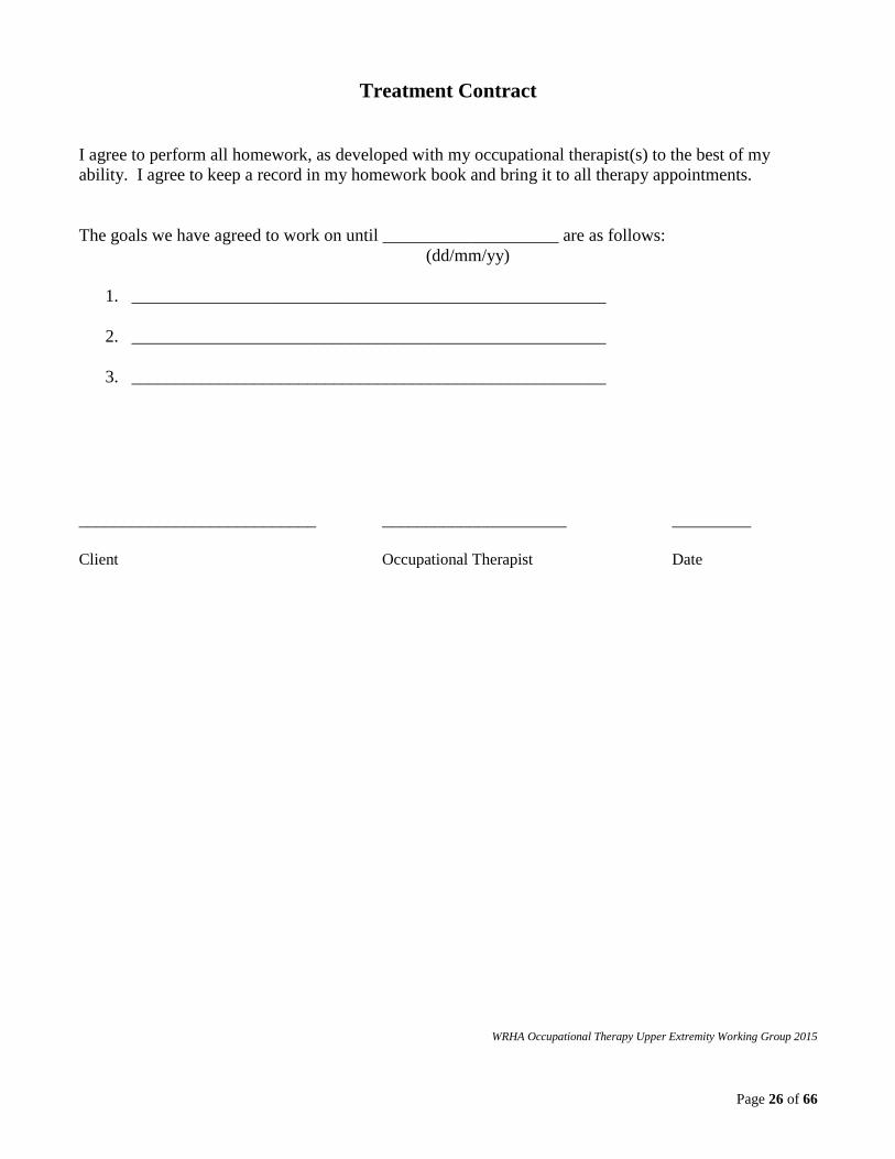
Treatment Contract
I agree to perform all homework, as developed with my occupational therapist(s) to the best of my
ability. I agree to keep a record in my homework book and bring it to all therapy appointments.
The goals we have agreed to work on until ____________________ are as follows:
(dd/mm/yy)
1. ______________________________________________________
2. ______________________________________________________
3. ______________________________________________________
___________________________ _____________________ _________
Client Occupational Therapist Date
WRHA Occupational Therapy Upper Extremity Working Group 2015

Page 27 of 66
8.1.2 Constraint Induced Movement Therapy
The Canadian Stroke Best Practice Recommendations 5.1.B.iv states: “Traditional or modified
constraint-induced movement therapy (CIMT) should be considered for a select group of patients who
demonstrate at least 20 degrees of active wrist extension and 10 degrees of active finger extension, with
minimal sensory or cognitive deficits (Evidence Level: Early-Level A; Late-Level A) (Hebert, 2016, p.
12).
“CIMT can be described as either: a) Traditional CIMT: 2-week training program, with 6 hours of
intensive upper-extremity training with restraint of the unaffected arm for at least 90% of waking hours.
b) Modified CIMT: often refers to less intense than traditional CIMT, with variable intensity, time of
constraint and duration of program” (Teasell & Hussein, 2016, p. 7 & 8).
Principles of CIMT:
Use the more affected upper extremity in frequent, intense, massed practice tasks.
Adapt the tasks for optimal challenge.
Use consistent “coaching” of client by occupational therapist, rehabilitation assistant or trained
family member (as able).
Constrain the less affected upper extremity with a mitt or splint for up to 90% of waking hours
(as negotiated between client and occupational therapist).
Focus on transfer of skills to daily tasks (use of treatment contract and homework).
For information regarding the CIMT program in Winnipeg, please contact the Health Sciences Centre
Occupational Therapy Department at 204-787-2786. Prior to acceptance into a CIMT program or in the
absence of a formal CIMT program, occupational therapists should incorporate CIMT principles into a
client’s daily therapy sessions and home programs as early as possible.
Page 28 of 66
8.1.3 Functional Dynamic Orthoses
The Canadian Stroke Best Practice Recommendations 5.1.c.iii states: “Functional dynamic orthoses are
an emerging therapy tool that may be offered to patients to facilitate repetitive task specific training
[Evidence Level B]” (Hebert et al., 2016, p. 12).
Using a dynamic wrist hand orthosis, that positions the wrist and hand functionally and assists with
finger / thumb extension (e.g. SaeboFlex or SaeboReach), may enable participation in repetitive task
oriented activities not otherwise possible. After the orthosis is removed in the daily training sessions
(with goal of two 45 minute sessions per day), continued use of the upper extremity in grasp / release
and functional activities is recommended.
Some functional dynamic orthoses, for those with minimal spasticity, can be worn for longer periods of
time during daily activities (e.g. SaeboGlove).
For eligibility criteria and information on Saebo functional dynamic orthoses, please see
http://www.saebo.com/.
Handouts are being developed to assist with screening for, assessing and treating with the SaeboFlex and
SaeboReach orthoses. These handouts are based on the Saebo arm training program guidelines.
Occupational therapists must be trained in order to prescribe and use Saebo orthoses with their clients.
Trained occupational therapists can contact the Toolkit authors for Saebo handout information.
Page 29 of 66
8.1.4 Functional Electrical Stimulation
The Canadian Stroke Best Practice Recommendations 5.1.B.iii states: “Functional Electrical Stimulation
(FES) targeted at the wrist and forearm muscles should be considered to reduce motor impairment and
improve function [Evidence Level: Early-Level A; Late-Level A]” (Hebert et al., 2016, p. 12).
The Canadian Stroke Best Practice Recommendations 5.3.A.ii states: “For patients with a flaccid arm
(i.e., Chedoke-McMaster Stroke Assessment < 3) electrical stimulation should be considered [Evidence
Levels: Early- Level B; Late- Level B]” (Hebert et al., 2016, p. 13).
The Evidence-Based Review of Stroke Rehabilitation states: “There is level 1a and level 2 evidence that
FES/NMES may improve upper limb motor function, range of motion, and manual dexterity when
offered in combination with conventional therapy or delivered alone in subacute stroke. The evidence is
also indicative of a beneficial effect on range of motion and manual dexterity when FES/NMES was
offered to chronic stroke patients either alone or in combination with other therapies. Despite
improvements in both stages of stroke recovery, level 1b evidence indicates that delivering FES early (<
6 months) may be more beneficial at recovering impaired motor function than delivering FES after 6
months post-stroke” (Foley et al., 2016, p. 88).
FES should be combined with task specific treatment activities whenever possible.
Some examples of treatment activities to combine with FES of the wrist extensors are:
Use the back of the hand to move a cup from one place to another on a table.
Wrap the hand around a cup when the muscle stimulation is off; let go of the cup when the
muscle stimulation is on.
Work on sit to stand using both arms on armrests of a chair. When the muscle stimulation comes
on, work on straightening wrist and pushing into standing position.
Use with the SaeboFlex orthosis to facilitate wrist / finger extension during the release of therapy
balls, water bottle, cup, etc.
Some examples of treatment activities to combine with FES of the shoulder girdle are:
Perform shoulder shrugs when the muscle stimulation is on.
Place hand on ball or pillow beside body and push down when the muscle stimulation is on.
Prior to providing this intervention, occupational therapists need to be trained regarding the use,
protocols and contraindications for functional electrical stimulation.
Page 30 of 66
8.1.5 Mental Imagery
The Canadian Stroke Best Practice Recommendations 5.1.B.ii states: “Following assessment to
determine if they are suitable candidates, patients should be encouraged to engage in mental imagery to
enhance upper-limb, sensorimotor recovery [Evidence Level: Early-Level A; Late-Level B]” (Hebert et
al., 2016, p. 12).
Page (2001) states: “. . . mental practice is a technique by which CVA patients can simulate repeated
practice using the affected arm. In so doing, activations occur as if the arm were actually being utilized,
which may restore some level of function in patients’ affected limbs” (p. 60).
Patients may have greater or lesser ability to perform mental imagery training, post stroke, depending on
the area of the brain affected. Patients with parietal lobe damage may have difficulty performing mental
imagery, as may patients with frontal lobe and basal ganglia involvement (McInnes, 2016).
Mental imagery is best done in a quiet environment so distractions are minimized. The client can be
instructed in progressive muscle relaxation techniques, which can be done prior to the mental imagery to
improve focus. Imagery is often done either immediately before or after practicing actual movements of
the affected upper extremity. The client can be instructed to imagine all the steps of a successful
functional activity. The affected upper extremity should be placed in the correct position for the start of
the movement that is to be imagined. The occupational therapist provides specific written instructions
or a voice recording describing the activity to be imagined, including the specific upper extremity
movements required to complete the task, the number of repetitions or the duration of the activity.
Mental imagery can be done several times a day. The imagery script should be graded as the client
improves.
Mental imagery scripts can be composed for many different activities depending on the client’s goals.
Examples include:
Picking up a pen and positioning it in the hand for writing
Reaching for a towel and drying the other arm with it
Grabbing a tissue and bringing it up to the nose
Squeezing water out of a washcloth
Wiping a counter with a towel
Using a knife to spread peanut butter onto bread
Throwing a ball
For an example of a mental imagery script, see page 31.
Page 31 of 66
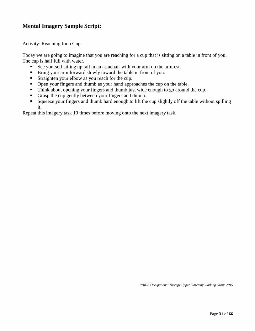
Mental Imagery Sample Script:
Activity: Reaching for a Cup
Today we are going to imagine that you are reaching for a cup that is sitting on a table in front of you.
The cup is half full with water.
See yourself sitting up tall in an armchair with your arm on the armrest.
Bring your arm forward slowly toward the table in front of you.
Straighten your elbow as you reach for the cup.
Open your fingers and thumb as your hand approaches the cup on the table.
Think about opening your fingers and thumb just wide enough to go around the cup.
Grasp the cup gently between your fingers and thumb.
Squeeze your fingers and thumb hard enough to lift the cup slightly off the table without spilling
it.
Repeat this imagery task 10 times before moving onto the next imagery task.
WRHA Occupational Therapy Upper Extremity Working Group 2015
Page 32 of 66
8.1.6 Joint Protection and Supports
The Canadian Stroke Best Practice Recommendations 5.3.A.i states: “Joint protection strategies should
be used during the early or flaccid stage of recovery to prevent or minimize shoulder pain. These
include: a) Positioning and supporting the arm during rest [Evidence Level B]. b) Protecting and
supporting the arm during functional mobility [Evidence Level C]. c) Protecting and supporting the arm
during wheelchair use by using a hemi-tray or arm trough [Evidence Level C]…” (Hebert et al., 2016, p.
13).
8.1.6a Positioning and Supporting the Arm in Lying and in Sitting
The Canadian Stroke Best Practice Recommendations 5.3.A.v states: “Healthcare staff, patients and
family should be educated to correctly handle the involved arm [Evidence Level A]. For example,
careful positioning and supporting the arm during assisted moves such as transfers; avoid pulling on the
affected arm [Evidence level C]” (Hebert et al., 2016, p. 13).
The Evidence-Based Review of Stroke Rehabilitation states: “The muscles around the hemiplegic
shoulder are often paralyzed, initially with flaccid tone and later with associated spasticity. Careful
positioning of the shoulder serves to minimize subluxation and later contractures as well as possibly
promote recovery, while poor positioning may adversely affect symmetry, balance and body image”
(Cotoi et al., 2016, p.15).
Optimal positioning in lying and sitting should maximize pain free degrees of shoulder abduction and
external rotation while maintaining shoulder joint alignment.
For an example of bed and chair positioning handouts, see pages 33 and 34.

Page 33 of 66
BED & CHAIR POSITIONING FOLLOWING A STROKE
CLIENT’S NAME: ___________________________________________ Affected side (shaded): RIGHT
Position affected shoulder forward with arm supported on pillow
Place pillow(s) between legs
Place a pillow behind back and ensure that they are not lying directly on hip bone
Lying on unaffected side
Position affected shoulder so that shoulder blade lies flat and arm appears slightly forward from trunk
Place unaffected leg forward on one or two pillows
Place a pillow behind back and ensure that they are not lying directly on hip bone
Lying on affected side**
Best position
Place pillow behind affected shoulder blade
Place affected hand on pillow above heart level
Place pillow beneath affected hip and/or beneath both knees (optional)
Lying on back (if desired)
Ensure client sits well back in the centre of chair or wheelchair
Place arms well forward onto two pillows on table or arm board if available
Ensure feet are flat on floor or footrests
Sitting up
ENSURE THAT YOU ASK CLIENT “ARE YOU COMFORTABLE?”
If you have any questions, please contact your Occupational Therapist or Physiotherapist
Name: ______________________________________ Phone: ___________________________________
(Adapted from: Chest Heart and Stroke Scotland, 2012) WRHA Occupational Therapy Upper Extremity Working Group 2013

Page 34 of 66
Position affected shoulder forward with arm supported on pillow
Place pillow(s) between legs
Place a pillow behind back and ensure that they are not lying directly on hip bone
Lying on unaffected side
Position affected shoulder so that shoulder blade lies flat and arm appears slightly forward from trunk
Place unaffected leg forward on one or two pillows
Place a pillow behind back and ensure that they are not lying directly on hip bone
Lying on affected side**
Best position
Place pillow behind affected shoulder blade
Place affected hand on pillow above heart level
Place pillow beneath affected hip and/or beneath both knees (optional)
Lying on back (if desired)
Ensure client sits well back in the centre of chair or wheelchair
Place arms well forward onto two pillows on table or arm board if available
Ensure feet are flat on floor or footrests
Sitting up
ENSURE THAT YOU ASK CLIENT “ARE YOU COMFORTABLE?”
BED & CHAIR POSITIONING FOLLOWING A STROKE
CLIENT’S NAME: ______________________________________________ Affected side (shaded): LEFT
If you have any questions, please contact your Occupational Therapist or Physiotherapist
Name: _______________________________________ Phone: ___________________________________
(Adapted from: Chest Heart and Stroke Scotland, 2012) WRHA Occupational Therapy Upper Extremity Working Group 2013
Page 35 of 66
8.1.6b Positioning and Supporting the Arm during Transfers and Mobility
The Canadian Stroke Best Practice Recommendations 5.3.A.d states: “The use of slings remains
controversial beyond the flaccid stage, as disadvantages outweigh advantages (such as encouraging
flexor synergies, discouraging arm use, inhibiting arm swing, contributing to contracture formation, and
decreasing body image) (Evidence Level C)” (Hebert et al., 2016, p. 13).
The Evidence-Based Review of Stroke Rehabilitation states: “….a sling remains the best method of
supporting the flaccid hemiplegic arm while the patient is standing or transferring. Ada et al. (2005a)
conducted a systematic Cochrane review evaluating the benefit of shoulder slings and supports, and
concluded that there is insufficient evidence that these devices reduce or prevent shoulder subluxation
following a stroke” (Cotoi et al., 2016, p. 16).
It is important that all positioning and supportive devices are evaluated each visit and that a client is not
discharged from an occupational therapist’s caseload without a plan in place for re-evaluation.
If a sling is required for short term use during ambulation and transfers, occupational therapists should
provide education regarding the purpose of the sling, donning methods, potential benefits and risks of
use, and the plan for monitoring use of and discontinuation of the sling. To determine if a client may
benefit from a sling for short term use, see page 36.
For information on various upper extremity positioning devices, see page 37.
Page 36 of 66
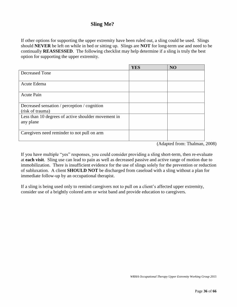
Sling Me?
If other options for supporting the upper extremity have been ruled out, a sling could be used. Slings
should NEVER be left on while in bed or sitting up. Slings are NOT for long-term use and need to be
continually REASSESSED. The following checklist may help determine if a sling is truly the best
option for supporting the upper extremity.
YES NO
Decreased Tone
Acute Edema
Acute Pain
Decreased sensation / perception / cognition
(risk of trauma)
Less than 10 degrees of active shoulder movement in
any plane
Caregivers need reminder to not pull on arm
(Adapted from: Thalman, 2008)
If you have multiple “yes” responses, you could consider providing a sling short-term, then re-evaluate
at each visit. Sling use can lead to pain as well as decreased passive and active range of motion due to
immobilization. There is insufficient evidence for the use of slings solely for the prevention or reduction
of subluxation. A client SHOULD NOT be discharged from caseload with a sling without a plan for
immediate follow-up by an occupational therapist.
If a sling is being used only to remind caregivers not to pull on a client’s affected upper extremity,
consider use of a brightly colored arm or wrist band and provide education to caregivers.
WRHA Occupational Therapy Upper Extremity Working Group 2015

Page 37 of 66
Positioning Devices
Positioning
Devices
Pros Cons
Arm Boards
(half lap tray or
arm trough)
Protects and supports a low tone
upper extremity during wheelchair
use
Places upper extremity in view of
client
Hand is “free” for functional
activity
Upper extremity may be at risk of
trauma secondary to falling off of
the arm board; strapping is not
advised due to possibility of
impingement
Requires height adjustable armrests
on a wheelchair to obtain ideal
position
GivMohr Sling Distal support promotes weight
bearing
Hand is not “free” for functional
activity
Hand piece can be uncomfortable
Hand piece may cause skin
breakdown
Difficult to don/doff independently
Omo Neurexa
Sling (Otto Bock)
Hand is “free” for functional
activity
May reinforce dependent edema of
upper extremity
Difficult to position sling for
optimal shoulder joint position (e.g.
humeral head elevation)
Difficult to don/doff independently
Hemi Sling
Hand is not “free” for functional
activity
Encourages flexor synergy patterns
Contributes to the development of
contractures
Restricts active and passive
movement
Inhibits arm swing
May impact functional balance and
ambulation
Difficult to don/doff independently
Other (e.g.:
pocket, belt,
shoulder bag,
waist pouch)
Low cost
Readily available
Easy transition from support to
functional use of arm
Trial and error for optimal support
and position
WRHA Occupational Therapy Upper Extremity Working Group 2015
Page 38 of 66
8.1.6c Positioning and Supporting the Hand
The Canadian Stroke Best Practice Recommendations 5.2.i states: “Spasticity and contractures may be
prevented or treated by antispastic pattern positioning, range-of-motion exercises, and/or stretching
[Evidence Levels: Early- Level C; Late-Level C]. Routine use of splints is not recommended in the
literature [Evidence Levels: Early-Level A; Late-Level B); however, optimal protocols for utilizing
splinting for improvement or preservation of tissue length and spasticity management have not yet been
determined. In some select patients, the use of splints may be useful and should be considered on an
individualized basis (Evidence Level C). A plan for monitoring the splint for effectiveness should be
provided (Evidence Level C)” (Hebert et al., 2016, p. 12).
Occupational therapists should assess each client individually to determine if splinting would be
beneficial to promote function, manage spasticity, prevent contracture, and/or assist with positioning for
pain and/or edema management. Splinting should always be seen as an adjunct to active task practice
and movement retraining. As with any treatment intervention, clear goals should be documented and
outcome measurement should occur (College of Occupational Therapists & Association of Chartered
Physiotherapists in Neurology, 2015).
The goal of splinting “should be about maintaining the improvement of range of motion and soft tissue
integrity” (Bondoc & Harmeyer, 2013, p. 11). “If muscles are biomechanically imbalanced, and soft
tissues shortened, functional motor recovery will be very challenging for the client” (Bondoc &
Harmeyer, 2013, p. 12).
Splinting Considerations:
“For acute stroke survivors, 35° of wrist extension with MCP’s, PIP’s and DIP’s in neutral” is
recommended (Saebo Inc., 2013, p. 37).
“For chronic stroke survivors, start with the wrist in flexion and finger joints in neutral.
Passively extend the wrist until resistance is felt (fingers begin to curl). This is the initial wrist
position for splinting (“catch one” or resistance, R1)” (Saebo Inc., 2013, p. 37). “The wrist may
be extended to a greater angle as long as the digits are maintained in composite extension to
achieve optimal stretch of the wrist and finger flexors” (Bondoc & Harmeyer, 2013, p. 11).
The thumb should be positioned “in abduction and extension” (Bondoc & Harmeyer, 2013,
p.11).
Occupational therapists should monitor for tingling in the fingers (thumb, index, middle and ring
fingers) if splinting the wrist in flexion, as the median nerve may be compressed. If median
nerve compression neuropathy occurs, wrist may need to be moved out of flexed position,
sacrificing finger extension.
Occupational therapists should “constantly monitor the progression of the client’s hand by
evaluating the range of motion, soft tissue and joint play, and the type of volitional control the
client has regained” (Bondoc & Harmeyer, 2013, p. 11). Occupational therapists should also
monitor skin integrity.
Occupational therapists should consider splinting with a flexible material that allows fingers to
move through flexion with increases in tone (e.g. Aquaplast 3/32), in order to provide a stretch to
the long finger and wrist flexors while maintaining joint integrity.
Page 39 of 66
Serial splinting could be used to progressively increase range of motion (e.g. elbow, forearm,
wrist and/or fingers).
Splinting that provides joint support to facilitate function may also be considered (e.g. opponens
splint or dorsal wrist cock-up splint) (Bondoc & Harmeyer, 2013).
The SaeboStretch is one option available for clients who are able to achieve at least neutral wrist
extension with all finger joints in composite extension. Occupational therapists must be trained
in order to prescribe and use Saebo orthoses with their clients. For eligibility criteria and
information on SaeboStretch orthoses, please see www.saebo.com.
Ensure education is provided regarding wearing schedules and precautions when a client is
provided with a splint. Occupational therapists should monitor the effectiveness of the splint in
regards to the specific goals and adjust or discharge the splint as required. Additional
information should be provided at the time of discharge, if the client still requires the use of a
splint. For an example of a splint instructions handout, please see page 40.
Page 40 of 66
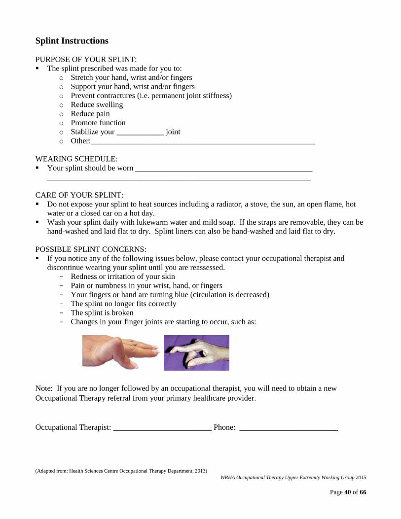
Splint Instructions
PURPOSE OF YOUR SPLINT:
The splint prescribed was made for you to:
o Stretch your hand, wrist and/or fingers
o Support your hand, wrist and/or fingers
o Prevent contractures (i.e. permanent joint stiffness)
o Reduce swelling
o Reduce pain
o Promote function
o Stabilize your ____________ joint
o Other:_________________________________________________________
WEARING SCHEDULE:
Your splint should be worn _____________________________________________
___________________________________________________________________
CARE OF YOUR SPLINT:
Do not expose your splint to heat sources including a radiator, a stove, the sun, an open flame, hot
water or a closed car on a hot day.
Wash your splint daily with lukewarm water and mild soap. If the straps are removable, they can be
hand-washed and laid flat to dry. Splint liners can also be hand-washed and laid flat to dry.
POSSIBLE SPLINT CONCERNS:
If you notice any of the following issues below, please contact your occupational therapist and
discontinue wearing your splint until you are reassessed.
- Redness or irritation of your skin
- Pain or numbness in your wrist, hand, or fingers
- Your fingers or hand are turning blue (circulation is decreased)
- The splint no longer fits correctly
- The splint is broken
- Changes in your finger joints are starting to occur, such as:
Note: If you are no longer followed by an occupational therapist, you will need to obtain a new
Occupational Therapy referral from your primary healthcare provider.
Occupational Therapist: _________________________ Phone: _________________________
(Adapted from: Health Sciences Centre Occupational Therapy Department, 2013)
WRHA Occupational Therapy Upper Extremity Working Group 2015
Page 41 of 66
8.1.6d Shoulder Girdle Taping
The Evidence-Based Review of Stroke Rehabilitation states: “Strapping / taping the hemiplegic shoulder
does not appear to improve upper limb function, but may reduce pain” (Cotoi et al., 2016, p. 19).
The Evidence-Based Review of Stroke Rehabilitation states: “Strapping the hemiplegic shoulder is used
as a method to prevent or reduce the severity of shoulder subluxation and may provide some sensory
stimulation” (Cotoi et al., 2016, p. 18).
There are various taping techniques that are used on the shoulder girdle that seek to optimize alignment
and reduce pain (e.g. McConnell approach, Tri-pull).
Page 42 of 66
8.1.7 Spasticity Management
The Canadian Stroke Best Practice Recommendations 5.2.ii states: “Chemodenervation using botulinum
toxin can be used to increase range of motion and decrease pain for patients with focal and/or
symptomatically distressing spasticity [Evidence Levels: Early-Level C; Late-Level A]” (Hebert, 2016,
p. 12).
The Evidence-Based Review of Stroke Rehabilitation states: “Botulinum toxin works by weakening
spastic muscles through blocking the release of acetylcholine at the neuromuscular junction. The
benefits of botulinum toxin injections are generally dose-dependent and last approximately 2 to 4
months (Bakheit et al. 2001; Brashear et al. 2002; Francisco et al. 2002; Simpson et al. 1996; Smith et
al. 2000)” (Foley et al., 2016, p. 68).
The Evidence-Based Review of Stroke Rehabilitation states: “There is Level I evidence that treatment
with botulinum toxin alone or in combination with therapy significantly reduces spasticity in the upper
extremity and overall disability in stroke survivors” (Foley et al., 2016, p. 72).
The United Kingdom’s National Guidelines for Spasticity in Adults: Management using Botulinum
Toxin states: “It is important to:
Assess the need for orthotics / splinting or review existing orthoses as appropriate once the
clinical effect of muscle weakening is observed (usually 7–14 days post-injection) and ensure
there is a system to review the orthotics / splinting provision, provide new orthoses as required
and assess patient compliance.
Provide patient education on stretching regimes and guidance on participating in activities . . .”
(Royal College of Physicians, British Society of Rehabilitation Medicine, Chartered Society of
Physiotherapy, Association of Chartered Physiotherapists Interested in Neurology, 2009, p. 21).
A review of arm function, including range of motion and tone, prior to injection will assist with
treatment planning and monitoring of outcomes.
It is best to combine botulinum toxin with therapy:
Occupational therapists should communicate with the physiatrist regarding functional goals,
outcome of previous injections and treatment plan.
Post injection, therapy and home programs can focus on strengthening the antagonist muscles as
new movement may now be possible. Active movement training can often be progressed.
“Splinting provides a prolonged stretch to a muscle and, when used together with BT [botulinum
toxin], aims to improve muscle length, correct and prevent contractures and maximise function”
(Royal College of Physicians, British Society of Rehabilitation Medicine, Chartered Society of
Physiotherapy, Association of Chartered Physiotherapists Interested in Neurology, 2009, p. 21).
Splints to help improve range of motion of the elbow, forearm, wrist, and hand as well as
functional splints can be considered. Refer to pages 38 and 39 for splinting considerations.
Splints should be reassessed frequently, including wrist and finger angles, resistance of springs
on dynamic splints, wearing schedule, skin integrity, and tolerance as well as changes in
functional ability.
Functional electrical stimulation may be used post injection to antagonist muscle groups.
Page 43 of 66
8.1.8 Supplementary Training Programs
The Canadian Stroke Best Practice Recommendations 5.1.B.viii states: “Therapists should consider
supplementary training programs aimed at increasing the active movement and functional use of the
affected arm between therapy sessions, e.g. Graded Repetitive Arm Supplementary Program (GRASP)
suitable for use during hospitalization and at home [Evidence Level: Early-Level B, Late-Level C]”
(Hebert et al., 2016, p. 12).
The GRASP program requires palpable or grade 1 wrist extension and active scapular elevation. A
client who is unable to partially open the hand is not appropriate for the GRASP program (Eng, Harris,
Dawson, & Miller, 2012). Please see the following resource for more details:
http://neurorehab.med.ubc.ca/grasp/.
Supplementary training programs should be provided so that strengthening, range of motion and
functional activities completed in therapy can be practiced between therapy sessions with the goal of
increasing the intensity and the number of repetitions being done. Since higher repetitions of upper
extremity use have been associated with better upper extremity outcomes post stroke (Birkenmeier,
Prager, & Lang, 2010), frequent use of the upper extremity between therapy sessions is critical.
Consider the use of a daily homework log or journal as a way of recording activities done at home, and
to increase compliance and accountability.
Page 44 of 66
8.1.9 Mirror Therapy
The Canadian Stroke Best Practice Recommendations 5.1.B.v states: “Mirror therapy should be
considered as an adjunct to motor therapy for select patients. It may help to improve upper extremity
motor function and ADL’s (Evidence Level: Early-Level A; Late-Level A)” (Hebert et al., 2016, p. 12).
The Evidence-Based Review of Stroke Rehabilitation states: “Mirror therapy is a technique that uses
visual feedback about motor performance to improve rehabilitation outcomes. Ramachandran et al.
(1995) first used this method to understand the effect of vision on phantom sensation in arm amputees.
This method has since been adapted from its original use (as a method to “re-train the brain”) as a means
to enhance upper-limb function following stroke and to reduce pain (Sathian et al. 2000). In mirror
therapy, patients place a mirror beside the unaffected limb, blocking their view of the affected limb and
creating an illusion of two limbs which are functioning normally. It is believed that by viewing the
reflection of the unaffected arm in the mirror, this may act as substitute for the decreased or absent
peripheral and proprioceptive input to the affected arm” (Foley et al., 2016, p. 40).
Mirror therapy involves the client placing their affected hand and forearm inside the mirror box and their
unaffected hand and forearm in front of the mirror. The client is then directed to perform a movement
with their unaffected hand and to simultaneously attempt to copy the movement with their hidden
affected hand. The client should be looking at the image in the mirror while attempting to move the
affected hand.
A “mirror box” can be purchased (e.g. http://www.mirrorboxtherapy.com). Alternatively, mirror boxes
can be made by bending cardboard into an inverted V (large enough for the affected hand to fit under) or
by using a box with a mirror attached on one side. Homemade versions have been effectively used with
many clients.
Mirror therapy can be provided as homework. Occupational therapists should provide specific written
instructions for the client, including the number of repetitions or duration of the activity. For an
example of a mirror therapy script, see page 45.
Therapists can also refer to the mirror therapy pocket booklet on Stroke Engine at:
http://www.strokengine.ca/wp-content/uploads/2016/01/Pocket_Card_mirror_therapy.pdf.
Page 45 of 66
Mirror Therapy Sample Script:
Watch the mirror as you complete the activities. Make sure you are trying to do these activities with
your affected (right / left) hand at the same time. Do these exercises 2 – 3 times a day. Go slowly!
1. Make a fist and then open your hand fully. Repeat 15 times.
2. Pretend to play the piano, pushing each finger on the table one at a time. Continue for 2
minutes.
3. Touch your thumb to the tip of each finger. Repeat 15 times for each finger.
4. Place a washcloth on the table. Wipe the table in a circular motion, back and forth, and up and
down, for 2 minutes.
5. Place a water bottle on the table. Grasp it with your hand, lift it up 2 inches, place it back on
the table and then let go. Repeat 15 times.
6. Place 5 coins on the table. Pick them up one at a time until they are all in your palm. Place
them back on the table, one at a time, using your thumb with your index and middle
fingertips. Repeat entire process 3 times.
WRHA Occupational Therapy Upper Extremity Working Group 2015

Page 46 of 66
8.1.10 Sensory Stimulation and Re-training
The Canadian Stroke Best Practice Recommendations 5.1.B.vi states: “It is uncertain whether sensory
stimulation (e.g., transcutaneous electrical nerve stimulation (TENS), acupuncture, muscle stimulation,
biofeedback) improves upper extremity motor function [Evidence Level B]” (Hebert et al., 2016. p. 12).
The Evidence-Based Review of Stroke Rehabilitation states: “Sensorimotor impairment is associated
with slower recovery following stroke; therefore, therapies to increase sensory stimulation may help to
improve motor performance” (Foley et al., 2016, p.23).
An occupational therapist involved in sensory retraining should:
Educate client / caregiver regarding the purpose of sensation, safety concerns, and upper
extremity protection.
Modify the environment for safety (e.g. adjust water temperature).
Introduce varied textures and sensations (e.g. wash cloth, rice, macaroni).
Use different weights, sizes, and shapes of objects to promote discrimination.
Use vision as a compensatory strategy, progressing to occluding vision if able and if safety
permits.
For sensation re-training practical examples, see page 47.
For safety tips that can be used with clients with decreased sensation, see page 48.
Page 47 of 66
Sensation Re-training Practical Examples
1. Take a washcloth and rub it over your affected hand in a circular motion (include forearm too as
necessary). Repeat this for 10 minutes. Keep watching the cloth and try to “feel” it on your
skin.
2. Lightly tap your affected hand with your less affected hand from your forearm to your fingertips.
Repeat for 10 minutes.
3. Use your affected fingertips to trace a textured maze (e.g. Velcro, string, cotton balls, felt, etc.)
with your eyes opened or closed.
4. Clap your hands together at shoulder level. Make sure you hear a loud “clap”. Repeat 10 times.
5. Place a butter knife on the table in front of you. Pick it up using only your affected hand. Get
the butter knife in a good position to cut, and then tap the table with the tip of it as if you are
cutting (or have an item to actually cut, e.g. theraputty). Put the knife down on the table. Repeat
10 times making sure that you have the correct grip each time you pick up the knife.
6. Place a pen or straw in your affected hand, holding it at the bottom. Work your way to the top of
the pen using only your fingertips. Repeat up and down 10 times.
7. Put 5 coins in your pocket. Use your affected hand to pull them out in order from smallest to
largest. Repeat 2 times.
8. Get a dark colored cloth bag. Place various items inside of it (screw, button, paperclip, spoon,
safety pin, coins, etc.), with a list of each item written out. Choose an item from the list and try
to find it in the bag one at a time. Keep track of how many you can find correctly each day.
9. Place a variety of items in a bowl of rice, macaroni, beans, or cereal, and remove them one at a
time with your eyes opened or closed.
WRHA Occupational Therapy Upper Extremity Working Group 2015

Page 48 of 66
Safety Tips For Decreased Sensation
After a stroke you may have decreased sensation (feeling) in certain areas of your body, especially your
hands and feet. Decreased sensation means that you may have more difficulty feeling pain, pressure,
and temperature, making you more at risk of injuring yourself. The following are tips on how to prevent
common injuries and increase safety:
Use your less affected hand to check water temperature (e.g. before having a shower or washing
the dishes).
Label water faucet handles for hot and cold (e.g. red for hot and blue for cold).
Use your less affected hand to handle sharp, hot or cold objects.
Look at the position of your affected arm:
When your affected arm is actively performing a task (e.g. look before reaching into a
cutlery drawer to ensure you do not cut your hand on a knife).
When your affected arm is at rest (e.g. look to ensure your hand is not near the stove
element).
Protect your affected arm during hot and cold seasons:
Wear mittens in cold weather to prevent frost bite.
Apply sunscreen in warmer weather to prevent sun burn.
Ensure you are not holding items too tightly which can damage your skin.
Check your skin daily to ensure there are no pressure (red / darker) areas. Report any changes to
your healthcare provider.
WRHA Occupational Therapy Upper Extremity Working Group 2015

Page 49 of 66
8.1.11 Range of Motion and Strength Training
The Canadian Stroke Best Practice Recommendations 5.1.B.i states: “Range of movement exercises
(passive and active assisted) should be provided that includes placement of the upper limb in a variety of
appropriate and safe positions within the patient’s visual field [Evidence Level C]” (Hebert et al., 2016,
p. 11).
The Canadian Stroke Best Practice Recommendations 5.3.A states that to help prevent hemiplegic
shoulder pain and subluxation, “Overhead pulleys should not be used [Evidence Level A]” and “The
arm should not be moved beyond 90 degrees of shoulder flexion or abduction, unless the scapula is
upwardly rotated and the humerus is laterally rotated [Evidence Level A]” (Hebert et al., 2016, p. 13).
The Canadian Stroke Best Practice Recommendations 5.3.C.i. states: “Treatment of hemiplegic shoulder
pain related to limitations in range of motion includes gentle stretching and mobilization techniques, and
typically involves increasing external rotation and abduction [Evidence Level B]. Active range of
motion should be increased gradually in conjunction with restoring alignment and strengthening weak
muscles in the shoulder girdle [Evidence Level B]” (Hebert et al., 2016, p. 13).
The Canadian Stroke Best Practice Recommendations 5.1.B.ix states: “Strength training should be
considered for persons with mild to moderate upper extremity function in both subacute and chronic
phases of recovery. Strength training does not aggravate tone or pain (Evidence Level A)” (Hebert et
al., 2016, p. 12). Section 5.2.iv states: “The presence of spasticity should not limit the use of strength
training in the arm [Evidence Level: Early-Level C; Late-Level C]” (Hebert et al., 2016, p. 13).
The Canadian Stroke Best Practice Recommendations 5.1.B.x states: “Bilateral arm training does not
appear to be superior to unilateral arm training in improving upper extremity motor function [Evidence
Level B]” (Hebert et al., 2016, p. 12).
The Canadian Stroke Best Practice Recommendations 5.3.E.i states: “Active, active assisted, or passive
range of motion exercises should be used to prevent Chronic Regional Pain Syndrome (CRPS)
[Evidence Level C]” (Hebert et al., 2016, p. 13).
Active range of motion should be encouraged as early as possible. Occupational therapists should
consider the use of gravity reduced positions to facilitate active movement (e.g. supine, side-lying).
Active assisted range of motion describes the client initiating maximal active movement with their
affected upper extremity; the movement is then completed by either the client using their less affected
upper extremity to assist their affected upper extremity or by the occupational therapist providing
assistance.
Passive range of motion should be provided by occupational therapists or trained caregivers to help
maintain full joint range of motion and to prevent contractures.
Self-range of motion is often provided to clients as homework to maintain or improve joint range of
motion. Self-range may not be appropriate if the client does not have the cognitive abilities to carry out
recommendations independently and safely.
For an example of a self-range of motion program, please see page 50.

Page 50 of 66
Self-Range of Motion Exercises for the Arm
What is self-range? Self-range of motion exercises can be used after a stroke when one arm or hand is unable to perform
exercises on its own. During self-range, the less affected arm is used to help the affected arm or hand
through the desired movement.
Why is it important? It is important to move the affected arm to keep the muscles mobile and the joints flexible. Other
benefits may include:
Prevention of stiffness
Improved movement within the joint
Improved sensory and body awareness
Reduced swelling
Most importantly, self-range of motion exercises can help make daily activities (e.g. dressing,
grooming) easier.
General Guidelines Participate in these exercises at least _________ time(s) a day.
Keep movements slow and controlled; avoid rapid and jerky movements.
Hold each position for at least 5 seconds, or as indicated by your therapist.
Do not “overdo it”; do not force the movement.
Exercises will cause a stretch but should not cause sharp pain.
If exercises cause sharp pain, stop until you are able to speak to your therapist.
If you have any questions, contact your therapist_________________ at___________________.
(Revised with permission. Copyright 2002, Ohio State University Wexner Medical Centre, Columbus, Ohio, Patient Education.)
WRHA Occupational Therapy Upper Extremity Working Group 2015
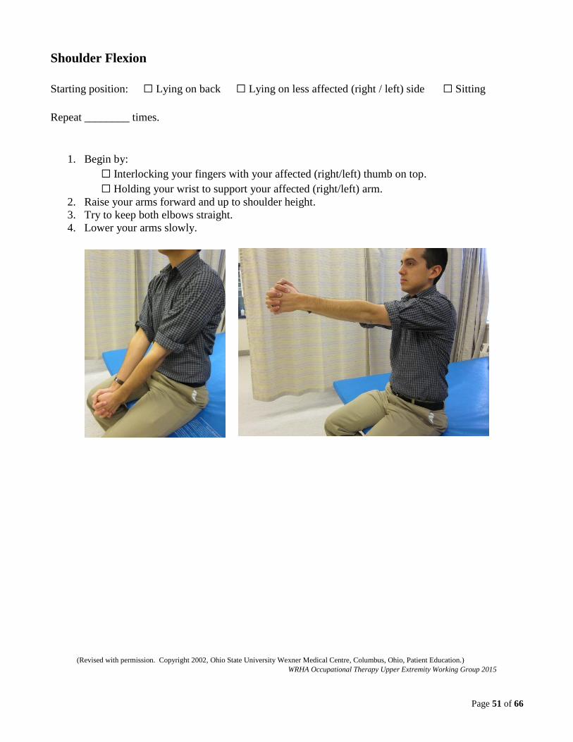
Page 51 of 66
Shoulder Flexion
Starting position: ☐ Lying on back ☐ Lying on less affected (right / left) side ☐ Sitting
Repeat ________ times.
1. Begin by:
☐ Interlocking your fingers with your affected (right/left) thumb on top.
☐ Holding your wrist to support your affected (right/left) arm.
2. Raise your arms forward and up to shoulder height.
3. Try to keep both elbows straight.
4. Lower your arms slowly.
(Revised with permission. Copyright 2002, Ohio State University Wexner Medical Centre, Columbus, Ohio, Patient Education.)
WRHA Occupational Therapy Upper Extremity Working Group 2015
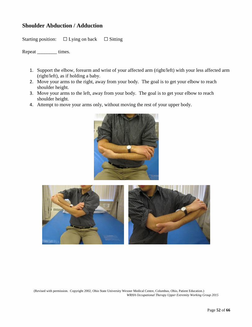
Page 52 of 66
Shoulder Abduction / Adduction
Starting position: ☐ Lying on back ☐ Sitting
Repeat ________ times.
1. Support the elbow, forearm and wrist of your affected arm (right/left) with your less affected arm
(right/left), as if holding a baby.
2. Move your arms to the right, away from your body. The goal is to get your elbow to reach
shoulder height.
3. Move your arms to the left, away from your body. The goal is to get your elbow to reach
shoulder height.
4. Attempt to move your arms only, without moving the rest of your upper body.
(Revised with permission. Copyright 2002, Ohio State University Wexner Medical Centre, Columbus, Ohio, Patient Education.)
WRHA Occupational Therapy Upper Extremity Working Group 2015

Page 53 of 66
Shoulder Internal / External Rotation
Starting position: ☐ Lying on back ☐ Lying on less affected (right / left) side ☐ Sitting
Repeat ________ times.
1. Begin by:
☐ Interlocking your fingers with your affected (right/left) thumb on top.
☐ Holding your wrist to support the affected (right/left) arm.
2. Keep your elbows bent and close to the sides of your body.
3. Use your less affected (right/left) arm to move your affected (right/left) arm across your body.
Keep the elbow of your affected (right/left) arm bent and close to your side.
4. Use your less affected (right/left) arm to move your affected (right/left) arm away from your
body. Keep the elbow of your affected (right/left) arm bent and close to your side.
(Revised with permission. Copyright 2002, Ohio State University Wexner Medical Centre, Columbus, Ohio, Patient Education.)
WRHA Occupational Therapy Upper Extremity Working Group 2015

Page 54 of 66
Elbow Flexion / Extension
Starting position: ☐ Lying on back ☐ Lying on less affected (right / left) side ☐ Sitting
Repeat ________ times.
1. Begin by:
☐ Interlocking your fingers with your affected (right/left) thumb on top.
☐ Holding your wrist to support your affected (right/left) arm.
2. Bend your elbows to bring your hands toward your chest. Try to touch your chin.
3. Straighten your elbows. Attempt to straighten both elbows fully.
(Revised with permission. Copyright 2002, Ohio State University Wexner Medical Centre, Columbus, Ohio, Patient Education.)
WRHA Occupational Therapy Upper Extremity Working Group 2015
(Revised with permission. Copyright 2002, Ohio State University Wexner Medical Centre, Columbus, Ohio, Patient Education.)
WRHA Occupational Therapy Upper Extremity Working Group 2015

Page 55 of 66
Forearm Supination / Pronation
Starting position: ☐ Lying on back ☐ Lying on less affected (right / left) side ☐ Sitting
Repeat ________ times.
1. Begin by:
☐ Interlocking your fingers with your affected (right/left) thumb on top.
☐ Holding your wrist to support the affected (right/left) arm.
2. Keep your affected (right/left) elbow bent and close to the side of your body, with your hands
pointing forward.
3. Use your less affected (right/left) arm to rotate your affected (right/left) arm so the palm faces
upward.
4. Use your less affected (right/left) arm to rotate your affected (right/left) arm so the palm faces
downward.
(Revised with permission. Copyright 2002, Ohio State University Wexner Medical Centre, Columbus, Ohio, Patient Education.)
WRHA Occupational Therapy Upper Extremity Working Group 2015

Page 56 of 66
Wrist Flexion / Extension
Starting position: ☐ Lying on back ☐ Lying on less affected (right / left) side ☐ Sitting
Repeat ________ times.
1. Begin by:
☐ Interlocking your fingers with your affected (right/left) thumb on top.
☐ Holding your wrist to support the affected (right/left) arm.
2. Keep your elbows bent and close to the side of your body, with your hands pointing forward.
3. Use your less affected (right/left) hand to bend your affected (right/left) wrist to the left.
4. Use your less affected (right/left) hand to bend your affected (right/left) wrist to the right.
(Revised with permission. Copyright 2002, Ohio State University Wexner Medical Centre, Columbus, Ohio, Patient Education.)
WRHA Occupational Therapy Upper Extremity Working Group 2015
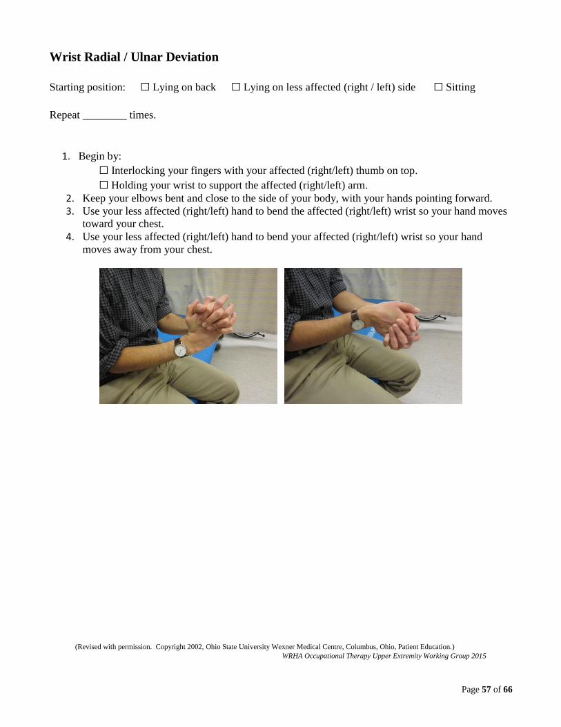
Page 57 of 66
Wrist Radial / Ulnar Deviation
Starting position: ☐ Lying on back ☐ Lying on less affected (right / left) side ☐ Sitting
Repeat ________ times.
1. Begin by:
☐ Interlocking your fingers with your affected (right/left) thumb on top.
☐ Holding your wrist to support the affected (right/left) arm.
2. Keep your elbows bent and close to the side of your body, with your hands pointing forward.
3. Use your less affected (right/left) hand to bend the affected (right/left) wrist so your hand moves
toward your chest.
4. Use your less affected (right/left) hand to bend your affected (right/left) wrist so your hand
moves away from your chest.
(Revised with permission. Copyright 2002, Ohio State University Wexner Medical Centre, Columbus, Ohio, Patient Education.)
WRHA Occupational Therapy Upper Extremity Working Group 2015

Page 58 of 66
Finger Flexion / Extension
Starting position: ☐ Lying on back ☐ Lying on less affected (right / left) side ☐ Sitting
Repeat ________ times.
1. Begin by placing your affected (right/left) arm on your lap or a table.
2. Use your less affected (right/left) hand to bend all the fingers of your affected (right/left) hand
until your fingertips touch your palm.
3. Use your less affected (right/left) hand to open all the fingers of your affected (right/left) hand so
they are straight.
(Revised with permission. Copyright 2002, Ohio State University Wexner Medical Centre, Columbus, Ohio, Patient Education.)
WRHA Occupational Therapy Upper Extremity Working Group 2015

Page 59 of 66
Thumb Flexion / Extension
Starting position: ☐ Lying on back ☐ Lying on less affected (right / left) side ☐ Sitting
Repeat ________ times.
1. Begin by placing your affected (right/left) arm on your lap or a table.
2. Use your less affected (right/left) hand to move your affected (right/left) thumb to touch the base
of your little finger.
3. Use your less affected (right/left) hand to move your affected (right/left) thumb away from your
hand.
(Revised with permission. Copyright 2002, Ohio State University Wexner Medical Centre, Columbus, Ohio, Patient Education.)
WRHA Occupational Therapy Upper Extremity Working Group 2015

Page 60 of 66
Thumb Abduction
Starting position: ☐ Lying on back ☐ Lying on less affected (right / left) side ☐ Sitting
Repeat ________ times.
1. Begin by placing your affected (right/left) arm on your lap or a table.
2. Use your less affected (right/left) hand to move your affected (right/left) thumb away from your
palm (stretch thumb and index finger apart).
(Revised with permission. Copyright 2002, Ohio State University Wexner Medical Centre, Columbus, Ohio, Patient Education.)
WRHA Occupational Therapy Upper Extremity Working Group 2015
Page 61 of 66
8.1.12 Edema Management
The Canadian Stroke Best Practice Recommendations 5.3.D.i states: “For patients with hand edema, the
following interventions may be considered: a) Active, active-assisted, or passive range of motion
exercises in conjunction with arm elevation [Evidence Level C]. b) Retrograde massage [Evidence
Level C] . . .” (Hebert et al., 2016, p. 13).
“Active and active-assisted extremity movement patterns produce muscular contractions that assist
venous and lymphatic return of the fluid” (Ryerson & Levit, 1997, p. 771).
In general, elevating the upper extremity with the hand above the heart can be beneficial in edema
management. When sitting or lying down, pillows can be used to assist with positioning the hand
(Kasch & Walsh, 2013).
“Despite a lack of evidence for the efficacy of light retrograde massage in stroke patients . . . it is often
used in clinical practice and . . . consensus exists about many components of the treatment method”
(Jackson, van Teijlingen & Bruce, 2012, p. 549).
“Compression bandaging may have benefits in the management of edema after stroke” (Gustafsson,
Walter, Bower, Slaughter, & Hoyle, 2014, p. 203). Compression gloves, sleeves, and wrapping for
finger edema (e.g. Coban) can be used and monitored frequently.
Splints may be considered and trialled to manage hand edema in the low level arm post stroke, however
they need to be frequently evaluated. They should not be long term interventions or used during the day
if they interfere with active movement. Refer to splinting considerations on pages 38 and 39 as needed.
Provide client handout regarding splint instructions on page 40 as needed.
A combination of interventions is typically used for edema management.

Page 62 of 66
8.1.13 Virtual Reality
The Canadian Stroke Best Practice Recommendations 5.1.B.vii states: “Virtual reality, including both
immersive technologies such as head mounted or robotic interfaces and non-immersive technologies
such as gaming devices can be used as adjunct tools to other rehabilitation therapies as a means to
provide additional opportunities for engagement, feedback, repetition, intensity and task-oriented
training (Evidence Level: Early-Level A; Late-Level A) (Hebert et al., 2016, p. 12).
Examples of non-immersive systems are the Nintendo Wii and the SaeboReJoyce.
Further research continues to emerge in this developing area of upper extremity rehabilitation.

Page 63 of 66
9.0 Reassessment Guidelines:
It is important to frequently reassess a client’s upper extremity function to monitor progress and modify
treatment plans. Reassessment helps to determine if a client’s goals have been met or need to be
revised. Reassessment can include re-administering initial assessment tools and reviewing SMART
goals with the client, as well as evaluating use of the upper extremity in activities of daily living.
Treatment plans may be modified based on reassessment results.
Page 64 of 66
10.0 References:
Birkenmeier, R. L., Prager, E. M., & Lang, C. E. (2010). Translating animal doses of task-specific
training to people with chronic stroke in 1-hour therapy sessions: A proof-of-concept study.
Neurorehabilitation and Neural Repair, 24(7), 620-635. doi: 10.1177/1545968310361957
Bondoc, S., & Harmeyer, F. (2013). Splinting the neurological hand. OT Practice, 18(8), 9-14.
Chest Heart & Stroke Scotland. (2012). Positioning for people affected by stroke. Retrieved from
http://www.chss.org.uk/documents/2013/08/f16_stroke_positioning_chart.pdf
College of Occupational Therapists and Association of Chartered Physiotherapists in Neurology.
(2015). Splinting for the prevention and correction of contractures in adults with neurological
dysfunction: Practice guideline for occupational therapists and physiotherapists. London, UK: College
of Occupational Therapists Ltd.
Cotoi, A., Viana, R., Wilson, R., Chae, J., Miller, T., Foley, N., & Teasell, R. (2016). Evidence-based
review of stroke rehabilitation: Painful hemiplegic shoulder. Retrieved from
http://www.ebrsr.com/evidence-review/11-painful-hemiplegic-shoulder
Eng, J., Harris, J., Dawson, A., & Miller, B. (2012). Graded repetitive arm supplementary program: A
home-work based program to improve arm and hand function in people living with stroke. Guidelines &
Manual. Vancouver, BC. Retrieved from http://med-fom-
neurorehab.sites.olt.ubc.ca/files/2012/10/GRASP-Instruction-Manual_2012.pdf
Foley, N., Cotoi, A., Serrato, J., Mirkowski, M., Harris, J., Dukelow, S., Sequeira, K., Knutson, J., Chae,
J., & Teasell, R. (2016). Evidence-based review of stroke rehabilitation: Upper extremity interventions.
Retrieved from http://www.ebrsr.com/evidence-review/10-upper-extremity-interventions
Gowland, C., VanHullenaar, S., Torresin, W., Moreland, J., Vanspall, B., Barrecca, S., Ward, M.,
Huijbregts, M., Stratford, P., & Barclay-Goddard, R. (1995). Chedoke-McMaster stroke assessment.
Hamilton, ON: Chedoke-McMaster Hospitals and McMaster University.
Gustafsson, L., Walter, A., Bower, K., Slaughter, A., & Hoyle, M. (2014). Single-case design
evaluation of compression therapy for edema of the stroke-affected hand. American Journal of
Occupational Therapy, 68, 203-211.
Harley, M. Y. (2013, April 29). The return of a second hand: Studies in neuroplasticity after stroke
[Webinar]. Retrieved from http://www.occupationaltherapy.com/ot-
ceus/search/term:the%20return%20of%20a%20second%20hand/
Health Sciences Centre Occupational Therapy Department. (2013). Splinting discharge instructions
[Client handout], Winnipeg, MB, Canada.
Hebert, D., Lindsay, M. P., McIntyre, A., Kirton, A., Rumney, P. G., Bagg, S. et al. (2016). Canadian
stroke best practice recommendations: Stroke rehabilitation practice guidelines, update 2015.
International Journal of Stroke, 11(4), 459-484. Retrieved
from http://journals.sagepub.com/doi/full/10.1177/1747493016643553

Page 65 of 66
Jackson, T., van Teijlingen, E. R., & Bruce, J. (2012). Light retrograde massage for the treatment of
post-stroke upper limb oedema : Clinical consensus using the delphi technique. British Journal of
Occupational Therapy, 75(12), 549-554. doi: 10.4276/030802212X13548955545495
Kasch, M. C., & Walsh, J. M. (2013). Hand and upper extremity injuries. In H. M. Pendleton, & W.
Schultz-Krohn (Eds.), Pedretti’s occupational therapy: Practice skills for physical dysfunction (7th ed.,
pp. 1037-1073). St. Louis, MS: Mosby, Inc.
Kleim, J. A., & Jones, T. A. (2008). Principles of experience-dependent neural plasticity: Implications
for rehabilitation after brain damage. Journal of Speech, Language, and Hearing Research, 51(1),
S225-S239. doi: 10.1044/1092-4388(2008/018)
Krueger, H., Koot, J., Hall, R., O’Callaghan, C., Bayley, M., & Corbett, D. (2015). Prevalence of
individuals experiencing the effects of stroke in Canada. Stroke, 46, 2226-2231.
Lang, C., & Birkenmeier, R. (2014). Upper-extremity task-specific training after stroke or disability: A
manual for occupational therapy and physical therapy. Bethesda, MD: American Occupational Therapy
Association Press.
Law, M., Baptiste, S., Carswell, A., McColl, M. A., Polatajko, H., & Pollock, N. (2014). Canadian
Occupational Performance Measure (5th ed.). Ottawa, ON: CAOT Publications ACE.
McInnes, K., Friesen, C., & Boe, S. (2016). Specific brain lesions impair explicit motor imagery
ability: a systematic review of the evidence. Archives of Physical Medicine and Rehabilitation, 97, 478-
489.
Ohio State University Wexner Medical Centre. (2002). Self-range of motion exercises for shoulders,
arms, wrists, fingers. Retrieved from http://www.sld.cu/galerias/pdf/sitios/rehabilitacion/self-
range_of_motion.pdf
Page, S. J. (2001). Mental practice: A promising restorative technique in stroke rehabilitation. Topics in
Stroke Rehabilitation, 8(3), 54-63. doi: 10.1310/7WDU-V2EA-76F8
Royal College of Physicians, British Society of Rehabilitation Medicine, Chartered Society of
Physiotherapy, Association of Chartered Physiotherapists Interested in Neurology. (2009). Spasticity in
adults: Management using botulinum toxin. National guidelines. London, UK: Royal College of
Physicians. Retrieved from https://www.rcplondon.ac.uk/sites/default/files/documents/spasticity-in-
adults-management-botulinum-toxin.pdf
Ryerson, S. and Levit, K. (1997). Functional movement reeducation: A contemporary model for stroke
rehabilitation. New York, NY: Churchill, Livingstone.
Saebo Inc. (2013). Improving upper extremity motor recovery following neurological injury
[Certification course manual]. Charlotte, NC: Saebo, Inc.
Page 66 of 66
Sirtori, V., Corbetta, D., Moja, L., & Gatti, R. (2009). Constraint-induced movement therapy for upper
extremities in stroke patients. Cochrane Database of Systemic Reviews, 4.
doi: 10.1002/14651858.CD004433.pub2
Stevenson, T., & Thalman, L. (2007). Rehabilitation of the hemiparetic upper extremity [Lecture
notes], School of Medical Rehabilitation, University of Manitoba, Winnipeg, MB, Canada.
Teasell, R. & Hussein, N. (2016). Stroke Rehabilitation Clinician Handbook: Motor Rehabilitation
(Upper Extremity). Retrieved from
http://www.ebrsr.com/sites/default/files/Chapter%204B_Upper%20Extremity%20Post%20Stroke_0.pdf
Thalman, L. (2002). Arm activity list [Client handout], St. Boniface General Hospital Occupational
Therapy Department, Winnipeg, MB, Canada.
Thalman, L. (2008). Treating the arm: A smorgasborg [Presentation], Saskatchewan Society of
Occupational Therapists Annual General Meeting, Regina, SK, Canada.
Related Documents