BioMed Central Page 1 of 20 (page number not for citation purposes) BMC Evolutionary Biology Open Access Research article An amphioxus orthologue of the estrogen receptor that does not bind estradiol: Insights into estrogen receptor evolution Mathilde Paris 1 , Katarina Pettersson 2 , Michael Schubert 1 , Stephanie Bertrand 3 , Ingemar Pongratz 2 , Hector Escriva 3 and Vincent Laudet* 1 Address: 1 Institut de Génomique Fonctionnelle de Lyon, Molecular Zoology team, Université de Lyon, Ecole Normale Supérieure de Lyon, Université Lyon 1, CNRS, INRA, Institut Fédératif 128 Biosciences Gerland Lyon Sud, France, 2 Karolinska Institutet, Department of Biosciences and Nutrition, S-141 57 Huddinge, Sweden and 3 CNRS, UMR 7628, Modèles en Biologie Cellulaire et Evolutive, Observatoire océanographique, F-66651, Banyuls/mer, France Email: Mathilde Paris - [email protected]; Katarina Pettersson - [email protected]; Michael Schubert - [email protected]; Stephanie Bertrand - [email protected]; Ingemar Pongratz - [email protected]; Hector Escriva - [email protected]; Vincent Laudet* - [email protected] * Corresponding author Abstract Background: The origin of nuclear receptors (NRs) and the question whether the ancestral NR was a liganded or an unliganded transcription factor has been recently debated. To obtain insight into the evolution of the ligand binding ability of estrogen receptors (ER), we comparatively characterized the ER from the protochordate amphioxus (Branchiostoma floridae), and the ER from lamprey (Petromyzon marinus), a basal vertebrate. Results: Extensive phylogenetic studies as well as signature analysis allowed us to confirm that the amphioxus ER (amphiER) and the lamprey ER (lampER) belong to the ER group. LampER behaves as a "classical" vertebrate ER, as it binds to specific DNA Estrogen Responsive Elements (EREs), and is activated by estradiol (E 2 ), the classical ER natural ligand. In contrast, we found that although amphiER binds EREs, it is unable to bind E 2 and to activate transcription in response to E 2 . Among the 7 natural and synthetic ER ligands tested as well as a large repertoire of 14 cholesterol derivatives, only Bisphenol A (an endocrine disruptor with estrogenic activity) bound to amphiER, suggesting that a ligand binding pocket exists within the receptor. Parsimony analysis considering all available ER sequences suggest that the ancestral ER was not able to bind E 2 and that this ability evolved specifically in the vertebrate lineage. This result does not support a previous analysis based on ancestral sequence reconstruction that proposed the ancestral steroid receptor to bind estradiol. We show that biased taxonomic sampling can alter the calculation of ancestral sequence and that the previous result might stem from a high proportion of vertebrate ERs in the dataset used to compute the ancestral sequence. Conclusion: Taken together, our results highlight the importance of comparative experimental approaches vs ancestral reconstructions for the evolutionary study of endocrine systems: comparative analysis of extant ERs suggests that the ancestral ER did not bind estradiol and that it gained the ability to be regulated by estradiol specifically in the vertebrate lineage, before lamprey split. Published: 25 July 2008 BMC Evolutionary Biology 2008, 8:219 doi:10.1186/1471-2148-8-219 Received: 3 January 2008 Accepted: 25 July 2008 This article is available from: http://www.biomedcentral.com/1471-2148/8/219 © 2008 Paris et al; licensee BioMed Central Ltd. This is an Open Access article distributed under the terms of the Creative Commons Attribution License (http://creativecommons.org/licenses/by/2.0 ), which permits unrestricted use, distribution, and reproduction in any medium, provided the original work is properly cited.

Welcome message from author
This document is posted to help you gain knowledge. Please leave a comment to let me know what you think about it! Share it to your friends and learn new things together.
Transcript
BioMed CentralBMC Evolutionary Biology
ss
Open AcceResearch articleAn amphioxus orthologue of the estrogen receptor that does not bind estradiol: Insights into estrogen receptor evolutionMathilde Paris1, Katarina Pettersson2, Michael Schubert1, Stephanie Bertrand3, Ingemar Pongratz2, Hector Escriva3 and Vincent Laudet*1Address: 1Institut de Génomique Fonctionnelle de Lyon, Molecular Zoology team, Université de Lyon, Ecole Normale Supérieure de Lyon, Université Lyon 1, CNRS, INRA, Institut Fédératif 128 Biosciences Gerland Lyon Sud, France, 2Karolinska Institutet, Department of Biosciences and Nutrition, S-141 57 Huddinge, Sweden and 3CNRS, UMR 7628, Modèles en Biologie Cellulaire et Evolutive, Observatoire océanographique, F-66651, Banyuls/mer, France
Email: Mathilde Paris - [email protected]; Katarina Pettersson - [email protected]; Michael Schubert - [email protected]; Stephanie Bertrand - [email protected]; Ingemar Pongratz - [email protected]; Hector Escriva - [email protected]; Vincent Laudet* - [email protected]
* Corresponding author
AbstractBackground: The origin of nuclear receptors (NRs) and the question whether the ancestral NR was aliganded or an unliganded transcription factor has been recently debated. To obtain insight into theevolution of the ligand binding ability of estrogen receptors (ER), we comparatively characterized the ERfrom the protochordate amphioxus (Branchiostoma floridae), and the ER from lamprey (Petromyzonmarinus), a basal vertebrate.
Results: Extensive phylogenetic studies as well as signature analysis allowed us to confirm that theamphioxus ER (amphiER) and the lamprey ER (lampER) belong to the ER group. LampER behaves as a"classical" vertebrate ER, as it binds to specific DNA Estrogen Responsive Elements (EREs), and is activatedby estradiol (E2), the classical ER natural ligand. In contrast, we found that although amphiER binds EREs,it is unable to bind E2 and to activate transcription in response to E2. Among the 7 natural and syntheticER ligands tested as well as a large repertoire of 14 cholesterol derivatives, only Bisphenol A (an endocrinedisruptor with estrogenic activity) bound to amphiER, suggesting that a ligand binding pocket exists withinthe receptor. Parsimony analysis considering all available ER sequences suggest that the ancestral ER wasnot able to bind E2 and that this ability evolved specifically in the vertebrate lineage. This result does notsupport a previous analysis based on ancestral sequence reconstruction that proposed the ancestralsteroid receptor to bind estradiol. We show that biased taxonomic sampling can alter the calculation ofancestral sequence and that the previous result might stem from a high proportion of vertebrate ERs inthe dataset used to compute the ancestral sequence.
Conclusion: Taken together, our results highlight the importance of comparative experimentalapproaches vs ancestral reconstructions for the evolutionary study of endocrine systems: comparativeanalysis of extant ERs suggests that the ancestral ER did not bind estradiol and that it gained the ability tobe regulated by estradiol specifically in the vertebrate lineage, before lamprey split.
Published: 25 July 2008
BMC Evolutionary Biology 2008, 8:219 doi:10.1186/1471-2148-8-219
Received: 3 January 2008Accepted: 25 July 2008
This article is available from: http://www.biomedcentral.com/1471-2148/8/219
© 2008 Paris et al; licensee BioMed Central Ltd. This is an Open Access article distributed under the terms of the Creative Commons Attribution License (http://creativecommons.org/licenses/by/2.0), which permits unrestricted use, distribution, and reproduction in any medium, provided the original work is properly cited.
Page 1 of 20(page number not for citation purposes)

BMC Evolutionary Biology 2008, 8:219 http://www.biomedcentral.com/1471-2148/8/219
BackgroundHormone signaling is a very important feature in metazo-ans, allowing communication between cells or organswithin the organism. Two components of these signalingsystems are of particular importance, the hormone and itsreceptor. The nuclear hormone receptor (NR) superfamilyincludes ligand dependent transcription factors that playa central role in various physiological processes as diverseas reproduction, development, and control of homeosta-sis [1,2]. They share a common structural organizationand exhibit a highly conserved DNA binding domain(DBD) and a moderately conserved ligand-bindingdomain (LBD). Some members of this superfamily are lig-anded receptors (24 among the 48 genes encoding NRs inthe human genome) but many lack identified ligand andare therefore called "orphan" [3]. Some orphan receptorsare 'true' orphans in the sense that they do not possess abona fide ligand-binding pocket (LBP), like the membersof the NR4 subfamily (for instance, NURR1, DHR38 orNGFI-B. For review, see [4]), and are regulated by othermechanisms [4]. Alternatively, the crystal structures ofseveral orphan receptors such as HNF4 were found tohave a phospholipid constitutively bound to a large lig-and binding pocket [5,6]. The functional and evolution-ary implications of these constitutive ligands remaindiscussed. Other orphan nuclear receptors have a ligandbinding pocket and thus have the potential to bind com-pounds. It is still not known whether those receptors havenatural ligands, still to be discovered. Undoubtedly, theexistence of such orphan receptors with physiological ordevelopmental activities constitutes both a major chal-lenge for understanding nuclear receptor evolution and apotential opportunity for pharmacology [1].
The existence of orphan and liganded members in the NRfamily raises the question of the evolution regarding theirligand binding ability. Whether the ancestral NR was lig-anded or orphan and more generally how NR ligand bind-ing ability evolved has been recently debated [7-14]. Ingeneral, it is still unclear if there is a correlation betweenthe evolution of the hormone repertoire and NRs. Moreo-ver the mechanisms underlying this coevolution are ofparticular interest [7,12,15-19].
Among the scenarios of NR evolution that have been pro-posed, one suggests that the ancestral NR was a ligand-independent transcription factor which acquired the abil-ity to be regulated by ligands several times during evolu-tion [7,18-20]. This hypothesis was based on theobservation that compounds of similar chemical naturebind to divergent NRs and on the contrary compounds ofvery different nature bind to closely related receptors. Forinstance, orphan receptors are found in all families ofNRs, and steroid receptors are not monophyletic but arelocated in two different subfamilies within the NR super-
family: the ecdysteroid as well as the sex steroid receptors.Interestingly, the evolution of sex steroid hormone recep-tors has also been used as an argument for an alternativehypothesis, the ligand exploitation model [8,11] (for analternative view, see [21,22]). Phylogenetic trees showthat sex steroid hormone receptors are grouped with ERRsas the NR3 subfamily, following the official nomenclature[23]. They contain receptors that bind estradiol (ERs), thatform the NR3A group as well as mineralocorticoids(MRs), glucocorticoids (GRs), progesterone (PRs), andandrogen (ARs) that form the NR3C group. All known lig-ands in this subfamily can be seen as variations aroundthe archetypical sterol skeleton. Consequently, Thorntonet al. suggested in the ligand exploitation model that theancestral steroid receptor was a high affinity estradiolreceptor [8,11] and the other steroid receptors that origi-nated later on, experienced, following gene duplication,shifts in their binding affinities to eventually bind to theirextant ligand. The model in fact suggests that the newlyduplicated receptors (here NR3C) exploit as ligandschemical species that serve as intermediary compounds inthe "ancestral ligand" synthesis pathway (here the estra-diol synthesis pathway) [8]. According to this view,orphan receptors, like ERRs, secondarily lost the ability tohave their activity regulated by a ligand and becameorphan. Interestingly, within the NR3 family, two receptorsubfamilies, ERRs and ERs, appear to be ancient since theyare found in a wide variety of metazoans including deuter-ostomes and protostomes, whereas, up to now, MRs, GRs,PRs and ARs have been found only in vertebrates. Theonly non-vertebrate ERs that have been described so farwere from mollusks and were shown to be unable to bindestradiol [11,12,24-26]. Since the ligand exploitationmodel implies an ancestral estradiol-binding ER and sinceall liganded ER found so far come from vertebrates, and toimprove taxonomic sampling, the ER orthologues fromthe basal vertebrate lamprey and the invertebrate chordateamphioxus were characterized here. Indeed, lamprey andamphioxus are located at key positions in the chordatephylum [27-30]. Moreover, amphioxus (Brachiostoma flor-idae) is much less derived than urochordates in its mor-phology as well as in its genome organization [30].Indeed, amphioxus and vertebrates share a similar generalbody plan whereas urochordate morphology is morederived. For instance, during metamorphosis of some uro-chordates, the tadpole-like larva transforms into an adultthat looks so different that it was first considered as a mol-lusk [31]. Moreover the urochordate genome is fast evolv-ing [27], with for instance the loss of the clustering of thehox genes [32]. There is no ER in the sequenced genomeof Ciona intestinalis [33] or in the sea urchin [34], one ERwas previously cloned in lamprey [8], only one ER wasfound in the amphioxus genome [35]. These reasonsmake lamprey and amphioxus excellent models to studythe evolution of estrogen signaling pathway at the origin
Page 2 of 20(page number not for citation purposes)

BMC Evolutionary Biology 2008, 8:219 http://www.biomedcentral.com/1471-2148/8/219
of vertebrates. In this study, we cloned the unique ER fromamphioxus (amphiER) and characterized it, as well as thepreviously cloned but uncharacterized lamprey ER (lam-pER). AmphiER is an orphan receptor, showing no affinityto the estrogen hormone estradiol, when in contrast, thelamprey ER behaves as a "classical" vertebrate ER. As noER from invertebrates studied so far binds estradiol, wepropose that the ancestral ER (and the ancestral steroidreceptor) was not a receptor for estradiol and gained lateron during evolution the ability to bind the hormone.
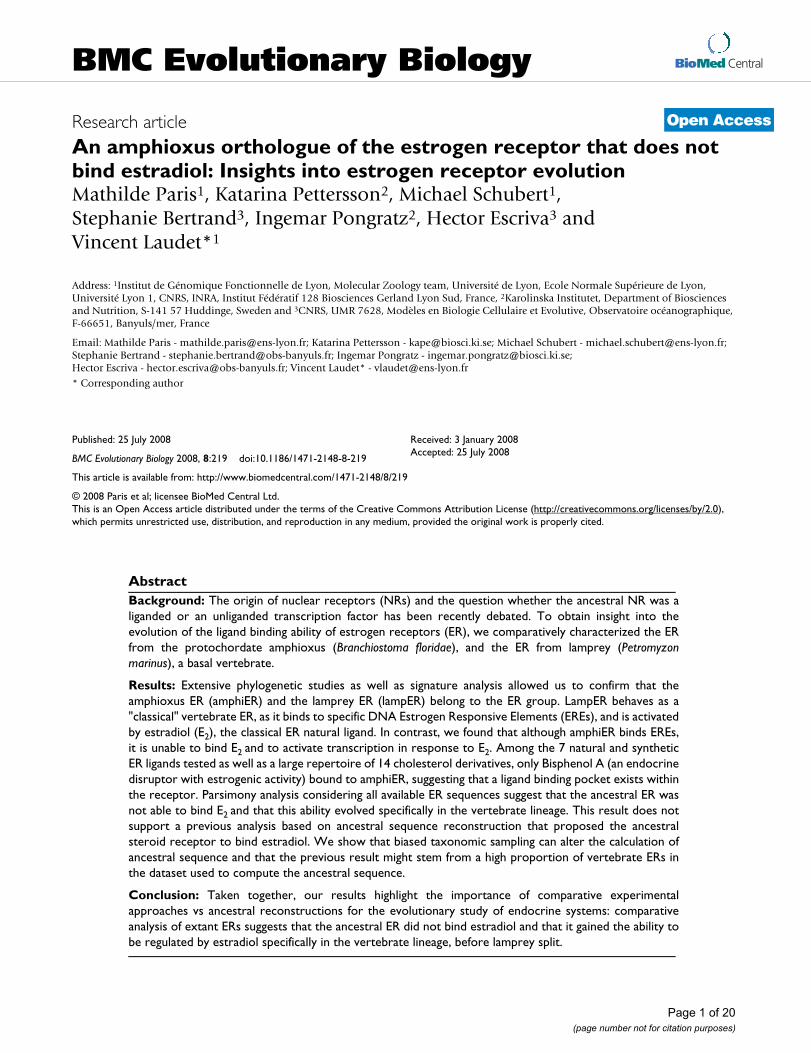
ResultsCloning of the ER from amphioxus (amphiER)Using degenerate primers designed to match motifs in themost conserved part of vertebrate ERs in the DNA bindingdomain, a single gene fragment from total RNA of anadult Branchiostoma floridae was amplified, cloned andsequenced. Rapid amplification of cDNA ends (RACE)was utilized to obtain the full-length cDNA. From thissequence, a new set of specific primers were designed andused to amplify the full length open reading frame of thisgene. The obtained cDNA [GenBank: ACF16007] is 2118bp long and encodes a 705 aa long putative protein (Fig-ure 1) that harbors the classical features of an ER with the5 main functional domains (Figure 2A), among which ahighly conserved DNA binding domain (DBD) and a lessconserved ligand binding domain (LBD). The DBD sharesan 82% sequence identity with the human ERα one (83%with human ERβ) and much less with the other NR3receptors (<62%). The same pattern is observed for theLBD, although this domain is less conserved since itexhibits only 34% amino acid identity with human ERα(35% for human ERβ) and about 20% with other steroidreceptors (Figure 2A). The three other domains, namelythe A/B region in the N-terminal part, the hinge betweenDBD and LBD, and the short C-terminal end of the pro-tein, are more divergent, which is a general pattern forNRs [2] (Figure 2A). The recent release of the amphioxusgenome confirmed the presence of a single ER gene [35].In contrast the previously described lamprey ER is moresimilar to the human ERα with its DBD sharing a 93%sequence identity (93% for human ERβ) and its LBD shar-ing 55% sequence identity (56% for human ERβ) [8].
Phylogenetic analysis of ERsThe orthology relationships of the amphioxus and lam-prey ER sequences were studied in a phylogenetic analysisof the NR3 family using an exhaustive dataset comprising69 members of the NR3 subfamily as well as sequences ofRXRs as an outgroup. The dataset included the 6 currentlyknown mollusk ER sequences (from Nucella lapillus,Crassostrea gigas, Marisa cornuarietis, Thais clavigera, Octo-pus vulgaris, Aplysia californica), as well as the 2 NR3sequences previously known from amphioxus (1 ERR[GenBank: AAU88062] and 1 NR3C [JGI: 201600],
retrieved from a previous work [36] or from the completegenome sequence [35]). In the resulting phylogenetic tree,the sequence of lamprey ER branches within the ER cladewith a high bootstrap support (95%), at the expectedposition before the split of vertebrate ERα and ERβ (Figure2B, and for a tree presenting all sequences, see Additionalfile 1A), as previously shown [8]. The sequence of amph-iER branches within the ERs and is located at the base ofthe vertebrate estrogen receptor group, before the split ofERα and ERβ but after the split of the mollusk ERs (boot-strap value of 81%, Figure 2B). However its precise posi-tion within the ER group is poorly supported (bootstrapvalue of 42%).
Such low bootstrap supports reveal either the weakness ofthe phylogenetic signal contained in ER proteins, or thepresence of two incompatible signals in the data, one sup-porting the observed position of amphiER within ERs,and the other supporting another position. Whereas theweakness of the signal is not testable, the long branchleading to amphiER in the tree suggests that sites that haveundergone a large number of substitutions may accountfor one of the two signals. Such sites may be saturated tothe point that phylogenetic methods are not able to cor-rectly recover their evolution, a situation leading to thelong branch attraction artifact [37]. It is therefore impor-tant to correctly characterize sites that support the ER posi-tion of amphiER: if only fast-evolving sites support thishypothesis, it is probably due to long branch attraction,and an alternative branching should be favored. Alterna-tively, if slowly-evolving sites support this position, onecan confidently identify amphiER as a bona fide ER. Tocharacterize sites with respect to their evolutionary ratesand the amphiER position they favor, both site likeli-hoods and site evolutionary rates were computed for allpossible positions of amphiER.
First, AmphiER was pruned from the tree shown in Figure2B, and then re-grafted in all 149 remaining branches.This yielded 149 topologies, for which site likelihoodsand site evolutionary rates could be computed usingPhyML-aLRT. This allowed us to obtain an evolutionaryrate per site averaged over all possible positions of amph-iER, and therefore independent from the precise positionof amphiER in the tree. Additionally, as likelihoods werecomputed for each of the 149 positions, these positionscould be compared according to the Approximately Unbi-ased test (AU test, implemented in Consel [38]). Out of allthe 149 resulting trees, 26 could not be distinguished withthe AU test and had a likelihood significantly better thanall the other ones (p-value > 0,05). Of these 26 topologies,all but three place amphiER within the ER clade ("ER"trees). The remaining topologies ("alter-ER" trees) placeamphiER either at the base of the NR3C clade (comprisingthe ARs, PRs, MRs and GRs), within the NR3C or at the
Page 3 of 20(page number not for citation purposes)

BMC Evolutionary Biology 2008, 8:219 http://www.biomedcentral.com/1471-2148/8/219
Page 4 of 20(page number not for citation purposes)
Sequence alignment of several ERs including the amphioxus ER and the lamprey ER, as well as ancestral steroid receptorsFigure 1Sequence alignment of several ERs including the amphioxus ER and the lamprey ER, as well as ancestral ster-oid receptors. The DBD is highlighted with light grey. The 12 helices from the LBD are indicated, based on the known 3D structure of human ERα [49]. Amino acids from human ERα making direct hydrogen bonds with E2 are indicated in green. Amino acids making hydrophobic bonds with E2 are highlighted in purple. Amino acids known to be involved in co-activator interaction have been indicated with a star on top of each site [55]. The more divergent A/B domain as well as the F domain have been omitted from the alignment. However, the numbering of the sites along the alignment starts at the beginning of each protein. The exon-intron limits of amphiER and humanERα have been indicated with small red strokes. The sequences of Anc-SRa and AncSRb have been inferred in this study. The sequence of AncSR1 was retrieved from a previous analysis [11].
DBD
H1
H10-H11 H12
H9 H10-H11
H7 H8H6
339226190210175 42
608217
404277237262227 86
659262
460333285318267120
705312
530393345378227187
765372
595449401434383243
822427
650515466500438299
881487
705582527554448304
933521
H3 H4-H5
1amphioxus ER DSPASSGSSI ENNQPQVKEL DNKARAVCRV CGDHASGFHY GVWSCEGCKA FFKRSIQQGQ TDYICPGTNQhuman ERa ------TNDK GSMAMESAK- --ET.-Y.A. .N.Y...Y.. .......... .......-.H N..M..A...human ERb ------VTGP G-----S.R- --D.H-F.A. .S.Y...Y.. .......... .......-.H N.....A...lamprey ER -E.RHVS.GS LPSSTRPER- --STQ-F.A. .S.Y...Y.. .......... .....T.-.H N..M..A...O.vulgaris ER ALLTTDFKKK YDSVVAGSST G.TT.-L.Q. .D.N...... .......... .......-.P V..V..A..Samphioxus NR3C ---------- ---------- -------.A. .HCPST.L.. ..YA.....S ..H.AHK-RA HP.V..AN.N
human PR YLRPD.EA.Q SPQYSFESL- --PQK-I.LI ...E...C.. ..LT.GS..V ....AME-.. HN.L.A.R.Dhuman ERRa ---------- -GKLVLSSL- --PK.-L.L. ...V...Y.. ..A...A... ....T..-.S IE.S..AS.E
71amphioxus ER CTIDRNRRKS CQACRYRKCL MVGMTKDGRR SGERRG-PRK KR----THNQ IDVSSTADSC KSSVSPLPSShuman ERa ....K..... .....L...Y E...M.G.I. KDR.G..RML .H----KRQR ---------- ----DDGEGRhuman ERb ....K..... .....L...Y E...V.C.S. RERCGY.RLV R.----QRSA ---------- ----DE----lamprey ER .......... .....L...Y E...V.G-V. KDRKGF.RGV .H----KRKR ---------- --PIPQKNGGO.vulgaris ER ....KH.... .....L...Y E...N.GSQ. KERKNS.--- SNQTKVKRSS ---------- ----ADFSD.amphioxus NR3C .V...RLK.N .P...LK... AM..SFE..G TIRAKK.--- ---------- ---------- --.HPDP.AE
human PR .IV.KI...N .P...L...C QA..VLG..K FKKFNKVRVV RA----LDAV ---------- -----A..QPhuman ERRa .E.TKR...A .....FT... R...L.E.V. LDRV...G.Q .Y----KRRP ---------- ----------
141amphioxus ER AS--AFDKSR S-ASPTENNS FDSDGDSSTG RELRTAS--- -------HQR LKALIDALDV K-EGEHRGEEhuman ERa GE--VGSAGD M-RAANLWP. PLMIKR.KKN SLALSLT--- -------ADQ MVSALLDAEP P-ILYSEYDPhuman ERb -Q--LHCAGK A-KRSGGHAP RV------RE LL.DAL.--- -------PEQ .VLTLLEAEP PHVLIS.--Plamprey ER EG--GAGGGQ D-V.E.RPQG ERPS.PRDRE SAVSSLE--- -------ADQ VISALLEAEP P-TVLSSYDPO.vulgaris RE TV--NST-.G N-QPA----- ---------- ---------- KSQRLSKSSS .VEELSKN.F A-VP.CKLNPamphioxus NR3C ETPPEQQLIV .PTL.LY.P- ---------- ---------- -------TVP .ISHLVSIEP N-PILTGYNP
human PR VG--VPNE.Q A-L.QRFTF. PGQ.IQL--- ---------- -------IPP .IN.LMSIEP D-VIYAGHDNhuman ERRa EV--DPLPFP G-PF.AGPLA VAGGPRKTAA P--------- -------VNA .VSHLLVVEP E-KLYAMPDP
211amphioxus ER NHPTGQQAGN WQEISNPELI ESVSSLVDRE LTGIICWGKK IPGYSKLSLN DQVLLMESTW LDLLILDLVWhuman ERa TR.------- ---F.EASMM GLLTN.A... .VHM.N.A.R V..FVD.T.H ...H.L.CA. .EI.MIG...human ERb SA.------- ---FTEASMM M.LTK.A.K. .VHM.S.A.. ...FVE...F ...R.L..C. MEV.MMG.M.lamprey ER DK.------- ---VTEAS.M AALT..A... .VHM.T.A.. ...FTAIG.S ...Q.L.CC. .EI..VG.I.O.vulgaris ER SI.------- ---LTKNYIL QLLIQVA.KD .VQL.N.A.H ....AD...S ...H.I.CC. ME.VL.NCAYamphioxus NR3C QCT------- ---PTEGY.M AL.TD.AN.. IE.LVD.AAR L...GM.PMD ...N.IRTV. ....M.G...
human PR TK.------- ---DTSSS.L T.LNQ.GE.Q .LSVVK.S.S L..FRN.HID ..IT.IQYS. MS.MVFG.G.human ERRa AG.------- ---DGHLPAV ATLCD.F... IVVT.S.A.S ...F.S...S ..MSVLQ.V. MEV.V.GVAQ
281amphioxus ER CSIR-HKGEK LLLSGGVLV N--RNTISNR -RNNSSGDDM EVLEMCDQIL SIATKFYEFD LQRREYLCLKhuman ERa R.MEH-PVK- ..FAPNL.L D--..QGKCV EGMVEIF.ML LATSSRFRMM NLQ------- --GE.FV...human ERb R..DH-PG.- .IFAPDLVL D--.DEGKCV EGILEIF.ML LATTSRFRE. KLQ------- --HK....V.lamprey ER R..DR-PGQ- .HFAPNLIL G--.EDAR.V EGMLDMF.ML L.TVSRFRE. HLR------- --.E..V...O.vulgaris ER R.MEY-EG.R .AFASNLIL E--KHHWEIL .GMTQILEQV AAVSEQLLQF G.N------- --.E.L.L.Eamphioxus NR3C R.MEH-R..W .VFAPDL.M D--.SLCRLS .GMEYICTP. LEFARQFAD. QVP------- --QEV.V...
human PR R.YKHVS.QM .YFAPDLIL .EQ.MKE.S- .-FY.LCLT. WQIPQEFVK. QVS------- --QE.F..M.human ERRa R.LPL.-QDE .AFAEDLVL D--EEGAR-A AGLGEL.AAL LQ.VRRL.A. RLE------- --.E..VL..
490
531
659
P-box D-box
AncSRa ---------- ---------- ---------- ---------- -------PDK ILSALLEAEP D-TLYASH-DAncSRb ---------- ---------- ---------- ---------- -------PDK ILSALLEAEP P-TLYASH-DAncSR1 ---------- ---------- ---------- ---------- ---------- -AEQLISALL EA.P.-TVYA
Anc SRa PNKP------ ---DTEAH.L T.LTD.A... .VHM.N.A.H ...FTD.... ...N.LQCC. ME....G.A.AncSRb PNKP------ ---DTEVK.L TALTN.A... .VHM.G.A.H ...FTD.... ..MH.LQCC. MEI...G.A.AncSR1 S.-DPDKP-- ---DTEVSML TTLTN.A... .VH..N.A.. ...FTD...G ..MH.LQCC. MEI...G...
AncSRa R..EHQD.KR .VFAPDLIL D--.EQAK-A AGMLEIF.H. LQ.ARRFRE .KLE------- --KE..V...AncSRb R..EHQD.KT .VFAPDFIL D--.EQAK-A AGMLEIF.HI LQ.VRRFRE .KLE------- --KE.FV...AncSR1 R..DHGE.KT .IFAPDLIL D--.EQGRCV AGMLELF.QL LQIVRRFRE .KLE------- --.E.FV...
AncSRa ..I.LNSDE. .......-GL EDHEKL.QKL R-.KIL...V .CI-GKSGAS .P-ENP..L. QL.....S..AncSRb ..I.LNSDE. .......-GL ENHEKL.QKL R-.KIL...V .CI-GKSGAQ .P-QDW..L. QL......I.AncSR1 .LV.LNSDVL PFLSDTLEEL ENLELI.QKM R-.KI....V .YIAGQRG.G SQ-QQW..F. QL......I.
AncSRa ...NK.IQYF YS--MKRE-G ....H.L..E M.DA.----- ---------- ---------- ----------AncSRb ..ANKGIEYF YS--MKME-G ....H.L..E M.DAH----- ---------- ---------- ----------AncSR1 .I.NKGIE.F YS--MKLQSG V...H.L..E M.DAH----- ---------- ---------- ----------
AncSRa ---------- ---------- ------L.A....... .Y....Y... ........T. .-.HI..M.. A..EAncSRb ---------- ---------- ------L.L....N.. .Y........ ........T. .-.HI..M.. A..EAncSR1 ---------- ---------- ------L.L. ...N...Y.. .......... .......-.H I..M..A..E
AncSR1 ....KH.... .....L...Y E...------ ---------- ---------- ---------- ----------AncSRb ....KR.... .....L.... E...------ ---------- ---------- ---------- ----------AncSRa ....KR.... .....L.... E...------ ---------- ---------- ---------- ----------
351amphioxus ER AITLVHGS-- -------LKG LESDTQ-VRQ LQDDLTDALM DV----CSER HA-LGSRRPA KMLLLLSHLRhuman ERa S.I.LNSGVY TFLSSTLKSL E.K.HI.H.V .-.KI..T.I HLM-AKAGLT LQ-QQHQ.L. QL..I...I.human ERb .MI.LNS.MY PLVTAT.QDA DS.RKL.AHL .-NAV....V W.I.AKSGIS SQ-QQ.M.L. NL.M....V.lamprey ER .MI.LNSGVF FCLSNSAGEQ TNVQLI.QQI .-EKVM...G STI.GHIEAS PP-QH...LS QL.....QI.O.vulgaris ER .TI..NAEVR R----..--L AGFSKI.DDI R-QIILN..I .T----AQKY .P-DNP.HVP SA......V.amphioxus NR3C .L..YTTAVS R----..--L QDYRQV.Q.L Q-HEINE..A EA----..ST FG-FSPGNI. RLMMIV.QVD
human PR VLL.LNTIPL E----..-GL RSQTQF.EEM R-SSYIRE.I KAI-GLRQKG VV-SS.Q.FY QLTK..DN.Hhuman ERRa .LA.ANSDSV H----..--I EDEPRLWSSC E-KL.HE..L EYEAGRAGPG GG-AER..AG RL..T.PL..
421amphioxus ER QVSARASSHL GA--VRNG-L KVPLYDILLD ILTDQVSEGQ RDQQAGHHEV ASSPEKER-- ----------human ERa HM.NKGME.. YS--MKCK-N V.....L..E M.DAHRLHAP TSRGGASV.E TDQSHLATAG STSSHSLQKYhuman ERb HA.NKGME.. LN--MKCK-N V..V..L..E M.NAH.LR.C KSSIT.SECS PAEDS.SKEG SQNPQSQ---lamprey ER HI.NKGIE.. NS--MKRK-N VI----L..E L.DAHSLQNT GLRTSPPPQD FRATLVP--- ----------O.vulgaris ER .A.D.SIIY. QK--QKDE-G H.TFCELITE M.EA.N.SND IVAPRADVIG MGT------- ----------amphioxus NR3C RWFIAQ---- ---------- ---------- ---------- ---------- ---------- ----------
human PR DLVKQLHLYC LNTFIQSRA. S.EFPEMMSE VIAA.LPKIL AGMVKPLLFH KK-------- ----------human ERRa .TAGKVLA.F YG--.KLE-G ...MHKLF.E M.EAMMD--- ---------- ---------- ----------
BMC Evolutionary Biology 2008, 8:219 http://www.biomedcentral.com/1471-2148/8/219
base of (ER, NR3C) (Figure 3A). Because site evolutionaryrates had been computed, sites having a higher likelihood
for the "alter-ER" trees could be compared with sites favor-ing the "ER" topologies with respect to their evolutionaryrates. Interestingly, the sites pleading for the "alter-ER"trees evolve significantly faster than the sites pleading forthe "ER" trees (mean evolutionary rates of 1.20 and 0.90,p-value < 10-5 with a Wilcoxon-test or p < 0.001 with a anunpaired t-test). This suggests that the "alter-ER" signal inthe alignment is probably due to long branch attraction tothe NR3C subtree, which might also be at the origin of thelow bootstrap support (42%) for the position of amph-iER. Conversely, this suggests that amphiER should beconsidered as an ER, as the signal at the origin of this posi-tion does not seem to be artifactual.
An additional test can be run to further confirm thishypothesis, and consists in reestimating the phylogenyusing only slowly-evolving sites. For that purpose, the dis-tribution of expected relative evolutionary rates acrosssites of the alignment was plotted, as found by phyml-aLRT [39,40] (Figure 3B). Fastest-evolving sites wereremoved from the dataset based on three different ratethresholds (2.5, 2 or 1.5, Figure 3B and 3C), and treeswere reconstructed based on the alignments containingonly the remaining slowly-evolving sites. These opera-tions did not impact the monophyly of ERs (Figure 3C) orthe statistical support. This shows that the clustering ofamphiER with vertebrate ERs does not come from satu-rated sites, which argues against long branch attractionbeing at the origin of this position [41]. Accordingly, com-plementary phylogenetic analyses with different methods(bayesian, Neighbor-joining, parsimony) gave similarresults (see Additional file 1). From these studies we con-clude that amphiER does indeed belong to the ER sub-family, which is confirmed by the general conservation ofthe exon-intron structure of amphiER with human ERα,especially at two exon-intron splice sites in the DBD andin the LBD after helix 3 (Figure 2C and short red strokes inFigure 1) [42].
Chordate ERs, including amphioxus ER and lamprey ER, are able to bind estrogen specific response elements (ERE)To test whether the lamprey ER and the amphioxus ER areable to bind DNA on specific estrogen response elements(EREs), electrophoresis mobility shift assays were per-formed using a radiolabeled consensus ERE sequence (seeAdditional file 2). These experiments show that, like ver-tebrate ERs, amphiER and lampER are able to bind DNAspecifically on a consensus ERE. This binding is specific,since a 100-fold excess of non-specific DNA was not ableto compete for binding, whereas a 100-fold excess of coldERE completely suppressed it (see Additional file 2, com-pare lanes 15 and 17, as well as lanes 19 and 21). ERs con-tain two major conserved signatures in the DBD, the P-box (CEGCKA), responsible for the binding specificity toresponse elements, and the D-box, also involved in the
There is a single ER in amphioxusFigure 2There is a single ER in amphioxus. (A) Schematic repre-sentation of the different domains of amphiER. Percent iden-tity of the amphioxus ER with other sequences from the NR3 subfamily in the DNA- and ligand-binding domains is indi-cated. Amino acid sequence of the highly conserved P-box and D-box in the DBD are shown. (B) Maximum likelihood (ML) tree obtained from the analysis of the amino acid sequences of the DBD and the LBD of a wide range of NR3 under a JTT+γ +i model. Bootstrap percentages obtained after 1,000 ML replicates are shown above selected branches. Scale bar indicates number of changes per site. The tree was rooted by selected RXR sequences. (C) The exon-intron structure of amphiER is conserved with that of human ERα, except two minor differences: the first human exon corresponds to the first two amphioxus exons and the last two human exons correspond to the last amphioxus exon.
DBD LBD436296 363 678 705
a/b d ec f
Amphioxus ER
Lamprey ER
Aplysia ER
Crassostrea ER
Marisa ER
Nucella ER
Octopus ER
Thais ER
Human ERa
Human ERb
Human ERRa
Human ERRb
Human ERRg
Amphioxus ERR
Human AR
Human PR
Human GR
Human MR
Amphioxus NR3C
CEGCKA
CEGCKA
CEGCKA
CEGCKA
CEGCKA
CEGCKA
CEGCKA
CEGCKA
CEGCKA
CEGCKA
CEACKA
CEACKA
CEACKA
CEACKA
CGSCKV
CGSCKV
CGSCKV
CGSCKV
CEGCKS
PGTNQ
PATNQ
PATNT
PATNT
PATNN
PATNN
PATNS
PATNN
PATNQ
PATNQ
PASNE
PASNE
PASNE
PATNE
ASRND
AGRND
AGRND
AGRND
PANNN
P-box% %D-box
---
82
77
77
77
77
77
77
82
83
68
71
68
73
54
58
62
61
54
---
33
27
31
28
30
26
30
34
35
22
27
27
26
20
19
19
19
35
shark MR
hagfish NR3Chagfish CR
lamprey CR
lamprey NR3C
0.3
MR
GR
AR
PR
7 5
5 1
7 6
7 9
6 68 1
7 6
1 0 0
1 0 0
1 0 0
9 9
9 9
9 7
1 0 0
1 0 0
1 0 0
9 5
9 5
1 0 0
RXR
ERR
mollusk ER
ER
ER
amphioxus ERlamprey ER
amphioxus NR3C
A
B
C
human ER
amphioxus ERexon 1 exon 2 exon 3 exon 4 exon 5 exon 6 exon 7 exon 8
Page 5 of 20(page number not for citation purposes)

BMC Evolutionary Biology 2008, 8:219 http://www.biomedcentral.com/1471-2148/8/219
DNA binding specificity of the ER dimers (Figure 2A). TheP-box is highly conserved in all known ERs, includingamphiER and lamprey ER and is different from other NR3members. AmphiER and lamprey ER also have a well con-served D-box, amphiER D-box containing just a few con-servative mutations, (e.g. a mutation of an alanine inglycine, Figure 2A). Since the three characterized molluskERs (from A. californica, O. vulgaris and Thais clavigera[11,12,24]) also bind EREs and since the P-box and D-boxare well conserved in all known ERs, including those from
mollusks, ERE binding appears to be a feature specific toall ERs.
Lamprey ER, but not amphioxus ER, is able to induce transactivation of a reporter gene in response to estradiol stimulationThe transactivation ability of lamprey ER and amphiERwas then compared with that of human ERα. AmphiERfailed to induce transcription of a reporter construct con-taining a consensus ERE in front of a minimal promoter
Phylogenetic analysis of amphiERFigure 3Phylogenetic analysis of amphiER. (A) Summary of the 26 best placements of amphiER within a phylogenetic tree com-prised of 69 NR3 and 8 RXR sequences. 23/26 topologies ("ER-tree") place amphiER within the ER clade, the 3 remaining topologies ("alter-ER tree") place amphiER either at the base of (ER, NR3C) or within the NR3C family (close to an AR) or at the base of the NR3 family. The mean evolutionary rate of the sites supporting one of the 23 "ER-tree" topologies (0,9) or for the "alter-ER tree" topologies (1,2) are indicated in (B). (B) Distribution of the site relative evolutionary rates. Rates were esti-mated using an 8 class discretized gamma distribution. The vertical dotted lines correspond to different tentative threshold (2.5, 2, 1.5), above which sites have been discarded due to their high evolutionary rate, before reestimating the phylogeny of the consecutive alignment. (C) Estimation of the minimum of Chi2-based and SH-like supports, available in the aLRT-PHYML software, for the branches defining the monophyly of ERs as well as the position of amphiER. 4 trees were inferred using an alignment on which the fastest evolving sites were removed (no site removed, 34, 53 and 82 sites removed out of 323, with a mean evolutionary rate threshold above 2.5, 2.0 and 1.5, respectively).
Distribution of site evolutionary rates
Site evolutionary rate
Fre
quen
cy
0.0 0.5 1.0 1.5 2.0 2.5 3.
05
1015
20
Sites < 2.5Sites < 2
Sites < 1.5
"alt
er-E
R t
rees
"
"ER
tre
es"
0.3RXR
ERR
ER amphioxus ER
NR3C
23 "ER trees"
"alter-ER tree 1"
"alter-ER tree 3"
"alter-ER tree 2"
0.3
All Sites: 0.87Sites < 2.5: 0.88Sites < 2.0: 0.90Sites < 1.5: 0.85
All Sites: 0.33Sites < 2.5: 0.41Sites < 2.0: 0.20Sites < 1.5: 0.35
RXR
ERR
ER
ER
amphioxus ERlamprey ER
NR3C
mollusk ER
A
B
C
Page 6 of 20(page number not for citation purposes)
BMC Evolutionary Biology 2008, 8:219 http://www.biomedcentral.com/1471-2148/8/219
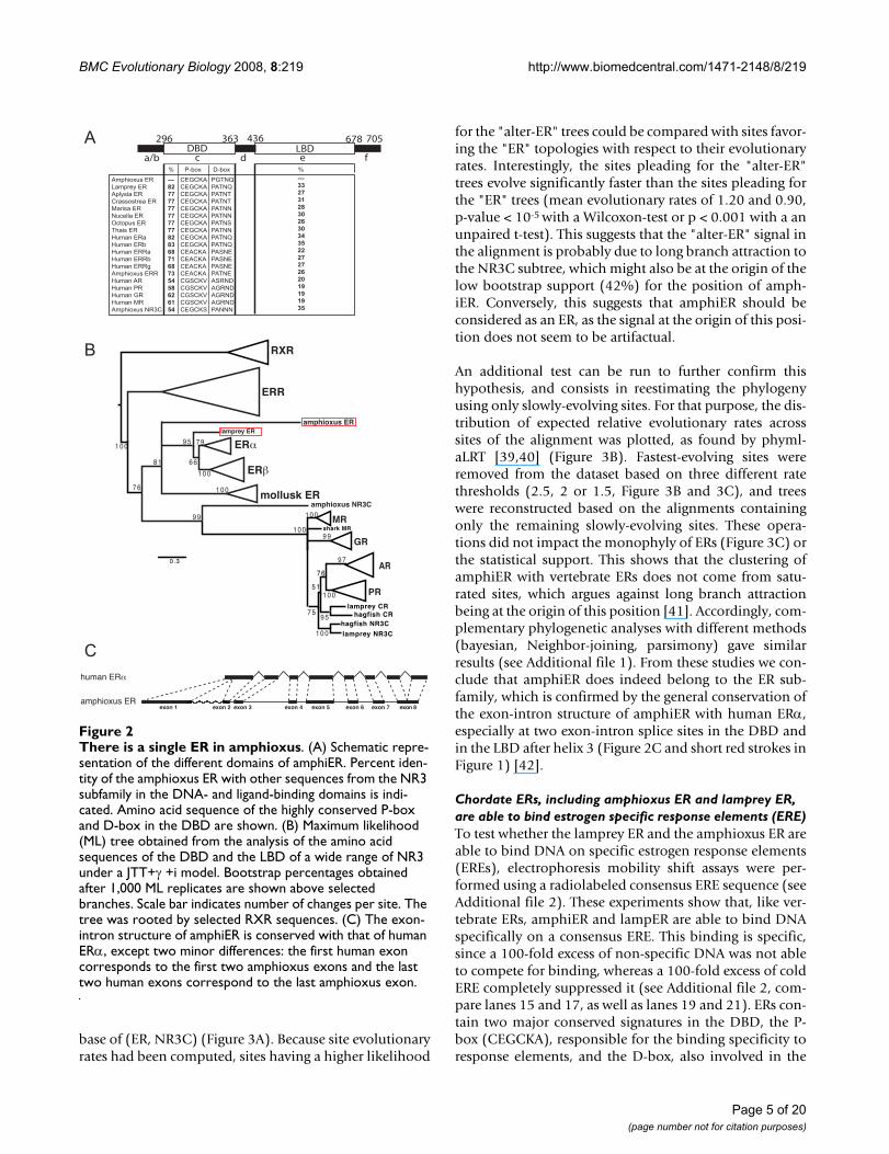
in transfected mammalian cells after stimulation by thenatural vertebrate ER ligand, estradiol (E2) as well as awide variety of other vertebrate ER ligands (the naturalagonist 3β-androstenediol [43], and the phytoestrogensresveratrol [44] and enterolactone [45]) (Figure 4A and4B). In order to improve the detection sensitivity, was alsotested the transactivation capacity of amphiER in responseto E2 as a construct containing only the LBD fused to theGAL4 DNA-binding domain. In this case again, no activa-tion was detected (Figure 4C). In agreement with thisresult, no recruitment of the coactivator SRC1 (an homo-logue of which is present in the amphioxus genome, see
Discussion) was detected in mammalian two-hybrid assay(Figure 4D). However lampER is activated by E2, with anintensity comparable to humanERα (Figure 4C), whichsuggests that the lamprey ER is a high affinity E2-depend-ant transcription factor.
Since amphiER is able to bind DNA but is unable to acti-vate transcription of a reporter gene, the dominant nega-tive capacity of the amphioxus protein was tested. A dose-dependent decrease in the reporter gene activity wasclearly visible in 2 different cell lines when increasingamounts of the amphiER plasmid were added together
LampER is activated by E2 whereas amphiER is not activated by ER agonistsFigure 4LampER is activated by E2 whereas amphiER is not activated by ER agonists. The pSG5 constructs containing either amphiER (A), human ERα or human ERβ (B), were tested in transfected Cos 7 cells for their ability to activate the co-trans-fected cognate ERE-luc reporter plasmid after E2 stimulation (10-6M). (C) GAL4-LBD constructs from several chordate ERs were tested in transfectec 293 cells for their ability to activate a (17m)5x-G-luc reporter plasmid in the presence of increasing doses of E2 (10-9M to 10-6M). (D) Mammalian two-hybrid SRC1 recruitment assay. The GAL4-amphiER-LBD chimera was used with the coactivator SRC1 fused to the strong activation domain VP16 to transfect 293 cells in the presence of increasing doses of E2 (10-9M to 10-6M).
Fol
d ac
tivat
ion
02468
101214161820
E2
vector human ER lamprey ER amphioxus ER
GAL4-amphioxus ERVP16-SRC1
Fol
d ac
tivat
ion
E2
0
1
2
0
100
200
300
400
500
600
700
800
0
0.2
0.4
0.6
0.8
1
1.2
1.4
1.6 -
E2
3b-Adiol
Resveratrol
Enterolactone
-
E2
3b-Adiol
Resveratrol
Enterolactone
Fol
d ac
tivai
on
Fol
d ac
tivat
ion
amphioxus ER vector human ER
consensus ERE consensus ERE
human ER
A
C
B
D
Page 7 of 20(page number not for citation purposes)
BMC Evolutionary Biology 2008, 8:219 http://www.biomedcentral.com/1471-2148/8/219
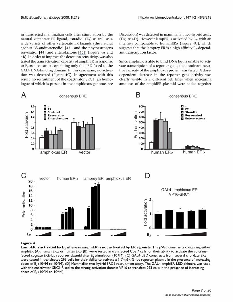
with constant amounts of human ERα or ERβ in transienttransfection experiments. This decrease was observedboth with synthetic consensus EREs (Figures 5A and 5B.See also Additional file 3) and with the natural EREpresent in the classical ER pS2 target gene (Figure 5C).Apparently, amphiER is able to compete with human ERαor ERβ for binding to the ERE sites present in the reporterconstructs, and in doing so, prevents ERα and ERβ frominducing transcription, which results in a decrease inreporter gene activity. Thus, in contrast to Aplysia, Octopusor Thais ER [11,12,24], amphiER does not display consti-tutive transcriptional activity under our experimental con-ditions and rather exhibits an inhibitory effect (Figure 4).This clearly shows that the absence of transcriptionalactivity observed here is not an artifact linked to a poorexpression of the construct but rather reflects the inabilityof amphiER to activate transcription in mammalian cells.
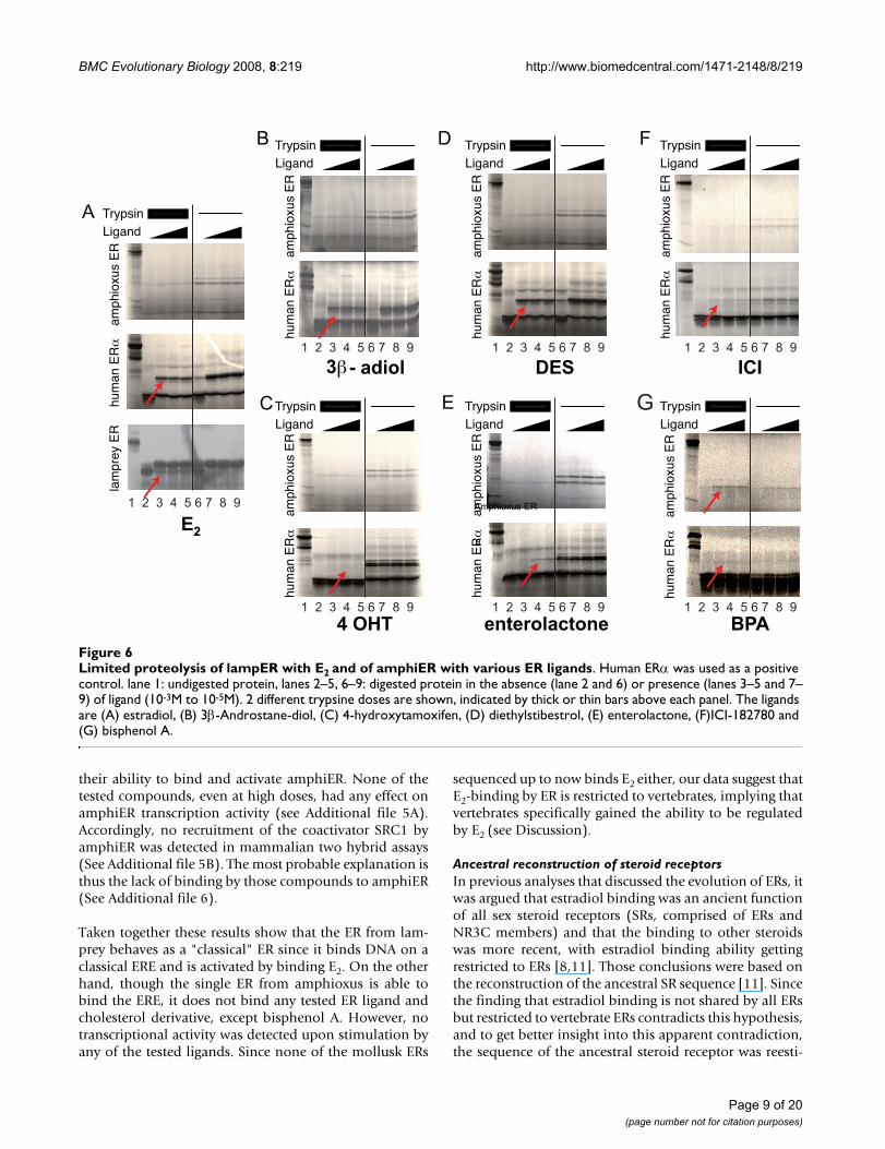
LampER is an estradiol receptor whereas amphiER is not able to bind ER ligands except the synthetic compound Bisphenol AIn order to confirm that lamprey ER is an E2 receptor andto better understand the molecular basis behind the ina-bility of the amphiER to become transcriptionally acti-vated by estradiol stimulation, E2 binding by lamprey ERand amphiER was tested in vitro. For that purpose, limitedproteolysis assay allows to assess whether addition of dif-ferent putative ligands can induce a conformationalchange in amphiER [13]. Using this method the ligandinduced conformational change of the LBD is revealed bythe alteration of the receptor sensitivity toward proteolyticdigestion by trypsin. As expected, E2 was able to protecthuman ERα from proteolysis (Figure 6A). Interestinglylamprey ER was also protected from proteolysis by E2 evenat the lowest concentration tested (Figure 6A), thus con-firming the results of the transactivation assays that thelamprey ER is a high affinity E2 receptor. In contrast noprotection of amphiER by estradiol was observed, even atvery high ligand concentrations (10-3M) (Figure 6A).Since estradiol does not protect amphiER from proteoly-sis, several other classical ER ligands were tested, such asthe synthetic ER agonists diethylstilbestrol [46], 4-hydroxy-tamoxifene [46] or bisphenol A (BPA) [47], thenatural agonist 3β-androstenediol [43], the phytoestro-gen enterolactone [45] or the synthetic ER antagonist ICI-182780 [46]. All compounds were able to bind to humanERα (Figure 6B to 6G) as expected, whereas none but BPAwas able to bind to amphiER (Figure 6G). However, BPAdid not induce transactivation by amphiER in mamma-lian cells reporter assay, and did not induce recruitment ofthe coactivator SRC1 either (see Additional file 4).
In order to rule out the possibility that amphiER is acti-vated by a compound related to E2, a large panel of 14other steroids and cholesterol derivatives were tested for
The amphioxus ER acts as a dominant negative estrogen receptorFigure 5The amphioxus ER acts as a dominant negative estrogen receptor. A pSG5 construct containing human ERα (A) or human ERβ (B) was tested in transfected HeLa cells for its ability to activate the co-transfected cognate ERE-luc reporter plasmid after E2 stimulation (10-6M) in presence of increasing doses of the amphiER construct. (C) A pSG5 construct containing human ERβ was tested in transfected HeLa cells for its ability to activate the co-transfected ps2 promoter after E2 stimulation (10-6M) in the presence of increasing doses of the amphiER construct.
0
200
400
600
800
1000
1200
-
E2
Fol
d ac
tivat
ion
human ER
amphioxus ER
-
E2
Fol
d ac
tivat
ion
human ER
amphioxus ER
-
E2
Fol
d ac
tivat
ion
human ER
amphioxus ER
0
20
40
60
80
100
120
140
0
1
2
3
4
5
6
7
8
consensus ERE
pS2
consensus ERE
A
B
C
Page 8 of 20(page number not for citation purposes)
BMC Evolutionary Biology 2008, 8:219 http://www.biomedcentral.com/1471-2148/8/219
their ability to bind and activate amphiER. None of thetested compounds, even at high doses, had any effect onamphiER transcription activity (see Additional file 5A).Accordingly, no recruitment of the coactivator SRC1 byamphiER was detected in mammalian two hybrid assays(See Additional file 5B). The most probable explanation isthus the lack of binding by those compounds to amphiER(See Additional file 6).
Taken together these results show that the ER from lam-prey behaves as a "classical" ER since it binds DNA on aclassical ERE and is activated by binding E2. On the otherhand, though the single ER from amphioxus is able tobind the ERE, it does not bind any tested ER ligand andcholesterol derivative, except bisphenol A. However, notranscriptional activity was detected upon stimulation byany of the tested ligands. Since none of the mollusk ERs
sequenced up to now binds E2 either, our data suggest thatE2-binding by ER is restricted to vertebrates, implying thatvertebrates specifically gained the ability to be regulatedby E2 (see Discussion).
Ancestral reconstruction of steroid receptorsIn previous analyses that discussed the evolution of ERs, itwas argued that estradiol binding was an ancient functionof all sex steroid receptors (SRs, comprised of ERs andNR3C members) and that the binding to other steroidswas more recent, with estradiol binding ability gettingrestricted to ERs [8,11]. Those conclusions were based onthe reconstruction of the ancestral SR sequence [11]. Sincethe finding that estradiol binding is not shared by all ERsbut restricted to vertebrate ERs contradicts this hypothesis,and to get better insight into this apparent contradiction,the sequence of the ancestral steroid receptor was reesti-
Limited proteolysis of lampER with E2 and of amphiER with various ER ligandsFigure 6Limited proteolysis of lampER with E2 and of amphiER with various ER ligands. Human ERα was used as a positive control. lane 1: undigested protein, lanes 2–5, 6–9: digested protein in the absence (lane 2 and 6) or presence (lanes 3–5 and 7–9) of ligand (10-3M to 10-5M). 2 different trypsine doses are shown, indicated by thick or thin bars above each panel. The ligands are (A) estradiol, (B) 3β-Androstane-diol, (C) 4-hydroxytamoxifen, (D) diethylstibestrol, (E) enterolactone, (F)ICI-182780 and (G) bisphenol A.
E2
4 OHT enterolactone
DES ICI3 - adiol
B
TrypsinLigand
hum
an E
Rla
mpr
ey E
Ram
phio
xus
ER
hum
an E
Ram
phio
xus
ER
hum
an E
Ram
phio
xus
ER
hum
an E
Ram
phio
xus
ER
hum
an E
Ram
phio
xus
ER
hum
an E
Ram
phio
xus
ER
hum
an E
Ram
phio
xus
ER
Amphioxus ER
a
A
BPA
C
D
E
FTrypsinLigand
TrypsinLigand
TrypsinLigand
TrypsinLigand
TrypsinLigand
TrypsinLigand
G
1 2 3 4 5 6 7 8 9
1 2 3 4 5 6 7 8 9
1 2 3 4 5 6 7 8 9
1 2 3 4 5 6 7 8 9
1 2 3 4 5 6 7 8 9
1 2 3 4 5 6 7 8 9
1 2 3 4 5 6 7 8 9
Page 9 of 20(page number not for citation purposes)
BMC Evolutionary Biology 2008, 8:219 http://www.biomedcentral.com/1471-2148/8/219
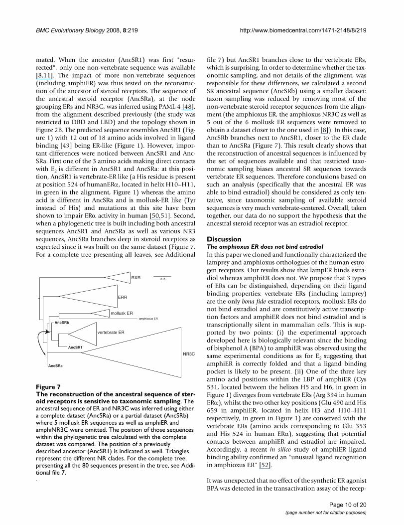
mated. When the ancestor (AncSR1) was first "resur-rected", only one non-vertebrate sequence was available[8,11]. The impact of more non-vertebrate sequences(including amphiER) was thus tested on the reconstruc-tion of the ancestor of steroid receptors. The sequence ofthe ancestral steroid receptor (AncSRa), at the nodegrouping ERs and NR3C, was inferred using PAML 4 [48],from the alignment described previously (the study wasrestricted to DBD and LBD) and the topology shown inFigure 2B. The predicted sequence resembles AncSR1 (Fig-ure 1) with 12 out of 18 amino acids involved in ligandbinding [49] being ER-like (Figure 1). However, impor-tant differences were noticed between AncSR1 and Anc-SRa. First one of the 3 amino acids making direct contactswith E2 is different in AncSR1 and AncSRa: at this posi-tion, AncSR1 is vertebrate-ER like (a His residue is presentat position 524 of humanERα, located in helix H10–H11,in green in the alignment, Figure 1) whereas the aminoacid is different in AncSRa and is mollusk-ER like (Tyrinstead of His) and mutations at this site have beenshown to impair ERα activity in human [50,51]. Second,when a phylogenetic tree is built including both ancestralsequences AncSR1 and AncSRa as well as various NR3sequences, AncSRa branches deep in steroid receptors asexpected since it was built on the same dataset (Figure 7.For a complete tree presenting all leaves, see Additional
file 7) but AncSR1 branches close to the vertebrate ERs,which is surprising. In order to determine whether the tax-onomic sampling, and not details of the alignment, wasresponsible for these differences, we calculated a secondSR ancestral sequence (AncSRb) using a smaller dataset:taxon sampling was reduced by removing most of thenon-vertebrate steroid receptor sequences from the align-ment (the amphioxus ER, the amphioxus NR3C as well as5 out of the 6 mollusk ER sequences were removed toobtain a dataset closer to the one used in [8]). In this case,AncSRb branches next to AncSR1, closer to the ER cladethan to AncSRa (Figure 7). This result clearly shows thatthe reconstruction of ancestral sequences is influenced bythe set of sequences available and that restricted taxo-nomic sampling biases ancestral SR sequences towardsvertebrate ER sequences. Therefore conclusions based onsuch an analysis (specifically that the ancestral ER wasable to bind estradiol) should be considered as only ten-tative, since taxonomic sampling of available steroidsequences is very much vertebrate-centered. Overall, takentogether, our data do no support the hypothesis that theancestral steroid receptor was an estradiol receptor.
DiscussionThe amphioxus ER does not bind estradiolIn this paper we cloned and functionally characterized thelamprey and amphioxus orthologues of the human estro-gen receptors. Our results show that lampER binds estra-diol whereas amphiER does not. We propose that 3 typesof ERs can be distinguished, depending on their ligandbinding properties: vertebrate ERs (including lamprey)are the only bona fide estradiol receptors, mollusk ERs donot bind estradiol and are constitutively active transcrip-tion factors and amphiER does not bind estradiol and istranscriptionally silent in mammalian cells. This is sup-ported by two points: (i) the experimental approachdeveloped here is biologically relevant since the bindingof bisphenol A (BPA) to amphiER was observed using thesame experimental conditions as for E2 suggesting thatamphiER is correctly folded and that a ligand bindingpocket is likely to be present. (ii) One of the three keyamino acid positions within the LBP of amphiER (Cys531, located between the helixes H5 and H6, in green inFigure 1) diverges from vertebrate ERs (Arg 394 in humanERα), whilst the two other key positions (Glu 490 and His659 in amphiER, located in helix H3 and H10–H11respectively, in green in Figure 1) are conserved with thevertebrate ERs (amino acids corresponding to Glu 353and His 524 in human ERα), suggesting that potentialcontacts between amphiER and estradiol are impaired.Accordingly, a recent in silico study of amphiER ligandbinding ability confirmed an "unusual ligand recognitionin amphioxus ER" [52].
It was unexpected that no effect of the synthetic ER agonistBPA was detected in the transactivation assay of the recep-
The reconstruction of the ancestral sequence of steroid receptors is sensitive to taxonomic samplingFigure 7The reconstruction of the ancestral sequence of ster-oid receptors is sensitive to taxonomic sampling. The ancestral sequence of ER and NR3C was inferred using either a complete dataset (AncSRa) or a partial dataset (AncSRb) where 5 mollusk ER sequences as well as amphiER and amphiNR3C were omitted. The position of those sequences within the phylogenetic tree calculated with the complete dataset was compared. The position of a previously described ancestor (AncSR1) is indicated as well. Triangles represent the different NR clades. For the complete tree, presenting all the 80 sequences present in the tree, see Addi-tional file 7.
0.3RXR
ERR
NR3C
mollusk ER
vertebrate ER
amphioxus ER
AncSRa
AncSRb
AncSR1
Page 10 of 20(page number not for citation purposes)

BMC Evolutionary Biology 2008, 8:219 http://www.biomedcentral.com/1471-2148/8/219
tor in mammalian cells since BPA induces a conforma-tional change of amphiER. This apparent absence ofcoactivator recruitment (see Additional file 4B) resultingin no transcriptional activity in response to BPA can beinterpreted in several ways: (i) because of the differentgeometry of the ligand-binding pocket in amphiER, BPAbehaves as an antiestrogen (partial agonist or even a par-tial antagonist) and blocks the transcriptional activationproperties of amphiER, for instance by inducing a confor-mational change that does not allow coactivator recruit-ment (like human ERα and 4-raloxifen, [53]) or byexcluding amphiER from nucleus (like ICI-182,780 withhuman ERα, [54]). (ii) Alternatively, the coactivator inter-face of amphiER does not fit with mammalian coactiva-tors, resulting in artifactual loss of activation. However,the conservation of the amino acids involved in co-activa-tor interaction, compared to human ERα does not supportthis hypothesis (sites indicated with a star in Figure 1, asdescribed in [55]). Among the divergent sites, at a posi-tion implicated in the charge clamp necessary for coacti-vator contact, amphiER contains an aspartate (D677)instead of a glutamate in humanERα (E542). Importantly,the divergence (E->D) is conservative and preserves thenegative charge of the amino acid, which is important forinteraction with the lysine from helix 3 (conserved inamphiER) to form this charge clamp [56]. In addition, aunique orthologue of the p160 family of coactivators wasfound in the amphioxus genome [35] and its overall con-servation with its 3 human ohnologues (genes that havebeen duplicated during the two rounds of whole genomeduplications in the chordate lineage [57]) is good. (iii)Interaction between mammalian chaperones like HSP90and amphiER is impaired, leading to improper binding tothe hormone [58]. Taking these results into account, itwill be interesting to test the effect of BPA on the subcel-lular localization of amphiER and to study if other relatedcompounds are able to bind and/or activate amphiER. Inaddition, it will be important, when cell cultures fromamphioxus are available, to check the activity of amphiERin a monospecific transient transfection assay. It shouldbe remembered that some orphan receptors such as ERRsare thought to have no natural ligands even if they are ableto bind synthetic compounds [59]. More generally, theprecise status of amphiER in terms of ligand bindingremains an open question. It is nevertheless clear, and thisis an important issue for the current evolutionary debate,that amphiER is not able to bind estradiol.
Is there any receptor for estradiol in amphioxus?The observation that amphiER does not bind E2 is indeeda surprising observation since E2 was detected in amphi-oxus by RIA, the hormonal production being correlatedwith breeding season [15]. Several aspects of steroidmetabolism were described in amphioxus [60] and thehomologues of many enzymes necessary for estradiol syn-
thesis in mammals were cloned from amphioxus ovaries[15,61]. Of particular interest is the report of an aromatasegene (CYP19) in amphioxus, which suggests that the cru-cial step in estradiol synthesis is indeed possible inamphioxus. These experimental data were recently con-firmed by the analysis of the complete amphioxusgenome sequence [35]. It may be that, in amphioxus, theactive sex hormone is an E2-derivative [62] or another sexhormone, like in the case of androgens in lamprey [63],and this derivative is still to be discovered. In a similarway, we recently demonstrated that the amphioxus TRorthologue does not bind T3 or T4, the classical thyroidhormones, but deaminated derivatives TRIAC and TET-RAC, which are able to induce amphioxus metamorphosis[64].
A second possibility is that E2 itself has a central role in sexmaturation in amphioxus, and that the functional estro-gen receptor in amphioxus is different from amphiER.Several candidates are possible. First, there is another ster-oid receptor in amphioxus (amphiNR3C in Figures 1 and2) [35] that exhibits several ER-like features. Its P- and D-boxes are closer to ERs than to vertebrate NR3C (Figure2A). The sequence identity of its LBD with human ERα(37%) and with NR3C members (35%) are similar. More-over, most of the amino acids involved in ligand bindingare more ER-like than AR-, PR- or MR-like (Figure 1).However it is the only NR3C receptor (orthologous to AR,PR, MR and GR) found in the amphioxus genome. Thus ifamphiNR3C plays the role of an estradiol receptor, thissuggests an absence of a "classical" steroid receptor able tobind testosterone, progesterone or corticoids. Alterna-tively, a non-nuclear receptor could mediate E2 action inamphioxus. Indeed, several non-genomic effects of estra-diol were reported in mammals involving GPCRs (forreviews see [65-67]). For instance, very recently, a highaffinity receptor for the steroid androstenedione linked tothe membrane, was described in lamprey [68] and aGPCR with high affinity for progestines was isolated fromsea trout [69].
Implications for the evolution of ERsThe absence of E2 binding by the amphioxus estrogenreceptor has interesting consequences for the evolution ofSRs and ERs. Indeed, only the well characterized gnathos-tome ERs and the lamprey ER (studied here) have beenshown to mediate E2 action. Outside vertebrates, all theERs studied so far (in mollusks and amphioxus) do notbind E2 [11,12,24-26]. Parsimony implies that the func-tion of estradiol in the bilaterian ancestor was not medi-ated by ER and that ER had another function. Only laterduring evolution, in the vertebrate lineage, ER would thenhave gained the ability to be activated by E2 and to medi-ate the hormonal action of this compound (Figure 8). Thealternative scenario (ancestral E2 binding and independ-
Page 11 of 20(page number not for citation purposes)

BMC Evolutionary Biology 2008, 8:219 http://www.biomedcentral.com/1471-2148/8/219
ent loss of either ER itself or E2 binding to ER in mollusksand invertebrate deuterostomes) is more costly in terms ofevolutionary events, even if the hypothesis of an NR3Corthologue binding E2 is taken into account. Thus, takentogether, our results do not support previous scenarios ofsteroid receptor evolution based on a reconstruction ofthe ancestral steroid hormone receptor AncSR1 [8,11].
To describe the evolution of a protein, being able to studyancestral sequences at different nodes of a phylogenywould obviously provide historically relevant informa-tion that is not available otherwise [70]. However suchsequences have disappeared long ago and can only be sta-tistically estimated. The accuracy and bias of these estima-tions therefore need to be investigated. Indeed, functionalstudies of ancestral sequences are of any value only if theancestral reconstruction is reliable enough. The confi-dence associated with the previously published ancestralsteroid receptor is quite low. Indeed, the overall accuracyof the reconstruction of the LBD (AncSR1) was only 62%[11]. This is similar for the ancestor inferred here (Anc-
SRa) on an enriched dataset, with an overall accuracy ofthe DBD+LBD of 70%. Moreover, amino acid uncertaintywas high at many sites of AncSRa and AncSR1: more than60 sites have more than 1 possible amino acid with aprobability superior to 0.2. If one were to make an exhaus-tive study, one would need to reconstruct and test morethan 1024 potential proteins (if all possible combinationsof amino acids with probability > 0.2 were tested). In factseveral of the sites involved in ligand binding have lowprobabilities. Examples of more reliable reconstructionsof nuclear receptors have been published, e.g. the ancestorof MR and GR (mineralocortoid and glucocorticoid recep-tors) in which the overall accuracy of the LBD was above99%, with no disrupting mutation at any site [71]. Thereconstructed ancestor of RARs also showed a high aver-age confidence (99% [13]). This discrepancy betweenresults obtained on the ancestor of all steroid receptors ormerely of MR and GR for instance, can be explained by thehigher sequence divergence observed among all SRs thansimply among subfamilies MR and GR (see branch lengthin Figure 2B). Consequently, the uncertainty associated tothe sequence of the ancestral steroid receptor as estimatedwith nowadays methods is probably too high to provide afirm basis for evolutionary conclusions. Moreover, thephylogenetic reconstruction of ancestral sequences hasbeen shown to be biased towards the most frequent (andmore stable) amino acids, resulting in an under-estima-tion of the less frequent amino acids (the stability of theancestral protein is then over-estimated [72]). In recon-structions of ancient proteins, where the evolutionary sig-nal has been lost due to a high number of substitutions,such biases might be problematic. Thus, current recon-struction methods do not seem powerful enough to infera biologically meaningful ancestral steroid receptor giventhe amount of divergence between sequences.
Nonetheless, all these reservations put aside, it is surpris-ing that the previously reconstructed ancestral SR, is verte-brate ER-like. As almost all the extant sequences used asmatrix for the reconstruction came from vertebrates andled to the estimation of a "vertebrate-like" ancestralsequence, the same ancestral steroid receptor as previ-ously published [11] was estimated, but adding moresequences from various taxa. This reconstruction wasdone using a phylogeny equivalent to the one previouslypublished [11]. Using this approach, the new AncSRa ismore divergent from vertebrate ERs than AncSR1. Interest-ingly AncSR1 was shown to bind E2 with a very low affin-ity (250 times lower affinity than human ERα [11]),suggesting that AncSRa may be an even worse estradiolreceptor.
The bias of AncSR1 towards vertebrate ERs is explained bya lack of non-vertebrate sequences used for the reconstruc-tion. Indeed, removing some non-vertebrate sequences
Model of evolution of the ligand binding ability of ERsFigure 8Model of evolution of the ligand binding ability of ERs. On a classical phylogenetic tree of bilaterians, data available on the binding ability of all known ERs have been indicated. Two hypotheses are compared in terms of parsimony, whether the ancestral ER was liganded (in blue) or not (ie an orphan receptor) (in red). This result displays different costs in terms of parsimony: one unique event of gain specifically in vertebrate for the "ancestral ER orphan" hypothesis against at least two parallel events of loss of binding for the "ances-tral ER binding E2" hypothesis. In addition, three events of loss of the ER gene in urochordates, echinoderms and insects+nematodes are implied by the current distribution of the gene across metazoans.
Page 12 of 20(page number not for citation purposes)
BMC Evolutionary Biology 2008, 8:219 http://www.biomedcentral.com/1471-2148/8/219
from our dataset leads to an estimation of an ancestralsteroid receptor that is more "vertebrate ER"-like (AncSRbin Figure 7). The clustering of AncSRb with the ER cladeand the exclusion of AncSRa from the ER clade were sup-ported with good statistical values (minimum of Chi2-based and SH-like supports of 0.83 and 0.89, respec-tively). Those data show that AncSR1 reconstruction wasprobably sensitive to the vertebrate bias in the data set(Figure 7). Overall, we suggest that analysis based onancestral reconstructions should be taken as tentative,especially in case of low statistical confidence and limitedtaxonomic sampling. In case of the ancestral steroid recep-tor, even if exhaustive taxonomic sampling is necessary,phylogenetic signal is weak and the resulting confidence isquite low. Thus we think that even if the ancestralsequence built here is biologically more relevant than pre-viously calculated ones (because of better taxonomic sam-pling), it remains quite uncertain. Consequentlyconclusions regarding the ancestral steroid receptorshould be based mostly on comparative characterizationof extant receptors. In that case, all the data based oninvertebrate ER receptors (from mollusks and amphioxus)support an ancestor of steroid receptors that was not ableto bind estradiol. This conclusion will obviously requirethe functional characterization of ERs from other proto-stome phyla in order to carefully check if this observationis general. Thus, available data converge towards a re-eval-uation of the ancestral status of estrogen receptors.
Sequence conservation reflects functional constraints: ligand binding ability is more recent than DNA-binding abilityFrom our and previous studies, only vertebrate ERs areable to bind and activate transcription under estradiolstimulation [11,12,24-26]. The LBD of amphiER is moredivergent from its vertebrate counterparts (ca. 34% aminoacid identities) than the LBD of other liganded amphi-oxus nuclear receptors such as amphiRAR (ca. 58%),which has been shown to bind the same ligand as its ver-tebrate homologue [7,13]. This suggests that a conservedfunctional feature (e.g. binding to the same ligand) isreflected in the sequence conservation of the LBD.
The same observation can be done concerning the DBDsince all ERs, including amphiER, have a highly conservedDBD and are able to bind EREs. Thus, for this domainalso, a conservation of the function is reflected insequence conservation.
Accordingly with this notion, the LBD of invertebrate ERsis highly divergent but their DNA binding domain, as wellas other functionally important domains not directlylinked to ligand binding such as the dimerization inter-face, or the amino acids responsible for interaction withthe co-activators [55] are well conserved. This is true foramphiER as well as mollusk ERs. This strongly suggests
that amphiER is a bona fide NR regulating ERE-containinggenes in an E2-independent manner. Post-translationalmodifications such as phosphorylation or the presence/absence of other receptor-interacting proteins such astranscriptional coactivators have been shown to regulateunliganded nuclear receptors [73]. Whether one of thesemechanisms acts to regulate the activity of invertebrateERs or if those receptors have unknown ligands still to beidentified remains to be explored. Anyway our observa-tions strongly suggest that for ERs, the DNA binding func-tion of the receptor as well as its interaction with co-regulators have been conserved due to selective pressure.Interestingly, when studying the AncSRa, the P- and D-boxes in the DBD are ER/ERR-like (Figure 1), suggestingthat ER/ERR DNA binding ability is ancestral, in accord-ance with the fact that these are the only receptors of theNR3 family found in invertebrates. This difference in theselection pressure between DBD and LBD has been pro-posed to be a general evolutionary pattern for the wholeNR family [7]. The plasticity of the ligand binding abilityof NRs was recently illustrated in the case of RXR-USPwhere the ability of the receptor to be regulated by a lig-and was suggested to have been subject to several succes-sive episodes of gain and loss during evolution [74].
Evolution of endocrine systems: refinement of the ligand exploitation modelThe ligand exploitation model hypothesizes how newhormones and new receptors appear during evolution. Itsuggests that the ancestral ligand is the last metabolite ofa synthesis pathway [8]. According to this model, theancestral steroid ligand was estradiol (and the ancestral SRbound estradiol). During evolution, other steroid recep-tors appeared by duplication of the ancestral ER andgained the ability to bind other steroids, intermediate inthe synthesis pathway (like testosterone or progesterone).
Our findings on the evolution of ERs do not support theligand exploitation model, since our data strongly suggestthat the ancestral ER did not bind estradiol. However, asestradiol has been detected in deuterostomes as well asprotostomes (for instance in vertebrates, amphioxus,echinoderms, mollusks, for review, see [75]), steroid sign-aling may have been already present in bilaterian ances-tor. However, up to now, the ancestral steroid moleculeremains to be determined. If estradiol is an ancient hor-mone, it then probably bound another receptor and lateron ER gained the ability to recognize it, as did other ster-oid receptors for their extant ligand. Thus the evolution ofsteroid system intermingles two distinct processes, theevolution of the receptor on one hand, and the evolutionof the ligand on the other.
The receptor can evolve by point mutations and change itsaffinity for a ligand towards another. This idea was con-vincingly exemplified in the case of corticoid receptors
Page 13 of 20(page number not for citation purposes)

BMC Evolutionary Biology 2008, 8:219 http://www.biomedcentral.com/1471-2148/8/219
(the ancestor of MR and GR) for which it was recentlydemonstrated that ability of the ancestral vertebrate corti-coid receptor to bind gnathostome-specific hormonealdosterone (a MR ligand) was a by-product of its abilityto bind the ancestral ligand 11-deoxycorticosterone(DOC) [71]. GR gained the ability to bind cortisol only inthe gnathostome lineage, in parallel to endogenous syn-thesis of the hormone [16,71]. This detailed study showsthat a receptor binding a given ligand can acquire affinityfor compounds present in the cell that are structurallyclose to its natural ligand: this refines the ligand exploita-tion model, since new ligands are not necessarily precur-sors of ancient ligands, simply compounds present in thecell and structurally close to the ancestral ligand. Similarconclusions were drawn previously in the case of RAR evo-lution [13]. It has to be emphasized that the pool of avail-able compounds is also subject to evolutionary changes inparallel. For instance, the spatiotemporal production ofestradiol is variable in the different vertebrate groups(reviewed in [76]). Glucocorticoids differ in mouse (corti-sol) and in human (corticosterone), with both hormonesbeing GR ligands [77]. There are several androgens in tel-eost fishes, with 11-ketotestosterone being teleost-specific[78]. As there are 2 androgen receptors (ARs) in teleostfishes, from a whole genome duplication [79], the studyof the ligand-binding ability of those ARs is a potentiallyinteresting case for the evolution of endocrine systems. Ashighlighted by Bridgham et al. (2006), lamprey does notproduce cortisol [71]. In accordance, their genomes donot contain the sequence corresponding to the enzymeresponsible for cortisol synthesis (11b-hydroxylase) andin general classical steroids except estradiol are rarelyfound in lamprey. This suggests that the steroids actuallyfound in lamprey are different from the ones found inmammals (reviewed in [80]). Those cases exemplify thelargely underestimated diversity of endocrine systems:except for lamprey and some teleost fishes, the hormonalpool of animals remains largely unknown. As proposedfor the study of steroid receptors, a comparative approachshould be applied to determine the metabolism of ster-oids in poorly studied animals. Indeed, the hormonalpool of such animals is usually evaluated from the pres-ence/absence of putative orthologues of mammalianenzymes. As the enzymatic machinery involved in hormo-nal metabolism has a very labile activity (reviewed in[81,82]), equating orthology with functional identitymight be unreliable.
The evolution of steroid receptors can be replaced in themore general context of ligand-nuclear receptor co-evolu-tion. The evolution of the NR1H subfamily, that includesreceptors for other steroidal compounds, like the majortranscriptional regulator of bile salt synthesis farnesoid ×receptor (FXR), the pregnane × receptor (PXR), the vita-
min D receptor (VDR) or liver × receptor (LXR)/ecdysonereceptor (Ecr), has been extensively studied and is not inline with the ligand exploitation model [83-85]. Forinstance, comparative functional studies of FXRs from var-ious chordate species showed that the vertebrate FXRsbind "late" cholesterol derivatives (from a complex syn-thesis pathway) but are thought to have evolved from anancestral FXR that bound early cholesterol derivatives(from a simpler synthesis pathway) [83].
In other cases, the evolution of ligand binding is more"chaotic" with close orthologs having a selective ligandbinding ability that varies extensively (vertebrate VDRs arevery well conserved when PXRs have the widest ligandrepertoire of all NRs) [83].
These complex histories are probably linked to specificfunction of some of those NRs, considered as xenotoxiccompounds "sensors". This tight relationship with theunstable environment probably makes receptors like FXRand especially PXR more prone to fast evolution [86]. Yetthey illustrate the impressive variety of scenarios of NRevolution.
ConclusionIn this article, we demonstrated that vertebrate ERs(including lamprey ER) are estradiol receptors whilst non-vertebrate ER (including amphioxus ER) are not. The mostparsimonous scenario proposes that the ancestral ER wasnot able to bind estradiol and that it had another func-tion. It later gained the ability to be regulated by estradiol,specifically in the vertebrate lineage. However, additionalcritical data remains to be discovered in poorly studiedtaxa [62]. To fully understand the evolution of steroid sig-naling pathway, a larger number of taxa need to be tar-geted for detailed comparative studies. More precisely,ERs and other steroid receptors should be cloned fromwidely distributed taxa, especially in protostomes.Enzymes involved in steroidogenesis should also becloned and characterized, to understand the evolution ofsteroid availability. In order to avoid the blinders of a"vertebrate-centered" view, it is of particular importanceto establish the steroid hormone repertoire of an enlargedanimal panel, including more protostomes. The descrip-tion of various endocrine systems will certainly be rele-vant to the early evolution of hormone signaling.
MethodsCloning of amphiERAn initial piece of amphiER was obtained by degeneratePCR on different RT reactions from total RNA extractedeither from developing B. floridae embryos and larvae (at13 h–15 h, 28 h, 36 h, 48 h or 3 d–4 d of development)or from B. floridae adults. The oligonucleotides used were
Page 14 of 20(page number not for citation purposes)

BMC Evolutionary Biology 2008, 8:219 http://www.biomedcentral.com/1471-2148/8/219
as follows: forward primer 5'-TGYGARGGITGYAARGCIT-TYTT-3' and reverse primer 5'-GTRCAYTSRTTIGTIGCIG-GRCA-3'.
The touchdown PCR program used was as follows:
5' 94 degrees
5× (30" 94 degrees, 1' 55 degrees, 1' 72 degrees)
5× the same cycle, but at 50 degrees annealing tempera-ture
5× the same cycle, but at 45 degrees annealing tempera-ture
5× the same cycle, but at 40 degrees annealing tempera-ture
25× the same cycle, but at 37 degrees annealing tempera-ture
7' 72 degrees
All degenerate PCRs irrespective of the RT reaction tem-plate used yielded a 83 bp fragment of amphiER. The frag-ment was sequenced on both strands and used for thedesign of oligonucleotides for 5' and 3' RACE experimentswith the Invitrogen GeneRacer Kit. The template for theRACE experiments was pooled total RNA from 13 h–15 hB. floridae embryos and from B. floridae adults. In addi-tion to the oligonucleotides provided by the kit, for the 3'RACE, the following primers were used:
3' RACE, 1st PCR: 5'-AACGGAGCATTCAGCAAGGTC-3'
3' RACE, 2nd PCR: 5'-GCATTCAGCAAGGTCAGACAG-3'
5' RACE, 1st strand cDNA synthesis: 5'-ATGTAATCT-GTCTGACCTTGC-3'
5' RACE, 1st PCR: 5'-CTGTCTGACCTTGCTGAATGC-3'
5' RACE, 2nd PCR: 5'-TCTGACCTTGCTGAATGCTCC-3'
The protocols for the 1st and 2nd round of PCR experi-ments are given in the Invitrogen GeneRacer Kit. The 3'and 5' RACE products were subsequently sequenced onboth strands and used for the design of oligonucleotidesfor the full-length cloning of amphiER: forward primer 5'-CGGCGAAGCGAAGAAGATCGAG-3' and reverse primer5'-CTTAACCGATACTAACGGAACAG-3'. The full-lengthamphiER was obtained by PCR on pooled RT reactionsfrom total RNA extracted from B. floridae 13 h–15 h
embryos, 3 d–4 d larvae and B. floridae adults. The PCRprotocol used was as follows:
10' 94 degrees
5× (30" 94 degrees, 30" 55 degrees, 2' 72 degrees)
35× the same cycle, but at 50 degrees annealing tempera-ture
10' 72 degrees
The full-length amphiER clone resulting from this PCR is2279 bp long, was cloned into the pCR2.1 vector (Invitro-gen) and subsequently sequenced on both strands.
Plasmid constructs and reagentsFull length amphiER were amplified by polymerase chainreaction (PCR) and the obtained fragments were insertedinto a pSG5 vector between EcoR1 sites. Lamprey ER wasa generous gift from JW Thornton. Human pSG5-ERα andpSG5-ERβ and the 3xERE-Luc luciferase reporter constructhave been described previously [87]. The pS2-Luc reporterconstruct encompasses an 1100 bp estrogen-responsiveregion of the human pS2 promoter inserted into the pGL3basic vector (Promega). Chimeras comprising the GAL4DNA-binding domain fused with the LBD of the humanERα (residues 251 to 595), the LBD of amphiER (residues364 to 705), the LBD of lampER (residues 234 to 554)have been cloned in the pG4MpolyII vector. 17β-estra-diol, genistein, 3β-androstenediol, resveratrol, choles-terol, cholic acid, chenodeoxycholic acid, 22®-hydroxycholesterol, 20-Hydroxyecdysone, pregnenolone,trans-Dehydroandrosterone (DHEA), corticosterone, pro-gesterone, 4-androstene-3,17-dione, estrone, testoster-one, 5α-androstan-17β-ol-3-one and 1a,25-Dihydroxyvitamin D3 (calcitriol) were purchased fromSigma. Enterolactone was a generous gift from Dr SariMäkelä [88].
Phylogenetic analysis of NR3Protein sequences of NR3 family members were obtainedfrom GenBank by BLAST search using Homo sapiens ERα asa query. Eight additional sequences from the closelyrelated RXR group were also obtained to serve as outgroupsequences. For accession numbers of the sequences used,see Additional file 8.
The retrieved sequences were aligned using the muscle 3.6program [89] and the resulting alignment was manuallycorrected with SEAVIEW [90]. Phylogenetic tree was calcu-lated by maximum likelihood as implemented in PhyMLversion 2.4.3 under a JTT substitution matrix plus a eight-category gamma rate correction (α estimated) and withthe proportion of invariant sites estimated. Both the DBD
Page 15 of 20(page number not for citation purposes)

BMC Evolutionary Biology 2008, 8:219 http://www.biomedcentral.com/1471-2148/8/219
and the LBD were used. Robustness was assessed by boot-strap analysis (1,000 repetitions) [91].
The Bayesian inference was done using the programMrBayes 3.1.2 [92]. Two simultaneous independent runswere performed. For each run, one chain was sampledevery 100 generations for 1,000,000 generations after theburn-in cycles, until the average SD of split frequencieswas <0.01; additionally, the potential scale reduction fac-tors of the parameters were close to or equal to 1, whichindicates that the runs had most probably converged. Theneighbour-joining (Poisson correction) and maximumparsimony trees were done with Phylo_win [90].
Likelihood-based tests of alternative topologies placing amphiER at all possible positions in the treeThe 149 trees were built by reconnecting amphiER fromthe maximum likelihood tree, into the 149 possible posi-tions. The branch length and the different parameters ofthe obtained trees were re-estimated using PhyML. Likeli-hood-based tests of the 149 alternative topologies werecalculated using CONSEL: site-wise log-likelihood values,available as output of PhyML, were used to calculate theP-values of the different positions according to the AU testwith the software R.
Ancestral sequence reconstructionThe aminoacid sequence of the ancestral AncSRa and Anc-SRb was inferred only for the most conserved part of thealignment, i.e. the DBD and LBD (defined as in Figure 1).The ancestral sequences were reconstructed by maximumlikelihood as implemented in PAML [48], under the JTTsubstitution model and a gamma distribution with 8 cat-egories of rates across sites, using the tree described inAdditional file 1A for AncSRa and a the same topologytruncated of the mollusk ER sequences and the amphi-oxus NR3 sequences, after reestimation of the branchlengths using phyml (JTT+γ) for AncSRb.
Electrophoretic Mobility Shift Assay (EMSA)EMSAs were performed as previously described [93].Where indicated, a 10- and 100-fold molar excess of 30-bp unlabeled oligonucleotides (a consensus ERE and anon-related probe) were added as competitors. Thesequence of the probe containing the consensus ERE is 5'-CGGGCCGAGGTCACAGTGACCTCGGCCCGT-3' andthe sequence of the non-related probe is 5'-CTAGTC-CTAGGTCTAGAGAATTCA-3'.
Cell culture and transfectionsHuman embryonic kidney 293 cell culture and transfec-tions using Lipofectamine Plus reagent (Invitrogen) weredone according to the manufacturer's recommendationsand as previously described [94]. Briefly, 200 ng of thechimeras comprising the GAL4 DNA-binding domain
fused with the LBD of either human ERα, lampER orAmphiER (or LBD for the control) were co-transfectedtogether with 100 ng of reporter plasmid and 10 ng of a β-galactosidase expression vector, included as a control fortransfection efficiency. For the mammalian double hybridassays, the GAL4-amphiER-LBD chimera was transfectedwith 200 ng of the coactivator SRC1 fused to the strongactivation domain VP16. Three to five hours post-transfec-tion, serum and hormones (as indicated in the figures)were added to the cells which were incubated for an addi-tional 48 hours before harvest and luciferase and β-galac-tosidase activities were determined. Results show themean ± s.e.m. (n = 3) of representative experiments.Human HeLa cervical cancer cells and CV-1 green monkeykidney cells were routinely maintained in Dulbecco'smodified eagle's medium (Invitrogen), supplementedwith 10% fetal bovine seum, 1% v/v L-glutamine and 1%v/v penicillin/streptomycin. Cells were seeded in 12 or 24well plates one day prior to transfection. Transient trans-fections were carried out using the Lipofectamine Plus rea-gent according to instructions of the manufacturer(Invitrogen) in culture media devoid of serum, phenol-red and antibiotics. Briefly, 1 ng of ERα, ERβ or AmphiERexpression vectors were co-transfected together with 100ng 3xERE-Luc (or 200 ng pS2-Luc where indicated) and 20ng of a β-galactosidase expression vector, included as acontrol for transfection efficiency. In the co-expressionexperiments, AmphiER was co-transfected together withERα or ERβ in ratios of 0.5:1, 1:1 and 1:5, respectively.Three hours post-transfection, serum and hormones (asindicated in the figures) were added to the cells whichwere thereafter incubated for an additional 48 hoursbefore harvest and luciferase and β-galactosidase activitieswere determined. Figures represent results from at leastthree independent experiments performed in duplicates.Data is presented as mean +/- SD of fold induction of rel-ative luciferase values corrected against β-galactosidaseactivity, where activity obtained from transfected reporterplasmid alone and treated with vehicle, was arbitrarily setto 1.
Limited proteolytic digestionThese assays were done as previously described [13].
Authors' contributionsMP, HE, MS, SB and VL contributed to the conception anddesign of the study. MS cloned amphiER. MP performedthe EMSA, limited proteolysis experiments, part of thetransactivaton assays and the bioinformatics study. KPand IP performed the rest of the transactivation assays. MPand VL wrote the manuscript.
Page 16 of 20(page number not for citation purposes)

BMC Evolutionary Biology 2008, 8:219 http://www.biomedcentral.com/1471-2148/8/219
Additional material
AcknowledgementsWe thank Bastien Boussau for help with phylogenetic analysis and for com-ments on the manuscript. We are grateful to Joseph Thornton for the gift of the lamprey ER clone, Gerard Benoit and Gabriel Markov for critical reading of the manuscript. This work was supported by the EU funded CASCADE network of excellence, CNRS, UCB Lyon 1, ENS de Lyon and MENRT.
Additional file 1Phylogenetic analysis of NR3 sequences using several methods. Phylo-genetical trees of an alignment comprising 69 NR3 sequences as well as RXR sequences were inferred using the maximum likelihood method (ML) (A), Bayesian analysis (B), neighbour-joining method (C) and maximum parsimony method (MP) (D) based on an elision alignment of the DBD and LBD of 77 NR3 and RXRs (accession numbers are given in Additional file 8). Labels above each branch show percentages of bootstrap values after 1000 replicates (A), posterior probabilities (B), percentages of bootstrap values after 500 replicates (C) or 100 replicates (D). The fastest evolving sites (with an evolutionary rape above 2, as indicated in the Figure 3A) were removed from the alignment before computing phyl-ogeny by maximum parsimony, to preserve the branching of mollusk ERs within the ER clade. In (A) nodes with bootstrap values below 50% are presented as polytomies, as in the Figure 2B.Click here for file[http://www.biomedcentral.com/content/supplementary/1471-2148-8-219-S1.pdf]
Additional file 2DNA binding characterization of chordate ERs. Various chordate mem-bers of the NR3 family, namely human ERα, human ERβ, mouse ERRα, amphiER and lamprey ER, were synthesized in vitro and allowed to bind to a 32P-labeled consensus ERE probe in an EMSA. Lane 1, empty vector (pSG5) reticulocytes lysates. Lanes 2–5, human ERα. Lanes 6–9, human ERβ. Lanes 10–13, mouse ERRα. Lanes 14–17, amphiER. Lanes 18–21, lamprey ER. Lanes 3–5, 7–9, 11–13, 15–17, 19–21, unlabeled non-spe-cific oligonucleotide (NS) or ERE were added at indicated molar excess as competitors to test the specificity of the binding. The arrows indicated the gel shift induced by amphiER binding the ERE probe. The asterisk indi-cates free ERE probe.Click here for file[http://www.biomedcentral.com/content/supplementary/1471-2148-8-219-S2.pdf]
Additional file 3The amphioxus ER acts as a dominant negative estrogen receptor in CV1 cells. A pSG5 construct containing human ERα (A) or human ERβ (B) was tested in transfected CV1 cells for its ability to activate the co-transfected cognate ERE-luc reporter plasmid after E2, genistein or β-Androstane-diol stimulation (10-6M) in presence of increasing doses of the amphiER construct.Click here for file[http://www.biomedcentral.com/content/supplementary/1471-2148-8-219-S3.pdf]
Additional file 4The amphioxus ER is not activated by BPA. (A) GAL4-LBD constructs from several chordate ERs were tested in transfected 293 cells for their ability to activate a (17 m)5x-G-luc reporter plasmid in the presence of increasing doses of BPA (10-9M to 10-6M). (B) Representation of the mammalian two-hybrid SRC1 recruitment assay. The GAL4-amphiER-LBD chimera was used with the coactivator SRC1 fused to the strong acti-vation domain VP16 to transfect 293 cells in the presence of increasing doses of BPA (10-9M to 10-6M).Click here for file[http://www.biomedcentral.com/content/supplementary/1471-2148-8-219-S4.pdf]
Additional file 5amphiER is not activated by cholesterol derivatives. (A) The GAL4-amphiER-LBD chimera was tested in transfected 293 cells for its ability to activate a (17 m)5x-G-luc reporter plasmid in the presence of various cholesterol derivatives at a high concentration (1 μM) (black). The empty vector (white) was used as a negative control and the GAL4-humanERα-LBD in the presence of E2 was used as a positive control (B) Representa-tion of the mammalian two-hybrid SRC1 recruitment assay. The GAL4-amphiER-LBD chimera was used with the coactivator SRC1 fused to the strong activation domain VP16 to transfect 293 cells in the presence of various cholesterol derivatives at 1 μM. The empty vector (white) was used as a negative control.Click here for file[http://www.biomedcentral.com/content/supplementary/1471-2148-8-219-S5.pdf]
Additional file 6Limited proteolysis of amphiER with various cholesterol derivatives. lane 1: undigested protein, lanes 2–4, 5–7: digested protein in the absence (lane 2 and 5) or presence (lanes 3–4 and 6–7) of ligand (10-3M and 10-4M). 2 different trypsine doses are shown, indicated by thick or thin bars above each panel. The ligands are cholic acid (A), Chenodeoxycholic acid (B), 22R-OH-cholesterol (C), cholesterol (D), 4-androstene-3,17-dione (E), DHEA (F), corticosterone (G), progesterone (H), pregne-nolone (I), estrone (J), testosterone (K), 5α-androstane-dione (L), 20-hydroxyecdysone (M) and calcitriol (N).Click here for file[http://www.biomedcentral.com/content/supplementary/1471-2148-8-219-S6.pdf]
Additional file 7Phylogenetic tree of NR3 sequences as well as ancestral sequences. Complete tree corresponding to the simplified one presented in the figure 7. The ancestral sequence of ER and NR3C was inferred using either a complete dataset (AncSRa) or a partial dataset (AncSRb) where 5 mol-lusk ER sequences as well as amphiER and amphiNR3C were omitted. The position of those sequences within the phylogenetic tree calculated with the complete dataset was compared. The position of a previously described ancestor (AncSR1) is indicated as well. Minimum of Chi2-based and SH-like supports are shown for each branch.Click here for file[http://www.biomedcentral.com/content/supplementary/1471-2148-8-219-S7.pdf]
Additional file 8Accession number of sequences used for phylogenetic analyses. AR: androgen receptor; ER: estrogen receptor; ERR: estrogen related receptor; GR: glucocorticoid receptor; MR: mineralocorticoid receptor; PR: proges-terone receptor; RXR: retinoid × receptor.Click here for file[http://www.biomedcentral.com/content/supplementary/1471-2148-8-219-S8.pdf]
Page 17 of 20(page number not for citation purposes)

BMC Evolutionary Biology 2008, 8:219 http://www.biomedcentral.com/1471-2148/8/219
References1. Gronemeyer H, Gustafsson JA, Laudet V: Principles for modula-
tion of the nuclear receptor superfamily. Nat Rev Drug Discov2004, 3:950-964.
2. Laudet V, Gronemeyer H: The Nuclear Receptor FacsBook.London: Academic Press;; 2005.
3. Germain P, Staels B, Dacquet C, Spedding M, Laudet V: Overview ofnomenclature of nuclear receptors. Pharmacological reviews2006, 58:685-704.
4. Benoit G, Cooney A, Giguere V, Ingraham H, Lazar M, Muscat G, Per-lmann T, Renaud JP, Schwabe J, Sladek F, Tsai MJ, Laudet V: Interna-tional Union of Pharmacology. LXVI. Orphan nuclearreceptors. Pharmacological reviews 2006, 58:798-836.
5. Dhe-Paganon S, Duda K, Iwamoto M, Chi YI, Shoelson SE: Crystalstructure of the HNF4 alpha ligand binding domain in com-plex with endogenous fatty acid ligand. The Journal of biologicalchemistry 2002, 277:37973-37976.
6. Wisely GB, Miller AB, Davis RG, Thornquest AD Jr, Johnson R,Spitzer T, Sefler A, Shearer B, Moore JT, Miller AB, Willson TM, Wil-liams SP: Hepatocyte nuclear factor 4 is a transcription factorthat constitutively binds fatty acids. Structure 2002,10:1225-1234.
7. Escriva H, Delaunay F, Laudet V: Ligand binding and nuclearreceptor evolution. Bioessays 2000, 22:717-727.
8. Thornton JW: Evolution of vertebrate steroid receptors froman ancestral estrogen receptor by ligand exploitation andserial genome expansions. Proceedings of the National Academy ofSciences of the United States of America 2001, 98:5671-5676.
9. Escriva H, Bertrand S, Laudet V: The evolution of the nuclearreceptor superfamily. Essays in biochemistry 2004, 40:11-26.
10. Krylova IN, Sablin EP, Moore J, Xu RX, Waitt GM, MacKay JA, Juzum-iene D, Bynum JM, Madauss K, Montana V, Lebedeva L, Suzawa M,Williams JD, Williams SP, Guy RK, Thornton JW, Fletterick RJ, Will-son TM, Ingraham HA: Structural analyses reveal phosphatidylinositols as ligands for the NR5 orphan receptors SF-1 andLRH-1. Cell 2005, 120:343-355.
11. Thornton JW, Need E, Crews D: Resurrecting the ancestralsteroid receptor: ancient origin of estrogen signaling. Science2003, 301(5640):1714-1717.
12. Keay J, Bridgham JT, Thornton JW: The Octopus vulgaris estrogenreceptor is a constitutive transcriptional activator: evolu-tionary and functional implications. Endocrinology 2006,147:3861-3869.
13. Escriva H, Bertrand S, Germain P, Robinson-Rechavi M, Umbhauer M,Cartry J, Duffraisse M, Holland L, Gronemeyer H, Laudet V: Neo-functionalization in vertebrates: the example of retinoic acidreceptors. PLoS genetics 2006, 2:e102.
14. Baker ME: Evolution of adrenal and sex steroid action in ver-tebrates: a ligand-based mechanism for complexity. Bioessays2003, 25:396-400.
15. Mizuta T, Kubokawa K: Presence of sex steroids and cyto-chrome P450 (CYP) genes in amphioxus. Endocrinology 2007.
16. Ortlund EA, Bridgham JT, Redinbo MR, Thornton JW: Crystalstructure of an ancient protein: evolution by conformationalepistasis. Science 2007, 317(5844):1544-1548.
17. Fox JE, Bridgham JT, Bovee TF, Thornton JW: An evolvable oestro-gen receptor activity sensor: development of a modular sys-tem for integrating multiple genes into the yeast genome.Yeast (Chichester, England) 2007, 24:379-390.
18. Escriva H, Safi R, Hanni C, Langlois MC, Saumitou-Laprade P, StehelinD, Capron A, Pierce R, Laudet V: Ligand binding was acquiredduring evolution of nuclear receptors. Proceedings of the NationalAcademy of Sciences of the United States of America 1997,94:6803-6808.
19. Laudet V: Evolution of the nuclear receptor superfamily: earlydiversification from an ancestral orphan receptor. Journal ofmolecular endocrinology 1997, 19:207-226.
20. Baker ME, Chandsawangbhuwana C: Analysis of 3D models ofoctopus estrogen receptor with estradiol: evidence for stericclashes that prevent estrogen binding. Biochem Biophys Res Com-mun 2007, 361:782-788.
21. Baker ME: Amphioxus, a primitive chordate, is on steroids:evidence for sex steroids and steroidogenic enzymes. Endo-crinology 2007, 148:3551-3553.
22. Baker ME: Recent insights into the origins of adrenal and sexsteroid receptors. Journal of molecular endocrinology 2002,28:149-152.
23. committee NRN: A unified nomenclature system for thenuclear receptor superfamily. Cell 1999, 97:161-163.
24. Kajiwara M, Kuraku S, Kurokawa T, Kato K, Toda S, Hirose H, Taka-hashi S, Shibata Y, Iguchi T, Matsumoto T, Miyata T, Miura T, Taka-hashi Y: Tissue preferential expression of estrogen receptorgene in the marine snail, Thais clavigera. Gen Comp Endocrinol2006, 148:315-326.
25. Matsumoto T, Nakamura AM, Mori K, Akiyama I, Hirose H, TakahashiY: Oyster estrogen receptor: cDNA cloning and immunolo-calization. Gen Comp Endocrinol 2007, 151:195-201.
26. Bannister R, Beresford N, May D, Routledge EJ, Jobling S, Rand-Weaver M: Novel estrogen receptor-related Transcripts inMarisa cornuarietis; a freshwater snail with reported sensi-tivity to estrogenic chemicals. Environmental science & technology2007, 41:2643-2650.
27. Delsuc F, Brinkmann H, Chourrout D, Philippe H: Tunicates andnot cephalochordates are the closest living relatives of ver-tebrates. Nature 2006, 439:965-968.
28. Marletaz F, Holland LZ, Laudet V, Schubert M: Retinoic acid sign-aling and the evolution of chordates. International journal of bio-logical sciences 2006, 2:38-47.
29. Blair JE, Hedges SB: Molecular phylogeny and divergence timesof deuterostome animals. Mol Biol Evol 2005, 22:2275-2284.
30. Schubert M, Escriva H, Xavier-Neto J, Laudet V: Amphioxus andtunicates as evolutionary model systems. Trends in ecology &evolution (Personal edition) 2006, 21:269-277.
31. Holland LZ, Gibson-Brown JJ: The Ciona intestinalis genome:when the constraints are off. Bioessays 2003, 25:529-532.
32. Ikuta T, Saiga H: Organization of Hox genes in ascidians:present, past, and future. Dev Dyn 2005, 233:382-389.
33. Dehal P, Satou Y, Campbell RK, Chapman J, Degnan B, De Tomaso A,Davidson B, Di Gregorio A, Gelpke M, Goodstein DM, Harafuji N,Hastings KE, Ho I, Hotta K, Huang W, Kawashima T, Lemaire P, Mar-tinez D, Meinertzhagen IA, Necula S, Nonaka M, Putnam N, Rash S,Saiga H, Satake M, Terry A, Yamada L, Wang HG, Awazu S, Azumi K,et al.: The draft genome of Ciona intestinalis: insights intochordate and vertebrate origins. Science 2002, 298:2157-2167.
34. Howard-Ashby M, Materna SC, Brown CT, Chen L, Cameron RA,Davidson EH: Gene families encoding transcription factorsexpressed in early development of Strongylocentrotus purpu-ratus. Developmental biology 2006, 300:90-107.
35. Holland LZ, Albalat R, Azumi K, Benito-Gutiérrez E, Blow MJ, Bron-ner-Fraser M, Brunet F, Butts T, Candiani S, Dishaw LJ, Ferrier DE,Garcia-Fernàndez J, Gibson-Brown JJ, Gissi C, Godzik A, Hallböök F,Hirose D, Hosomichi K, Ikuta T, Inoko H, Kasahara M, Kasamatsu J,Kawashima T, Kimura A, Kobayashi M, Kozmik Z, Kubokawa K, Lau-det V, Litman GW, Mchardy AC, Meulemans D, Nonaka M, OlinskiRP, Pancer Z, Pennacchio LA, Rast JP, Rigoutsos I, Robinson-RechaviM, Roch G, Saiga H, Sasakura Y, Satake M, Satou Y, Schubert M, Sher-wood N, Shiina T, Takatori N, Tello J, Vopalensky P, Wada S, Xu A,Ye Y, Yoshida K, Yoshizaki F, Yu JK, Zhang Q, Zmasek CM, de JongPJ, Osoegawa K, Putnam NH, Rokhsar DS, Satoh N, Holland PW:The amphioxus genome illuminates vertebrate origins andcephalochordate biology. Genome Res 2008, 18:1100-1111.
36. Bardet PL, Schubert M, Horard B, Holland LZ, Laudet V, Holland ND,Vanacker JM: Expression of estrogen-receptor related recep-tors in amphioxus and zebrafish: implications for the evolu-tion of posterior brain segmentation at the invertebrate-to-vertebrate transition. Evolution & development 2005, 7:223-233.
37. Felsenstein J: Cases in which parsimony or compatibility meth-ods will be positively misleading. Systematic zoology 1978,27:401-410.
38. Shimodaira H, Hasegawa M: CONSEL: for assessing the confi-dence of phylogenetic tree selection. Bioinformatics (Oxford, Eng-land) 2001, 17:1246-1247.
39. Anisimova M, Gascuel O: Approximate likelihood-ratio test forbranches: A fast, accurate, and powerful alternative. System-atic biology 2006, 55:539-552.
40. Guindon S, Gascuel O: A simple, fast, and accurate algorithmto estimate large phylogenies by maximum likelihood. Sys-tematic biology 2003, 52:696-704.
Page 18 of 20(page number not for citation purposes)
http://www.ncbi.nlm.nih.gov/entrez/query.fcgi?cmd=Retrieve&db=PubMed&dopt=Abstract&list_uids=9192646
http://www.ncbi.nlm.nih.gov/entrez/query.fcgi?cmd=Retrieve&db=PubMed&dopt=Abstract&list_uids=9192646
http://www.ncbi.nlm.nih.gov/entrez/query.fcgi?cmd=Retrieve&db=PubMed&dopt=Abstract&list_uids=9460643

BMC Evolutionary Biology 2008, 8:219 http://www.biomedcentral.com/1471-2148/8/219
41. Lartillot N, Brinkmann H, Philippe H: Suppression of long-branchattraction artefacts in the animal phylogeny using a site-het-erogeneous model. BMC evolutionary biology 2007, 7(Suppl 1):S4.
42. Zhang Z, Burch PE, Cooney AJ, Lanz RB, Pereira FA, Wu J, Gibbs RA,Weinstock G, Wheeler DA: Genomic analysis of the nuclearreceptor family: new insights into structure, regulation, andevolution from the rat genome. Genome research 2004,14:580-590.
43. Kuiper GG, Carlsson B, Grandien K, Enmark E, Haggblad J, Nilsson S,Gustafsson JA: Comparison of the ligand binding specificityand transcript tissue distribution of estrogen receptors alphaand beta. Endocrinology 1997, 138:863-870.
44. Gehm BD, McAndrews JM, Chien PY, Jameson JL: Resveratrol, apolyphenolic compound found in grapes and wine, is an ago-nist for the estrogen receptor. Proceedings of the National Acad-emy of Sciences of the United States of America 1997, 94:14138-14143.
45. Wang LQ: Mammalian phytoestrogens: enterodiol and enter-olactone. Journal of chromatography 2002, 777:289-309.
46. Dahlman-Wright K, Cavailles V, Fuqua SA, Jordan VC, Katzenellenbo-gen JA, Korach KS, Maggi A, Muramatsu M, Parker MG, GustafssonJA: International Union of Pharmacology. LXIV. Estrogenreceptors. Pharmacological reviews 2006, 58:773-781.
47. Wetherill YB, Akingbemi BT, Kanno J, McLachlan JA, Nadal A, Sonn-enschein C, Watson CS, Zoeller RT, Belcher SM: In vitro molecu-lar mechanisms of bisphenol A action. Reprod Toxicol 2007,24:178-198.
48. Yang Z: PAML 4: phylogenetic analysis by maximum likeli-hood. Mol Biol Evol 2007, 24:1586-1591.
49. Tanenbaum DM, Wang Y, Williams SP, Sigler PB: Crystallographiccomparison of the estrogen and progesterone receptor's lig-and binding domains. Proceedings of the National Academy of Sci-ences of the United States of America 1998, 95:5998-6003.
50. Ekena K, Weis KE, Katzenellenbogen JA, Katzenellenbogen BS: Iden-tification of amino acids in the hormone binding domain ofthe human estrogen receptor important in estrogen binding.The Journal of biological chemistry 1996, 271:20053-20059.
51. Aliau S, Mattras H, Richard E, Bonnafous JC, Borgna JL: Differentialinteractions of estrogens and antiestrogens at the 17 beta-hydroxyl or counterpart hydroxyl with histidine 524 of thehuman estrogen receptor alpha. Biochemistry 2002,41:7979-7988.
52. Baker ME, Chandsawangbhuwana C: Motif analysis of amphioxus,lamprey and invertebrate estrogen receptors: toward a bet-ter understanding of estrogen receptor evolution. BiochemBiophys Res Commun 2008, 371:724-728.
53. Brzozowski AM, Pike AC, Dauter Z, Hubbard RE, Bonn T, EngstromO, Ohman L, Greene GL, Gustafsson JA, Carlquist M: Molecularbasis of agonism and antagonism in the oestrogen receptor.Nature 1997, 389:753-758.
54. Pick H, Jankevics H, Vogel H: Distribution plasticity of thehuman estrogen receptor alpha in live cells: distinct imagingof consecutively expressed receptors. Journal of molecular biology2007, 374:1213-1223.
55. Shiau AK, Barstad D, Loria PM, Cheng L, Kushner PJ, Agard DA,Greene GL: The structural basis of estrogen receptor/coacti-vator recognition and the antagonism of this interaction bytamoxifen. Cell 1998, 95:927-937.
56. Nolte RT, Wisely GB, Westin S, Cobb JE, Lambert MH, Kurokawa R,Rosenfeld MG, Willson TM, Glass CK, Milburn MV: Ligand bindingand co-activator assembly of the peroxisome proliferator-activated receptor-gamma. Nature 1998, 395:137-143.
57. Ohno S: Evolution by gene duplication. Berlin, New York,:Springer-Verlag;; 1970.
58. Pratt WB, Toft DO: Steroid receptor interactions with heatshock protein and immunophilin chaperones. Endocrine reviews1997, 18:306-360.
59. Greschik H, Wurtz JM, Sanglier S, Bourguet W, van Dorsselaer A,Moras D, Renaud JP: Structural and functional evidence for lig-and-independent transcriptional activation by the estrogen-related receptor 3. Molecular cell 2002, 9:303-313.
60. Callard GV, Pudney JA, Kendall SL, Reinboth R: In vitro conversionof androgen to estrogen in amphioxus gonadal tissues. GenComp Endocrinol 1984, 56:53-58.
61. Castro LF, Santos MM, Reis-Henriques MA: The genomic environ-ment around the Aromatase gene: evolutionary insights.BMC evolutionary biology 2005, 5:43.
62. Baker ME: Co-evolution of steroidogenic and steroid-inacti-vating enzymes and adrenal and sex steroid receptors. MolCell Endocrinol 2004, 215:55-62.
63. Bryan MB, Young BA, Close DA, Semeyn J, Robinson TC, Bayer J, LiW: Comparison of synthesis of 15 alpha-hydroxylated ster-oids in males of four North American lamprey species. GenComp Endocrinol 2006, 146:149-156.
64. Paris M, Escriva H, Schubert M, Brunet F, Brtko J, Ciesielski F, Roeck-lin D, Vivat-Hannah V, Jamin EL, Cravedi J-P, Scanlan TS, Renaud J-P,Holland ND, Laudet V: Amphioxus Postembryonic Develop-ment Reveals the Homology of Chordate Metamorphosis.Current Biology 2008, 18:825-830.
65. Losel R, Wehling M: Nongenomic actions of steroid hormones.Nature reviews 2003, 4:46-56.
66. Wehling M, Losel R: Non-genomic steroid hormone effects:membrane or intracellular receptors? J Steroid Biochem Mol Biol2006, 102:180-183.
67. Losel RM, Falkenstein E, Feuring M, Schultz A, Tillmann HC, Rossol-Haseroth K, Wehling M: Nongenomic steroid action: contro-versies, questions, and answers. Physiological reviews 2003,83:965-1016.
68. Bryan MB, Scott AP, Li W: The sea lamprey (Petromyzon mari-nus) has a receptor for androstenedione. Biology of reproduction2007, 77:688-696.
69. Thomas P, Pang Y, Dong J, Groenen P, Kelder J, de Vlieg J, Zhu Y,Tubbs C: Steroid and G protein binding characteristics of theseatrout and human progestin membrane receptor alphasubtypes and their evolutionary origins. Endocrinology 2007,148:705-718.
70. Thornton JW: Resurrecting ancient genes: experimental anal-ysis of extinct molecules. Nat Rev Genet 2004, 5:366-375.
71. Bridgham JT, Carroll SM, Thornton JW: Evolution of hormone-receptor complexity by molecular exploitation. Science 2006,312(5770):97-101.
72. Williams PD, Pollock DD, Blackburne BP, Goldstein RA: Assessingthe accuracy of ancestral protein reconstruction methods.PLoS computational biology 2006, 2:e69.
73. Volakakis N, Malewicz M, Kadkhodai B, Perlmann T, Benoit G: Char-acterization of the Nurr1 ligand-binding domain co-activatorinteraction surface. Journal of molecular endocrinology 2006,37:317-326.
74. Iwema T, Billas IM, Beck Y, Bonneton F, Nierengarten H, Chaumot A,Richards G, Laudet V, Moras D: Structural and functional char-acterization of a novel type of ligand-independent RXR-USPreceptor. The EMBO journal 2007, 26:3770-3782.
75. Lafont R, Mathieu M: Steroids in aquatic invertebrates. Ecotoxi-cology (London, England) 2007, 16:109-130.
76. Lange IG, Hartel A, Meyer HH: Evolution of oestrogen functionsin vertebrates. J Steroid Biochem Mol Biol 2002, 83:219-226.
77. Lu NZ, Wardell SE, Burnstein KL, Defranco D, Fuller PJ, Giguere V,Hochberg RB, McKay L, Renoir JM, Weigel NL, Wilson EM, McDon-nell DP, Cidlowski JA: International Union of Pharmacology.LXV. The pharmacology and classification of the nuclearreceptor superfamily: glucocorticoid, mineralocorticoid,progesterone, and androgen receptors. Pharmacological reviews2006, 58:782-797.
78. Olsson PE, Berg AH, von Hofsten J, Grahn B, Hellqvist A, Larsson A,Karlsson J, Modig C, Borg B, Thomas P: Molecular cloning andcharacterization of a nuclear androgen receptor activatedby 11-ketotestosterone. Reprod Biol Endocrinol 2005, 3:37.
79. Jaillon O, Aury JM, Brunet F, Petit JL, Stange-Thomann N, Mauceli E,Bouneau L, Fischer C, Ozouf-Costaz C, Bernot A, Nicaud S, Jaffe D,Fisher S, Lutfalla G, Dossat C, Segurens B, Dasilva C, Salanoubat M,Levy M, Boudet N, Castellano S, Anthouard V, Jubin C, Castelli V,Katinka M, Vacherie B, Biemont C, Skalli Z, Cattolico L, Poulain J, etal.: Genome duplication in the teleost fish Tetraodon nigro-viridis reveals the early vertebrate proto-karyotype. Nature2004, 431:946-957.
80. Bryan MB, Scott AP, Li W: Sex steroids and their receptors inlampreys. Steroids 2008, 73:1-12.
81. Markov G, Paris M, Bertrand S, Laudet V: The evolution of the lig-and/receptor couple: A long road from comparative endo-crinology to comparative genomics. Molecular and CellularEndocrinology 2008 in press.
Page 19 of 20(page number not for citation purposes)
http://www.ncbi.nlm.nih.gov/entrez/query.fcgi?cmd=Retrieve&db=PubMed&dopt=Abstract&list_uids=9048584
http://www.ncbi.nlm.nih.gov/entrez/query.fcgi?cmd=Retrieve&db=PubMed&dopt=Abstract&list_uids=9048584
http://www.ncbi.nlm.nih.gov/entrez/query.fcgi?cmd=Retrieve&db=PubMed&dopt=Abstract&list_uids=9048584
http://www.ncbi.nlm.nih.gov/entrez/query.fcgi?cmd=Retrieve&db=PubMed&dopt=Abstract&list_uids=9391166
http://www.ncbi.nlm.nih.gov/entrez/query.fcgi?cmd=Retrieve&db=PubMed&dopt=Abstract&list_uids=9391166
http://www.ncbi.nlm.nih.gov/entrez/query.fcgi?cmd=Retrieve&db=PubMed&dopt=Abstract&list_uids=9391166
http://www.ncbi.nlm.nih.gov/entrez/query.fcgi?cmd=Retrieve&db=PubMed&dopt=Abstract&list_uids=9600906
http://www.ncbi.nlm.nih.gov/entrez/query.fcgi?cmd=Retrieve&db=PubMed&dopt=Abstract&list_uids=9600906
http://www.ncbi.nlm.nih.gov/entrez/query.fcgi?cmd=Retrieve&db=PubMed&dopt=Abstract&list_uids=9600906
http://www.ncbi.nlm.nih.gov/entrez/query.fcgi?cmd=Retrieve&db=PubMed&dopt=Abstract&list_uids=8702724
http://www.ncbi.nlm.nih.gov/entrez/query.fcgi?cmd=Retrieve&db=PubMed&dopt=Abstract&list_uids=8702724
http://www.ncbi.nlm.nih.gov/entrez/query.fcgi?cmd=Retrieve&db=PubMed&dopt=Abstract&list_uids=9338790
http://www.ncbi.nlm.nih.gov/entrez/query.fcgi?cmd=Retrieve&db=PubMed&dopt=Abstract&list_uids=9338790
http://www.ncbi.nlm.nih.gov/entrez/query.fcgi?cmd=Retrieve&db=PubMed&dopt=Abstract&list_uids=9875847
http://www.ncbi.nlm.nih.gov/entrez/query.fcgi?cmd=Retrieve&db=PubMed&dopt=Abstract&list_uids=9875847
http://www.ncbi.nlm.nih.gov/entrez/query.fcgi?cmd=Retrieve&db=PubMed&dopt=Abstract&list_uids=9875847
http://www.ncbi.nlm.nih.gov/entrez/query.fcgi?cmd=Retrieve&db=PubMed&dopt=Abstract&list_uids=9744270
http://www.ncbi.nlm.nih.gov/entrez/query.fcgi?cmd=Retrieve&db=PubMed&dopt=Abstract&list_uids=9744270
http://www.ncbi.nlm.nih.gov/entrez/query.fcgi?cmd=Retrieve&db=PubMed&dopt=Abstract&list_uids=9744270
http://www.ncbi.nlm.nih.gov/entrez/query.fcgi?cmd=Retrieve&db=PubMed&dopt=Abstract&list_uids=9183567
http://www.ncbi.nlm.nih.gov/entrez/query.fcgi?cmd=Retrieve&db=PubMed&dopt=Abstract&list_uids=9183567
http://www.ncbi.nlm.nih.gov/entrez/query.fcgi?cmd=Retrieve&db=PubMed&dopt=Abstract&list_uids=6541606
BMC Evolutionary Biology 2008, 8:219 http://www.biomedcentral.com/1471-2148/8/219
Publish with BioMed Central and every scientist can read your work free of charge
"BioMed Central will be the most significant development for disseminating the results of biomedical research in our lifetime."
Sir Paul Nurse, Cancer Research UK
Your research papers will be:
available free of charge to the entire biomedical community
peer reviewed and published immediately upon acceptance
cited in PubMed and archived on PubMed Central
yours — you keep the copyright
Submit your manuscript here:http://www.biomedcentral.com/info/publishing_adv.asp
BioMedcentral
82. Markov G, Lecointre G, Demeneix B, Laudet V: The «street lightsyndrome», or how protein taxonomy can bias experimentalmanipulations. Bioessays 2008, 30:349-357.
83. Reschly EJ, Ai N, Ekins S, Welsh WJ, Hagey LR, Hofmann AF, Kra-sowski MD: Evolution of the bile salt nuclear receptor FXR invertebrates. Journal of lipid research 2008.
84. Reschly EJ, Ai N, Welsh WJ, Ekins S, Hagey LR, Krasowski MD: Lig-and specificity and evolution of liver × receptors. J Steroid Bio-chem Mol Biol 2008.
85. Reschly EJ, Bainy AC, Mattos JJ, Hagey LR, Bahary N, Mada SR, Ou J,Venkataramanan R, Krasowski MD: Functional evolution of thevitamin D and pregnane × receptors. BMC evolutionary biology2007, 7:222.
86. Iyer M, Reschly EJ, Krasowski MD: Functional evolution of thepregnane × receptor. Expert opinion on drug metabolism & toxicology2006, 2:381-397.
87. Delaunay F, Pettersson K, Tujague M, Gustafsson JA: Functional dif-ferences between the amino-terminal domains of estrogenreceptors alpha and beta. Molecular pharmacology 2000,58:584-590.
88. Penttinen P, Jaehrling J, Damdimopoulos AE, Inzunza J, Lemmen JG,Saag P van der, Pettersson K, Gauglitz G, Makela S, Pongratz I: Diet-derived polyphenol metabolite enterolactone is a tissue-spe-cific estrogen receptor activator. Endocrinology 2007,148:4875-4886.
89. Edgar RC: MUSCLE: multiple sequence alignment with highaccuracy and high throughput. Nucleic acids research 2004,32:1792-1797.
90. Galtier N, Gouy M, Gautier C: SEAVIEW and PHYLO_WIN:two graphic tools for sequence alignment and molecularphylogeny. Comput Appl Biosci 1996, 12:543-548.
91. Felsenstein J: Confidence limits on phylogenies: An approachusing the bootstrap. Evolution 1985, 39:783-791.
92. Swofford DL, Waddell PJ, Huelsenbeck JP, Foster PG, Lewis PO, Rog-ers JS: Bias in phylogenetic estimation and its relevance to thechoice between parsimony and likelihood methods. System-atic biology 2001, 50:525-539.
93. Wada H, Escriva H, Zhang S, Laudet V: Conserved RARE localiza-tion in amphioxus Hox clusters and implications for Hoxcode evolution in the vertebrate neural crest. Dev Dyn 2006,235:1522-1531.
94. Wang Z, Benoit G, Liu J, Prasad S, Aarnisalo P, Liu X, Xu H, WalkerNP, Perlmann T: Structure and function of Nurr1 identifies aclass of ligand-independent nuclear receptors. Nature 2003,423:555-560.
Page 20 of 20(page number not for citation purposes)
http://www.ncbi.nlm.nih.gov/entrez/query.fcgi?cmd=Retrieve&db=PubMed&dopt=Abstract&list_uids=9021275
http://www.ncbi.nlm.nih.gov/entrez/query.fcgi?cmd=Retrieve&db=PubMed&dopt=Abstract&list_uids=9021275
Related Documents