401 DEVELOPMENT AND DISEASE RESEARCH ARTICLE INTRODUCTION Acute myelogenous leukemia (AML) is the most common form of leukemia. Each year, more than ten thousand new cases of AML are diagnosed in the United States and approximately a quarter of a million new cases are reported in the world (Stone et al., 2004). The (8;21)(q22;q22) chromosomal translocation can be found in 12-15% of AML patients. This translocation joins the AML1 (also known as CBF2, RUNX1 and PEBPB) gene and the ETO (also known as MTG8) gene (Downing, 1999; Peterson and Zhang, 2004). A majority of the patients carrying the AML1- ETO fusion gene have the M2 subtype of AML, according to the French-American-British classification, which is characterized by overproduction of granulocytic precursors. Granulocytes, like the cells of all blood lineages, are derived from multipotent hematopoietic stems cells (HSCs) through a series of cell fate decisions. It is thought that many leukemic oncogenes, including AML1-ETO, may contribute to the development of AML by affecting the specification and maturation of hematopoietic cells (Tenen, 2003). AML1 by itself plays an important role in hematopoiesis. Normally, AML1 forms a complex with CBF, called the core- binding factor (CBF) complex. This complex binds the enhancer core motif and activates tissue-specific expression of a number of hematopoietic genes (Borregaard et al., 2001; Lutterbach and Hiebert, 2000). By contrast, ETO normally functions by recruiting the nuclear receptor co-repressor (N-CoR)/mSin3/histone deacetylase (HDAC) complex (Licht, 2001). The chromosomal translocation juxtaposes the region encoding the DNA-binding domain of the AML1 protein to the region encoding almost all of the ETO protein. Thus, the AML1-ETO fusion product is thought to antagonize the functions of AML1. In addition, previous studies indicate that this fusion protein has additional activities other than antagonizing AML1 function. For example, AML1-ETO knock-in mouse embryos contain abnormal hematopoietic progenitor cells that are not found in AML1-deficient mouse embryos (Okuda et al., 1998; Yergeau et al., 1997). Moreover, many of the AML1-ETO target genes that have been identified are not affected by AML1 (Shimada et al., 2000). At present, the full range of AML1-ETO target genes and their roles in AML pathogenesis remain poorly understood. Many lines of evidence indicate that the (8;21) chromosomal translocation is likely to occur in a primitive hematopoietic progenitor cell capable of generating all hematopoietic lineages. For example, transcripts of the AML1-ETO fusion gene can be found in hematopoietic cells of non-myeloid lineages in some patients (Miyamoto et al., 2000). Moreover, it has been demonstrated that the cells capable of initiating leukemia are CD34 + CD38 – , a characteristic of non-committed primitive hematopoietic progenitor cells (Bonnet and Dick, 1997). Thus, it is important to know how AML1-ETO affects the specification of various hematopoietic lineages from multipotent progenitor cells. Unfortunately, AML1- ETO knock-in mouse embryos die in early gestation, making it difficult to analyze the effect of AML1-ETO in these models (Okuda et al., 1998; Yergeau et al., 1997). Interestingly, it has been shown that AML1-ETO can promote myelopoiesis in adult mice (de Guzman et al., 2002; Fenske et al., 2004; Schwieger et al., 2002). However, these mouse models manifest phenotypes only after AML1-ETO reprograms hematopoietic cell fate by downregulating scl expression Jing-Ruey J. Yeh 1,2,3, *, Kathleen M. Munson 1,2,3 , Yvonne L. Chao 1,2 , Quinn P. Peterson 1,2 , Calum A. MacRae 1,2 and Randall T. Peterson 1,2,3, * AML1-ETO is one of the most common chromosomal translocation products associated with acute myelogenous leukemia (AML). Patients carrying the AML1-ETO fusion gene exhibit an accumulation of granulocyte precursors in the bone marrow and the blood. Here, we describe a transgenic zebrafish line that enables inducible expression of the human AML1-ETO oncogene. Induced AML1- ETO expression in embryonic zebrafish causes a phenotype that recapitulates some aspects of human AML. Using this highly tractable model, we show that AML1-ETO redirects myeloerythroid progenitor cells that are developmentally programmed to adopt the erythroid cell fate into the granulocytic cell fate. This fate change is characterized by a loss of gata1 expression and an increase in pu.1 expression in myeloerythroid progenitor cells. Moreover, we identify scl as an early and essential mediator of the effect of AML1-ETO on hematopoietic cell fate. AML1-ETO quickly shuts off scl expression, and restoration of scl expression rescues the effects of AML1-ETO on myeloerythroid progenitor cell fate. These results demonstrate that scl is an important mediator of the ability of AML1-ETO to reprogram hematopoietic cell fate decisions, suggesting that scl may be an important contributor to AML1- ETO-associated leukemia. In addition, treatment of AML1-ETO transgenic zebrafish embryos with a histone deacetylase inhibitor, Trichostatin A, restores scl and gata1 expression, and ameliorates the accumulation of granulocytic cells caused by AML1-ETO. Thus, this zebrafish model facilitates in vivo dissection of AML1-ETO-mediated signaling, and will enable large-scale chemical screens to identify suppressors of the in vivo effects of AML1-ETO. KEY WORDS: Hematopoiesis, Myeloid, Leukemia, Zebrafish Development 135, 401-410 (2008) doi:10.1242/dev.008904 1 Developmental Biology Laboratory, Cardiovascular Research Center, Massachusetts General Hospital, Charlestown, MA 02129, USA. 2 Department of Medicine, Harvard Medical School, Boston, MA 02115, USA. 3 The Broad Institute of MIT and Harvard, Cambridge, MA 02142, USA. *Authors for correspondence (e-mails: [email protected]; [email protected]) Accepted 18 October 2007 DEVELOPMENT

Welcome message from author
This document is posted to help you gain knowledge. Please leave a comment to let me know what you think about it! Share it to your friends and learn new things together.
Transcript

401DEVELOPMENT AND DISEASE RESEARCH ARTICLE
INTRODUCTIONAcute myelogenous leukemia (AML) is the most common formof leukemia. Each year, more than ten thousand new cases ofAML are diagnosed in the United States and approximately aquarter of a million new cases are reported in the world (Stone etal., 2004). The (8;21)(q22;q22) chromosomal translocation canbe found in 12-15% of AML patients. This translocation joins theAML1 (also known as CBF�2, RUNX1 and PEBP�B) gene andthe ETO (also known as MTG8) gene (Downing, 1999; Petersonand Zhang, 2004). A majority of the patients carrying the AML1-ETO fusion gene have the M2 subtype of AML, according to theFrench-American-British classification, which is characterized byoverproduction of granulocytic precursors. Granulocytes, like thecells of all blood lineages, are derived from multipotenthematopoietic stems cells (HSCs) through a series of cell fatedecisions. It is thought that many leukemic oncogenes, includingAML1-ETO, may contribute to the development of AML byaffecting the specification and maturation of hematopoietic cells(Tenen, 2003).
AML1 by itself plays an important role in hematopoiesis.Normally, AML1 forms a complex with CBF�, called the core-binding factor (CBF) complex. This complex binds the enhancercore motif and activates tissue-specific expression of a number ofhematopoietic genes (Borregaard et al., 2001; Lutterbach and
Hiebert, 2000). By contrast, ETO normally functions by recruitingthe nuclear receptor co-repressor (N-CoR)/mSin3/histonedeacetylase (HDAC) complex (Licht, 2001). The chromosomaltranslocation juxtaposes the region encoding the DNA-bindingdomain of the AML1 protein to the region encoding almost all of theETO protein. Thus, the AML1-ETO fusion product is thought toantagonize the functions of AML1. In addition, previous studiesindicate that this fusion protein has additional activities other thanantagonizing AML1 function. For example, AML1-ETO knock-inmouse embryos contain abnormal hematopoietic progenitor cellsthat are not found in AML1-deficient mouse embryos (Okuda et al.,1998; Yergeau et al., 1997). Moreover, many of the AML1-ETOtarget genes that have been identified are not affected by AML1(Shimada et al., 2000). At present, the full range of AML1-ETOtarget genes and their roles in AML pathogenesis remain poorlyunderstood.
Many lines of evidence indicate that the (8;21) chromosomaltranslocation is likely to occur in a primitive hematopoieticprogenitor cell capable of generating all hematopoietic lineages. Forexample, transcripts of the AML1-ETO fusion gene can be found inhematopoietic cells of non-myeloid lineages in some patients(Miyamoto et al., 2000). Moreover, it has been demonstrated that thecells capable of initiating leukemia are CD34+CD38–, acharacteristic of non-committed primitive hematopoietic progenitorcells (Bonnet and Dick, 1997). Thus, it is important to know howAML1-ETO affects the specification of various hematopoieticlineages from multipotent progenitor cells. Unfortunately, AML1-ETO knock-in mouse embryos die in early gestation, making itdifficult to analyze the effect of AML1-ETO in these models (Okudaet al., 1998; Yergeau et al., 1997). Interestingly, it has been shownthat AML1-ETO can promote myelopoiesis in adult mice (deGuzman et al., 2002; Fenske et al., 2004; Schwieger et al., 2002).However, these mouse models manifest phenotypes only after
AML1-ETO reprograms hematopoietic cell fate bydownregulating scl expressionJing-Ruey J. Yeh1,2,3,*, Kathleen M. Munson1,2,3, Yvonne L. Chao1,2, Quinn P. Peterson1,2, Calum A. MacRae1,2
and Randall T. Peterson1,2,3,*
AML1-ETO is one of the most common chromosomal translocation products associated with acute myelogenous leukemia (AML).Patients carrying the AML1-ETO fusion gene exhibit an accumulation of granulocyte precursors in the bone marrow and the blood.Here, we describe a transgenic zebrafish line that enables inducible expression of the human AML1-ETO oncogene. Induced AML1-ETO expression in embryonic zebrafish causes a phenotype that recapitulates some aspects of human AML. Using this highlytractable model, we show that AML1-ETO redirects myeloerythroid progenitor cells that are developmentally programmed toadopt the erythroid cell fate into the granulocytic cell fate. This fate change is characterized by a loss of gata1 expression and anincrease in pu.1 expression in myeloerythroid progenitor cells. Moreover, we identify scl as an early and essential mediator of theeffect of AML1-ETO on hematopoietic cell fate. AML1-ETO quickly shuts off scl expression, and restoration of scl expression rescuesthe effects of AML1-ETO on myeloerythroid progenitor cell fate. These results demonstrate that scl is an important mediator of theability of AML1-ETO to reprogram hematopoietic cell fate decisions, suggesting that scl may be an important contributor to AML1-ETO-associated leukemia. In addition, treatment of AML1-ETO transgenic zebrafish embryos with a histone deacetylase inhibitor,Trichostatin A, restores scl and gata1 expression, and ameliorates the accumulation of granulocytic cells caused by AML1-ETO. Thus,this zebrafish model facilitates in vivo dissection of AML1-ETO-mediated signaling, and will enable large-scale chemical screens toidentify suppressors of the in vivo effects of AML1-ETO.
KEY WORDS: Hematopoiesis, Myeloid, Leukemia, Zebrafish
Development 135, 401-410 (2008) doi:10.1242/dev.008904
1Developmental Biology Laboratory, Cardiovascular Research Center, MassachusettsGeneral Hospital, Charlestown, MA 02129, USA. 2Department of Medicine, HarvardMedical School, Boston, MA 02115, USA. 3The Broad Institute of MIT and Harvard,Cambridge, MA 02142, USA.
*Authors for correspondence (e-mails: [email protected];[email protected])
Accepted 18 October 2007 DEVELO
PMENT

402
several months of latency. Thus, it is difficult to identify theimmediate transcriptional and cytological changes that lead to theobserved AML1-ETO effects.
During embryonic development of mammals, hematopoiesisstarts and continues as two successive waves. The first wave,named primitive hematopoiesis, begins in the blood islands of theyolk sac at embryonic day 7 (E7.0) in the mouse. Although it isgenerally thought that only erythrocytes and macrophages areproduced during primitive hematopoiesis in mammals,multilineage precursors are detected in the later, but still pre-circulation, yolk sac (Palis et al., 2001). The second wave ofhematopoiesis, named definitive hematopoiesis, begins at E8.5in the aorta-gonad-mesonephros (AGM) region. Definitivehematopoiesis produces HSCs that not only give rise to all bloodlineages, but also possess self-renewal capabilities. Studies ofzebrafish embryonic development have also identified two wavesof hematopoiesis, a primitive wave beginning at 12 hours post-fertilization (hpf), and a definitive wave beginning at 24 hpf(Davidson and Zon, 2004). There is some evidence that themyeloerythoid progenitor cells (MPCs) arising from zebrafishprimitive hematopoiesis are functionally equivalent to thecommon myeloid progenitors (CMPs) arising from mammaliandefinitive hematopoeisis, and many of the pathways governinghematopoietic cell fate decisions may be shared between thesecells (Galloway et al., 2005; Rhodes et al., 2005). The MPCs ofthe primitive wave of hematopoiesis reside in two distinctembryonic locations that appear to influence their ultimate cellfates. MPCs of the rostral blood island (RBI) express the myeloid-specific transcription factor Pu.1 (Spi1 - Zebrafish InformationNetwork) and produce cells of the myeloid lineage includingmacrophages and granulocytes, whereas MPCs of theintermediate cell mass (ICM) region express the erythroid-specific transcription factor Gata1 and produce erythrocytes(Davidson and Zon, 2004). It has been shown that abrogation ofpu.1 expression in zebrafish embryos results in erythropoiesis inthe RBI. Conversely, abrogation of gata1 expression results inmyelopoiesis in the ICM region (Galloway et al., 2005; Rhodes etal., 2005). These results suggest that the primitive hematopoieticcells of the RBI and ICM are multipotent MPCs.
The zebrafish may be a useful model for uncovering the in vivoeffects of AML1-ETO expression. Zebrafish MPCs arise inpredictable locations and differentiate in synchrony, making itpossible to assess the effects of AML1-ETO on reprogramming cellfate decisions. Manipulation of gene expression and tracking ofspecific cell lineages are straightforward in zebrafish, making itpossible to identify the direct effects of AML1-ETO expression invivo. In addition, zebrafish embryos can be readily adapted for high-throughput chemical screens to identify compounds that modify theeffects of AML1-ETO in vivo. Here, we report the generation of aninducible zebrafish AML1-ETO model. Previously, it had beenshown that injection of the human AML1-ETO cDNA can causehematopoietic defects in zebrafish embryos (Kalev-Zylinska et al.,2002). However, the effects of AML1-ETO expression on cell fatedecisions had not been explored, and temporal control of AML1-ETO expression was not possible. Using our stable, inducibletransgenic line, we demonstrate that AML1-ETO expression inzebrafish disrupts both primitive and definitive hematopoiesis. Weshow that AML1-ETO reprograms erythropoiesis to granulopoiesis,resulting in a robust phenotype that exhibits cytological andtranscriptional characteristics similar to those seen in human AML.We also show that the cell fate changes caused by AML1-ETOexpression are preceded by and dependent upon a rapid
downregulation of the hematopoietic stem cell factor Scl (alsoknown as Tal1), and that a small molecule histone deacetylaseinhibitor is able to rescue many of the effects of AML1-ETOexpression on hematopoiesis.
MATERIALS AND METHODSZebrafish care and embryo collectionAll zebrafish experiments were approved by the Massachusetts GeneralHospital Subcommittee on Research Animal Care. Zebrafish embryos werecollected in Petri dishes and kept in a 23.0-28.5°C incubator until reachingthe desired stages. The stages (hours post-fertilization) described in thisreport are based on the developmental stages of normal zebrafish embryosat 28.5°C (Kimmel et al., 1995).
Generation of the Tg(hsp:AML1-ETO) zebrafish lineTo construct pHSP/AML1-ETO, we first amplified a 1.5-kb zebrafish hsp70promoter fragment from pHSP70/4 (Xiao et al., 2003) and cloned it into theHindIII and PstI sites of the pG1 vector. Subsequently, the GFP fragment inpG1 was removed and replaced with the XbaI fragment containing thehuman AML1-ETO gene from pCS2cmv-RUNX1-CBF2T1 (Kalev-Zylinska et al., 2002). The transgenic line is maintained in TL strainbackground. The zebrafish carrying the transgene were identified by finclipping and genotyping, using PCR primers AML1-f (5�-GGA AGA -GGGAAAAGCTTCAC-3�) and ETO-r (5�-GAGTAGTTG GGGGAG -GTGG-3�).
Heat treatmentsInitially, zebrafish embryos were heat shocked in a 37-42°C incubator for 1hour. The heat treatment was repeated three to four times every 12 hours from4 hpf (Fig. 1, Fig. 2B,C, Figs 3, 4; Fig. S1 in the supplementary material). Lateron, we found that incubation at 38°C for 1 hour once between 14 to 19.5 hpfis enough to reproducibly induce the AML1-ETO phenotypes, and, thus, thisregimen was used in all subsequent experiments (Fig. 2A, Figs 5-9; Fig. S2,Figs S3-S6, Table S1 in the supplementary material).
Morpholino oligonucleotides and microinjectionThe morpholino antisense oligonucleotides hAML1-MO (5�-CTGGC -ATCTACGGGGATACGCATCA-3�) and pu.1-MO (5�-GATGATGTACC -CCTCCATTCTGTAC-3�) were obtained from Gene-Tools, LLC. TheGata1-MO was purchased from Open Biosystems. For microinjection,500 �M hAML1-MO, 200 �M pu.1-MO or 250 �M Gata1-MO in0.3�Danieau’s buffer were prepared and injected as described (Naseviciusand Ekker, 2000). For scl mRNA injection, scl mRNA was transcribed fromDanio rerio full-length IMAGE cDNA clone (catalog number MDR1734,Open Biosystems) using SP6 mMESSAGE mMACHINE (Ambion),subjected to a Poly (A) tailing reaction (Ambion), and was then injected at100-200 ng/�l concentrations.
Fluorescence microangiographyFluorescence microangiography was performed as described (Weinstein etal., 1995).
Isolation of hematopoietic cells from zebrafish embryosFor embryos older than 26 hpf, hematopoietic cells were isolated as follows.Anesthetized embryos were transferred into phosphate buffered saline (PBS)containing 50 U/ml heparin, 1% bovine serum albumin and 0.006% tricaine.Tails were excised posterior to the yolk extension using a scalpel, and bloodcells were extruded from the site of excision with the scalpel and collectedusing a micropipette.
For embryos younger than 26 hpf, hematopoietic cells were isolated asfollows. Homozygous gata1-DsRed transgenic fish were crossed with wild-type or Tg(hsp:AML1-ETO) fish. Embryos were dechorionated anddeyolked as described (Westerfield, 2000). The embryos were then rinsed incalcium-free Ringer’s solution (116 mM NaCl, 2.9 mM KCl and 5 mMHEPES, pH 7.2), and incubated in 0.05% Trypsin-EDTA solution (GIBCO)at 28.5°C for 30 minutes. Finally, dissociated cells were rinsed with PBS andfiltered through 40 �m meshes to obtain a single cell suspension. The cellsuspension was processed with a flow cytometer and fluorescent cells werecollected at the Massachusetts General Hospital Flow Cytometry Core.
RESEARCH ARTICLE Development 135 (2)
DEVELO
PMENT

CytologyFor cytological analyses, blood cells collected from the zebrafish embryoswere transferred onto glass slides by cytospin and stained by Protocol®
Wright-Giemsa stain (Fisher Diagnostics) following the manufacturer’sinstructions.
Microarray analysisFor hematopoietic cells collected from the flow cytometer, total RNA wasisolated with Trizol (Invitrogen) from 5�104 cells. The samples were thenprocessed and hybridized to Zebrafish Oligo Microarrays (AgilentTechnologies) at the Whitehead Institute Center for Microarray Technology.For blood cells extracted from 38- to 40-hour-old zebrafish embryos, totalRNA was isolated with RNAqueous®-Micro (Ambion) and was used tosynthesize fluorescent probes using the Agilent Low RNA Input FluorescentLinear Amplification kit. The probes were hybridized to Zebrafish OligoMicroarrays (Agilent Technologies) following the manufacturer’sprocessing protocol. After washing and drying steps, the microarrays werescanned in an Agilent DNA Microarray Scanner and the images wereprocessed using Feature Extraction software.
In situ hybridizationDigoxigenin-labeled antisense riboprobes for gata1, mpo, l-plastin, scl, flk1,aml1, cmyb and c/ebp� were made according to previous publications(Bennett et al., 2001; Burns et al., 2005; Lyons et al., 2001; Thompson et al.,1998). Whole-mount in situ hybridization was performed as described(Ransom et al., 1996).
RESULTSInduced expression of AML1-ETO causes a rapidaccumulation of hematopoietic cells and aconcomitant loss of circulating blood cells inzebrafish embryosTo create a zebrafish model of AML1-ETO expression, we generatedan inducible transgenic zebrafish line Tg(hsp:AML1-ETO) in whichthe human AML1-ETO transgene is under the control of thezebrafish hsp70 promoter (Fig. 1A). To test the effect of AML1-ETOexpression in zebrafish, we first crossed homozygousTg(hsp:AML1-ETO) fish with wild-type fish and induced transgeneexpression at various times (see Materials and methods for heat-shock conditions). We then screened the embryos for any visiblephenotypes at 24-40 hours post-fertilization (hpf). Wild-typeembryos that have been subjected to the heat treatments do not showany abnormality and establish blood circulation (Fig. 1B,D). Bycontrast, Tg(hsp:AML1-ETO) embryos have no circulating bloodcells even though their hearts are beating. Moreover, the majority ofthe blood cells in these embryos accumulate in a region that liesalong the trunk ventral to the dorsal aorta but above the yolkextension, and in a region posterior to the yolk extension (Fig. 1C,E).The observed phenotype is consistent with the previously reportedphenotype caused by injections of AML1-ETO DNA into zebrafishembryos (Kalev-Zylinska et al., 2002).
Using an antisense morpholino oligonucleotide against AML1-ETO, we have confirmed that this phenotype is dependent on theinduced expression of AML1-ETO (see Fig. S1 in the supplementarymaterial). In addition, we find that inducing AML1-ETO expressionafter 21 hpf greatly diminishes its effect (see Fig. S2 in thesupplementary material).
AML1-ETO transgenic zebrafish possess afunctional cardiovascular system despite a lack ofcirculating cellsIn order to determine whether the loss-of-circulation and blood cellaccumulation phenotypes are caused by cardiovascular defects inthese embryos, we first analyzed the expression of the endothelial
lineage marker flk1 at 21 and 36 hpf. We found that, at 21 hpf, flk1expression is slightly decreased in Tg(hsp:AML1-ETO) embryos(Fig. 2A). However, at 36 hpf, there are no significant differencesbetween the expression levels and patterns of flk1 in wild-typeembryos and those in Tg(hsp:AML1-ETO) embryos (Fig. 2A). Wealso crossed Tg(hsp:AML1-ETO) fish with an endothelial reporterline carrying the fli1-EGFP transgene (Lawson and Weinstein,
403RESEARCH ARTICLEAML1-ETO reprograms blood cell fate
Fig. 1. Expression of AML1-ETO in zebrafish embryos causes anaccumulation of hematopoietic cells. (A) Schematic diagram of theDNA fragment used to generate the Tg(hsp:AML1-ETO) zebrafish line.(B,C) Bright-light images of 1-dpf embryos that have been subjected toheat treatment. The accumulated hematopoietic cells in Tg(hsp:AML1-ETO) embryos are indicated with red arrows (C). The areas in the boxesare shown at a higher magnification in D and E.
Fig. 2. AML1-ETO transgenic zebrafish possess functionalcardiovascular systems. (A) flk1 in situ hybridization of heat-treatedwild-type and Tg(hsp:AML1-ETO) embryos harvested at designatedstages as indicated. (B) Bright-light and fluorescent images of fli1-EGFPtransgenic embryos in wild-type or Tg(hsp:AML1-ETO) background.These embryos have been subjected to the same heat treatment.The arrowhead indicates the site of the accumulated blood cells.(C) Fluorescent microangiography shows that the vasculature iscontinuous and the site of the accumulated blood cells is connected tothe rest of the circulatory system. D
EVELO
PMENT

404
2002) and found that, in the double transgenic embryos, fli1-EGFPexpression is largely normal, except for a mildly reducedexpression in the intersomitic vessels and an expansion inthe ventral tail region (Fig. 2B). These results indicate that, ifvascular development is affected in Tg(hsp:AML1-ETO) embryos,the defect is subtle. Finally, we employed fluorescentmicroangiography to test cardiovascular structure and function,and showed that fluorescein-coupled latex beads injected into theinflow tract of the atrium are able to perfuse the whole vascularsystem of Tg(hsp:AML1-ETO) embryos (Fig. 2C). These dataindicate that Tg(hsp:AML1-ETO) embryos possess functionalhearts, as well as lumenized and patterned circulatory systems. Incontrast to our results, Kalev-Zylinska et al. have shown thatinjections of DNA containing the AML1-ETO cDNA elicit avascular patterning defect (Kalev-Zylinska et al., 2002). Thediscrepancy between our results and those previously reported maybe due to differences in the timing and duration of AML1-ETOexpression in these two models.
AML1-ETO expression leads to an accumulation ofimmature hematopoietic blast cellsTo investigate the blood phenotype induced by AML1-ETOexpression, we extracted blood cells at 40 hpf from wild-type andTg(hsp:AML1-ETO) embryos that had been subjected to the sameheat treatment. Using cytology, we determined that the accumulatedblood cells in Tg(hsp:AML1-ETO) embryos are dramaticallyenriched for immature blast-like cells. As shown in Fig. 3, bloodfrom both wild-type and transgenic fish contains a mixture ofindividual cells and clusters of cells, although cell clusters are moreprevalent in samples from the transgenic fish than in samplesfrom wild-type fish. The blood cells from wild-type fish arepredominantly erythrocytes, with myeloid cells only occasionallyobserved (Fig. 3A,D,E). By contrast, the blood cells from thetransgenic fish contain abundant blast-like cells, which are largerthan the erythrocytes and have high nuclear to cytoplasmic ratios(Fig. 3B,C,F,G). Surprisingly, these blast-like cells remained in theimmature state even 4 days after the heat treatment (see Fig. S3 inthe supplementary material). Therefore, AML-ETO expressionresults in an accumulation of immature hematopoietic blast cells inzebrafish embryos.
Transcriptional changes in the blood ofTg(hsp:AML1-ETO) embryos parallel thoseobserved in human AMLTo test whether transcriptional profiles in the zebrafish AML1-ETOmodel are similar to expression signatures of human AML, weperformed microarray analysis using blood cells from wild-type andTg(hsp:AML1-ETO) embryos isolated at 38-40 hpf. We found thatthe scl, gata1, cmyb, nfe2 and znfn1a1 genes are downregulated inTg(hsp:AML1-ETO) zebrafish blood (Fig. 4). The scl, gata1 andcmyb genes are all expressed in primitive erythroid cells. Thereduction of their expression is likely to reflect a reduction oferythropoiesis in Tg(hsp:AML1-ETO) zebrafish. Decreases in SCLexpression have also been shown in leukemia samples harboring thet(8;21) translocation (Shimamoto et al., 1995). In addition, it has beenshown that MYB expression is downregulated in human AMLpatients but upregulated in acute lymphoblastic leukemia (ALL)patients (Golub et al., 1999). The NFE2 and ZNFN1A1 (also calledIkaros) genes, both involved in hematopoietic differentiation, aredownregulated by AML1-ETO in a human mononuclear cell line(Alcalay et al., 2003).
The lmo1, hoxa9, hoxa10, robo1 and caveolin 1 genes areupregulated in Tg(hsp:AML1-ETO) zebrafish blood (Fig. 4).lmo1 upregulation by AML1-ETO has been shown in a cellculture study (Alcalay et al., 2003). In addition, it has beenshown that both HOXA9 and HOXA10 genes are stronglyupregulated in myeloid but not lymphoid leukemias (Golub et al.,1999; Lawrence et al., 1995). Moreover, the upregulation of
RESEARCH ARTICLE Development 135 (2)
Fig. 3. The accumulated hematopoietic cells in Tg(hsp:AML1-ETO)embryos are enriched for immature blast cells. (A-G) Cytology ofhematopoietic cells from wild-type (A,D,E) and Tg(hsp:AML1-ETO)(B,C,F,G) embryos at 40 hpf. Low magnification (A,B); highmagnification (C-G). Arrowheads, blast-like cells (C); black arrow, a bi-nucleated heterophil/neutrophil (D).
≥≥≥
Fig. 4. The transcriptional changes in hematopoietic cells ofTg(hsp:AML1-ETO) embryos correlate with the transcriptionalchanges caused by AML1-ETO expression in human patients andin human cell lines. The embryos used for array #1 were heat shockedtwo times at 40 and 42°C for one hour each time at 16 and 24 hpfrespectively. The blood cells were extracted at 38 hpf. The embryos forarrays #2 and #3 were heat-shocked three times at 40, 42 and 42°C forone hour each time at 16, 24 and 38 hpf respectively. The blood cellswere extracted at 40 hpf. The ratios between the signals fromTg(hsp:AML1-ETO) and wild-type samples are shown in the heat map.The corresponding changes in the human studies are indicated in theReferences column.
DEVELO
PMENT

ROBO1 and Caveolin 1 have been demonstrated as expressionsignatures of pediatric AML associated with AML1-ETO (Rosset al., 2004).
In all, these results show that in addition to the cytologicalsimilarities between our model and human AML, AML1-ETOexerts comparable transcriptional changes in key hematopoieticgenes in zebrafish and in humans.
AML1-ETO disrupts definitive hematopoiesisIn zebrafish, definitive hematopoiesis begins between 24-48 hpf,as the hematopoietic stem cell markers aml1 and cmyb begin to beexpressed in the ventral wall of the dorsal aorta. It has been shownthat AML1-ETO disrupts definitive hematopoiesis in mice (Okudaet al., 1998; Yergeau et al., 1997). Thus, we examined whetherAML1-ETO also affects definitive hematopoiesis in zebrafish. Weincubated wild-type and Tg(hsp:AML1-ETO) embryos at 38°C for1 hour at 18 hpf, and fixed the embryos at 33 hpf for aml1 andcmyb in situ hybridization. Although both aml1 and cmyb areexpressed in wild-type embryos, they fail to be expressed inTg(hsp:AML1-ETO) embryos (Fig. 5). These results indicate thatAML1-ETO disrupts definitive hematopoiesis in zebrafishembryos.
AML1-ETO rapidly shuts down gata1 expressionand converts erythropoiesis to granulopoiesis inthe myeloerythroid progenitor cellsNext, we sought to understand how AML1-ETO exerts the observedhematopoietic phenotype. Interestingly, the blood accumulation sitein Tg(hsp:AML1-ETO) embryos corresponds to the posterior bloodisland, or intermediate cell mass (ICM) of zebrafish embryos. TheICM contains multipotent MPCs capable of producing cells of theerythroid and myeloid lineages. However, for reasons still to beidentified, MPCs in the ICM homogeneously express Gata1, atranscription factor essential for erythropoiesis, and aredevelopmentally programmed to adopt erythroid cell fates(Galloway et al., 2005; Rhodes et al., 2005). By in situ hybridization,we found that gata1 expression is completely abolished in the ICMregion of Tg(hsp:AML1-ETO) embryos one hour after AML1-ETOinduction (Fig. 6A). The reduction in gata1 expression suggests thatAML1-ETO inhibits the normal erythropoietic process (Fig. 6B). Inaddition, three hours after we observed the downregulation of gata1,we detected increased pu.1 expression in the ICM region ofTg(hsp:AML1-ETO) embryos, as shown by an increase in thenumber and intensity of the fluorescent cells in AML1-ETO andzpu.1-EGFP double transgenic embryos (Hsu et al., 2004) (Fig. 6A).
405RESEARCH ARTICLEAML1-ETO reprograms blood cell fate
Fig. 5. AML1-ETO disrupts definitive hematopoiesis.(A,B) In situ hybridization of (A) aml1 and (B) cmyb at 33hpf indicates that definitive hematopoiesis is blocked inTg(hsp:AML1-ETO) embryos.
Fig. 6. AML1-ETO reprograms hematopoietic cell fate, converting erythropoiesis to granulopoiesis. (A) In situ hybridization of gata1,fluorescent images of zpu.1-EGFP transgenic fish, and in situ hybridization of mpo and l-plastin. AML1-ETO expression results in gata1downregulation. Subsequently, pu.1 expression is increased. Finally, the accumulated blood cells express the granulocytic cell marker mpo but notthe monocytic cell marker l-plastin. All embryos were subjected to the heat treatment and then were collected at designated stages as indicated.(B) Proposed effects of AML1-ETO in hematopoietic progenitor cells. MPC, myeloerythoid progenitor cell; GMP, granulocyte/monocyte progenitor;MEP, megakaryocyte/erythroid progenitor. The red crosses indicate the steps suppressed by AML1-ETO. The parentheses indicate the markers foreach cell type or the genes involved in the processes. These data suggest that AML1-ETO reprograms hematopoietic cell fate, resulting in anenrichment of myeloblasts that express mpo (boxed and shaded). D
EVELO
PMENT
406
Pu.1 is a master regulator and marker of myeloid cells. Thus, theseresults suggest that AML1-ETO reprograms the cell fate decision ofmany of the multipotent hematopoietic progenitor cells, convertingthe erythroid cell fate to the myeloid cell fate (Fig. 6B).
We then sought to determine whether the pu.1+ myeloid cellsarising in the ICM after AML1-ETO induction would go on to adoptthe granulocytic and/or monocytic cell fates. After allowingembryogenesis to proceed to 33 hpf, we looked for the presence ofgranulocytes, which express the myeloid-specific peroxidase (Mpo),and monocytes, which express l-plastin. While both Mpo+ and l-plastin+ cells were observed in wild-type embryos, Tg(hsp:AML1-ETO) embryos exhibited a dramatic expansion in Mpo+ cells and acomplete loss of l-plastin+ cells in the trunk region (Fig. 6A). Wehave confirmed that these Mpo+ cells are unlikely to be immatureerythroid progenitors based on their lack of hemoglobin expression(see Fig. S4 in the supplementary material). Because there is nocirculation in Tg(hsp:AML1-ETO) embryos, the abundant Mpo+
cells detected in the trunk region are likely to be derived from MPCsin the ICM of these embryos. In addition, the loss of the monocyticlineage appears specific to the ICM, because l-plastin+ cells are seenin the RBI, and some of them migrate into various tissues in
Tg(hsp:AML1-ETO) embryos (Fig. 6A). These data suggest thatAML1-ETO expression directs primitive hematopoietic progenitorcells to the granulocytic cell fate at the expense of erythropoietic andmonocytic cell fates (Fig. 6B).
Moreover, it has been shown that the CCAAT/enhancer-bindingprotein � (C/ebp�) plays a crucial role in the maturation ofgranulocytes, and that human t(8;21) blasts express very low levelsof CEBPA (Pabst et al., 2001a; Pabst et al., 2001b). We find thathematopoietic cells in the trunk region of Tg(hsp:AML1-ETO)embryos do not express C/ebp� (see Fig. S5 in the supplementarymaterial). Thus, the loss of cebpa expression may cause a defect inthe maturation of myeloblasts (Fig. 6B), resulting in theaccumulation of hematopoietic blasts observed in Tg(hsp:AML1-ETO) embryos (Fig. 3).
AML1-ETO suppresses the monocytic cell fateIt has been shown that, in zebrafish embryos, injections of gata1antisense morpholino oligonucleotides result in increased expressionof pu.1 and myelopoiesis in the ICM (Galloway et al., 2005; Rhodeset al., 2005). These results suggest that pu.1 expression in the ICMis normally inhibited by Gata1. Subsequently, the hematopoieticcells in the ICM of gata1 morpholino-injected embryos (morphants)express both the granulocytic cell marker mpo and the monocyticcell marker l-plastin (Galloway et al., 2005; Rhodes et al., 2005). Wehave shown that AML1-ETO also inhibits gata1 expression, leadingto increased expression of pu.1 (Fig. 6A). However, inTg(hsp:AML1-ETO) embryos, the accumulated hematopoietic cellsin the ICM express only mpo and not l-plastin. Thus, it is temptingto hypothesize that AML1-ETO actively suppresses thespecification of the monocytic cell fate in hematopoietic progenitorcells. To test this hypothesis, we injected gata1 morpholinos intoTg(hsp:AML1-ETO) embryos and heat shocked half of the injectedembryos only to induce AML1-ETO expression. We find thatinduced expression of AML1-ETO not only reduces l-plastinexpression in the gata1 morphants, but also enhances mpoexpression in these embryos (Fig. 7A). These effects can be seen inall of the embryos treated. In addition, it has been shown thatinjections of chordin antisense morpholino oligonucleotides resultin an expanded pool of blood cells consisting of mostly erythrocytesand some monocytes in the ICM (Leung et al., 2005). We find thatinduced expression of AML1-ETO in the chordin morphants alsoresults in downregulation of gata1 and l-plastin, but upregulation ofmpo (see Fig. S6 in the supplementary material). These data stronglysuggest that AML1-ETO suppresses the monocytic cell fate andpromotes the granulocytic cell fate.
Interestingly, when we examined pu.1 expression, we found thatgata1 morphants express very high levels of pu.1, but inducedexpression of AML1-ETO reduces the extent of pu.1 induction ingata1 morphants (Fig. 7A). Thus, pu.1 is kept at a moderate level ofinduction in the ICM of Tg(hsp:AML1-ETO) embryos compared
RESEARCH ARTICLE Development 135 (2)
Fig. 7. AML1-ETO suppresses the monocytic cell fate. (A) In situhybridization of l-plastin, mpo and pu.1. Tg(hsp:AML1-ETO) embryoswere injected with gata1 morpholino (MO). Half of the injectedembryos were heat-treated to induce AML1-ETO expression. AML1-ETOsuppresses the expression of pu.1 and l-plastin but promotes theexpression of mpo in gata1 morphants. (B) In situ hybridization of mpo.Injections of pu.1 MO decreased the expression of mpo in AML1-ETO-expressing embryos, inicating that Pu.1 is essential for the specificationof granulocytes.
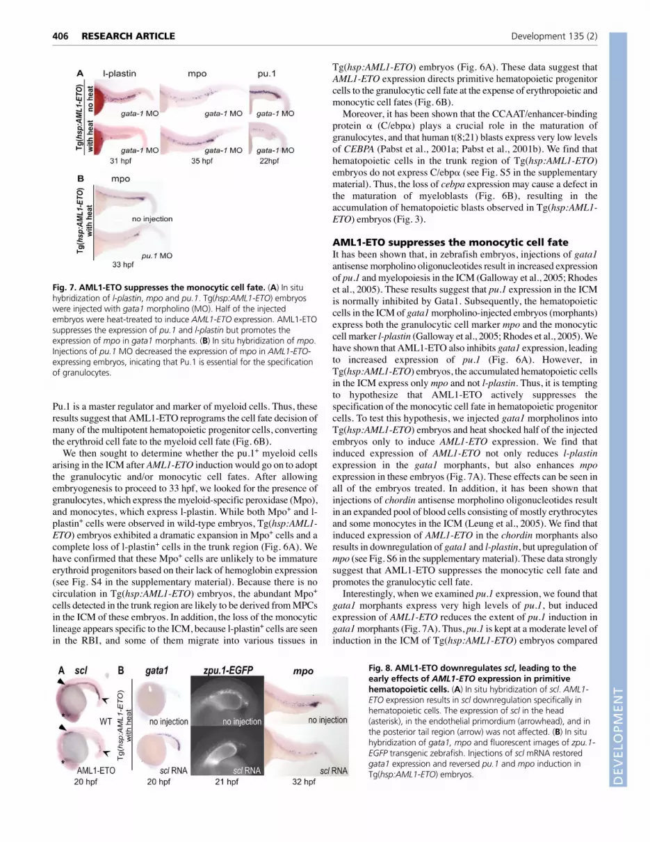
Fig. 8. AML1-ETO downregulates scl, leading to theearly effects of AML1-ETO expression in primitivehematopoietic cells. (A) In situ hybridization of scl. AML1-ETO expression results in scl downregulation specifically inhematopoietic cells. The expression of scl in the head(asterisk), in the endothelial primordium (arrowhead), and inthe posterior tail region (arrow) was not affected. (B) In situhybridization of gata1, mpo and fluorescent images of zpu.1-EGFP transgenic zebrafish. Injections of scl mRNA restoredgata1 expression and reversed pu.1 and mpo induction inTg(hsp:AML1-ETO) embryos.
DEVELO
PMENT

with that of gata1 morphants. It has been shown that Pu.1 isexpressed at a higher level in monocytic cells than its level ingranulocytic cells (Dahl et al., 2003). Thus, a moderate level of pu.1induction in Tg(hsp:AML1-ETO) embryos may not be able tosupport the monocytic cell specification in these embryos. To testwhether this moderate level of pu.1 induction is required for theMPCs to adopt the granulocytic cell fate in Tg(hsp:AML1-ETO)embryos, we injected pu.1 morpholinos into Tg(hsp:AML1-ETO)embryos. We found that expression of the granulocytic lineagemarker mpo in the ICM is reduced in pu.1 morphants, suggestingthat pu.1 expression is required for granulocytic cell specification inTg(hsp:AML1-ETO) embryos (Fig. 7B).
AML1-ETO downregulates scl expression inprimitive hematopoietic cellsTo identify the immediate downstream targets of AML1-ETO, weexamined the transcriptional profiles of hematopoietic cells only twohours after the heat treatment. To isolate blood cells in the earlyembryos (~22 hpf), we crossed wild-type and Tg(hsp:AML1-ETO)fish to the gata1-DsRed transgenic zebrafish line (Traver et al.,2003). The gata1-DsRed transgene is expressed transiently in allhematopoietic cells in the ICM before the heat treatment. Becauseof the stability of DsRed protein, the hematopoietic cells remainfluorescent after the heat treatment. We incubated embryos at 38°Cfor 1 hour at 19 hpf and isolated fluorescent cells using a flowcytometer. The experiments were done twice and the transcriptionprofiles of these samples were analyzed and compared using DNAmicroarrays. We found that the most strongly downregulated genein Tg(hsp:AML1-ETO) embryos is the hematopoietic stem cell genescl, which is reduced 11-fold (see Table S1 in the supplementarymaterial).
Overexpression of scl reverses the effects ofAML1-ETO inductionThe scl gene encodes a basic helix-loop-helix (bHLH)transcription factor involved in the specification of bothhematopoietic and endothelial cells (Gering et al., 1998; Liao etal., 1998). It has also been shown that Scl plays important roles inboth primitive and definitive hematopoiesis in mice and inzebrafish (Begley and Green, 1999). The rapid and dramaticdownregulation of scl observed in our microarray analysesprompted us to investigate whether it mediates some of the earlyeffects of AML1-ETO expression. To confirm that scl isdownstream of AML1-ETO, we heat-shocked wild-type andTg(hsp:AML1-ETO) embryos at 18 hpf for 1 hour and harvested
the embryos 1 hour after the heat treatment for scl in situhybridization. As shown in Fig. 8A, scl expression is rapidlydownregulated in Tg(hsp:AML1-ETO) embryos. Interestingly,this downregulation seems to be specific to hematopoietic cells inthe ICM region, as scl expression in the head (Fig. 8A, asterisk),which will contribute to both cranial vasculature and primitivemacrophages, in the trunk above the yolk (Fig. 8A, arrowhead),which is likely to be the precursors of the endothelium of the ductsof Cuvier, and in the posterior tail region (Fig. 8A, arrow) is notaffected (Zhang and Rodaway, 2007).
To investigate whether scl downregulation may mediate some ofthe early effects of AML1-ETO expression that we observed, weinjected scl mRNA into Tg(hsp:AML1-ETO) embryos and inducedAML1-ETO expression in these embryos. We found that injectionsof scl mRNA restores gata1 expression (in 47 out of 52 embryosinjected; Fig. 8B). Next, we tested if scl expression could also rescuethe effect of AML1-ETO on pu.1 and mpo expression. As shown inFig. 8B,C, we found that injections of scl mRNA can abolish theupregulation of pu.1 and mpo in the presence of AML1-ETOexpression (Fig. 8B, 39 out of 46 embryos injected; Fig. 8C, 20 outof 56 embryos injected). These data indicate that AML1-ETO leadsto the downregulation of gata1 and the upregulation of pu.1 and mpothrough scl, and that scl is an important mediator of the effect ofAML1-ETO on the specification of multipotent hematopoieticprogenitors.
Trichostatin A (TSA) suppresses the effect ofAML1-ETO in zebrafish embryosThe recruitment of histone deacetylase (HDAC) by the ETOdomain of AML1-ETO is believed to play an important role inAML1-ETO-mediated pathogenesis (Wang et al., 1999). To testwhether the observed AML1-ETO effects can be suppressedpharmacologically, we added TSA, an HDAC inhibitor, to theembryo media two hours before the heat treatment. We found that0.5 �M TSA blocks AML1-ETO-mediated downregulation ofboth scl and gata1, as shown by in situ hybridization (Fig. 9A,B).In addition, adding 0.5 �M TSA to embryos before (data notshown) or after 1-hour heat treatment also reverses the ability ofAML1-ETO to induce the accumulation of Mpo+ cells (Fig. 9C).These results indicate that the zebrafish AML1-ETO phenotypemay be dependent on HDAC activity. Moreover, the zebrafishAML1-ETO phenotype can be reversed by treatment with smallmolecules and may enable facile identification by high-throughput screens of novel compounds that suppress the effectof AML1-ETO in vivo.
407RESEARCH ARTICLEAML1-ETO reprograms blood cell fate
Fig. 9. Trichostatin A suppresses the effects of AML1-ETO in zebrafish embryos. (A-C) DMSO or 0.5 �M TSAwas added to Tg(hsp:AML1-ETO) embryos 2 hours before(A,B) or at the end of a 1-hour heat treatment (C) at 38°C at18 hpf. Tg(hsp:AML1-ETO) embryos that had not beensubjected to the heat treatment were used as a control. TSAtreatment rescues scl and gata1 downregulation and blocksthe accumulation of Mpo+ cells caused by AML1-ETOexpression.
DEVELO
PMENT

408
DISCUSSIONWe report here the generation of a highly tractable in vivo model ofAML1-ETO expression. We show that induced expression of AML1-ETO in zebrafish embryos results in rapid manifestation of a robustphenotype that exhibits cytological and transcriptional hallmarks ofhuman AML, suggesting that AML1-ETO signaling pathways arelikely to be conserved between human and zebrafish. Mostimportantly, using the zebrafish model of AML1-ETO expressionenabled us to track the molecular changes that take place well beforemorphological phenotypes can be detected, and to determine theroles of candidate AML1-ETO target genes. We demonstrate thatAML1-ETO regulates scl and several lineage-specific transcriptionfactors, reprogramming hematopoietic cell fate in vivo.
Induced expression of AML1-ETO in zebrafish results in anaccumulation of non-circulating hematopoietic cells. Our resultssuggest that the loss-of-circulation phenotype is likely to be due tointrinsic defects in hematopoietic cells rather than to a generaldisruption of cardiovascular function. By histological analyses, weknow that at least some hematopoietic cells are able to leave theirniche and enter the vasculature (data not shown). In addition, weoccasionally observe some circulating cells bypassing a pool of non-circulating blood cells. Interestingly, granulocytic sarcomas(chloromas or myeloblastomas) are a common clinical manifestationof t(8;21) AML (Schwyzer et al., 1998; Tallman et al., 1993).Granulocytic sarcoma is a solid tumor composed of myeloblasts. Inthis situation, the myeloblasts are very adhesive and have a hightendency to form aggregates. Whether the blood cells inTg(hsp:AML1-ETO) embryos are more adhesive than those in wild-type embryos is not clear at present.
We show that induced expression of AML1-ETO results in gata1downregulation and pu.1 upregulation in multipotent hematopoieticprogenitor cells, suppressing erythropoiesis and promotingmyelopoiesis. These results corroborate the finding that AML1-ETOinhibits erythropoiesis of purified human hematopoietic progenitorcells (Choi et al., 2006), and the finding that it suppresseserythropoiesis and stimulates granulopoiesis in mice (Schwieger etal., 2002). Our results indicate that the level of pu.1 expression isdetermined by the ability of AML1-ETO to regulate both gata1 andpu.1. AML1-ETO downregulates gata1. As a result, pu.1 is induced.Conversely, AML1-ETO may directly suppress pu.1 expression. Ithas been shown that AML1-ETO can bind to Pu.1 and inhibit itsfunction (Vangala et al., 2003). Such interaction may pose aninhibitory effect on the autoregulation of the pu.1 gene (Chen et al.,1995). The ability of AML1-ETO to fine tune the level of pu.1induction is likely to be very important for its leukemogenic effect.Supporting this idea, it has been shown that the hypomorphic allele,but not the null allele, of the Pu.1 gene in mice leads to AML-likephenotypes, suggesting the importance of Pu.1 gene dose inleukemogenesis (Rosenbauer et al., 2005).
Although we demonstrate that AML1-ETO causes cell fatechanges, converting erythropoiesis to myelopoiesis, there are clearlystill some erythrocytes in Tg(hsp:AML1-ETO) embryos (Fig. 3). Wehave observed that both induced expression of AML1-ETO and theloss of gata1 expression last only for a few hours after the heat shock(data not shown). Thus, some hematopoietic cells may eventuallycommit to the erythroid cell fate as a result of the restoration of gata1expression.
During primitive hematopoiesis, zebrafish mpo expression initiatesbetween 18 and 20 hpf, first in the ICM and then in the RBI. At 24 hpf,myeloblasts can be identified morphologically only in the RBI and notin the ICM, although granulocytes are reliably found in the circulationby 48 hpf (Bennett et al., 2001; Lieschke et al., 2001). The source of
these circulating granulocytes is not totally clear at this moment. Forexample, it is not known whether the Mpo+ cells observed in the ICMat 20 hpf can become mature in situ. In our model, induced expressionof AML1-ETO causes an enrichment of Mpo+ cells andmorphologically immature hematopoietic blasts in the ICM. Theaccumulation of hematopoietic blasts is only partially reversed asAML1-ETO expression ceases (see Fig. S3 in the supplementarymaterial). What causes the long-lasting effect of transient induction ofAML1-ETO on the blockade of maturation of these cells is not clear.It could be that these cells cannot migrate to the environment thatsupports their maturation, that the environmental signals that inducethe maturation of these cells no longer exist, or that a factor that isrequired for maturation, such as C/ebp�, can no longer be expressedafter AML1-ETO expression diminishes.
We were able to identify several early downstream targets ofAML1-ETO by isolating hematopoietic cells only two hours afterthe induction of AML1-ETO expression. We show that inducedexpression of AML1-ETO rapidly downregulates scl expression.Even though decreased scl expression has been shown in t(8;21)leukemia samples, a direct link between AML1-ETO and sclexpression had not been established previously (Shimamoto et al.,1995). We have not been able to identify potential Aml1 bindingsites upstream of the coding region of scl, indicating that AML1-ETO may inhibit scl expression through binding with other factors.Interestingly, the downregulation of scl is seen only in hematopoieticprogenitor cells and not in cells that will give rise to the endotheliallineage in Tg(hsp:AML1-ETO) embryos. It has been shown that theheat shock promoter used to drive the human AML1-ETO generesults in ubiquitous expression upon induction (Xiao et al., 2003).Thus, these results suggest that other hematopoietic-specificcofactors may be required for AML1-ETO function.
Overexpression of scl can block the ability of AML1-ETO toreprogram hematopoietic cell fate, as shown by the reversal ofgata1, pu.1 and mpo dysregulation in Tg(hsp:AML1-ETO) embryos.However, Scl-mediated rescue of the early effects of AML1-ETOon hematopoiesis does not completely rescue the circulation defectin Tg(hsp:AML1-ETO) embryos. This may be because of additionaldefects caused by ubiquitous scl overexpression, or by non-Scl-mediated effects of AML1-ETO at later stages of development.Nevertheless, these results indicate that scl is an important effectorthat mediates the earliest observable effects of AML1-ETO inhematopoietic progenitor cells.
Induced expression of AML1-ETO in zebrafish embryos before21 hpf disrupts definitive hematopoiesis but also enables us to studythe in vivo effects of AML1-ETO in MPCs generated duringprimitive hematopoiesis. The transcriptional profile ofTg(hsp:AML1-ETO) embryo blood at 40 hpf presents expressionalsignatures of human AML, suggesting that the regulatorymechanisms of cell specification and maturation in these cellsresemble those in progenitors of adult human blood. However,MPCs in zebrafish embryos may not have the self-renewalcapability that is also essential for leukemogenesis. Thus, the effectsof AML1-ETO on the self-renewal capability of hematopoieticprogenitor cells may not be accessible using this model.Additionally, in humans and mice, AML1-ETO is not sufficient toinduce leukemia in the absence of secondary mutations, so the blastcells observed in AML1-ETO-expressing fish are unlikely topossess full leukemogenic potential. In future studies, it will beinteresting to combine AML1-ETO expression with knowncollaborating mutations, and to characterize the effects of AML1-ETO expression in hematopoietic stem cells by inducing AML1-ETO expression at later stages of development, or in adults.
RESEARCH ARTICLE Development 135 (2)
DEVELO
PMENT

Understanding the molecular mechanisms by which AML1-ETOexerts its influence on hematopoietic progenitor cells may help usdevelop targeted therapeutics. For example, compounds that blockSCL or GATA1 downregulation in the presence of AML1-ETOexpression may prove useful in treating AML associated with t(8;21)translocation. It has been shown that compounds that enhanceCebpa (C/ebp�) transcriptional activity induce differentiation ofAML cell lines (Jiang et al., 2005). Moreover, high-throughputscreening is feasible in the zebrafish (Yeh and Crews, 2003; Zon andPeterson, 2005). We have shown that some of the effects of AML1-ETO can be suppressed by a histone deacetylase inhibitorTrichostatin A. Therefore, in addition to the fundamental insightsinto the mechanism of AML1-ETO function already provided bythis model, it may ultimately provide a unique opportunity toconduct whole-organism chemical suppressor screens to identifycompounds that can reverse AML1-ETO function in vivo.
We thank Drs K. E. Crosier and J. Y. Kuwada for providing AML1-ETO andzebrafish hsp70 plasmids; and Drs A. T. Look and L. I. Zon for providingTg(zpu.1:EGFP) and Tg(gata1:DsRed) fish. We also thank Elma Feric fortechnical assistance; and Drs K. R. Chien, I. A. Drummond, D. A. Haber, D. A.Sweetser and Developmental Biology Laboratory members for helpfulsuggestions on this work and on the preparation of this manuscript. We aregrateful to Brian Patterson of Agilent Technologies for assistance with themicroarray experiments. The authors received financial support from the NedSahin Research Fund for Restoring Developmental Plasticity, the NovartisInstitutes for BioMedical Research, and the National Cancer Institute. J.-R.J.Y.was supported by a Tosteson Fellowship from the Massachusetts BiomedicalResearch Corporation.
Supplementary materialSupplementary material for this article is available athttp://dev.biologists.org/cgi/content/full/135/2/401/DC1
ReferencesAlcalay, M., Meani, N., Gelmetti, V., Fantozzi, A., Fagioli, M., Orleth, A.,
Riganelli, D., Sebastiani, C., Cappelli, E., Casciari, C. et al. (2003). Acutemyeloid leukemia fusion proteins deregulate genes involved in stem cellmaintenance and DNA repair. J. Clin. Invest. 112, 1751-1761.
Begley, C. G. and Green, A. R. (1999). The SCL gene: from case report to criticalhematopoietic regulator. Blood 93, 2760-2770.
Bennett, C. M., Kanki, J. P., Rhodes, J., Liu, T. X., Paw, B. H., Kieran, M. W.,Langenau, D. M., Delahaye-Brown, A., Zon, L. I., Fleming, M. D. et al.(2001). Myelopoiesis in the zebrafish, Danio rerio. Blood 98, 643-651.
Bonnet, D. and Dick, J. E. (1997). Human acute myeloid leukemia is organized asa hierarchy that originates from a primitive hematopoietic cell. Nat. Med. 3, 730-737.
Borregaard, N., Theilgaard-Monch, K., Sorensen, O. E. and Cowland, J. B.(2001). Regulation of human neutrophil granule protein expression. Curr. Opin.Hematol. 8, 23-27.
Burns, C. E., Traver, D., Mayhall, E., Shepard, J. L. and Zon, L. I. (2005).Hematopoietic stem cell fate is established by the Notch-Runx pathway. GenesDev. 19, 2331-2342.
Chen, H., Ray-Gallet, D., Zhang, P., Hetherington, C. J., Gonzalez, D. A.,Zhang, D. E., Moreau-Gachelin, F. and Tenen, D. G. (1995). PU.1 (Spi-1)autoregulates its expression in myeloid cells. Oncogene 11, 1549-1560.
Choi, Y., Elagib, K. E., Delehanty, L. L. and Goldfarb, A. N. (2006). Erythroidinhibition by the leukemic fusion AML1-ETO is associated with impairedacetylation of the major erythroid transcription factor GATA-1. Cancer Res. 66,2990-2996.
Dahl, R., Walsh, J. C., Lancki, D., Laslo, P., Iyer, S. R., Singh, H. and Simon,M. C. (2003). Regulation of macrophage and neutrophil cell fates by thePU.1:C/EBPalpha ratio and granulocyte colony-stimulating factor. Nat. Immunol.4, 1029-1036.
Davidson, A. J. and Zon, L. I. (2004). The ‘definitive’ (and ‘primitive’) guide tozebrafish hematopoiesis. Oncogene 23, 7233-7246.
de Guzman, C. G., Warren, A. J., Zhang, Z., Gartland, L., Erickson, P.,Drabkin, H., Hiebert, S. W. and Klug, C. A. (2002). Hematopoietic stem cellexpansion and distinct myeloid developmental abnormalities in a murine modelof the AML1-ETO translocation. Mol. Cell. Biol. 22, 5506-5517.
Downing, J. R. (1999). The AML1-ETO chimaeric transcription factor in acutemyeloid leukaemia: biology and clinical significance. Br. J. Haematol. 106, 296-308.
Fenske, T. S., Pengue, G., Mathews, V., Hanson, P. T., Hamm, S. E., Riaz, N.
and Graubert, T. A. (2004). Stem cell expression of the AML1/ETO fusionprotein induces a myeloproliferative disorder in mice. Proc. Natl. Acad. Sci. USA101, 15184-15189.
Galloway, J. L., Wingert, R. A., Thisse, C., Thisse, B. and Zon, L. I. (2005). Lossof gata1 but not gata2 converts erythropoiesis to myelopoiesis in zebrafishembryos. Dev. Cell 8, 109-116.
Gering, M., Rodaway, A. R., Gottgens, B., Patient, R. K. and Green, A. R.(1998). The SCL gene specifies haemangioblast development from earlymesoderm. EMBO J. 17, 4029-4045.
Golub, T. R., Slonim, D. K., Tamayo, P., Huard, C., Gaasenbeek, M., Mesirov,J. P., Coller, H., Loh, M. L., Downing, J. R., Caligiuri, M. A. et al. (1999).Molecular classification of cancer: class discovery and class prediction by geneexpression monitoring. Science 286, 531-537.
Hsu, K., Traver, D., Kutok, J. L., Hagen, A., Liu, T. X., Paw, B. H., Rhodes, J.,Berman, J. N., Zon, L. I., Kanki, J. P. et al. (2004). The pu.1 promoter drivesmyeloid gene expression in zebrafish. Blood 104, 1291-1297.
Jiang, Y., Xue, Z. H., Shen, W. Z., Du, K. M., Yan, H., Yu, Y., Peng, Z. G., Song,M. G., Tong, J. H., Chen, Z. et al. (2005). Desferrioxamine induces leukemiccell differentiation potentially by hypoxia-inducible factor-1 alpha that augmentstranscriptional activity of CCAAT/enhancer-binding protein-alpha. Leukemia 19,1239-1247.
Kalev-Zylinska, M. L., Horsfield, J. A., Flores, M. V., Postlethwait, J. H., Vitas,M. R., Baas, A. M., Crosier, P. S. and Crosier, K. E. (2002). Runx1 is requiredfor zebrafish blood and vessel development and expression of a human RUNX1-CBF2T1 transgene advances a model for studies of leukemogenesis.Development 129, 2015-2030.
Kimmel, C. B., Ballard, W. W., Kimmel, S. R., Ullmann, B. and Schilling, T. F.(1995). Stages of embryonic development of the zebrafish. Dev. Dyn. 203, 253-310.
Lawrence, H. J., Sauvageau, G., Ahmadi, N., Lopez, A. R., LeBeau, M. M.,Link, M., Humphries, K. and Largman, C. (1995). Stage- and lineage-specificexpression of the HOXA10 homeobox gene in normal and leukemichematopoietic cells. Exp. Hematol. 23, 1160-1166.
Lawson, N. D. and Weinstein, B. M. (2002). In vivo imaging of embryonicvascular development using transgenic zebrafish. Dev. Biol. 248, 307-318.
Leung, A. Y., Mendenhall, E. M., Kwan, T. T., Liang, R., Eckfeldt, C., Chen, E.,Hammerschmidt, M., Grindley, S., Ekker, S. C. and Verfaillie, C. M. (2005).Characterization of expanded intermediate cell mass in zebrafish chordinmorphant embryos. Dev. Biol. 277, 235-254.
Liao, E. C., Paw, B. H., Oates, A. C., Pratt, S. J., Postlethwait, J. H. and Zon,L. I. (1998). SCL/Tal-1 transcription factor acts downstream of cloche to specifyhematopoietic and vascular progenitors in zebrafish. Genes Dev. 12, 621-626.
Licht, J. D. (2001). AML1 and the AML1-ETO fusion protein in the pathogenesis oft (8;21) AML. Oncogene 20, 5660-5679.
Lieschke, G. J., Oates, A. C., Crowhurst, M. O., Ward, A. C. and Layton, J. E.(2001). Morphologic and functional characterization of granulocytes andmacrophages in embryonic and adult zebrafish. Blood 98, 3087-3096.
Lutterbach, B. and Hiebert, S. W. (2000). Role of the transcription factor AML-1in acute leukemia and hematopoietic differentiation. Gene 245, 223-235.
Lyons, S. E., Shue, B. C., Lei, L., Oates, A. C., Zon, L. I. and Liu, P. P. (2001).Molecular cloning, genetic mapping, and expression analysis of four zebrafishc/ebp genes. Gene 281, 43-51.
Miyamoto, T., Weissman, I. L. and Akashi, K. (2000). AML1/ETO-expressingnonleukemic stem cells in acute myelogenous leukemia with 8;21 chromosomaltranslocation. Proc. Natl. Acad. Sci. USA 97, 7521-7526.
Nasevicius, A. and Ekker, S. C. (2000). Effective targeted gene ‘knockdown’ inzebrafish. Nat. Genet. 26, 216-220.
Okuda, T., Cai, Z., Yang, S., Lenny, N., Lyu, C. J., van Deursen, J. M., Harada,H. and Downing, J. R. (1998). Expression of a knocked-in AML1-ETO leukemiagene inhibits the establishment of normal definitive hematopoiesis and directlygenerates dysplastic hematopoietic progenitors. Blood 91, 3134-3143.
Pabst, T., Mueller, B. U., Harakawa, N., Schoch, C., Haferlach, T., Behre, G.,Hiddemann, W., Zhang, D. E. and Tenen, D. G. (2001a). AML1-ETOdownregulates the granulocytic differentiation factor C/EBPalpha in t (8;21)myeloid leukemia. Nat. Med. 7, 444-451.
Pabst, T., Mueller, B. U., Zhang, P., Radomska, H. S., Narravula, S.,Schnittger, S., Behre, G., Hiddemann, W. and Tenen, D. G. (2001b).Dominant-negative mutations of CEBPA, encoding CCAAT/enhancer bindingprotein-alpha (C/EBPalpha), in acute myeloid leukemia. Nat. Genet. 27, 263-270.
Palis, J., Chan, R. J., Koniski, A., Patel, R., Starr, M. and Yoder, M. C. (2001).Spatial and temporal emergence of high proliferative potential hematopoieticprecursors during murine embryogenesis. Proc. Natl. Acad. Sci. USA 98, 4528-4533.
Peterson, L. F. and Zhang, D. E. (2004). The 8;21 translocation inleukemogenesis. Oncogene 23, 4255-4262.
Ransom, D. G., Haffter, P., Odenthal, J., Brownlie, A., Vogelsang, E., Kelsh,R. N., Brand, M., van Eeden, F. J., Furutani-Seiki, M., Granato, M. et al.(1996). Characterization of zebrafish mutants with defects in embryonichematopoiesis. Development 123, 311-319.
409RESEARCH ARTICLEAML1-ETO reprograms blood cell fate
DEVELO
PMENT

410
Rhodes, J., Hagen, A., Hsu, K., Deng, M., Liu, T. X., Look, A. T. and Kanki,J. P. (2005). Interplay of pu.1 and gata1 determines myelo-erythroid progenitorcell fate in zebrafish. Dev. Cell 8, 97-108.
Rosenbauer, F., Koschmieder, S., Steidl, U. and Tenen, D. G. (2005). Effect oftranscription-factor concentrations on leukemic stem cells. Blood 106, 1519-1524.
Ross, M. E., Mahfouz, R., Onciu, M., Liu, H. C., Zhou, X., Song, G., Shurtleff,S. A., Pounds, S., Cheng, C., Ma, J. et al. (2004). Gene expression profiling ofpediatric acute myelogenous leukemia. Blood 104, 3679-3687.
Schwieger, M., Lohler, J., Friel, J., Scheller, M., Horak, I. and Stocking, C.(2002). AML1-ETO inhibits maturation of multiple lymphohematopoieticlineages and induces myeloblast transformation in synergy with ICSBPdeficiency. J. Exp. Med. 196, 1227-1240.
Schwyzer, R., Sherman, G. G., Cohn, R. J., Poole, J. E. and Willem, P. (1998).Granulocytic sarcoma in children with acute myeloblastic leukemia and t (8;21).Med. Pediatr. Oncol. 31, 144-149.
Shimada, H., Ichikawa, H., Nakamura, S., Katsu, R., Iwasa, M., Kitabayashi,I. and Ohki, M. (2000). Analysis of genes under the downstream control ofthe t (8;21) fusion protein AML1-MTG8: overexpression of the TIS11b (ERF-1,cMG1) gene induces myeloid cell proliferation in response to G-CSF. Blood 96,655-663.
Shimamoto, T., Ohyashiki, K., Ohyashiki, J. H., Kawakubo, K., Fujimura, T.,Iwama, H., Nakazawa, S. and Toyama, K. (1995). The expression pattern oferythrocyte/megakaryocyte-related transcription factors GATA-1 and the stemcell leukemia gene correlates with hematopoietic differentiation and isassociated with outcome of acute myeloid leukemia. Blood 86, 3173-3180.
Stone, R. M., O’Donnell, M. R. and Sekeres, M. A. (2004). Acute myeloidleukemia. Hematology Am. Soc. Hematol. Educ. Program 2004, 98-117.
Tallman, M. S., Hakimian, D., Shaw, J. M., Lissner, G. S., Russell, E. J. andVariakojis, D. (1993). Granulocytic sarcoma is associated with the 8;21translocation in acute myeloid leukemia. J. Clin. Oncol. 11, 690-697.
Tenen, D. G. (2003). Disruption of differentiation in human cancer: AML showsthe way. Nat. Rev. Cancer 3, 89-101.
Thompson, M. A., Ransom, D. G., Pratt, S. J., MacLennan, H., Kieran, M. W.,Detrich, H. W., 3rd, Vail, B., Huber, T. L., Paw, B., Brownlie, A. J. et al.(1998). The cloche and spadetail genes differentially affect hematopoiesis andvasculogenesis. Dev. Biol. 197, 248-269.
Traver, D., Paw, B. H., Poss, K. D., Penberthy, W. T., Lin, S. and Zon, L. I.(2003). Transplantation and in vivo imaging of multilineage engraftment inzebrafish bloodless mutants. Nat. Immunol. 4, 1238-1246.
Vangala, R. K., Heiss-Neumann, M. S., Rangatia, J. S., Singh, S. M., Schoch,C., Tenen, D. G., Hiddemann, W. and Behre, G. (2003). The myeloid masterregulator transcription factor PU.1 is inactivated by AML1-ETO in t (8;21)myeloid leukemia. Blood 101, 270-277.
Wang, J., Saunthararajah, Y., Redner, R. L. and Liu, J. M. (1999). Inhibitors ofhistone deacetylase relieve ETO-mediated repression and induce differentiationof AML1-ETO leukemia cells. Cancer Res. 59, 2766-2769.
Weinstein, B. M., Stemple, D. L., Driever, W. and Fishman, M. C. (1995).Gridlock, a localized heritable vascular patterning defect in the zebrafish. Nat.Med. 1, 1143-1147.
Westerfield, M. (2000). The Zebrafish Book. A Guide for the Laboratory use ofZebrafish (Danio rerio). Eugene: University of Oregon Press.
Xiao, T., Shoji, W., Zhou, W., Su, F. and Kuwada, J. Y. (2003). Transmembranesema4E guides branchiomotor axons to their targets in zebrafish. J. Neurosci.23, 4190-4198.
Yeh, J. R. and Crews, C. M. (2003). Chemical genetics: adding to thedevelopmental biology toolbox. Dev. Cell 5, 11-19.
Yergeau, D. A., Hetherington, C. J., Wang, Q., Zhang, P., Sharpe, A. H.,Binder, M., Marin-Padilla, M., Tenen, D. G., Speck, N. A. and Zhang, D. E.(1997). Embryonic lethality and impairment of haematopoiesis in miceheterozygous for an AML1-ETO fusion gene. Nat. Genet. 15, 303-306.
Zhang, X. Y. and Rodaway, A. R. (2007). SCL-GFP transgenic zebrafish: in vivoimaging of blood and endothelial development and identification of the initialsite of definitive hematopoiesis. Dev. Biol. 307, 179-194.
Zon, L. I. and Peterson, R. T. (2005). In vivo drug discovery in the zebrafish. Nat.Rev. Drug Discov. 4, 35-44.
RESEARCH ARTICLE Development 135 (2)
DEVELO
PMENT
Related Documents