Biochem. J. (1997) 326, 205–213 (Printed in Great Britain) 205 Altered regulation of cholesterol and cholesteryl ester synthesis in Chinese- hamster ovary cells overexpressing the oxysterol-binding protein is dependent on the pleckstrin homology domain Thomas A. LAGACE, David M. BYERS, Harold W. COOK and Neale D. RIDGWAY 1 Atlantic Research Centre and Departments of Pediatrics and Biochemistry, Clinical Research Center, 5849 University Avenue, Dalhousie University, Halifax, Nova Scotia, Canada B3H 4H7 Oxysterol-binding protein (OSBP) is a high-affinity receptor for a variety of oxysterols, such as 25-hydroxycholesterol, that down-regulate cholesterol synthesis and stimulate cholesterol esterification. To examine a potential role for OSBP in regulating cholesterol metabolism, we stably overexpressed this protein in Chinese-hamster ovary (CHO)-K1 cells. Compared with mock- transfected controls, several cell lines overexpressing wild-type OSBP (CHO-OSBP) displayed a 50 % decrease in cholesteryl ester synthesis when cultured in medium with delipidated serum, 25-hydroxycholesterol or low-density lipoprotein (LDL) . CHO- OSBP cells showed a 40–60 % decrease in acyl-CoA :cholesterol acyltransferase activity and mRNA, a 50 % elevation in mRNA for three sterol-regulated genes [LDL receptor, 3-hydroxy-3- methylgluraryl (HMG)-CoA reductase and HMG-CoA synthase], and an 80 % increase in ["%C]acetate incorporation into INTRODUCTION Oxysterols have a wide range of effects on cellular physiology that are poorly defined in terms of precise intracellular targets and primary versus non-specific responses (reviewed in [1,2]). An extensively studied aspect of oxysterol action is the putative role of these molecules in regulating cholesterol metabolism [3]. Oxysterol treatment of cultured cells reproduces many of the regulatory responses of low-density lipoprotein (LDL)-derived cholesterol such as transcriptional suppression of sterol-regulated genes, stimulation of acyl-CoA :cholesterol acyltransferase (ACAT) and enhanced degradation of 3-hydroxy-3-methyl- glutaryl (HMG)-CoA reductase [4]. One hypothesis for the action of oxysterols is that they are generated in cells when the cholesterol content is elevated and suppress sterol synthesis by interaction with a regulatory protein(s). Kandutsch and co- workers [5,6] identified a high-affinity (K D 10 nM) oxysterol- binding protein (OSBP) in the cytosol of cultured cells and various tissues. It was postulated that OSBP mediated some regulatory effects of oxysterols on cholesterol metabolism. This conclusion was based primarily on evidence of a positive corre- lation between oxysterol suppression of cholesterol synthesis and HMG-CoA reductase activity in cultured cells, and affinity for OSBP [5,7]. cDNA cloning of human [8] and rabbit [9] OSBPs revealed highly conserved proteins that migrated on SDS}PAGE as a doublet of 97 and 101 kDa [9,10]. Purified [10] or overexpressed OSBP from COS cells [11] had a native molecular mass consistent with a homodimer. Immunofluo- Abbreviations used : ACAT, acyl-CoA :cholesterol acyltransferase ; CHO, Chinese-hamster ovary ; DMEM, Dulbecco’s modified Eagle’s medium ; LDL, low-density lipoprotein ; FCS, fetal-calf serum ; HMG, 3-hydroxy-3-methylglutaryl ; OSBP, oxysterol-binding protein ; PH, pleckstrin homology. 1 To whom correspondence should be addressed. cholesterol. CHO-K1 cells overexpressing two OSBP mutants with a complete or N-terminal deletion of the pleckstrin hom- ology (PH) domain had cholesterol esterification and synthesis rates that were similar to those shown by mock-transfected controls. Unlike wild-type OSBP, both PH domain mutants displayed diffuse cytoplasmic immunofluorescence staining and did not translocate to the Golgi apparatus in the presence of 25- hydroxycholesterol. CHO-K1 cells overexpressing OSBP have pronounced alterations in cholesterol esterification and synthesis, indicating a potential role for this receptor in cholesterol homoeostasis. The phenotype observed in cells overexpressing OSBP is dependent on the PH domain, which appears to be necessary for ligand-dependent localization of OSBP to the Golgi apparatus. rescence localization in overexpressing Chinese-hamster ovary (CHO)-K1 cells indicated that OSBP was predominately cyto- plasmic or vesicle-associated in the absence of oxysterol, but underwent rapid localization to the Golgi apparatus when treated with 25-hydroxycholesterol [11]. The N-terminal region of OSBP was shown to be important for localization to the Golgi apparatus [11], and contains a pleckstrin homology (PH) domain, a motif with a putative role in intracellular signalling [12,13]. A family of OSBP homologues has been identified in Saccharomyces cereisiae [14]. Yeast harbouring double or triple deletions of members of this gene family displayed cold-sensitive growth and nystatin resistance, as well as small cumulative reductions in ergosterol synthesis [14]. Interestingly, deletion of one member of this OSBP-related family (KES 1) was found to bypass a SEC 14 temperature-sensitive mutation, suggesting that the kes 1 protein may be involved in Golgi function and vesicle trafficking [15]. Because of its apparent association with the Golgi}vesicular pathway, OSBP and related proteins could be involved in aspects of cholesterol or oxysterol trafficking and thereby modify downstream regulatory events by affecting the sterol content at regulatory sites in the endoplasmic reticulum. To assess the role of OSBP in regulation of cellular cholesterol synthesis and esterification, we studied cholesterol homoeostasis in CHO-K1 cells overexpressing wild-type rabbit OSBP. Over- expression of OSBP resulted in a decrease in ACAT activity and mRNA and elevated mRNA for sterol-regulated genes and cholesterol synthesis in cells grown in lipoprotein-free medium. Additional evidence is presented demonstrating that this altered

Welcome message from author
This document is posted to help you gain knowledge. Please leave a comment to let me know what you think about it! Share it to your friends and learn new things together.
Transcript
Biochem. J. (1997) 326, 205–213 (Printed in Great Britain) 205
Altered regulation of cholesterol and cholesteryl ester synthesis in Chinese-hamster ovary cells overexpressing the oxysterol-binding protein isdependent on the pleckstrin homology domainThomas A. LAGACE, David M. BYERS, Harold W. COOK and Neale D. RIDGWAY1
Atlantic Research Centre and Departments of Pediatrics and Biochemistry, Clinical Research Center, 5849 University Avenue, Dalhousie University, Halifax, Nova Scotia,Canada B3H 4H7
Oxysterol-binding protein (OSBP) is a high-affinity receptor for
a variety of oxysterols, such as 25-hydroxycholesterol, that
down-regulate cholesterol synthesis and stimulate cholesterol
esterification. To examine a potential role for OSBP in regulating
cholesterol metabolism, we stably overexpressed this protein in
Chinese-hamster ovary (CHO)-K1 cells. Compared with mock-
transfected controls, several cell lines overexpressing wild-type
OSBP (CHO-OSBP) displayed a 50% decrease in cholesteryl
ester synthesis when cultured in medium with delipidated serum,
25-hydroxycholesterol or low-density lipoprotein (LDL) . CHO-
OSBP cells showed a 40–60% decrease in acyl-CoA:cholesterol
acyltransferase activity and mRNA, a 50% elevation in mRNA
for three sterol-regulated genes [LDL receptor, 3-hydroxy-3-
methylgluraryl (HMG)-CoA reductase and HMG-CoA
synthase], and an 80% increase in ["%C]acetate incorporation into
INTRODUCTION
Oxysterols have a wide range of effects on cellular physiology
that are poorly defined in terms of precise intracellular targets
and primary versus non-specific responses (reviewed in [1,2]). An
extensively studied aspect of oxysterol action is the putative role
of these molecules in regulating cholesterol metabolism [3].
Oxysterol treatment of cultured cells reproduces many of the
regulatory responses of low-density lipoprotein (LDL)-derived
cholesterol such as transcriptional suppression of sterol-regulated
genes, stimulation of acyl-CoA:cholesterol acyltransferase
(ACAT) and enhanced degradation of 3-hydroxy-3-methyl-
glutaryl (HMG)-CoA reductase [4]. One hypothesis for the action
of oxysterols is that they are generated in cells when the
cholesterol content is elevated and suppress sterol synthesis by
interaction with a regulatory protein(s). Kandutsch and co-
workers [5,6] identified a high-affinity (KD
10 nM) oxysterol-
binding protein (OSBP) in the cytosol of cultured cells and
various tissues. It was postulated that OSBP mediated some
regulatory effects of oxysterols on cholesterol metabolism. This
conclusion was based primarily on evidence of a positive corre-
lation between oxysterol suppression of cholesterol synthesis
and HMG-CoA reductase activity in cultured cells, and affinity
for OSBP [5,7]. cDNA cloning of human [8] and rabbit [9]
OSBPs revealed highly conserved proteins that migrated on
SDS}PAGE as a doublet of 97 and 101 kDa [9,10]. Purified [10]
or overexpressed OSBP from COS cells [11] had a native
molecular mass consistent with a homodimer. Immunofluo-
Abbreviations used: ACAT, acyl-CoA:cholesterol acyltransferase ; CHO, Chinese-hamster ovary ; DMEM, Dulbecco’s modified Eagle’s medium; LDL,low-density lipoprotein ; FCS, fetal-calf serum; HMG, 3-hydroxy-3-methylglutaryl ; OSBP, oxysterol-binding protein ; PH, pleckstrin homology.
1 To whom correspondence should be addressed.
cholesterol. CHO-K1 cells overexpressing two OSBP mutants
with a complete or N-terminal deletion of the pleckstrin hom-
ology (PH) domain had cholesterol esterification and synthesis
rates that were similar to those shown by mock-transfected
controls. Unlike wild-type OSBP, both PH domain mutants
displayed diffuse cytoplasmic immunofluorescence staining and
did not translocate to the Golgi apparatus in the presence of 25-
hydroxycholesterol. CHO-K1 cells overexpressing OSBP have
pronounced alterations in cholesterol esterification and synthesis,
indicating a potential role for this receptor in cholesterol
homoeostasis. The phenotype observed in cells overexpressing
OSBP is dependent on the PH domain, which appears to be
necessary for ligand-dependent localization of OSBP to the
Golgi apparatus.
rescence localization in overexpressing Chinese-hamster ovary
(CHO)-K1 cells indicated that OSBP was predominately cyto-
plasmic or vesicle-associated in the absence of oxysterol, but
underwent rapid localization to the Golgi apparatus when treated
with 25-hydroxycholesterol [11]. The N-terminal region of OSBP
was shown to be important for localization to theGolgi apparatus
[11], and contains a pleckstrin homology (PH) domain, a motif
with a putative role in intracellular signalling [12,13].
A family of OSBP homologues has been identified in
Saccharomyces cere�isiae [14]. Yeast harbouring double or triple
deletions of members of this gene family displayed cold-sensitive
growth and nystatin resistance, as well as small cumulative
reductions in ergosterol synthesis [14]. Interestingly, deletion of
one member of this OSBP-related family (KES 1) was found to
bypass a SEC 14 temperature-sensitive mutation, suggesting that
the kes 1 protein may be involved in Golgi function and vesicle
trafficking [15]. Because of its apparent association with the
Golgi}vesicular pathway, OSBP and related proteins could be
involved in aspects of cholesterol or oxysterol trafficking and
thereby modify downstream regulatory events by affecting the
sterol content at regulatory sites in the endoplasmic reticulum.
To assess the role of OSBP in regulation of cellular cholesterol
synthesis and esterification, we studied cholesterol homoeostasis
in CHO-K1 cells overexpressing wild-type rabbit OSBP. Over-
expression of OSBP resulted in a decrease in ACAT activity and
mRNA and elevated mRNA for sterol-regulated genes and
cholesterol synthesis in cells grown in lipoprotein-free medium.
Additional evidence is presented demonstrating that this altered
206 T. A. Lagace and others
regulatory phenotype observed in cells overexpressing wild-type
OSBP requires the PH domain.
MATERIALS AND METHODS
Materials
25-Hydroxycholesterol and cholesterol were purchased from
Steraloids (Wilton, NH, U.S.A.). Other oxysterols, fatty acid-
free BSA and oleate were from Sigma Chemical Co. [α-$#P]dATP,
[9,10-$H]oleate, [$H]25-hydroxycholesterol, [1-"%C]acetate and
[1-"%C]oleoyl-CoA were from Dupont-NEN. Silica-gel G TLC
plates were from BDH. Tissue-culture reagents were from
Gibco–BRL. FITC-labelled rabbit anti-mouse IgG was from
Organon Teknika (Westchester, PA, U.S.A.). Goat anti-mouse
IgG–horseradish peroxidase conjugate was purchased from
Bio-Rad.
Cell culture and transfections
CHO-K1 cells were cultured in Dulbecco’s modified Eagle’s
medium (DMEM) with 5% fetal-calf serum (FCS) (medium A).
CHO-K1 cells (100 mm diam. dishes) were transfected with
10 µg of wild-type or mutant OSBP cDNA in pCMV and 1 µg of
pSV3Neo by the calcium phosphate precipitation method [11].
Mock (control)-transfected cells received equivalent amounts of
pCMV and pSV3Neo. Clones resistant to 600 µg of G418
(Geneticin)}ml were selected and expression of OSBP was
determined by immunoblotting (see below). Stock cultures of
OSBP-expressing and mock transfected cells were maintained in
medium A containing 350 µg G418 (Geneticin)}ml, but were
subcultured for experiments in medium A without G418. Cells
were cultured in DMEM with 5% lipoprotein-free FCS 18 h
before the start of experiments. OSBP expression was routinely
monitored by immunoblotting and immunofluorescence to
ensure that 90% of the cells were stably overexpressing.
COS 7 cells were cultured in DMEM containing 10% FCS.
COS 7 cells were transiently transfected with wild-type and
mutant OSBP cDNAs by the DEAE-dextran method [16] and
harvested 48 h later.
Site-directed mutagenesis
The 1230 bp SmaI–XbaI fragment of the rabbit OSBP cDNA [9]
was subcloned into pAlter-1 and mutagenized according to the
manufacturer’s instructions (Altered Sites II system; Promega).
Two deletion mutants of OSBP were generated; the entire PH
domain was deleted (OSBP ∆PH, amino acids 92–182) and the
N-terminal region of this domain was removed (OSBP ∆N-PH,
amino acids 92–125). Both mutations were confirmed by
sequencing.
Immunoblotting and immunofluorescence
Stably overexpressing CHO-K1 cells were harvested in ice-cold
PBS and collected by centrifugation (2000 g for 5 min at 4 °C).
Cell pellets were solubilized in 10 mM sodium phosphate
(pH 7.4)}150 mM NaCl}2 mM EDTA}2 mM EGTA}10 mM
NaF}1 mM sodium pyrophosphate}1 mM β-glycerophosphate}100 µM PMSF}aprotinin (2 µg}ml)}leupeptin (2.5 µg}ml)}0.3% (w}v) Triton X-100 (buffer A) on ice for 20 min, followed
by centrifugation for 15 min at 4 °C and 10000 g (rav.
6 cm). The
supernatant fraction, which contained all immunoreactive OSBP,
was collected and proteins separated on SDS}6%-PAGE and
transferred to nitrocellulose filters. Filters were probed with
OSBP monoclonal antibody 11H9 [11] (kindly provided by Dr.
Joseph Goldstein, University of Texas Southwestern Medical
Center, Dallas, TX, U.S.A.) followed by goat anti-mouse IgG
coupled to horseradish peroxidase. The filter was developed by
the enhanced chemiluminescence technique according to the
manufacturer’s instructions (ECL; Amersham). Indirect
immunofluorescence localization of OSBP was as previously
described [11] using an Olympus microscope and 100¬-
magnification oil-immersion objective.
[3H]25-Hydroxycholesterol binding assays
Monolayers of COS 7 cells overexpressing wild-type and mutant
OSBP were washed once with cold PBS, scraped into PBS and
collected by centrifugation (2000 g for 5 min). Cells were
homogenized in 10 mM Hepes (pH 7.4)}50 mM KCl}5 mM
dithiothreitol}1 mM EDTA}pepstatin A (0.5 µg}ml)}aprotinin
(0.5 µg }ml)}50 µM leupeptin}0.6 mM PMSF}10 mM NaF}1 mM β-glycerophosphate}1 mM sodium pyrophosphate by 25
passages through a 23-gauge needle and sedimented at 100000 g
(rav
6 cm) for 45 min at 4 °C. The cytosol fraction was collected
and equivalent amounts of protein (0.3 mg}ml) were assayed for
specific binding of [$H]25-hydroxycholesterol (199 d.p.m.}fmol)
by the charcoal}dextran method [5].
Cholesterol esterification and ACAT assays
Measurement of cholesterol esterification in monolayers of CHO-
K1 cells was performed using 100 µM [$H]oleate}BSA in the
culture medium [17]. [$H]Oleate-labelled cholesteryl ester, triacyl-
glycerol and phospholipid were separated by TLC and quantified
by liquid-scintillation counting.
For in �itro ACAT assays, cells were harvested in cold PBS
and homogenized in 20 mM Tris}HCl (pH 7.7)}1 mM EDTA
(Tris}EDTA buffer) by 20 passages through a 23-gauge needle
[18]. Homogenates were subjected to centrifugation at 4 °C and
100000 g (rav
6 cm) for 1 h, and the membrane pellet was
suspended in Tris}EDTA buffer. Assays contained 25–50 µg of
protein and 2 mg of fatty-acid-free BSA}ml in 90 µl of
Tris}EDTA buffer. Samples were preincubated for 2 min at
37 °C and the reaction was initiated by the addition of 10 µl of
0.5 mM [1-"%C]oleoyl-CoA (100–120 d.p.m.}pmol). The reaction
was terminated after 10 min by the addition of 2 ml of
chloroform}methanol (1 :1, v}v). Lipids were extracted,
separated by TLC in hexane}diethyl ether}acetic acid (90:30:1,
by vol.) and the plates were briefly exposed to iodine vapour.
Cholesteryl esters were scraped into vials and radioactivity was
quantified by liquid-scintillation counting.
Other methods
mRNA for HMG-CoA reductase, HMG-CoA synthase, ACAT
and the LDL receptor was quantified by S1 nuclease protection
assays [19]. The ACAT S1 probe corresponding to nucleotides
25–339 of the hamster sequence [20] was obtained by PCR CHO-
K1 cDNA. Cholesterol synthesis in mock- and OSBP-transfected
CHO-KI cells was measured by [1-"%C]acetate (7.5 µCi}ml,
55 mCi}mmol) incorporation for 2 h. Cells and medium were
pooled and saponified in 50% ethanol}0.7 M KOH [21]. Chol-
esterol and lanosterol were resolved by TLC in light petroleum
ether (b.p. 38.1–52.5 °C)}diethyl ether}acetic acid (60:40:1, by
vol.) and identified by fluorography and co-migration with
authentic standards.
207Oxysterol-binding protein and cholesterol regulation
RESULTS
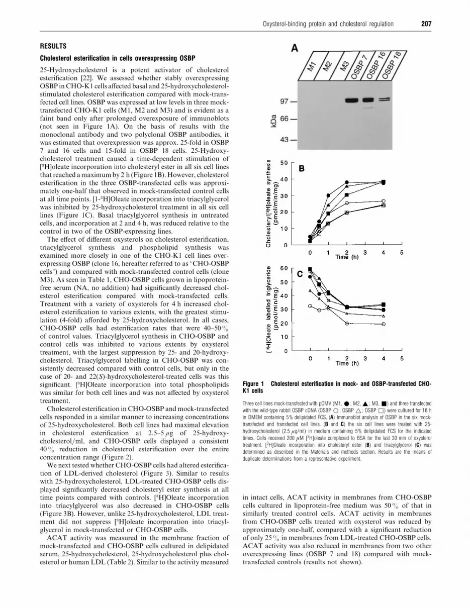
Cholesterol esterification in cells overexpressing OSBP
25-Hydroxycholesterol is a potent activator of cholesterol
esterification [22]. We assessed whether stably overexpressing
OSBP in CHO-K1 cells affected basal and 25-hydroxycholesterol-
stimulated cholesterol esterification compared with mock-trans-
fected cell lines. OSBP was expressed at low levels in three mock-
transfected CHO-K1 cells (M1, M2 and M3) and is evident as a
faint band only after prolonged overexposure of immunoblots
(not seen in Figure 1A). On the basis of results with the
monoclonal antibody and two polyclonal OSBP antibodies, it
was estimated that overexpression was approx. 25-fold in OSBP
7 and 16 cells and 15-fold in OSBP 18 cells. 25-Hydroxy-
cholesterol treatment caused a time-dependent stimulation of
[$H]oleate incorporation into cholesteryl ester in all six cell lines
that reached a maximum by 2 h (Figure 1B). However, cholesterol
esterification in the three OSBP-transfected cells was approxi-
mately one-half that observed in mock-transfected control cells
at all time points. [1-$H]Oleate incorporation into triacylglycerol
was inhibited by 25-hydroxycholesterol treatment in all six cell
lines (Figure 1C). Basal triacylglycerol synthesis in untreated
cells, and incorporation at 2 and 4 h, was reduced relative to the
control in two of the OSBP-expressing lines.
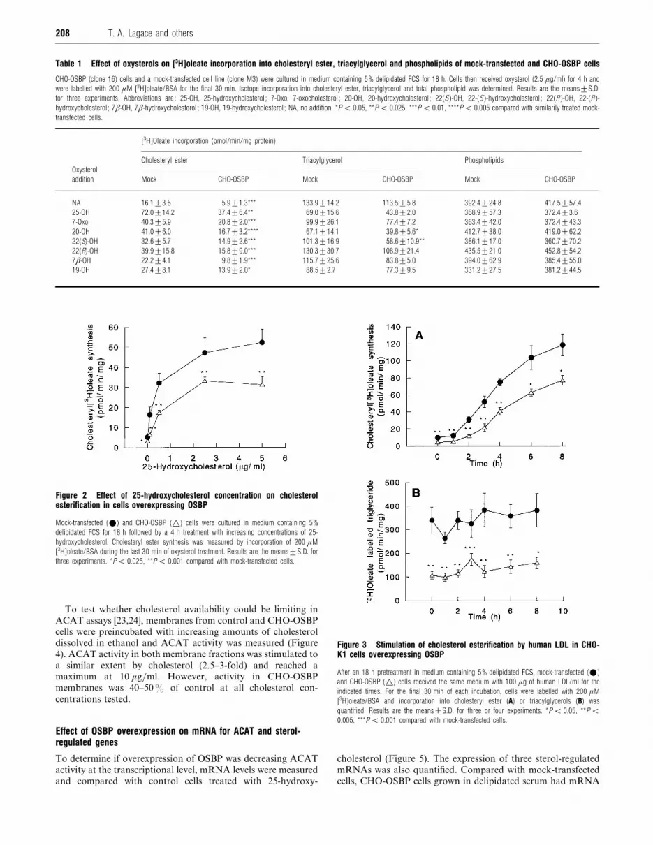
The effect of different oxysterols on cholesterol esterification,
triacylglycerol synthesis and phospholipid synthesis was
examined more closely in one of the CHO-K1 cell lines over-
expressing OSBP (clone 16, hereafter referred to as ‘CHO-OSBP
cells ’) and compared with mock-transfected control cells (clone
M3). As seen in Table 1, CHO-OSBP cells grown in lipoprotein-
free serum (NA, no addition) had significantly decreased chol-
esterol esterification compared with mock-transfected cells.
Treatment with a variety of oxysterols for 4 h increased chol-
esterol esterification to various extents, with the greatest stimu-
lation (4-fold) afforded by 25-hydroxycholesterol. In all cases,
CHO-OSBP cells had esterification rates that were 40–50%
of control values. Triacylglycerol synthesis in CHO-OSBP and
control cells was inhibited to various extents by oxysterol
treatment, with the largest suppression by 25- and 20-hydroxy-
cholesterol. Triacylglycerol labelling in CHO-OSBP was con-
sistently decreased compared with control cells, but only in the
case of 20- and 22(S)-hydroxycholesterol-treated cells was this
significant. [$H]Oleate incorporation into total phospholipids
was similar for both cell lines and was not affected by oxysterol
treatment.
Cholesterol esterification in CHO-OSBP and mock-transfected
cells responded in a similar manner to increasing concentrations
of 25-hydroxycholesterol. Both cell lines had maximal elevation
in cholesterol esterification at 2.5–5 µg of 25-hydroxy-
cholesterol}ml, and CHO-OSBP cells displayed a consistent
40% reduction in cholesterol esterification over the entire
concentration range (Figure 2).
We next tested whether CHO-OSBP cells had altered esterifica-
tion of LDL-derived cholesterol (Figure 3). Similar to results
with 25-hydroxycholesterol, LDL-treated CHO-OSBP cells dis-
played significantly decreased cholesteryl ester synthesis at all
time points compared with controls. [$H]Oleate incorporation
into triacylglycerol was also decreased in CHO-OSBP cells
(Figure 3B). However, unlike 25-hydroxycholesterol, LDL treat-
ment did not suppress [$H]oleate incorporation into triacyl-
glycerol in mock-transfected or CHO-OSBP cells.
ACAT activity was measured in the membrane fraction of
mock-transfected and CHO-OSBP cells cultured in delipidated
serum, 25-hydroxycholesterol, 25-hydroxycholesterol plus chol-
esterol or human LDL (Table 2). Similar to the activity measured
Figure 1 Cholesterol esterification in mock- and OSBP-transfected CHO-K1 cells
Three cell lines mock-transfected with pCMV (M1, E ; M2, _ ; M3, +) and three transfected
with the wild-type rabbit OSBP cDNA (OSBP D ; OSBP ^ ; OSBP *) were cultured for 18 h
in DMEM containing 5% delipidated FCS. (A) Immunoblot analysis of OSBP in the six mock-
transfected and transfected cell lines. (B and C) the six cell lines were treated with 25-
hydroxycholesterol (2.5 µg/ml) in medium containing 5% delipidated FCS for the indicated
times. Cells received 200 µM [3H]oleate complexed to BSA for the last 30 min of oxysterol
treatment. [3H]Oleate incorporation into cholesteryl ester (B) and triacylglycerol (C) was
determined as described in the Materials and methods section. Results are the means of
duplicate determinations from a representative experiment.
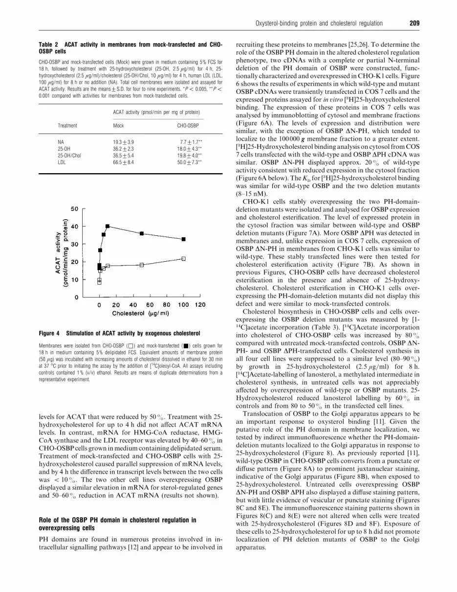
in intact cells, ACAT activity in membranes from CHO-OSBP
cells cultured in lipoprotein-free medium was 50% of that in
similarly treated control cells. ACAT activity in membranes
from CHO-OSBP cells treated with oxysterol was reduced by
approximately one-half, compared with a significant reduction
of only 25% in membranes from LDL-treated CHO-OSBP cells.
ACAT activity was also reduced in membranes from two other
overexpressing lines (OSBP 7 and 18) compared with mock-
transfected controls (results not shown).
208 T. A. Lagace and others
Table 1 Effect of oxysterols on [3H]oleate incorporation into cholesteryl ester, triacylglycerol and phospholipids of mock-transfected and CHO-OSBP cells
CHO-OSBP (clone 16) cells and a mock-transfected cell line (clone M3) were cultured in medium containing 5% delipidated FCS for 18 h. Cells then received oxysterol (2.5 µg/ml) for 4 h and
were labelled with 200 µM [3H]oleate/BSA for the final 30 min. Isotope incorporation into cholesteryl ester, triacylglycerol and total phospholipid was determined. Results are the means³S.D.
for three experiments. Abbreviations are : 25-OH, 25-hydroxycholesterol ; 7-Oxo, 7-oxocholesterol ; 20-OH, 20-hydroxycholesterol ; 22(S )-OH, 22-(S )-hydroxycholesterol ; 22(R )-OH, 22-(R )-
hydroxycholesterol ; 7β-OH, 7β-hydroxycholesterol ; 19-OH, 19-hydroxycholesterol ; NA, no addition. *P ! 0.05, **P ! 0.025, ***P ! 0.01, ****P ! 0.005 compared with similarily treated mock-
transfected cells.
[3H]Oleate incorporation (pmol/min/mg protein)
Cholesteryl ester Triacylglycerol Phospholipids
Oxysterol
addition Mock CHO-OSBP Mock CHO-OSBP Mock CHO-OSBP
NA 16.1³3.6 5.9³1.3*** 133.9³14.2 113.5³5.8 392.4³24.8 417.5³57.4
25-OH 72.0³14.2 37.4³6.4** 69.0³15.6 43.8³2.0 368.9³57.3 372.4³3.6
7-Oxo 40.3³5.9 20.8³2.0*** 99.9³26.1 77.4³7.2 363.4³42.0 372.4³43.3
20-OH 41.0³6.0 16.7³3.2**** 67.1³14.1 39.8³5.6* 412.7³38.0 419.0³62.2
22(S)-OH 32.6³5.7 14.9³2.6*** 101.3³16.9 58.6³10.9** 386.1³17.0 360.7³70.2
22(R)-OH 39.9³15.8 15.8³9.0*** 130.3³30.7 108.9³21.4 435.5³21.0 452.8³54.2
7β-OH 22.2³4.1 9.8³1.9*** 115.7³25.6 83.8³5.0 394.0³62.9 385.4³55.0
19-OH 27.4³8.1 13.9³2.0* 88.5³2.7 77.3³9.5 331.2³27.5 381.2³44.5
Figure 2 Effect of 25-hydroxycholesterol concentration on cholesterolesterification in cells overexpressing OSBP
Mock-transfected (E) and CHO-OSBP (^) cells were cultured in medium containing 5%
delipidated FCS for 18 h followed by a 4 h treatment with increasing concentrations of 25-
hydroxycholesterol. Cholesteryl ester synthesis was measured by incorporation of 200 µM
[3H]oleate/BSA during the last 30 min of oxysterol treatment. Results are the means³S.D. for
three experiments. *P! 0.025, **P ! 0.001 compared with mock-transfected cells.
To test whether cholesterol availability could be limiting in
ACAT assays [23,24], membranes from control and CHO-OSBP
cells were preincubated with increasing amounts of cholesterol
dissolved in ethanol and ACAT activity was measured (Figure
4). ACAT activity in both membrane fractions was stimulated to
a similar extent by cholesterol (2.5–3-fold) and reached a
maximum at 10 µg}ml. However, activity in CHO-OSBP
membranes was 40–50% of control at all cholesterol con-
centrations tested.
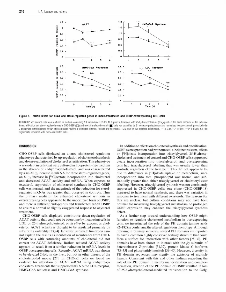
Effect of OSBP overexpression on mRNA for ACAT and sterol-regulated genes
To determine if overexpression of OSBP was decreasing ACAT
activity at the transcriptional level, mRNA levels were measured
and compared with control cells treated with 25-hydroxy-
Figure 3 Stimulation of cholesterol esterification by human LDL in CHO-K1 cells overexpressing OSBP
After an 18 h pretreatment in medium containing 5% delipidated FCS, mock-transfected (E)
and CHO-OSBP (^) cells received the same medium with 100 µg of human LDL/ml for the
indicated times. For the final 30 min of each incubation, cells were labelled with 200 µM
[3H]oleate/BSA and incorporation into cholesteryl ester (A) or triacylglycerols (B) was
quantified. Results are the means³S.D. for three or four experiments. *P ! 0.05, **P !0.005, ***P ! 0.001 compared with mock-transfected cells.
cholesterol (Figure 5). The expression of three sterol-regulated
mRNAs was also quantified. Compared with mock-transfected
cells, CHO-OSBP cells grown in delipidated serum had mRNA
209Oxysterol-binding protein and cholesterol regulation
Table 2 ACAT activity in membranes from mock-transfected and CHO-OSBP cells
CHO-OSBP and mock-transfected cells (Mock) were grown in medium containing 5% FCS for
18 h, followed by treatment with 25-hydroxycholesterol (25-OH, 2.5 µg/ml) for 4 h, 25-
hydroxycholesterol (2.5 µg/ml)/cholesterol (25-OH/Chol, 10 µg/ml) for 4 h, human LDL (LDL,
100 µg/ml) for 8 h or no addition (NA). Total cell membranes were isolated and assayed for
ACAT activity. Results are the means³S.D. for four to nine experiments. *P ! 0.005, **P !0.001 compared with activities for membranes from mock-transfected cells.
ACAT activity (pmol/min per mg of protein)
Treatment Mock CHO-OSBP
NA 19.3³3.9 7.7³1.7**
25-OH 36.2³2.3 18.0³4.3**
25-OH/Chol 36.5³5.4 19.8³4.0**
LDL 66.5³8.4 50.0³7.3**
Figure 4 Stimulation of ACAT activity by exogenous cholesterol
Membranes were isolated from CHO-OSBP (*) and mock-transfected (+) cells grown for
18 h in medium containing 5% delipidated FCS. Equivalent amounts of membrane protein
(50 µg) was incubated with increasing amounts of cholesterol dissolved in ethanol for 30 min
at 37 °C prior to initiating the assay by the addition of [14C]oleoyl-CoA. All assays including
controls contained 1% (v/v) ethanol. Results are means of duplicate determinations from a
representative experiment.
levels for ACAT that were reduced by 50%. Treatment with 25-
hydroxycholesterol for up to 4 h did not affect ACAT mRNA
levels. In contrast, mRNA for HMG-CoA reductase, HMG-
CoA synthase and the LDL receptor was elevated by 40–60% in
CHO-OSBP cells grown inmedium containing delipidated serum.
Treatment of mock-transfected and CHO-OSBP cells with 25-
hydroxycholesterol caused parallel suppression of mRNA levels,
and by 4 h the difference in transcript levels between the two cells
was ! 10%. The two other cell lines overexpressing OSBP
displayed a similar elevation in mRNA for sterol-regulated genes
and 50–60% reduction in ACAT mRNA (results not shown).
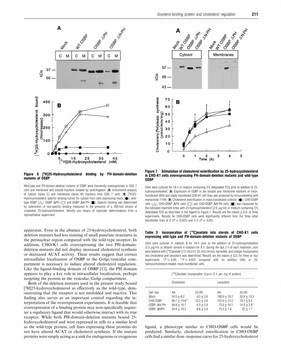
Role of the OSBP PH domain in cholesterol regulation inoverexpressing cells
PH domains are found in numerous proteins involved in in-
tracellular signalling pathways [12] and appear to be involved in
recruiting these proteins to membranes [25,26]. To determine the
role of the OSBP PH domain in the altered cholesterol regulation
phenotype, two cDNAs with a complete or partial N-terminal
deletion of the PH domain of OSBP were constructed, func-
tionally characterized and overexpressed inCHO-K1 cells. Figure
6 shows the results of experiments in which wild-type and mutant
OSBP cDNAs were transiently transfected in COS 7 cells and the
expressed proteins assayed for in �itro [$H]25-hydroxycholesterol
binding. The expression of these proteins in COS 7 cells was
analysed by immunoblotting of cytosol and membrane fractions
(Figure 6A). The levels of expression and distribution were
similar, with the exception of OSBP ∆N-PH, which tended to
localize to the 100000 g membrane fraction to a greater extent.
[$H]25-Hydroxycholesterol binding analysis on cytosol fromCOS
7 cells transfected with the wild-type and OSBP ∆PH cDNA was
similar. OSBP ∆N-PH displayed approx. 20% of wild-type
activity consistent with reduced expression in the cytosol fraction
(Figure 6A below). TheKD
for [$H]25-hydroxycholesterol binding
was similar for wild-type OSBP and the two deletion mutants
(8–15 nM).
CHO-K1 cells stably overexpressing the two PH-domain-
deletion mutants were isolated and analysed for OSBP expression
and cholesterol esterification. The level of expressed protein in
the cytosol fraction was similar between wild-type and OSBP
deletion mutants (Figure 7A). More OSBP ∆PH was detected in
membranes and, unlike expression in COS 7 cells, expression of
OSBP ∆N-PH in membranes from CHO-K1 cells was similar to
wild-type. These stably transfected lines were then tested for
cholesterol esterification activity (Figure 7B). As shown in
previous Figures, CHO-OSBP cells have decreased cholesterol
esterification in the presence and absence of 25-hydroxy-
cholesterol. Cholesterol esterification in CHO-K1 cells over-
expressing the PH-domain-deletion mutants did not display this
defect and were similar to mock-transfected controls.
Cholesterol biosynthesis in CHO-OSBP cells and cells over-
expressing the OSBP deletion mutants was measured by [1-"%C]acetate incorporation (Table 3). ["%C]Acetate incorporation
into cholesterol of CHO-OSBP cells was increased by 80%
compared with untreated mock-transfected controls, OSBP ∆N-
PH- and OSBP ∆PH-transfected cells. Cholesterol synthesis in
all four cell lines were suppressed to a similar level (80–90%)
by growth in 25-hydroxycholesterol (2.5 µg}ml) for 8 h.
["%C]Acetate-labelling of lanosterol, a methylated intermediate in
cholesterol synthesis, in untreated cells was not appreciably
affected by overexpression of wild-type or OSBP mutants. 25-
Hydroxycholesterol reduced lanosterol labelling by 60% in
controls and from 80 to 50% in the transfected cell lines.
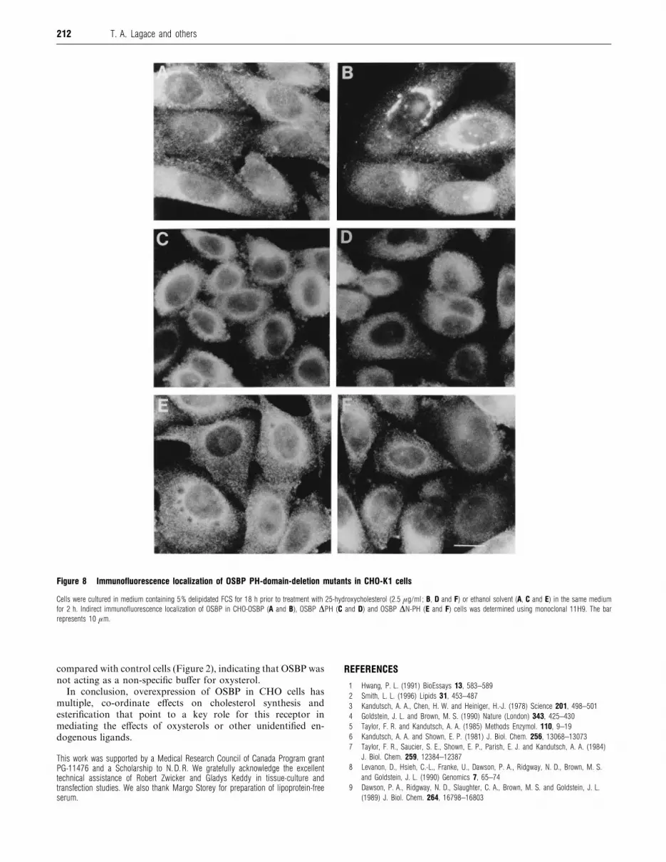
Translocation of OSBP to the Golgi apparatus appears to be
an important response to oxysterol binding [11]. Given the
putative role of the PH domain in membrane localization, we
tested by indirect immunofluorescence whether the PH-domain-
deletion mutants localized to the Golgi apparatus in response to
25-hydroxycholesterol (Figure 8). As previously reported [11],
wild-type OSBP in CHO-OSBP cells converts from a punctate or
diffuse pattern (Figure 8A) to prominent juxtanuclear staining,
indicative of the Golgi apparatus (Figure 8B), when exposed to
25-hydroxycholesterol. Untreated cells overexpressing OSBP
∆N-PH and OSBP ∆PH also displayed a diffuse staining pattern,
but with little evidence of vesicular or punctate staining (Figures
8C and 8E). The immunofluorescence staining patterns shown in
Figures 8(C) and 8(E) were not altered when cells were treated
with 25-hydroxycholesterol (Figures 8D and 8F). Exposure of
these cells to 25-hydroxycholesterol for up to 8 h did not promote
localization of PH deletion mutants of OSBP to the Golgi
apparatus.
210 T. A. Lagace and others
Figure 5 mRNA levels for ACAT and sterol-regulated genes in mock-transfected and OSBP-overexpressing CHO cells
CHO-OSBP and control cells were cultured in medium containing 5% delipidated FCS for 18 h prior to treatment with 25-hydroxycholesterol (2.5 µg/ml) in the same medium for the indicated
times. mRNA for four sterol-regulated genes in CHO-OSBP (*) and mock-transfected control (+) cells was quantified by S1 nuclease protection assays, normalized to expression of glyceraldehyde-
3-phosphate dehydrogenase mRNA and expressed relative to untreated controls. Results are the means³S.D. four or five separate experiments. *P ! 0.05, **P ! 0.01, ***P ! 0.005, n.s (not
significant) compared with mock-transfected cells.
DISCUSSION
CHO-OSBP cells displayed an altered cholesterol regulation
phenotype characterized by up-regulation of cholesterol synthesis
and down-regulation of cholesterol esterification. This phenotype
was evident in cells that were cultured in lipoprotein-free medium
in the absence of 25-hydroxycholesterol, and was characterized
by a 40–60% increase in mRNA for three sterol-regulated genes,
an 80% increase in ["%C]acetate incorporation into cholesterol
and decreased ACAT activity and mRNA. When exposed to
oxysterol, suppression of cholesterol synthesis in CHO-OSBP
cells was normal, and the magnitude of the reduction for sterol-
regulated mRNAs was greater than observed in controls. Thus
the primary mediator for enhanced cholesterol synthesis in
overexpressing cells appears to be the unoccupied form of OSBP,
and there is sufficient endogenous and transfected rabbit OSBP
to ensure a normal or slightly exaggerated response to oxysterol
treatment.
CHO-OSBP cells displayed constitutive down-regulation of
ACAT activity that could not be overcome by incubating cells in
LDL or 25-hydroxycholesterol, or in �itro by exogenous chol-
esterol. ACAT activity is thought to be regulated primarily by
substrate availability [23,24]. However, substrate limitation can-
not explain the results as incubation of membranes from CHO-
OSBP cells with saturating amounts of cholesterol did not
correct the ACAT deficiency. Rather, reduced ACAT activity
appears to result from a similar reduction in mRNA levels in
OSBP overexpressing cells. Recently, ACAT mRNA was shown
to be elevated 2-fold in the liver, but not in other tissues, of the
cholesterol-fed mouse [27]. In CHO-K1 cells we found no
evidence for alteration of ACAT mRNA using 25-hydroxy-
cholesterol treatments that suppressed mRNA for LDL receptor,
HMG-CoA reductase and HMG-CoA synthase.
In addition to effects on cholesterol synthesis and esterification,
OSBP overexpression had pronounced, albeit inconsistent, effects
on [$H]oleate incorporation into triacylglycerol. 25-Hydroxy-
cholesterol treatment of control and CHO-OSBP cells suppressed
oleate incorporation into triacylglycerol, and overexpressing
cells had triacylglycerol labelling that was usually lower than
controls, regardless of the treatment. This did not appear to be
due to differences in [$H]oleate uptake or metabolism, since
incorporation into total phospholipid was normal and sub-
stantially greater than either triacylglycerol or cholesteryl ester
labelling. However, triacylglycerol synthesis was not consistently
suppressed in CHO-OSBP cells ; one clone (CHO-OSBP-18)
appeared to have normal synthesis, and there was variation in
response to treatment with different oxysterols. The reasons for
this are unclear, but culture conditions may not have been
optimal for measuring triacylglycerol metabolism or prolonged
OSBP expression may enhance the triacylglycerol synthesis
defect.
As a further step toward understanding how OSBP might
function to regulate cholesterol metabolism in overexpressing
cells, we investigated the role of the PH domain (amino acids
92–182) in conferring the altered regulation phenotype. Although
differing in primary sequence, several PH domains are reported
to have a common highly conserved tertiary structure that could
form a surface for interaction with other factors [28–30]. PH
domains have been shown to interact with the βγ subunits of
heterotrimeric G-proteins [31,32], protein kinase C isoforms
[33–35] and phosphatidylinositols [36–40]. However, diversity in
PH domain sequences may signify the existence of multiple
ligands. Consistent with this and other findings regarding the
role of the PH domain in membrane localization and complex-
formation, deletion of the PH domain of OSBP resulted in loss
of 25-hydroxycholesterol-mediated translocation to the Golgi
211Oxysterol-binding protein and cholesterol regulation
Figure 6 [3H]25-Hydroxycholesterol binding by PH-domain-deletionmutants of OSBP
Wild-type and PH-domain-deletion mutants of OSBP were transiently overexpressed in COS 7
cells and membrane and soluble fractions isolated by centrifugation. (A) Immunoblot analysis
of cytosol (lanes C) and membrane (lanes M) fractions from COS 7 cells. (B) [3H]25-
Hydroxycholesterol specific binding curves for cytosol from cells expressing mock (E), wild-
type OSBP (^), OSBP ∆PH (*) and OSBP ∆N-PH (+). Specific binding was determined
by subtraction of non-specific binding measured in the presence of a 200-fold excess of
unlabelled 25-hydroxycholesterol. Results are means of duplicate determinations from a
representative experiment.
apparatus. Even in the absence of 25-hydroxycholesterol, both
deletion mutants had less staining of small punctate structures in
the perinuclear region compared with the wild-type receptor. In
addition, CHO-K1 cells overexpressing the two PH-domain-
deletion mutants did not display increased cholesterol synthesis
or decreased ACAT activity. These results suggest that correct
intracellular localization of OSBP in the Golgi}vesicular com-
partment is necessary to induce altered cholesterol regulation.
Like the ligand-binding domain of OSBP [11], the PH domain
appears to play a key role in intracellular localization, perhaps
targeting the protein in the vesicular}Golgi compartment.
Both of the deletion mutants used in the present study bound
[$H]25-hydroxycholesterol as effectively as the wild-type, dem-
onstrating that the receptor is not misfolded and inactive. This
finding also serves as an important control regarding the in-
terpretation of the overexpression experiments. It is feasible that
overexpression of a binding protein may non-specifically seques-
ter a regulatory ligand that would otherwise interact with its true
receptor. While both PH-domain-deletion mutants bound 25-
hydroxycholesterol and were expressed in cells to a similar level
as the wild-type protein, cell lines expressing these proteins do
not have altered ACAT or cholesterol synthesis. If the mutant
proteinswere simply acting as a sink for endogenous or exogenous
Figure 7 Stimulation of cholesterol esterification by 25-hydroxycholesterolin CHO-K1 cells overexpressing PH-domain-deletion mutants and wild-typeOSBP
Cells were cultured for 18 h in medium containing 5% delipidated FCS prior to addition of 25-
hydroxycholesterol. (A) Expression of OSBP in the soluble and membrane fractions of mock-
transfected (M3) and stably transfected CHO-K1 cell lines was assessed by immunoblotting with
monoclonal 11H9. (B) Cholesterol esterification in mock-transfected controls (E), CHO-OSBP
cells (^), CHO-OSBP ∆PH cells (*) and CHO-OSBP ∆N-PH cells (+) was measured for
the indicated treatment times with 25-hydroxycholesterol (2.5 µg/ml) in medium containing 5%
delipidated FCS as described in the legend to Figure 1. Results are the means³S.D. of three
experiments. Results for CHO-OSBP cells were significantly different from the three other
transfected lines at 0 (P ! 0.025) and 4 h (P ! 0.05).
Table 3 Incorporation of [14C]acetate into sterols of CHO-K1 cellsexpressing wild-type and PH-domain-deletion mutants of OSBP
Cells were cultured in medium B for 18 h prior to the addition of 25-hydroxycholesterol
(2.5 µg/ml) or ethanol solvent in medium for 8 h. During the last 2 h of each treatment, cells
were labelled with [14C]acetate (7.5 mCi/ml, 55 mCi/mmol), harvested, and isotope incorporation
into cholesterol and lanosterol was determined. Results are the means³S.D. for three or four
experiments. *P ! 0.05, **P ! 0.025 compared with no addition (NA) or 25-
hydroxycholesterol-treated mock-transfected cells.
[14C]Acetate incorporation (d.p.m./2 h per mg of protein)
Cholesterol Lanosterol
Cell line NA 25-OH NA 25-OH
Mock 54.5³8.2 6.5³3.0 100.0³16.2 35.9³15.2
CHO-OSBP 89.7³15.0** 10.3³3.6 102.0³13.2 19.7³8.2
OSBP ∆N-PH 48.8³16.7 6.3³3.9 72.0³16.1 14.9³2.6*
OSBP ∆6PH 50.4³16.2 8.8³5.9 75.3³1.6 32³7.7
ligand, a phenotype similar to CHO-OSBP cells would be
predicted. Similarly, cholesterol esterification in CHO-OSBP
cells had a similar dose–response curve for 25-hydroxycholesterol
212 T. A. Lagace and others
Figure 8 Immunofluorescence localization of OSBP PH-domain-deletion mutants in CHO-K1 cells
Cells were cultured in medium containing 5% delipidated FCS for 18 h prior to treatment with 25-hydroxycholesterol (2.5 µg/ml ; B, D and F) or ethanol solvent (A, C and E) in the same medium
for 2 h. Indirect immunofluorescence localization of OSBP in CHO-OSBP (A and B), OSBP ∆PH (C and D) and OSBP ∆N-PH (E and F) cells was determined using monoclonal 11H9. The bar
represents 10 µm.
compared with control cells (Figure 2), indicating that OSBP was
not acting as a non-specific buffer for oxysterol.
In conclusion, overexpression of OSBP in CHO cells has
multiple, co-ordinate effects on cholesterol synthesis and
esterification that point to a key role for this receptor in
mediating the effects of oxysterols or other unidentified en-
dogenous ligands.
This work was supported by a Medical Research Council of Canada Program grantPG-11476 and a Scholarship to N.D.R. We gratefully acknowledge the excellenttechnical assistance of Robert Zwicker and Gladys Keddy in tissue-culture andtransfection studies. We also thank Margo Storey for preparation of lipoprotein-freeserum.
REFERENCES
1 Hwang, P. L. (1991) BioEssays 13, 583–589
2 Smith, L. L. (1996) Lipids 31, 453–487
3 Kandutsch, A. A., Chen, H. W. and Heiniger, H.-J. (1978) Science 201, 498–501
4 Goldstein, J. L. and Brown, M. S. (1990) Nature (London) 343, 425–430
5 Taylor, F. R. and Kandutsch, A. A. (1985) Methods Enzymol. 110, 9–19
6 Kandutsch, A. A. and Shown, E. P. (1981) J. Biol. Chem. 256, 13068–13073
7 Taylor, F. R., Saucier, S. E., Shown, E. P., Parish, E. J. and Kandutsch, A. A. (1984)
J. Biol. Chem. 259, 12384–12387
8 Levanon, D., Hsieh, C.-L., Franke, U., Dawson, P. A., Ridgway, N. D., Brown, M. S.
and Goldstein, J. L. (1990) Genomics 7, 65–74
9 Dawson, P. A., Ridgway, N. D., Slaughter, C. A., Brown, M. S. and Goldstein, J. L.
(1989) J. Biol. Chem. 264, 16798–16803
213Oxysterol-binding protein and cholesterol regulation
10 Dawson, P. A., Van Der Wethuyzen, D. R., Goldstein, J. L. and Brown, M. S. (1989)
J. Biol. Chem. 264, 9046–9052
11 Ridgway, N. D., Dawson, P. A., Ho, Y. K., Brown, M. S. and Goldstein, J. L. (1992)
J. Cell Biol. 116, 307–319
12 Gibson, T. J., Hyvonen, M., Musacchio, A. and Saraste, M. (1994) Trends Biochem.
Sci. 19, 349–353
13 Haslam, R. J., Kolde, H. B. and Hemmings, B. A. (1993) Nature (London) 363,309–310
14 Jiang, B., Brown, J. L., Sheraton, J., Fortin, N. and Bussey, H. (1994) Yeast 10,341–353
15 Fang, M., Kearns, B. G., Gedvitaite, A., Kagiwada, S., Kearns, M., Fung, M. K. Y. and
Bankaitis, V. A. (1996) EMBO J. 15, 6447–6459
16 Esser, V., Limbird, L. E., Brown, M. S., Goldstein, J. L. and Russell, D. W. (1988)
J. Biol. Chem. 263, 13283–13290
17 Goldstein, J. L., Basu, S. K. and Brown, M. S. (1983) Methods Enzymol. 98,241–260
18 Metherall, J. E., Ridgway, N. D., Dawson, P. A., Goldstein, J. L. and Brown, M. S.
(1991) J. Biol. Chem. 266, 12734–12740
19 Ridgway, N. D. and Lagace, T. A. (1995) J. Biol. Chem. 270, 8023–8031
20 Cao, G., Goldstein, J. L. and Brown, M. S. (1996) J. Biol. Chem. 271, 14642–14648
21 Brown, M. S., Faust, J. R., Goldstein, J. L., Kandeko, I. and Endo, A. (1978) J. Biol.
Chem. 253, 1121–1128
22 Brown, M. S., Dana, S. E. and Goldstein, J. L. (1975) J. Biol. Chem. 250,4025–4027
23 Cheng, D., Chang, C. C. Y., Qu, X. M. and Chang, T. Y. (1995) J. Biol. Chem. 270,685–695
24 Myant, N. B. (1990) in Cholesterol Metabolism, LDL and LDL Receptor, (Myant,
N. B., ed.), pp. 85–98, Academic Press, San Diego
25 Inglese, J., Koch, J. W., Kazushige, T. and Lefkowitz, R. J. (1995) Trends Biochem.
Sci. 20, 151–156
Received 23 January 1997/15 April 1997 ; accepted 22 April 1997
26 Lemmon, M. A., Ferguson, K. M. and Schlessinger, J. (1996) Cell 85, 621–624
27 Uleman, P. J., Oka, K., Sullivan, M., Chang, C. C. Y., Chang, T. Y. and Chan, L.
(1995) J. Biol. Chem. 270, 26192–26201
28 Ferguson, K. M., Lemmon, M. A., Schlessinger, J. and Sigler, P. B. (1994) Cell 79,199–209
29 Yoon, S.-H., Hadjuk, P. J., Petros, A. M., Olejniczak, E. T., Meadows, R. P. and Fesik,
S. W. (1994) Nature (London) 369, 672–677
30 Ferguson, K. M., Lemmon, M. A., Schlessinger, J. and Sigler, P. B. (1995) Cell 83,1037–1046
31 Touhara, K., Inglese, J., Pitcher, J. A., Shaw, G. and Lefkowitz, R. J. (1994) J. Biol.
Chem. 269, 10217–10220
32 Touhara, K., Koch, W. J., Hawes, B. E. and Lefkowitz, R. J. (1995) J. Biol. Chem.
270, 17000–17005
33 Yao, L., Kawakami, Y. and Kawakami, T. (1994) Proc. Natl. Acad. Sci. U.S.A. 91,9175–9179
34 Konishi, H., Kuroda, S. and Kikkawa, U. (1994) Biochem. Biophys. Res. Commun.
205, 1770–1775
35 Wang, D. S., Shaw, R., Hattori, M., Arai, H., Inoue, K. and Shaw, G. (1995) Biochem.
Biophys. Res. Commun. 209, 622–629
36 Paterson, H. F., Savopoulos, J. W., Perisisc, O., Cheung, R., Ellis, M. V., Williams,
R. L. and Katan, M. (1995) Biochem. J. 312, 661–666
37 Essen, L. O., Pewrisic, O., Cheung, R., Katan, M. and Williams, R. L. (1996) Nature
(London) 380, 595–602
38 Lemmon, M. A., Ferguson, K. M., O ’Brien, R., Sigler, P. B. and Schlessinger, J.
(1995) Proc. Natl. Acad. Sci. U.S.A. 92, 10472–10476
39 Hyvo$ nen, M., Macias, M. J., Nilges, M., Oschkinat, H., Saraste, M. and Wilmanns,
M. (1995) EMBO J. 14, 4676–4685
40 Harlan, J. E., Hadjuk, P. J., Yoon, H.-S. and Fesik, S. W. (1994) Nature (London)
371, 168–170
Related Documents