Altered brain function underlying verbal memory encoding and retrieval in psychotic major depression Ryan Kelley a,b , Amy Garrett a,b,n , Jeremy Cohen a , Rowena Gomez b , Anna Lembke b , Jennifer Keller b , Allan L. Reiss a,b,c , Alan Schatzberg b a Center for Interdisciplinary Brain Sciences Research, Stanford University School of Medicine, Stanford, CA 94305, USA b Department of Psychiatry and Behavioral Sciences, Stanford University School of Medicine, Stanford, CA 94305, USA c Department of Radiology, Stanford University School of Medicine, Stanford, CA 94305, USA article info Article history: Received 15 July 2011 Received in revised form 10 May 2012 Accepted 12 June 2012 Keywords: Depression Psychosis Functional imaging Verbal declarative memory Hippocampus Encoding deficits abstract Psychotic major depression (PMD) is associated with deficits in verbal memory as well as other cognitive impairments. This study investigated brain function in individuals with PMD during a verbal declarative memory task. Participants included 16 subjects with PMD, 15 subjects with non-psychotic major depression (NPMD) and 16 healthy controls (HC). Functional magnetic resonance imaging (fMRI) data were acquired while subjects performed verbal memory encoding and retrieval tasks. During the explicit encoding task, subjects semantically categorized words as either ‘‘man-made’’ or ‘‘not man- made.’’ For the retrieval task, subjects identified whether words had been presented during the encoding task. Functional MRI data were processed using SPM5 and a group by condition ANOVA. Clusters of activation showing either a significant main effect of group or an interaction of group by condition were further examined using t-tests to identify group differences. During the encoding task, the PMD group showed lower hippocampus, insula, and prefrontal activation compared to HC. During the retrieval task, the PMD group showed lower recognition accuracy and higher prefrontal and parietal cortex activation compared to both HC and NPMD groups. Verbal retrieval deficits in PMD may be associated with deficient hippocampus function during encoding. Increased brain activation during retrieval may reflect an attempt to compensate for encoding deficits. & 2012 Elsevier Ireland Ltd. All rights reserved. 1. Introduction Depression can be regarded as a heterogeneous condition, likely consisting of various subtypes associated with family psychiatric history, cognitive profiles and clinical features (Keller et al., 2007; Carragher et al., 2009). It has been estimated that 1 in 6 individuals with major depression also demonstrate psychotic features (psychotic major depression; PMD) (Johnson et al., 1991; Parker et al., 1992; Ohayon and Schatzberg, 2002). Nosological data have emerged since the early 1980s suggesting PMD and non-psychotic major depression (NPMD) are, indeed, separate subtypes of depression (Keller et al., 2007). PMD is associated with greater duration and severity of symptoms compared to NPMD (Nelson and Charney, 1981; Rothschild et al., 1989; Fleming et al., 2004; Keller et al., 2006; Maj et al., 2007). Additionally, individuals with PMD show greater cognitive deficits in verbal memory, attention and executive functioning compared to NPMD (Basso and Bornstein, 1999; Schatzberg et al., 2000; Fleming et al., 2004; Gomez et al., 2006). A recent functional brain imaging study (Garrett et al., 2011) identified aberrant working memory networks in PMD compared to NPMD and healthy control (HC) groups. Several studies have identified verbal memory deficits in PMD (Schatzberg et al., 2000; Fleming et al., 2004; Hill et al., 2004; Gomez et al., 2006; Zanelli et al., 2010). Two recent studies suggest that impaired encoding of verbal memory may be responsible for recognition memory deficits in PMD (Gomez et al., 2006; Zanelli et al., 2010). Similarly, a review of verbal declarative memory studies in schizophrenia concluded that deficits occur primarily during encoding, and are likely to involve prefrontal-hippocampal circuits (Cirillo and Seidman, 2003). It is possible that deficits in similar brain circuits during encoding underlie verbal memory deficits in PMD as well. The hippocampus is heavily resourced during verbal declara- tive memory encoding (Schacter and Wagner, 1999; Menon et al., 2002; Greicius et al., 2003; Parsons et al., 2006; Spaniol et al., 2009) and, to a lesser extent, during verbal declarative memory retrieval (Spaniol et al., 2009). Across the literature on hippocampus function during memory tasks in subjects with Contents lists available at SciVerse ScienceDirect journal homepage: www.elsevier.com/locate/psychresns Psychiatry Research: Neuroimaging 0925-4927/$ - see front matter & 2012 Elsevier Ireland Ltd. All rights reserved. http://dx.doi.org/10.1016/j.pscychresns.2012.06.008 n Corresponding author at: 401 Quarry Rd, Stanford, CA 94305-5795, USA. Tel.: þ1 650 736 1874; fax: þ1 650 724 4794. E-mail address: [email protected] (A. Garrett). Psychiatry Research: Neuroimaging 211 (2013) 119–126

Welcome message from author
This document is posted to help you gain knowledge. Please leave a comment to let me know what you think about it! Share it to your friends and learn new things together.
Transcript
Psychiatry Research: Neuroimaging 211 (2013) 119–126
Contents lists available at SciVerse ScienceDirect
Psychiatry Research: Neuroimaging
0925-49
http://d
n Corr
USA. Te
E-m
journal homepage: www.elsevier.com/locate/psychresns
Altered brain function underlying verbal memory encoding and retrievalin psychotic major depression
Ryan Kelley a,b, Amy Garrett a,b,n, Jeremy Cohen a, Rowena Gomez b, Anna Lembke b, Jennifer Keller b,Allan L. Reiss a,b,c, Alan Schatzberg b
a Center for Interdisciplinary Brain Sciences Research, Stanford University School of Medicine, Stanford, CA 94305, USAb Department of Psychiatry and Behavioral Sciences, Stanford University School of Medicine, Stanford, CA 94305, USAc Department of Radiology, Stanford University School of Medicine, Stanford, CA 94305, USA
a r t i c l e i n f o
Article history:
Received 15 July 2011
Received in revised form
10 May 2012
Accepted 12 June 2012
Keywords:
Depression
Psychosis
Functional imaging
Verbal declarative memory
Hippocampus
Encoding deficits
27/$ - see front matter & 2012 Elsevier Irelan
x.doi.org/10.1016/j.pscychresns.2012.06.008
esponding author at: 401 Quarry Rd, Stanfor
l.: þ1 650 736 1874; fax: þ1 650 724 4794.
ail address: [email protected] (A. Garrett
a b s t r a c t
Psychotic major depression (PMD) is associated with deficits in verbal memory as well as other
cognitive impairments. This study investigated brain function in individuals with PMD during a verbal
declarative memory task. Participants included 16 subjects with PMD, 15 subjects with non-psychotic
major depression (NPMD) and 16 healthy controls (HC). Functional magnetic resonance imaging (fMRI)
data were acquired while subjects performed verbal memory encoding and retrieval tasks. During the
explicit encoding task, subjects semantically categorized words as either ‘‘man-made’’ or ‘‘not man-
made.’’ For the retrieval task, subjects identified whether words had been presented during the
encoding task. Functional MRI data were processed using SPM5 and a group by condition ANOVA.
Clusters of activation showing either a significant main effect of group or an interaction of group by
condition were further examined using t-tests to identify group differences. During the encoding task,
the PMD group showed lower hippocampus, insula, and prefrontal activation compared to HC. During
the retrieval task, the PMD group showed lower recognition accuracy and higher prefrontal and parietal
cortex activation compared to both HC and NPMD groups. Verbal retrieval deficits in PMD may be
associated with deficient hippocampus function during encoding. Increased brain activation during
retrieval may reflect an attempt to compensate for encoding deficits.
& 2012 Elsevier Ireland Ltd. All rights reserved.
1. Introduction
Depression can be regarded as a heterogeneous condition,likely consisting of various subtypes associated with familypsychiatric history, cognitive profiles and clinical features(Keller et al., 2007; Carragher et al., 2009). It has been estimatedthat 1 in 6 individuals with major depression also demonstratepsychotic features (psychotic major depression; PMD) (Johnsonet al., 1991; Parker et al., 1992; Ohayon and Schatzberg, 2002).Nosological data have emerged since the early 1980s suggestingPMD and non-psychotic major depression (NPMD) are, indeed,separate subtypes of depression (Keller et al., 2007). PMD isassociated with greater duration and severity of symptomscompared to NPMD (Nelson and Charney, 1981; Rothschildet al., 1989; Fleming et al., 2004; Keller et al., 2006; Maj et al.,2007). Additionally, individuals with PMD show greater cognitivedeficits in verbal memory, attention and executive functioning
d Ltd. All rights reserved.
d, CA 94305-5795,
).
compared to NPMD (Basso and Bornstein, 1999; Schatzberg et al.,2000; Fleming et al., 2004; Gomez et al., 2006). A recentfunctional brain imaging study (Garrett et al., 2011) identifiedaberrant working memory networks in PMD compared to NPMDand healthy control (HC) groups.
Several studies have identified verbal memory deficits in PMD(Schatzberg et al., 2000; Fleming et al., 2004; Hill et al., 2004;Gomez et al., 2006; Zanelli et al., 2010). Two recent studiessuggest that impaired encoding of verbal memory may beresponsible for recognition memory deficits in PMD (Gomezet al., 2006; Zanelli et al., 2010). Similarly, a review of verbaldeclarative memory studies in schizophrenia concluded thatdeficits occur primarily during encoding, and are likely to involveprefrontal-hippocampal circuits (Cirillo and Seidman, 2003). It ispossible that deficits in similar brain circuits during encodingunderlie verbal memory deficits in PMD as well.
The hippocampus is heavily resourced during verbal declara-tive memory encoding (Schacter and Wagner, 1999; Menon et al.,2002; Greicius et al., 2003; Parsons et al., 2006; Spaniol et al.,2009) and, to a lesser extent, during verbal declarative memoryretrieval (Spaniol et al., 2009). Across the literature onhippocampus function during memory tasks in subjects with

R. Kelley et al. / Psychiatry Research: Neuroimaging 211 (2013) 119–126120
depression, activation deficits have been observed during encoding(Bremner et al., 2004; Fairhall et al., 2010; Milne et al., 2011), exceptfor an associative learning paradigm that instead found increasedparahippocampal activation (Werner et al., 2009). While less com-mon, memory retrieval in depression has also been associated withlower hippocampus activation (Werner et al., 2009; Fairhall et al.,2010; Milne et al., 2011). Hippocampal dysfunction could also play arole in verbal memory deficits in PMD (Gomez et al., 2006).Although, gross hippocampal volume reductions have not beenobserved in PMD (Keller et al., 2008) hippocampal dysfunctioncould still exist (Czeh and Lucassen, 2007). Alternately, othercomponents of the verbal memory encoding network may beresponsible for deficits in recognition, such as the prefrontal cortex.Reviews of the memory encoding literature conclude that activationof the ventrolateral prefrontal cortex is associated with selecting andmaintaining incoming information while activation of the dorsolat-eral prefrontal cortex is associated with organizing and formingassociations between items during encoding (Blumenfeld andRanganath, 2007; Binder et al., 2009; Spaniol et al., 2009).
Using functional magnetic resonance imaging (fMRI), wehypothesized that individuals with PMD would demonstratealtered profiles of activation during encoding (but not retrieval)of verbal information, particularly within the hippocampus andprefrontal cortex. Such findings would suggest that verbal mem-ory retrieval deficits in PMD are primarily associated withencoding deficits.
2. Methods
2.1. Subjects
The initial sample comprised 24 subjects with PMD, 19 subjects with NPMD,
and 21 HC subjects. After screening for exclusionary criteria (see below), 16
subjects with PMD, 15 subjects with NPMD, and 16 HC subjects were included in
Table 1Demographic variables.
Descriptor mean (SD) Psychotic major depression
group (PMD) N¼16
Non-psychoti
depression gr
N¼15
Age 35.57 (11.70) range¼18–54 35.78 (11.81)
Education, years 16.15 (3.97) range¼9–23
(N¼13)
14.45 (1.50) r
(N¼11)
Left handed 4 (N¼13) 2
WTAR predictedb 111.80 (12.72) (N¼10) 100.18 (25.03
Hamilton Depression Rating Scale 29.06 (4.12) range¼21–37 24.53 (3.09) r
Brief Psychiatric Rating Scale—positive
symptom subscale score (�4)
8.06 (3.33) range¼3–14 0.13 (0.35) ra
Endogenomorphic scale 9.69 (1.88) 8.13 (1.40)
Age at onset 25.11 (12.86) range¼10–51
(N¼9)
31.77 (11.89)
(N¼9)
Gender distribution 8 F/8 M 8 F/7 M
Current medicationc 2 none; 4 anx; 7 AD; 9 AP;
1 MS (N¼12)
8 none; 1 anx
Comorbid diagnoses 6 none, 4 anxiety disorderd
(N¼10)
8 none, 7 anx
a Only significant post hoc t-test p-values presented.b Wechsler test of reading (WTAR) estimate of premorbid full scale IQ.c For medication; anx¼anxiolytics; AD¼antidepressant; AP¼antipsychotic, MS¼md Anxiety disorders in the PMD group included 1 subject with panic disorder, 2 with
1 with social phobia.e Anxiety disorders in the NPMD group included 1 subjects with OCD, 1 with agorn po0.001.
the final fMRI analysis. The Stanford University Institutional Review Board
approved the study and all subjects gave written consent before participation.
Subjects with PMD and NPMD were recruited through inpatient and outpatient
facilities at Stanford University Medical Center or self-referred from advertise-
ments. Healthy controls were recruited from the community through
advertisements.
Subjects with PMD and NPMD met the following inclusion criteria: (1) a
minimum score of 21 on the 21-item Hamilton Depression Rating Scale (HDRS)
(Hamilton, 1960), (2) a score of 7 or higher on 7 items of the core endogen-
omorphic scale (Thase et al., 1983), and (3) current unipolar major depressive
episode, with or without psychotic features, based on the DSM-IV criteria. More-
over, PMD was differentiated from NPMD by a minimum score of 5 on the positive
symptoms subscale of the Brief Psychiatric Rating Scale (BPRS) (Gorham and
Overall, 1961). When relevant, subjects were permitted to continue medications
for ethical and safety reasons; all medications were required to be stable for one-
week prior to study entry (Table 1). Healthy controls met the following inclusion
criteria: (1) score of less than 6 on the HDRS, (2) no psychotic symptoms as
measured by the BPRS, and (3) no past or present psychiatric disorders as
determined by the Structural Clinical Interview for DSM-IV-TR Axis I Disorders
(First MB et al., 1997).
Subjects were excluded from the study if they had a major medical illness,
a history of seizures, previous head trauma, unstable or untreated hypertension,
a history of substance abuse, were actively suicidal, met criteria for obsessive
compulsive disorder or bipolar I or II disorder, were pregnant or lactating, were less
than 18 years of age, or had electroconvulsive therapy within the last 6 months.
Subjects taking estrogen supplements or hormonal contraceptives were excluded
because of known interactions between cortisol and estrogen and potential con-
founding effects on brain function during semantic retrieval (Kuhlmann and Wolf,
2005; Konrad et al., 2008).
Subjects who were recruited into the study, were later excluded from the fMRI
analysis if performance was below chance (50% accuracy) on the recognition of
encoded words during the retrieval task. A subject’s functional MRI data were
excluded if more than 25% of the time points exceeded a 0.5 mm/TR motion
threshold or had global signal greater than 3% from the mean global signal of all
images as determined by ArtRepair (http://cibsr.stanford.edu/tools/ArtRepair/
ArtRepair.htm).
2.2. Verbal declarative memory tasks
The encoding task presented alternating blocks of encoding and control
epochs and had a duration of 5’56’’. The five encoding epochs each presented
c major
oup (NPMD)
Healthy control
group (HC) N¼16
ANOVA and post hoc T-testsa
range¼20–59 32.74 (13.55)
range¼18–57
F(2,43)¼0.283, p¼0.753
ange¼12–16 15.46 (2.40)
range¼12–20
(N¼13)
F(2,33)¼1.042, p¼0.364
1 cb(1,44)¼3.336, p¼0.189
) (N¼11) 110.94 (8.98)
(N¼16)
F(2,34)¼1.818, p¼0.178
ange¼21–31 0.47 (0.52)
range¼0–1
F(2,43)¼457.2, po0.001
PMD4NPMD,n PMD4HC,n
NPMD4HC,n
nge¼0–1 0 F(2,43)¼88.06, po0.001
PMD4NPMD,n PMD4HC,n
0 F(2,43)¼237.18, po0.001,
PMD4HC,n NPMD4HC,n
range¼17–51 N/A t(16)¼1.224, p¼0.285
7 F/9 M cb(1,47)¼0.296, p¼0.863
; 7 AD; 0 AP; 1 MS N/A AD use is similar but AP use in PMD
group only
iety disordere N/A Similar; however, limited data
available
ood stabilizer.
agoraphobia, 1 with GAD, 2 with PTSD, 1 with anxiety NOS, 1 with specific phobia,
aphobia, 1 with GAD, 2 with social phobia, 4 with specific phobia.
R. Kelley et al. / Psychiatry Research: Neuroimaging 211 (2013) 119–126 121
eight nouns for 2.5 s with a 0.5 s interstimulus interval. To enhance encoding
(Craik et al., 1994), subjects were instructed to classify each noun into one of two
semantic categories, ‘‘man-made’’ or ‘‘not man-made’’. Subjects indicated their
response by pressing one of two buttons. The two semantic categories were
represented equally across each encoding epoch. Subjects were aware that there
would be a subsequent retrieval task. The five control epochs alternated the
presentation of the same two words 8 times subjects were instructed to alternate
pressing one of two buttons for each word presented without making a semantic
discrimination. Each epoch was preceded by a 4 s written instruction. A 24-s rest
epoch with fixation occurred at the beginning, end, and mid-point of the task. This
task has been described in previous studies from our group (Greicius et al., 2003;
Carrion et al., 2009).
The retrieval task presented alternating blocks of retrieval and control epochs
and had a duration of 6’52’’. In total, 32 previously-encoded words and 16 novel
words were shown across 6 retrieval epochs. During each retrieval epoch nouns
were presented for 2.5 s with a 0.5 s interstimulus interval. Subjects were
instructed to press one of two buttons to identify whether the word had or had
not been presented during the encoding task. Each retrieval epoch was followed
by a control epoch presenting the same two alternating words from the encoding
task. Subjects were instructed to alternate between the two buttons for each word
presented regardless of recognition judgment. A 24 s rest epoch with fixation was
presented at the beginning, end, and mid-point of the task. Subjects completed a
5-min scan (with gonogo task) between the encoding and retrieval tasks in order
to reduce the likelihood of rehearsal. The tasks were presented using Psyscope
software (http://psy.ck.sissa.it).
2.3. Behavioral data analysis
Measures of task performance collected during scanning included response
accuracy and response time for each stimulus (Table 2). For the encoding task,
accuracy was the percentage of correct semantic categorizations; corrected
accuracy for the retrieval task was calculated by hit rate minus false alarm rate,
where hit rate¼number correctly recognized words/ total number encoding words
and false alarm rate¼number of novel words incorrectly recognized/ total number
of encoding words. A signal detection applet (http://wise.cgu.edu/sdtmod/signa
l_applet.asp) was used to calculate recognition discriminability (d’). Behavioral
data were analyzed using SPSS 17 (http://www.spss.com/software/statistics).
A 3-group ANOVA was used to test for group differences in accuracy and response
time. Significant ANOVAs (po0.05) were followed by 2-group post hoc analyses.
2.4. fMRI data acquisition
Functional data were collected on a 3T GE Signa scanner using a GE whole head
coil (Lx platform, gradients 40 mT/m, 150 T/m/s: GE Medical Systems, Milwaukee,
Table 2Task accuracy and reaction times.
Descriptor mean (SE) Psychotic major depression
group (PMD) N¼16
Non-psychotic major d
group (NPMD) N¼15
Encoding condition
Percent accuracy 89.40712.77 94.4078.40
Response time for correct
responses, ms
1238.447237.41 1114.937206.81
Control condition
Percent accuracy 97.65673.19 93.2678.71
Response time for correct
responses, ms
586.097202.38 589.687192.36
Retrieval condition
Percent accuracy:
recognized words
68.28713.63 79.10712.60
Percent accuracy: novel
words
83.59714.76 79.79712.98
Correct response time:
recognized words, ms
1195.897215.92 1153.727135.09
Correct response time: novel
words, ms
1274.357236.06 1183.48 132.59
False alarm rate 0.07270.063 0.06570.048
Recognition discriminability
(d’)
2.2570.80 2.6870.67
Control condition
Percent accuracy 93.7779.28 96.3976.72
Response time for correct
responses, ms
533.477180.73 538.227138.09
a Only significant post hoc t-test p-values presented.
Wisconsin). Twenty-eight axial slices (4 mm thick, 0.05 mm skip), parallel to the
axis of anterior and posterior commisures and covering the entire brain
(FOV¼200 mm2, 64�64 matrix with an inplane spatial resolution¼3.125 mm2)
were acquired using a T2 weighted gradient echo spiral pulse sequence with the
following parameters: TR¼2000 ms, TE¼30 ms, flip angle¼89 degrees and one
interleave (Glover and Lai, 1998). Magnetic field inhomogeneities were reduced by
a shimming protocol used before acquiring fMRI scans (Spielman et al., 1998).
2.5. fMRI data analysis
Functional data were preprocessed and statistically analyzed using SPM5
(http://www.fil.ion.ucl.ac.uk/spm). After scanning, images were reconstructed,
resampled to 2 mm isotropic voxels and spatially realigned to the third volume,
which was chosen to avoid magnetic susceptibility artifacts. Interpolation errors
caused during image realignment were corrected using voxel-wise amplitude-
motion correlations (Grootoonk et al., 2000). The image distortion and spin history
errors caused by abrupt motions were repaired by interpolation from the nearest
unaffected volumes. These methods were implemented in the ArtRepair software
toolbox for SPM (http://cibsr.stanford.edu/tools/ArtRepair/ArtRepair.htm). Data
were spatially normalized into standard stereotactic space using the MNI
(Montreal Neurological Institute) echo-planar image template provided by SPM5.
Normalized images were then smoothed with a 7 mm FWHM Gaussian filter.
Statistics at the individual level used a fixed effects block design model to
define experimental (encoding and retrieval) and control conditions. A high-pass
filter of 120 s was applied to the data. Contrasts included encoding minus control
and retrieval minus control for the encoding and retrieval tasks, respectively.
Functional region of interest (ROI) clusters were initially defined using a random
effects model and a multivariate ANOVA analysis as implemented in SPM5.
A 2-way ANOVA with factors group (PMD, NPMD, and HC) and task (encoding
minus control and retrieval minus control) was used to identify activation
differences among the three groups (main effect of group) and regions that
showed different task effects per group (interaction between group and task). For
this initial step, we applied an exploratory threshold (height po0.001 and extent
of 10 voxels), intended to identify activation differences among the 3 groups. We
expect the PMD and NPMD groups to share some abnormalities compared to the
healthy control group, and a more stringent cluster corrected threshold would be
likely to eliminate these common abnormalities. In the second step, we conducted
follow-up t-tests of clusters identified in the first step. Clusters were required to
reach significance criteria and also to be relevant to previous neuroimaging
studies of patients with PMD or NPMD. Activation (mean T-score) was extracted
using the MarsBar toolbox (http://marsbar.sourceforge.net/) and exported to SPSS
software, where independent group t-tests, covaried for task accuracy, were used
to identify group differences, with Bonferroni correction for multiple comparisons.
epression Healthy control group
(HC) N¼16
ANOVA results and
post hoc T-testsa
93.3476.51 F(2,43)¼1.175, p¼0.318
1131.207153.19 F(2,43)¼1.738, p¼0.188
95.0075.40 F(2,43)¼2.026, p¼0.144
578.187150.87 F(2,43)¼0.002, p¼0.998
79.36712.16 F(2,47)¼3.852, p¼0.029, HC4PMD
p¼0.02, NPMD4PMD p¼0.03
77.74714.47 F(2,43)¼0.708, p¼0.498
1089.127139.67 F(2,43)¼1.63, p¼0.207
1197.567273.14 F(2,43)¼0.753, p¼0.477
0.05970.041 F(2,43)¼0.277, p¼0.759
2.8170.80 F(2,43)¼2.418, p¼0.101, HC4PMD
p¼0.041
93.25710.97 F(2,43)¼0.512, p¼0.603
536.507125.94 F(2,43)¼0.004, p¼0.996
R. Kelley et al. / Psychiatry Research: Neuroimaging 211 (2013) 119–126122
To identify neuroanatomical locations, activation clusters were superimposed
on a single-subject high-resolution T1-weighted image. Additionally, activation
cluster coordinates were converted from MNI space into Talairach space (http://
imaging.mrc-cbu.cam.ac.uk/imaging/MniTalairach/) and input into a Talairach
daemon (http://www.nitrc.org/projects/tal-daemon/) to help identify neuroana-
tomical locations.
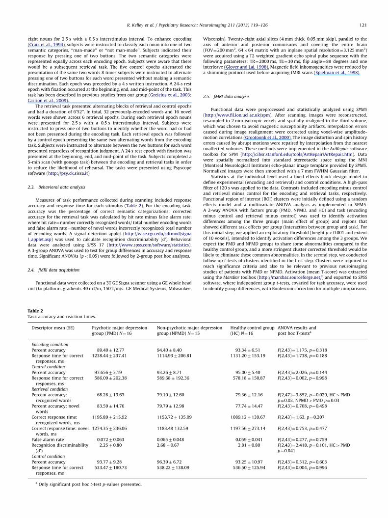
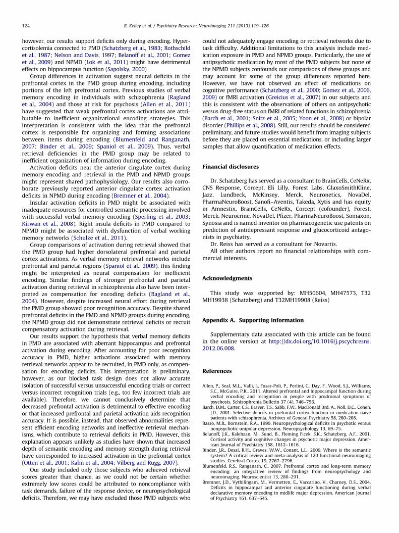
Fig. 1. Main effect of group: activation clusters. Main effect of group results of
2-way group by task analysis of variance: two clusters were significant, including
the sub-gyral right anterior cingulate and left parietal lobule. Extracted clusters
were adjusted for recognition accuracy using unstandardized residuals and means
plotted using bar graphs including 72 standard errors. Blue bars represent means
extracted from memory encoding task and green represent means extracted from
the memory retrieval task. Statistical results are presented for each cluster in
Table 3.
3. Results
3.1. Subjects
Five subjects with PMD, two with NPMD and two HC wereexcluded from the analysis due to retrieval task accuracy below50%. Three subjects with PMD, two with NPMD and three HC wereexcluded due to excessive motion during one of the functionalimaging verbal memory tasks. Included in the final analysis are 16PMD (8 females), 15 NPMD (8 females), and 16 HC (7 females)subjects. The groups were not significantly different in age,gender, handedness, or years of education. Additionally, thegroups had similar IQ (p¼0.60), as measured by the WechslerTest of Adult Reading (WTAR) (2001) full scale estimate ofpremorbid intellectual function.
3.2. Verbal declarative memory performance
Significant group differences were observed for correctedaccuracy of recognized words (F(1,47)¼4.80, p¼0.013). Two-group post hoc analyses indicated significantly lower correctedaccuracy for recognized words in the PMD group compared to theHC group (t(30)¼�2.91, p¼0.007) and the NPMD group(t(29)¼�2.15, p¼0.04) (Table 2). Therefore, fMRI analyses ofverbal encoding and retrieval were covaried for recognitionaccuracy. No other measures of task performance were signifi-cantly different between groups including false alarm rate and d’.
3.3. fMRI—main effect of group
The main effect of group across retrieval and encoding con-trasts identified 2 clusters of activation: right anterior cingulateand left parietal (Table 3, Fig. 1). Post-hoc t-tests found that sub-gyral right anterior cingulate differences was attributed to higheractivation in the HC group compared to both PMD (p¼0.001 andp¼0.009) and NPMD (p¼0.010 and p¼0.003) groups in bothencoding and retrieval tasks, respectively. No significant right
Table 3Significant clusters of activation from the main effect of group for encoding minus con
Neuroanatomy BA Cluster
size
Talairach
coordinates
F
value
Post-hoc T
ROI analysis: main effect of group
Left parietal lobule 40 24 �46, �33, 38 8.80 Encoding;
Sub-gyral, right anterior
cingulate
32 19 22, 39, 2 8.93 Encoding;
HC4NPMD
ROI analysis: interaction of group� task
Right insula 13 23 42, 2, 2 9.02 Encoding;
Left insula 13 93 �36, 1, 20 12.39 Encoding;
Right hippocampus 19 24, �7, �18 9.93 Encoding;
Left hippocampus 26 �20, �11, �18 8.98 Encoding;
Left middle frontal gyrus 8 132 �20, 19, 38 11.53 Encoding;
Right dorsolateral prefrontal
cortex
10 49 26, 59, 14 8.98 Encoding;
Left mid-cingulate gyrus 31 163 �18, �22, 34 10.22
Left mid-cingulate gyrus 24 71 �8, 6, 33 12.30
Right precentral gyrus 44 11 63, 8, 11 8.54
Right superior frontal gyrus 8 14 22, 18, 41 8.31
n Cluster significance within two 2-group t-tests; clusters from main effect of group
anterior cingulate activation differences were observed betweenPMD and NPMD groups. Group comparisons of left parietal lobuleactivation found the PMD group to have significantly higheractivation compared to both HC (po0.001) and NPMD (p¼0.013)groups exclusively in the retrieval task. No significant left parietallobule activation differences were observed between HC and NPMDgroups in either encoding or retrieval tasks.
3.4. fMRI—interaction of group by task
The interaction of group by task identified by the ANOVAresulted in 10 significant clusters of activation (Table 3, Fig. 2).
trol and retrieval minus control contrasts.
-test*
no significance, retrieval; PMD4HC, po0.001, PMD4NPMD, p¼0.013
HC4PMD, p¼0.001, HC4NPMD, p¼0.010, retrieval; HC4PMD, p¼0.009,
, p¼0.003
NPMD4PMD, p¼0.003, HC4PMD, p¼0.016, retrieval; no significance
HC4PMD, p¼0.018, HC4NPMD, p¼0.044, retrieval; no significance
HC4PMD, p¼0.007, retrieval; no significance
HC4PMD, p¼0.027, HC4NPMD, p¼0.009, retrieval; no significance
HC4PMD, p¼0.001, HC4NPMD, p¼0.038, retrieval; PMD4NPMD, p¼0.032
no significance, retrieval; PMD4HC, p¼0.031
defined at p¼0.025, clusters from interaction of group� task defined at p¼0.0083.
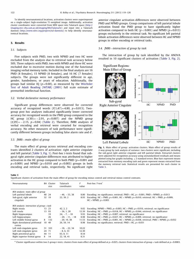
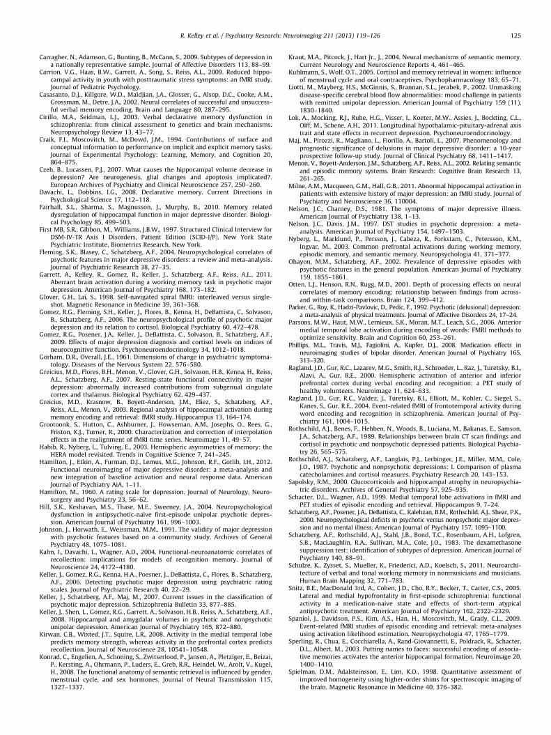
Fig. 2. Interaction group by task: activation clusters. Interaction of group by task
results of 2-way group by task analysis of variance: 6 of 10 significant clusters
were analyzed, including the right and left insula and hippocampus, left middle
frontal gyrus, and right dorsolateral prefrontal cortex. Extracted clusters were
adjusted for recognition accuracy using unstandardized residuals and means
plotted using bar graphs including 72 standard errors. Blue bars represent means
extracted from memory encoding task and green represent means extracted from
the memory retrieval task. Statistical results are presented for each cluster in
Table 3.
R. Kelley et al. / Psychiatry Research: Neuroimaging 211 (2013) 119–126 123
Post-hoc t-tests were conducted on 6 clusters that were selectedbased on previous literature implicating aberrant hippocampus,prefrontal cortex and insular function in depression (Liotti et al.,2002; Werner et al., 2009; Fairhall et al., 2010; Sprengelmeyeret al., 2011; Garrett et al., 2011; Milne et al., 2011; Hamilton et al.,2012). During the encoding task the PMD group had significantlylower activation compared to HC in the right hippocampus(t¼3.15, p¼0.007), and left middle frontal gyrus (t¼3.80, p¼
0.001), while activation differences from left insula (p¼0.018),right insula (p¼0.016), left hippocampus (t¼2.18, p¼0.027)approached significance. Additionally, the PMD group had sig-nificantly lower activation in the right insula (t¼3.00, p¼0.003)compared to the NPMD group during encoding. Higher activationin HC group compared to NPMD group approached significance inthe left hippocampus, (t¼2.57, p¼0.009), left insula (p¼0.044),and left superior dorsolateral frontal gyrus (p¼0.038) duringencoding. The PMD group exhibited higher activation approach-ing significance during the retrieval task in the right dorsolateralprefrontal cortex compared to HC (p¼0.031) and in the leftmiddle frontal gyrus compared to NPMD (p¼0.032).
4. Discussion
In this study we investigated brain function related to encod-ing and retrieval of verbal information in carefully characterizedgroups of individuals with PMD compared to those with NPMDand HC. As expected, the PMD group had impaired memoryretrieval compared to the HC and NPMD groups. After adjustingfor recognition accuracy, fMRI results indicated hippocampus,insula and prefrontal cortical deficits in the PMD group duringencoding, possibly associated with inefficient organization andassociation of incoming information. The PMD group also showedrelatively greater prefrontal and parietal cortex activation duringretrieval, possibly reflecting a compensatory response in the faceof encoding deficits. These findings support the hypothesis thatverbal memory retrieval deficits in PMD stem from aberrant brainfunction during encoding.
The profile of activation within the HC group during theencoding task is largely compatible with previously describedmodels of verbal and semantic memory, including left hemi-sphere activation in the inferior frontal cortex, hippocampus,fusiform gyrus, and middle occipital gyrus (Wagner et al., 1998;Casasanto et al., 2002; Kraut et al., 2004; Davachi and Dobbins,2008; Spaniol et al., 2009) (Table S2, online supplemental mate-rial). Thus, we are confident that the task was valid for testing ourprimary hypothesis. The PMD and NPMD groups also showedactivation in these networks, including the left prefrontal cortexand visual areas. For the retrieval task, all three groups activatedthe bilateral inferior frontal gyri and the visual cortex, consistentwith previous literature on verbal memory retrieval (Raglandet al., 2000; Habib et al., 2003; Spaniol et al., 2009) (Table S3,online supplemental material). Several brain regions, includingthe left inferior frontal gyrus, are activated during both encodingand retrieval and may represent an overarching semantic mem-ory network (Nyberg et al., 2003; Binder et al., 2009).
Hippocampal activation during encoding was observed withinthe HC group, consistent with previous studies from our lab usingthe same task (Menon et al., 2002; Greicius et al., 2003). Our resultsof hippocampus dysfunction during memory encoding in PMD andNPMD support previous findings (Bremner et al., 2004; Fairhallet al., 2010). These activation deficits are located in the anteriorhippocampal head, which has been shown to be associated withsuccessful memory encoding in subjects with depression (Fairhallet al., 2010). Hippocampus activation deficits in depression have alsobeen observed during memory retrieval (Milne et al., 2011);
R. Kelley et al. / Psychiatry Research: Neuroimaging 211 (2013) 119–126124
however, our results support deficits only during encoding. Hyper-cortisolemia connected to PMD (Schatzberg et al., 1983; Rothschildet al., 1987; Nelson and Davis, 1997; Belanoff et al., 2001; Gomezet al., 2009) and NPMD (Lok et al., 2011) might have detrimentaleffects on hippocampus function (Sapolsky, 2000).
Group differences in activation suggest neural deficits in theprefrontal cortex in the PMD group during encoding, includingportions of the left prefrontal cortex. Previous studies of verbalmemory encoding in individuals with schizophrenia (Raglandet al., 2004) and those at risk for psychosis (Allen et al., 2011)have suggested that weak prefrontal cortex activations are attri-butable to inefficient organizational encoding strategies. Thisinterpretation is consistent with the idea that the prefrontalcortex is responsible for organizing and forming associationsbetween items during encoding (Blumenfeld and Ranganath,2007; Binder et al., 2009; Spaniol et al., 2009). Thus, verbalretrieval deficiencies in the PMD group may be related toinefficient organization of information during encoding.
Activation deficits near the anterior cingulate cortex duringmemory encoding and retrieval in the PMD and NPMD groupsmight represent shared pathophysiology. Our results also corro-borate previously reported anterior cingulate cortex activationdeficits in NPMD during encoding (Bremner et al., 2004).
Insular activation deficits in PMD might be associated withinadequate resources for controlled semantic processing involvedwith successful verbal memory encoding (Sperling et al., 2003;Kirwan et al., 2008). Right insula deficits in PMD compared toNPMD might be associated with dysfunction of verbal workingmemory networks (Schulze et al., 2011).
Group comparisons of activation during retrieval showed thatthe PMD group had higher dorsolateral prefrontal and parietalcortex activations. As verbal memory retrieval networks includeprefrontal and parietal regions (Spaniol et al., 2009), this findingmight be interpreted as neural compensation for inefficientencoding. Similar findings of stronger prefrontal and parietalactivation during retrieval in schizophrenia also have been inter-preted as compensation for encoding deficits (Ragland et al.,2004). However, despite increased neural effort during retrievalthe PMD group showed poor recognition accuracy. Despite sharedprefrontal deficits in the PMD and NPMD groups during encoding,the NPMD group did not demonstrate retrieval deficits or recruitcompensatory activation during retrieval.
Our results support the hypothesis that verbal memory deficitsin PMD are associated with aberrant hippocampus and prefrontalactivation during encoding. After accounting for poor recognitionaccuracy in PMD, higher activations associated with memoryretrieval networks appear to be recruited, in PMD only, as compen-sation for encoding deficits. This interpretation is preliminary,however, as our blocked task design does not allow accurateisolation of successful versus unsuccessful encoding trials or correctversus incorrect recognition trials (e.g., too few incorrect trials areavailable). Therefore, we cannot conclusively determine thatdecreased prefrontal activation is detrimental to effective encodingor that increased prefrontal and parietal activation aids recognitionaccuracy. It is possible, instead, that observed abnormalities repre-sent efficient encoding networks and ineffective retrieval mechan-isms, which contribute to retrieval deficits in PMD. However, thisexplanation appears unlikely as studies have shown that increaseddepth of semantic encoding and memory strength during retrievalhave corresponded to increased activation in the prefrontal cortex(Otten et al., 2001; Kahn et al., 2004; Vilberg and Rugg, 2007).
Our study included only those subjects who achieved retrievalscores greater than chance, as we could not be certain whetherextremely low scores could be attributed to noncompliance withtask demands, failure of the response device, or neuropsychologicaldeficits. Therefore, we may have excluded those PMD subjects who
could not adequately engage encoding or retrieval networks due totask difficulty. Additional limitations to this analysis include med-ication exposure in PMD and NPMD groups. Particularly, the use ofantipsychotic medication by most of the PMD subjects but none ofthe NPMD subjects confounds our comparisons of these groups andmay account for some of the group differences reported here.However, we have not observed an effect of medications oncognitive performance (Schatzberg et al., 2000; Gomez et al., 2006,2009) or fMRI activation (Greicius et al., 2007) in our subjects andthis is consistent with the observations of others on antipsychoticversus drug-free status on fMRI of related functions in schizophrenia(Barch et al., 2001; Snitz et al., 2005; Yoon et al., 2008) or bipolardisorder (Phillips et al., 2008). Still, our results should be consideredpreliminary, and future studies would benefit from imaging subjectsbefore they are placed on essential medications, or including largersamples that allow quantification of medication effects.
Financial disclosures
Dr. Schatzberg has served as a consultant to BrainCells, CeNeRx,CNS Response, Corcept, Eli Lilly, Forest Labs, GlaxoSmithKline,Jazz, Lundbeck, McKinsey, Merck, Neuronetics, NovaDel,PharmaNeuroBoost, Sanofi–Aventis, Takeda, Xytis and has equityin Amnestix, BrainCells, CeNeRx, Corcept (cofounder), Forest,Merck, Neurocrine, NovaDel, Pfizer, PharmaNeuroBoost, Somaxon,Synosia and is named inventor on pharmacogenetic use patents onprediction of antidepressant response and glucocorticoid antago-nists in psychiatry.
Dr. Reiss has served as a consultant for Novartis.All other authors report no financial relationships with com-
mercial interests.
Acknowledgments
This study was supported by: MH50604, MH47573, T32MH19938 (Schatzberg) and T32MH19908 (Reiss)
Appendix A. Supporting information
Supplementary data associated with this article can be foundin the online version at http://dx.doi.org/10.1016/j.pscychresns.2012.06.008.
References
Allen, P., Seal, M.L., Valli, I., Fusar-Poli, P., Perlini, C., Day, F., Wood, S.J., Williams,S.C., McGuire, P.K., 2011. Altered prefrontal and hippocampal function duringverbal encoding and recognition in people with prodromal symptoms ofpsychosis. Schizophrenia Bulletin 37 (4), 746–756.
Barch, D.M., Carter, C.S., Braver, T.S., Sabb, F.W., MacDonald 3rd, A., Noll, D.C., Cohen,J.D., 2001. Selective deficits in prefrontal cortex function in medication-naivepatients with schizophrenia. Archives of General Psychiatry 58, 280–288.
Basso, M.R., Bornstein, R.A., 1999. Neuropsychological deficits in psychotic versusnonpsychotic unipolar depression. Neuropsychology 13, 69–75.
Belanoff, J.K., Kalehzan, M., Sund, B., Fleming Ficek, S.K., Schatzberg, A.F., 2001.Cortisol activity and cognitive changes in psychotic major depression. Amer-ican Journal of Psychiatry 158, 1612–1616.
Binder, J.R., Desai, R.H., Graves, W.W., Conant, L.L., 2009. Where is the semanticsystem? A critical review and meta-analysis of 120 functional neuroimagingstudies. Cerebral Cortex 19, 2767–2796.
Blumenfeld, R.S., Ranganath, C., 2007. Prefrontal cortex and long-term memoryencoding: an integrative review of findings from neuropsychology andneuroimaging. Neuroscientist 13, 280–291.
Bremner, J.D., Vythilingam, M., Vermetten, E., Vaccarino, V., Charney, D.S., 2004.Deficits in hippocampal and anterior cingulate functioning during verbaldeclarative memory encoding in midlife major depression. American Journalof Psychiatry 161, 637–645.
R. Kelley et al. / Psychiatry Research: Neuroimaging 211 (2013) 119–126 125
Carragher, N., Adamson, G., Bunting, B., McCann, S., 2009. Subtypes of depression ina nationally representative sample. Journal of Affective Disorders 113, 88–99.
Carrion, V.G., Haas, B.W., Garrett, A., Song, S., Reiss, A.L., 2009. Reduced hippo-campal activity in youth with posttraumatic stress symptoms: an fMRI study.Journal of Pediatric Psychology.
Casasanto, D.J., Killgore, W.D., Maldjian, J.A., Glosser, G., Alsop, D.C., Cooke, A.M.,Grossman, M., Detre, J.A., 2002. Neural correlates of successful and unsuccess-ful verbal memory encoding. Brain and Language 80, 287–295.
Cirillo, M.A., Seidman, L.J., 2003. Verbal declarative memory dysfunction inschizophrenia: from clinical assessment to genetics and brain mechanisms.Neuropsychology Review 13, 43–77.
Craik, F.I., Moscovitch, M., McDowd, J.M., 1994. Contributions of surface andconceptual information to performance on implicit and explicit memory tasks.Journal of Experimental Psychology: Learning, Memory, and Cognition 20,864–875.
Czeh, B., Lucassen, P.J., 2007. What causes the hippocampal volume decrease indepression? Are neurogenesis, glial changes and apoptosis implicated?.European Archives of Psychiatry and Clinical Neuroscience 257, 250–260.
Davachi, L., Dobbins, I.G., 2008. Declarative memory. Current Directions inPsychological Science 17, 112–118.
Fairhall, S.L., Sharma, S., Magnusson, J., Murphy, B., 2010. Memory relateddysregulation of hippocampal function in major depressive disorder. Biologi-cal Psychology 85, 499–503.
First MB, S.R., Gibbon, M., Williams, J.B.W., 1997. Structured Clinical Interview forDSM-IV-TR Axis I Disorders, Patient Edition (SCID-I/P). New York StatePsychiatric Institute, Biometrics Research, New York.
Fleming, S.K., Blasey, C., Schatzberg, A.F., 2004. Neuropsychological correlates ofpsychotic features in major depressive disorders: a review and meta-analysis.Journal of Psychiatric Research 38, 27–35.
Garrett, A., Kelley, R., Gomez, R., Keller, J., Schatzberg, A.F., Reiss, A.L., 2011.Aberrant brain activation during a working memory task in psychotic majordepression. American Journal of Psychiatry 168, 173–182.
Glover, G.H., Lai, S., 1998. Self-navigated spiral fMRI: interleaved versus single-shot. Magnetic Resonance in Medicine 39, 361–368.
Gomez, R.G., Fleming, S.H., Keller, J., Flores, B., Kenna, H., DeBattista, C., Solvason,B., Schatzberg, A.F., 2006. The neuropsychological profile of psychotic majordepression and its relation to cortisol. Biological Psychiatry 60, 472–478.
Gomez, R.G., Posener, J.A., Keller, J., DeBattista, C., Solvason, B., Schatzberg, A.F.,2009. Effects of major depression diagnosis and cortisol levels on indices ofneurocognitive function. Psychoneuroendocrinology 34, 1012–1018.
Gorham, D.R., Overall, J.E., 1961. Dimensions of change in psychiatric symptoma-tology. Diseases of the Nervous System 22, 576–580.
Greicius, M.D., Flores, B.H., Menon, V., Glover, G.H., Solvason, H.B., Kenna, H., Reiss,A.L., Schatzberg, A.F., 2007. Resting-state functional connectivity in majordepression: abnormally increased contributions from subgenual cingulatecortex and thalamus. Biological Psychiatry 62, 429–437.
Greicius, M.D., Krasnow, B., Boyett-Anderson, J.M., Eliez, S., Schatzberg, A.F.,Reiss, A.L., Menon, V., 2003. Regional analysis of hippocampal activation duringmemory encoding and retrieval: fMRI study. Hippocampus 13, 164–174.
Grootoonk, S., Hutton, C., Ashburner, J., Howseman, A.M., Josephs, O., Rees, G.,Friston, K.J., Turner, R., 2000. Characterization and correction of interpolationeffects in the realignment of fMRI time series. Neuroimage 11, 49–57.
Habib, R., Nyberg, L., Tulving, E., 2003. Hemispheric asymmetries of memory: theHERA model revisited. Trends in Cognitive Science 7, 241–245.
Hamilton, J., Etkin, A., Furman, D.J., Lemus, M.G., Johnson, R.F., Gotlib, I.H., 2012.Functional neuroimaging of major depressive disorder: a meta-analysis andnew integration of baseline activation and neural response data. AmericanJournal of Psychiatry AiA, 1–11.
Hamilton, M., 1960. A rating scale for depression. Journal of Neurology, Neuro-surgery and Psychiatry 23, 56–62.
Hill, S.K., Keshavan, M.S., Thase, M.E., Sweeney, J.A., 2004. Neuropsychologicaldysfunction in antipsychotic-naive first-episode unipolar psychotic depres-sion. American Journal of Psychiatry 161, 996–1003.
Johnson, J., Horwath, E., Weissman, M.M., 1991. The validity of major depressionwith psychotic features based on a community study. Archives of GeneralPsychiatry 48, 1075–1081.
Kahn, I., Davachi, L., Wagner, A.D., 2004. Functional-neuroanatomic correlates ofrecollection: implications for models of recognition memory. Journal ofNeuroscience 24, 4172–4180.
Keller, J., Gomez, R.G., Kenna, H.A., Poesner, J., DeBattista, C., Flores, B., Schatzberg,A.F., 2006. Detecting psychotic major depression using psychiatric ratingscales. Journal of Psychiatric Research 40, 22–29.
Keller, J., Schatzberg, A.F., Maj, M., 2007. Current issues in the classification ofpsychotic major depression. Schizophrenia Bulletin 33, 877–885.
Keller, J., Shen, L., Gomez, R.G., Garrett, A., Solvason, H.B., Reiss, A., Schatzberg, A.F.,2008. Hippocampal and amygdalar volumes in psychotic and nonpsychoticunipolar depression. American Journal of Psychiatry 165, 872–880.
Kirwan, C.B., Wixted, J.T., Squire, L.R., 2008. Activity in the medial temporal lobepredicts memory strength, whereas activity in the prefrontal cortex predictsrecollection. Journal of Neuroscience 28, 10541–10548.
Konrad, C., Engelien, A., Schoning, S., Zwitserlood, P., Jansen, A., Pletziger, E., Beizai,P., Kersting, A., Ohrmann, P., Luders, E., Greb, R.R., Heindel, W., Arolt, V., Kugel,H., 2008. The functional anatomy of semantic retrieval is influenced by gender,menstrual cycle, and sex hormones. Journal of Neural Transmission 115,1327–1337.
Kraut, M.A., Pitcock, J., Hart Jr., J., 2004. Neural mechanisms of semantic memory.Current Neurology and Neuroscience Reports 4, 461–465.
Kuhlmann, S., Wolf, O.T., 2005. Cortisol and memory retrieval in women: influenceof menstrual cycle and oral contraceptives. Psychopharmacology 183, 65–71.
Liotti, M., Mayberg, H.S., McGinnis, S., Brannan, S.L., Jerabek, P., 2002. Unmaskingdisease-specific cerebral blood flow abnormalities: mood challenge in patientswith remitted unipolar depression. American Journal of Psychiatry 159 (11),1830–1840.
Lok, A., Mocking, R.J., Ruhe, H.G., Visser, I., Koeter, M.W., Assies, J., Bockting, C.L.,Olff, M., Schene, A.H., 2011. Longitudinal hypothalamic-pituitary-adrenal axistrait and state effects in recurrent depression. Psychoneuroendocrinology.
Maj, M., Pirozzi, R., Magliano, L., Fiorillo, A., Bartoli, L., 2007. Phenomenology andprognostic significance of delusions in major depressive disorder: a 10-yearprospective follow-up study. Journal of Clinical Psychiatry 68, 1411–1417.
Menon, V., Boyett-Anderson, J.M., Schatzberg, A.F., Reiss, A.L., 2002. Relating semanticand episodic memory systems. Brain Research: Cognitive Brain Research 13,261–265.
Milne, A.M., Macqueen, G.M., Hall, G.B., 2011. Abnormal hippocampal activation inpatients with extensive history of major depression: an fMRI study. Journal ofPsychiatry and Neuroscience 36, 110004.
Nelson, J.C., Charney, D.S., 1981. The symptoms of major depressive illness.American Journal of Psychiatry 138, 1–13.
Nelson, J.C., Davis, J.M., 1997. DST studies in psychotic depression: a meta-analysis. American Journal of Psychiatry 154, 1497–1503.
Nyberg, L., Marklund, P., Persson, J., Cabeza, R., Forkstam, C., Petersson, K.M.,Ingvar, M., 2003. Common prefrontal activations during working memory,episodic memory, and semantic memory. Neuropsychologia 41, 371–377.
Ohayon, M.M., Schatzberg, A.F., 2002. Prevalence of depressive episodes withpsychotic features in the general population. American Journal of Psychiatry159, 1855–1861.
Otten, L.J., Henson, R.N., Rugg, M.D., 2001. Depth of processing effects on neuralcorrelates of memory encoding: relationship between findings from across-and within-task comparisons. Brain 124, 399–412.
Parker, G., Roy, K., Hadzi-Pavlovic, D., Pedic, F., 1992. Psychotic (delusional) depression:a meta-analysis of physical treatments. Journal of Affective Disorders 24, 17–24.
Parsons, M.W., Haut, M.W., Lemieux, S.K., Moran, M.T., Leach, S.G., 2006. Anteriormedial temporal lobe activation during encoding of words: FMRI methods tooptimize sensitivity. Brain and Cognition 60, 253–261.
Phillips, M.L., Travis, M.J., Fagiolini, A., Kupfer, D.J., 2008. Medication effects inneuroimaging studies of bipolar disorder. American Journal of Psychiatry 165,313–320.
Ragland, J.D., Gur, R.C., Lazarev, M.G., Smith, R.J., Schroeder, L., Raz, J., Turetsky, B.I.,Alavi, A., Gur, R.E., 2000. Hemispheric activation of anterior and inferiorprefrontal cortex during verbal encoding and recognition: a PET study ofhealthy volunteers. Neuroimage 11, 624–633.
Ragland, J.D., Gur, R.C., Valdez, J., Turetsky, B.I., Elliott, M., Kohler, C., Siegel, S.,Kanes, S., Gur, R.E., 2004. Event-related fMRI of frontotemporal activity duringword encoding and recognition in schizophrenia. American Journal of Psy-chiatry 161, 1004–1015.
Rothschild, A.J., Benes, F., Hebben, N., Woods, B., Luciana, M., Bakanas, E., Samson,J.A., Schatzberg, A.F., 1989. Relationships between brain CT scan findings andcortisol in psychotic and nonpsychotic depressed patients. Biological Psychia-try 26, 565–575.
Rothschild, A.J., Schatzberg, A.F., Langlais, P.J., Lerbinger, J.E., Miller, M.M., Cole,J.O., 1987. Psychotic and nonpsychotic depressions: I. Comparison of plasmacatecholamines and cortisol measures. Psychiatry Research 20, 143–153.
Sapolsky, R.M., 2000. Glucocorticoids and hippocampal atrophy in neuropsychia-tric disorders. Archives of General Psychiatry 57, 925–935.
Schacter, D.L., Wagner, A.D., 1999. Medial temporal lobe activations in fMRI andPET studies of episodic encoding and retrieval. Hippocampus 9, 7–24.
Schatzberg, A.F., Posener, J.A., DeBattista, C., Kalehzan, B.M., Rothschild, A.J., Shear, P.K.,2000. Neuropsychological deficits in psychotic versus nonpsychotic major depres-sion and no mental illness. American Journal of Psychiatry 157, 1095–1100.
Schatzberg, A.F., Rothschild, A.J., Stahl, J.B., Bond, T.C., Rosenbaum, A.H., Lofgren,S.B., MacLaughlin, R.A., Sullivan, M.A., Cole, J.O., 1983. The dexamethasonesuppression test: identification of subtypes of depression. American Journal ofPsychiatry 140, 88–91.
Schulze, K., Zysset, S., Mueller, K., Friederici, A.D., Koelsch, S., 2011. Neuroarchi-tecture of verbal and tonal working memory in nonmusicians and musicians.Human Brain Mapping 32, 771–783.
Snitz, B.E., MacDonald 3rd, A., Cohen, J.D., Cho, R.Y., Becker, T., Carter, C.S., 2005.Lateral and medial hypofrontality in first-episode schizophrenia: functionalactivity in a medication-naive state and effects of short-term atypicalantipsychotic treatment. American Journal of Psychiatry 162, 2322–2329.
Spaniol, J., Davidson, P.S., Kim, A.S., Han, H., Moscovitch, M., Grady, C.L., 2009.Event-related fMRI studies of episodic encoding and retrieval: meta-analysesusing activation likelihood estimation. Neuropsychologia 47, 1765–1779.
Sperling, R., Chua, E., Cocchiarella, A., Rand-Giovannetti, E., Poldrack, R., Schacter,D.L., Albert, M., 2003. Putting names to faces: successful encoding of associa-tive memories activates the anterior hippocampal formation. Neuroimage 20,1400–1410.
Spielman, D.M., Adalsteinsson, E., Lim, K.O., 1998. Quantitative assessment ofimproved homogeneity using higher-order shims for spectroscopic imaging ofthe brain. Magnetic Resonance in Medicine 40, 376–382.

R. Kelley et al. / Psychiatry Research: Neuroimaging 211 (2013) 119–126126
Sprengelmeyer, R., Steele, J.D., Mwangi, B., Kumar, P., Christmas, D., Milders, M.,Matthews, K., 2011. The insular cortex and the neuroanatomy of majordepression. Journal of Affective Disorders 133 (1-2), 120–127.
Thase, M.E., Hersen, M., Bellack, A.S., Himmelhoch, J.M., Kupfer, D.J., 1983.Validation of a Hamilton subscale for endogenomorphic depression. Journalof Affective Disorders 5, 267–278.
Vilberg, K.L., Rugg, M.D., 2007. Dissociation of the neural correlates of recognitionmemory according to familiarity, recollection, and amount of recollectedinformation. Neuropsychologia 45, 2216–2225.
Wagner, A.D., Poldrack, R.A., Eldridge, L.L., Desmond, J.E., Glover, G.H., Gabrieli, J.D.,1998. Material-specific lateralization of prefrontal activation during episodicencoding and retrieval. Neuroreport 9, 3711–3717.
Wechsler Test of Adult Reading (WTAR), 2001. WMS-III, WTAR Manual. ThePsychological Corporation, Third ed. Harcourt, Toronto, CAN.
Werner, N.S., Meindl, T., Materne, J., Engel, R.R., Huber, D., Riedel, M., Reiser, M.,Hennig-Fast, K., 2009. Functional MRI study of memory-related brain regions
in patients with depressive disorder. Journal of Affective Disorders 119,
124–131.Yoon, J.H., Minzenberg, M.J., Ursu, S., Ryan Walter, B.S., Wendelken, C., Ragland,
J.D., Carter, C.S., 2008. Association of dorsolateral prefrontal cortex dysfunction
with disrupted coordinated brain activity in schizophrenia: relationship with
impaired cognition, behavioral disorganization, and global function. American
Journal of Psychiatry 165, 1006–1014.Zanelli, J., Reichenberg, A., Morgan, K., Fearon, P., Kravariti, E., Dazzan, P., Morgan,
C., Zanelli, C., Demjaha, A., Jones, P.B., Doody, G.A., Kapur, S., Murray, R.M.,
2010. Specific and generalized neuropsychological deficits: a comparison of
patients with various first-episode psychosis presentations. American Journal
of Psychiatry 167, 78–85.
Related Documents