Alcohol increases the permeability of airway epithelial tight junctions in Beas-2B and NHBE cells Samantha. M. Simet, PhD 1 , Todd A. Wyatt, PhD 1,2,3 , Jane DeVasure, BS 1 , Daniel Yanov, BS 1 , and Joseph H. Sisson, MD 1 1 University of Nebraska Medical Center, Department of Internal Medicine, Pulmonary, Critical Care, Sleep & Allergy Division, Omaha, NE 68198-5910 2 University of Nebraska College of Public Health, Department of Environmental, Agricultural, and Occupational Health, Omaha, NE 68198-7850 3 Research Service, Department of Veterans Affairs Medical Center, 4101 Woolworth Avenue, Omaha, NE 68105 Abstract BACKGROUND—Tight junctions form a continuous belt-like structure between cells and act to regulate paracellular signaling. Protein kinase C (PKC) has been shown to regulate tight junction assembly and disassembly and is activated by alcohol. Previous research has shown that alcohol increases the permeability of tight junctions in lung alveolar cells. However, little is known about alcohol’s effect on tight junctions in epithelium of the conducting airways. We hypothesized that long-term alcohol exposure reduces zonnula occluden-1 (ZO-1) and claudin-1 localization at the cell membrane and increases permeability through a PKC-dependent mechanism. METHODS—To test this hypothesis, we exposed normal human bronchial epithelial cells (NHBE), cells from a human bronchial epithelial transformed cell line (Beas-2B), and Beas-2B expressing a PKCα dominant negative (DN) to alcohol (20, 50, and 100 mM) for up to 48 hours. Immunofluorescence was used to assess changes in ZO-1, claudin-1, claudin-5 and claudin-7 localization. Electrical cell substrate impedance sensing (ECIS) was used to measure permeability of tight junctions between monolayers of NHBE, Beas-2B, and DN cells. RESULTS—Alcohol increased tight junction permeability in a concentration-dependent manner and decreased ZO-1, claudin-1, claudin-5 and claudin-7 localization at the cell membrane. To determine a possible signaling mechanism, we measured the activity of PKC isoforms (alpha, delta, epsilon, zeta). PKCα activity significantly increased in Beas-2B cells from 1–6 hours of 100 mM alcohol exposure, while PKCζ activity significantly decreased at 1 hour and increased at 3 hrs. Inhibiting PKCα with Gö-6976 prevented the alcohol-induced protein changes of both ZO-1 and claudin-1 at the cell membrane. PKCα dominant negative Beas-2B cells were resistant to alcohol-induced protein alterations. CONCLUSIONS—These results suggest that alcohol disrupts ZO-1, claudin-1, claudin-5 and claudin-7 through the activation of PKCα, leading to an alcohol-induced “leakiness” in bronchial epithelial cells. Such alcohol-induced airway leak state likely contributes to the impaired airway host defenses associated with acute and chronic alcohol ingestion. Address Correspondence and Reprint Requests to: Joseph H. Sisson, MD, Department of Internal Medicine, Pulmonary, Critical Care, Sleep & Allergy Division, University of Nebraska Medical Center, 985910 Nebraska Medical Center, Omaha, NE 68198-5910, TEL: 402-559-8834, FAX: 402-559-6584, [email protected]. NIH Public Access Author Manuscript Alcohol Clin Exp Res. Author manuscript; available in PMC 2013 March 1. Published in final edited form as: Alcohol Clin Exp Res. 2012 March ; 36(3): 432–442. doi:10.1111/j.1530-0277.2011.01640.x. NIH-PA Author Manuscript NIH-PA Author Manuscript NIH-PA Author Manuscript

Welcome message from author
This document is posted to help you gain knowledge. Please leave a comment to let me know what you think about it! Share it to your friends and learn new things together.
Transcript
Alcohol increases the permeability of airway epithelial tightjunctions in Beas-2B and NHBE cells
Samantha. M. Simet, PhD1, Todd A. Wyatt, PhD1,2,3, Jane DeVasure, BS1, Daniel Yanov,BS1, and Joseph H. Sisson, MD1
1University of Nebraska Medical Center, Department of Internal Medicine, Pulmonary, CriticalCare, Sleep & Allergy Division, Omaha, NE 68198-59102University of Nebraska College of Public Health, Department of Environmental, Agricultural, andOccupational Health, Omaha, NE 68198-78503Research Service, Department of Veterans Affairs Medical Center, 4101 Woolworth Avenue,Omaha, NE 68105
AbstractBACKGROUND—Tight junctions form a continuous belt-like structure between cells and act toregulate paracellular signaling. Protein kinase C (PKC) has been shown to regulate tight junctionassembly and disassembly and is activated by alcohol. Previous research has shown that alcoholincreases the permeability of tight junctions in lung alveolar cells. However, little is known aboutalcohol’s effect on tight junctions in epithelium of the conducting airways. We hypothesized thatlong-term alcohol exposure reduces zonnula occluden-1 (ZO-1) and claudin-1 localization at thecell membrane and increases permeability through a PKC-dependent mechanism.
METHODS—To test this hypothesis, we exposed normal human bronchial epithelial cells(NHBE), cells from a human bronchial epithelial transformed cell line (Beas-2B), and Beas-2Bexpressing a PKCα dominant negative (DN) to alcohol (20, 50, and 100 mM) for up to 48 hours.Immunofluorescence was used to assess changes in ZO-1, claudin-1, claudin-5 and claudin-7localization. Electrical cell substrate impedance sensing (ECIS) was used to measure permeabilityof tight junctions between monolayers of NHBE, Beas-2B, and DN cells.
RESULTS—Alcohol increased tight junction permeability in a concentration-dependent mannerand decreased ZO-1, claudin-1, claudin-5 and claudin-7 localization at the cell membrane. Todetermine a possible signaling mechanism, we measured the activity of PKC isoforms (alpha,delta, epsilon, zeta). PKCα activity significantly increased in Beas-2B cells from 1–6 hours of 100mM alcohol exposure, while PKCζ activity significantly decreased at 1 hour and increased at 3hrs. Inhibiting PKCα with Gö-6976 prevented the alcohol-induced protein changes of both ZO-1and claudin-1 at the cell membrane. PKCα dominant negative Beas-2B cells were resistant toalcohol-induced protein alterations.
CONCLUSIONS—These results suggest that alcohol disrupts ZO-1, claudin-1, claudin-5 andclaudin-7 through the activation of PKCα, leading to an alcohol-induced “leakiness” in bronchialepithelial cells. Such alcohol-induced airway leak state likely contributes to the impaired airwayhost defenses associated with acute and chronic alcohol ingestion.
Address Correspondence and Reprint Requests to: Joseph H. Sisson, MD, Department of Internal Medicine, Pulmonary, Critical Care,Sleep & Allergy Division, University of Nebraska Medical Center, 985910 Nebraska Medical Center, Omaha, NE 68198-5910, TEL:402-559-8834, FAX: 402-559-6584, [email protected].
NIH Public AccessAuthor ManuscriptAlcohol Clin Exp Res. Author manuscript; available in PMC 2013 March 1.
Published in final edited form as:Alcohol Clin Exp Res. 2012 March ; 36(3): 432–442. doi:10.1111/j.1530-0277.2011.01640.x.
NIH
-PA Author Manuscript
NIH
-PA Author Manuscript
NIH
-PA Author Manuscript
KeywordsAlcohol; tight junctions; ZO-1; Claudin-1; airway epithelium; lung
BackgroundAlcohol is the most commonly abused drug in the United States (Stinson et al., 2005). Morethan half the general adult population of the United States consumes alcohol andapproximately 17 million individuals are alcoholics (Grant et al., 2004). Alcohol affectsmany tissues throughout the body including the liver, brain, heart (Lieber, 1995), lung, andconducting airways (Joshi and Guidot, 2007). Heavy alcohol consumption has long beenknown to impair lung defenses. Therefore, it is no surprise that alcoholics are also morelikely to suffer from pulmonary diseases (Jerrells et al., 2007; Sisson, 2007) such as acuterespiratory distress syndrome (ARDS; Moss and Burnham, 2003), chronic bronchitis(Suadicani et al., 2001), and pneumonia (Samokhvalov et al., 2010).
Within the airways, tight junctions play an important role in maintaining cellular polarityand homeostasis by selectively regulating the passage of water, ions, neutral molecules andinflammatory cells through the paracellular pathway (Godfrey, 1997), providing a site forcell-cell signaling (Matter et al., 2005; Schneeberger and Lynch, 2004; Shin et al., 2006) andprotecting tissues from pathogenic infection (Fu et al., 2009). Tight junction disruption leadsto an imbalance of fluid and ions, loss of cell polarity, impaired neural responsiveness of theairways (Godfrey, 1997), and increased exposure to pathogens (Fu et al., 2009). Importantly,impaired tight junctions have been linked to various diseases in the conducting airways suchas chronic bronchitis (Wilson et al., 1996), pneumonia (Asgrimsson et al., 2006) and asthma(Holgate, 2007).
Based on the sites of common lung diseases, it is useful to divide the lung into two separatecompartments: the conducting airways and the parenchyma. The conducting airways includethe trachea, bronchi, and terminal bronchi. The parenchyma, or gas exchange region, iscomposed mainly of alveolar air spaces. Each of these compartments contain specializedcells unique to their environment. The effect of alcohol on tight junctions within thesedifferent compartments has focused predominantly on the lung parenchyma while little isknown about the impact of alcohol on tight junctions in the conducting airways.
Airway “leak” characterized by increased fluid in the conducting airway epithelium, is acardinal feature of airway diseases such as bronchitis (Thompson et al., 1993) and asthma(Aas, 1981; Fahy et al., 2000). Increased airway leak contributes to airway edema, which isa classic finding of these diseases and results in airway obstruction and impairedmucociliary clearance. Results for these studies will improve our understanding of theimpact alcohol has on airway epithelial barrier functions, which are critical for theunderstanding and treatment of airway diseases common among alcohol abusers.
Tight junction strands are composed of both transmembrane and cytoplasmic proteins. Morethan 40 proteins that make up or associate with tight junction strands have been identified.The main proteins comprising the tight junction complex include the transmembraneproteins (occludins, claudins, junctional-associated adhesion molecules (JAMs) and thecytoplasmic proteins [zonnula occludens (ZO-1, ZO-2, ZO-3)], Steed et al.) For thepurposes of these studies, we focus on the regulation of ZO-1, claudin-5 and claudin-7 inalcohol-induced airway epithelial leak.
Simet et al. Page 2
Alcohol Clin Exp Res. Author manuscript; available in PMC 2013 March 1.
NIH
-PA Author Manuscript
NIH
-PA Author Manuscript
NIH
-PA Author Manuscript
Tight junction permeability is regulated through a variety of mechanisms including themodulation of protein kinase C (PKC) activity. PKC has long been known to play a role inboth increasing and decreasing tight junction permeability (see for review [Gonzalez-Mariscal et al., 2008]). However, many studies demonstrate that PKC regulation of tightjunctions is cell/tissue type specific (Gonzalez-Mariscal et al., 2008) rendering it difficult touniversally characterize. Adding to this complexity is the fact that airway epithelium isknown to express several PKC isoforms: α, ε, ζ, δ (Liedtke et al., 1997; Wyatt et al., 2009;Wyatt et al., 1998). Several studies show that chronic alcohol intake activates PKC, thustranslocating it from the cytoplasm to the membrane (Newton, 2009). Previous research hassuggested that alcohol, in low levels, may enhance PKCα activation through Rho GTPases(Slater et al., 2003).
Based on previous evidence demonstrating alcohol’s ability to increase the permeability oftight junctions within the lungs making the cells “leakier” and alcohol’s ability to enhancePKCα activation, we hypothesize that alcohol exposure increases the permeability of tightjunctions in airway epithelial cells through a PKCα-dependent mechanism. To test thishypothesis, we exposed Beas-2B, Beas-2B cells expressing a PKCα dominant negative(DN), and normal human bronchial epithelial cells (NHBE) to alcohol and measuredtransepithelial resistance, PKC isoform activity, and tight junction protein localization.
MethodsCell Culture
Beas-2B cells (American Tissue Culture Collection (ATCC), Manassas, VA) (passage 10–25), Beas-2B PKCα DN, or NHBE (passage 1–5) were grown to confluence in 75cm2 tissueculture flasks (Corning, Corning, NY) pre-coated with Vitrogen 30 (Invitrogen by LifeTechnologies, Carlsbad, CA) in Bronchial Epithelial Basal Media (BEBM; Lonza,Walkersville, MD) supplemented with the following additives (with the exception ofgentamycin-amphotericin B) from Lonza/Clonetics Corporation as a kit: bovine pituitaryextract, epinephrine, triiodothyronine, epidermal growth factor, transferrin, insulin,hydrocortisone, and retinoic acid. In addition, BEBM was supplemented with 50 U/mlpenicillin and 50 U/ml streptomycin (Invitrogen, Carlsbad, CA). PKCα DN selectionmedium is as described above with the addition of doxycycline (1μg/ml) and G418 (100μg/ml). NHBE cells were isolated from human lung donors (International Institute for theAdvancement of Medicine, Jessup, PA). Beas-2B PKCα DN cells were constructed aspreviously reported and characterized (Wyatt et al., 2007).
Cytotoxicity AssayTo determine cell viability of alcohol-treated and non-treated cells, LDH-Cytotoxicity AssayKit (BioVision Incorporated, Mountain View, CA) was performed according to themanufacturer’s protocol. Briefly, 50 μl supernatant was pipetted into corresponding wells ofa 90-well plate. 50 μl of reaction mixture was added to each well and incubated for 15 minat room temperature. The absorbance of all samples was taken at 490–500 nm using amicroplate reader (BioRad, Hercules, CA). An additional cell viability assay was performedusing trypan blue exclusion test of cell viability. Beas-2B cells were grown to confluence on60 mm culture dishes as described above. Cells were treated with 100 mM alcohol for 24hrs. Following 24 hrs of alcohol exposure, cells were rinsed in PBS, trypsinized, centrifugedand resuspended in 3 ml of BEBM media. 100 μl trypan blue (0.4%) and 500 μl cell solutionwas added to a microfuge tube and mixed thoroughly. Solution was allowed to incubate atroom temperature for 5 min. Viable and non-viable cells were counted using ahemocytometer.
Simet et al. Page 3
Alcohol Clin Exp Res. Author manuscript; available in PMC 2013 March 1.
NIH
-PA Author Manuscript
NIH
-PA Author Manuscript
NIH
-PA Author Manuscript
Alcohol TreatmentTo minimize alcohol evaporation, 90% of the area on each dish was sealed with paraffin,allowing the exchange of CO2. Alcohol levels in the media were measured using HPLC toensure minimal alcohol evaporation. For experiments carried out longer than 24 hours, freshmedia with alcohol was added every 24 hours.
Alcohol concentrations used in this study were based on biologically relevant levels (100mM). For instance, an individual is legally impaired to drive when blood alcoholconcentration reaches 0.08%, which equates to a concentration of 22 mM alcohol. Higherblood alcohol concentrations, such as 50 mM (equates to 0.20% concentration) arecommonly observed by individuals presenting to emergency rooms for intoxication. Thehighest alcohol dose we use is 100 mM, which is consistent with blood alcoholconcentrations observed in patients admitted to hospitals and those who chronically abusealcohol (Teplin et al., 1989). To control for any ethanol-induced changes in cell viability,cytotoxicity, and cell death, Beas-2B cells were exposed to alcohol (100 mM) for 24 hours.Both LDH and trypan blue cell viability assays were then performed. No significantcytotoxicity or cell death occurred in cells treated with 100 mM alcohol compared to controlcells (LDH release data not shown). To further confirm cell viability, trypan blue cellviability assays were performed. Results confirmed no significant differences in cellviability of alcohol-treated cells when compared to controls cells. Both alcohol and controlgroups were at least 95% viable (data not shown).
ImmunofluorescenceBeas-2B, Beas-2B PKCα DN, or NHBE cells were plated onto sterile glass coverslipsprecoated with Vitrogen 30 for indirect immunofluorescence as follows: coverslips wererinsed with PBS, fixed in 4% paraformaldehyde, permeabilized and blocked for 15 min(0.1% Triton X-100, 1% BSA, 0.02% Sodium Azide). Cells were incubated in primaryantibodies (mouse anti-claudin-1, rabbit anti-claudin-7, mouse anti-claudin-5, mouse anti-ZO1, and rabbit anti-ZO-1 mid; Invitrogen by Life Technologies, Carlsbad, CA) overnightat 4°C. Following primary antibody incubation, cells were rinsed in PBS and coverslipsincubated with secondary antibodies (Alexa 488 and Alexa 647 against rabbit or mouse;Invitrogen by Life Technologies, Carlsbad, CA) for 1 hr at room temperature in ahumidified chamber. Following incubation, coverslips were rinsed in PBS and mountedusing ProLong Gold with DAPI (Invitrogen by Life Technologies, Carlsbad, CA). Imagingwas performed using an LSM 510 Meta Laser Scanning Confocal Microscope (Zeiss,Thornwood, New York). All images were collected as Z-stacks using the set parameters(laser strength, gain, etc.) for all treatments. Z sections were obtained through cells at 0.5μm. Z-stacks were compressed for fluorescence measurement analysis.
Image J analysisAll images were obtained as Z-stacks (0.5 μm slices) using set parameters for allexperimental conditions. Images were compressed using Zeiss microscope software (Zeiss,Thornwood, New York) and saved as jpeg images. Images were uploaded into ImageJ(Rasband, 1997–2009). Image analysis was completed by first subtracting backgroundfluorescence. Then regions of interest were selected to measure fluorescent mean intensity atthe membrane only. Data are presented as the mean ± SEM. Statistics were performed withGraphPad Prism software (GraphPad, La Jolla, CA) using a one-way ANOVA or t-test.
Epithelial Barrier FunctionEpithelial barrier function was measured using the electric cell-substrate impedance sensingsystem (ECIS; Applied BioPhysics, Troy, NY). This is a non-invasive technique that allows
Simet et al. Page 4
Alcohol Clin Exp Res. Author manuscript; available in PMC 2013 March 1.
NIH
-PA Author Manuscript
NIH
-PA Author Manuscript
NIH
-PA Author Manuscript
us to measure and record transepithelial resistance (TER) changes in real time. It is alsosensitive enough to determine the cell-cell interactions (tight junctions) (Hartmann et al.,2007; Wegener et al., 2000). With ECIS, cells are cultured onto gold electrodes. As cellsgrow and become confluent, they constrict current flow and alter impedance (Hartmann etal., 2007). ECIS is able to report impedance values or translate impedance measurements toboth resistance and capacitance. This system has become a useful tool to monitor barrierfunction (the resistance of the paracellular pathway between cells; cell permeability) (Kilaniet al., 2004; Sidhaye et al.; Tiruppathi et al., 1992).
Cell ResistanceCell resistance was measured using ECIS continuously for as long as 7 days (alcohol andmedia were replenished every two days). All cells including primary cells were grown onVitrogen 30-coated 8EW10+ arrays. 8EW10+ arrays contain 40 circular 250 μm diameterelectrodes, and are capable of measuring between 2000–4000 cells (confluent monolayers).Each well has a substrate area of 0.8cm2. Cells (5×10⁵) were seeded into each well, grownto confluence, and treated with alcohol (0 mM, 20 mM, 50 mM, 100 mM) for up to 96 hrs.Cell resistance measurements were based on changes in resistance/capacitance to currentflow applied to the electrode arrays at multiple frequencies. A frequency scan wasperformed to test for the frequency at which the largest difference in transepithelialresistance (TER) values were obtained between the cell-covered and cell-free electrodes.The optimal frequency to study resistance in Beas-2B and NHBE cells appeared to be 250and 400 Hz. Baseline values were established with culture media alone and compared toelectrodes covered with a monolayer of cells. Experimental conditions were run in duplicateon each plate. Three separate experiments were run for each cell line.
Data were analyzed using ECIS software, resistance normalized (subsequent values dividedby initial values) and compiled for each condition and presented as the mean normalizedresistance ± SEM (n=3). Resistance was normalized by dividing the impedance values fromelectrodes confluent with cells by the corresponding quantities for the cell-free electrodesusing ECIS software (Lo et al., 1995). Statistics performed were a one-way ANOVA.
PKC ActivityBeas-2B and NHBE cells were grown to confluence in 60 mm Petri dishes containingBEBM. The medium was removed, the cells gently rinsed in PBS, and 250 μl of cell lysisbuffer added (35 mM Tris-HCl, 0.4 mM EGTA, 10 mM MgCl2, 0.01% Triton-X 100, 1 mMPMSF, 1 mM Sigma Protease Inhibitor). The epithelial lysates were then immediately flash-frozen in liquid nitrogen for kinase assay. Epithelial lysates were used to determine PKCactivity (ε, ζ, δ, α) as previously described (Wyatt et al., 2009). PKC activity was assayed inboth the supernatant and particulate fractions. Total PKC activity was measured in cytosolicfractions, which contain soluble proteins and unactivated PKC, and in particulate fractions,containing nonsoluble material (cell membranes, nuclear particles, and cytoskeletalelements) as well as all translocated and activated PKC. PKC activity assays wereperformed using 900 μM PKC substrate peptide (Peninsula, Belmont, CA), 24 μg/mlphorbol 12-myristate 13-acetate, 12 mM calcium-acetate, 30 mM dithiothreitol, 8 μMphosphatidyl-L-serine, 150 μM ATP, 45 mM magnesium acetate, and 10 μCi/ml [γ-32P]ATP (MP Biomedicals, Irvine, CA) in a Tris-HCl buffer (pH 7.5). Each sample (20 μl) wasadded to 40 μl of the reaction mixture, and incubated for 15 minutes at 30°C. Next, 50 μl ofeach sample was spotted onto P-81 phosphocellulose papers (Whatman, Clifton, NJ) andincubations halted. Phosphocellulose papers were then washed five times for 5 minutes inphosphoric acid (75 mM), washed in ethanol, allowed to dry, and counted in nonaqueousscintillant, as previously described (Wyatt et al., 2009). Kinase activity was standardized inrelation to total cellular protein assayed, and was calculated as picomoles of phosphate
Simet et al. Page 5
Alcohol Clin Exp Res. Author manuscript; available in PMC 2013 March 1.
NIH
-PA Author Manuscript
NIH
-PA Author Manuscript
NIH
-PA Author Manuscript
incorporated per minute per milligram. For standardized comparison between isoforms, datawas expressed as fold activity changes from control media.
StatisticsECIS data are presented as the mean normalized resistance (subsequent values divided byinitial values) ± SEM (n=3). Statistics performed were a one-way ANOVA for PKC activityand ECIS measurements to determine significant changes among treatment groups, usingGraphPad Prism software (GraphPad, La Jolla, CA). Fluorescence mean measurements werecompleted using ImageJ software from NIH. Data are presented as the mean ± SEM.Statistics were performed using a one-way ANOVA or t-test.
ResultsEffect of alcohol on bronchial epithelial cell permeability
To determine the effect of biologically relevant alcohol exposure on airway epithelial“leak”, cells (Beas-2B, Beas-2B PKCα DN or NHBE) were plated on specialized ECISarrays (5×10⁵) and treated with 0, 20, 50, or 100 mM alcohol. Barrier function andpermeability measurements were obtained by measuring resistance (Kilani et al., 2004; Lo etal., 1999; Qiao et al., 2003) using ECIS. Resistance changes were maximal within the first24–48 hours of alcohol exposure beyond which no significant increases or decreases wereobserved. Therefore, alcohol exposure experiments were limited to 48 hrs in subsequentexperiments.
Resistance data was normalized to time zero (start of alcohol treatment) and compiled togenerate ECIS graphs. Alcohol decreased TER in both Beas-2B (Figure 1A) and NHBE(Figure 1B) cells in a concentration-dependent manner, inducing significant decreases inepithelial barrier function, therefore increasing cellular “leak” in cells exposed to 50 mMand 100 mM alcohol compared to control (P<0.01).
Effect of alcohol exposure on PKC Isoform activityAirway cells (Beas-2B, Beas-2B DN and NHBE) were treated with 100 mM alcohol for 1,3, 6, and 24 hours to determine the activity of various PKC isoforms in response to alcohol.Following alcohol exposure, PKC α, ε, ζ, δ levels were measured as described in Materialsand Methods. Activity assays revealed a significant time-dependent increase in PKCαactivity at 1, 3, and 6 hrs after treatment with 100 mM alcohol (Figure 2) in Beas-2B cells.PKCα activity trended higher at 3 and 6 hrs and was significantly increased at 24 hrs inNHBE cells (Figure 3). PKCζ activity significantly decreased at 1 hr, followed by asignificant increase at 3 hrs in alcohol-treated Beas-2B cells. Although we did not observean initial decrease in PKCζ in alcohol-exposed NHBE cells, a significant increase was seenat 6 hrs. No significant activity changes were observed in PKCε or PKCδ isoforms (Figures2 & 3) in any of the cell types tested.
To further define PKCα as a potential mechanism for the observed alcohol-induced decreasein TER, PKC isoform activity was performed on PKCα dominant negative engineeredBeas-2B cells (PKCα DN). There were no significant changes in any of the PKC isoforms inBeas-2B PKCα DN cells exposed to 100 mM alcohol at any time point measured (Figure 4),further supporting a critical role for the PKCα isoform in mediating alcohol’s effect onairway permeability. As a positive control for DN cell function, 100 ng/mL phorbol 12-myristate 13-acetate (PMA) significantly activated both PKCε and PKCδ in the PKCα DNcells (Figure 4). While PKCζ is a PMA-insensitive isoform, we observed a significant PKCζactivation in response to 10 μg/mL lipopolysaccharide (LPS) in both PKCα DN and normal
Simet et al. Page 6
Alcohol Clin Exp Res. Author manuscript; available in PMC 2013 March 1.
NIH
-PA Author Manuscript
NIH
-PA Author Manuscript
NIH
-PA Author Manuscript
Beas-2B, thus defining the suppression of only the alpha isoform in this cell line (data notshown).
Alcohol-induced barrier disruption requires PKCα activationTo determine the possible role of PKCα in regulating tight junction permeability, Beas-2Bcells expressing a dominant negative PKCα (Beas-2B PKCα DN) were exposed to 0, 20, 50,and 100 mM alcohol and resistance measurements were recorded (Figure 5A). Nosignificant changes in TER measurements were detected in Beas-2B PKCα DN.Furthermore, Beas-2B cells pretreated with a PKCα-specific inhibitor, Gö-6976, were alsoresistant to alcohol-induced permeability changes (Figure 5B).
Effect of Alcohol on ZO-1 and Claudin-1, Claudin-5 and Claudin-7 LocalizationImmunofluorescence microscopy was used to determine the effect of varying concentrationsof alcohol at numerous time points on the localization of ZO-1 and claudin-1 and claudin-7in Beas-2B, Beas-2B PKCα DN, and NHBE cells. Claudin-5 localization was investigated inNHBE cells only, due to the known absence of claudin-5 in Beas-2B cells (data not shown;Merikallio et al.). No changes in protein localization occurred from 1–6 hrs in any of thealcohol concentrations (20–100 mM; data not shown). By 24 hrs, exposure to 20–100 mMalcohol reduced ZO-1, claudin-1, and claudin-7 and localization at the plasma membrane inBeas-2B (Figure 7) and NHBE cells (Figure 6). Claudin-5 was also reduced in NHBE cells.In addition, ZO-1, claudin-1, and claudin-7 appear to shift from normal membranelocalization, observed in control cells, to a cytosolic localization after alcohol treatment inthe NHBE cells. In contrast, no observable changes in ZO-1, claudin-1, or claudin-7localization were detected in Beas-2B PKCα DN exposed to alcohol at any time point(Figure 9).
Because PKC isoform activity assays revealed significant changes in PKCα and PKCζactivity, we sought to determine if PKCζ was also regulating alcohol-induced decreases inTER. To determine the impact of PKCα and PKCζ signaling in alcohol-impaired proteintranslocation, cells were pretreated with 1 μM PKCα inhibitor (Gö-6976) or 1 μMmyristolated PKCζ, a cell-permeable PKCζ specific inhibitor, prior to alcohol exposure.Images revealed an alcohol-induced decrease in membrane protein localization in cellspretreated with myristolated PKCζ comparable to alcohol-only-exposed cells (Figure 8).Alternatively, ZO-1, claudin-1, and claudin-7 remained at the cell membrane in alcohol-stimulated Beas-2B and NHBE cells pretreated with Gö-6976 comparable to control cells(Figure 6 & 7). No increased membrane translocation of ZO-1, claudin-1, or claudin-7 wasobserved in alcohol-stimulated Beas-2B PKCα DN pretreated with Gö-6976 or myristolatedPKCζ (data not shown). Quantification of membrane fluorescence determined that thesignificant (P<0.05) alcohol-induced translocation of ZO-1 and claudin-1 was inhibited byGö-6976 (Figure 8).
DiscussionTo date, studies focused on the lung alveolar compartment have shown that chronic alcoholingestion (6 weeks) in rats increases lung epithelial permeability ~5–6 fold in vivo andpromotes the flooding of alveolar spaces with proteinaceous fluid upon stress, such as sepsis(Fernandez et al., 2007). Although, the mechanisms involved in alcohol-induced alveolarleak are still not fully understood, Fernandez et al. (2007) observed decreased claudin-1 and-7 in whole lung and alveolar epithelial monolayers in alcohol-fed rats and increasedclaudin-5. Our observed reduction in claudin-1 and claudin-7 in our airway epithelial cellsresembles those of Fernandez et al. (2007). Contrary to Fernandez’s findings of increasedclaudin-5, we observed a decrease in claudin-5 in the NHBE cells. Fernandez et al. were
Simet et al. Page 7
Alcohol Clin Exp Res. Author manuscript; available in PMC 2013 March 1.
NIH
-PA Author Manuscript
NIH
-PA Author Manuscript
NIH
-PA Author Manuscript
utilizing alveolar epithelial monolayers from alcohol-fed rats in their studies, while ourstudies involved primary human conducting airway epithelial cells treated with alcohol. Thisobserved difference in claudin-5’s response to alcohol is likely due to differences that areoften observed between methods of alcohol administration (i.e. drinking rats vs. alcohol-exposed cells), species and cell types.
While both ZO-1 and claudin proteins are important in the formation and maintenance offunctional tight junctions (Fanning et al., 2007; Lal-Nag and Morin, 2009; Tsukita et al.,2009), it is normal for cells and tissues to exhibit various permeability properties through theaddition or reduction of tight junctions (Mitic et al., 2000; Tsukita et al., 2001). Whentissues or cells begin to lose the ability to manipulate these tight junction propertiesappropriately, they become more susceptible to various injuries, such as inflammation, viraland bacterial infections, and injury due to inhaled particles (Holgate et al., 2009).
Our goal was to determine the effect of alcohol on tight junction permeability in airwayepithelial cells and the primary signaling mechanism involved. We initially found thatalcohol increases permeability in a concentration-dependent manner in Beas-2B and NHBEcells; whereas, alcohol had no effect on permeability in Beas-2B cells expressing a PKCαDN phenotype. These results indicated that alcohol is likely acting through a PKCα-dependent mechanism. The inability of alcohol to induce changes in cells expressing a DNPKCα or wild type Beas-2B pretreated with an inhibitor of PKCα suggests that alcoholrequires the presence of activatable PKCα to increase tight junction permeability. Indeed,PKC isoform assays in Beas-2B and NHBE cells revealed a significant increase in PKCαactivity occurring at 1–6 hrs and 24 hrs in alcohol-treated cells. Interestingly, we did observedifferences in the time points of maximal PKC activity in Beas-2B vs. NHBE cells. Inaddition, NHBE cells demonstrated more alcohol-induced cytoplasmic localization of ZO-1and claudin-1 than did Beas-2B cells. These observations are likely due to inherentdifferences between immortalized and primary cells. However, the trend of alcohol-inducedactivation of PKC and redistribution of ZO-1 and claudin-1 were consistent for both thesedifferent cells. We suspect that PKCζ is an important component in tight junction assemblyin airway epithelial cells. The initial decrease in PKCζ activity at 1 hour suggests that thecells are responding to an insult, thus reducing tight junction assembly promotion. IncreasedPKCζ activity at 3 hours is likely a compensatory mechanism in response to the insult.While our understanding of tight junction regulation in general is poorly understood, we canonly speculate as to the regulation of tight junctions under altered conditions. Although wedetected modest changes in PKCζ activity, we hypothesize that PKCα is the majormechanism through which alcohol is acting due to its ability to indirectly activate PKCα(Slater et al., 2003) and other studies implicating PKCα as a major component in increasingtight junction permeability (Clarke et al., 2000; Mullin et al., 1997). Our protein localizationstudies using PKCα and PKCζ-specific inhibitors prior to alcohol exposure support thishypothesis.
To determine if alcohol disrupts the translocation of ZO-1 and claudin-1 to the plasmamembrane, cells were stained for ZO-1 and claudin-1. Fluorescence microscopy revealed asignificant decrease in ZO-1 and claudin-1 at the plasma membrane in alcohol-treated cells.This alcohol-impaired membrane protein localization did not occur in cells pretreated with aPKCα inhibitor. No changes were observed in ZO-1 or claudin-1 in cells pretreated withmyristolated-PKCζ prior to alcohol exposure compared to alcohol-only-treated cells(without pretreatment). Knowing that ZO-1 and claudin-1 proteins are important for theproper formation of tight junctions and permeability control, we think that the observeddecrease in junction protein localization at the membrane corresponds to alcohol-inducedairway “leak.”
Simet et al. Page 8
Alcohol Clin Exp Res. Author manuscript; available in PMC 2013 March 1.
NIH
-PA Author Manuscript
NIH
-PA Author Manuscript
NIH
-PA Author Manuscript
PKCα and PKCζ have been shown in other cell types to both increase and decrease tightjunction permeability (Gonzalez-Mariscal et al., 2008). While our PKC isoform-specifickinase activity data suggest that both PKCα and PKCζ can be activated by alcohol, ourinhibitor experiments indicate that PKCα is the more likely mechanism through whichalcohol is inducing epithelial leak. Increased epithelial permeability and PKCα activity, aswell as altered ZO-1 and claudin-1 protein localization, occurred at the plasma membrane inalcohol-treated cells. Most importantly, all of these alcohol-induced changes were preventedin Beas-2B cells pre-treated with Gö-6976. Because our pharmacologic inhibition of PKCαby Gö-6976 may also result in the activation of other calcium-dependent PKC isoforms,such as PKCβ, we chose to confirm our findings using Beas-2B cells transfected to expressonly a dominant negative (and thus catalytically inactive) PKCα. Likewise, we observed noalcohol-induced permeability in the Beas-2B DN cells. The increase in PKCα activity, itsability to increase permeability, and the inhibition of alcohol-induced changes by PKCαinhibition all implicate this kinase as an important regulator of alcohol-induced airway leak.
In summary, our results demonstrate that alcohol increases the permeability of tightjunctions in airway epithelial cells in a concentration-dependent manner, interferes withmembrane localization of ZO-1, claudin-1, claudin-7 and claudin-5 proteins and increasesthe activities of PKCα and PKCζ. Beas-2B cells expressing a dominant negative PKCαphenotype were unaffected by alcohol, implicating this PKC isoform as the criticalregulator. Furthermore, Beas-2B and NHBE cells pretreated with a PKCα inhibitor prior toalcohol exposure were also unaffected by alcohol-induced changes, while PKCζ inhibitordid not prevent alcohol-induced changes. These findings suggest that alcohol increases tightjunction permeability in airway epithelial cells through a PKCα-dependent mechanism. Wespeculate that alcohol-induced airway permeability impairs bronchial airflow and interfereswith airway host defenses, thus potentially increasing alcohol-exposed individualssusceptibility to airway infection.
AcknowledgmentsGrant support: Funding: NIH 5R37AA008769-20 (JHS), 5R01AA017993-02 (TAW), VA Merit (TAW)
We thank Janice A. Taylor and James R. Talaska of the Confocal Laser Scanning Microscope Core Facility at theUniversity of Nebraska Medical Center for providing assistance with confocal microscopy and the NebraskaResearch Initiative and the Eppley Cancer Center for their support of the Core Facility. We thank Ms. LisaChudomelka for expert editorial assistance in preparing the manuscript.
ReferencesAas K. Heterogeneity of bronchial asthma. Sub-populations--or different stages of the disease. Allergy.
1981; 36(1):3–14. [PubMed: 7013557]Asgrimsson V, Gudjonsson T, Gudmundsson GH, Baldursson O. Novel effects of azithromycin on
tight junction proteins in human airway epithelia. Antimicrob Agents Chemother. 2006; 50(5):1805–12. [PubMed: 16641453]
Clarke H, Ginanni N, Laughlin KV, Smith JB, Pettit GR, Mullin JM. The transient increase of tightjunction permeability induced by bryostatin 1 correlates with rapid downregulation of protein kinaseC-alpha. Exp Cell Res. 2000; 261(1):239–49. [PubMed: 11082294]
Fahy JV, Corry DB, Boushey HA. Airway inflammation and remodeling in asthma. Curr Opin PulmMed. 2000; 6(1):15–20. [PubMed: 10608420]
Fanning AS, Little BP, Rahner C, Utepbergenov D, Walther Z, Anderson JM. The unique-5 and -6motifs of ZO-1 regulate tight junction strand localization and scaffolding properties. Mol Biol Cell.2007; 18(3):721–31. [PubMed: 17182847]
Fernandez AL, Koval M, Fan X, Guidot DM. Chronic alcohol ingestion alters claudin expression inthe alveolar epithelium of rats. Alcohol. 2007; 41(5):371–9. [PubMed: 17889313]
Simet et al. Page 9
Alcohol Clin Exp Res. Author manuscript; available in PMC 2013 March 1.
NIH
-PA Author Manuscript
NIH
-PA Author Manuscript
NIH
-PA Author Manuscript
Fu LS, Ko YH, Lin KW, Hsu JY, Chu JJ, Chi CS. Dioscorin protects tight junction protein expressionin A549 human airway epithelium cells from dust mite damage. J Microbiol Immunol Infect. 2009;42(6):457–63. [PubMed: 20422129]
Godfrey RW. Human airway epithelial tight junctions. Microsc Res Tech. 1997; 38(5):488–99.[PubMed: 9376652]
Gonzalez-Mariscal L, Tapia R, Chamorro D. Crosstalk of tight junction components with signalingpathways. Biochim Biophys Acta. 2008; 1778(3):729–56. [PubMed: 17950242]
Grant BF, Dawson DA, Stinson FS, Chou SP, Dufour MC, Pickering RP. The 12-month prevalenceand trends in DSM-IV alcohol abuse and dependence: United States, 1991–1992 and 2001–2002.Drug Alcohol Depend. 2004; 74(3):223–34. [PubMed: 15194200]
Hartmann C, Zozulya A, Wegener J, Galla HJ. The impact of glia-derived extracellular matrices on thebarrier function of cerebral endothelial cells: an in vitro study. Exp Cell Res. 2007; 313(7):1318–25. [PubMed: 17346702]
Holgate ST. Epithelium dysfunction in asthma. J Allergy Clin Immunol. 2007; 120(6):1233–44. quiz1245–6. [PubMed: 18073119]
Holgate ST, Roberts G, Arshad HS, Howarth PH, Davies DE. The role of the airway epithelium and itsinteraction with environmental factors in asthma pathogenesis. Proc Am Thorac Soc. 2009; 6(8):655–9. [PubMed: 20008870]
Jerrells TR, Pavlik JA, DeVasure J, Vidlak D, Costello A, Strachota JM, Wyatt TA. Association ofchronic alcohol consumption and increased susceptibility to and pathogenic effects of pulmonaryinfection with respiratory syncytial virus in mice. Alcohol. 2007; 41(5):357–69. [PubMed:17889312]
Joshi PC, Guidot DM. The alcoholic lung: epidemiology, pathophysiology, and potential therapies.Am J Physiol Lung Cell Mol Physiol. 2007; 292(4):L813–23. [PubMed: 17220370]
Kilani MM, Mohammed KA, Nasreen N, Hardwick JA, Kaplan MH, Tepper RS, Antony VB.Respiratory syncytial virus causes increased bronchial epithelial permeability. Chest. 2004;126(1):186–91. [PubMed: 15249461]
Lal-Nag M, Morin PJ. The claudins. Genome Biol. 2009; 10(8):235. [PubMed: 19706201]Lieber CS. Medical disorders of alcoholism. N Engl J Med. 1995; 333(16):1058–65. [PubMed:
7675050]Liedtke CM, Cole T, Ikebe M. Differential activation of PKC-delta and -zeta by alpha 1-adrenergic
stimulation in human airway epithelial cells. Am J Physiol. 1997; 273(3 Pt 1):C937–43. [PubMed:9316415]
Lo CM, Keese CR, Giaever I. Impedance analysis of MDCK cells measured by electric cell-substrateimpedance sensing. Biophys J. 1995; 69(6):2800–7. [PubMed: 8599686]
Lo CM, Keese CR, Giaever I. Cell-substrate contact: another factor may influence transepithelialelectrical resistance of cell layers cultured on permeable filters. Exp Cell Res. 1999; 250(2):576–80. [PubMed: 10413610]
Matter K, Aijaz S, Tsapara A, Balda MS. Mammalian tight junctions in the regulation of epithelialdifferentiation and proliferation. Curr Opin Cell Biol. 2005; 17(5):453–8. [PubMed: 16098725]
Merikallio H, Kaarteenaho R, Paakko P, Lehtonen S, Hirvikoski P, Makitaro R, Harju T, Soini Y.Impact of smoking on the expression of claudins in lung carcinoma. Eur J Cancer. 47(4):620–30.[PubMed: 21106366]
Moss M, Burnham EL. Chronic alcohol abuse, acute respiratory distress syndrome, and multiple organdysfunction. Crit Care Med. 2006; 31(4 Suppl):S207–12. [PubMed: 12682442]
Mitic LL, Van Itallie CM, Anderson JM. Molecular physiology and pathophysiology of tight junctionsI. Tight junction structure and function: lessons from mutant animals and proteins. Am J PhysiolGastrointest Liver Physiol. 2000; 279(2):G250–4. [PubMed: 10915631]
Mullin JM, Kampherstein JA, Laughlin KV, Saladik DT, Soler AP. Transepithelial paracellularleakiness induced by chronic phorbol ester exposure correlates with polyp-like foci andredistribution of protein kinase C-alpha. Carcinogenesis. 1997; 18(12):2339–45. [PubMed:9450479]
Newton AC. Protein kinase C: poised to signal. Am J Physiol Endocrinol Metab. 2009; 298(3):E395–402. [PubMed: 19934406]
Simet et al. Page 10
Alcohol Clin Exp Res. Author manuscript; available in PMC 2013 March 1.
NIH
-PA Author Manuscript
NIH
-PA Author Manuscript
NIH
-PA Author Manuscript
Qiao J, Huang F, Lum H. PKA inhibits RhoA activation: a protection mechanism against endothelialbarrier dysfunction. Am J Physiol Lung Cell Mol Physiol. 2003; 284(6):L972–80. [PubMed:12588708]
Rasband, WS. ImageJ. U.S. National Institutes of Health; Bethesda, Maryland, USA: 1997–2009.Samokhvalov AV, Irving HM, Rehm J. Alcohol consumption as a risk factor for pneumonia: a
systematic review and meta-analysis. Epidemiol Infect. 2010:1–7.Schneeberger EE, Lynch RD. The tight junction: a multifunctional complex. Am J Physiol Cell
Physiol. 2004; 286(6):C1213–28. [PubMed: 15151915]Shin K, Fogg VC, Margolis B. Tight junctions and cell polarity. Annu Rev Cell Dev Biol. 2006;
22:207–35. [PubMed: 16771626]Sidhaye VK, Chau E, Breysse P, King LS. Septin-2 Mediates Airway Epithelial Barrier Function in
Physiologic and Pathologic Conditions. Am J Respir Cell Mol Biol.Sisson JH. Alcohol and airways function in health and disease. Alcohol. 2007; 41(5):293–307.
[PubMed: 17764883]Slater SJ, Cook AC, Seiz JL, Malinowski SA, Stagliano BA, Stubbs CD. Effects of ethanol on protein
kinase C alpha activity induced by association with Rho GTPases. Biochemistry. 2003; 42(41):12105–14. [PubMed: 14556642]
Steed E, Balda MS, Matter K. Dynamics and functions of tight junctions. Trends Cell Biol. 20(3):142–9. [PubMed: 20061152]
Stinson FS, Grant BF, Dawson DA, Ruan WJ, Huang B, Saha T. Comorbidity between DSM-IValcohol and specific drug use disorders in the United States: results from the NationalEpidemiologic Survey on Alcohol and Related Conditions. Drug Alcohol Depend. 2005; 80(1):105–16. [PubMed: 16157233]
Suadicani P, Hein HO, Meyer HW, Gyntelberg F. Exposure to cold and draught, alcohol consumption,and the NS-phenotype are associated with chronic bronchitis: an epidemiological investigation of3387 men aged 53–75 years: the Copenhagen Male Study. Occup Environ Med. 2001; 58(3):160–4. [PubMed: 11171928]
Teplin LA, Abram KM, Michaels SK. Blood alcohol level among emergency room patients: amultivariate analysis. J Stud Alcohol. 1989; 50(5):441–7. [PubMed: 2779246]
Thompson AB, Huerta G, Robbins RA, Sisson JH, Spurzem JR, von Essen S, Rickard KA, RombergerDJ, Rubinstein I, Ghafouri M, et al. The bronchitis index. A semiquantitative visual scale for theassessment of airways inflammation. Chest. 1993; 103(5):1482–8. [PubMed: 8486031]
Tiruppathi C, Malik AB, Del Vecchio PJ, Keese CR, Giaever I. Electrical method for detection ofendothelial cell shape change in real time: assessment of endothelial barrier function. Proc NatlAcad Sci U S A. 1992; 89(17):7919–23. [PubMed: 1518814]
Tsukita S, Furuse M, Itoh M. Multifunctional strands in tight junctions. Nat Rev Mol Cell Biol. 2001;2(4):285–93. [PubMed: 11283726]
Tsukita S, Katsuno T, Yamazaki Y, Umeda K, Tamura A. Roles of ZO-1 and ZO-2 in establishment ofthe belt-like adherens and tight junctions with paracellular permselective barrier function. Ann NY Acad Sci. 2009; 1165:44–52. [PubMed: 19538286]
Wegener J, Hakvoort A, Galla HJ. Barrier function of porcine choroid plexus epithelial cells ismodulated by cAMP-dependent pathways in vitro. Brain Res. 2000; 853(1):115–24. [PubMed:10627315]
Wilson R, Dowling RB, Jackson AD. The effects of bacterial products on airway cells and theirfunction. Am J Respir Crit Care Med. 1996; 154(4 Pt 2):S197–201. [PubMed: 8876542]
Wyatt TA, Slager RE, Devasure J, Auvermann BW, Mulhern ML, Von Essen S, Mathisen T, FloreaniAA, Romberger DJ. Feedlot dust stimulation of interleukin-6 and -8 requires protein kinaseCepsilon in human bronchial epithelial cells. Am J Physiol Lung Cell Mol Physiol. 2007;293(5):L1163–70. [PubMed: 17720876]
Wyatt TA, Slager RE, Heires AJ, Devasure JM, Vonessen SG, Poole JA, Romberger DJ. Sequentialactivation of protein kinase C isoforms by organic dust is mediated by tumor necrosis factor. Am JRespir Cell Mol Biol. 2009; 42(6):706–15. [PubMed: 19635931]
Simet et al. Page 11
Alcohol Clin Exp Res. Author manuscript; available in PMC 2013 March 1.
NIH
-PA Author Manuscript
NIH
-PA Author Manuscript
NIH
-PA Author Manuscript

Wyatt TA, Spurzem JR, May K, Sisson JH. Regulation of ciliary beat frequency by both PKA andPKG in bovine airway epithelial cells. Am J Physiol. 1998; 275(4 Pt 1):L827–35. [PubMed:9755116]
Simet et al. Page 12
Alcohol Clin Exp Res. Author manuscript; available in PMC 2013 March 1.
NIH
-PA Author Manuscript
NIH
-PA Author Manuscript
NIH
-PA Author Manuscript

Figure 1.Normalized Resistance (ohms) (subsequent values divided by initial values) of Beas-2B (A)and NHBE (B) cells treated with 0, 20, 50, 100mM alcohol for 48 hours. Lower resistancemeasurements in Beas-2B and NHBE cells indicates an increased permeability betweencells, which occurs in an alcohol dose-dependent manner. Barrier function is significantlydecreased at 50 mM and 100 mM alcohol concentrations in Beas-2B and NHBE cells (n=6;* P= 0.01) compared to control.
Simet et al. Page 13
Alcohol Clin Exp Res. Author manuscript; available in PMC 2013 March 1.
NIH
-PA Author Manuscript
NIH
-PA Author Manuscript
NIH
-PA Author Manuscript
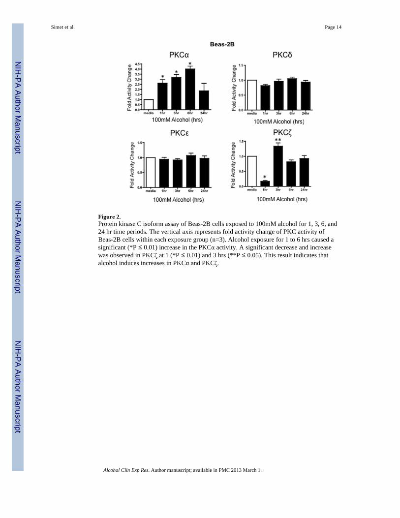
Figure 2.Protein kinase C isoform assay of Beas-2B cells exposed to 100mM alcohol for 1, 3, 6, and24 hr time periods. The vertical axis represents fold activity change of PKC activity ofBeas-2B cells within each exposure group (n=3). Alcohol exposure for 1 to 6 hrs caused asignificant (*P ≤ 0.01) increase in the PKCα activity. A significant decrease and increasewas observed in PKCζ at 1 (*P ≤ 0.01) and 3 hrs (**P ≤ 0.05). This result indicates thatalcohol induces increases in PKCα and PKCζ.
Simet et al. Page 14
Alcohol Clin Exp Res. Author manuscript; available in PMC 2013 March 1.
NIH
-PA Author Manuscript
NIH
-PA Author Manuscript
NIH
-PA Author Manuscript
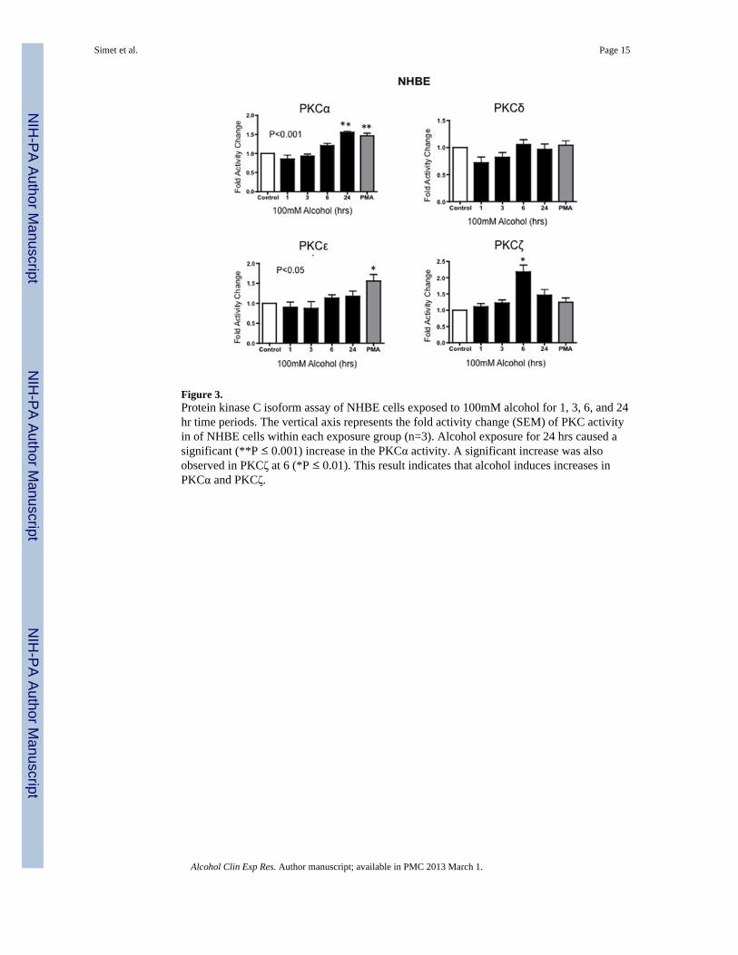
Figure 3.Protein kinase C isoform assay of NHBE cells exposed to 100mM alcohol for 1, 3, 6, and 24hr time periods. The vertical axis represents the fold activity change (SEM) of PKC activityin of NHBE cells within each exposure group (n=3). Alcohol exposure for 24 hrs caused asignificant (**P ≤ 0.001) increase in the PKCα activity. A significant increase was alsoobserved in PKCζ at 6 (*P ≤ 0.01). This result indicates that alcohol induces increases inPKCα and PKCζ.
Simet et al. Page 15
Alcohol Clin Exp Res. Author manuscript; available in PMC 2013 March 1.
NIH
-PA Author Manuscript
NIH
-PA Author Manuscript
NIH
-PA Author Manuscript
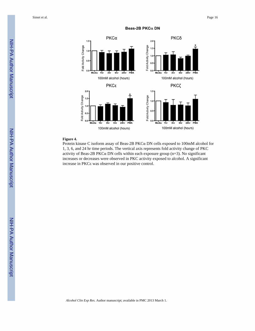
Figure 4.Protein kinase C isoform assay of Beas-2B PKCα DN cells exposed to 100mM alcohol for1, 3, 6, and 24 hr time periods. The vertical axis represents fold activity change of PKCactivity of Beas-2B PKCα DN cells within each exposure group (n=3). No significantincreases or decreases were observed in PKC activity exposed to alcohol. A significantincrease in PKCε was observed in our positive control.
Simet et al. Page 16
Alcohol Clin Exp Res. Author manuscript; available in PMC 2013 March 1.
NIH
-PA Author Manuscript
NIH
-PA Author Manuscript
NIH
-PA Author Manuscript

Figure 5.Normalized Resistance (ohms) (subsequent values divided by initial values) of Beas-2BPKCα (A) cells treated with 0, 20, 50, 100mM alcohol for 48 hrs. (B) NormalizedResistance of Beas-2B cells pretreated for 1 hr with a PKCα inhibitor, Gö-6976, and/or100mM alcohol. Lower resistance measurements in Beas-2B cells indicate a decreasedbarrier function between cells. Beas-2B PKCα DN cells are resistant to alcohol-induceddecreased barrier function. Similarly, wt Beas-2B cells pretreated with Gö-6976 are resistantto alcohol-induced decreased barrier function. Barrier function is significantly decreased(p<0.01) in Beas-2B cells treated with 100 mM alcohol (n=3;* P= 0.01) compared to controland Gö-6976 + EtOH treated cells.
Simet et al. Page 17
Alcohol Clin Exp Res. Author manuscript; available in PMC 2013 March 1.
NIH
-PA Author Manuscript
NIH
-PA Author Manuscript
NIH
-PA Author Manuscript
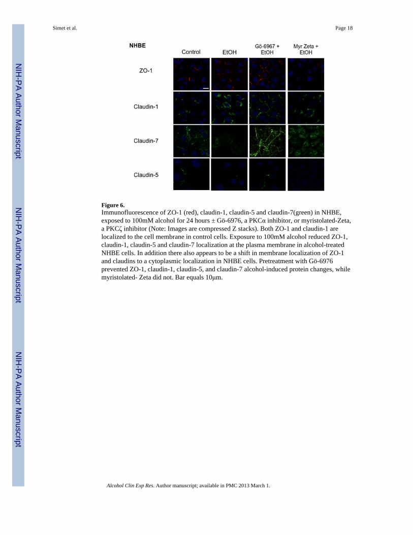
Figure 6.Immunofluorescence of ZO-1 (red), claudin-1, claudin-5 and claudin-7(green) in NHBE,exposed to 100mM alcohol for 24 hours ± Gö-6976, a PKCα inhibitor, or myristolated-Zeta,a PKCζ inhibitor (Note: Images are compressed Z stacks). Both ZO-1 and claudin-1 arelocalized to the cell membrane in control cells. Exposure to 100mM alcohol reduced ZO-1,claudin-1, claudin-5 and claudin-7 localization at the plasma membrane in alcohol-treatedNHBE cells. In addition there also appears to be a shift in membrane localization of ZO-1and claudins to a cytoplasmic localization in NHBE cells. Pretreatment with Gö-6976prevented ZO-1, claudin-1, claudin-5, and claudin-7 alcohol-induced protein changes, whilemyristolated- Zeta did not. Bar equals 10μm.
Simet et al. Page 18
Alcohol Clin Exp Res. Author manuscript; available in PMC 2013 March 1.
NIH
-PA Author Manuscript
NIH
-PA Author Manuscript
NIH
-PA Author Manuscript
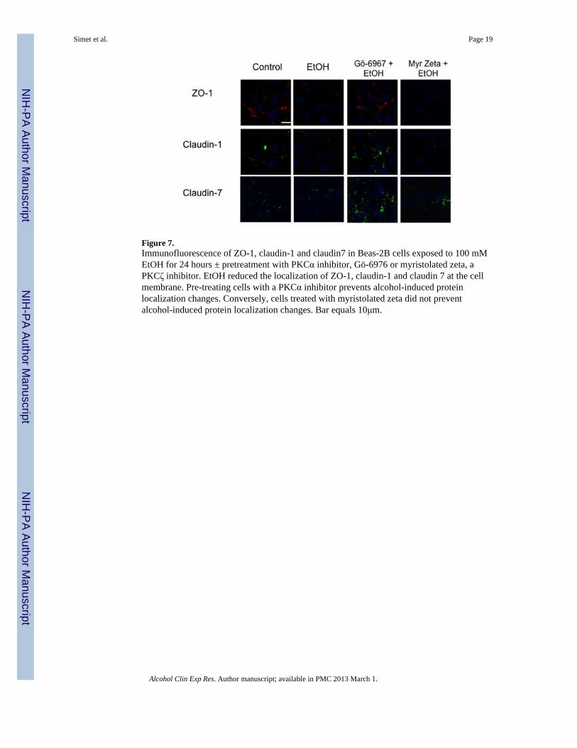
Figure 7.Immunofluorescence of ZO-1, claudin-1 and claudin7 in Beas-2B cells exposed to 100 mMEtOH for 24 hours ± pretreatment with PKCα inhibitor, Gö-6976 or myristolated zeta, aPKCζ inhibitor. EtOH reduced the localization of ZO-1, claudin-1 and claudin 7 at the cellmembrane. Pre-treating cells with a PKCα inhibitor prevents alcohol-induced proteinlocalization changes. Conversely, cells treated with myristolated zeta did not preventalcohol-induced protein localization changes. Bar equals 10μm.
Simet et al. Page 19
Alcohol Clin Exp Res. Author manuscript; available in PMC 2013 March 1.
NIH
-PA Author Manuscript
NIH
-PA Author Manuscript
NIH
-PA Author Manuscript
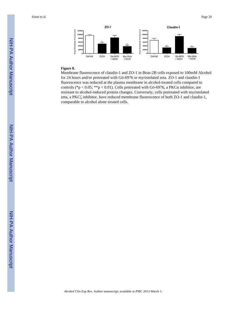
Figure 8.Membrane fluorescence of claudin-1 and ZO-1 in Beas-2B cells exposed to 100mM Alcoholfor 24 hours and/or pretreated with Gö-6976 or myristolated zeta. ZO-1 and claudin-1fluorescence was reduced at the plasma membrane in alcohol-treated cells compared tocontrols (*p < 0.05; **p < 0.01). Cells pretreated with Gö-6976, a PKCα inhibitor, areresistant to alcohol-induced protein changes. Conversely, cells pretreated with myristolatedzeta, a PKCζ inhibitor, have reduced membrane fluorescence of both ZO-1 and claudin-1,comparable to alcohol alone treated cells.
Simet et al. Page 20
Alcohol Clin Exp Res. Author manuscript; available in PMC 2013 March 1.
NIH
-PA Author Manuscript
NIH
-PA Author Manuscript
NIH
-PA Author Manuscript
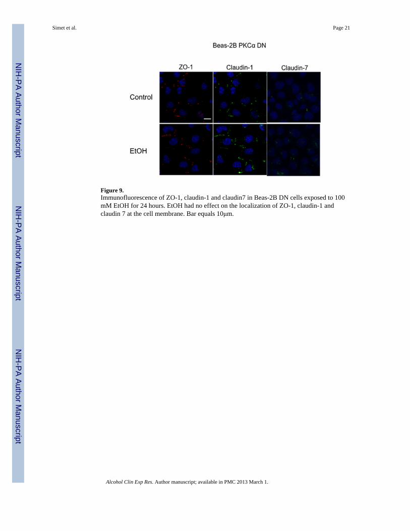
Figure 9.Immunofluorescence of ZO-1, claudin-1 and claudin7 in Beas-2B DN cells exposed to 100mM EtOH for 24 hours. EtOH had no effect on the localization of ZO-1, claudin-1 andclaudin 7 at the cell membrane. Bar equals 10μm.
Simet et al. Page 21
Alcohol Clin Exp Res. Author manuscript; available in PMC 2013 March 1.
NIH
-PA Author Manuscript
NIH
-PA Author Manuscript
NIH
-PA Author Manuscript
Related Documents