Air-Liquid Interface (ALI) Culture of Human Bronchial Epithelial Cell Monolayers as an In Vitro Model for Airway Drug Transport Studies HONGXIA LIN, 1 HONG LI, 1 HYUN-JONG CHO, 1 SHENGJIE BIAN, 2 HWAN-JUNG ROH, 3 MIN-KI LEE, 3 JUNG SUN KIM, 4 SUK-JAE CHUNG, 1 CHANG-KOO SHIM, 1 DAE-DUK KIM 1 1 College of Pharmacy and Research Institute of Pharmaceutical Sciences, Seoul National University, Seoul 151-742, South Korea 2 Department of Biology, Pusan National University, Busan 609-735, South Korea 3 College of Medicine, Pusan National University, Busan 602-739, South Korea 4 Department of Biotechnology, Dongseo University, Busan 617-716, South Korea Received 4 April 2006; revised 9 July 2006; accepted 10 September 2006 Published online in Wiley InterScience (www.interscience.wiley.com). DOI 10.1002/jps.20803 ABSTRACT: Serially passaged normal human bronchial epithelial (NHBE) cell mono- layers were established on Transwell 1 inserts via an air-liquid interface (ALI) culture method. NHBE cells were seeded on polyester Transwell 1 inserts, followed by an ALI culture from day 3, which resulted in peak TEER value of 766 154 O cm 2 on the 8th day. Morphological characteristics were observed by light microscopy and SEM, while the formation of tight junctions was visualized by actin staining, and confirmed successful formation of a tight monolayer. The transepithelial permeability (P app ) of model drugs significantly increased with the increase of lipophilicity and showed a good linear relationship, which indicated that lipophilicity is an important factor in determining the P app value. The expression of P-gp transporter in NHBE cell monolayers was confirmed by the significantly higher basolateral to apical permeability of rhodamine123 than that of reverse direction and RT-PCR of MDR1 mRNA. However, the symmetric transport of fexofenadine HCl in this NHBE cell monolayers study seems to be due to the low expression of P-gp transporter and/or to its saturation with high concentration of fexofenadine HCl. Thus, the development of tight junction and the expression of P-gp in the NHBE cell monolayers in this study imply that they could be a suitable in vitro model for evaluation of systemic drug absorption via airway delivery, and that they reflect in vivo condition better than P-gp over-expressed cell line models. ß 2006 Wiley-Liss, Inc. and the American Pharmacists Association J Pharm Sci 96:341– 350, 2007 Keywords: drug transport; in vitro model; bronchial epithelial cell; P-glycoprotein; permeability INTRODUCTION Pulmonary drug absorption has been extensively investigated as an attractive route for systemic drug delivery since peptides and protein drugs with poor bioavailability via the oral route apparently show excellent bioavailability when delivered by inhalation as pharmaceutical aero- sols. 1–4 In addition to targeting to the deep lung for systemic absorption, aerosols are also routi- nely used for drug delivery to the bronchial regions of the airways for the treatment of acute or chronic lung diseases, such as asthma, cystic JOURNAL OF PHARMACEUTICAL SCIENCES, VOL. 96, NO. 2, FEBRUARY 2007 341 Correspondence to: Dae-Duk Kim (Telephone: þ82-2-880- 7870; Fax: þ82-2-873-9177; E-mail: [email protected]) Journal of Pharmaceutical Sciences, Vol. 96, 341–350 (2007) ß 2006 Wiley-Liss, Inc. and the American Pharmacists Association

Welcome message from author
This document is posted to help you gain knowledge. Please leave a comment to let me know what you think about it! Share it to your friends and learn new things together.
Transcript

Air-Liquid Interface (ALI) Culture of HumanBronchial Epithelial Cell Monolayers as an In VitroModel for Airway Drug Transport Studies
HONGXIA LIN,1 HONG LI,1 HYUN-JONG CHO,1 SHENGJIE BIAN,2 HWAN-JUNG ROH,3 MIN-KI LEE,3
JUNG SUN KIM,4 SUK-JAE CHUNG,1 CHANG-KOO SHIM,1 DAE-DUK KIM1
1College of Pharmacy and Research Institute of Pharmaceutical Sciences, Seoul National University,Seoul 151-742, South Korea
2Department of Biology, Pusan National University, Busan 609-735, South Korea
3College of Medicine, Pusan National University, Busan 602-739, South Korea
4Department of Biotechnology, Dongseo University, Busan 617-716, South Korea
Received 4 April 2006; revised 9 July 2006; accepted 10 September 2006
Published online in Wiley InterScience (www.interscience.wiley.com). DOI 10.1002/jps.20803
ABSTRACT: Serially passaged normal human bronchial epithelial (NHBE) cell mono-layers were established on Transwell1 inserts via an air-liquid interface (ALI) culturemethod. NHBE cells were seeded on polyester Transwell1 inserts, followed by an ALIculture from day 3, which resulted in peak TEER value of 766� 154 O� cm2 on the 8thday.Morphological characteristicswere observedby lightmicroscopy andSEM,while theformation of tight junctions was visualized by actin staining, and confirmed successfulformation of a tight monolayer. The transepithelial permeability (Papp) of model drugssignificantly increased with the increase of lipophilicity and showed a good linearrelationship, which indicated that lipophilicity is an important factor in determining thePapp value. The expression of P-gp transporter in NHBE cell monolayers was confirmedby the significantly higher basolateral to apical permeability of rhodamine123 than thatof reverse direction and RT-PCR of MDR1 mRNA. However, the symmetric transportof fexofenadine �HCl in this NHBE cell monolayers study seems to be due to the lowexpression of P-gp transporter and/or to its saturation with high concentration offexofenadine �HCl. Thus, the development of tight junction and the expression of P-gp intheNHBE cell monolayers in this study imply that they could be a suitable in vitromodelfor evaluation of systemic drug absorption via airway delivery, and that they reflectin vivo condition better than P-gp over-expressed cell line models. � 2006 Wiley-Liss, Inc.
and the American Pharmacists Association J Pharm Sci 96:341–350, 2007
Keywords: drug transport; in vitro model; bronchial epithelial cell; P-glycoprotein;permeability
INTRODUCTION
Pulmonary drug absorption has been extensivelyinvestigated as an attractive route for systemic
drug delivery since peptides and protein drugswith poor bioavailability via the oral routeapparently show excellent bioavailability whendelivered by inhalation as pharmaceutical aero-sols.1–4 In addition to targeting to the deep lungfor systemic absorption, aerosols are also routi-nely used for drug delivery to the bronchialregions of the airways for the treatment of acuteor chronic lung diseases, such as asthma, cystic
JOURNAL OF PHARMACEUTICAL SCIENCES, VOL. 96, NO. 2, FEBRUARY 2007 341
Correspondence to: Dae-Duk Kim (Telephone: þ82-2-880-7870; Fax: þ82-2-873-9177; E-mail: [email protected])
Journal of Pharmaceutical Sciences, Vol. 96, 341–350 (2007)� 2006 Wiley-Liss, Inc. and the American Pharmacists Association

fibrosis, and chronic obstructive pulmonary dis-ease (COPD). Formulations of aerosols have beendesigned to comprise small spherical dropletsor particles suitable for penetrating into theairways or lung periphery, which lead to optimaltherapeutic effect both for local and systemictherapeutic delivery.5,6 It is also known thattracheobronchial disposition of inhaled particu-late formulation (5–15 mm) is significantly greaterthan alveolar deposition.7 Therefore, it would beequally important to understand the character-istics of absorption barrier in this region.
In vitro cell culture models of absorptiveepithelia based on primary culture or immorta-lized cells have been proven extremely useful forthe study of respiratory epithelial permeabilityand drug absorption.8 Advantages of cell culturemodels based on continuous cell lines are welldemonstrated by the popularity of the Caco-2model of the gastrointestinal epithelium. How-ever, there is no well-established in vitro respira-tory epithelium model up to date, and efforts todevelop bronchial/trachea culture systems for drugmetabolism and permeation studies are still intheir infancy. Recently, two cell lines, 16HBE14o�
andCalu-3,were identified as promising candidat-es as drug absorption models of the airways.9–11
The 16HBE14o� cell line was originally developedby SV40 large T antigen transformation of cul-tured bronchial-surface epithelial cells for study-ing the chloride channel activity of cystic fibrosistransmembrane conductance (CFTR) regulator.12
The Calu-3 cell line was derived from a bronchialadenocarcinoma which is available from ATCC.The usefulness of 16HBE14o� and Calu-3 cells forthe study of bronchial epithelial transport proper-ties has recently been reported.10,13–15 However,although cell lines from cancer tissues are easilymaintained in culture, they often do not have themorphology or biochemical characteristics of theoriginal tissue.16
Comparedwith immortalized cell lines, primaryairway epithelial cells are composed of mixedtypes, such as goblet cell, basal cell, cilia epithelialcell, and noncilia epithelial cell, and thus providethe closest in vitro representation of the airwayepithelium. However, major limitations of normalhumanairway tissue are knownas follows: (1) lackof donor sources, (2) limited amount of cells, and (3)large donor variation. To overcome the shortage ofhuman airway tissues, researchers have investi-gated passage culture of primary epithelialcells.17–19 Previously, we have established an air-liquid interface (ALI) culture method of passaged
human nasal epithelial (HNE) cell monolayer fordrug transport studies.20 Optimizing the culturecondition, such as selecting the appropriatemedia,is known to be critical for airway epithelial cellculture to promote the differentiation and to formsuitable epithelial barriers with tight junctions.
In this study, the normal human bronchialepithelial (NHBE) cells, which come from non-diseased human tissue without immortalization,were selected to establish the passaged humanbronchial cell monolayers. After characterizingthe NHBE cell monolayers, the permeability ofvarious anti-allergic drugs were investigated toevaluate the feasibility of the monolayers forin vitro transport studies.
EXPERIMENTAL
Materials
Normal human bronchial epithelial (NHBE)(CloneticsTM, 1st passage) cells were obtainedfrom Cambrex Bio Science, Inc. (Walkersville,MD). Budesonide, Hank’s balanced salt solution(HBSS), rhodamine123, and D-(þ)-glucose werepurchased from Sigma Chemical Co. (St. Louis,MO). Triamcinolone acetonide and fexofenadi-ne �HCl were gifts from Handok-Aventis Pharma-ceutical Co. (Seoul, Korea). Other cell culturereagents and supplies were obtained from Invi-trogen Co. (Grand Island, NY). Bronchial epithe-lial cell growth medium (BEGM) bullet kit wasobtained from Cambrex Bio Science, Inc. Trans-wells1 (0.4 mm, 12 mm diameter, polyester) wereobtained from Costar Co. (Cambridge, MA). Allother materials were of analytical grade orhigher.
Air-Liquid Interface Cell Culture Method forHuman Bronchial Epithelial Cell
A reported method on the passaged ALI culture ofHNE cell monolayers was slightly modified.20
Briefly, frozen passage-1 stocks were thawedand cultured in T-flask using BEGM at 378C inan atmosphere of 5% CO2 and 95% relativehumidity. The medium was changed every 2 days.When cultures reached approximately 70%–80%confluency, the cells were detached with 0.1%trypsin-EDTA and were seeded at densities of2� 105 to 3� 105 cells/cm2 on Transwell1 insert.Both apical side (0.5 mL) and basolateral side(1.5 mL) were filled with BEGM:DME/F12 (50:50)
342 LIN ET AL.
JOURNAL OF PHARMACEUTICAL SCIENCES, VOL. 96, NO. 2, FEBRUARY 2007 DOI 10.1002/jps
supplemented with hydrocortisone (0.5 mg/mL),insulin (5 mg/mL), transferrin (10 mg/mL), epine-phrine (0.5 mg/mL), triiodothyronine (6.5 mg/mL),gentamycin (50 mg/mL), amphotericin-B (50 mg/mL), retinoic acid (0.1 ng/mL), and epidermalgrowth factor (0.5 ng/mL human recombinant)(all supplied by Cambrex Bio Science, Inc.). Mediaof both sides were changed after 24 h, and thenthe apical side was directly exposed to ambientcondition on day 3 for ALI culture, after which themedium of the basolateral side was changedeveryday. Remaining cells were subcultured forthe next passage at a density of 500 cells/cm2, andthe medium (BEGM) was changed every 2 days.
Transepithelial Electrical Resistance(TEER) Measurement
The transepithelial electrical resistance (TEER)value was measured daily using an EVOMvoltohmmeter device (WPI, Sarasota, FL), andwas corrected by subtracting the background dueto the blank Transwell1 insert and medium. Pre-equilibrated medium (BEGM:DME/F12, 50:50)were added to the apical (0.5 mL) and basolateral(1.5 mL) side, after which the monolayers wereallowed to attain a steady potential for about5 min prior to reading of TEER values.
Actin Staining
The filamentous actin (F-actin) was stained withFITC-labeled phalloidin (Sigma chemical Co.).The passage 2 human bronchial epithelial cellmonolayers grown on permeable supports for7 days were rinsed three times with PBS, andwere fixed for 10 min in 3.7% formaldehyde inPBS. After rinsing three times with PBS, theywere treated with 1% Triton X-100 (InvitrogenCo.) on ice for 5 min. Then, they were rinsed withPBS twice and air-dried. The monolayers werestained with FITC-phalloidin for 20 min in thedark. Themonolayers were rinsed again with PBSthree times and then mounted on glass slideswith gelvatol. The samples were observed under afluorescence microscope at 200� magnification(Diaphot 300, Nikon Co., Tokyo, Japan).
Transport Studies Across the NHBE Cell Monolayers
Bronchial epithelial cell monolayers of passage 2and 3 were used for drug transport studies after6� 8 days of seeding when TEER value exceeded500 O� cm2. All the transport experiments wereperformed at 378C in a shaking water bath. Prior
to the transport studies, the cell culture mediumwas removed and the cell monolayers were allow-ed to equilibrate in transport medium (HBSSbuffered with 10 mM HEPES and 10 mM D-(þ)-glucose) for 30min. For the transport studies fromthe apical side to the basolateral side (A!B),0.4 mL of various anti-allergic drugs in transportmedium was applied in the apical compartment,and 1.0 mL of blank transport medium was addedin the basolateral chamber. Samples of 1.0 mLwere withdrawn from the basolateral chamber at30, 60, 90, and 120 min, and quickly replaced withequal volume of fresh transport medium. Trans-port studies in reverse direction (B!A) wereperformed by adding 0.4 mL of transport mediumin apical side, and 1.0 mL of drug solution inbasolateral side. At predetermined time intervals,samples of 0.3 mL were withdrawn from theapical side and quickly replaced with an equalvolume of fresh transport medium.
The transport study of P-gp substrate, rho-damine 123 (5 mM) was also performed in twodirections, with and without P-gp inhibitor, ver-apamil. Verapamil (50 mM) was added in bothapical and basolateral side to ensure a continuousinhibition of the P-gp efflux pump. The TEERvalue was measured at the beginning and end ofeach transport experiment in order to monitorintegrity of the cultured epithelial cellmonolayers.
Analytical Method
The samples of anti-allergic drugs from transportstudies were directly analyzed using a WatersHPLC system (Waters Co., Milford, MA) equippedwith a pump (Waters 515), an automatic injector(Waters 717plus) and UV detector (Waters 2487).A reversed phase C-18 column (Lichrospher1 100,RP-18, 125� 4 mm, 5 mm, Merck Darmstadt,Germany) was used, except for albuterol, whichwas determined on a C-8 column (Lichrospher1
100, RP-8, 250� 4 mm, 5 mm). Rhodamine123 wasdetermined using Waters HPLC system with fluor-escence detector (lex 485 nm and lem 525 nm) (Series200, PerkinElmer Instrument, Norwalk, CT). Theflow rate was 1.0 mL with the injection volume of50 mL.
Data Analysis and Statistics
The apparent permeability coefficients (Papp, cm/s)were calculated using the following equation:
Papp ¼ dQ
dt
1
A � C0
ALI CULTURE OF HUMAN BRONCHIAL EPITHELIAL CELL MONOLAYERS 343
DOI 10.1002/jps JOURNAL OF PHARMACEUTICAL SCIENCES, VOL. 96, NO. 2, FEBRUARY 2007

where, dQ/dt is the solute flux, A is the surfacearea across which transport studies were measur-ed (1.0 cm2), and C0 is the initial drug concentra-tion. Each study was performed at least intriplicates and the data were expressed as themean� standard deviation (SD). A two-tailedstudent’s t-test was performed at p< 0.05.
Reverse Transcription-Polymerase Chain Reaction
Reverse transcription-polymerase chain reaction(RT-PCR) was performed for the detection ofMDR1 gene mRNAs in the monolayers. Themethods for detection of MDR1 mRNAs havepreviously been described in detail.21 Total RNAsof NHBE cell monolayer on day 7, 14, 21 wereextracted with RNAeasy Mini kit according to themanufacturer’s recommendations. An AccuPowerRT premix (Bioneer Co., Daejeon, Korea) wasused for the reverse transcription, which wasperformed under the following conditions: 708Cfor 5 min (incubation), 428C for 60 min (cDNAsynthesis), and 948C for 5 min (RTase inactiva-tion). The synthesized first strand cDNAwas usedfor PCR using a set of MDR1 primer (50-CGA AACCGT ATC AGT CCT CG-30; 50-CTT GAG TCTGAG AGA CCA CC-30). cDNA template (2.5 mL)and 50 pmol of each primer were added to theAccuPower HotStart PCR Premix (Bioneer Co.).Standard PCR amplification was performed usingthe MJ Mini Thermal Cycler (Bio-Rad Labora-tories, Inc., Hercules, CA). Amplification cycleswere as following: 958C for 15 min, then 32 cyclesat 948C for 1 min, 568C for 1 min, 728C for 1 min,followed by 728C for 7 min. Aliquots (10 mL) ofPCR product were electrophoresed on 1.5%agarose gels, and PCR fragments were visualizedby GelRedTM (Biotium, Inc., Hayward, CA) stain-ing. In order to verify that the amplified productswere from mRNA and not genomic DNA con-tamination, negative controls were performed byomitting reverse transcriptase from RT-PCTreaction. In the absence of reverse transcriptase,no PCR products were observed.
RESULTS AND DISCUSSION
Bioelectric Parameters and Morphological Studies
Passage 2 and 3 of NHBE cells with serum-freeBEGM was successfully cultured on T-flask with-out collagen coating, as shown in Figure 1A. TheNHBE cells showed epitheliumlike morphologyand contact inhibition. When these cells were
Figure 1. Morphology of normal human bronchialepithelial (NHBE) cells under light microscopy. (A)Passage 3 cells on T-flask with 90% confluency (100�),(B) Monolayer of passage 3 cells from (A) after 3 days ofseeding on Transwell1 (100�). [Color figure can be seenin the online version of this article, available on thewebsite, www.interscience.wiley.com.]
Figure 2. Change of TEERvalues of passagedhumanbronchial epithelial cell monolayer cultured by the air-liquid interface (ALI) culture method on Transwell1 for2 weeks (n¼ 9).
344 LIN ET AL.
JOURNAL OF PHARMACEUTICAL SCIENCES, VOL. 96, NO. 2, FEBRUARY 2007 DOI 10.1002/jps

seeded on Transwell1 at a density of 2.5� 105
cells/cm2 in serum-free ALI culture condition, theepithelial cell monolayers of passage 2 and 3appeared to reach confluency (Fig. 1B) and beganto exhibit a measurable transepithelial resistancefrom day 3. Figure 2 shows the change of TEERvalue of NHBE cell monolayer on Transwell1
measured by using the EVOM voltohmmeterdevice for 14 days. The maximum TEER valuewas 766� 154 O� cm2 on day 8 after seeding andmaintained higher than 500O� cm2 for 5�6 days.The high TEER value implies the formation oftight junction that is an impermeable junctionlocated at the apical side. The formation of tightjunction was also confirmed by the F-actin stain-ing, as shown in Figure 3A. However, comparedwith primary human nasal cell monolayers,20
SEM study showed less prominent cilia or
denuded ciliated cells even after 21 days of ALIculture (Fig. 3B), indicating incomplete ciliadifferentiation.
Tight junction is envisioned as a zone of densehydrophobic, intercellular material that wouldform a seal when two adjacent cells were heldclose. There are many molecular components ofthe tight junction, such as occludin, acludins,junctional adhesionmolecule (JAM), zonula occlu-dens, and actin. Among these components, peri-junctional actin is known to play a major role incontrolling the paracellular permeability.22 Therelatively high TEER in this study was in agree-ment with previously published data, reporting800–1000 O� cm2 in primary cultured trachealand bronchial epithelial cell monolayers of rabbitand human, respectively.23,24 The maximumTEER appearing on about day 7 after seedingwas also consistent with data reported on rabbittracheal epithelial cell monolayer,23 primary humanalveolar epithelial cells,25 and air-interfaced pri-mary rabbit conjunctival epithelial culture.26 Themorphological and functional characteristics inthe bronchial epithelial cell layers grown underALI condition is due to the improved delivery ofoxygen across the liquid film to the cells, whichmay change the cellular respiration to a moreaerobic nature.23 Our previous study on therelationship between Papp and TEER values27
showed that consistent permeation results wereobtained when TEER values were higher than500 O� cm2, which implies that the NHBE cellmonolayer could be useful for in vitro transportstudies.
Figure 3. (A) F-Actin staining of passage 3 cells onTranswell1 (200�) after 7 days of seeding, (B) scanningelectron microscopy (SEM) of passage 2 cells after21 days of seeding. [Color figure can be seen in theonline version of this article, available on the website,www.interscience.wiley.com.]
Figure 4. Transport profiles of anti-allergic drugsacross the NHBE cell monolayers from apical tobasolateral side. Each point represents the mean�SD(n¼ 3).
ALI CULTURE OF HUMAN BRONCHIAL EPITHELIAL CELL MONOLAYERS 345
DOI 10.1002/jps JOURNAL OF PHARMACEUTICAL SCIENCES, VOL. 96, NO. 2, FEBRUARY 2007
Transport Studies of Anti-Allergic Drugs
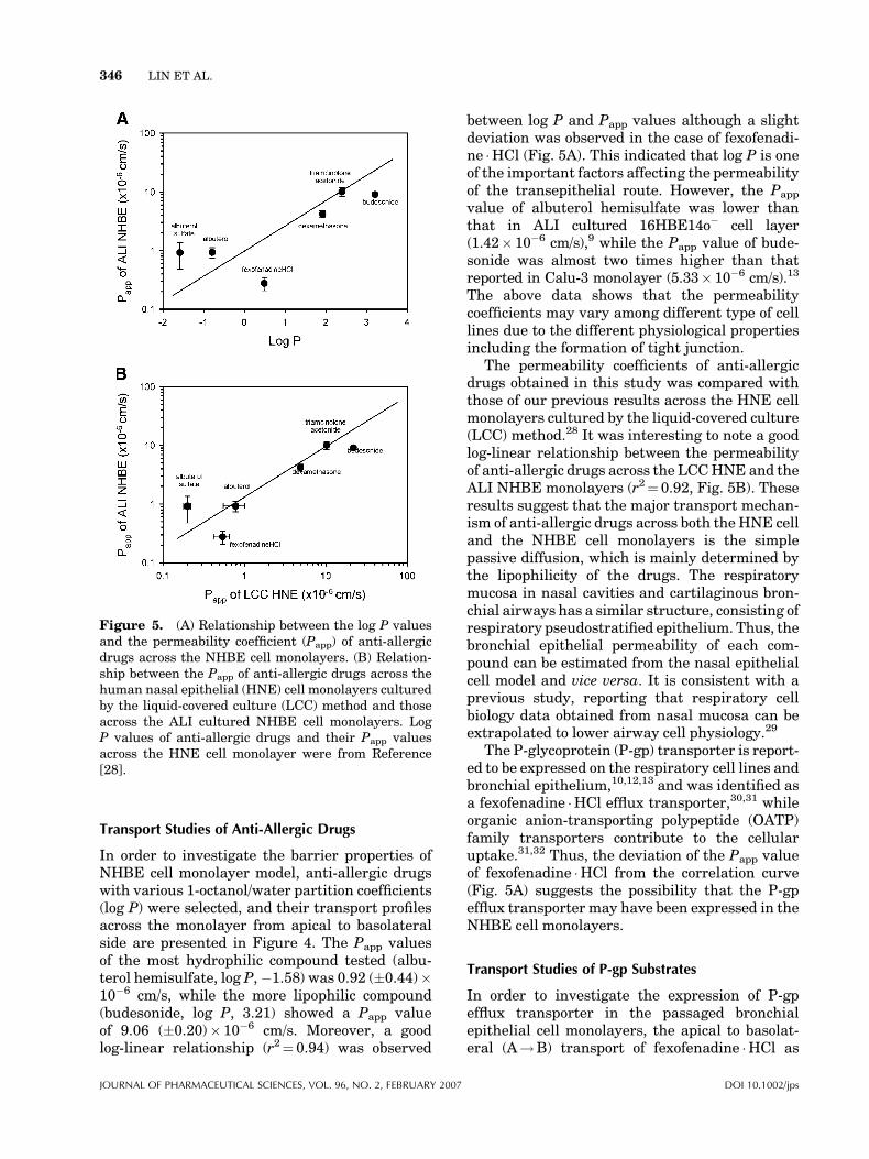
In order to investigate the barrier properties ofNHBE cell monolayer model, anti-allergic drugswith various 1-octanol/water partition coefficients(log P) were selected, and their transport profilesacross the monolayer from apical to basolateralside are presented in Figure 4. The Papp valuesof the most hydrophilic compound tested (albu-terol hemisulfate, log P,�1.58) was 0.92 (�0.44)�10�6 cm/s, while the more lipophilic compound(budesonide, log P, 3.21) showed a Papp valueof 9.06 (�0.20)� 10�6 cm/s. Moreover, a goodlog-linear relationship (r2¼ 0.94) was observed
between log P and Papp values although a slightdeviation was observed in the case of fexofenadi-ne �HCl (Fig. 5A). This indicated that log P is oneof the important factors affecting the permeabilityof the transepithelial route. However, the Papp
value of albuterol hemisulfate was lower thanthat in ALI cultured 16HBE14o� cell layer(1.42� 10�6 cm/s),9 while the Papp value of bude-sonide was almost two times higher than thatreported in Calu-3 monolayer (5.33� 10�6 cm/s).13
The above data shows that the permeabilitycoefficients may vary among different type of celllines due to the different physiological propertiesincluding the formation of tight junction.
The permeability coefficients of anti-allergicdrugs obtained in this study was compared withthose of our previous results across the HNE cellmonolayers cultured by the liquid-covered culture(LCC) method.28 It was interesting to note a goodlog-linear relationship between the permeabilityof anti-allergic drugs across the LCCHNE and theALI NHBE monolayers (r2¼ 0.92, Fig. 5B). Theseresults suggest that the major transport mechan-ism of anti-allergic drugs across both the HNE celland the NHBE cell monolayers is the simplepassive diffusion, which is mainly determined bythe lipophilicity of the drugs. The respiratorymucosa in nasal cavities and cartilaginous bron-chial airways has a similar structure, consisting ofrespiratory pseudostratified epithelium. Thus, thebronchial epithelial permeability of each com-pound can be estimated from the nasal epithelialcell model and vice versa. It is consistent with aprevious study, reporting that respiratory cellbiology data obtained from nasal mucosa can beextrapolated to lower airway cell physiology.29
The P-glycoprotein (P-gp) transporter is report-ed to be expressed on the respiratory cell lines andbronchial epithelium,10,12,13 and was identified asa fexofenadine �HCl efflux transporter,30,31 whileorganic anion-transporting polypeptide (OATP)family transporters contribute to the cellularuptake.31,32 Thus, the deviation of the Papp valueof fexofenadine �HCl from the correlation curve(Fig. 5A) suggests the possibility that the P-gpefflux transporter may have been expressed in theNHBE cell monolayers.
Transport Studies of P-gp Substrates
In order to investigate the expression of P-gpefflux transporter in the passaged bronchialepithelial cell monolayers, the apical to basolat-eral (A!B) transport of fexofenadine �HCl as
Figure 5. (A) Relationship between the log P valuesand the permeability coefficient (Papp) of anti-allergicdrugs across the NHBE cell monolayers. (B) Relation-ship between the Papp of anti-allergic drugs across thehuman nasal epithelial (HNE) cell monolayers culturedby the liquid-covered culture (LCC) method and thoseacross the ALI cultured NHBE cell monolayers. LogP values of anti-allergic drugs and their Papp valuesacross the HNE cell monolayer were from Reference[28].
346 LIN ET AL.
JOURNAL OF PHARMACEUTICAL SCIENCES, VOL. 96, NO. 2, FEBRUARY 2007 DOI 10.1002/jps

well as rhodamine123, as P-gp substrates, wascompared with the basolaterial to apical (B!A)transport of both compounds. Rhodamine123 is afluorescent dye, and has been extensively used asan index of P-gp mediated transport in rodent andtissue culture models.33 Fexofenadine �HCl trans-
port studies in Caco-2 cells also supported itsutility as a probe compound reflecting the P-gpactivity in vivo.30 As shown in Figure 6A andTable 1, no significant difference in Papp values inboth directions was observed in the transportstudies of fexofenadine �HCl. The ratio of (B!A)/(A!B) was 1.0, consistent with results from ourprevious human nasal cell monolayer study,28
indicating that the transport of fexofenadine �HClwas not significantly affected by P-gp. Possibleexplanations for this symmetric transport couldinclude the following: (1) the paracellular diffu-sion is the major translocation mechanism con-sidering the hydrophilicity of fexofenadine �HCl,and thus Papp of fexofenadine �HCl may not beaffected by the existence of the efflux system;(2) the P-gp transporter is saturated with fexofe-nadine �HCl; (3) P-gp expression in NHBE cellmonolayers is low.
Although fexofenadine �HCl may mainlypermeate through the paracellular pathway oftheNHBEcellmonolayer, it still canbea substrateof the efflux system on the apical side of the cellsafter translocated to the basolateral side, whichwould result in higher Papp value of B!Adirection than that of A!B direction if the effluxsystemon the apical side isworking. Thus, thefirsthypothesis indicating that the paracellular routeis the sole pathway for this drug is not convincingenough.
In the transport study of rhodamine123(Fig. 6B), the Papp value of A!B direction(2.82� 0.81� 10�7 cm/s) was significantly lowerthan that of B!A direction (8.31� 2.15�10�7 cm/s). Moreover, a P-gp inhibitor, verapamil(50 mM), significantly increased the Papp value ofA!B direction transport, while that of B!Adirection was slightly decreased (Tab. 1). Thus,P-gp was undoubtedly expressed in NHBE cell
Figure 6. Transport profiles of (A) feoxfenadine �HCl(500 mg/mL) and (B) rhodamine 123 (5 mM) across thehuman bronchial epithelial cell monolayers. Each pointrepresents the mean�SD (n> 3).
Table 1. Transepithelial Permeability Coefficient (Papp) of Fexofenadine �HCl and Rhodamine123 across theNormal Human Bronchial Epithelial Cell Monolayers
Drug (Loading Dose)
Papp (�10�7 cm/s)
(B!A)/(A!B)Apical to Basolateral
(A!B)Basolateral to Apical
(B!A)
Fexofenadine �HCl (500 mg/mL) 2.76� 0.69 2.77� 0.34 1.00Rhodamine123 (5 mM) 2.82� 0.81 8.31� 2.15** 2.95Rhodamine123 (5 mM) þ Verapamil (50 mM) 4.77� 0.53*** 7.85� 0.94* 1.65
Each data represents the mean�SD (n> 3).*p< 0.05.**p< 0.01, compared to A to B transport.***p<0.01, compared to without verapamil.
ALI CULTURE OF HUMAN BRONCHIAL EPITHELIAL CELL MONOLAYERS 347
DOI 10.1002/jps JOURNAL OF PHARMACEUTICAL SCIENCES, VOL. 96, NO. 2, FEBRUARY 2007

monolayers. However, since the concentration offexofenadine �HCl was higher than that of ver-apamil, P-gp transporter inNHBEcellmonolayersmay have been saturated with fexofenadine �HCland/or the expression of P-gp transporter maynot have been as high as inCaco-2 cellmonolayers.In other words, P-gp was expressed in NHBE,enough to affect rhodamine123 transport, but in amuch lower extent than in cancerous cells likeCaco-2 cells, not enough to affect fexofenadine �HCl transport.
RT-PCR Assay of mRNA Expressionof MDR1 Gene
In order to confirm the expression of the P-gp inthe NHBE cell monolayers, the levels of mRNAexpression of MDR1 genes were measured by RT-PCR analysis after 7, 14, and 21 days of ALIculture. MDR1 gene is known to code for a 170–180 kD plasma membrane P-glycoprotein.34 Asshown in Figure 7, MDR1 mRNA was expressedin ALI cultured NHBE cell monolayer, whichindicated the expression of the P-gp transporterin the monolayer. However, the level of P-gpexpression does not seem to have been completeon the 7th day after seeding, which was when thetransport study was conducted, while strongerbands are observed on the 14th and 21st days.Significantly higher Papp value of basolateral toapical direction (B!A) than that of apical tobasolateral direction (A!B) in rhodamine123transport study suggested that the P-gp trans-porter was localized on the apical side of the ALINHBE cell monolayer. However, in the NHBE cellmonolayer transport study of rhodamine123, thePapp ratio of (B!A)/(A!B) direction was 2.95(Tab. 1), which is lower than that reported inCaco-2-cell layers (greater than 10-fold).35 RT-PCR and transport studies suggest that P-gp was
expressed on the apical side of ALI NHBEmonolayers, yet the expression level seems to belower than that in Caco-2 cell monolayers, whichare known to over-express P-gp.30 A previousimmunohistochemistry study reported that inhuman bronchus, the expression of P-gp waslower than in cultured cell lines.34 Therefore,the NHBE cell monolayer seems to be closer inmimicking the physiological condition of humanbronchus compared to other cultured cells.
The investigation on the expression of varioustransporters in human respiratory epitheliumincluding the super family of ABC transporter,MDR1, MRP, and amino transporters haveattracted the attention of pharmaceuticalresearchers.10,12,13 MDR1 (P-gp, 170 kD) is acell-membrane ATP-dependent efflux pump withdiverse substrate specificities. Although the phy-siological role of P-gp in various tissues is stillunclear, it was consistently detected at the apicalsurface of ciliated epithelial cells from the surfaceepithelium or ciliated collecting ducts.34 However,since the activity P-gp efflux system that arerelevant for drug transport at the respiratoryepithelium has been examined mostly in P-gpover-expressed cell lines, such as Calu-3,13–15
Caco-2,30 and L-MDR131 cells, the effect of P-gpefflux transporter in respiratory epithelium couldhave been over-estimated. Thus, the symmetrictransport of fexofenadine �HCl in this NHBE cellmonolayers study could be due to the low expres-sion of P-gp transporter and/or to the saturation ofP-gp transporter with fexofenadine �HCl. How-ever, it would be more meaningful to define theeffect of P-gp efflux transporter in NHBE cellsthan in over-expressed cells. Therefore, the devel-opment of tight junction and the low expression ofP-gp in theNHBEcellmonolayers in this study arepromising results that can be useful for the in vitrodrug transport studies.
CONCLUSIONS
The NHBE cells were successfully subcultured upto passage 3 and each passage culture formed atight monolayer with high TEER value sufficientfor drug transport studies. Polarized transportacross the NHBE cell monolayers was observedfor P-gp substrate (i.e., rhodamine 123), whichindicated the expression of P-gp in this culturesystem. However, since the level of expression inthe NHBE cell monolayers seems to be lower thanthat in immortalized cells (i.e., Caco-2 and16HBE14o� cells), it was not sufficient to inves-
Figure 7. The expression of mRNA of MDR1 by RT-PCR after 7, 14, and 21 days of seeding on transwells.[Color figure can be seen in the online version of thisarticle, available on the website, www.interscience.wiley.com.]
348 LIN ET AL.
JOURNAL OF PHARMACEUTICAL SCIENCES, VOL. 96, NO. 2, FEBRUARY 2007 DOI 10.1002/jps
tigate the polarized transport of fexofenadi-ne �HCl. On the other hand, the permeability ofanti-allergic drugs across the monolayer waslinearly dependent on their lipophilicity. Also, agood correlation was obtained between the ALINHBE and LCC HNE monolayer culture model,which implies that simple passive diffusion is thepredominant factor in respiratory transepithelialtransport. Even though the differentiations ofcilia were not complete, the NHBE cell monolayercould be a suitable in vitro model for rapidevaluation of transport properties of drugs tar-geted for systemic absorption via airway delivery.
ACKNOWLEDGMENTS
This work was supported by the research grantfrom the Ministry of Health and Welfare in Korea(02-PJ2-PG1-CH12-0002).
REFERENCES
1. Kwon JH, Lee BH, Lee JJ, Kim CW. 2004. Insulinmicrocrystal suspension as a long-acting formula-tion for pulmonary delivery. Eur J Pharm Sci 22:107–116.
2. Surendrakumar K, Martyn GP, Hodgers EC,Jansen M, Blair JA. 2003. Sustained release ofinsulin from sodium hyaluronate based dry powderformulations after pulmonary delivery to beagledogs. J Control Release 91:385–394.
3. Mahesh KT, Misra A. 2004. Pulmonary absorptionenhancement of salmon calcitonin. J Drug Target12:135–144.
4. Wang ZY, Zhang Q. 2003. Study on pulmonarydelivery of peptide drugs in rats: Effects of absorp-tion enhancers on cellular membrane fluidity. YaoXue Xue Bao 38:957–961.
5. Edwards DA, Ben-Jebria A, Langer R. 1998. Recentadvances in pulmonary drug delivery using large,porous inhaled particles. J Appl Physiol 85:379–385.
6. Labiris NR, Dolovich MB. 2003. Pulmonary drugdelivery. Part I: Physiological factors affectingtherapeutic effectiveness of aerosolized medica-tions. Br J Clin Pharmacol 56:588–599.
7. Chan T, Lippmann M. 1980. Experimental mea-surements and empirical modeling of the regionaldeposition of inhaled particles in humans. Am IndHyg Assoc J 41:399–408.
8. Neil RM, Fumiyoshi Y, Lee VH. 1996. Respiratoryepithelial cell culture models for evaluation of ionand drug transport. Adv Drug Dev Rev 22:215–249.
9. Forbes B, Shah A, Martin GP, Lansley AB.2003. The human bronchial epithelial cell line16HBE14o� as a model system of the airways forstudying drug transport. Int J Pharm 257:161–167.
10. Ehrhardt C, Kneuer C, Laue M, Schaefer UF, KimKJ, Lehr CM. 2003. 16HBE14o� human bronchialepithelial cell layers express P-glycoprotein, lungresistance-related protein, and caveolin-1. PharmRes 20:545–551.
11. Forbes B, Ehrhardt C. 2005. Human respiratoryepithelial cell culture for drug delivery applica-tions. Eur J Pharm Biopharm 60:193–205.
12. Cozens AL, Yezzi MJ, Kunzelmann K, Ohrui T,Chin L, Eng K, Finkbeiner WE, Widdicombe JH,Gruenert DC. 1994. CFTR expression and chloridesecretion in polarized immortal human bronchialepithelial cells. Am J Respir Cell Mol Biol 10:38–47.
13. Borchard G, Cassara ML, Roemele PE, Florea BI,Junginger HE. 2002. Transport and local metabo-lism of budesonide and fluticasone propionate ina human bronchial epithelial cell line (Calu-3).J Pharm Sci 91:1561–1567.
14. Florea BI, van der Sandt ICJ, Schrier SM, KooimanK, Deryckere K, de Boer AG, Junginger HE,Borchard G. 2001. Evidence of p-glycoproteinmediated apical to basolateral transport of fluniso-lide in human broncho-tracheal epithelial cells(Calu-3). Br J Pharmacol 134:1555–1563.
15. Foster KA, Avery ML, Yazdanian M, Audus KL.2000. Characterization of the Calu-3 cell line asa tool to screen pulmonary drug delivery. Int JPharm 208:1–11.
16. Schmidt MC, Peter H, Lang SR, Ditzinger G,Merkle HP. 1998. In vitro cell models to studynasal mucosal permeability and metabolism. AdvDrug Deliv Rev 29:51–79.
17. Kim KS, Shin JH, Baek SJ, Yoon JH. 2003.Expression of non-steroidal anti-inflammatorydrug-activated gene-1 in human nasal mucosaand cultured nasal epithelial cells: A preliminaryinvestigation. Acta Otolaryngol 123:857–861.
18. Mattinger C, Nyugen T, Schafer D, Hormann K.2003. Evaluation of serum-free culture conditionsfor primary human nasal epithelial cells. ActaOtolaryngol 123:857–861.
19. Yoon JH, Kim KS, Kim SS, Lee JG, Park IY. 2000.Secretory differentiation of serially passaged nor-mal human nasal epithelial cells by retinoic acid:Expression of mucin and lysozyme. Ann OtolRhinol Laryngol 109:594–601.
20. Lee MK, Yoo JW, Lin H, Kim YS, Kim DD, ChoiYM, Roh HJ. 2005. Air-liquid interface (ALI)culture of serially passaged human nasal epithelialcell monolayer for in vitro drug transport studies.Drug Deliv 12:305–311.
21. Wu H, Hait WN, Yang JM. 2003. Small interferingRNA-Induced suppression ofMDR1 (P-glycoprotein)restores sensitivity to multidrug-resistant cancercells. Cancer Res 63:1515–1519.
ALI CULTURE OF HUMAN BRONCHIAL EPITHELIAL CELL MONOLAYERS 349
DOI 10.1002/jps JOURNAL OF PHARMACEUTICAL SCIENCES, VOL. 96, NO. 2, FEBRUARY 2007
22. Anderson JM, Van Itallie CM. 1995. Tight junc-tions and themolecular basis for regulation of para-cellular permeability. Am J Physiol 269:G467–475.
23. Mathias NR, Kim KJ, Robison TW, Lee VH. 1995.Development and characterization of rabbit tra-cheal epithelial cell monolayer models for drugtransport studies. Pharm Res 12:1499–1505.
24. Galietta LJ, Lantero S, Gazzolo A, Sacco O,Romano L, Rossi GA, Zegarra-Moran O. 1998. Animproved method to obtain highly differentiatedmonolayers of human bronchial epithelial cells. InVitro Cell Dev Biol Anim 34:478–481.
25. Elbert KJ, Schafer UF, Schafers HJ, Kim KJ, LeeVH, Lehr CM. 1999. Monolayers of human alveolarepithelial cells in primary culture for pulmonaryabsorption and transport studies. Pharm Res 16:601–608.
26. Yang JJ, Ueda H, Kim K, Lee VH. 2000. Meetingfuture challenges in topical ocular drug delivery:Development of an air-interfaced primary cultureof rabbit conjunctival epithelial cells on a perme-able support for drug transport studies. J ControlRelease 65, 1–11.
27. Yoo JW, Kim YS, Lee SH, Lee MK, Roh HJ, JhunBH, Lee CH, Kim DD. 2003. Serially passagedhuman nasal epithelial cell monolayer for in vitrodrug transport studies. Pharm Res 20:1690–1696.
28. Lin H, Yoo JW, Roh HJ, Lee MK, Chung SJ, ShimCK, Kim DD. 2005. Transport of anti-allergic drugsacross the passage cultured human nasal epithelialcell monolayer. Eur J Pharm Sci 26:203–210.
29. Wioland MA, Fleury-Feith J, Corlieu P, Commo F,Monceaux G, Lacau-St-Guily J, Bernaudin JF.2000. CFTR, MDR1, and MRP1 immunolocaliza-tion in normal human nasal respiratory mucosa.J Histochem Cytochem 48:1215–1222.
30. Perloff MD, von Moltke LL, Greenblatt DJ. 2002.Fexofenadine transport in Caco-2 cells: Inhibitionwith verapamil and ritonavir. J Clin Pharmacol 42:1269–1274.
31. Cvetkovic M, Leake B, Fromm MF, Wilkinson GR,Kim RB. 1999. OATP and P-glycoprotein transpor-ters mediate the cellular uptake and excretion offexofenadine. Drug Metab Dispos 27:866–871.
32. Shimizu M, Fuse K, Okudaira K, Nishigaki R,Maeda K, Kusuhara H, Sugiyama Y. 2005. Con-tribution of OATP (organic anion-transportingpolypeptide) family transporters to the hepaticuptake of fexofenadine in humans. Drug MetabDispos 33:1477–1481.
33. Thiebaut F, Tsuruo T, Hamada H, Gottesman MM,Pastan I, Willingham MC. 1987. Cellular localiza-tion of the multidrug-resistance gene product P-glycoprotein in normal human tissues. Proc NatlAcad Sci USA 84:7735–7738.
34. Lechapt-Zalcman E, Hurbain I, Lacave R, CommoF, Urban T, Antoine M, Milleron B, Bernaudin JF.1997. MDR1-Pgp 170 expression in humanbronchus. Eur Respir J 10:1837–1843.
35. Troutman ME, Thakker DR. 2003. Rhodamine 123requires carrier-mediated influx for its activity as ap-glycoprotein substrate in Caco-2 cell. Pharm Res20:1192–1199.
350 LIN ET AL.
JOURNAL OF PHARMACEUTICAL SCIENCES, VOL. 96, NO. 2, FEBRUARY 2007 DOI 10.1002/jps
Related Documents