193 HORTSCIENCE VOL. 41(1) FEBRUARY 2006 HORTSCIENCE 41(1):193–201. 2006. Received for publication 25 July 2005. Accepted for publication 21 Sept. 2005. From a thesis submitted by Ana Carolina Espinosa in partial fulfillment of the requirements for the MS degree. We gratefully acknowledge the Indiana DNR-Jasper Pulaski Nursery for donation of plant material for this study; and Matthew Jenks, Charles Maynard, and Robert Thomas for their constructive review and suggestions for improvement of this manuscript. Mention of a trademark, proprietary product, or vendor does not constitute a guarantee or warranty of the product by the U.S. Dept. of Agriculture and does not imply its approval to the exclusion of other products or vendors that may also be suitable. 1 To whom reprint requests may be addressed; e-mail [email protected]. Adventitious Shoot Regeneration and Rooting of Prunus serotina In Vitro Cultures Ana Carolina Espinosa Purdue University, Department of Forestry and Natural Resources, Hardwood Tree Improvement and Regeneration Center, 715 West State Street, West Lafayette, IN 47907 Paula M. Pijut 1 and Charles H. Michler U.S. Department of Agriculture Forest Service, North Central Research Station, Hardwood Tree Improvement and Regeneration Center, 715 West State Street, West Lafayette, IN 47907 Additional index words. black cherry, micropropagation, organogenesis, leaf explants, tissue culture Abstract. A complete regeneration protocol was developed for Prunus serotina Ehrh., an important hardwood species for timber and sawlog production in the central and eastern United States. Nodal sections were cultured on Murashige and Skoog (MS) medium supple- mented with 4.44 μM 6-benzylaminopurine (BA), 0.49 μM indole-3-butyric acid (IBA), and 0.29 μM gibberellic acid (GA 3 ). In vitro leaf explants of three genotypes were placed on woody plant medium (WPM) supplemented with 0, 2.27, 4.54, or 6.81 μM thidiazuron (TDZ) in combination with 0, 0.54, 1.07, or 5.37 μM naphthaleneacetic acid (NAA), and on WPM supplemented with 0, 4.44, 8.88, or 13.32 μM BA in combination with 0, 0.54, 1.07, or 5.37 μM NAA. Cultures were maintained either in continuous darkness for 5 weeks, or in the dark for 3 weeks and then transferred to a 16-hour photoperiod. TDZ and the genotype had a significant effect on the number of shoots regenerated. The maximum mean number of shoots regenerated per explant (5.05 ± 1.14) was obtained with 2.27 μM TDZ plus 0.54 μM NAA with the 3-week dark period then light treatment. The highest percent shoot regeneration (38.3) and mean number of shoots (4.13 ± 0.97) was obtained with 6.81 μM TDZ plus 1.07 μM NAA. The highest rooting (27%) of adventitious shoots and number of roots per shoot (2.3 ± 0.2) was obtained with 2.5 μM IBA when shoots were maintained for 7 days in the dark on rooting medium before transfer to a 16-hour photoperiod. The highest rooting (70%) of nodal explant-derived stock cultures and number of roots per shoot (2.7 ± 0.9) was also obtained with 2.5 μM IBA, but when shoots were maintained for 4 days in the dark before transfer to a 16-hour photoperiod. In total, 86% of the plantlets survived acclimatization to the greenhouse and 100% survival after overwintering in cold-storage. Black cherry (Prunus serotina Ehrh.) is an economically important species for timber and sawlog production in the central and eastern U.S. Its value has been increasing with higher sawlog and veneer log prices compared to other fine hardwood trees such as black walnut (Hoover and Preston, 2003). The light to dark reddish-brown, often figured wood is valued for cabinetry, furniture, veneer, and architec- tural woodwork (Forest Products Laboratory, easily adapted will facilitate the delivery of genetically engineered trees to the market- place (Brunner et al., 1998). The critical step for successful genetic transformation is the development of an efficient in vitro regenera- tion system (Ahuja, 2000; Perez-Tornero et al., 2000). Adventitious shoot induction, in vitro rooting, and acclimatization of plantlets needs to be investigated to determine the regenera- tion protocols useful for many genotypes. An efficient adventitious regeneration protocol is essential for the application of genetic engineer- ing for the improvement of trees (Perez-Tornero et al., 2000). Adventitious shoot regeneration from leaf explants has been successful for various Prunus species. A complete protocol, for P. avium, with a high percentage of regeneration was achieved by optimizing hormone combinations, leaf position, and wounding (Bhagwat and Lane, 2004). Research emphasizing hormone com- binations have allowed the successful regen- eration of P. cerasus and P. dulcis (Ainsley et al., 2000; Tang et al., 2002). Studies have been conducted to observe the effect of maturity of the explant, size of leaf explants, and geno- types on shoot regeneration. For P. persica, P. armeniaca, and P. avium results indicated that under identical conditions the shoot regenera- tion percentage varied depending on the source and type of explants used (Gentile et al., 2002; Grant and Hammatt, 2000; Perez-Tornero et al., 2000). In P. dulcis a higher percentage of shoot regeneration was attained from juvenile leaf explants compared to adult leaves (Miguel et al., 1996) and a higher percentage of differ- entiation on leaves and cotyledons compared to other explants (Mehra and Mehra, 1974). The effect of medium composition, surfactants, and genetic transformation components in adventitious regeneration has been reported for P. domestica, P. armeniaca, and P. padus (Burgos and Alburquerque, 2003; Grant and Hammatt, 2000; Hammatt, 1993; Nowak et al., 2004). Micropropagation and rooting of microshoots of several Prunus species has been achieved, including P. dulcis (Ainsley et al., 2001a; Channuntapipat et al., 2003), P. avium (AL-Sabbagh et al., 1999; Grant and Hammatt, 1999; Ranjit and Kester, 1988), P. armeniaca (Perez-Tornero and Burgos, 2000; Perez- Tornero et al., 2000), P. cerasus (Borkowska, 1985; Theiler-Hedtrich and Feucht, 1985), P. mume (Harada and Murai, 1996), P. persica (Antonelli and Chiariotti, 1988; Fouad et al., 1995; Morini and Concetti, 1985), P. virginiana and P. pensylvanica (Pruski et al., 2000; Zhang et al., 2000), P. sargentii and P. verecunda (Sasaki, 2003). Several studies have been conducted on micropropagation and rooting of black cherry. Shoot multiplication and rooting from mature black cherry trees was achieved (Tricoli et al., 1985) and other reports have described the improvement of rooting and acclimatization of plantlets (Eliasson et al., 1994; Fuernkranz et al., 1990; Kavanagh et al., 1993). The first successful regeneration of adventitious shoots from P. serotina leaves was reported by Ham- matt and Grant (1998). A complete protocol for regeneration has not been established, 1999; Maynard et al., 1991). Despite good natural regeneration, it is difficult to locate large numbers of veneer quality black cherry trees in a natural stand. As few as 10% of black cherry trees in a natural stand may be straight and free of defects and thus qualify for veneer (Maynard, 1994). Black cherry tree improve- ment and clonal propagation would help in- crease the availability and commercialization of selected genotypes carrying desired traits. Production of high quality black cherry trees and establishment in plantations may also ease the pressure of cutting high quality trees from natural forests. There is great interest in genetic modifica- tion to produce trees with herbicide tolerance, disease and pest resistance, lignin modification, and reproductive manipulation for commercial plantations (Ahuja, 2000). For black cherry, there is an interest to increase vegetative growth, confer flowering control, and to obtain resistance to borers and beetles that cause gum- mosis, a factor that reduces the wood quality for veneer (Rexrode and Baumgras, 1984). Development of an effective gene transfer and efficient regeneration system that can be PROPAGATION AND TISSUE CULTURE

Welcome message from author
This document is posted to help you gain knowledge. Please leave a comment to let me know what you think about it! Share it to your friends and learn new things together.
Transcript

193HORTSCIENCE VOL. 41(1) FEBRUARY 2006
HORTSCIENCE 41(1):193–201. 2006.
Received for publication 25 July 2005. Accepted for publication 21 Sept. 2005. From a thesis submitted by Ana Carolina Espinosa in partial fulfi llment of the requirements for the MS degree. We gratefully acknowledge the Indiana DNR-Jasper Pulaski Nursery for donation of plant material for this study; and Matthew Jenks, Charles Maynard, and Robert Thomas for their constructive review and suggestions for improvement of this manuscript. Mention of a trademark, proprietary product, or vendor does not constitute a guarantee or warranty of the product by the U.S. Dept. of Agriculture and does not imply its approval to the exclusion of other products or vendors that may also be suitable.1To whom reprint requests may be addressed; e-mail [email protected].
Adventitious Shoot Regeneration and Rooting of Prunus serotina In Vitro CulturesAna Carolina EspinosaPurdue University, Department of Forestry and Natural Resources, Hardwood Tree Improvement and Regeneration Center, 715 West State Street, West Lafayette, IN 47907
Paula M. Pijut1 and Charles H. MichlerU.S. Department of Agriculture Forest Service, North Central Research Station, Hardwood Tree Improvement and Regeneration Center, 715 West State Street, West Lafayette, IN 47907
Additional index words. black cherry, micropropagation, organogenesis, leaf explants, tissue culture
Abstract. A complete regeneration protocol was developed for Prunus serotina Ehrh., an important hardwood species for timber and sawlog production in the central and eastern United States. Nodal sections were cultured on Murashige and Skoog (MS) medium supple-mented with 4.44 µM 6-benzylaminopurine (BA), 0.49 µM indole-3-butyric acid (IBA), and 0.29 µM gibberellic acid (GA
3). In vitro leaf explants of three genotypes were placed
on woody plant medium (WPM) supplemented with 0, 2.27, 4.54, or 6.81 µM thidiazuron (TDZ) in combination with 0, 0.54, 1.07, or 5.37 µM naphthaleneacetic acid (NAA), and on WPM supplemented with 0, 4.44, 8.88, or 13.32 µM BA in combination with 0, 0.54, 1.07, or 5.37 µM NAA. Cultures were maintained either in continuous darkness for 5 weeks, or in the dark for 3 weeks and then transferred to a 16-hour photoperiod. TDZ and the genotype had a signifi cant effect on the number of shoots regenerated. The maximum mean number of shoots regenerated per explant (5.05 ± 1.14) was obtained with 2.27 µM TDZ plus 0.54 µM NAA with the 3-week dark period then light treatment. The highest percent shoot regeneration (38.3) and mean number of shoots (4.13 ± 0.97) was obtained with 6.81 µM TDZ plus 1.07 µM NAA. The highest rooting (27%) of adventitious shoots and number of roots per shoot (2.3 ± 0.2) was obtained with 2.5 µM IBA when shoots were maintained for 7 days in the dark on rooting medium before transfer to a 16-hour photoperiod. The highest rooting (70%) of nodal explant-derived stock cultures and number of roots per shoot (2.7 ± 0.9) was also obtained with 2.5 µM IBA, but when shoots were maintained for 4 days in the dark before transfer to a 16-hour photoperiod. In total, 86% of the plantlets survived acclimatization to the greenhouse and 100% survival after overwintering in cold-storage.
Black cherry (Prunus serotina Ehrh.) is an economically important species for timber and sawlog production in the central and eastern U.S. Its value has been increasing with higher sawlog and veneer log prices compared to other fi ne hardwood trees such as black walnut (Hoover and Preston, 2003). The light to dark reddish-brown, often fi gured wood is valued for cabinetry, furniture, veneer, and architec-tural woodwork (Forest Products Laboratory,
easily adapted will facilitate the delivery of genetically engineered trees to the market-place (Brunner et al., 1998). The critical step for successful genetic transformation is the development of an effi cient in vitro regenera-tion system (Ahuja, 2000; Perez-Tornero et al., 2000). Adventitious shoot induction, in vitro rooting, and acclimatization of plantlets needs to be investigated to determine the regenera-tion protocols useful for many genotypes. An effi cient adventitious regeneration protocol is essential for the application of genetic engineer-ing for the improvement of trees (Perez-Tornero et al., 2000).
Adventitious shoot regeneration from leaf explants has been successful for various Prunus species. A complete protocol, for P. avium, with a high percentage of regeneration was achieved by optimizing hormone combinations, leaf position, and wounding (Bhagwat and Lane, 2004). Research emphasizing hormone com-binations have allowed the successful regen-eration of P. cerasus and P. dulcis (Ainsley et al., 2000; Tang et al., 2002). Studies have been conducted to observe the effect of maturity of the explant, size of leaf explants, and geno-types on shoot regeneration. For P. persica, P. armeniaca, and P. avium results indicated that under identical conditions the shoot regenera-tion percentage varied depending on the source and type of explants used (Gentile et al., 2002; Grant and Hammatt, 2000; Perez-Tornero et al., 2000). In P. dulcis a higher percentage of shoot regeneration was attained from juvenile leaf explants compared to adult leaves (Miguel et al., 1996) and a higher percentage of differ-entiation on leaves and cotyledons compared to other explants (Mehra and Mehra, 1974). The effect of medium composition, surfactants, and genetic transformation components in adventitious regeneration has been reported for P. domestica, P. armeniaca, and P. padus (Burgos and Alburquerque, 2003; Grant and Hammatt, 2000; Hammatt, 1993; Nowak et al., 2004). Micropropagation and rooting of microshoots of several Prunus species has been achieved, including P. dulcis (Ainsley et al., 2001a; Channuntapipat et al., 2003), P. avium (AL-Sabbagh et al., 1999; Grant and Hammatt, 1999; Ranjit and Kester, 1988), P. armeniaca (Perez-Tornero and Burgos, 2000; Perez-Tornero et al., 2000), P. cerasus (Borkowska, 1985; Theiler-Hedtrich and Feucht, 1985), P. mume (Harada and Murai, 1996), P. persica (Antonelli and Chiariotti, 1988; Fouad et al., 1995; Morini and Concetti, 1985), P. virginiana and P. pensylvanica (Pruski et al., 2000; Zhang et al., 2000), P. sargentii and P. verecunda (Sasaki, 2003).
Several studies have been conducted on micropropagation and rooting of black cherry. Shoot multiplication and rooting from mature black cherry trees was achieved (Tricoli et al., 1985) and other reports have described the improvement of rooting and acclimatization of plantlets (Eliasson et al., 1994; Fuernkranz et al., 1990; Kavanagh et al., 1993). The fi rst successful regeneration of adventitious shoots from P. serotina leaves was reported by Ham-matt and Grant (1998). A complete protocol for regeneration has not been established,
1999; Maynard et al., 1991). Despite good natural regeneration, it is diffi cult to locate large numbers of veneer quality black cherry trees in a natural stand. As few as 10% of black cherry trees in a natural stand may be straight and free of defects and thus qualify for veneer (Maynard, 1994). Black cherry tree improve-ment and clonal propagation would help in-crease the availability and commercialization of selected genotypes carrying desired traits. Production of high quality black cherry trees and establishment in plantations may also ease the pressure of cutting high quality trees from natural forests.
There is great interest in genetic modifi ca-tion to produce trees with herbicide tolerance, disease and pest resistance, lignin modifi cation, and reproductive manipulation for commercial plantations (Ahuja, 2000). For black cherry, there is an interest to increase vegetative growth, confer fl owering control, and to obtain resistance to borers and beetles that cause gum-mosis, a factor that reduces the wood quality for veneer (Rexrode and Baumgras, 1984). Development of an effective gene transfer and effi cient regeneration system that can be
PROPAGATION AND TISSUE CULTURE
FebruaryBook 1 193FebruaryBook 1 193 12/14/05 10:57:33 AM12/14/05 10:57:33 AM

HORTSCIENCE VOL. 41(1) FEBRUARY 2006194
particularly for the induction of adventitious shoots from leaves, in vitro rooting, and ac-climatization of plantlets.
Genetic improvement of Prunus spp. has been widely studied, but little has been achieved in P. serotina. One reason for this is the lack of a reliable clonal regeneration system. The only report of Agrobacterium infection of black cherry was by Maynard and Fuernkranz (1989) using leaf pieces for regeneration and fi ve Agrobacterium strains for gall formation tests.
Research for improving valuable timber species is only recently fl ourishing. Particularly in black cherry, further research is needed to establish a reliable regeneration method to initi-ate genetic transformation and the introduction of desired traits. The objective of this study was to regenerate and root adventitious shoots of black cherry using in vitro leaf explants in order to establish a regeneration system for use in genetic transformation.
Materials and Methods
Establishment and maintenance of in vitro shoot cultures. One-year-old P. serotina seed-lings were obtained from the Jasper Pulaski Nursery in Medaryville, Ind., and established as stock plants in the greenhouse. These seedlings have a high probability of being genetically diverse, since the nursery collects seed from different stands for seedling production. The seedlings were potted in 15 × 41 cm Treepots (Stuewe and Sons, Inc., Corvallis, Ore.) con-taining 366-P Scotts Coir growing medium (The Scotts Co., Marysville, Ohio), watered daily with a 15N–5P–15K commercial fertilizer (Miracle Gro Excel Cal-Mag; The Scotts Co.), fertilized with Osmocote (13N–5.7P–10.8K) every 6 months, and light was supplemented with 1000-W high-pressure sodium lamps (100 µmol·m–2·s–1, at bench level) for 7 months (16 h-photoperiod). For practical purposes the three seedlings used in the experiments were desig-nated genotype A, D, and F. Nodal sections (2 cm in length) from actively growing seedlings were excised and surface disinfected in 70% (v/v) ethanol for 30 s, then in 15% (v/v) com-mercial bleach (5.25 % sodium hypochlorite) for 20 min, followed by four rinses in sterile, deionized water. Nodal sections were placed vertically in 50 × 70-mm glass jars contain-ing 30 mL of Murashige and Skoog (MS) medium (1962) supplemented with 20 g·L–1 sucrose, 4.44 µM 6-benzyladenine (BA), 0.49 µM indole-3-butyric acid (IBA), 0.29 µM gib-berellic acid (GA
3) (Tricoli et al.,1985), and
solidifi ed with 7 g·L–1 Difco-Bacto agar (shoot culture medium). The pH of the medium was adjusted to 5.7 before the addition of agar and autoclaved at 121 °C for 20 min. Cultures were incubated at 25 °C under a 16-h photoperiod provided by cool-white fl uorescent lamps (65.5 µmol·m–2·s–1) and transferred to fresh media every week. After bud break and shoot elongation, shoots (1 to 2 cm in length) were excised from nodal explants and transferred to fresh media every 3 weeks until shoots were 3 to 4 cm in length, with three or more axillary buds. Shoot tips, nodal sections, and axillary
shoots were then excised routinely and used for multiplication of shoot cultures. Shoot cultures were maintained on this medium, transferred every 3 weeks, and additional explants from greenhouse stock plants were initiated into culture to have a constant source of in vitro leaf explants for adventitious shoot regenera-tion experiments.
Adventitious shoot induction from leaf ex-plants. Leaf explants with midrib (5 to 7 mm2; leaf margins wounded by complete removal of leaf edges) from in vitro shoots were excised and fi rmly placed abaxial side up in 100 × 15 mm petri dishes containing 25 to 30 mL woody plant medium (WPM) (Lloyd and McCown, 1981). The medium was supplemented with 30 g·L–1 sucrose, 0, 2.27, 4.54, or 6.81 µM thidiazuron (TDZ) in combination with 0, 0.54, 1.07, or 5.37 µM naphthaleneacetic acid (NAA), and solidifi ed with 7 g·L–1 Difco-Bacto agar. For each genotype (A, D, and F), 20 leaf explants per treatment combination were randomly cultured in four petri dishes (fi ve ex-plants per petri dish). Two culture dishes were maintained in continuous darkness for 5 weeks at 26 °C and two culture dishes were placed in the dark for 3 weeks and then transferred to a 16-h photoperiod (65.5 µmol·m–2·s–1), at 25 °C for 2 weeks. The experiment was replicated six times (total of 60 explants per treatment, per photoperiod). Callus percentage data were taken after 5 weeks on WPM treatment medium. Callus was categorized on a scale from 0 to 4; 0 = 0% callus, 1 = 25% callus (at midvein ends), 2 = 50% callus (leaf perimeter covered with callus), 3 = 75% of leaf covered with callus, and 4 = 100% callus. Adventi-tious bud clusters or explants with callus were transferred after the 5 weeks to MS medium (shoot culture medium) for shoot elongation. Cultures on MS medium were maintained at 25 °C under a 16-h photoperiod for 2 months. The experimental procedure and environmental conditions described above were repeated, but the shoot induction medium was supple-mented with 0, 4.44, 8.88, or 13.32 µM BA in combination with 0, 0.54, 1.07, or 5.37 µM NAA for the three genotypes. The number of adventitious shoots per explant was recorded after 2 months on MS medium. Adventitious shoots were maintained on MS medium and transferred to fresh media every 3 to 4 weeks. Shoots were maintained for root induction ex-periments; the number of shoots varied among treatments and genotypes.
Rooting of in vitro shoot cultures from nodal explants. Shoots, 2.0 to 3.5 cm in length from genotypes A, D, and F, with at least two ex-panded leaves were excised and placed in Ma-genta GA-7 vessels (Magenta Corp., Chicago, Ill.) containing 75 mL root induction medium consisting of MS basal medium supplemented with 20 g·L–1 sucrose, 0, 2.5, 5.0, 7.5, or 10 µM IBA, and solidifi ed with 7 g·L–1 Difco-Bacto agar. Cultures were placed in the dark at 26 °C for 0, 4, 7, 10, or 14 d before transfer to a 16-hour photoperiod (63 µmol·m–2·s–1) at 25 °C. The experiment was replicated three times (for genotypes D and F) with 10 shoots per treatment, per replication.
Microshoots from genotype A did not reach
a proper length or multiply effi ciently in order to obtain an adequate number of microshoots to carry out three replicates of the experiment. The experiment was done once and treatments 7.5 and 10.0 µM IBA were omitted, based on results with genotypes D and F. Number of roots and root length were recorded 4 to 6 weeks after shoots were placed on root induc-tion medium.
Rooting of adventitious shoots from leaf explants. After 4 to 5 months, elongated adven-titious shoots from genotype F were pooled and randomly placed on root induction medium. Treatments that had regenerated more adven-titious shoots contributed more shoots to the root induction study. In total, 56 shoots were placed on MS root induction medium with 0, 2.5, or 5.0 µM IBA and maintained in the dark for 0, 4, or 7 d (based on results from rooting in vitro shoot cultures from nodal explants). The experiment was conducted only once. Number and length of roots were recorded 4 to 6 weeks after shoots were placed on root induction medium. Genotypes A and D were not tested because not enough adventitious shoots were available.
Acclimatization of rooted plantlets from nodal and leaf explants. After 4 to 6 weeks on rooting medium, rooted plantlets were trans-planted into 10 × 9-cm plastic pots containing moist, autoclaved 366-P Scotts Coir growing medium and placed in 1-gal Zip-loc plastic bags to provide a high relative humidity. Potted plants in bags were placed in growth chambers under a 16-h photoperiod (65.5 µmol·m–2·s–1)
and 25 °C. Plantlets were watered weekly to maintain the soil moisture, and gradually acclimatized (over a period of 4 to 6 weeks) to room temperature and humidity conditions by progressively opening the bag until plants were ready for transfer to the greenhouse. In the greenhouse, plantlets were maintained in the small pots and watered every 4 d with 15N–5P–15K commercial fertilizer (Miracle Gro Excel Cal-Mag; The Scotts Co.). After 3 to 4 weeks the plantlets were repotted in tall Treepots (10 × 36-cm) and watered as needed to avoid water stress. In mid-October plantlets were placed in outdoor conditions (15 °C or less) for hardening off before overwintering in a controlled cold-storage environment (3 to 4 °C in darkness). After overwintering, the pots were returned to the greenhouse the following year (April) allowed to acclimatize to this environment, initiate new growth, and survival data were recorded.
Data analysis. Since the data did not fol-low a normal distribution, number of shoots regenerated was square root transformed and percent shoot and root regeneration were arc-sine transformed. Following transformation, the data were analyzed with an analysis of variance using the GLM procedure of SAS (Software Version 8) in a split plot design where all the units were assumed to be completely randomized within repeat of each genotype.
Results
Establishment and maintenance of in vitro shoot cultures from nodal explants. Budbreak
FebruaryBook 1 194FebruaryBook 1 194 12/14/05 10:57:36 AM12/14/05 10:57:36 AM

195HORTSCIENCE VOL. 41(1) FEBRUARY 2006
from nodal explants occurred 1 to 2 weeks after placing the explants on MS medium containing 4.44 µM BA, 0.49 µM IBA, and 0.29 µM GA
3.
After 5 to 6 weeks, shoots were 3 to 4 cm in length and were excised into shoot tips and nodal sections for multiplication. Genotype F had the best performance when micropropa-gated on MS medium. After sectioning the microshoots into shoot tips and nodal sections, explants from genotype F elongated faster and had larger (width) leaves as compared to microshoots from genotype A or D. After about 4 months of subculturing, most of the microshoots from genotype A were internally contaminated with bacteria, had lower axillary shoot formation, and slower elongation com-pared to the other two genotypes. Therefore,
multiplication and rooting of genotype A was diffi cult. The percent of explants that developed into shoot cultures varied among genotypes. In general, genotype D and F had >90% of explants that developed into shoot cultures, and genotype A had <50% of explants that developed into viable shoot cultures (data not shown).
Adventitious shoot induction from leaf ex-plants. Callus began to form on the wounded margins of leaf explants as early as 7 d after the explants were placed on WPM supplemented with TDZ and NAA, with subsequent regenera-tion of clusters of shoot bud primordia after 14 d. Clusters of shoot bud primordia usually regenerated from the margins of the leaf explant or at the leaf midrib (Fig. 1A–C). Shoot bud pri-
mordia were observed for all three genotypes, but genotype D had a very low regeneration (highest = 6%). For this reason, genotype D was excluded from the analysis.
Callus was observed on the midvein ends or around the wounded leaf edges. From 50% to 75% of the leaf or the entire leaf (100%) was covered with callus when explants were placed on 5.37 µM NAA plus 2.27, 4.54, or 6.81 µM TDZ, but no shoot regeneration was observed (Table 1). No adventitious shoot regeneration was observed when leaf explants from geno-type A and F were placed on medium without TDZ. Although similar coverage of callus was observed between the dark and dark-plus-light photoperiods, greater coverage of callus was observed in some of the TDZ plus NAA treat-ments maintained in the dark for 5 weeks as compared to the cultures maintained in the dark for 3 weeks and then transferred to the light for 2 weeks (Table 1). At 5 weeks, the callus on explants maintained in the dark had a yellowish to brown appearance and the shoot bud primordia were etiolated, as compared to a mixture of red (anthocyanin production), green (photosynthetic), and brown (dead) on the explants that were exposed to a 3 week dark plus 2 week 16-h photoperiod. The leaf pieces that were visible after callus formation for both photoperiods were still green and healthy after 5 weeks.
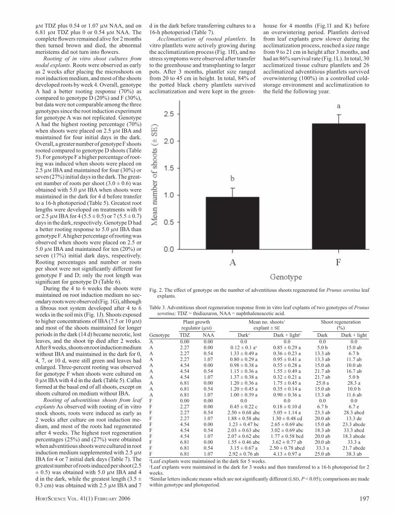
Two weeks after the explants with callus or with shoot bud primordia were transferred to elongation medium, the callus turned brown, shoot bud primordia turned green, and shoots started to differentiate and elongate (Fig.1D, E). The adventitious shoots were phenotypi-cally identical to the original explant shoots from each genotype. The genotype had a signifi cant effect (P > 0.0264) on the mean number of shoots regenerated, but there was no signifi cant effect (P > 0.1907) of the genotype on the percentage of regeneration (Table 2). Mean number of shoots for genotype F (2.33 ± 0.16) was 2.5-fold the mean number of shoots for genotype A (0.93 ± 0.08) (Fig. 2).
The effect of maintaining leaf cultures either in the dark for 5 weeks or in dark for 3 weeks followed by transfer to light for 2 weeks was not signifi cantly different for the mean number of shoots regenerated (P > 0.4964) or for the percentage of regeneration (P > 0.8958) (Table 2). The maximum mean number of shoots (5.05 ± 1.14) regenerated for genotype F was attained when cultures were maintained in dark for 3 weeks and then transferred to the light for 2 weeks on 2.27 µM TDZ plus 0.54 µM NAA (Table 3). For the same photoperiod, the greatest percent shoot regeneration (38.3) was obtained when cultures were placed on 6.81 µM TDZ plus 1.07 µM NAA. The high-est mean number of shoots (3.15 ± 0.67) and the highest percent shoot regeneration (33.3) occurred when explants were maintained in the dark for 5 weeks with 6.81 µM TDZ plus 0.54 µM NAA (Table 3).
For genotype A, although the treatments were not statistically different, the highest percent shoot regeneration (28.3) and the high-est mean number of shoots (1.75 ± 0.45) were observed when cultures were maintained in the
Fig. 1. Adventitious shoot regeneration from in vitro cultured leaves, in vitro rooting, and acclimatiza-tion of Prunus serotina plantlets. (A, B, and C) Adventitious bud primordia regenerated from callus on the midvein and margins of leaf explants (arrows). (D and E) Adventitious shoots on Murashige and Skoog (MS) medium in process of differentiation and elongation. (F) Flower from genotype A adventitious shoot after 3 months on MS medium. (G) Rooted microshoot 4 weeks after root induc-tion. (H) Plantlet in process of acclimatization (4 weeks after potting). I) Plantlets in the greenhouse (8 weeks after potting). (J) Fibrous root system of black cherry microshoot (6 weeks after potting). (K) Plantlets after transplanting to tall pots. (L) Adventitious shoots acclimatized to the greenhouse (12 weeks after potting).
FebruaryBook 1 195FebruaryBook 1 195 12/14/05 10:57:39 AM12/14/05 10:57:39 AM

HORTSCIENCE VOL. 41(1) FEBRUARY 2006196
dark for 3 weeks and transferred to the light for 2 weeks on media supplemented with 6.81 µM TDZ plus 0 µM NAA. The highest percent shoot regeneration (25) attained when cultures were maintained in the dark for 5 weeks occurred on media supplemented with 6.81 µM TDZ plus 0 µM NAA, and the greatest mean number of shoots (1.37 ± 0.38) was attained with 4.54 µM TDZ plus 1.07 µM NAA (Table 3).
Thidiazuron had a signifi cant effect on the number of shoots (P < 0.0183) and shoot regeneration percentages (P < 0.0013) for both genotypes (Table 2). The percent regeneration for the TDZ concentrations 2.27, 4.54, and 6.81 µM were signifi cantly different from each other. The regeneration increased with an increasing concentration of TDZ (Fig. 3). The mean number of shoots regenerated per explant also increased with an increasing concentration of TDZ. Thidiazuron at 6.81 µM was signifi cantly different from 2.27 µM, but not from 4.54 µM (Fig. 3).
A high variability of shoot regeneration from leaf segments was observed within each treatment. The greatest mean number of shoots per explant for genotype F (5.05 ± 1.14; from 60 leaf explants) occurred on media supplemented with 2.27 µM TDZ plus 0.54 µM NAA (Table 3) when cultures were maintained for 3 weeks in the dark and 2 weeks in a 16-h photoperiod, as compared to the maximum number of shoots regenerated (34) from an individual leaf.
The BA plus NAA factorial experiment produced no shoot regeneration for genotype D (data not shown) and very low shoot re-generation for genotype A and F (Table 4). A cluster of shoot bud primordia was observed when genotype F explants were placed on the highest treatment combination, 13.32 µM BA plus 5.37 µM NAA, and maintained in the dark for 3 weeks and then transferred to a 16-h photoperiod for 2 weeks. Genotype A was more
responsive compared to genotype F. Shoot bud primordia were regenerated for genotype A when explants were placed on 13.32 µM BA plus 0.54, 1.07, or 5.37 µM NAA (Table 4). A maximum of six differentiated and elongated shoots from 60 explants were obtained when leaf explants were placed on media supple-mented with 13.32 µM BA plus 1.07 µM NAA and maintained in the dark for 3 weeks before transfer to the light for 2 weeks.
For both A and F genotypes, cultures on media supplemented with BA plus NAA had less callus (percent coverage) compared to cultures on media supplemented with TDZ plus NAA (Tables 1 and 4). Less callus was observed when 0 µM BA in combination with any level of NAA or when 0 µM NAA with
any BA level was incorporated into the me-dia. An increase in callus was obtained with increasing concentrations of auxin with each BA level (Table 4).
Nine weeks after transfer to shoot elonga-tion medium, adventitious shoots of genotype A fl owered in vitro. Abnormal meristems, complete (Fig. 1F), and incomplete fl owers were observed after the second subculture for replicate six and after the seventh subculture for replicates three and four. Complete and incom-plete fl owers were observed on adventitious shoots induced on 2.27 µM TDZ or 4.54 µM TDZ plus 0.54 µM NAA. Incomplete fl owers, abnormal apical and axillary meristems were also observed from adventitious shoots induced on 2.27 µM TDZ plus 0 or 1.07 µM NAA, 4.54
Table 1. Callus induction and adventitious shoot regeneration response for two genotypes of Prunus serotina in vitro leaf explants as affected by thidiazuron (TDZ) and naphthaleneacetic acid (NAA).
Plant growth Genotype A Genotype F regulator Callus coverage Shoot Callus coverage Shoot (µM) (0 to 4)z regenerationy (0 to 4) regenerationBA NAA Darkx Dark + light Dark Dark + light Dark Dark + light Dark Dark + light0.00 0.00 0 w 0 – – 0 0 – –0.00 0.54 1 1 – – 1 1 – –0.00 1.07 2 2 – – 2 2 – –0.00 5.37 2 2 – – 2 1,2 – –2.27 0.00 2 2 + + 2 2 + +2.27 0.54 4 4 + + 3 3,4 + +2.27 1.07 4 4 + + 4 3,4 + +2.27 5.37 2,3 2 – – 4 4 – –4.54 0.00 3 3 + + 3 3 + +4.54 0.54 4 4 + + 4 3,4 + +4.54 1.07 4 3,4 + + 4 3,4 + +4.54 5.37 2,4 2,3 – – 4 3,4 – –6.81 0.00 3 2,3 + + 3 2,3 + +6.81 0.54 4 4 + + 4 4 + +6.81 1.07 4 4 + + 4 4 + +6.81 5.37 3,4 2,3,4 – – 4 3,4 – –zClassifi cation of callus coverage on leaf explants: 0 = 0% callus, 1= 25% callus (at midvein ends), 2 = 50% callus (leaf perimeter covered with callus), 3 = 75% of leaf covered with callus, and 4 = 100% callus.yShoot regeneration (+); No shoot regeneration (–).xDark = cultures maintained in the dark for 5 weeks; Dark + Light = cultures maintained in the dark for 3 weeks then transferred for 2 weeks to a 16-h photo-period.wCallus coverage observed on leaf explants for each plant growth regulator combination; n = 60.
Table 2. Analysis of variance for mean number of adventitious shoots regenerated and percentage of regeneration for Prunus serotina leaf explants; TDZ = thidiazuron, NAA = naphthaleneacetic acid.
Analysis of varianceSource Shoots Regenerationof (no.) (%)variation df Mean square F Mean square FGenotype 1 61.5879 6.78 * 1.3669 1.97Error (a) 10 9.0898 3.35 ** 0.6957 5.23 **NAA 2 5.2723 1.94 0.1343 1.01Genotype × NAA 2 5.7072 2.10 0.2974 2.24TDZ 2 10.9789 4.04 * 0.8961 6.74 **Genotype × TDZ 2 2.6935 0.99 0.1269 0.95TDZ × NAA 4 5.1766 1.91 0.2983 2.24Genotype × NAA × TDZ 4 3.2307 1.19 0.1089 0.82Light 1 1.2592 0.46 0.0022 0.02Genotype × light 1 7.0213 2.58 0.2261 1.70NAA × light 2 2.6367 0.97 0.1301 0.98Genotype × NAA × light 2 0.2536 0.09 0.0198 0.15TDZ × Light 2 0.1757 0.06 0.0387 0.29Genotype × TDZ × light 2 1.0574 0.39 0.1291 0.97TDZ × NAA × light 4 3.4061 1.25 0.1117 0.84Genotype × TDZ × NAA × light 4 2.7544 1.01 0.0786 0.59Error (b) 386 2.7165 2.90 ** 0.1329Leaf error (b') 1728 0.9366*,**Signifi cant at P < 0.05 or 0.01 respectively; n = 60.
FebruaryBook 1 196FebruaryBook 1 196 12/14/05 10:57:43 AM12/14/05 10:57:43 AM
197HORTSCIENCE VOL. 41(1) FEBRUARY 2006
µM TDZ plus 0.54 or 1.07 µM NAA, and on 6.81 µM TDZ plus 0 or 0.54 µM NAA. The complete fl owers remained alive for 2 months then turned brown and died, the abnormal meristems did not turn into fl owers.
Rooting of in vitro shoot cultures from nodal explants. Roots were observed as early as 2 weeks after placing the microshoots on root induction medium, and most of the shoots developed roots by week 4. Overall, genotype A had a better rooting response (70%) as compared to genotype D (20%) and F (30%), but data were not comparable among the three genotypes since the root induction experiment for genotype A was not replicated. Genotype A had the highest rooting percentage (70%) when shoots were placed on 2.5 µM IBA and maintained for four initial days in the dark. Overall, a greater number of genotype F shoots rooted compared to genotype D shoots (Table 5). For genotype F a higher percentage of root-ing was induced when shoots were placed on 2.5 µM IBA and maintained for four (30%) or seven (27%) initial days in the dark. The great-est number of roots per shoot (3.0 ± 0.6) was obtained with 5.0 µM IBA when shoots were maintained in the dark for 4 d before transfer to a 16-h photoperiod (Table 5). Greatest root lengths were developed on treatments with 0 or 2.5 µM IBA for 4 (5.5 ± 0.5) or 7 (5.5 ± 0.7) days in the dark, respectively. Genotype D had a better rooting response to 5.0 µM IBA than genotype F. A higher percentage of rooting was observed when shoots were placed on 2.5 or 5.0 µM IBA and maintained for ten (20%) or seven (17%) initial dark days, respectively. Rooting percentages and number or roots per shoot were not signifi cantly different for genotype F and D; only the root length was signifi cant for genotype D (Table 6).
During the 4 to 6 weeks the shoots were maintained on root induction medium no sec-ondary roots were observed (Fig. 1G), although a fi brous root system developed after 4 to 6 weeks in the soil mix (Fig. 1J). Shoots exposed to higher concentrations of IBA (7.5 or 10 µM) and most of the shoots maintained for longer periods in the dark (14 d) became necrotic, lost leaves, and the shoot tip died after 2 weeks. After 8 weeks, shoots on root induction medium without IBA and maintained in the dark for 0, 4, 7, or 10 d, were still green and leaves had enlarged. Three-percent rooting was observed for genotype F when shoots were cultured on 0 µM IBA with 4 d in the dark (Table 5). Callus formed at the basal end of all shoots, except on shoots cultured on medium without IBA.
Rooting of adventitious shoots from leaf explants As observed with rooting of in vitro stock shoots, roots were induced as early as 2 weeks after culture on root induction me-dium, and most of the roots had regenerated after 4 weeks. The highest root regeneration percentages (25%) and (27%) were obtained when adventitious shoots were cultured in root induction medium supplemented with 2.5 µM IBA for 4 or 7 initial dark days (Table 7). The greatest number of roots induced per shoot (2.5 ± 0.5) was obtained with 5.0 µM IBA and 4 d in the dark, while the greatest length (3.5 ± 0.3 cm) was obtained with 2.5 µM IBA and 7
d in the dark before transferring cultures to a 16-h photoperiod (Table 7).
Acclimatization of rooted plantlets. In vitro plantlets were actively growing during the acclimatization process (Fig. 1H), and no stress symptoms were observed after transfer to the greenhouse and transplanting to larger pots. After 3 months, plantlet size ranged from 20 to 45 cm in height. In total, 84% of the potted black cherry plantlets survived acclimatization and were kept in the green-
house for 4 months (Fig.1I and K) before an overwintering period. Plantlets derived from leaf explants grew slower during the acclimatization process, reached a size range from 9 to 21 cm in height after 3 months, and had an 86% survival rate (Fig.1L). In total, 30 acclimatized tissue culture plantlets and 26 acclimatized adventitious plantlets survived overwintering (100%) in a controlled cold-storage environment and acclimatization to the fi eld the following year.
Fig. 2. The effect of genotype on the number of adventitious shoots regenerated for Prunus serotina leaf explants.
Table 3. Adventitious shoot regeneration response from in vitro leaf explants of two genotypes of Prunus serotina; TDZ = thidiazuron, NAA = naphthaleneacetic acid.
Plant growth Mean no. shoots/ Shoot regeneration regulator (µM) explant ± SE (%)Genotype TDZ NAA Darkz Dark + lighty Dark Dark + lightA 0.00 0.00 0.0 0.0 0.0 0.0A 2.27 0.00 0.12 ± 0.1 ax 0.85 ± 0.29 a 5.0 b 15.0 abA 2.27 0.54 1.33 ± 0.49 a 0.36 ± 0.23 a 13.3 ab 6.7 bA 2.27 1.07 0.80 ± 0.29 a 0.95 ± 0.41 a 13.3 ab 11.7 abA 4.54 0.00 0.98 ± 0.38 a 0.55 ± 0.28 a 15.0 ab 10.0 abA 4.54 0.54 1.15 ± 0.36 a 1.55 ± 0.49 a 21.7 ab 16.7 abA 4.54 1.07 1.37 ± 0.38 a 0.32 ± 0.21 a 21.7 ab 5.0 bA 6.81 0.00 1.20 ± 0.36 a 1.75 ± 0.45 a 25.0 a 28.3 aA 6.81 0.54 1.20 ± 0.45 a 0.35 ± 0.14 a 15.0 ab 10.0 bA 6.81 1.07 1.00 ± 0.39 a 0.90 ± 0.36 a 13.3 ab 11.6 abF 0.00 0.00 0.0 0.0 0.0 0.0F 2.27 0.00 0.45 ± 0.22 c 0.18 ± 0.10 d 6.7 b 6.7 eF 2.27 0.54 2.50 ± 0.68 abc 5.05 ± 1.14 a 23.3 ab 28.3 abcdF 2.27 1.07 1.88 ± 0.58 abc 1.30 ± 0.48 cd 20.0 ab 13.3 deF 4.54 0.00 1.23 ± 0.47 bc 2.65 ± 0.69 abc 15.0 ab 23.3 abcdeF 4.54 0.54 2.03 ± 0.63 abc 3.02 ± 0.69 abc 18.3 ab 33.3 abcdF 4.54 1.07 2.07 ± 0.62 abc 1.77 ± 0.58 bcd 20.0 ab 18.3 abcdeF 6.81 0.00 1.55 ± 0.46 abc 3.62 ± 0.77 ab 20.0 ab 33.3 aF 6.81 0.54 3.15 ± 0.67 a 2.50 ± 0.78 abcd 33.3 a 21.7 abcdeF 6.81 1.07 2.92 ± 0.76 ab 4.13 ± 0.97 a 25.0 ab 38.3 abzLeaf explants were maintained in the dark for 5 weeks.yLeaf explants were maintained in the dark for 3 weeks and then transferred to a 16-h photoperiod for 2 weeks.xSimilar letters indicate means which are not signifi cantly different (LSD, P < 0.05); comparisons are made within genotype and photoperiod.
FebruaryBook 1 197FebruaryBook 1 197 12/14/05 10:57:46 AM12/14/05 10:57:46 AM

HORTSCIENCE VOL. 41(1) FEBRUARY 2006198
Discussion
Adventitious shoots were regenerated from Prunus serotina leaf explants and rooted in vitro. Rooted microshoots from nodal explant-derived stock cultures and leaf explant-derived adventitious shoots actively grew throughout the acclimatization period, survived under greenhouse conditions, and were success-fully overwintered. This established protocol demonstrates that black cherry may be used for clonal propagation of selected genotypes and gives a starting point for research on genetic transformation and regeneration for improve-ment of this high value hardwood.
A difference in adventitious shoot regen-eration was observed between plant growth regulator combinations of BA plus NAA (2%) and TDZ plus NAA (38.3%). Although a wider range and higher levels of BA were used com-
pared to previous studies on Prunus species, a poor effect of BA on shoot regeneration was observed. A signifi cantly higher shoot regen-eration from P. serotina leaves was obtained by Hammatt and Grant (1998) with 4.44 µM TDZ (70%) compared to 4.44 µM BA (29%). Medium supplemented with TDZ successfully regenerated (40%) P. dulcis adventitious shoots compared to medium supplemented with BA, which had no effect on regeneration (Miguel et al., 1996). The same trend was observed with P. armeniaca and P. avium, where TDZ had a greater effect of promoting regeneration (24%, 71%) compared to BA (3%, 20%) (Bhagwat and Lane, 2004; Perez-Tornero et al., 2000). The greatest percent regeneration (38.3%) in our study was obtained when cultures were placed on 6.81 µM TDZ plus 1.07 µM NAA. Although TDZ proves to be a more effective and potent cytokinin in our study, Tang et al.
(2002) reported that BA (40%) was more effec-tive than TDZ (8%) on shoot regeneration of P. cerasus and P. avium. Similarly, Gentile et al. (2002) reported that BA was highly effective compared to TDZ in promoting organogenesis from all P. persica genotypes tested.
The signifi cant effect of TDZ observed on percent regeneration and number of shoots regenerated, as well as the nonsignifi cant ef-fect of NAA agrees with results reported by Perez-Tornero et al. (2000) with P. armeniaca. This contrasts with the results of leaf segment experiments of Bhagwat and Lane (2004), where NAA had a signifi cant effect and TDZ did not. These varied results may be a result of an effect of different species, genotypes, various levels of auxin plus cytokinin combinations, and the interactions with the environmental conditions. Because the highest percent regen-eration (38.3%) and number of shoots (4.13 ± 0.97) regenerated were observed when the highest level of TDZ (6.81 µM) was used, a higher concentration of TDZ may increase the regeneration percent and the number of shoots regenerated per explant. For P. armeniaca, the highest regeneration (24.3 ± 5.1) was attained with 9 µM TDZ (Perez-Tornero et al., 2000). Ainsley et al. (2001b) reported that increasing concentrations of TDZ resulted in increasing regeneration frequencies and number of shoots regenerated for P. dulcis, but the highest level (20 µM) inhibited shoot elongation and increased the probability of obtaining hyperhydric shoots. It may be necessary to study the higher levels of TDZ that can be used without sacrifi cing other aspects such as shoot elongation or shoot multiplication.
Photoperiod is another important factor that could account for differences in regeneration of adventitious shoots. The highest percentage of regeneration (38.3%) and number of shoots regenerated per explant (5.05 ± 1.14) were observed when cultures were maintained in the dark for 3 weeks and then transferred to a 16-h photoperiod for 2 weeks. These results are in agreement with previous reports on adventi-tious shoot regeneration of Prunus species. Previous studies have shown the dark period before a 16-h photoperiod to be critical for regeneration, because it can stimulate organo-genesis by infl uencing the levels of endogenous hormones such as indoleacetic acid to interact with exogenously applied growth regulators and promote adventitious shoot regeneration (Gentile et al., 2002, 2003; Miguel et al., 1996; Pooler and Scorza, 1995). Perez-Tornero et al. (2000) also reported a signifi cant effect of the dark period on the regeneration percentage, and higher regeneration occurred with 2 or 3 weeks (30%) compared to 1 or 4 weeks (<10%) of dark incubation before a 16-h photoperiod.
It is widely known that regeneration percent-ages are affected by the genetics of the stock plant. The differences observed in regenera-tion between genotypes A, D, and F was also reported for fi ve genotypes of P. serotina leaves cultured on WPM and on Driver and Kuniyuki walnut medium (Hammatt and Grant, 1998). Genotypic effects were also reported for other Prunus species (Ainsley et al., 2000, 2001b; Bassi and Cossio, 1991; Escalettes and Dosba,
Fig. 3. The effect of TDZ concentrations on the number of adventitious shoots regenerated and on percent regeneration for Prunus serotina leaf explants.
FebruaryBook 1 198FebruaryBook 1 198 12/14/05 10:57:50 AM12/14/05 10:57:50 AM

199HORTSCIENCE VOL. 41(1) FEBRUARY 2006
1993; Gentile et al., 2002; Grant and Hammatt, 2000; Perez-Tornero et al., 2000). For this rea-son, there is the need for experimenting with various genotypes under identical conditions in order to be able to select the genotype that gives the best response.
Adventitious shoots regenerated from fi rm, compact callus, but not from friable callus, that had developed on the wounded and midrib surfaces. Grant and Hammatt (2000) reported
that leaf explants that produced only friable callus had no shoot regeneration. Our results also showed that no shoot regeneration was obtained from leaf explants on treatments that induced only friable callus, and low regenera-tion from genotype D which produced a type of callus more friable as compared to the other two genotypes.
A higher regeneration from genotype F may be attributed not only to a genetic effect, but
to a leaf size effect. In general, microshoots from genotype F had larger leaves compared to genotype A or D. The effect on regeneration attributed to leaf size was reported for P. avium (Grant and Hammatt, 2000). Very small leaves (<3 mm2) were found to produce only friable callus and no regeneration of adventitious shoots, whereas leaves (3 to 5 mm2) resulted in 70% regeneration. Larger leaves (5 to 10 mm2) had a low percent of regeneration (1%). In other
Table 4. Callus induction and adventitious shoot regeneration response for two genotypesof Prunus serotina in vitro leaf explants as affected by 6-benzylaminopurine (BA) plus naphthaleneacetic acid (NAA).
Plant growth Genotype A Genotype F regulator Callus coverage Shoot Callus coverage Shoot (µM) (0 to 4)z regenerationy (0 to 4) regenerationBA NAA Darkx Dark + light Dark Dark + light Dark Dark + light Dark Dark + light0.00 0.54 1w 1 – – 0,1 0 – –0.00 1.07 1 1 – – 0,1 0 – –0.00 5.37 1,2 1,2 – – 0,1 0,1 – –4.44 0.00 1 1 – – 1,2 1 – –4.44 0.54 2,3 2,3 – – 2,3 2,3 – –4.44 1.07 2,3 2,3 – – 3 2,3 – –4.44 5.37 3,4 3,4 – – 3,4 3 – –8.88 0.00 1 1 – – 1,2 1 – –8.88 0.54 2,3 2,3 – – 2,3 2 – –8.88 1.07 4 3 – – 3,4 2,4 – –8.88 5.37 3,4 3,4 – – 3,4 3,4 – –13.32 0.00 1 1 – – 1 1 – –13.32 0.54 2,3 2,3 + – 2,3 2 – –13.32 1.07 3,4 2,3 + + 3 2,3 – –13.32 5.37 3,4 3 + – 3,4 3 – +zClassifi cation of callus coverage on leaf explants: 0 = 0% callus, 1= 25% callus (at midvein ends), 2 = 50% callus (leaf perimeter covered with callus), 3 = 75% of leaf covered with callus, and 4 = 100% callus.yShoot regeneration (+); No shoot regeneration (–).xDark = cultures maintained in the dark for 5 weeks; Dark + Light = cultures maintained in the dark for 3 weeks then transferred for 2 weeks to a 16-h photo-period.wCallus coverage observed on leaf explants for each plant growth regulator combination; n = 60.
Table 5. In vitro rooting response for Prunus serotina microshoots from nodal explants; IBA = indole-3-butyric acid.
Rooting Roots/shoot Root length (%) ± SE (cm) ± SE
IBA Dark daysz Genotype(µM) (no.) Ay D F A D F A D F0.0 0 ---x --- --- --- --- --- --- --- ---0.0 4 --- --- 3 --- - 2.0 --- --- 5.5 ± 0.50.0 7 --- --- --- --- --- --- --- --- ---0.0 10 --- --- --- --- --- --- --- --- ---0.0 14 --- --- --- --- --- --- --- --- ---2.5 0 20 --- 10 1.0 --- 1.7 ± 0.3 2.2 ± 1.0 --- 3.7 ± 1.22.5 4 70 10 30 2.7 ± 0.9 2.7 ± 1.7 1.9 ± 0.4 2.9 ± 1.0 4.6 ± 0.8 4.0 ± 0.52.5 7 60 7 27 3.4 ± 1.8 1.0 1.4 ± 0.3 4.0 ± 0.4 1.8 ± 0.2 5.5 ± 0.72.5 10 20 20 13 2.5 ± 0.5 2.7 ± 0.6 1.5 ± 0.3 3.5 ± 0.5 2.6 ± 0.5 4.2 ± 1.02.5 14 50 7 13 3.4 ± 0.9 4.0 ± 2.0 1.3 ± 0.3 1.3 ± 0.4 1.0 ± 0.2 2.8 ± 0.95.0 0 10 --- --- 1.0 --- --- 4.0 --- ---5.0 4 --- 3 10 --- 2.0 3.0 ± 0.6 --- 0.6 2.3 ± 0.45.0 7 --- 17 --- --- 2.2 ± 0.5 --- --- 0.8 ± 0.2 ---5.0 10 --- 10 --- --- 2.0 ± 0.6 --- --- 1.0 ± 0.2 ---5.0 14 --- --- --- --- --- --- --- --- ---7.5 0 NAw --- --- NA --- --- NA --- ---7.5 4 NA --- --- NA --- --- NA --- ---7.5 7 NA --- --- NA --- --- NA --- ---7.5 10 NA --- --- NA --- --- NA --- ---7.5 14 NA --- --- NA --- --- NA --- ---10.0 0 NA --- --- NA --- --- NA --- ---10.0 4 NA --- --- NA --- --- NA --- ---10.0 7 NA --- --- NA --- --- NA --- ---10.0 10 NA --- --- NA --- --- NA --- ---10.0 14 NA --- --- NA --- --- NA --- ---zShoots were placed on IBA media and maintained in the dark for 0, 4, 7, 10, or 14 d before transfer to a 16-h photoperiod.yAs a result of the number of shoots obtained from genotype A that were available for root induction, the experiment was only conducted once.xNo response.wNA = does not apply, shoots were not placed on these treatments because of the number of shoots available for root induction and based on results from geno-types D and F; n = 30.
FebruaryBook 1 199FebruaryBook 1 199 12/14/05 10:57:53 AM12/14/05 10:57:53 AM

HORTSCIENCE VOL. 41(1) FEBRUARY 2006200
studies wounded, small, immature leaves from shoot tips produced callus, but no adventitious shoots (Bhagwat and Lane, 2004).
The high variability of shoot regeneration observed within each petri plate may be the result of the variation of organogenic capacity of leaves depending on their position on the shoot and on segments from different parts of the leaf. The leaves were randomly chosen from different parts of the microshoots, as op-posed to using only the youngest leaves that may have higher organogenic competence. Using only the fi rst expanded leaves has been reported previously for Prunus species (da Camara Machado et al., 1989; Druart, 1990; Gentile et al., 2002, 2003). A 2-fold increase in regeneration was reported for P. armeniaca when using young leaves compared to older leaves (Perez-Tornero et al., 2000). Because the regenerative capacity of cotyledon segments from P. cerasus was higher at the proximal end compared to segments median and distal to the shoot, a higher hormone level was needed for the latter (Tang et al., 2000). For P. avium, whole leaves wounded along the midrib and placed on medium supplemented with TDZ and NAA had a better response to shoot regeneration (71%) as compared to leaf segments (36%) (Bhagwat and Lane, 2004). Other factors that may increase the regenera-tion response may be the use of petiole tissue as part of the explant. It has been reported that leaf explants from several Prunus species that included the petiole tissue resulted in higher adventitious shoot regeneration (Escalettes and Dosba, 1993; Gentile et al., 2002, 2003; Miguel et al., 1996). A higher regeneration has been attained from the cut near the petiole of the leaf compared to apical, middle, and petiole cuts of P. canescens (Druart, 1990; Perez-Tornero et al., 2000). Given that the adventitious shoots
regenerated not only from the midvein, but from the wounded margins of leaf explants, inducing adventitious shoots of P. serotina using com-plete leaves including the petiole, wounding across the midvein, and along the edges of the leaf may confer a higher regeneration suitable for genetic transformation. Prunus avium and P. cerasus adventitious shoots regenerated directly from the midvein or in association with vascular tissues, whereas few shoots regenerated from callus (Tang et al., 2002). Contrasting, in our study as in other reports (Antonelli and Druart, 1990; Perez-Tornero et al., 2000), the adventitious shoots regenerated from callus that formed on the leaf segment wounded margins or on the midvein. For this reason, transferring callus with or without visible clusters of shoot bud primordia to MS medium and separation of the shoots from the clusters was essential for full development and elongation of the adventitious shoots. Evidence of this practice that resulted in a higher number of elongated shoots was reported by Bhagwat and Lane (2004), where they obtained only a few shoots to elongate when clusters were not separated, as compared to 43 shoots obtained when the clusters were separated.
The highest rooting percentage (70%) and number of roots per shoot (2.7 ± 0.9) were comparable to other reports for P. serotina in vitro root induction. Fuernkranz et al. (1990) reported 70% to 74% rooting and 2.5 to 2.8 roots per shoot with different light intensities of white light. These authors also report a greater percentage of rooting (97%) was attained when cultures were maintained under yellow, low intensity light. Tricoli et al. (1985) attained a higher rooting percentage (94%) with P. serotina as compared to our study. Rooting response variations may be affected by dif-ferent conditions of the shoots used for root
Table 7. In vitro rooting response for Prunus serotina genotype F adventitious shoots; IBA = indole-3-butyric acid.
IBA Dark days Rooting Roots/shoot Root length(µM) (no.)z (%) (no.) ± SE (cm) ± SE
0.0 0 0.0 0.0 0.02.5 0 2 1.0 ± 0 2.0 ± 02.5 4 25 1.5 ± 0.2 2.6 ± 0.42.5 7 27 2.3 ± 0.2 3.5 ± 0.35.0 0 0 0.0 0.05.0 4 4 2.5 ± 0.5 2.3 ± 0.55.0 7 9 1.2 ± 0.2 2.9 ± 0.2zShoots were placed on IBA media and maintained in the dark for 0, 4, or 7 d before transfer to a 16-h photoperiod.
Table 6. Analysis of variance for rooting percent, number of roots per shoot, and root length for shoots from nodal explants of genotypes D and F of Prunus serotina.
Genotype D Genotype FVariable df Mean squares F P df Mean squares F PRooting (%) Replication 2 0.4263 25.70 <0.0001 2 0.0435 0.83 0.461 Treatment 6 0.0399 2.41 0.0918 6 0.0605 1.03 0.449 Error 12 0.0166 12 0.0585Roots per shoot (no.) Replication 2 1.3457 0.44 0.6556 2 0.9181 1.38 0.272 Treatment 5 1.7547 0.57 0.7228 5 1.1157 1.67 0.181 Error 13 3.0853 23 0.6671Root length Replication 2 0.0227 0.03 0.9746 2 2 15.535 7.84 Treatment 5 7.0112 7.95 0.0013 5 5 4.2751 2.16 Error 13 0.8823 23 1.9820
induction, variations in the medium used for multiplication before root induction, number of subcultures before root induction, and the number of days maintained on the multiplica-tion medium before transfer to root induction medium. The protocol for rooting P. serotina recommended by Gerhold and Barbour (1991) includes the careful selection of shoots 60 to 90 d after culture initiation. The differences in rooting response among reports may also be a result of the genotype or culture conditions. Fuernkranz et al. (1990) reported that light intensity and spectral quality of the light had a signifi cant effect on root formation and growth. In contrast to other reports, we obtained a better response with lower concentration of IBA as opposed to higher concentrations. The high-est percent rooting was attained on medium supplemented with 2.5 µM IBA for 4 (70%) or 7 (60%) d of dark incubation before a 16-h photoperiod. Research on root induction of microshoots report the highest percent rooting after 14 d on root induction medium (94% or 80%) using higher concentrations of IBA (4.9 or 7.35 µM) and maintained in continuous dark-ness or in a 16-h photoperiod plus 10–3 M rutin (Tricoli et al., 1985). Fuernkranz et al. (1990) and Eliasson et al. (1994) report responses to light and paclobutrazol respectively, based on 5 µM IBA for root induction.
The percent of rooted plantlets that sur-vived transfer to the greenhouse in our study was greater (84% to 86%) compared to 10% (Tricoli et al., 1985), or <60% reported by Eliasson et al. (1994) without paclobutrazol. Treatments reported to increase stem elonga-tion, and increase the percent survival of black cherry, such as paclobutrazol, cold treatments, and GA
3 (Eliasson et al., 1994; Kavanagh
et al., 1993; Maynard et al., 1991) were not used in this study. A higher percent survival and continuous growth of the plantlets during acclimatization were attained for both rooted in vitro microshoots and rooted adventitious shoots, proving that black cherry can be ac-climatized without complications.
The in vitro fl owering observed on the adventitious shoots regenerated for genotype A was not expected. Although the shoots were maintained and subcultured for 8 months, the phenomenon did not reoccur. Not all the fl owers appeared to be complete fl owers. The in vitro fl owering phenomenon from committed fl ower parts or from non-committed parts has been recorded for herbaceous and some woody spe-cies (George, 1993). Several factors that may promote or inhibit in vitro fl owering, such as cytokinins, auxins, gibberellins, carbohydrates, photoperiod, vernalization, and the physical state of the medium have been described; suggesting that no substance alone causes fl owering, but may result from an interaction of the promoters, inhibitors, and plant species (Scorza, 1982).
This report of a complete protocol for adventitious shoot regeneration, rooting, and acclimatization of black cherry plantlets is a key step for future research on genetic transformation and clonal propagation. As it was revealed in this research and previously reported (Antonelli and Druart, 1990; Bassi and
FebruaryBook 1 200FebruaryBook 1 200 12/14/05 10:57:55 AM12/14/05 10:57:55 AM

201HORTSCIENCE VOL. 41(1) FEBRUARY 2006
Cossio, 1991; Gentile et al., 2002) adventitious shoot regeneration of Prunus species is affected by numerous factors. Further research needs to be conducted to study these factors involved in adventitious shoot regeneration and rooting of P. serotina in order to optimize protocols for use in genetic transformation. In vitro shoot cultures from elite, mature black cherry selec-tions have been established and will be used to further optimize this regeneration system.
Literature Cited
Ahuja, M.R. 2000. Genetic engineering in forest trees: State of the art and future perspectives, p. 31–49. In: S.M. Jain and S.C. Minocha (eds.). Molecular biology of woody plants. vol.1. Klu-wer Academic Publ., The Netherlands.
Ainsley, P.J., G.C. Collins, and M. Sedgley. 2000. Adventitious shoot regeneration from leaf ex-plants of almond (Prunus dulcis Mill.). In Vitro Cell. Dev. Biol.–Plant 36:470–474.
Ainsley, P.J., G.G. Collins, and M. Sedgley. 2001a. In vitro rooting of almond (Prunus dulcis Mill.). In Vitro Cell. Dev. Biol.–Plant 37:778–785.
Ainsley, P.J., F.A. Hammerschlag, T. Bertozzi, G.G. Collins, and M. Sedgley. 2001b. Regeneration of almond from immature seed cotyledons. Plant Cell Tiss. Org. Cult. 67:221–226.
AL-Sabbagh, M., A. Abdul-Kader, M. Khoder, and A.R. Kalhout. 1999. In vitro propagation of a semi-dwarfi ng cherry rootstock. Plant Cell Tiss. Org. Cult. 59:203–208.
Antonelli, M. and A. Chiariotti. 1988. In vitro root-ing of different peach genotypes. Acta Hort. 227:405–407.
Antonelli, M. and P. Druart. 1990. The use of a brief 2, 4-D treatment to induce leaf regeneration on Prunus canescens bois. Acta Hort. 280:45–50.
Bassi, G. and F. Cossio. 1991. In vitro shoot regen-eration of ‘Bluefre’ and ‘Susina di Dro’ prune cultivars (Prunus domestica L.). Acta Hort. 289:81–82.
Bhagwat, B. and W.D. Lane. 2004. In vitro shoot regeneration from leaves of sweet cherry (Prunus avium) ‘Lapins’ and ‘Sweetheart’. Plant Cell Tiss. Org. Cult. 78:173–181.
Borkowska, B. 1985. Micropropagation of sour cherry, cultivar Schattenmorelle. Acta Hort. 169:329–333.
Brunner, A.M., R. Mohamed, R. Meilan, L.A. Shep-pard, W.H. Rottmann, and S.H. Strauss. 1998. Genetic engineering of sexual sterility in shade trees. J. Arboricult. 24(5):263–272.
Burgos, L. and N. Alburquerque. 2003. Ethylene inhibitors and low kanamycin concentrations improve adventitious regeneration from apricot leaves. Plant Cell Rpt. 21:1167–1174.
Channuntapipat, C., M. Sedgley, and G. Collins. 2003. Micropropagation of almond cultivars Nonpareil and Ne Plus Ultra and the hybrid rootstock Titan × Nemaguard. Scientia Hort. 98:473–484.
da Camara Machado, L.M., V. Hanzer, D. Mat-tanovich, D. Himmler, and H. Katinger. 1989. Regeneration of shoots from leaf discs and stem microcuttings of fruit trees as a tool for transformation. Acta Hort. 235:85–92.
Druart, P.H. 1990. Effect of culture conditions and leaf selection on organogenesis of Malus domes-tica cv. Mcintosh “wijcik” and Prunus canescens bois GM79. Acta Hort. 280:117–124.
Eliasson, M.K., C.A. Beyl, and P.A. Barker. 1994.
In vitro responses and acclimatization of Prunus serotina with paclobutrazol. J. Plant Growth Regulat. 13:137–142.
Escalettes, V. and F. Dosba. 1993. In vitro adventi-tious shoot regeneration from leaves of Prunus spp. Plant Sci. 90(2):201–209.
Forest Products Laboratory. 1999. Wood handbook-Wood as an engineering material. Gen. Tech. Rpt. FPL-GTR-113. U.S. Dept. of Agr., For. Serv., For. Prod. Lab., Madison, Wis.
Fouad, M.M., A.H. Goma, and M.H.A. El Zhar. 1995. Factors infl uencing rooting of peach shoots cultured in vitro. Acta Hort. 409:197–202.
Fuernkranz, H.A., C.A. Nowak, C.A. Maynard. 1990. Light effects on in vitro adventitious root formation in axillary shoots of mature Prunus serotina. Physiol. Plant. 80:337–341.
Gentile, A., S. Monticelli, and C. Damiano. 2002. Adventitious shoot regeneration in peach [Prunus persica (L.) Batsch]. Plant Cell Rpt. 20:1011–1016.
Gentile, A., S. Monticelli, and C. Damiano. 2003. Adventitious shoot regeneration for Agrobacte-rium-mediated transformation in Prunus. Acta Hort. 616:331–333.
George, E.F. 1993. Plant propagation by tissue culture. part 1, p. 243–245. Exegatics Ltd., Great Britain.
Gerhold, H.D. and W.W. Barbour. 1991. Selection of Prunus serotina for cloning by tissue culture. Proc. 1st N. For. Gen. Conf. 158–168.
Grant, N.J. and N. Hammatt. 1999. Increased root and shoot production during micropropagation of cherry and apple rootstocks: Effect of subculture frequency. Tree Physiol. 19:899–903.
Grant, N.J. and N. Hammatt. 2000. Adventitious shoot development from wild cherry (Prunus avium L.) leaves. New For. 20:287–295.
Hammatt, N. 1993. Micropropagation of fastigate bird cherry (Prunus padus L.) and adventi-tious shoot formation from leaves. J. Hort. Sci. 68(6):975–981.
Hammatt, N. and N.J. Grant. 1998. Shoot regenera-tion from leaves of Prunus serotina Ehrh. (black cherry) and P. avium (wild cherry). Plant Cell Rpt. 17:526–530.
Harada, H. and Y. Murai. 1996. Micropropagation of Prunus mume. Plant Cell Tiss. Org. Cult. 46:265–267.
Hoover, W.L. and G. Preston. 2003. Indiana forest products price report and trend analysis. Purdue Univ., Dept. For. Nat. Resour. October 2004. FNR-177-W. http://www.ces.purdue.edu/extme-dia/FNR/FNR-177-W.pdf.
Kavanagh, K.L., D.H. Lee, A.P. Drew, and C.A. Maynard. 1993. The effects of GA
3 and organic
solvents on acclimatization of tissue culture propagated black cherry (Prunus serotina Ehrh.) plantlets. For. Sci. 39(4):644–654.
Lloyd, G. and B. McCown. 1981. Commercially-feasible micropropagation of mountain laurel, Kalmia latifolia, by use of shoot-tip culture. Proc. Intl. Plant Prop. Soc. 30:421–427.
Maynard, C.A. 1994. Six-year fi eld test results of micropropagated black cherry (Prunus serotina). In Vitro Cell. Dev. Biol.–Plant 30:64–69.
Maynard, C.A. and H.A. Fuernkranz. 1989. Shoot regeneration and Agrobacterium infection of Prunus serotina leaves. In Vitro Cell. Dev. Biol. –Plant 25(3–part II):30A.
Maynard, C.A., K. Kavanagh, H. Fuernkranz, and A.P. Drew. 1991. Black cherry (Prunus serotina Ehrh), p. 3–22. In: Y.P.S. Bajaj (ed.). Biotechnol-ogy in agriculture and forestry. vol. 16. Springer,
New York.Mehra, A. and P.N. Mehra. 1974. Organogenesis
and plantlet formation in vitro in almond. Bot. Gaz. 135(1):61–73.
Miguel, C.M., P. Duart, and M.M. Oliveira. 1996. Shoot regeneration from adventitious buds induced on juvenile and adult almond (Prunus dulcis Mill.) explants. In Vitro Cell. Dev. Biol.–Plant 32:148–153.
Morini, S. and S. Concetti. 1985. In vitro propa-gation of P.S B2 peach rootstock. Acta Hort. 173:205–209.
Murashige, T. and F. Skoog. 1962. A revised medium for rapid growth and bio assays with tobacco tissue cultures. Physiol. Plant. 15:473–497.
Nowak, B., K. Miczynski, and L. Hudy. 2004. Sugar uptake and utilization during adventitious bud differentiation on in vitro leaf explants of ‘We-gierka Zwykla’ plum (Prunus domestica). Plant Cell Tiss. Org. Cult. 76:225–260.
Perez-Tornero, O. and L. Burgos. 2000. Different media requirements for micropropagation of apricot cultivars. Plant Cell Tiss. Org. Cult. 63:133–141.
Perez-Tornero, O., J. Egea, A. Vanoostende, and L. Burgos. 2000. Assessment of factors affecting adventitious shoot regeneration from in vitro cul-tured leaves of apricot. Plant Sci. 158:61–70.
Pooler, M.R. and R. Scorza. 1995. Regeneration of peach [Prunus persica (L.) Batsch] rootstock cultivars from cotyledons of mature stored seed. HortScience 30(2):355–356.
Pruski, K.W., T. Lewis, T. Astatkie, and J. Nowak. 2000. Micropropagation of chokecherry and pincherry cultivars. Plant Cell Tiss. Org. Cult. 63:93–100.
Ranjit, M. and D.E. Kester. 1988. Micropropaga-tion of cherry rootstocks: II. Invigoration and enhanced rooting of ’46-1 Mazzard’ by co-culture with ‘Colt’. J. Amer. Soc. Hort. Sci 113(1):150–154.
Rexrode, C.O. and J.E. Baumgras. 1984. Distribution of gum spots by causal agent in black cherry and effects on log and tree quality. S. J. Appl. For. 8(1):22–28.
Sasaki, Y. 2003. Micropropagation of Prunus sargen-tii REHDER and P. verecunda (KOIDZ) Koehe, p. 507–518. In: S.M. Jain and K. Ishii (eds.). Mi-cropropagation of woody trees and fruits. Kluwer Academic Publishers, The Netherlands.
Scorza, R. 1982. In vitro fl owering. Hort. Rev. 4:106–127.
SAS Institute. 1999. The SAS system for Windows. v. 8.2. SAS Inst., Cary, N.C.
Tang, H., Z. Ren, and G. Krczal. 2000. Somatic em-bryogenesis and organogenesis from immature embryo cotyledons of three sour cherry cultivars (Prunus cerasus L.). Sci. Hort. 83:109–126.
Tang, H., Z. Ren, G. Reustle, and G. Krczal. 2002. Plant regeneration from leaves of sweet and sour cherry cultivars. Sci. Hort. 93:235–244.
Theiler-Hedtrich, C.M. and W. Feucht. 1985. Micro-propagation of Prunus cerasus rootstocks-infl u-ence of culture medium constituents on growth stage I and II. Acta Hort. 169:335–340.
Tricoli, D.M., C.A. Maynard, and A. Drew. 1985. Tissue culture of propagation of mature trees of Prunus serotina Ehrh. I. Establishment, multiplication, and rooting in vitro. For. Sci. 31(1):201–208.
Zhang, Z., W.H. Dai, Z.M. Cheng, and J.A. Walla. 2000. A shoot tip-culture micropropagation system for chokecherry. J. Environ. Hort. 18(4):234–237.
FebruaryBook 1 201FebruaryBook 1 201 12/14/05 10:57:58 AM12/14/05 10:57:58 AM
Related Documents


![PLANTE - [ 7 ] OG - Prunus Serotina](https://static.cupdf.com/doc/110x72/55cf8e3e550346703b900f20/plante-7-og-prunus-serotina.jpg)