Chapter 63 Advantages and Limitations of Models for Cancer and Malignant Cell Progression in Breast Cancer Garth L. Nicolson and Marwan Y. Nasralla Institute for Molecular Medicine, Huntington Beach, CA, USA INTRODUCTION TO CANCER MODELS 63:1 Since the beginning of studies on the pathogenesis of various cancers, it was recognized that little progress would be made in understanding the molecular and genetic events that define the precancerous states, the inception of cancers, sometimes called transformation, and its further progression to invasive and metastatic phenotypes, unless these events could be duplicated in some reproducible way. Thus the need for cancer models was born. There are many different types of cancer models, and our purpose here is not to review them. Rather, we will summarize the various scientific and medical needs for cancer models and what has been accomplished with them and use as an example some models for breast cancer. This in no way is meant to overemphasize the importance of breast cancers over other types of cancers. It simply reflects an example of an important need to understand various molecular and genetic aspects of an important class of cancers because it is a leading cause of death of women in the Western world. 63:2 Cancer models are useful for accurately and repro- ducibly studying a variety of pathogenic, biochemical, immunological, pharmacological, genetic and other prop- erties of cancer cells. Although it would be useful if we all agreed on using one appropriate model system, with the use of only one model it is extremely difficult to study accurately all of the above properties. This is due to the obvious fact that cancer models are ‘models’ and not ‘real’ cancers obtained directly from cancer patients. As such, they are only approximations of reality, and they should not be confused with the real world, although they may have been directly derived from real cancers growing in patients. In addition, certain models are particularly useful for certain types of experiments but not for others, and it is particularly difficult to find one model that can substitute for the number of models that are currently in use to study the properties of breast cancers or any other type of cancer. For hundreds of years pathologists have warned us of the diversity and heterogeneity of clinical cancers, and this diversity cannot be easily reflected in one or even a few models of clinical cancers (Nicolson and Poste, 1983). 63:3 The worth of a cancer model is reflected in how accu- rately and reproducibly the model is able to mimic the ‘real world,’ with the understanding that it may not accurately reflect all of the properties of the in situ cancer in its natural human host. In addition, one of the commonly found properties of malignant cancers is that they tend to be quite heterogeneous at the cellular level, and the known instabilities of cancers and cancer cells can result in the continuous diversification and evolution of cancers and, in particular, the cells that comprise them (Nowell, 1976; Poste, 1982; Nicolson, 1986, 1987). Fortunately, or unfortunately depending on the need for exacting repro- ducibility, most if not all cancer models share with legiti- mate clinical cancers the properties of cellular instability and diversification into heterogeneous cellular populations during growth, and this is even more apparent in malignant cancers that have progressed to the metastatic phenotype. This instability, although a critical property for any model of human cancers, is also the bane of the experimentalist. CONTENTS Introduction to Cancer Molecules The Use of Breast Cancer Models to Study Tumour Progression Oncogenes and Suppressor Genes in Tumour Progression Host Surveillance Mechanisms and Tumour Progression Cancer Progression and Breast Cancer Gene Expression Intercellular Communication and Tumour Cell Diversification Cancer Progression and Cellular Diversification

Welcome message from author
This document is posted to help you gain knowledge. Please leave a comment to let me know what you think about it! Share it to your friends and learn new things together.
Transcript
Chapter 63
Advantages and Limitations of Modelsfor Cancer and Malignant CellProgression in Breast CancerGarth L. Nicolson and Marwan Y. NasrallaInstitute for Molecular Medicine, Huntington Beach, CA, USA
INTRODUCTION TO CANCER MODELS
63:1 Since the beginning of studies on the pathogenesis ofvarious cancers, it was recognized that little progresswould be made in understanding the molecular and geneticevents that define the precancerous states, the inception ofcancers, sometimes called transformation, and its furtherprogression to invasive and metastatic phenotypes, unlessthese events could be duplicated in some reproducibleway. Thus the need for cancer models was born. There aremany different types of cancer models, and our purposehere is not to review them. Rather, we will summarize thevarious scientific and medical needs for cancer models andwhat has been accomplished with them and use as anexample some models for breast cancer. This in no way ismeant to overemphasize the importance of breast cancersover other types of cancers. It simply reflects an exampleof an important need to understand various molecular andgenetic aspects of an important class of cancers because itis a leading cause of death of women in the Western world.
63:2 Cancer models are useful for accurately and repro-ducibly studying a variety of pathogenic, biochemical,immunological, pharmacological, genetic and other prop-erties of cancer cells. Although it would be useful if we allagreed on using one appropriate model system, with theuse of only one model it is extremely difficult to studyaccurately all of the above properties. This is due to theobvious fact that cancer models are `models' and not `real'cancers obtained directly from cancer patients. As such,they are only approximations of reality, and they should
not be confused with the real world, although they mayhave been directly derived from real cancers growing inpatients. In addition, certain models are particularly usefulfor certain types of experiments but not for others, and it isparticularly difficult to find one model that can substitutefor the number of models that are currently in use to studythe properties of breast cancers or any other type of cancer.For hundreds of years pathologists have warned us of thediversity and heterogeneity of clinical cancers, and thisdiversity cannot be easily reflected in one or even a fewmodels of clinical cancers (Nicolson and Poste, 1983).
63:3The worth of a cancer model is reflected in how accu-rately and reproducibly the model is able to mimic the `realworld,' with the understanding that it may not accuratelyreflect all of the properties of the in situ cancer in itsnatural human host. In addition, one of the commonlyfound properties of malignant cancers is that they tend tobe quite heterogeneous at the cellular level, and the knowninstabilities of cancers and cancer cells can result in thecontinuous diversification and evolution of cancers and, inparticular, the cells that comprise them (Nowell, 1976;Poste, 1982; Nicolson, 1986, 1987). Fortunately, orunfortunately depending on the need for exacting repro-ducibility, most if not all cancer models share with legiti-mate clinical cancers the properties of cellular instabilityand diversification into heterogeneous cellular populationsduring growth, and this is even more apparent in malignantcancers that have progressed to the metastatic phenotype.This instability, although a critical property for any modelof human cancers, is also the bane of the experimentalist.
C O N T E N T S� Introduction to Cancer Molecules� The Use of Breast Cancer Models to Study Tumour Progression� Oncogenes and Suppressor Genes in Tumour Progression� Host Surveillance Mechanisms and Tumour Progression� Cancer Progression and Breast Cancer Gene Expression� Intercellular Communication and Tumour Cell Diversification� Cancer Progression and Cellular Diversification

Thus some of the most important properties of cancers,such as their abilities to undergo change, progression,diversification and evolution, are also properties that makethe reproducible study of cancers and especially cancercells so difficult (Poste, 1982; Nicolson, 1987).
63:4 Notwithstanding the problems associated with thedevelopment and use of cancer models and also the inter-pretation of data obtained using them, they have provedtheir worth over the years. In fact, we would know con-siderably less about the detailed biochemical and immu-nological and other properties of cancers if we wereentirely dependent on using clinical biopsies for experi-mental studies. Thus cancer models have proved to be anessential element in our research programmes, and withoutthem our knowledge of human cancers, particularly theirmolecular properties, would be significantly less than it istoday. But even with that it should be obvious even to thecasual observer that knowing the limitations of cancermodels is essential, and cancer models are likely to be asheterogeneous and thus problematic in their utility andusefulness as the cancers from which they were derived(Table 1).
63:5 It has been said that not all cancer models are usefulbecause they do not closely mimic all of the properties ofhuman cancers. On the other hand, even a model that doesnot closely mimic human cancer can be useful for someexperimental uses, particularly those that are not designedto be applied eventually to clinical use (Table 1). Thus itdepends greatly on the use for which the model is to beemployed. One model may be useful as a cellular reposi-tory of certain critical molecules or biochemical platformfor certain types of experiments but not entirely useful forother purposes. Hence it depends greatly on the use of themodel. The trend in recent years, however, has been to tryto develop and use models that reflect as accurately aspossible the in vivo and in vitro characteristics of humancancers so that the information gathered may at some later
date be applied to clinical cancers. Of critical need is theuse of models to develop new therapeutic approaches fortreating highly malignant tumour cells (Frei, 1982).
63:6Cancer models are especially useful for studying certainspecific aspects of the individual steps of the cancer pro-gression process, and these models for the most part arein vitro and animal models. For example, various modelshave been developed to study specific properties of cancercells that are related to progression to the metastatic phe-notype. For example, malignant cell adhesion, invasion,survival and growth have been successfully studied in vitroand in animal models (Table 2). The utility of thesein vitro models is that detailed biochemical studies can beperformed that could not be easily done with in vivo sys-tems. Although such models do not exactly mimic theevents that occur in vivo during the process of metastasis,they are extremely useful for obtaining detailed bio-chemical information on cellular processes that are relatedto cancer progression and metastasis.
63:7Models for human cancers have proved useful in avariety of settings where comparisons are necessary. Forexample, we developed an animal model for breast cancerthat was based on a transplantable rat mammary 13762NFadenocarcinoma (Neri et al., 1982). This model not onlyfulfills the criteria that it displays similar pathogenic pro-perties to human breast adenocarcinomas, but it also pos-sesses similar biochemical, immunological and enzymaticproperties, cytocellular structures and other characteristics,such as dynamic heterogeneity in drug, radiation and heatsensitivities associated with tumour cell heterogeneity andinstability (review: Nicolson, 1994). When cells from thisadenocarcinoma model were injected into the mammaryfat pads of syngeneic rats, they first metastasized region-ally to axial lymph nodes and then to lungs in a relativelyreproducible pattern (Neri and Nicolson, 1981). Interest-ingly, when the properties of metastatic clones derivedfrom this model were compared with nonmetastatic cell
Table 1 Models for cancer and cancer progression: which models are best?
1. What questions are being asked? Do these involve eventual application to the clinic?2. Should human cancers or cancer cells be used, or will animal tumours or cells suffice?3. If human cancers/cells are being used, are these representative of the original cancers?4. If animal tumours/cells are being used, are they of similar histology, natural history, etc., to the human tumours/cells?5. If animal tumours/cells are being used, do they grow and undergo progression similar to human tumours/cells?6. If animal tumours/cells are being used, do they have similar changes in molecular genetics?7. If the tumours/cells are to be assayed in vivo, is a syngeneic tumour/host system required?8. Can human cancers/cells be assayed effectively in immune-compromised animal hosts?9. Are counterpart normal, preneoplastic, benign or invasive/metastatic tumours/cells necessary for comparison?
10. Are counterpart tumours/cells of similar genetic background necessary for comparison?11. Will in vivo or in vitro assays, or both, be used? Is in vitro growth required?12. Are the cancer/tumour cells relatively stable during growth in vivo and/or in vitro?13. Are the cancer or tumour cells of similar biological phenotype to the original tumour?14. Are the cancer or tumour cells of similar biochemistry, immunology, drug sensitivity and other properties to the original tumour?15. Is the pathogenesis of progression and metastasis similar to the original tumour?16. Can the model be used to develop relevant new therapies that can be applied to the clinic?
I812
2 In vitro and Animal Models for Human Cancer
clones, differences in various biochemical and immuno-logical properties were found to be similar to the moleculardifferences found in human breast carcinomas derivedfrom primary and metastatic sites (Nakajima et al., 1987;Steck et al., 1987a,b). Hence this model can be used fora variety of experiments that cannot be done easily withhuman cancers or biopsies. Similarly, rat mammarytumour models have proved to be very useful in studyingthe process of development and differentiation of mam-mary gland tissue (Rudland et al., 1998). In this case theinteractions of epithelial, myoepithelial and other celltypes in the developmental formation of glandular struc-tures were studied in vitro and in vivo and then related tosimilar events that occur in human mammary tissues.
63:8Models for breast cancer have also been developedbased on murine mammary tumour cell lines (Heppner,1989). Using various cloned cell lines developed from thesame mammary tumour clonal interactions influenced suchproperties as growth, drug sensitivity and malignancy(Miller and Heppner, 1990). Although these mouse modelsdo not spread regionally before haematogenous coloniza-tion, they have other attributes that make them useful asmodels for breast cancer. The rat and mouse mammarytumour models show many similarities to human breastcancer cells that have been sequentually selected forinvasive properties (Thompson et al., 1993).
63:9Of course, not all of the properties of human and animalmammary cells will be the same, and not all properties ofin situ tumours can be mimicked by the same tumour cellsgrowing in vitro. The reasons for this are probably relatedto differences in the cellular compositions of tumourscompared with cell populations growing in vitro anddifferences in environment that can affect many of thecellular properties of tumour and normal cells (Miller andHeppner, 1990; Rudland et al., 1998). Important in thisconsideration is when human cancer cells are grown inanimals (usually in nude mice) that they are implanted intothe appropriate site (orthotropic implantation) (Fidler et al.,1990). For breast cancer cells or tissue, this would usuallybe the mammary fat pads of rodents, but for other cancersarising in other tissues the tissue of origin would apply.Unfortunately, for human breast cancer cells growing inthe mammary fat pads of rodents the microenvironment isnot the same as in the human breast, and differences mayensue. In this brief introduction we will discuss the use ofbreast cancer and other models in ascertaining the criticalmolecular determinants of various steps of the metastaticprocess and some of the general properties of malignantbreast tumours.
THE USE OF BREAST CANCER MODELSTO STUDY TUMOUR PROGRESSION
63:10There are many important questions in cancer research thatcan best be answered with the use of tumour models. Oneof most important is explaining the process of tumourprogression or the tendency of tumours to undergo gradualchange to more malignant phenotypes, and the use ofcancer models to study this process will be summarized.Tumour progression is characterized by specific geneticchanges, and these can be studied using animal tumourmodels that undergo progression. The accumulation ofrare qualitative genetic changes is thought to drive tumourprogression (Nowell, 1976; Nicolson, 1986, 1991;Moustafa and Nicolson, 1998). These changes include butare not exclusive to DNA sequence alterations, such asgene amplifications, mutations, deletions, translocationsand other changes. The accumulation of genetic changes or
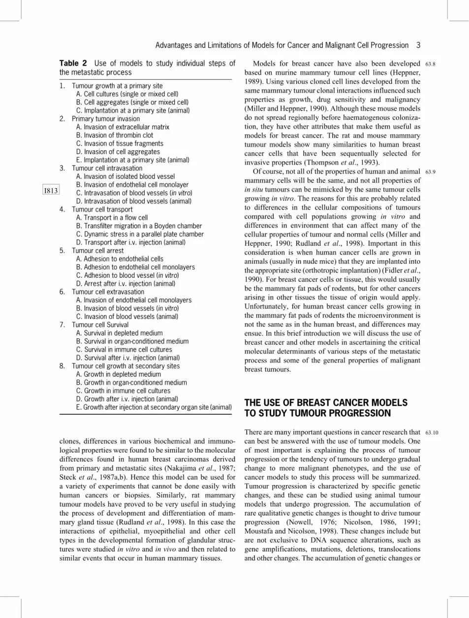
Table 2 Use of models to study individual steps ofthe metastatic process
1. Tumour growth at a primary siteA. Cell cultures (single or mixed cell)B. Cell aggregates (single or mixed cell)C. Implantation at a primary site (animal)
2. Primary tumour invasionA. Invasion of extracellular matrixB. Invasion of thrombin clotC. Invasion of tissue fragmentsD. Invasion of cell aggregatesE. Implantation at a primary site (animal)
3. Tumour cell intravasationA. Invasion of isolated blood vesselB. Invasion of endothelial cell monolayerC. Intravasation of blood vessels (in vitro)D. Intravasation of blood vessels (animal)
4. Tumour cell transportA. Transport in a flow cellB. Transfilter migration in a Boyden chamberC. Dynamic stress in a parallel plate chamberD. Transport after i.v. injection (animal)
5. Tumour cell arrestA. Adhesion to endothelial cellsB. Adhesion to endothelial cell monolayersC. Adhesion to blood vessel (in vitro)D. Arrest after i.v. injection (animal)
6. Tumour cell extravasationA. Invasion of endothelial cell monolayersB. Invasion of blood vessels (in vitro)C. Invasion of blood vessels (animal)
7. Tumour cell SurvivalA. Survival in depleted mediumB. Survival in organ-conditioned mediumC. Survival in immune cell culturesD. Survival after i.v. injection (animal)
8. Tumour cell growth at secondary sitesA. Growth in depleted mediumB. Growth in organ-conditioned mediumC. Growth in immune cell culturesD. Growth after i.v. injection (animal)E. Growth after injection at secondary organ site (animal)
I813
Advantages and Limitations of Models for Cancer and Malignant Cell Progression 3
alterations may initiate important events that eventuallylead to tumour progression, but it is likely that other eventsare also involved that do not depend on genomic DNAsequence changes (Nicolson, 1986, 1991; Moustafa andNicolson, 1998). One of the uses of cancer models is to beable to determine the importance of specific genetic eventsin transformation and progression by replicating theseevents in an experimental setting. In addition, once can-didate genes have been identified, their role in the patho-genesis of cancer can be confirmed using transgenic andgene knockout mice to model the changes associated withspecific genes (review: Viney, 1995).
63:11 The progression of breast cells to the malignant phe-notype is mainly typified by quantitative changes in geneexpression rather than many qualitative changes (muta-tions, translocations, truncations, etc.) in gene structures,although the latter changes are known to occur with pro-gression (Nicolson, 1991; Moustafa and Nicolson, 1998).In breast cancer models such qualitative changes have alsobeen documented, especially in genes associated withhereditary breast cancer, such as BRCA-1 and BRCA-2(Lynch et al., 1998). Using breast cancer models, parallelstudies can be used to examine the role of multiple qua-litative cytogenetic and genetic changes and determine ifthey are important in the process of transformation andprogression (Pearce et al., 1984).
63:12 Qualitative genomic modifications may occur onlyrarely in tumours, and the rates of appearance of qualita-tive alterations may vary widely among different tumours.Hence it is important to determine if there are (multiple)critical changes in specific genes that must occur in orderfor transformation and progression to occur. If qualitativegenomic changes are critical to tumour transformation,progression and clonal dominance, then it is likely that themost successful cells in a tumour that have these changeswill overgrow the other cells and eventually almost all ofthe tumour cells within a tumour would display similargenomic modifications (Nowell, 1976). Probably not allgenes that undergo change are important in the process oftumour progression, and the types of genes that are mostlikely to be important have been found to be the genes thatencode oncogenes, suppressor genes, differentiation genesand genes associated with growth, invasion, survival andmetastasis (Nicolson, 1991; Moustafa and Nicolson,1998). These are also the genes that are likely to also bechanged in their expression (Volpe, 1988; Moustafa andNicolson, 1998). Often important qualitative changes thatare associated with early states of oncogenesis, such ascellular transformation and tumour initiation, are not thesame changes that are associated with progression to moremalignant states (Nicolson, 1986, 1991; Volpe, 1988;Moustafa and Nicolson, 1998).
63:13 The changes in gene expression and gene structure thatoccur in breast cancer cells as they progress are not the onlyimportant events that typify tumour progression. There arealso signals from the host tissue microenvironment (cells,
extracellular matrix, soluble glyoproteins and other sig-nals) that are important in this process (Nicolson, 1986;1991; Volpe, 1988; Moustafa and Nicolson, 1998). Thesesignals provide tumours with soluble and insoluble medi-ators that can modulate tumour cell properties and res-ponses to host tissue, cell and extracellular matrix signals(Bissell and Barcellos-Hoff, 1987). In some tissues, suchas breast eipthelium, there are also unique regulators, suchas the ionic and metabolic signals that are presented toadjacent cells, either at their surfaces or through junctionalcommunication in epithelium. There are both external andcellular signals that elicit both positive and negative sig-nals that usually control the normal aspects of epithelialtissue differentiation, proliferation and death, and these areimportant in tumour progression and growth (Nicolson,1989, 1993; Trosko et al., 1993).
63:14As tumours progress, particularly breast and mammarytumours (Foulds, 1975), they are thought to be lessresponsive to host microenvironments and cellular con-trols. The end result of this is that they eventually gainautonomy from cell and tissue regulation as they progressto the late or end stages of their life histories (Nicolson,1986, 1987, 1993). An important property of malignanttumours is their ability to undergo cellular diversificationinto heterogeneous phenotypes. Heterogeneity in cellularproperties is found in virtually all malignant tumour cellpopulations, but in highly malignant tumours, such asinvasive breast carcinomas, it is usually more pronouncedthan that found in the cells of benign or normal breasttissues. In normal tissues intercellular, cellular and matrixinteractions probably combine to stabilize cellular pheno-types into more narrow states of diversity than seen inisolated single cells or tumour cells derived from the sametissue (Nicolson, 1986, 1987, 1993). Once normal cellshave been removed from their normal interactions, theyshow increased diversity in their cellular properties (Rubin,1990). Such diversity may be due to adoptive changesthat individually affect each cell and result in individualquantitative differences in gene expression. In malignantcell populations diversification occurs irrespective of orat least less dependent on the host microenvironment.Eventually, heterogeneous cellular phenotypes arise thatare less regulated by normal host cell, matrix and otherinteractions (Nicolson, 1987, 1993).
63:15In mammary as in other tumour cell populations, singlecells or a few cells undergo diversification to more het-erogeneous phenotypes, and as this happens these cellpopulations also face host immune and nonimmuneselection pressures. Eventually differences in cellularresponses to host mediators, growth stimulators or inhibi-tors allow certain subpopulations to become dominant inthe tumour cell population, or by active processes, such asimmune or nonimmune host responses, certain sub-populations arise in the population to become the pre-dominant cellular type (Nowell, 1976; Nicolson, 1987,1991; Kerbel et al., 1988; Frost and Chernajovsky, 1990).
4 In vitro and Animal Models for Human Cancer
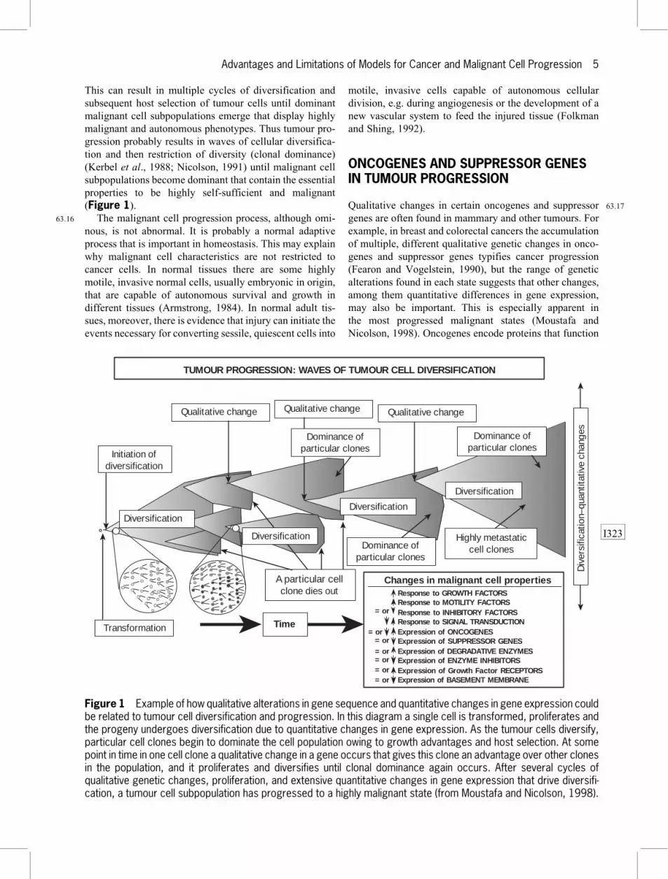
This can result in multiple cycles of diversification andsubsequent host selection of tumour cells until dominantmalignant cell subpopulations emerge that display highlymalignant and autonomous phenotypes. Thus tumour pro-gression probably results in waves of cellular diversifica-tion and then restriction of diversity (clonal dominance)(Kerbel et al., 1988; Nicolson, 1991) until malignant cellsubpopulations become dominant that contain the essentialproperties to be highly self-sufficient and malignant(Figure 1).
63:16 The malignant cell progression process, although omi-nous, is not abnormal. It is probably a normal adaptiveprocess that is important in homeostasis. This may explainwhy malignant cell characteristics are not restricted tocancer cells. In normal tissues there are some highlymotile, invasive normal cells, usually embryonic in origin,that are capable of autonomous survival and growth indifferent tissues (Armstrong, 1984). In normal adult tis-sues, moreover, there is evidence that injury can initiate theevents necessary for converting sessile, quiescent cells into
motile, invasive cells capable of autonomous cellulardivision, e.g. during angiogenesis or the development of anew vascular system to feed the injured tissue (Folkmanand Shing, 1992).
ONCOGENES AND SUPPRESSOR GENESIN TUMOUR PROGRESSION
63:17Qualitative changes in certain oncogenes and suppressorgenes are often found in mammary and other tumours. Forexample, in breast and colorectal cancers the accumulationof multiple, different qualitative genetic changes in onco-genes and suppressor genes typifies cancer progression(Fearon and Vogelstein, 1990), but the range of geneticalterations found in each state suggests that other changes,among them quantitative differences in gene expression,may also be important. This is especially apparent inthe most progressed malignant states (Moustafa andNicolson, 1998). Oncogenes encode proteins that function
Transformation
Initiation ofdiversification
Dominance ofparticular clones
Highly metastaticcell clones
Div
ersi
ficat
ion–
quan
titat
ive
chan
ges
A particular cellclone dies out
Qualitative change
TUMOUR PROGRESSION: WAVES OF TUMOUR CELL DIVERSIFICATION
Time
Response to GROWTH FACTORS Response to MOTILITY FACTORS Response to INHIBITORY FACTORS Response to SIGNAL TRANSDUCTION Expression of ONCOGENES Expression of SUPPRESSOR GENES Expression of DEGRADATIVE ENZYMES Expression of ENZYME INHIBITORS Expression of Growth Factor RECEPTORS Expression of BASEMENT MEMBRANE
= or
Changes in malignant cell properties
Diversification Diversification
Diversification
Dominance ofparticular clones
Qualitative change Qualitative change
= or = or = or = or = or = or
Dominance ofparticular clones
Diversification
Figure 1 Example of how qualitative alterations in gene sequence and quantitative changes in gene expression couldbe related to tumour cell diversification and progression. In this diagram a single cell is transformed, proliferates andthe progeny undergoes diversification due to quantitative changes in gene expression. As the tumour cells diversify,particular cell clones begin to dominate the cell population owing to growth advantages and host selection. At somepoint in time in one cell clone a qualitative change in a gene occurs that gives this clone an advantage over other clonesin the population, and it proliferates and diversifies until clonal dominance again occurs. After several cycles ofqualitative genetic changes, proliferation, and extensive quantitative changes in gene expression that drive diversifi-cation, a tumour cell subpopulation has progressed to a highly malignant state (from Moustafa and Nicolson, 1998).
I323
Advantages and Limitations of Models for Cancer and Malignant Cell Progression 5

abnormally, inappropriately or at improper concentrations,resulting in the circumvention of the normal cellular con-trols that regulate cellular proliferation and the state ofdifferentiation (Klein and Klein, 1986; Nicolson, 1987).Although qualitative changes in oncogenes have been foundin transformed cells, a more common finding is a change inoncogene expression due to chromosome translocations,gene amplifications and other changes, particularly inbreast cancer (Yokota et al., 1986). Such single events bythemselves are unlikely to be the underlying cause ofneoplastic transformation, because further cellular changesare usually necessary.
63:18 Specific changes in oncogenes are often important intumour progression. For example, oncogene amplificationis an important mechanism (Gitelman et al., 1987).Although amplification of oncogenes has frequently beenseen in various cancers, it is not universally seen in alltumours (Yokota et al., 1986). Oncogene amplificationmay be indicative of other, unrecognized genetic changes,or the amplification of oncogenes and other genes couldcontribute more directly to cancer progression (Nicolson,1991; Moustafa and Nicolson, 1998). Since the expressionof oncogenes can differ between primary breast cancersand their metastases, oncogene expression has been pro-posed to be important in breast tumour progression.However, examination of a variety of primary and secon-dary tumours reveals that oncogenes can be overexpres-sed, underexpressed or equally expressed in metastasescompared with primary tumours (Yokota et al., 1986;Nicolson, 1987). Hence, the qualitative changes seen inoncogenes or the quantitative changes in their expressionmay contribute to progression, but they are unlikely to bethe universal determinants.
63:19 Although oncogenes are important in tumour progres-sion, the data are not convincing in support of a universalcausative role for oncogenes in the progression of breastand other tumours to the metastatic state. In most studiesmetastases were compared with advanced primary tumoursthat may have already undergone all of the changesnecessary to become metastatic (Yokota et al., 1986;Nicolson, 1987). Experimentally, the insertion of dom-inantly acting oncogenes into a suitable recipient cell canresult in acquisition of the metastatic phenotype (Kerbelet al., 1987; Nicolson et al., 1990; Tuck et al., 1990). Oftensuch experiments have been performed using aneuploid,unstable, easily spontaneously transformable animal cellsas recipients, like unstable mouse fibroblast cell lines. Insome untransformed cells conversion to the metastaticstate only occurred when two different dominantly actingoncogenes were simultaneously inserted, an event rarelyseen in spontaneous tumours. These rapid qualitativechanges are unlike the slow, sequential changes thatcharacterize spontaneous transformation and tumour pro-gression to the metastatic state in vivo.
63:20 Some normal cells or even benign tumour cells arehighly resistant to oncogene-mediated conversion to the
metastatic phenotype (Kerbel et al., 1987; Nicolson et al.,1990; Tuck et al., 1990). Even within the same cell typethere appears to be heterogeneity in the ability of dom-inantly acting activated oncogenes to cause metastaticconversion. For example, in rat mammary cells there wasa considerable difference in the ability of an activated rasoncogene to convert benign cells to the metastatic phe-notype (Muschel and Liotta, 1988; Nicolson et al., 1990).Moreover, in some systems the gene transfer techniquesthemselves may be as important as the transferred gene incausing metastatic conversion (Kerbel et al., 1987;Nicolson et al., 1990). Often multiple gene copies areinserted, and the effects of their accompanying strongpromoter/enhancer elements are not considered. It isusually assumed that oncogene constructs are randomlyincorporated into the genome, but just the opposite is likelyto be the case. In addition, nonrandom cytogenetic changesmay occur concomitant with gene transfer (Muschel andMcKenna, 1989). Hence it is difficult to conclude fromexperimental studies that the insertion of an oncogene isthe only event required for conversion of benign cells tothe invasive, metastatic state. In some cases oncogene-mediated conversion of a cell to the invasive, metastaticstate may be dependent on the resulting concentration of anoncogene-encoded product (Muschel and Liotta, 1988;Greenberg et al., 1989); however, there was no obviouscorrelation between the expression level of an oncogene-encoded product and metastatic conversion in mammarycells (Nicolson et al., 1990). In addition to oncogene inser-tion and expression, other changes are probably necessary.Some of these changes may involve other oncogenes,suppressor genes, chromosomal structural alterations, andeventually cellular diversification.
63:21Suppressor genes can affect mammary tumour pro-gression and malignancy (Muschel and McKenna, 1989;Sobel, 1990). Metastasis suppressor genes were origi-nally identified on the basis of cell fusion experimentsthat resulted in suppression of metastatic properties(Sidebottom and Clark, 1983). By examining differencesin gene expression in nonmetastatic and metastatic celllines, several candidate metastasis suppressor genes wereidentified and eventually isolated (Sobel, 1990). The bestknown of these in breast cancer is the nm23 gene family.Steeg et al. (1988) cloned the nm23 candidate metastasissuppressor gene, and low expression if this gene wasassociated with lymph node metastasis of breast cancers(Bevilacqua et al., 1989). The encoded product of thenm23 gene is known. The predicted sequence of the nm23protein was found to be identical with the Drosophiladevelopmental gene awd product (Rosengard et al., 1989).The awd gene, in turn, was found to have a high degree ofhomology with nucleotide diphosphate kinase (Kimuraet al., 1990), suggesting a possible role for the nm23 geneproduct in microtubule assembly/disassembly, signaltransduction and/or regulation of G proteins. An alterednm23 protein or changes in its expression could result in
6 In vitro and Animal Models for Human Cancer
modified signal transduction, gene expression and possiblyprogression in breast cancer.
HOST SURVEILLANCE MECHANISMSAND TUMOUR PROGRESSION
63:22 Other differentially expressed genes have been found inhighly metastatic cells. For the most part, the precise rolesof most of these differentially expressed genes or theirgene products in tumour progression have yet to bedetermined. Some differentially expressed genes could beinvolved in tumour progression by acting on intracellularsignalling in a manner that results in an increase in cellsurvival or an inhibition of apoptosis or programmed celldeath. Gene products that act on host surveillance oftumour cells could also affect tumour progression. Astumour cells proliferate and diversify, they are under hostsurveillance pressures that continuously seek to inhibit orkill aberrant cells. Components that allow malignant cellsto escape host surveillance mechanisms could be importantin tumour progression by allowing more malignant cellsto become dominant in a tumour cell population. Forexample, the cells that escape macrophage surveillancemechanisms might be expected to be more malignant byvirtue of their ability to grow in the face of host antitumourresponses. These less host-sensitive cells could then over-grow the more host-sensitive tumour cells and become thedominant cell population in the tumour. In breast cancermodels the most malignant cell populations are alsoapparently those which are less susceptible to host sur-veillance systems, suggesting that in vivo selection occursduring the progression of mammary tumours to moremalignant states (North and Nicolson, 1985). Moreover,in contrast to what has been seen in primary tumours, asmetastases grow there are fewer host effector cells thatinfiltrate into the secondary tumour mass (Bugelski et al.,1987). This suggests that metastases may appear to bemore resistant to host defenses by virtue of the fact thatfewer host effector cells can apparently penetrate intometastases.
CANCER PROGRESSION AND BREASTCANCER GENE EXPRESSION
63:23 Several differentially expressed genes associated withmammary tumour metastasis have been identified. Highlymetastatic cells appear to over- or underexpress a num-ber of genes, including the overexpressed mts1 gene(Ebralidze et al., 1989). The mts1 gene has a high hom-ology with calcium-binding proteins but is of unknownfunction. This gene has been found to be part of the S100gene family that encodes calcium-binding, cytoskeletal-binding proteins. Transfection of the S100A4 gene into
benign mammary cells resulted in these cells acquiring themetastatic phenotype (Davies et al., 1993). The mts1 genemay be related to signal transduction systems, and itsoverexpression may lead to increased signalling of cellmovement and growth.
63:24Other differentially expressed genes have been found inmammary adenocarcinoma cells (Pencil et al., 1993). Themost differentially expressed genes could, for the mostpart, be identified as known genes that had already beenidentified in the metastatic process, e.g. some encodeddegradative enzymes, such as type IV collagenases, thatare known to be differentially expressed in highly meta-static cells. Other genes encoded transcription or transla-tion protein factors that could be involved in the metastaticprocess by increasing the ability of more malignant cells toproliferate under limiting growth conditions (Pencil et al.,1993). One of the overexpressed novel genes, mta1, wasidentified as overexpressed both in spontaneous rat mam-mary tumour metastases and in human breast cancer cellsof high metastatic potential (Toh et al., 1994). This geneappears to function in signal transduction mechanisms butits exact role in maintenance of the malignant phenotype isnot known. We have now identified and cloned the humanMTA1 gene, also a novel gene that appears to be involvedin human breast epithelial cell motility and growth regu-lation. We sought to investigate the role of the MTA1 geneby blocking its expression using an antisense oligo-nucleotide to the precise sequence at the transcription startsite in the gene. In preliminary experiments, the antisenseoligonucleotide but not a sense oligonucleotide blockedexpression of the gene and inhibited human breast cancercell motility and proliferation. The mta1/MTA1 gene has asa part of its structure an Src homology or SH3-bindingdomain at its C-terminal region. This domain may functionin Src and other related signal transduction pathways. Themta1/MTA1 gene may act to modulate signal transductionpathways, and its overexpression in highly metastatic cellscould result in an increased ability to circumvent hostcontrols on cell growth and movement.
INTERCELLULAR COMMUNICATION ANDTUMOUR CELL DIVERSIFICATION
63:25The communication via intercellular junctions is animportant cellular controlling mechanism in epithelialcells. These structures, in particular gap junctions, allowadjacent epithelial cells to be metabolically and elec-trically coupled, and this could be important in theirnormal maintenance of cellular proliferation and diversity.Using nonmalignant epithelial cells in culture we havefound that cellular diversification and heterogeneity can bestimulated by oncogene transfer (Nicolson et al., 1992).This was demonstrated by transfecting relatively stablebenign cell clones with the dominantly acting oncogene
Advantages and Limitations of Models for Cancer and Malignant Cell Progression 7
construct and observing the diversification of subclonesderived from single transfected cells. We found that stable,benign mammary epithelial cell clones that acquired anoncogene construct diversified rapidly concomitant withthe cells acquiring the metastatic phenotype. In addition toincreased diversity in metastatic properties, the transfectedcells also showed increased diversity in the expression of ametastasis-associated cell surface mucin-like glycoproteinthat may be involved in cell adhesion. In contrast, most ofthe cell clones that received the control gene constructremained relatively more stable and their subclones werefor the most part nonmetastatic. The results suggest thatrapid cellular diversification is an important property ofhighly malignant cells. A qualitative genetic change, suchas sequence alteration of a dominantly acting oncogene,would be expected to occur at a low rate in vivo; however,the results demonstrated the relationships between quali-tative gene changes, cellular phenotypic diversificationand malignancy. This suggests that stimulation of cellulardiversification could be an important step in tumourprogression.
63:26 Environmental signals can also regulate cellular diver-sification and heterogeneity. The dynamic regulation ofelectrical, ionic and metabolic coupling between epithelialcells is mediated by gap junctions. This form of cellularcommunication plays an important role in cell prolifera-tion, differentiation, physiological responses and carcino-genesis (Dotto et al., 1989). Overexpression of oncogenescan disrupt gap junctional communication, and over-expression of tumour-suppressor genes is associated withan increase in gap junctional communication. When weexamined the ability of a dominantly acting oncogene tocause diversification in the junctional communicationproperties of transfected benign mammary cells, we foundthat similar to their metastatic properties, their gap junc-tional communication was inhibited and more diverseamong subclones obtained from single cell clones at vari-ous times during subculture (Nicolson et al., 1992).Intercellular junctional communication and its interferenceduring tumour progression may be important in releasingcells from microenvironmental controls that regulate cel-lular diversification.
CANCER PROGRESSION ANDCELLULAR DIVERSIFICATION
63:27 Highly progressed malignant cells exhibit rapid rates ofdiversification and phenotypic change, mainly due toquantitative differences in gene expression. This probablyresults in tumours that express a diversity of differentimmunological, biochemical, enzymological, structuraland other cellular phenotypes. The cellular and extra-cellular mechanisms that control tumour and normal cel-lular stability and diversity have not been readily
appreciated or extensively studied, hence it is difficult toascribe precise molecular mechanisms for this process.Volpe (1988) proposed that certain genes are present in thegenome that control cellular stability, and these may bealtered during tumour progression, resulting in extensivetumour cell diversity, evolution and eventually acquisitionof more malignant and metastatic cellular phenotypes. Hetermed these `stability genes,' and such genes could beinvolved in karyokinesis and the repair, recombination andreplication of DNA. These genes would be good candi-dates as genes controlling mechanisms that regulate cel-lular diversification. In addition to stability and othergenes, a wide variety of epigenetic factors, such as thosethat control tissue and stromal organization, could alsocontrol cellular diversification mechanisms (Nicolson,1987; Moustafa and Nicolson, 1988; Miller and Heppner,1990). The removal of cells from their usual micro-environments could also result in loss of tissue-specificcontrols. In this scheme cells are in a constant dynamicflux due to changes in their microenvironments.
63:28There are apparently complex mechanisms that controlcellular differentiation and the diversity of cells withintissues. Both normal cells and tumour cells are exposed tovariations in the concentrations of various regulatory fac-tors. Although normal cells may be more stable thantumour cells in their microenvironments, under certainconditions normal cells can undergo rapid change anddiversification. In contrast, malignant cells can be mademore stable in their cellular phenotypes. Thus, as malig-nant cells progress, they can reach states where they are nolonger regulated by paracrine growth factors and inhibi-tors. Once this occurs, they can grow in a variety ofmicroenvironments that would usually not be permissivefor growth (Nicolson, 1993).
REFERENCES
Armstrong, P. B. (1984). Invasion: Experimental and Clinical
Implications. 126--167 (Oxford University Press, Oxford).
Bevilacqua, G., et al. (1989). Association of low nm23 RNA
levels in human primary infilitrating ductal breast carcinomas
with lymph node involvement and other histopathological
indicators of high metastatic potential. Cancer Research, 49,
5185--5190.
Bissell, M. J. and Barcellos-Hoff, M. H. (1987). The influence of
extracellular matrix on gene expression: is structure the
message? Journal of Cell Science, 8, Suppl., 327--343.
Bugelski, P. J., et al. (1987). The macrophage content of spon-
taneous metastases at different stages of their growth. Cancer
Research, 47, 4141--4145.
Davies, B. R., et al. (1993). Induction of the metastatic phenotype
by transfection of a benign rat mammary epithelial cell line
with the gene for p9Ka, a rat calcium-binding protein, but not
with the oncogene EJ-ras-1. Oncogene, 8, 999--1008.
8 In vitro and Animal Models for Human Cancer

Dotto, G. P., et al. (1989). Similar and synergistic inhibition of
gap-junctional communication by ras transformation and
tumor promoter treatment of mouse primary keratinocytes.
Oncogene, 4, 637--641.
Ebralidze, A. K., et al. (1989). Isolation and characterization of
a gene specifically expressed in different metastatic cells and
whose deduced gene product has a high degree of homology
to a Ca2�-binding protein family. Genes and Development, 3,
1086--1093.
Fearon, E. R. and Vogelstein, B. (1990). A genetic model for
colorectal tumorigenesis. Cell, 61, 759--767.
Fidler, I. J., et al. (1990). Orthotopic implantation is essential for
the selection, growth and metastasis of human cancer cells in
nude mice. Cancer and Metastasis Reviews, 9, 149--165.
Folkman, J. and Shing, Y. (1992). Angiogenesis. Journal of
Biological Chemistry, 267, 10931--10934.
Foulds, L. (1975). Neoplastic Development (Academic Press,
New York).
Frei, E. (1982). Models and the clinical dilemma. In: Fidler, I. J.
and White, R. J. (eds), Design of Models for Testing Cancer
Therapeutic Agents. 248--259 (Van Nostrand, New York).
Frost, P. and Chernajovsky, Y. (1990). Transformation injury and
the unicellular phenotype of malignant cells. Cancer and
Metastasis Reviews, 9, 93--98.
Gitelman, I., et al. (1987). DNA amplification and metastasis of
the human melanoma cell line MeWo. Cancer Research, 47,
3851--3855.
Greenberg, A. H., et al. (1989). Oncogenes and metastatic pro-
gression. Invasion and Metastasis, 9, 360--378.
Heppner, G. H. (1989). Tumor cell societies. Journal of the
National Cancer Institute, 81, 648--649.
Kerbel, R. A., et al. (1987). Alteration of the tumorigenic and
metastatic properties of neoplastic cells is associated with the
process of calcium phosphate-mediated DNA transfection.
Proceedings of the National Academy of Sciences of the USA,
84, 1263--1267.
Kerbel, R. A., et al. (1988). Clonal dominance of primary tumors
by metastatic cells: Genetic analysis and biological implica-
tions. Cancer Surveys, 7, 597--629.
Kimura, N., et al. (1990). Isolation and characterization of a
cDNA clone encoding rat nucleoside diphosphate kinase.
Journal of Biological Chemistry, 265, 15744--15749.
Klein, G. and Klein, E. (1986). Conditional tumorigenicity of
activated oncogenes. Cancer Research, 46, 3211--3224.
Lynch, et al. (1998). Breast cancer genetics: heterogeneity,
molecular genetics, syndrome diagnosis and genetic coun-
seling. In: Bland, K. I. and Copeland, E. M., III (eds), The
Breast: Comprehensive Management of Benign and Malig-
nant Diseases. 370--394 (W. B. Saunders, Philadelphia).
Miller, F. R. and Heppner, G. H. (1990). Cellular interactions in
metastasis. Cancer and Metastasis Reviews, 9, 21--34.
Moustafa, A. F. and Nicolson, G. L. (1998). Breast cancer
metastasis-associated genes: prognostic significance and
therapeutic implications. Oncology Research, 9, 505--525.
Muschel, R. and Liotta, L. A. (1988). Role of oncogenes in
metastasis. Carcinogenesis, 9, 705--710.
Muschel, R. J. and McKenna, W. G. (1989). Oncogenes and tumor
progression. Anticancer Research, 9, 1395--1406.
Nakajima, M., et al. (1987). Degradation of basement membrane
type IV collagen and lung subendothelial matrix by rat
mammary adenocarcinoma cell clones of differing metastatic
potentials. Cancer Research, 47, 4869--4876.
Neri, A. and Nicolson, G. L. (1981). Phenotypic drift of metastatic
and cell surface properties of mammary adenocarcinoma cell
clones during growth in vitro. International Journal of
Cancer, 28, 731--738.
Neri, A., et al. (1982). Development and biologic properties of
malignant cell sublines and clones of a spontaneously
metastasizing rat mammary adenocarcinoma. Journal of the
National Cancer Institute, 68, 507--517.
Nicolson, G. L. (1986). Oncogenes, genetic instability and the
evolution of the metastatic phenotype. Advances in Viral
Oncology, 6, 143--167.
Nicolson, G. L. (1987). Tumor cell instability, diversification and
progression to the metastatic phenotype: from oncogene to
oncofetal expression. Cancer Research, 47, 1473--1487.
Nicolson, G. L. (1989). Metastatic tumor cell interactions with
endothelium, basement membrane and tissue. Current Opi-
nions in Cell Biology, 1, 1009--1019.
Nicolson, G. L. (1991). Gene expression and tumor progression to
the metastatic phenotype. Bioessays, 13, 337--342.
Nicolson, G. L. (1993). Pacarine/autocrine growth mechanisms in
tumor metastasis. Oncology Research, 4, 389--399.
Nicolson, G. L. and Poste, G. (1983). Tumor implantation and
invasion at metastatic sites. International Review of Experi-
mental Pathology, 25, 77--181.
Nicolson, G. L., et al. (1990). Intercellular junctional commu-
nication, p21rasEJ expression, and spontaneous metastatic
properties of rat mammary cells after transfection with
c-H-rasEJ or neo genes. Oncogene, 5, 747--753.
Nicolson, G. L., et al. (1992). Transfection of activated c-H-rasEJ/
pSV2neo or pSV2neo genes into rat mammary cells: rapid
stimulation of clonal diversification in spontaneous metastatic
and cell surface properties. Oncogene, 7, 1127--1135.
North, S. M. and Nicolson, G. L. (1985). Effect of host immune
status on the spontaneous metastasis of cloned cell lines of the
13762NF rat mammary adenocarcinoma. British Journal of
Cancer, 52, 747--755.
Nowell, P. C. (1976). The clonal evolution of tumor cell popu-
lations. Science, 194, 23--28.
Pearce, V., et al. (1984). Chromosome and DNA analysis of
rat 13762NF mammary adenocarcinoma cell lines and clones
of different metastatic potentials. Clinical and Experimental
Metastasis, 2, 271--286.
Pencil, S. D., et al. (1993). Candidate metastasis-associated genes
of rat 13762NF mammary adenocarcinoma. Breast Cancer
Research and Treatment, 25, 165--174.
Poste, G. (1982). Experimental systems for analysis of the malig-
nant phenotype. Cancer and Metastasis Reviews, 1, 141--199.
Rosengard, A. M., et al. (1989). Reduced Nm23/Awd protein in
tumor metastasis and aberrant Drosophila development.
Nature, 342, 177--180.
Advantages and Limitations of Models for Cancer and Malignant Cell Progression 9

Rubin, H. (1990). The significance of biological heterogeneity.
Cancer and Metastasis Reviews, 9, 1--20.
Rudland, P. S., et al. (1998). Growth and differentiation of the
normal mammary gland and its tumours. Biochemical Society
Symposia, 63, 1--20.
Sidebottom, E. and Clark, S. R. (1983). Cell fusion segregates
progressive growth from metastasis. British Journal of
Cancer, 47, 399--406.
Sobel, M. E. (1990). Metastasis suppressor genes. Journal of the
National Cancer Institute, 82, 267--275.
Steck, P. A., et al. (1987a). Altered expression of glycos-
aminoglycans in metastatic 13762NF rat mammary adeno-
carcinoma. Biochemistry, 26, 1020--1028.
Steck, P. A., et al. (1987b). Purification and partial character-
ization of a tumour metastasis-associated high Mr glycopro-
tein from rat 13762NF mammary adenocarcinoma cells.
Biochemical Journal, 242, 779--787.
Steeg, P. S., et al. (1988). Evidence for a novel gene associated
with low tumour metastatic potential. Journal of the National
Cancer Institute, 80, 200--204.
Toh, Y., et al. (1994). A novel candidate metastasis-associated
gene mta1 differentially expressed in highly metastatic
mammary adenocarcinoma cell lines: cDNA cloning,
expression and protein analyses. Journal of Biological
Chemistry, 269, 22958--22963.
Trosko, J. E., et al. (1993). Endogenous and exogenous modula-
tion of gap junctional intercellular communication: Toxi-
cological and pharmacological implications. Life Sciences,
53, 1--19.
Thompson, E. W., et al. (1993). The invasive and metastatic
properties of hormone-independent but hormone-responsive
variants of MCF-7 human breast cancer cells. Clinical and
Experimental Metastasis, 11, 15--26.
Tuck, A. B., et al. (1990). Ras transfection and expression does
not induce progression from tumorigenicity to metastatic
ability in mouse LTA cells. Clinical and Experimental
Metastasis, 8, 417--431.
Viney, J. L. (1995). Transgenic and gene knockout mice in cancer
research. Cancer and Metastasis Reviews, 14, 77--90.
Volpe, J. P. G. (1988). Genetic instability of cancer. Why a
metastatic tumor is unstable and a benign tumor is stable.
Cancer Genetics and Cytogenetics, 34, 125--134.
Yokota, J., et al. (1986). Alterations of myc, myb and rasHa proto-
oncogenes in cancers are frequent and show clinical correla-
tion. Science, 231, 261--265.
FURTHER READING
1. Clarke, R. (1996). Animal models of breast cancer: their
diversity and role in biomedical. Breast Cancer Research and
Treatment, 39, 1--6.
2. Welch, D. R. (1997). Technical considerations for studying
cancer metastasis in vivo. Clinical and Experimental Metas-
tasis, 15, 272--306.
3. Edwards, P. A. (1999). The impact of developmental biology
on cancer research: an overview. Cancer and Metastasis
Reviews, 18, 175--180.
4. Mattern, J., et al. (1988). Human tumor xenografts as model
for drug testing. Cancer and Metastasis Reviews, 7, 263--284.
5. Nicolson, G. L. (1981). The use of animal tumor models
to study the metastatic process. In: Stroehlein, J. R. and
Romsdahl, M. M. (eds), Gastrointestinal Cancer. 427--441
(Raven Press, New York).
6. Nicolson, G. L. (1994). The use of animal tumor models to
study the role of the tumor microenvironment and paracrine
and autocrine growth mechanisms in metastasis to specific
sites. In: Fusenig, N. E. and Graf, H. (eds), Cell Culture in
Pharmaceutical Research. 103--123 (Springer, Berlin).
7. Poste, G. (1982). Experimental systems for analysis of the
malignant phenotype. Cancer and Metastasis Reviews, 1,
141--199.
8. Rudland, P. S., et al. (1998). Growth and differentiation of the
normal mammary gland and its tumours. Biochemical Society
Symposia, 63, 1--20.
10 In vitro and Animal Models for Human Cancer

KEYWORDS
animal modelstumour progressiongenetic changesgene expressionmetastasisbreast cancercell communicationgrowthinvasionimmune responsetumour diversificationmalignancyoncogenessuppressor genesmetastasis-associated genes
GLOSSARY TERMS
Cancer progression The process whereby a cancerbecomes more malignant and for some tumours moremetastatic.
Transformation The process where by a cell changesfrom one that undergoes density-dependent growth, con-tact-dependent inhibition of movement and the lack ofability to grow in semisolid suspension to one that grows tomuch higher cell densities, moves without regulation byother cells and grows in suspension without initial contact
with other cells. This is usually considered to be the firstimportant step in the eventual formation of a cancer cell.
Tumour heterogeneity The intertumoural or cell-to-celldifferences in a variety of properties.
Cell communication The process where cells send sig-nals to other cells.
Metastasis The spread of cancer cells from a primary siteof origin to secondary sites in different locations and tissuesat near and distant sites.
Tumour diversification The generation of tumour cellheterogeneity.
Tumour invasion The penetration of tissues or extra-cellular matrix by tumour cells.
Oncogenes Genes that contribute to transformation andcancer.
Suppressor genes Genes that suppress tranformation,tumour formation or metastasis.
Malignancy The property of cell to kill its host, usuallyby invasion and/or metastasis.
Gene expression The ability of a gene to generate mes-senger RNA that can be transcribed into a protein.
Clonal dominance The dominance of individual or a fewcell clones in a heterogeneous cell population that results inthe dominant clone(s) overgrowth of other cells.
Host surveillance The ability of a host to recognize andinhibit or destroy foreign or altered cells and microorgan-isms.
Epigenetic All nongenetic factors or factors that are notrelated to gene sequence or organization.
Advantages and Limitations of Models for Cancer and Malignant Cell Progression 11
Related Documents

![cells inhibits angiogenesis in glioblastoma · cells * glioma Downregulation INTRODUCTION Angiogenesis is a key event in the progression of malignant gliomas [1,2]. It is a highly](https://static.cupdf.com/doc/110x72/5ecd7b084c46b638be2fbb49/cells-inhibits-angiogenesis-in-glioblastoma-cells-glioma-downregulation-introduction.jpg)