Identification of Liver Cancer Progenitors Whose Malignant Progression Depends on Autocrine IL-6 Signaling Guobin He, 1,13 Debanjan Dhar, 1,13 Hayato Nakagawa, 1,11,13 Joan Font-Burgada, 1,13 Hisanobu Ogata, 1,12,13 Yuhong Jiang, 1 Shabnam Shalapour, 1 Ekihiro Seki, 2 Shawn E. Yost, 4,5 Kristen Jepsen, 5 Kelly A. Frazer, 5,6,7,8 Olivier Harismendy, 5,6,7 Maria Hatziapostolou, 9 Dimitrios Iliopoulos, 9 Atsushi Suetsugu, 3,10 Robert M. Hoffman, 3,10 Ryosuke Tateishi, 11 Kazuhiko Koike, 11 and Michael Karin 1,6, * 1 Laboratory of Gene Regulation and Signal Transduction, Departments of Pharmacology and Pathology 2 Department of Medicine 3 Department of Surgery 4 Bioinformatics Graduate Program 5 Rady’s Children’s Hospital and Department of Pediatrics 6 Moores UCSD Cancer Center 7 Clinical and Translational Research Institute 8 Institute for Genomic Medicine University of California San Diego, School of Medicine, 9500 Gilman Drive, San Diego, CA 92093, USA 9 Center for Systems Biomedicine, Division of Digestive Diseases and Institute for Molecular Medicine, David Geffen School of Medicine, University of California Los Angeles, Los Angeles, CA 90095, USA 10 AntiCancer, Inc., San Diego, CA 92111, USA 11 Department of Gastroenterology, University of Tokyo, Tokyo 113-8655, Japan 12 Department of Medicine and Clinical Science, Graduate School of Medical Sciences, Kyushu University, Fukuoka 812-8582, Japan 13 These authors contributed equally to this work *Correspondence: karinoffi[email protected] http://dx.doi.org/10.1016/j.cell.2013.09.031 SUMMARY Hepatocellular carcinoma (HCC) is a slowly devel- oping malignancy postulated to evolve from pre- malignant lesions in chronically damaged livers. However, it was never established that premalig- nant lesions actually contain tumor progenitors that give rise to cancer. Here, we describe isola- tion and characterization of HCC progenitor cells (HcPCs) from different mouse HCC models. Unlike fully malignant HCC, HcPCs give rise to cancer only when introduced into a liver undergoing chronic damage and compensatory proliferation. Although HcPCs exhibit a similar transcriptomic profile to bipotential hepatobiliary progenitors, the latter do not give rise to tumors. Cells resembling HcPCs reside within dysplastic lesions that appear several months before HCC nodules. Unlike early hepatocarcinogenesis, which depends on paracrine IL-6 production by inflammatory cells, due to upre- gulation of LIN28 expression, HcPCs had acquired autocrine IL-6 signaling that stimulates their in vivo growth and malignant progression. This may be a general mechanism that drives other IL-6-producing malignancies. INTRODUCTION Every malignant tumor is probably derived from a single progen- itor that had acquired growth and survival advantages through genetic and epigenetic changes, allowing clonal expansion (Nowell, 1976). Tumor progenitors are not necessarily identical to cancer stem cells (CSCs), which maintain and renew fully established malignancies (Nguyen et al., 2012). However, clonal evolution and selective pressure may cause some descendants of the initial progenitor to cross the bridge of no return and form a premalignant lesion. Cancer genome sequencing indicates that most cancers require at least five genetic changes to evolve (Wood et al., 2007). How these changes affect the properties of tumor progenitors and control their evolution into a CSC is not entirely clear, as it has been difficult to isolate and propagate can- cer progenitors prior to detection of tumor masses. Given these difficulties, it is also not clear whether cancer progenitors are the precursors for the more malignant CSC isolated from fully established cancers. An answer to these critical questions de- pends on identification and isolation of cancer progenitors, which may also enable definition of molecular markers and signaling pathways suitable for early detection and treatment. This is espe- cially important in cancers of the liver and pancreas, which evolve over the course of many years but, once detected, are extremely difficult to treat (El-Serag, 2011; Hruban et al., 2007). Hepatocellular carcinoma (HCC), the most common liver can- cer, is the end product of chronic liver diseases, requiring 384 Cell 155, 384–396, October 10, 2013 ª2013 Elsevier Inc.

Welcome message from author
This document is posted to help you gain knowledge. Please leave a comment to let me know what you think about it! Share it to your friends and learn new things together.
Transcript
Identification of Liver CancerProgenitors Whose Malignant ProgressionDepends on Autocrine IL-6 SignalingGuobin He,1,13 Debanjan Dhar,1,13 HayatoNakagawa,1,11,13 Joan Font-Burgada,1,13 HisanobuOgata,1,12,13 Yuhong Jiang,1
Shabnam Shalapour,1 Ekihiro Seki,2 Shawn E. Yost,4,5 Kristen Jepsen,5 Kelly A. Frazer,5,6,7,8 Olivier Harismendy,5,6,7
Maria Hatziapostolou,9 Dimitrios Iliopoulos,9 Atsushi Suetsugu,3,10 Robert M. Hoffman,3,10 Ryosuke Tateishi,11
Kazuhiko Koike,11 and Michael Karin1,6,*1Laboratory of Gene Regulation and Signal Transduction, Departments of Pharmacology and Pathology2Department of Medicine3Department of Surgery4Bioinformatics Graduate Program5Rady’s Children’s Hospital and Department of Pediatrics6Moores UCSD Cancer Center7Clinical and Translational Research Institute8Institute for Genomic MedicineUniversity of California San Diego, School of Medicine, 9500 Gilman Drive, San Diego, CA 92093, USA9Center for Systems Biomedicine, Division of Digestive Diseases and Institute for Molecular Medicine, David Geffen School of Medicine,
University of California Los Angeles, Los Angeles, CA 90095, USA10AntiCancer, Inc., San Diego, CA 92111, USA11Department of Gastroenterology, University of Tokyo, Tokyo 113-8655, Japan12Department of Medicine and Clinical Science, Graduate School of Medical Sciences, Kyushu University, Fukuoka 812-8582, Japan13These authors contributed equally to this work*Correspondence: [email protected]
http://dx.doi.org/10.1016/j.cell.2013.09.031
SUMMARY
Hepatocellular carcinoma (HCC) is a slowly devel-oping malignancy postulated to evolve from pre-malignant lesions in chronically damaged livers.However, it was never established that premalig-nant lesions actually contain tumor progenitorsthat give rise to cancer. Here, we describe isola-tion and characterization of HCC progenitor cells(HcPCs) from different mouse HCC models. Unlikefully malignant HCC, HcPCs give rise to canceronly when introduced into a liver undergoingchronic damage and compensatory proliferation.Although HcPCs exhibit a similar transcriptomicprofile to bipotential hepatobiliary progenitors, thelatter do not give rise to tumors. Cells resemblingHcPCs reside within dysplastic lesions that appearseveral months before HCC nodules. Unlike earlyhepatocarcinogenesis, which depends on paracrineIL-6 production by inflammatory cells, due to upre-gulation of LIN28 expression, HcPCs had acquiredautocrine IL-6 signaling that stimulates their in vivogrowth and malignant progression. This may be ageneral mechanism that drives other IL-6-producingmalignancies.
384 Cell 155, 384–396, October 10, 2013 ª2013 Elsevier Inc.
INTRODUCTION
Every malignant tumor is probably derived from a single progen-
itor that had acquired growth and survival advantages through
genetic and epigenetic changes, allowing clonal expansion
(Nowell, 1976). Tumor progenitors are not necessarily identical
to cancer stem cells (CSCs), which maintain and renew fully
established malignancies (Nguyen et al., 2012). However, clonal
evolution and selective pressure may cause some descendants
of the initial progenitor to cross the bridge of no return and form
a premalignant lesion. Cancer genome sequencing indicates
that most cancers require at least five genetic changes to evolve
(Wood et al., 2007). How these changes affect the properties of
tumor progenitors and control their evolution into a CSC is not
entirely clear, as it hasbeendifficult to isolate andpropagate can-
cer progenitors prior to detection of tumor masses. Given these
difficulties, it is also not clear whether cancer progenitors are
the precursors for the more malignant CSC isolated from fully
established cancers. An answer to these critical questions de-
pends on identification and isolation of cancer progenitors, which
may also enable definition of molecular markers and signaling
pathways suitable for early detection and treatment. This is espe-
cially important in cancers of the liver andpancreas, which evolve
over the course of many years but, once detected, are extremely
difficult to treat (El-Serag, 2011; Hruban et al., 2007).
Hepatocellular carcinoma (HCC), the most common liver can-
cer, is the end product of chronic liver diseases, requiring
several decades to evolve (El-Serag, 2011). Currently, HCC is
the third most deadly and fifth most common cancer worldwide,
and in the United States its incidence has doubled in the past
two decades. Furthermore, 8% of the world’s population are
chronically infected with hepatitis B or C viruses (HBV and
HCV) and are at a high risk of new HCC development (El-Serag,
2011). Up to 5% of HCV patients will develop HCC in their life-
time, and the yearly HCC incidence in patients with cirrhosis is
3%–5%. These tumors may arise from premalignant lesions,
ranging from dysplastic foci to dysplastic hepatocyte nodules
that are often seen in damaged and cirrhotic livers and are
more proliferative than the surrounding parenchyma (Hytiroglou
et al., 2007). However, the tumorigenic potential of these lesions
was never examined, and it is unknown whether they contain
any genetic alterations. Given that there is no effective treat-
ment for HCC and, upon diagnosis, most patients with
advanced disease have a remaining lifespan of 4–6 months, it
is important to detect HCC early, while it is still amenable to sur-
gical resection or chemotherapy. Premalignant lesions, called
foci of altered hepatocytes (FAH), were also described in chem-
ically induced HCC models (Pitot, 1990), but it was questioned
whether these lesions harbor tumor progenitors or result from
compensatory proliferation (Sell and Leffert, 2008). The aim of
this study was to determine whether HCC progenitor cells
(HcPCs) exist and if so, to isolate these cells and identify
some of the signaling networks that are involved in their mainte-
nance and progression.
We now describe HcPC isolation from mice treated with the
procarcinogen diethyl nitrosamine (DEN), which induces poorly
differentiated HCC nodules within 8 to 9 months (Verna et al.,
1996). Although these tumors do not evolve in the context of
cirrhosis, the use of a chemical carcinogen is justified because
the finding of up to 121 mutations per HCC genome suggests
that carcinogens may be responsible for human HCC induction
(Guichard et al., 2012). Furthermore, 20%–30% of HCC, espe-
cially in HBV-infected individuals, evolve in noncirrhotic livers
(El-Serag, 2011). Nonetheless, we also isolated HcPCs from
Tak1Dhep mice, which develop spontaneous HCC as a result of
progressive liver damage, inflammation, and fibrosis caused by
ablation of TAK1 (Inokuchi et al., 2010). Although the etiology
of each model is distinct, both contain HcPCs that express
marker genes and signaling pathways previously identified
in human HCC stem cells (Marquardt and Thorgeirsson, 2010)
long before visible tumors are detected. Furthermore, DEN-
induced premalignant lesions and HcPCs exhibit autocrine IL-6
production that is critical for tumorigenic progression. Circu-
lating IL-6 is a risk indicator in several human pathologies and
is strongly correlatedwith adverse prognosis in HCCand cholan-
giocarcinoma (Porta et al., 2008; Soresi et al., 2006). IL-6 pro-
duced by in-vitro-induced CSCs was suggested to be important
for their maintenance (Iliopoulos et al., 2009). Furthermore, auto-
crine IL-6 was detected in several cancers, but its origin is poorly
understood (Grivennikov and Karin, 2008). In particular, little is
known about the source of IL-6 in HCC. In early stages of
hepatocarcinogenesis, IL-6 is produced by Kupffer cells or mac-
rophages (Maeda et al., 2005; Naugler et al., 2007). However,
paracrine IL-6 production is transient and does not explain its
expression by HCC cells.
RESULTS
DEN-Induced Collagenase-Resistant Aggregates ofHCC ProgenitorsA single intraperitoneal (i.p.) injection of DEN into 15-day-old
BL/6 mice induces HCC nodules first detected 8 to 9 months
later. However, hepatocytes prepared from macroscopically
normal livers 3 months after DEN administration already contain
cells that progress toHCCwhen transplanted into the permissive
liver environment of MUP-uPA mice (He et al., 2010), which ex-
press urokinase plasminogen activator (uPA) from a mouse
liver-specific major urinary protein (MUP) promoter and undergo
chronic liver damage and compensatory proliferation (Rhim
et al., 1994). Collagenase digestion of DEN-treated livers gener-
ated a mixture of monodisperse hepatocytes and aggregates of
tightly packed small hepatocytic cells (Figure 1A). Aggregated
cells were also present—but in lower abundance—in digests of
control livers (Figure S1A available online). HCC markers such
as a fetoprotein (AFP), glypican 3 (Gpc3), and Ly6D, whose
expression in mouse liver cancer was reported (Meyer et al.,
2003), were upregulated in aggregates from DEN-treated livers,
but not in nonaggregated hepatocytes or aggregates from con-
trol livers (Figure S1A). Thus, control liver aggregates may result
from incomplete collagenase digestion, whereas aggregates
from DEN-treated livers may contain HcPC. DEN-induced
aggregates became larger and more abundant 5 months after
carcinogen exposure, when they consisted of 10–50 cells that
were smaller than nonaggregated hepatocytes. Using 70 mm
and 40 mm sieves, we separated aggregated from nonaggre-
gated hepatocytes (Figure 1A) and tested their tumorigenic
potential by transplantation into MUP-uPA mice (Figure 1B). To
facilitate transplantation, the aggregates were mechanically
dispersed and suspended in Dulbecco’s modified Eagle’s
medium (DMEM). Five months after intrasplenic (i.s.) injection of
104 viable cells, mice receiving cells from aggregates developed
about 18 liver tumors per mouse, whereasmice receiving nonag-
gregated hepatocytes developed less than 1 tumor each (Fig-
ure 1B). The tumors exhibited typical trabecularHCCmorphology
andcontained cells that abundantly expressAFP (FigureS1B). To
confirm that the HCCs were derived from transplanted cells, we
measured their relative MUP-uPA DNA copy number and found
that they contained much less MUP-uPA transgene DNA than
the surrounding parenchyma (Figure S1C). Transplantation of
aggregated cells from livers of DEN-treatedactin-GFP transgenic
mice resulted in GFP-positive HCCs (Figure S1D). Both experi-
ments strongly suggest that the HCCs were derived from the
transplantedcells.No tumorswereeverobservedafter transplan-
tation of control hepatocytes (nonaggregated or aggregated).
Only liver tumors were formed by the transplanted cells. Other
organs, including the spleen into which the cells were injected,
remained tumor free (Figure 1B), suggesting that HcPCs prog-
ress to cancer only in the proper microenvironment. Indeed, no
tumors appeared after HcPC transplantation into normal BL/6
mice. But, if BL/6 mice were first treated with retrorsine (a chem-
ical that permanently inhibits hepatocyte proliferation [Laconi
et al., 1998]), intrasplenically transplanted with HcPC-containing
aggregates, and challenged with CCl4 to induce liver injury and
compensatory proliferation (Guo et al., 2002), HCCs readily
Cell 155, 384–396, October 10, 2013 ª2013 Elsevier Inc. 385
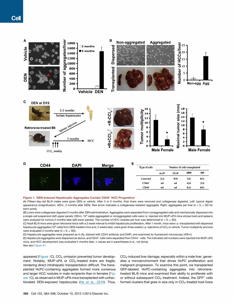
Figure 1. DEN-Induced Hepatocytic Aggregates Contain CD44+ HCC Progenitors
(A) Fifteen-day-old BL/6 males were given DEN or vehicle. After 3 or 5 months, their livers were removed and collagenase digested. Left: typical digest
appearance (magnification: 4003; 3 months after DEN). Red arrow indicates a collagenase-resistant aggregate. Right: aggregates per liver (n = 5; ± SD for
each point).
(B) Livers were collagenase digested 5 months after DEN administration. Aggregates were separated from nonaggregated cells and mechanically dispersed into
a single-cell suspension (left upper panels; 2003). 104 viable aggregated or nonaggregated cells were i.s. injected into MUP-uPA mice whose livers and spleens
were analyzed for tumors 5 months later (left lower panels). The number of HCC nodules per liver was determined (n = 5; ± SD).
(C) Adult BL/6 mice were given retrorsine twice with a 2 week interval to inhibit hepatocyte proliferation. After 1 month, mice were i.s. transplanted with dispersed
hepatocyte aggregates (104 cells) from DEN-treated mice and, 2 weeks later, were given three weekly i.p. injections of CCl4 or vehicle. Tumor multiplicity and size
were evaluated 5 months later (n = 5; ± SD).
(D) Hepatocyte aggregates were prepared as in (A), stained with CD44 antibody and DAPI, and examined by fluorescent microscopy (4003).
(E) Hepatocyte aggregates were dispersed as above, and CD44+ cells were separated fromCD44� cells. The indicated cell numbers were injected intoMUP-uPA
mice, and HCC development was evaluated 5 months later. n values are in parentheses (n.d., not done).
See also Figure S1.
appeared (Figure 1C). CCl4 omission prevented tumor develop-
ment. Notably, MUP-uPA or CCl4-treated livers are fragile,
rendering direct intrahepatic transplantation difficult. The trans-
planted HcPC-containing aggregates formed more numerous
and larger HCC nodules in male recipients than in females (Fig-
ure 1C), as observed inMUP-uPAmice transplanted with unfrac-
tionated DEN-exposed hepatocytes (He et al., 2010). Thus,
386 Cell 155, 384–396, October 10, 2013 ª2013 Elsevier Inc.
CCl4-induced liver damage, especially within a male liver, gener-
ates a microenvironment that drives HcPC proliferation and
malignant progression. To examine this point, we transplanted
GFP-labeled HcPC-containing aggregates into retrorsine-
treated BL/6 mice and examined their ability to proliferate with
or without subsequent CCl4 treatment. Indeed, the GFP+ cells
formed clusters that grew in size only in CCl4-treated host livers
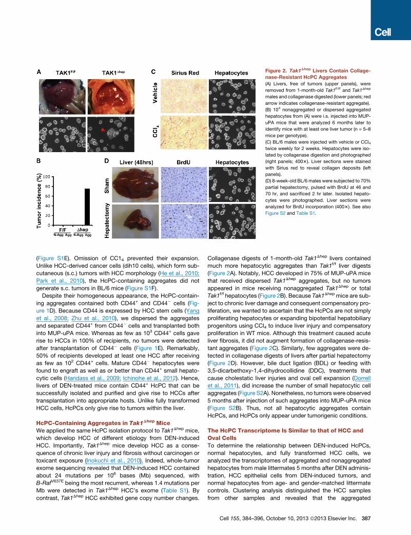
Figure 2. Tak1Dhep Livers Contain Collage-
nase-Resistant HcPC Aggregates
(A) Livers, free of tumors (upper panels), were
removed from 1-month-old Tak1F/F and Tak1Dhep
males and collagenase digested (lower panels; red
arrow indicates collagenase-resistant aggregate).
(B) 104 nonaggregated or dispersed aggregated
hepatocytes from (A) were i.s. injected into MUP-
uPA mice that were analyzed 6 months later to
identify mice with at least one liver tumor (n = 5–8
mice per genotype).
(C) BL/6 males were injected with vehicle or CCl4twice weekly for 2 weeks. Hepatocytes were iso-
lated by collagenase digestion and photographed
(right panels; 4003). Liver sections were stained
with Sirius red to reveal collagen deposits (left
panels).
(D) 8-week-old BL/6 males were subjected to 70%
partial hepatectomy, pulsed with BrdU at 46 and
70 hr, and sacrificed 2 hr later. Isolated hepato-
cytes were photographed. Liver sections were
analyzed for BrdU incorporation (4003). See also
Figure S2 and Table S1.
(Figure S1E). Omission of CC14 prevented their expansion.
Unlike HCC-derived cancer cells (dih10 cells), which form sub-
cutaneous (s.c.) tumors with HCC morphology (He et al., 2010;
Park et al., 2010), the HcPC-containing aggregates did not
generate s.c. tumors in BL/6 mice (Figure S1F).
Despite their homogeneous appearance, the HcPC-contain-
ing aggregates contained both CD44+ and CD44� cells (Fig-
ure 1D). Because CD44 is expressed by HCC stem cells (Yang
et al., 2008; Zhu et al., 2010), we dispersed the aggregates
and separated CD44+ from CD44� cells and transplanted both
into MUP-uPA mice. Whereas as few as 103 CD44+ cells gave
rise to HCCs in 100% of recipients, no tumors were detected
after transplantation of CD44� cells (Figure 1E). Remarkably,
50% of recipients developed at least one HCC after receiving
as few as 102 CD44+ cells. Mature CD44� hepatocytes were
found to engraft as well as or better than CD44+ small hepato-
cytic cells (Haridass et al., 2009; Ichinohe et al., 2012). Hence,
livers of DEN-treated mice contain CD44+ HcPC that can be
successfully isolated and purified and give rise to HCCs after
transplantation into appropriate hosts. Unlike fully transformed
HCC cells, HcPCs only give rise to tumors within the liver.
HcPC-Containing Aggregates in Tak1Dhep MiceWe applied the same HcPC isolation protocol to Tak1Dhep mice,
which develop HCC of different etiology from DEN-induced
HCC. Importantly, Tak1Dhep mice develop HCC as a conse-
quence of chronic liver injury and fibrosis without carcinogen or
toxicant exposure (Inokuchi et al., 2010). Indeed, whole-tumor
exome sequencing revealed that DEN-induced HCC contained
about 24 mutations per 106 bases (Mb) sequenced, with
B-RafV637E being the most recurrent, whereas 1.4 mutations per
Mb were detected in Tak1Dhep HCC’s exome (Table S1). By
contrast, Tak1Dhep HCC exhibited gene copy number changes.
Collagenase digests of 1-month-old Tak1Dhep livers contained
much more hepatocytic aggregates than Tak1f/f liver digests
(Figure 2A). Notably, HCC developed in 75% of MUP-uPA mice
that received dispersed Tak1Dhep aggregates, but no tumors
appeared in mice receiving nonaggregated Tak1Dhep or total
Tak1f/f hepatocytes (Figure 2B). Because Tak1Dhep mice are sub-
ject to chronic liver damage and consequent compensatory pro-
liferation, we wanted to ascertain that the HcPCs are not simply
proliferating hepatocytes or expanding bipotential hepatobiliary
progenitors using CCl4 to induce liver injury and compensatory
proliferation in WT mice. Although this treatment caused acute
liver fibrosis, it did not augment formation of collagenase-resis-
tant aggregates (Figure 2C). Similarly, few aggregates were de-
tected in collagenase digests of livers after partial hepatectomy
(Figure 2D). However, bile duct ligation (BDL) or feeding with
3,5-dicarbethoxy-1,4-dihydrocollidine (DDC), treatments that
cause cholestatic liver injuries and oval cell expansion (Dorrell
et al., 2011), did increase the number of small hepatocytic cell
aggregates (Figure S2A). Nonetheless, no tumors were observed
5 months after injection of such aggregates into MUP-uPA mice
(Figure S2B). Thus, not all hepatocytic aggregates contain
HcPCs, and HcPCs only appear under tumorigenic conditions.
The HcPC Transcriptome Is Similar to that of HCC andOval CellsTo determine the relationship between DEN-induced HcPCs,
normal hepatocytes, and fully transformed HCC cells, we
analyzed the transcriptomes of aggregated and nonaggregated
hepatocytes from male littermates 5 months after DEN adminis-
tration, HCC epithelial cells from DEN-induced tumors, and
normal hepatocytes from age- and gender-matched littermate
controls. Clustering analysis distinguished the HCC samples
from other samples and revealed that the aggregated
Cell 155, 384–396, October 10, 2013 ª2013 Elsevier Inc. 387
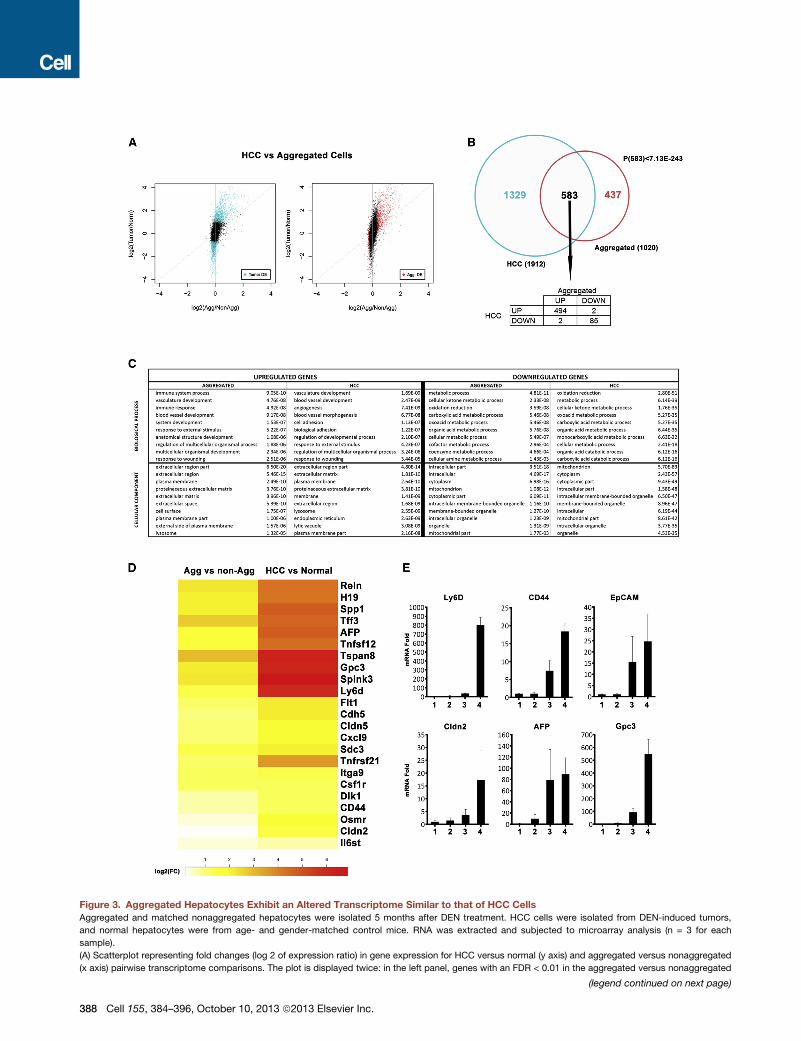
Figure 3. Aggregated Hepatocytes Exhibit an Altered Transcriptome Similar to that of HCC Cells
Aggregated and matched nonaggregated hepatocytes were isolated 5 months after DEN treatment. HCC cells were isolated from DEN-induced tumors,
and normal hepatocytes were from age- and gender-matched control mice. RNA was extracted and subjected to microarray analysis (n = 3 for each
sample).
(A) Scatterplot representing fold changes (log 2 of expression ratio) in gene expression for HCC versus normal (y axis) and aggregated versus nonaggregated
(x axis) pairwise transcriptome comparisons. The plot is displayed twice: in the left panel, genes with an FDR < 0.01 in the aggregated versus nonaggregated
(legend continued on next page)
388 Cell 155, 384–396, October 10, 2013 ª2013 Elsevier Inc.
hepatocyte samples did not cluster with each other but rather
with nonaggregated hepatocytes derived from the same mouse
(Figure S3A). Interestingly, the aggregated cell transcriptome ap-
peared closer to that of normal hepatocytes than to the HCC
profile. This similarity may be due to the presence of �70%
nontumorigenic (or CD44�) hepatocytes within the purified ag-
gregates (Figure 1D). Comparison of the HCC and normal hepa-
tocyte transcriptomes revealed 1,912 differentially expressed
genes (false discovery rate [FDR] < 0.01; Figure 3A, left, cyan
dots). A similar comparison revealed 1,020 genes that are differ-
entially expressed between aggregated and nonaggregated
hepatocytes (FDR < 0.01; Figure 3A, right, red dots). The range
of differential expression is wider for the HCC and normal hepa-
tocyte pair than the aggregate versus nonaggregate pair, reflect-
ing presence of normal, nontransformed hepatocytes within the
aggregates, resulting in signal dilution. Interestingly, 57% (583/
1,020) of genes differentially expressed in aggregated relative
to nonaggregated hepatocytes are also differentially expressed
in HCC relative to normal hepatocytes (Figure 3B, top), a value
that is highly significant (p < 7.13 3 10�243). More specifically,
85% (494/583) of these genes are overexpressed in both HCC
and HcPC-containing aggregates (Figure 3B, bottom table).
Thus, hepatocyte aggregates isolated 5 months after DEN injec-
tion contain cells that are related in their gene expression profile
to HCC cells isolated from fully developed tumor nodules.
To gain insight into the functional differences between the
transcriptomes of the four populations, we examined which bio-
logical processes or cellular compartments were significantly
overrepresented in the induced or repressed genes in both pair-
wise comparisons (Gene Ontology Analysis). As expected, pro-
cesses and compartments that were enriched in aggregated
hepatocytes relative to nonaggregated hepatocytes were almost
identical to those that were enriched in HCC relative to normal
hepatocytes (Figure 3C). Upregulated genes were related to
immune response, angiogenesis, development, andwound heal-
ing, and many encoded plasma membrane or secreted proteins.
By contrast, downregulated genes were highly enriched for
metabolic processes, and many of them encoded mitochondrial
proteins or had functions associated with differentiated hepato-
cytes (Figure 3C). Several human HCC markers, including AFP,
Gpc3 and H19, were upregulated in aggregated hepatocytes
(Figures 3D and 3E). Aggregated hepatocytes also expressed
more Tetraspanin 8 (Tspan8), a cell-surface glycoprotein that
complexes with integrins and is overexpressed in human carci-
comparison are highlighted in red, and in the right panel, genes with an FDR < 0.01
expressed.
(B) Venn diagram showing overlap between genes that are differentially expressed
and normal hepatocytes with an FDR < 0.01 (cyan and red dots from A). The proba
genes, only 4 behaved differently.
(C) The tenmost enriched biological processes (upper table) and cellular compartm
panel) or downregulated (right panel) in HCC relative to normal hepatocytes (HC
(D) Heatmap displaying positive fold changes (FC) in expression of genes of intere
hepatocytes (right).
(E) Expression of selected genes was examined by real-time PCR and is depicte
(n = 3; ± SD). (1) Normal hepatocytes; (2) nonaggregated hepatocytes from D
induced HCCs.
See also Figure S3.
nomas (Zoller, 2009). Another cell-surface molecule highly
expressed in aggregated cells is Ly6D (Figures 3D and 3E).
Immunofluorescence (IF) analysis revealed that Ly6D was unde-
tectable in normal liver but was elevated in FAH and ubiquitously
expressed in most HCC cells (Figure S3C). A fluorescent-labeled
Ly6D antibody injected into HCC-bearing mice specifically
stained tumor nodules (FigureS3D).Other cell-surfacemolecules
that were upregulated in aggregated cells included syndecan 3
(Sdc3), integrin a 9 (Itga9), claudin 5 (Cldn5), and cadherin 5
(Cdh5) (Figure 3D). Aggregated hepatocytes also exhibited
elevated expression of extracellular matrix proteins (TIF3 and
Reln1) and a serine protease inhibitor (Spink3). Elevated expres-
sion of such proteins may explain aggregate formation. Aggre-
gated hepatocytes also expressed progenitor cell markers,
including the epithelial cell adhesion molecule (EpCAM) (Fig-
ure 3E) and Dlk1 (Figure 3D). Elevated expression of cytokines
and cytokine receptors was also detected, including tumor ne-
crosis factor superfamilymembers 12 and 21, colony-stimulating
factor 1 receptor, FMS-like tyrosine kinase 1, chemokine (C-X-C
motif) ligand 9, the STAT3-activating cytokine osteopontin, IL-6
receptor (IL-6R) signal transducing subunit (gp130), andoncosta-
tin M (OSM) receptor, which also activates STAT3 (Figure 3D).
Aggregated hepatocytes expressed albumin, albeit less than
nonaggregated hepatocytes (Figure 4A). Some aggregated cells
were positive for cytokeratin 19 (CK19) and A6, markers for bile
duct epithelium and oval cells (Figure 4A). Most cells in the DEN-
induced aggregates were AFP positive, and some of them
expressed EpCAM (Figure 4A). However, not all markers were
expressed by every cell within a given aggregate, suggesting
that the aggregates contain liver cells that are related to bipoten-
tial hepatobiliary progenitors/oval cells as well as more differen-
tiated progeny and normal hepatocytes. To confirm these
observations, we compared the HcPC and HCC (Figure 3A) to
the transcriptome of DDC-induced oval cells (Shin et al., 2011).
This analysis revealed a striking similarity between the HCC,
HcPC, and the oval cell transcriptomes (Figure S3B). Despite
these similarities, some genes that were upregulated in HcPC-
containing aggregates and HCC were not upregulated in oval
cells. Such genes may account for the tumorigenic properties
of HcPC and HCC.
We examined the aggregates for signaling pathways and
transcription factors involved in hepatocarcinogenesis. Many
aggregated cells were positive for phosphorylated c-Jun and
STAT3 (Figure 4A), transcription factors involved in DEN-induced
in the HCC versus normal comparison are highlighted in cyan. DE, differentially
between aggregated and nonaggregated hepatocytes and between HCC cells
bility to find 583 overlapping genes is <7.133 10�243. From these 583 common
ents (lower panel) represented by genes that are significantly upregulated (left
C) or in aggregated relative to nonaggregated hepatocytes (aggregated).
st in aggregated versus nonaggregated HcPCs (left) and in HCC versus normal
d as fold change relative to normal hepatocytes given an arbitrary value of 1.0
EN-treated liver; (3) HcPC aggregates from DEN-treated liver; and (4) DEN-
Cell 155, 384–396, October 10, 2013 ª2013 Elsevier Inc. 389
B
50µm
A
Bright DAPI BrdU Merge
Bright
Bright
Bright
Bright
Bright
DAPI
DAPI
DAPI
DAPI
DAPI
Merge
Merge
Merge
Merge
Merge
MergeCK19
AFP
EpCAM
A6
p-STAT3
AlbuminDAPI
DAPI
DAPI
DAPI
Bright
Bright
Bright
Bright
p-cJUN Merge
Merge
Merge
SOX9
p-cMet
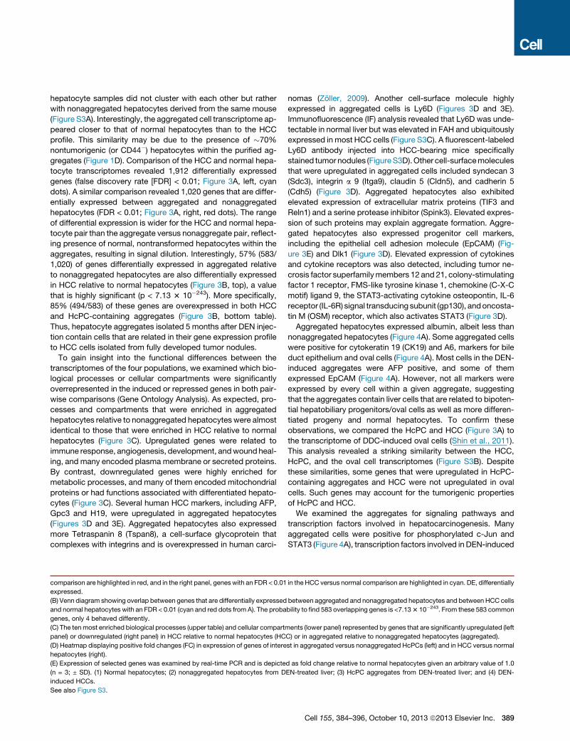
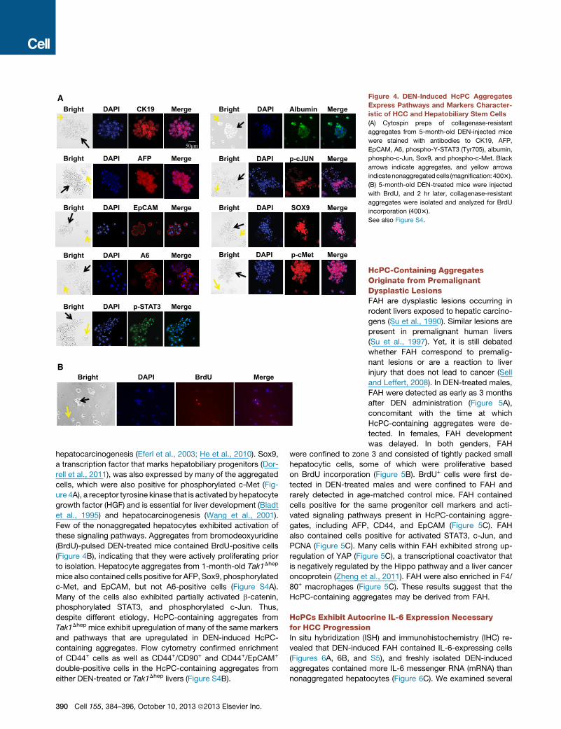
Figure 4. DEN-Induced HcPC Aggregates
Express Pathways and Markers Character-
istic of HCC and Hepatobiliary Stem Cells
(A) Cytospin preps of collagenase-resistant
aggregates from 5-month-old DEN-injected mice
were stained with antibodies to CK19, AFP,
EpCAM, A6, phospho-Y-STAT3 (Tyr705), albumin,
phospho-c-Jun, Sox9, and phospho-c-Met. Black
arrows indicate aggregates, and yellow arrows
indicatenonaggregatedcells (magnification: 4003).
(B) 5-month-old DEN-treated mice were injected
with BrdU, and 2 hr later, collagenase-resistant
aggregates were isolated and analyzed for BrdU
incorporation (4003).
See also Figure S4.
hepatocarcinogenesis (Eferl et al., 2003; He et al., 2010). Sox9,
a transcription factor that marks hepatobiliary progenitors (Dor-
rell et al., 2011), was also expressed by many of the aggregated
cells, which were also positive for phosphorylated c-Met (Fig-
ure 4A), a receptor tyrosine kinase that is activated by hepatocyte
growth factor (HGF) and is essential for liver development (Bladt
et al., 1995) and hepatocarcinogenesis (Wang et al., 2001).
Few of the nonaggregated hepatocytes exhibited activation of
these signaling pathways. Aggregates from bromodeoxyuridine
(BrdU)-pulsed DEN-treated mice contained BrdU-positive cells
(Figure 4B), indicating that they were actively proliferating prior
to isolation. Hepatocyte aggregates from 1-month-old Tak1Dhep
mice also contained cells positive for AFP, Sox9, phosphorylated
c-Met, and EpCAM, but not A6-positive cells (Figure S4A).
Many of the cells also exhibited partially activated b-catenin,
phosphorylated STAT3, and phosphorylated c-Jun. Thus,
despite different etiology, HcPC-containing aggregates from
Tak1Dhep mice exhibit upregulation of many of the same markers
and pathways that are upregulated in DEN-induced HcPC-
containing aggregates. Flow cytometry confirmed enrichment
of CD44+ cells as well as CD44+/CD90+ and CD44+/EpCAM+
double-positive cells in the HcPC-containing aggregates from
either DEN-treated or Tak1Dhep livers (Figure S4B).
390 Cell 155, 384–396, October 10, 2013 ª2013 Elsevier Inc.
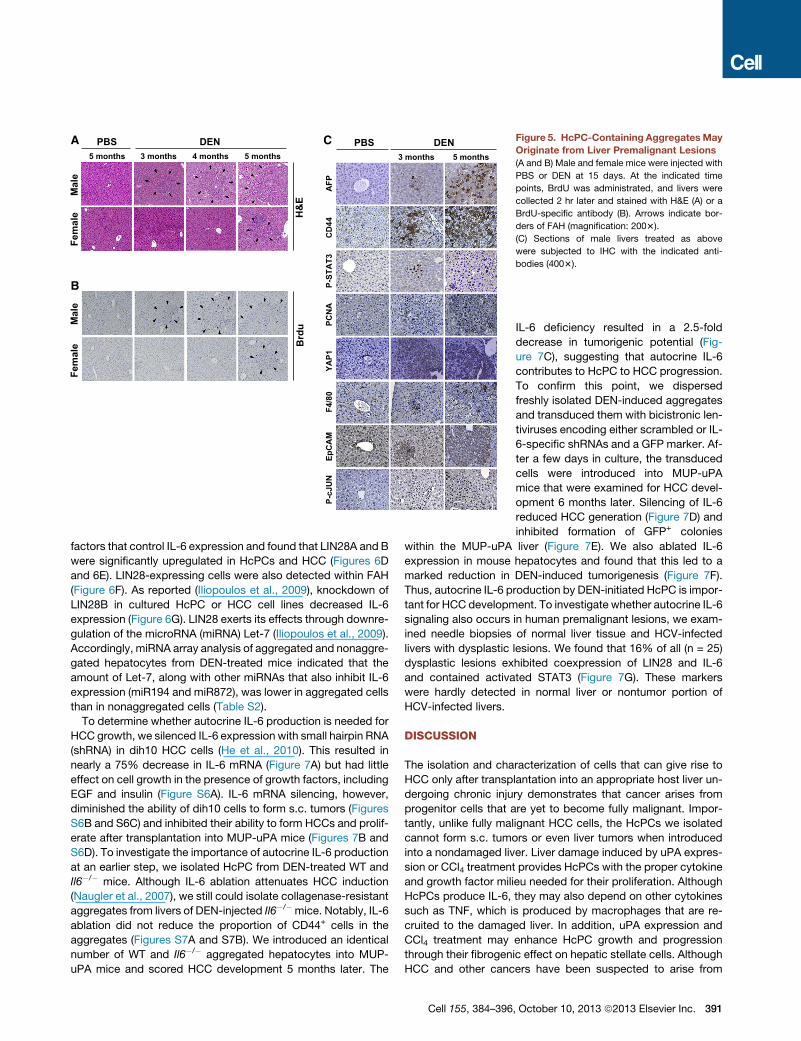
HcPC-Containing AggregatesOriginate from PremalignantDysplastic LesionsFAH are dysplastic lesions occurring in
rodent livers exposed to hepatic carcino-
gens (Su et al., 1990). Similar lesions are
present in premalignant human livers
(Su et al., 1997). Yet, it is still debated
whether FAH correspond to premalig-
nant lesions or are a reaction to liver
injury that does not lead to cancer (Sell
and Leffert, 2008). In DEN-treated males,
FAH were detected as early as 3 months
after DEN administration (Figure 5A),
concomitant with the time at which
HcPC-containing aggregates were de-
tected. In females, FAH development
was delayed. In both genders, FAH
were confined to zone 3 and consisted of tightly packed small
hepatocytic cells, some of which were proliferative based
on BrdU incorporation (Figure 5B). BrdU+ cells were first de-
tected in DEN-treated males and were confined to FAH and
rarely detected in age-matched control mice. FAH contained
cells positive for the same progenitor cell markers and acti-
vated signaling pathways present in HcPC-containing aggre-
gates, including AFP, CD44, and EpCAM (Figure 5C). FAH
also contained cells positive for activated STAT3, c-Jun, and
PCNA (Figure 5C). Many cells within FAH exhibited strong up-
regulation of YAP (Figure 5C), a transcriptional coactivator that
is negatively regulated by the Hippo pathway and a liver cancer
oncoprotein (Zheng et al., 2011). FAH were also enriched in F4/
80+ macrophages (Figure 5C). These results suggest that the
HcPC-containing aggregates may be derived from FAH.
HcPCs Exhibit Autocrine IL-6 Expression Necessaryfor HCC ProgressionIn situ hybridization (ISH) and immunohistochemistry (IHC) re-
vealed that DEN-induced FAH contained IL-6-expressing cells
(Figures 6A, 6B, and S5), and freshly isolated DEN-induced
aggregates contained more IL-6 messenger RNA (mRNA) than
nonaggregated hepatocytes (Figure 6C). We examined several
A
B
C Figure 5. HcPC-Containing AggregatesMay
Originate from Liver Premalignant Lesions
(A and B) Male and female mice were injected with
PBS or DEN at 15 days. At the indicated time
points, BrdU was administrated, and livers were
collected 2 hr later and stained with H&E (A) or a
BrdU-specific antibody (B). Arrows indicate bor-
ders of FAH (magnification: 2003).
(C) Sections of male livers treated as above
were subjected to IHC with the indicated anti-
bodies (4003).
factors that control IL-6 expression and found that LIN28A and B
were significantly upregulated in HcPCs and HCC (Figures 6D
and 6E). LIN28-expressing cells were also detected within FAH
(Figure 6F). As reported (Iliopoulos et al., 2009), knockdown of
LIN28B in cultured HcPC or HCC cell lines decreased IL-6
expression (Figure 6G). LIN28 exerts its effects through downre-
gulation of the microRNA (miRNA) Let-7 (Iliopoulos et al., 2009).
Accordingly, miRNA array analysis of aggregated and nonaggre-
gated hepatocytes from DEN-treated mice indicated that the
amount of Let-7, along with other miRNAs that also inhibit IL-6
expression (miR194 and miR872), was lower in aggregated cells
than in nonaggregated cells (Table S2).
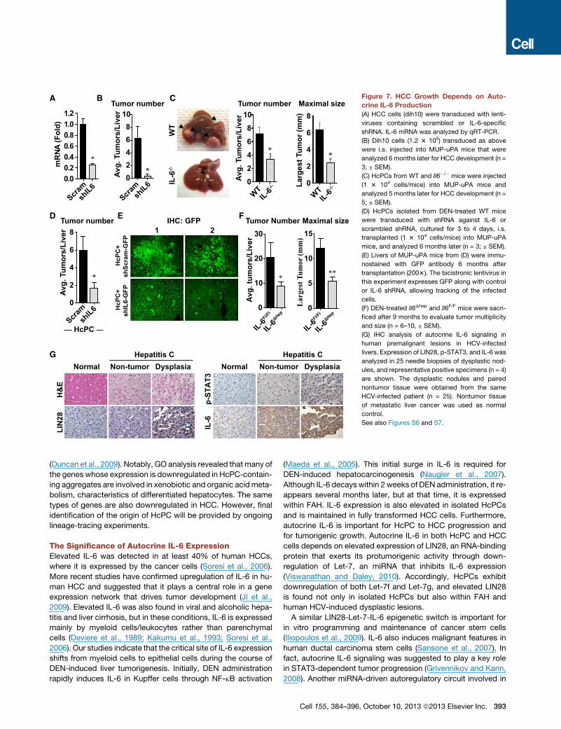
To determine whether autocrine IL-6 production is needed for
HCC growth, we silenced IL-6 expression with small hairpin RNA
(shRNA) in dih10 HCC cells (He et al., 2010). This resulted in
nearly a 75% decrease in IL-6 mRNA (Figure 7A) but had little
effect on cell growth in the presence of growth factors, including
EGF and insulin (Figure S6A). IL-6 mRNA silencing, however,
diminished the ability of dih10 cells to form s.c. tumors (Figures
S6B and S6C) and inhibited their ability to form HCCs and prolif-
erate after transplantation into MUP-uPA mice (Figures 7B and
S6D). To investigate the importance of autocrine IL-6 production
at an earlier step, we isolated HcPC from DEN-treated WT and
Il6�/� mice. Although IL-6 ablation attenuates HCC induction
(Naugler et al., 2007), we still could isolate collagenase-resistant
aggregates from livers of DEN-injected Il6�/�mice. Notably, IL-6
ablation did not reduce the proportion of CD44+ cells in the
aggregates (Figures S7A and S7B). We introduced an identical
number of WT and Il6�/� aggregated hepatocytes into MUP-
uPA mice and scored HCC development 5 months later. The
Cell 155, 384–396
IL-6 deficiency resulted in a 2.5-fold
decrease in tumorigenic potential (Fig-
ure 7C), suggesting that autocrine IL-6
contributes to HcPC to HCC progression.
To confirm this point, we dispersed
freshly isolated DEN-induced aggregates
and transduced them with bicistronic len-
tiviruses encoding either scrambled or IL-
6-specific shRNAs and a GFP marker. Af-
ter a few days in culture, the transduced
cells were introduced into MUP-uPA
mice that were examined for HCC devel-
opment 6 months later. Silencing of IL-6
reduced HCC generation (Figure 7D) and
inhibited formation of GFP+ colonies
within the MUP-uPA liver (Figure 7E). We also ablated IL-6
expression in mouse hepatocytes and found that this led to a
marked reduction in DEN-induced tumorigenesis (Figure 7F).
Thus, autocrine IL-6 production by DEN-initiated HcPC is impor-
tant for HCC development. To investigate whether autocrine IL-6
signaling also occurs in human premalignant lesions, we exam-
ined needle biopsies of normal liver tissue and HCV-infected
livers with dysplastic lesions. We found that 16% of all (n = 25)
dysplastic lesions exhibited coexpression of LIN28 and IL-6
and contained activated STAT3 (Figure 7G). These markers
were hardly detected in normal liver or nontumor portion of
HCV-infected livers.
DISCUSSION
The isolation and characterization of cells that can give rise to
HCC only after transplantation into an appropriate host liver un-
dergoing chronic injury demonstrates that cancer arises from
progenitor cells that are yet to become fully malignant. Impor-
tantly, unlike fully malignant HCC cells, the HcPCs we isolated
cannot form s.c. tumors or even liver tumors when introduced
into a nondamaged liver. Liver damage induced by uPA expres-
sion or CCl4 treatment provides HcPCs with the proper cytokine
and growth factor milieu needed for their proliferation. Although
HcPCs produce IL-6, they may also depend on other cytokines
such as TNF, which is produced by macrophages that are re-
cruited to the damaged liver. In addition, uPA expression and
CCl4 treatment may enhance HcPC growth and progression
through their fibrogenic effect on hepatic stellate cells. Although
HCC and other cancers have been suspected to arise from
, October 10, 2013 ª2013 Elsevier Inc. 391
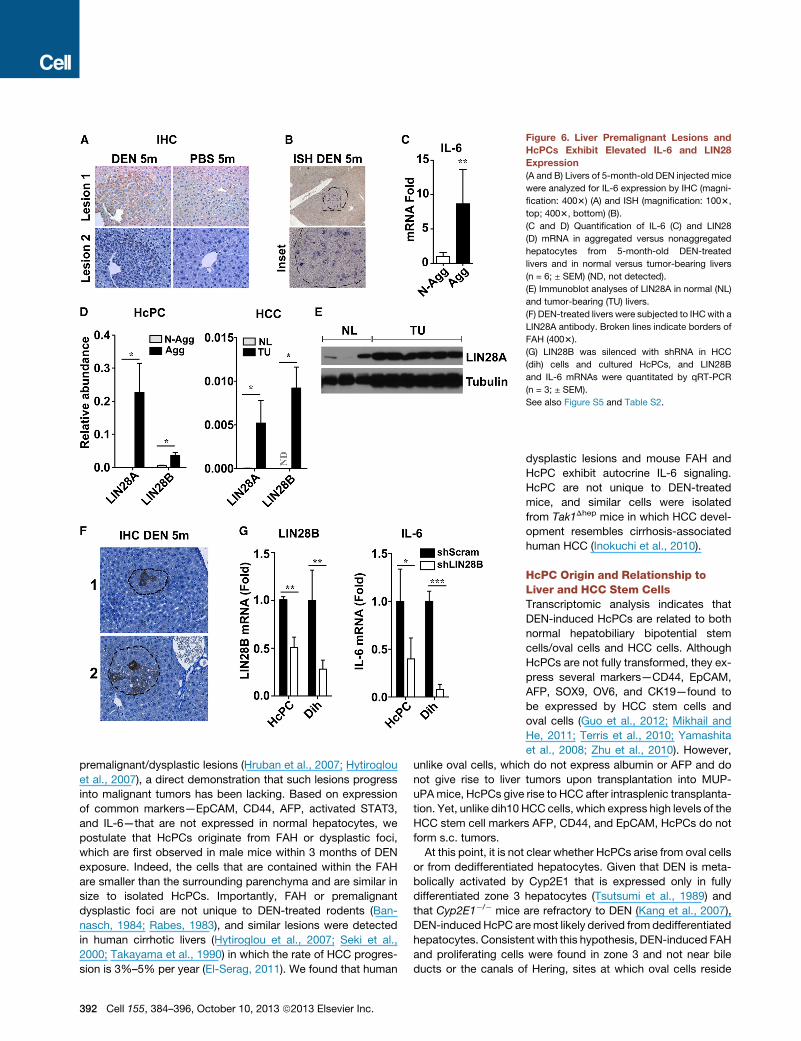
Figure 6. Liver Premalignant Lesions and
HcPCs Exhibit Elevated IL-6 and LIN28
Expression
(A and B) Livers of 5-month-old DEN injected mice
were analyzed for IL-6 expression by IHC (magni-
fication: 4003) (A) and ISH (magnification: 1003,
top; 4003, bottom) (B).
(C and D) Quantification of IL-6 (C) and LIN28
(D) mRNA in aggregated versus nonaggregated
hepatocytes from 5-month-old DEN-treated
livers and in normal versus tumor-bearing livers
(n = 6; ± SEM) (ND, not detected).
(E) Immunoblot analyses of LIN28A in normal (NL)
and tumor-bearing (TU) livers.
(F) DEN-treated livers were subjected to IHCwith a
LIN28A antibody. Broken lines indicate borders of
FAH (4003).
(G) LIN28B was silenced with shRNA in HCC
(dih) cells and cultured HcPCs, and LIN28B
and IL-6 mRNAs were quantitated by qRT-PCR
(n = 3; ± SEM).
See also Figure S5 and Table S2.
premalignant/dysplastic lesions (Hruban et al., 2007; Hytiroglou
et al., 2007), a direct demonstration that such lesions progress
into malignant tumors has been lacking. Based on expression
of common markers—EpCAM, CD44, AFP, activated STAT3,
and IL-6—that are not expressed in normal hepatocytes, we
postulate that HcPCs originate from FAH or dysplastic foci,
which are first observed in male mice within 3 months of DEN
exposure. Indeed, the cells that are contained within the FAH
are smaller than the surrounding parenchyma and are similar in
size to isolated HcPCs. Importantly, FAH or premalignant
dysplastic foci are not unique to DEN-treated rodents (Ban-
nasch, 1984; Rabes, 1983), and similar lesions were detected
in human cirrhotic livers (Hytiroglou et al., 2007; Seki et al.,
2000; Takayama et al., 1990) in which the rate of HCC progres-
sion is 3%–5% per year (El-Serag, 2011). We found that human
392 Cell 155, 384–396, October 10, 2013 ª2013 Elsevier Inc.
dysplastic lesions and mouse FAH and
HcPC exhibit autocrine IL-6 signaling.
HcPC are not unique to DEN-treated
mice, and similar cells were isolated
from Tak1Dhep mice in which HCC devel-
opment resembles cirrhosis-associated
human HCC (Inokuchi et al., 2010).
HcPC Origin and Relationship toLiver and HCC Stem CellsTranscriptomic analysis indicates that
DEN-induced HcPCs are related to both
normal hepatobiliary bipotential stem
cells/oval cells and HCC cells. Although
HcPCs are not fully transformed, they ex-
press several markers—CD44, EpCAM,
AFP, SOX9, OV6, and CK19—found to
be expressed by HCC stem cells and
oval cells (Guo et al., 2012; Mikhail and
He, 2011; Terris et al., 2010; Yamashita
et al., 2008; Zhu et al., 2010). However,
unlike oval cells, which do not express albumin or AFP and do
not give rise to liver tumors upon transplantation into MUP-
uPAmice, HcPCs give rise to HCC after intrasplenic transplanta-
tion. Yet, unlike dih10 HCC cells, which express high levels of the
HCC stem cell markers AFP, CD44, and EpCAM, HcPCs do not
form s.c. tumors.
At this point, it is not clear whether HcPCs arise from oval cells
or from dedifferentiated hepatocytes. Given that DEN is meta-
bolically activated by Cyp2E1 that is expressed only in fully
differentiated zone 3 hepatocytes (Tsutsumi et al., 1989) and
that Cyp2E1�/� mice are refractory to DEN (Kang et al., 2007),
DEN-inducedHcPC aremost likely derived fromdedifferentiated
hepatocytes. Consistent with this hypothesis, DEN-induced FAH
and proliferating cells were found in zone 3 and not near bile
ducts or the canals of Hering, sites at which oval cells reside
A
Scramsh
IL602468
10
B
mR
NA (F
old)
0.00.20.40.60.81.01.2
Scramsh
IL6
C
WT
**
IL-6
-/-
0
2
4
6
8
10
0
2
4
6
8
D
Scramsh
IL60
2
4
6
8
Avg.
Tum
ors/
Live
r
Avg.
Tum
ors/
Live
r
Avg.
Tum
ors/
Live
r
Tumor number Maximal size
Tumor number
*
WTIL-6
-/-
WTIL-6
-/-
Tumor number
**
HcP
C+
shSc
ram
-GFP
H
cPC
+sh
IL6-
GFP
IHC: GFPE
1 2
IL-6Fl/F
l
Avg.
tum
ors/
Live
r
Tumor Number Maximal size
IL-6ΔHep
**
Lar
gest
Tum
or (m
m)
F
IL-6ΔHep
IL-6Fl/F
l0
10
20
30
*
0
5
10
15
Larg
est T
umor
(mm
)
Non-tumor Dysplasia
LIN
28
IL-6
p-ST
AT3
H&
E
NormalHepatitis CG
HcPC
Non-tumor DysplasiaNormalHepatitis C
Figure 7. HCC Growth Depends on Auto-
crine IL-6 Production
(A) HCC cells (dih10) were transduced with lenti-
viruses containing scrambled or IL-6-specific
shRNA. IL-6 mRNA was analyzed by qRT-PCR.
(B) Dih10 cells (1.2 3 105) transduced as above
were i.s. injected into MUP-uPA mice that were
analyzed 6 months later for HCC development (n =
3; ± SEM).
(C) HcPCs from WT and Il6�/� mice were injected
(1 3 104 cells/mice) into MUP-uPA mice and
analyzed 5 months later for HCC development (n =
5; ± SEM).
(D) HcPCs isolated from DEN-treated WT mice
were transduced with shRNA against IL-6 or
scrambled shRNA, cultured for 3 to 4 days, i.s.
transplanted (1 3 104 cells/mice) into MUP-uPA
mice, and analyzed 6 months later (n = 3; ± SEM).
(E) Livers of MUP-uPA mice from (D) were immu-
nostained with GFP antibody 6 months after
transplantation (2003). The bicistronic lentivirus in
this experiment expresses GFP along with control
or IL-6 shRNA, allowing tracking of the infected
cells.
(F) DEN-treated Il6Dhep and Il6F/F mice were sacri-
ficed after 9 months to evaluate tumor multiplicity
and size (n = 6–10, ± SEM).
(G) IHC analysis of autocrine IL-6 signaling in
human premalignant lesions in HCV-infected
livers. Expression of LIN28, p-STAT3, and IL-6 was
analyzed in 25 needle biopsies of dysplastic nod-
ules, and representative positive specimens (n = 4)
are shown. The dysplastic nodules and paired
nontumor tissue were obtained from the same
HCV-infected patient (n = 25). Nontumor tissue
of metastatic liver cancer was used as normal
control.
See also Figures S6 and S7.
(Duncan et al., 2009). Notably, GO analysis revealed that many of
the genes whose expression is downregulated in HcPC-contain-
ing aggregates are involved in xenobiotic and organic acidmeta-
bolism, characteristics of differentiated hepatocytes. The same
types of genes are also downregulated in HCC. However, final
identification of the origin of HcPC will be provided by ongoing
lineage-tracing experiments.
The Significance of Autocrine IL-6 ExpressionElevated IL-6 was detected in at least 40% of human HCCs,
where it is expressed by the cancer cells (Soresi et al., 2006).
More recent studies have confirmed upregulation of IL-6 in hu-
man HCC and suggested that it plays a central role in a gene
expression network that drives tumor development (Ji et al.,
2009). Elevated IL-6 was also found in viral and alcoholic hepa-
titis and liver cirrhosis, but in these conditions, IL-6 is expressed
mainly by myeloid cells/leukocytes rather than parenchymal
cells (Deviere et al., 1989; Kakumu et al., 1993; Soresi et al.,
2006). Our studies indicate that the critical site of IL-6 expression
shifts from myeloid cells to epithelial cells during the course of
DEN-induced liver tumorigenesis. Initially, DEN administration
rapidly induces IL-6 in Kupffer cells through NF-kB activation
(Maeda et al., 2005). This initial surge in IL-6 is required for
DEN-induced hepatocarcinogenesis (Naugler et al., 2007).
Although IL-6 decays within 2 weeks of DEN administration, it re-
appears several months later, but at that time, it is expressed
within FAH. IL-6 expression is also elevated in isolated HcPCs
and is maintained in fully transformed HCC cells. Furthermore,
autocrine IL-6 is important for HcPC to HCC progression and
for tumorigenic growth. Autocrine IL-6 in both HcPC and HCC
cells depends on elevated expression of LIN28, an RNA-binding
protein that exerts its protumorigenic activity through down-
regulation of Let-7, an miRNA that inhibits IL-6 expression
(Viswanathan and Daley, 2010). Accordingly, HcPCs exhibit
downregulation of both Let-7f and Let-7g, and elevated LIN28
is found not only in isolated HcPCs but also within FAH and
human HCV-induced dysplastic lesions.
A similar LIN28-Let-7-IL-6 epigenetic switch is important for
in vitro programming and maintenance of cancer stem cells
(Iliopoulos et al., 2009). IL-6 also induces malignant features in
human ductal carcinoma stem cells (Sansone et al., 2007). In
fact, autocrine IL-6 signaling was suggested to play a key role
in STAT3-dependent tumor progression (Grivennikov and Karin,
2008). Another miRNA-driven autoregulatory circuit involved in
Cell 155, 384–396, October 10, 2013 ª2013 Elsevier Inc. 393
hepatocarcinogenesis accounts for elevated IL-6R expression
(Hatziapostolou et al., 2011). Yet, HcPC-containing aggregates
also express several other STAT3-activiting cytokines and re-
ceptors. Accordingly, silencing or ablation of IL-6 results in
incomplete inhibition of HcPC to HCCprogression. Nonetheless,
our results demonstrate that autoregulatory circuits/epigenetic
switches play an important role in the very early stages of tumor-
igenesis. Given that such circuits are already activated in prema-
lignant cells, pharmacological agents that disrupt their function
may be useful in cancer prevention. Prevention is of particular
importance in cancers such as HCC, which is often detected
at a stage that is refractory to currently available therapeutics.
EXPERIMENTAL PROCEDURES
Mice, HCC Induction, HcPC Isolation, and Transplantation
MUP-uPA transgenic mice (Weglarz et al., 2000) were maintained on a pure
BL/6 background. Because homozygous females frequently die when preg-
nant, MUP-uPA heterozygotes were generated by backcrossing homozygous
MUP-uPA males with BL/6 females to be used as recipients for hepatic trans-
plantation. Tak1Dhep (Inokuchi et al., 2010) and Il6F/F (Quintana et al., 2013)
mice were also in the BL/6 background. Il6Dhep mice were generated by
crossing Il6F/F and Alb-Cre mice. C57BL/6 actin-GFP mice were from the
Jackson Laboratories. BL/6 mice were purchased from Charles River
Laboratories.
To induce HCC, 15-day-old mice were injected i.p. with 25 mg/kg DEN
(Sigma). A pool of DEN-injected BL/6 mice was maintained and used in
most experiments. Hepatocytes were isolated using a two-step procedure
(He et al., 2010). Cell aggregates were isolated by filtration through 70 and
40 mmsieves. To disperse the aggregates into single cells, theywere subjected
to gentle pipetting in Ca/Mg-free PBS on ice. Single-cell suspensions of aggre-
gated and nonaggregated hepatocytes were transplanted via an i.s. injection
into 21-day-old male MUP-uPA mice (He et al., 2010). Alternatively, single-
cell suspensions of aggregated hepatocytes were enriched for CD44+ HcPC
using magnetic beads. As few as 100 viable CD44+ cells mixed with 1 3 105
normal hepatocytes from normal males were transplanted into MUP-uPA
mice. Alternatively, BL/6 mice were pretreated with retrorsine (70 mg/kg i.p.)
(Sigma), a cell-cycle inhibitor, 1 month prior to transplantation. Transplanted
mice were allowed to recover for 1 week and then injected weekly with 33
0.5 ml/kg CCl4 i.p. to induce liver injury and hepatocyte proliferation (Guo
et al., 2002). Mice were sacrificed 5 to 6 months later, and tumors bigger
than 1 mm in diameter on the liver surface were counted. Tumors bigger
than 5 mm across were dissected for biochemical and molecular analyses.
ACCESSION NUMBERS
Raw gene expression array data have been deposited to NCBI’s Gene
Expression Omnibus under the GSE50431 study.
SUPPLEMENTAL INFORMATION
Supplemental Information includes Extended Experimental Procedures, seven
figures, and three tables and can be found with this article online at http://dx.
doi.org/10.1016/j.cell.2013.09.031.
AUTHOR CONTRIBUTIONS
G.H. identified, isolated, and characterized HcPCs; D.D. and H.N. optimized
the HcPC isolation and purification procedure; D.D. found the mechanism of
their dependence on autocrine IL-6 controlled by LIN28, characterized them
using flow cytometry (with S.S.), and conducted miR analyses (with M.H.
and D.I.); H.N. and D.D. used Il6Dhep mice to demonstrate in vivo HCC depen-
dency on autocrine IL-6; H.N. (with R.T. and K.K.) found IL-6, LIN28, and
P-STAT3 in human dysplastic lesions; J.F.-B. conducted the transcriptome
394 Cell 155, 384–396, October 10, 2013 ª2013 Elsevier Inc.
analysis and exome sequencing (with S.E.Y., K.J., and O.H.) and with H.O.
examined oncogenic potential of oval cells; H.O. examined HcPC proliferative
potential and performed IF analysis of isolated HcPC (with A.S. and R.M.H.);
Y.J. assisted with IHC and ISH staining; E.S. contributed to the experiments
involving Tak1Dhep mice; G.H., D.D., J.F.-B., H.O., and M.K. wrote the
manuscript.
ACKNOWLEDGMENTS
We acknowledge the Biogem facility at UCSD for their assistance with tran-
scriptome analysis and A. Arian, K. Iwaisako, Y. Hiroshima, and H. Matsui
for technical assistance. We thank Dr. J. Hidalgo (Universitat Autonoma de
Barcelona, Spain) for the Il6F/F mice. Research was supported by the Super-
fund Basic Research Program (P42ES010337), NIH (CA118165 and
CA155120), Wellcome Trust (WT086755), American Diabetes Association (7-
08-MN-29), the Center for Translational Science (UL1RR031980 and
UL1TR000100), the National Center for Research Resources IMAT program
(N12R1CA155615), and postdoctoral research fellowships from the Damon
Runyon Cancer Research Foundation (G.H.), American Liver Foundation
(D.D.), Daiichi Sankyo Foundation of Life Science (H.N.), California Institute
for Regenerative Medicine Stem Cell Training Grant II (TG2-01154) fellowship
(J.F.-B.), KanzawaMedical Research Foundation (H.O.), theGermanResearch
Foundation (DFG, SH721/1-1 to S.S.), and a Young Investigator Award from
the National Childhood Cancer Foundation, ‘‘CureSearch’’ (D.D.). M.K. is an
ACS Research Professor and is a recipient of the Ben and Wanda Hildyard
Chair for Mitochondrial and Metabolic Diseases.
Received: December 11, 2012
Revised: June 4, 2013
Accepted: September 19, 2013
Published: October 10, 2013
REFERENCES
Bannasch, P. (1984). Sequential cellular changes during chemical carcinogen-
esis. J. Cancer Res. Clin. Oncol. 108, 11–22.
Bladt, F., Riethmacher, D., Isenmann, S., Aguzzi, A., and Birchmeier, C. (1995).
Essential role for the c-met receptor in the migration of myogenic precursor
cells into the limb bud. Nature 376, 768–771.
Deviere, J., Content, J., Denys, C., Vandenbussche, P., Schandene, L.,
Wybran, J., and Dupont, E. (1989). High interleukin-6 serum levels and
increased production by leucocytes in alcoholic liver cirrhosis. Correlation
with IgA serum levels and lymphokines production. Clin. Exp. Immunol. 77,
221–225.
Dorrell, C., Erker, L., Schug, J., Kopp, J.L., Canaday, P.S., Fox, A.J., Smirnova,
O., Duncan, A.W., Finegold, M.J., Sander, M., et al. (2011). Prospective isola-
tion of a bipotential clonogenic liver progenitor cell in adult mice. Genes Dev.
25, 1193–1203.
Duncan, A.W., Dorrell, C., and Grompe, M. (2009). Stem cells and liver regen-
eration. Gastroenterology 137, 466–481.
Eferl, R., Ricci, R., Kenner, L., Zenz, R., David, J.P., Rath, M., andWagner, E.F.
(2003). Liver tumor development. c-Jun antagonizes the proapoptotic activity
of p53. Cell 112, 181–192.
El-Serag, H.B. (2011). Hepatocellular carcinoma. N. Engl. J. Med. 365, 1118–
1127.
Grivennikov, S., and Karin, M. (2008). Autocrine IL-6 signaling: a key event in
tumorigenesis? Cancer Cell 13, 7–9.
Guichard, C., Amaddeo, G., Imbeaud, S., Ladeiro, Y., Pelletier, L., Maad, I.B.,
Calderaro, J., Bioulac-Sage, P., Letexier, M., Degos, F., et al. (2012). Inte-
grated analysis of somatic mutations and focal copy-number changes iden-
tifies key genes and pathways in hepatocellular carcinoma. Nat. Genet. 44,
694–698.
Guo, D., Fu, T., Nelson, J.A., Superina, R.A., and Soriano, H.E. (2002). Liver
repopulation after cell transplantation in mice treated with retrorsine and car-
bon tetrachloride. Transplantation 73, 1818–1824.
Guo, X., Xiong, L., Sun, T., Peng, R., Zou, L., Zhu, H., Zhang, J., Li, H., and
Zhao, J. (2012). Expression features of SOX9 associate with tumor progression
and poor prognosis of hepatocellular carcinoma. Diagn. Pathol. 7, 44.
Haridass, D., Yuan, Q., Becker, P.D., Cantz, T., Iken, M., Rothe, M., Narain, N.,
Bock, M., Norder, M., Legrand, N., et al. (2009). Repopulation efficiencies of
adult hepatocytes, fetal liver progenitor cells, and embryonic stem cell-derived
hepatic cells in albumin-promoter-enhancer urokinase-type plasminogen
activator mice. Am. J. Pathol. 175, 1483–1492.
Hatziapostolou, M., Polytarchou, C., Aggelidou, E., Drakaki, A., Poultsides,
G.A., Jaeger, S.A., Ogata, H., Karin, M., Struhl, K., Hadzopoulou-Cladaras,
M., and Iliopoulos, D. (2011). An HNF4a-miRNA inflammatory feedback circuit
regulates hepatocellular oncogenesis. Cell 147, 1233–1247.
He, G., Yu, G.Y., Temkin, V., Ogata, H., Kuntzen, C., Sakurai, T., Sieghart, W.,
Peck-Radosavljevic, M., Leffert, H.L., and Karin, M. (2010). Hepatocyte
IKKbeta/NF-kappaB inhibits tumor promotion and progression by preventing
oxidative stress-driven STAT3 activation. Cancer Cell 17, 286–297.
Hruban, R.H., Maitra, A., Kern, S.E., and Goggins, M. (2007). Precursors to
pancreatic cancer. Gastroenterol. Clin. North Am. 36, 831–849, vi.
Hytiroglou, P., Park, Y.N., Krinsky, G., and Theise, N.D. (2007). Hepatic
precancerous lesions and small hepatocellular carcinoma. Gastroenterol.
Clin. North Am. 36, 867–887, vii.
Ichinohe, N., Kon, J., Sasaki, K., Nakamura, Y., Ooe, H., Tanimizu, N., and
Mitaka, T. (2012). Growth ability and repopulation efficiency of transplanted
hepatic stem cells, progenitor cells, and mature hepatocytes in retrorsine-
treated rat livers. Cell Transplant. 21, 11–22.
Iliopoulos, D., Hirsch, H.A., and Struhl, K. (2009). An epigenetic switch
involving NF-kappaB, Lin28, Let-7 MicroRNA, and IL6 links inflammation to
cell transformation. Cell 139, 693–706.
Inokuchi, S., Aoyama, T., Miura, K., Osterreicher, C.H., Kodama, Y., Miyai, K.,
Akira, S., Brenner, D.A., and Seki, E. (2010). Disruption of TAK1 in hepatocytes
causes hepatic injury, inflammation, fibrosis, and carcinogenesis. Proc. Natl.
Acad. Sci. USA 107, 844–849.
Ji, J., Shi, J., Budhu, A., Yu, Z., Forgues, M., Roessler, S., Ambs, S., Chen, Y.,
Meltzer, P.S., Croce, C.M., et al. (2009). MicroRNA expression, survival, and
response to interferon in liver cancer. N. Engl. J. Med. 361, 1437–1447.
Kakumu, S., Shinagawa, T., Ishikawa, T., Yoshioka, K., Wakita, T., and Ida, N.
(1993). Interleukin 6 production by peripheral blood mononuclear cells in
patients with chronic hepatitis B virus infection and primary biliary cirrhosis.
Gastroenterol. Jpn. 28, 18–24.
Kang, J.S., Wanibuchi, H., Morimura, K., Gonzalez, F.J., and Fukushima, S.
(2007). Role of CYP2E1 in diethylnitrosamine-induced hepatocarcinogenesis
in vivo. Cancer Res. 67, 11141–11146.
Laconi, E., Oren, R., Mukhopadhyay, D.K., Hurston, E., Laconi, S., Pani, P.,
Dabeva, M.D., and Shafritz, D.A. (1998). Long-term, near-total liver replace-
ment by transplantation of isolated hepatocytes in rats treated with retrorsine.
Am. J. Pathol. 153, 319–329.
Maeda, S., Kamata, H., Luo, J.L., Leffert, H., and Karin, M. (2005). IKKbeta
couples hepatocyte death to cytokine-driven compensatory proliferation
that promotes chemical hepatocarcinogenesis. Cell 121, 977–990.
Marquardt, J.U., and Thorgeirsson, S.S. (2010). Stem cells in hepatocarcino-
genesis: evidence from genomic data. Semin. Liver Dis. 30, 26–34.
Meyer, K., Lee, J.S., Dyck, P.A., Cao,W.Q., Rao, M.S., Thorgeirsson, S.S., and
Reddy, J.K. (2003). Molecular profiling of hepatocellular carcinomas devel-
oping spontaneously in acyl-CoAoxidase deficient mice: comparisonwith liver
tumors induced in wild-typemice by a peroxisome proliferator and a genotoxic
carcinogen. Carcinogenesis 24, 975–984.
Mikhail, S., and He, A.R. (2011). Liver cancer stem cells. Int. J. Hepatol. 2011,
486954.
Naugler, W.E., Sakurai, T., Kim, S., Maeda, S., Kim, K., Elsharkawy, A.M., and
Karin, M. (2007). Gender disparity in liver cancer due to sex differences in
MyD88-dependent IL-6 production. Science 317, 121–124.
Nguyen, L.V., Vanner, R., Dirks, P., and Eaves, C.J. (2012). Cancer stem cells:
an evolving concept. Nat. Rev. Cancer 12, 133–143.
Nowell, P.C. (1976). The clonal evolution of tumor cell populations. Science
194, 23–28.
Park, E.J., Lee, J.H., Yu, G.Y., He, G., Ali, S.R., Holzer, R.G., Osterreicher,
C.H., Takahashi, H., and Karin, M. (2010). Dietary and genetic obesity promote
liver inflammation and tumorigenesis by enhancing IL-6 and TNF expression.
Cell 140, 197–208.
Pitot, H.C. (1990). Altered hepatic foci: their role in murine hepatocarcinogen-
esis. Annu. Rev. Pharmacol. Toxicol. 30, 465–500.
Porta, C., De Amici, M., Quaglini, S., Paglino, C., Tagliani, F., Boncimino, A.,
Moratti, R., and Corazza, G.R. (2008). Circulating interleukin-6 as a tumor
marker for hepatocellular carcinoma. Ann. Oncol. 19, 353–358.
Quintana, A., Erta, M., Ferrer, B., Comes, G., Giralt, M., and Hidalgo, J. (2013).
Astrocyte-specific deficiency of interleukin-6 and its receptor reveal specific
roles in survival, body weight and behavior. Brain Behav. Immun. 27, 162–173.
Rabes, H.M. (1983). Development and growth of early preneoplastic lesions
induced in the liver by chemical carcinogens. J. Cancer Res. Clin. Oncol.
106, 85–92.
Rhim, J.A., Sandgren, E.P., Degen, J.L., Palmiter, R.D., and Brinster, R.L.
(1994). Replacement of diseased mouse liver by hepatic cell transplantation.
Science 263, 1149–1152.
Sansone, P., Storci, G., Tavolari, S., Guarnieri, T., Giovannini, C., Taffurelli, M.,
Ceccarelli, C., Santini, D., Paterini, P., Marcu, K.B., et al. (2007). IL-6 triggers
malignant features in mammospheres from human ductal breast carcinoma
and normal mammary gland. J. Clin. Invest. 117, 3988–4002.
Seki, S., Sakaguchi, H., Kitada, T., Tamori, A., Takeda, T., Kawada, N., Habu,
D., Nakatani, K., Nishiguchi, S., and Shiomi, S. (2000). Outcomes of dysplastic
nodules in human cirrhotic liver: a clinicopathological study. Clin. Cancer Res.
6, 3469–3473.
Sell, S., and Leffert, H.L. (2008). Liver cancer stem cells. J. Clin. Oncol. 26,
2800–2805.
Shin, S., Walton, G., Aoki, R., Brondell, K., Schug, J., Fox, A., Smirnova, O.,
Dorrell, C., Erker, L., Chu, A.S., et al. (2011). Foxl1-Cre-marked adult hepatic
progenitors have clonogenic and bilineage differentiation potential. Genes
Dev. 25, 1185–1192.
Soresi, M., Giannitrapani, L., D’Antona, F., Florena, A.M., La Spada, E., Terra-
nova, A., Cervello, M., D’Alessandro, N., and Montalto, G. (2006). Interleukin-6
and its soluble receptor in patients with liver cirrhosis and hepatocellular car-
cinoma. World J. Gastroenterol. 12, 2563–2568.
Su, Y., Kanamoto, R., Miller, D.A., Ogawa, H., and Pitot, H.C. (1990). Regula-
tion of the expression of the serine dehydratase gene in the kidney and liver of
the rat. Biochem. Biophys. Res. Commun. 170, 892–899.
Su, Q., Benner, A., Hofmann, W.J., Otto, G., Pichlmayr, R., and Bannasch, P.
(1997). Human hepatic preneoplasia: phenotypes and proliferation kinetics of
foci and nodules of altered hepatocytes and their relationship to liver cell
dysplasia. Virchows Arch. 431, 391–406.
Takayama, T., Makuuchi, M., Hirohashi, S., Sakamoto, M., Okazaki, N.,
Takayasu, K., Kosuge, T., Motoo, Y., Yamazaki, S., and Hasegawa, H.
(1990). Malignant transformation of adenomatous hyperplasia to hepatocellu-
lar carcinoma. Lancet 336, 1150–1153.
Terris, B., Cavard, C., and Perret, C. (2010). EpCAM, a new marker for cancer
stem cells in hepatocellular carcinoma. J. Hepatol. 52, 280–281.
Tsutsumi, M., Lasker, J.M., Shimizu, M., Rosman, A.S., and Lieber, C.S.
(1989). The intralobular distribution of ethanol-inducible P450IIE1 in rat and
human liver. Hepatology 10, 437–446.
Verna, L., Whysner, J., and Williams, G.M. (1996). N-nitrosodiethylamine
mechanistic data and risk assessment: bioactivation, DNA-adduct formation,
mutagenicity, and tumor initiation. Pharmacol. Ther. 71, 57–81.
Viswanathan, S.R., and Daley, G.Q. (2010). Lin28: AmicroRNA regulator with a
macro role. Cell 140, 445–449.
Wang, R., Ferrell, L.D., Faouzi, S., Maher, J.J., and Bishop, J.M. (2001). Acti-
vation of the Met receptor by cell attachment induces and sustains hepatocel-
lular carcinomas in transgenic mice. J. Cell Biol. 153, 1023–1034.
Cell 155, 384–396, October 10, 2013 ª2013 Elsevier Inc. 395
Weglarz, T.C., Degen, J.L., and Sandgren, E.P. (2000). Hepatocyte transplan-
tation into diseased mouse liver. Kinetics of parenchymal repopulation and
identification of the proliferative capacity of tetraploid and octaploid hepato-
cytes. Am. J. Pathol. 157, 1963–1974.
Wood, L.D., Parsons, D.W., Jones, S., Lin, J., Sjoblom, T., Leary, R.J., Shen,
D., Boca, S.M., Barber, T., Ptak, J., et al. (2007). The genomic landscapes of
human breast and colorectal cancers. Science 318, 1108–1113.
Yamashita, T., Forgues, M., Wang, W., Kim, J.W., Ye, Q., Jia, H., Budhu, A.,
Zanetti, K.A., Chen, Y., Qin, L.X., et al. (2008). EpCAM and alpha-fetoprotein
expression defines novel prognostic subtypes of hepatocellular carcinoma.
Cancer Res. 68, 1451–1461.
396 Cell 155, 384–396, October 10, 2013 ª2013 Elsevier Inc.
Yang, Z.F., Ho, D.W., Ng, M.N., Lau, C.K., Yu, W.C., Ngai, P., Chu, P.W., Lam,
C.T., Poon, R.T., and Fan, S.T. (2008). Significance of CD90+ cancer stem cells
in human liver cancer. Cancer Cell 13, 153–166.
Zheng, T., Wang, J., Jiang, H., and Liu, L. (2011). Hippo signaling in oval cells
and hepatocarcinogenesis. Cancer Lett. 28, 91–99.
Zhu, Z., Hao, X., Yan, M., Yao, M., Ge, C., Gu, J., and Li, J. (2010). Cancer
stem/progenitor cells are highly enriched in CD133+CD44+ population in
hepatocellular carcinoma. Int. J. Cancer 126, 2067–2078.
Zoller, M. (2009). Tetraspanins: push and pull in suppressing and promoting
metastasis. Nat. Rev. Cancer 9, 40–55.
Related Documents