Acute CVA and TIA Robert Dachs, MD, FAAFP Clinical Assistant Professor Ellis Hospital Family Medicine Residency Program Albany Medical College Albany, New York Learning Objectives 1. Assess patients with underlying risk factors for stroke. 2. State the 2009 AHA/ASA definition of TIA and describe the recommended evaluation. 3. Formulate plans to assist patients in making behavioral modifications (such as smoking, lowering high blood pressure) to decrease their risk of having a stroke. 4. Propose appropriate treatment options to improve outcomes in patients who suffer a stroke. 5. Assist patients and caregivers in identifying resources and dealing with the after effects of stroke. In 30 Minutes … The Plan • CVA risk factors and prevention • Acute CVA care • TIA - everything has changed 1. Acute stroke events are most often the result of which of the following pathological process? A. Acute thrombosis B. Acute embolic event C. Acute intracerebral hemorrhage D. Acute subarachnoid hemorrhage 1. Acute stroke events are most often the result of which of the following pathological process? A. Acute thrombosis B. Acute embolic event C. Acute intracerebral hemorrhage D. Acute subarachnoid hemorrhage 0% 56% 41% 3% Stroke Type/Subtypes Ischemic (80%-85%) / \ Thrombotic Embolic (30%) Large-vessel (20%-25%) Small-vessel (20%-25%) / \ Hemorrhagic (15%-20%) \ / Intracerebral (10%) Subarachnoid (6%) Acute CVA and TIA © American Academy of Family Physicians. All Rights Reserved.

Welcome message from author
This document is posted to help you gain knowledge. Please leave a comment to let me know what you think about it! Share it to your friends and learn new things together.
Transcript

Acute CVA and TIARobert Dachs, MD, FAAFP
Clinical Assistant ProfessorEllis Hospital Family Medicine Residency Program
Albany Medical CollegeAlbany, New York
Learning Objectives
1. Assess patients with underlying risk factors for stroke.
2. State the 2009 AHA/ASA definition of TIA and describe the recommended evaluation.
3. Formulate plans to assist patients in making behavioral modifications (such as smoking, lowering high blood pressure) to decrease their risk of having a stroke.
4. Propose appropriate treatment options to improve outcomes in patients who suffer a stroke.
5. Assist patients and caregivers in identifying resources and dealing with the after effects of stroke.
In 30 Minutes … The Plan
• CVA risk factors and prevention
• Acute CVA care
• TIA - everything has changed
1. Acute stroke events are most often the result of which of the following pathological process?
A. Acute thrombosis
B. Acute embolic event
C. Acute intracerebral hemorrhage
D. Acute subarachnoid hemorrhage
1. Acute stroke events are most often the result of which of the following pathological process?
A. Acute thrombosis
B. Acute embolic event
C. Acute intracerebral hemorrhage
D. Acute subarachnoid hemorrhage0%
56%
41%
3%
Stroke Type/Subtypes
Ischemic (80%-85%) /\
Thrombotic
Embolic (30%)
Large-vessel(20%-25%)
Small-vessel(20%-25%)
/\
Hemorrhagic (15%-20%)\/ Intracerebral (10%)
Subarachnoid (6%)
Acute CVA and TIA
© American Academy of Family Physicians. All Rights Reserved.

Stroke Risk Factors
• Traditional vs. novel
• Modifiable vs. non-modifiable-Age-Sex-Family Hx-Ethnicity
2. Which of the following risk factors is associated with the greatest risk for developing a stroke?
A. Hypertension
B. Smoking
C. Physical inactivity
D. Elevated LDL
2. Which of the following risk factors is associated with the greatest risk for developing a stroke?
A. Hypertension
B. Smoking
C. Physical inactivity
D. Elevated LDL0%
46%
54%
0%
Traditional Risk Factors and CVA
0
10
20
30
40
50
60
70
HTN Afib DM Physicalinactivity
Smoking
# of strokes/100,000
Accounts for 2/3 of all strokes
Stroke Type/Subtypes and Risk Factor: HTN
Ischemic (80%-85%)/\
Thrombotic
Embolic (30%)
Large-vessel(20%-25%)
Small-vessel(20%-25%)
/\
Hemorrhagic (15%-20%)\/ Intracerebral (10%)
Subarachnoid (6%)
If you can control BP, can you reduce CVAs?
Modifiable Risk Factors: HTN(the Placebo-Controlled Trials)
MRC, 1985
SHEP, 1991
STOP, 1991
MRC-2, 1992
17,354 ptsAge: 36-64Diastolic: 90-109
4,736 ptsAge: 60+BP:160-219/90+
1,627 ptsAge:70-84BP: 180-230/90+
4,396 ptsAge: 65-74BP: 160-209/<115
a) Bendrofluazide orb) Propranolol vs.placebo
Chlorthalidone vs placebo
a) B-Blocker, orb) HCTZ + amiloride, orc) ACE-I vs. placebo
a) HCTZ + amiloride, orb) atenolol vsplacebo
90/5.5yrs192/5.5yrs
43/4.5yrs
34/4.0yrs
53/5.8yrs103/5.8yrs
Population Treatment NNT/yrs*
Acute CVA and TIA
© American Academy of Family Physicians. All Rights Reserved.

Which Antihypertensive(for 10 Prevention)?
• STOP-2, 1999 6,614 pts, Age: 70-84, BP > 180/105
HCTZ + amiloride = B-Blockers = ACE-I = Ca++ blocker
• ALLHAT, 2002 33,357 pts, Age: >55, (+)CHD risk
Chlorthalidone = amlodipine > lisinopril doxazosin
• LIFE, 2002 9,193 pts, Age: 55-80, BP: 160-200/95-11+ LVH!!!!
4-yr stroke rate: losartan (5%) vs. atenolol (7%)
manufacturer supported
*Stroke Type/Subtypes and
Risk Factor: DM
Ischemic (80%-85%) /\
Thrombotic
Embolic (30%)
Large-vessel(20%-25%)
Small-vessel(20%-25%)
/\
Hemorrhagic (15%-20%)\/ Intracerebral (10%)
Subarachnoid (6%)
Which risk factor for which type of stroke?
Stroke Type/Subtypes and Risk Factor: Afib
Ischemic (80%-85%) /\
Thrombotic
Embolic (30%)
Large-vessel(20%-25%)
Small-vessel(20%-25%)
/\
Hemorrhagic (15%-20%)\/ Intracerebral (10%)
if anticoagulated
Subarachnoid (6%)
Which risk factor for which type of stroke?
Atrial Fibrillation and Stroke
• The older the patient with atrial fibrillation, the higher the risk of cardioembolic stroke.
• Strokes due to Afib have higher mortality and morbidity.
• Warfarin decreases absolute annual risk from 4.5% --> 1.4% (NNT=30).
0
1
2
3
4
5
6
7
8
< 65 yrs 65-75yrs > 75 yrs
CVA rate(% per yr)
Atrial Fibrillation: Who Gets Warfarin? ACC/AHA/ESC Guideline (2006)
• No risk factors……………….• One moderate risk factor……• Any high-risk factor OR
> 2 moderate risk factors…….
Risk Category Recommended Therapy
ASA 81-325mg q dASA or warfarin
Warfarin (INR 2.0-3.0)
Moderate-risk factorsAge > 75yrs
HTNCHF
LV ejection fraction < 35%DM
High-risk factorsPrevious CVA,TIA,embolism
Mitral stenosisProsthetic heart valve
Atrial Fibrillation: Who Gets Warfarin? Would the CHADS2 Score Help?
• Prior Stroke or TIA• Age >75 yrs• HTN• DM• CHF
CHADS2 Risk Criteria Score21111
Risk Category0: Low-risk (ASA)1: Moderate (ASA or warfarin)2+: High-risk (warfarin)
Pts. (N=1733) CVA Rate (%/yr) (95%CI) CHAD2 Score
120 1.9 (1.2 - 3.0) 0
463 2.8 (2.0 - 3.8) 1
523 4.0 (3.1 - 5.1) 2
337 5.9 (4.6 - 7.3) 3
220 8.5 (6.3 - 11.1) 4
65 12.5 (8.2 - 17.5) 5
5 18.2 (10.5 -27.4) 6
Acute CVA and TIA
© American Academy of Family Physicians. All Rights Reserved.

Atrial Fibrillation: Warfarin Risks/Benefits
• Decreases CVA by 64% (vs. ASA 22%)– Absolute reduction approx. 3%/yr
• Rate of ICH 0.1 - 0.6%– Increased with advanced age, HTN
• Major bleeding rates: 1.2%/yr
What About Clopidogrel + ASA vsWarfarin? Don’t Go There!!!
• Methods: 6,706 pts with Afib– Randomized double blind to:
• Results: ASA + Clopidogrel vs Warfarin
Rate of CVA (%/yr) 2.39% 1.4%
CVA/embolus/MI, 5.6% 3.9%vascular death
Hemorrhage 15.4% 13.2%Total mortality No difference
ACTIVE W Writing Group, et al. Lancet. 2006;367(9526):1903-1912.
Trial stopped early because of superiority of warfarin!!
What about Dabigatran (Pradaxa)?• RE-LY trial: NEJM 2009; 361: 1139-51.
• Methods: 18,113 pts with afib, randomized to:
dabigatran dabigatran warfarin110mg BID 150mg BID
• Results– CVA/embolism 1.53% 1.11%* 1.69%– Major bleeding/yr 2.71% 3.11% 3.36%
– Mortality rate/yr 3.75% 3.64% 4.13%
Followed for 2yrs
Cost: Pradexa = $230 per month, $2760 per yearPrice accessed @ drugstore.com - 3/25/11
NNT=172
*Traditional Risk Factors and CVA
0
10
20
30
40
50
60
70
HTN Afib DM Physicalinactivity
Smoking
# of strokes/100,000
Accounts for 2/3 of all strokes
_____
Physical Activity: Just Do It!
• Methods: Women’s Health Study– 39,315 women, reported physical activity at
baseline, followed 11.9 yrs
• Results: compared to sedentary (nonwalkers)
– Walk > 2 hrs/week ==> lowered CVA risk 30%
… and speed (ie, vigorous) did not matter!!
Sattelmair, JR, Kurth T, Buring JE, et al. Physical activity and risk of stroke in women.Stroke. 2010 [Epub ahead of print].
Traditional Risk Factors and CVA
0
10
20
30
40
50
60
70
HTN Afib DM Physicalinactivity
Smoking
# of strokes/100,000
Accounts for 2/3 of all strokes
______
What aboutlipids?
Acute CVA and TIA
© American Academy of Family Physicians. All Rights Reserved.

Do Lower Lipids Decrease CVA Risk?
• Studies that say “yes” have all been done on patients with CV disease or have multiple risk factors,
and
• “Stroke” was always a secondary end point in these trials.
The Last “Traditional” Risk Factor: What About Family History?
• Documented parental stroke by 65 yrs of age is associated with a 3-fold increase in stroke in offspring.
Based on 8-year follow-up of 3,443 stroke-free Framingham offspring
Seshadri S, et al. Stroke. 2010
Stroke Risk Factors
• Traditional vs. novel
• Modifiable vs. non-modifiable-Age-Sex-Family Hx-Ethnicity
Novel CVA Risk Factors: Antidepressants
• Methods: 136,293 post-menopausal women– From WHI, prospectively followed, avg 5.9 yrs
– 5,496 were taking antidepressant at start
• Results: Hazard ratio (95%CI)
– SSRI 1.40 (1.09-1.80)
• Hemorrhagic stroke 2.12 (1.1 - 4.07)
• Ischemic stroke 1.21 (0.8 - 1.83)
• All cause mortality 1.32 (1.1 - 1.59)
• TCA All cause mortality 1.67 (1.33-2.09)Smoller JW, et al. Arch Intern Med. 2009;169(22):2128-2139.
Stroke Type/Subtypes and Risk Factor: Intracerebral Bleed
Ischemic (80%-85%) /\
Thrombotic
Embolic (30%)
Large-vessel(20%-25%)
Small-vessel(20%-25%)
/\
Hemorrhagic (15%-20%)\/ Intracerebral (10%)
Subarachnoid (6%)
Risk Factors: IntracerebralHemorrhage
• Hypertension
• Amyloid angiopathy
• AVMs
• Brain tumors
• Bleeding disorders
• Vasculitis
• CNS infection
• Septic embolism
• High-risk groups– Older age
– Ethnicity(African American, Asian,
Mexican American)
• Drugs– Anticoagulants
– Cocaine
– Amphetamines
– SSRIs
Acute CVA and TIA
© American Academy of Family Physicians. All Rights Reserved.

Subarachnoid Hemorrhage
• 80% due to saccular aneurysms
• Who is at risk?– Hypertension
– Smoking
– Vasculitis, SLE
– Genetic
• Peak age 500
10
20
30
40
50
60
70
80
< 10 yrs 10-20 yrs 20-40 yrs 40-65 yrs
% of SAH per 100 cases
In 30 Minutes … The Plan
• CVA risk factors and prevention
• Acute CVA care
• TIA - everything has changed
3. An 82 y/o male developed sudden dysarthria and right upper extremity weakness at 8am. He arrives at the ED at 11a. At 12 noon, the labs and head CT are reported as “normal”. Which of the following is true?
A. Is not a candidate for thrombolytic therapy (tPA) because therapy is not started within 3 hours of onset of symptoms.
B. Is a candidate for thrombolytic therapy (tPA) because treatment can be initiated within 4.5 hours of onset of symptoms
C. Is not a candidate for thrombolytic therapy (tPA) because the “normal” CT scan suggests the patient does not have a stroke.
D. Is not a candidate for thrombolytic therapy (tPA) regardless of the timing because his age is > 80 years.
26%
33%
36%
5%
What Is the Data Supporting tPA for Stroke?
Trial # of pts Time to drug Drug and dose Result
ECASS(JAMA, 10/4/95)
620 pts 6 hours TPA: 1.1mg/kg Increased mortality
NINDS(NEJM, 12/14/95)
624 pts 3 hours TPA: 0.9mg/kg Neurologic improvement at 3 months
ECASSII(Lancet, 10/17/98)
800 pts 6 hours TPA: 0.9mg/kg No neurologic change Increased ICH & mortality
ATLANTIS(JAMA 12/1/99)
547 pts 3-5 hours TPA: 0.9mg/kg No neurologic change Increased ICH & mortality
The NINDS Trial
Location # of Pts Time of CVA Drug and DoseUnited 624 3 hours 1) TPA 0.9 mg/kg States vs
2) Placebo
RESULTS
A. Mortality: No difference!!!
B. Part 1: At 24-hour neurologic assessment (291 pts) No difference!!!
C. Part 2: 3-month neurologic assessment (333 pts)(+) Significant difference***
The NINDS TrialPositive Results• 50% of pts with minimal/no disability at 3 months
(with tPA)
vs• 38% of pts with minimal/no disability at 3 months
(with placebo)– Number needed to treat (to experience benefit): 1
in 8Absolute risk reduction (ARR) = 12% (NNT=8)
Number needed to treat (NNT) = 1/ARRExample: 1/ .12 = 8.3
Acute CVA and TIA
© American Academy of Family Physicians. All Rights Reserved.

The NINDS Trial
Positive Results
• 50% of pts with minimal or no disability at 3 months (with tPA)
vs
• 38% of pts with minimal or no disability at 3 months (with placebo)
Number needed to treat (to experience benefit): 1 in 8
Negative Results
• 6.4 % of pts develop intracranial hemorrhage (with tPA)
vs• 0.6% of pts develop intracranial hemorrhage (with placebo)
Number needed to harm: 1 in 16
I Heard That tPA Can Now Be Given Up to 4.5 Hrs After Onset of Stroke?
ECASS 3, NEJM, 9/25/08: tPA 3 - 4.5 hoursResults: (+) tPA (n=418) Placebo (n=403)
mRankin score (0-1) 52.4% 45.2%
Symptomatic ICH 2.4% 0.2%
Absolute difference = 7%, NNT = 14
Absolute difference = 2.2%, NNH = 45
*** But only patients < 80 years of age are eligible!!!!
3. An 82 y/o male developed sudden dysarthria and right upper extremity weakness at 8am. He arrives at the ED at 11a. At 12 noon, the labs and head CT are reported as “normal”. Which of the following is true?
A. Is not a candidate for thrombolytic therapy (tPA) because therapy is not started within 3 hours of onset of symptoms.
B. Is a candidate for thrombolytic therapy (tPA) because treatment can be initiated within 4.5 hours of onset of symptoms
C. Is not a candidate for thrombolytic therapy (tPA) because the “normal” CT scan suggests the patient does not have a stroke.
D. Is not a candidate for thrombolytic therapy (tPA) regardless of the timing because his age is > 80 years.
26%
33%
36%
5%
What About Heparin (UFH) and Low-Molecular Weight Heparin
(LMWH)?
• AHA/ASA 2003, 2007 recommend “against”– IST (Lancet, 1997): 19,435 pts, UFH => no benefit
– LMH - initial trials promising, subsequent disappoint
– TOAST trial (JAMA,1998) LMH ==> no benefit
• Cochrane Review: 24 trials (23,748 pts) ==>– 9 fewer ischemic strokes (per 1,000) with heparin– 9 more intracerebral hemorrhages (per 1,000)
Just say “NO.”
What About Aspirin? Say, “Yes”• CAST trial: Lancet. 1997;349(9066):1641-1649.Methods: 20,000 pts with acute ischemic CVA,within 48 hrs of onset,
randomized to:Results at 4 weeks: Aspirin 160 mg q d Placebo
Fully recovered 38.7% 37.7%Independent, not full recovered 33.3% 33.6%Dependent 28.0% 28.7%Mortality 3.6% 4.2%Dead or dependent 30.5% 31.6%
• Cochrane Review, Issue 3, 2008– 9 trials (41,399 pts) – 13 pts; alive and independent (per 1,000) with ASA– 10 more pts: made complete recovery (per 1,000) – 2 more pts: intracerebral hemorrhage (per 1,000)
Acute Ischemic Stroke
• What about warfarin?– Just say, “NO!”
• What about clopidogrel (Plavix)?– Just say, “NO!”
• What about glycoprotein IIB/IIIA inhibitors?– Just say, “NO!”
• What about prophylactic antiseizure meds?– Just say, “NO!”
AHA/ASA Ischemic Stroke Guidelines, 2007
Acute CVA and TIA
© American Academy of Family Physicians. All Rights Reserved.

Acute Ischemic Stroke
• What about antipyretics (in fever)?– Just say, “YES!”
• What about treating glucose (>140mg/dL)?– Just say, “YES! ”
• Assessment of swallowing before feeding?– Just say, “YES! ”
• What about anticoagulants to prevent VTE??– Just say, “YES!”– … But ideal timing of starting is not clear!!!!
AHA/ASA Ischemic Stroke Guidelines, 2007
Stroke Units• CVA accounts for 4% of all hospital admissions• Cochrane Review: 23 trials reviewed ===>
Decreases odds of death or dependency by 20% at 1 year!!!
• Why? It’s not the high-tech stuff!!!!
1) Aspiration prevention, use of oxygen, and use of acetaminophen (for fever) were more commonly used in stroke units than general wards.
2) Less use of urinary catheters were noted in stroke units.
3) Stroke units experienced less stroke progression or recurrence, chest infections, other infections, falls, and pressure sores.
4) A review of death certificates suggests that stroke units do not prevent neurologic deaths, but deaths from stroke complications such as infections
Stroke. 2007;38(9):2536-2540.
4) Your patient with an acute CVA …you start aspirin, but the blood pressure remains 195/100.
You should:
A. Start IV labetolol
B. Start IV nitroprusside (Nipride)
C. Start sublingual nifedipine
D. Continue to monitor
4) Your patient with an acute CVA …you start aspirin, but the blood pressure remains 195/100.
You should:
A. Start IV labetolol
B. Start IV nitroprusside (Nipride)
C. Start sublingual nifedipine
D. Continue to monitor68%
25%
7%
1%
Blood Pressure Control:
CAUTION in Acute CVA!!!• Elevated BP is body’s desire to maintain cerebral perfusion
• AHA guidelines: treat BP systolic >220(2003, 2007) treat BP diastolic >120
• Recommended meds:1) labetalol: 10 mg q 10-20 min2) nicardipine: 5 mg/hr, titrate q 5 min
AHA Stroke Guideline, 2007
5. TIA (Transient Ischemic Attack) is defined as:
A. Sudden focal neurologic deficit caused by focal brain ischemia of vascular origin that completely resolves in 24 hours
B. A brief episode of neurologic dysfunction caused by focal brain or retinal ischemia, with clinical symptoms typically lasting <1 hour, and without evidence of acute infarction
C. A brief episode of neurologic dysfunction caused by focal brain or retinal ischemia, with clinical symptoms typically lasting <1 hour, and with hyperacute changes on MRI
D. A transient episode of neurologic dysfunction caused by focal brain, spinal cord, or retinal ischemia, without acute infarction
Acute CVA and TIA
© American Academy of Family Physicians. All Rights Reserved.

5. TIA (Transient Ischemic Attack) is defined as:
A. Sudden focal neurologic deficit caused by focal brain ischemia of vascular origin that completely resolves in 24 hours
B. A brief episode of neurologic dysfunction caused by focal brain or retinal ischemia, with clinical symptoms typically lasting <1 hour, and without evidence of acute infarction
C. A brief episode of neurologic dysfunction caused by focal brain or retinal ischemia, with clinical symptoms typically lasting <1 hour, and with hyperacute changes on MRI
D. A transient episode of neurologic dysfunction caused by focal brain, spinal cord, or retinal ischemia, without acute infarction
20%
64%
16%
1%
TIA: The Definition Has Changed!!!!
• Classic definition: sudden focal neurologic deficit caused by focal brain ischemia of vascular origin that completely resolves in 24 hours
• 2002 TIA Working Group“A brief episode of neurologic dysfunction caused by focal brain or retinal ischemia, with clinical symptoms typically lasting less than 1 hour, and without evidence of acute infarction”
TIA: New DefinitionAHA/ASA Statement: June 2009
“A transient episode of neurologic
dysfunction caused by focal brain, spinal
cord, or retinal ischemia, without acute
infarction”
Note 1: No time limitation
Note 2: A tissue-based definition (no evidence of acute infarction)
Why No Time Limits?
0 - 1 33.6%1 - 2 29.5%2 - 3 39.5%3 - 6 30.0%6 - 12 51.1%12 - 18 50.0%18 - 24 49.5%
Duration of symptoms, hrs DWI hypersensitivity
Frequency of DWI abnormalities in patients with TIA of different durations: Pooled data from 10 MRI studies enrolling 818 patients. Shah SH, Saver JL, Kidwell CS, et al. A multicenter pooled, patient-level data analysis of diffusion-weighted MRI in TIA patients. Stroke. 2007;38:463.
What Does DWI-MRI Tell Us?
The longer duration of symptoms, the greater the likelihood of (+) DWI-MRI (ischemia)
0%
10%
20%
30%
40%
50%
60%
70%
80%
0-1 hr 1-3 hr 3-6 hr 6-12 hr 12 -24 hr
Duration of Symptoms
% of (+)DWI-MRI
Acute Coronary Syndrome (ACS)non-diagnostic EKG
Unstable Angina Myocardial Infarction
Transient neurologic changesand has returned to baseline
/ \(+) troponin
Troponin
(-) troponin
Acute CVA and TIA
© American Academy of Family Physicians. All Rights Reserved.

Acute Coronary Syndrome (ACS)non-diagnostic EKG
Unstable Angina Myocardial Infarction
TIA CVA
/ \
/ \
(+) troponin
MRI
Troponin
(-) troponin
(+)MRI(-)MRI
Transient neurologic changesand has returned to baseline
Acute Coronary Syndrome (ACS)non-diagnostic EKG
Unstable Angina Myocardial Infarction
TIA CVA
/ \
/ \
(+) troponin
MRI
Troponin
(-) troponin
(+)MRI(-)MRI
Transient neurologic changesand has returned to baseline
“Acute neurovascular syndrome”
\/Acute Coronary Syndrome (ACS)
Unstable Angina Myocardial Infarction
“Acute neurovascular syndrome”
TIA CVA/ \
It’s a clinical diagnosis!!!|
|
Acute Coronary Syndrome (ACS)
“Acute neurovascular syndrome”
TIA CVA
/ \
/ \
|
It’s a clinical diagnosis!!!
Unstable Angina Myocardial Infarction
It’s a clinical diagnosis!!!
So My Patient Has a Neg (-) MRIWas It a TIA?
• Hemiparesis
• Unilateral sensory loss
• Visual field deficit
• Gaze preference
• Aphasia
• Left-sided spatial neglect
• Loss of consciousness**
• Dizziness
• Generalized weakness
• Mental confusion
• Vision: wavy lines, flashing lights (retina)
• Limb shaking or “tingling”
• Incontinence
TIA: Anterior CirculationNot Associated With TIA:
Non-focal Symptoms
“negative” or “lost”
Does This Patient Have a TIA?
• Hemiparesis• “Crossed deficits”• Diplopia• Disconjugate gaze• Gaze palsy• Nystagmus• Dysarthria with dysphagia• Vertigo• Intractable vomiting• Limb/gait ataxia
• Sensory symptoms confined to part of 1 limb
• Loss of balance• Diplopia• Scintillating scotomas• Amnesia• Drop attack• Dysphagia• Vertigo• Tinnitus
TIA: Posterior Circulation(Several usually present)
Unlikely to be TIA(If symptoms are isolated)
Acute CVA and TIA
© American Academy of Family Physicians. All Rights Reserved.

The Differential Diagnosis of TIA
• Structural brain lesion (tumor, hemorrhage, AVM, aneurysm)
• Infection (focal abscess, septic emboli)• Seizure/Todd’s paralysis• Complicated migraine• Hypoglycemia• Syncope from any cause (especially arrhythmia)• Labyrinthine disorders• Temporal arteritis• Multiple sclerosis (flare)
6. Mr. X is a 72-year-old male with a history of Type 2 DM, HTN; presents to the ED complaining that he had uncontrollable slurring of words for 15 minutes. This resolved 45 minutes ago, and currently he has no complaints. BP: 160/92It is 1 am; labs and EKG are normal. CT is also normal.
The AHA/ASA recommend which scoring system to calculate this patient’s short-term risk of developing a CVA?
A. ABCD score
B. ABCD2 score
C. The California scoring system
D. The Oxfordshire scoring system
6. Mr. X is a 72-year-old male with a history of Type 2 DM, HTN; presents to the ED complaining that he had uncontrollable slurring of words for 15 minutes. This resolved 45 minutes ago, and currently he has no complaints. BP: 160/92It is 1 am; labs and EKG are normal. CT is also normal.
The AHA/ASA recommend which scoring system to calculate this patient’s short-term risk of developing a CVA?
A. ABCD score
B. ABCD2 score
C. The California scoring system
D. The Oxfordshire scoring system3%
13%
74%
10%
ABCD2 Score
• Age: greater than or equal to 60 (1 pt)• Blood pressure: SBP >140 or DBP >90 (1 pt)• Clinical Features:
• Focal weakness (2 pt) or • Speech impairment without focal weakness (1 pt)
• Duration of symptoms:• >60 minutes (2 pt) or• <59 minutes (1 pt)
• Diabetes (1 pt)
Risk of CVA at 2 days
• 0-3 points = 1% risk
• 4-5 points = 4.1% risk
• 6-7 points = 8.1% risk
Johnston SC, et al. Lancet. 2007;369:283-292.
What Do You Do Withthe ABCD2 Score?
In 2009, the AHA/ASA Recommended:
“It is reasonable to hospitalize patients with TIAif they present within 72 hours of the event andany of the following criteria are present:
a. ABCD2 score of >3
b. ABCD2 score of 0-2 and uncertainty that diagnostic workup can be completed within 2 days as an outpatient
c. ABCD2 score of 0-2 and other evidence that indicates the patient’s event was caused by focal ischemia”All Class IIa recommendations, Level of evidence C
What Do You Do Withthe ABCD2 Score?
In 2009, the AHA/ASA Recommended:
“It is reasonable to hospitalize patients with TIAif they present within 72 hours of the event andany of the following criteria are present:
a. ABCD2 score of >3
b. ABCD2 score of 0-2 and uncertainty that diagnostic workup can be completed within 2 days as an outpatient
c. ABCD2 score of 0-2 and other evidence that indicates the patient’s event was caused by focal ischemia”
All Class IIa “reasonable,” “GOBSAT”
Acute CVA and TIA
© American Academy of Family Physicians. All Rights Reserved.
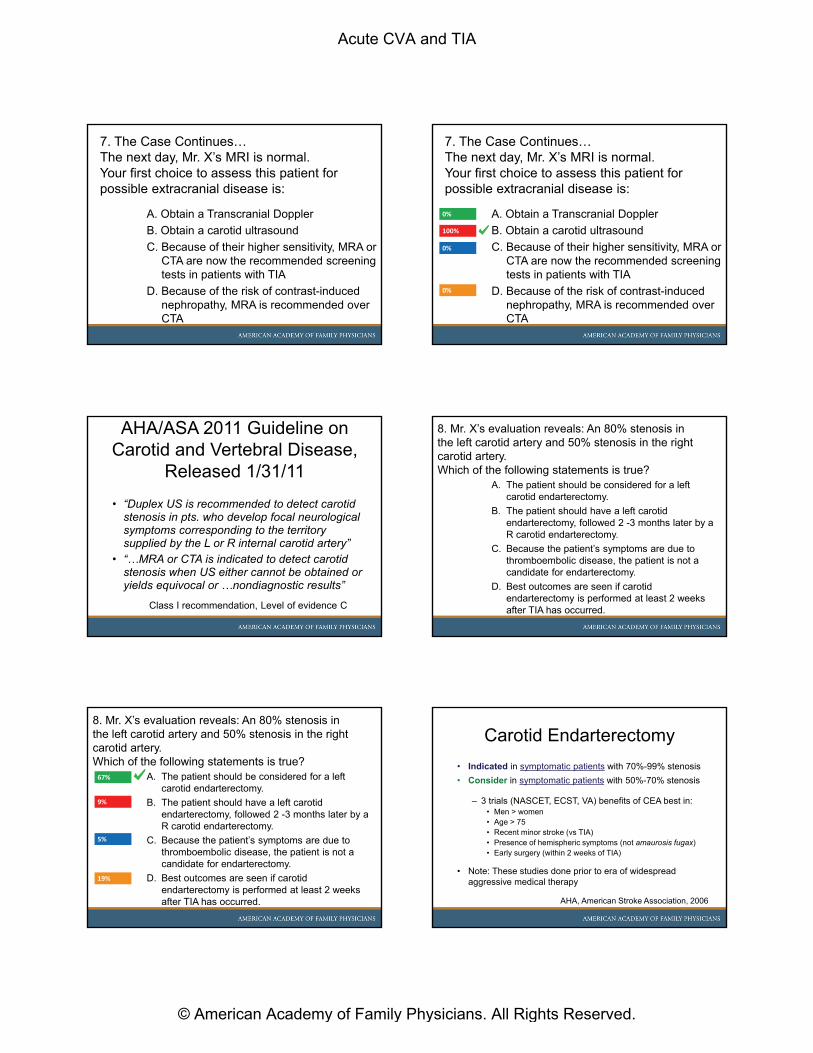
7. The Case Continues…The next day, Mr. X’s MRI is normal. Your first choice to assess this patient for possible extracranial disease is:
A. Obtain a Transcranial Doppler
B. Obtain a carotid ultrasound
C. Because of their higher sensitivity, MRA or CTA are now the recommended screening tests in patients with TIA
D. Because of the risk of contrast-induced nephropathy, MRA is recommended over CTA
0%
0%
100%
0%
AHA/ASA 2011 Guideline on Carotid and Vertebral Disease,
Released 1/31/11
• “Duplex US is recommended to detect carotid stenosis in pts. who develop focal neurological symptoms corresponding to the territory supplied by the L or R internal carotid artery”
• “…MRA or CTA is indicated to detect carotid stenosis when US either cannot be obtained or yields equivocal or …nondiagnostic results”
Class I recommendation, Level of evidence C
8. Mr. X’s evaluation reveals: An 80% stenosis in the left carotid artery and 50% stenosis in the right carotid artery.Which of the following statements is true?
A. The patient should be considered for a left carotid endarterectomy.
B. The patient should have a left carotid endarterectomy, followed 2 -3 months later by a R carotid endarterectomy.
C. Because the patient’s symptoms are due to thromboembolic disease, the patient is not a candidate for endarterectomy.
D. Best outcomes are seen if carotid endarterectomy is performed at least 2 weeks after TIA has occurred.
19%
67%
9%
5%
Carotid Endarterectomy
• Indicated in symptomatic patients with 70%-99% stenosis
• Consider in symptomatic patients with 50%-70% stenosis
– 3 trials (NASCET, ECST, VA) benefits of CEA best in:• Men > women• Age > 75• Recent minor stroke (vs TIA) • Presence of hemispheric symptoms (not amaurosis fugax)• Early surgery (within 2 weeks of TIA)
• Note: These studies done prior to era of widespread aggressive medical therapy
AHA, American Stroke Association, 2006
Acute CVA and TIA
© American Academy of Family Physicians. All Rights Reserved.

9. In 2007, the USPSTF stated:
A. All adults >65 years of age should have ultrasound screening for carotid artery disease.
B. Adults >65 years of age with diabetes should have ultrasound screening for carotid artery disease.
C. Adults >65 years of age with any of the common risk factors for atherosclerosis (DM, HTN, smoking, family history, or hypercholesterolemia) should have ultrasound screening for carotid artery disease.
D. Adults should not be screened for carotid artery disease with ultrasound or other tests.
91%
0%
0%
9%
Carotid Artery Screening …(Asymptomatic Population)
• The bottom line: Prevalence of disease in age >65 = 1%
• 4,348 persons would need screening to prevent 1 CVA in 5 years.
• 8,696 persons would need screening to prevent 1 disabling CVA in 5 years.
Answers1. A2. A3. A4. D5. D6. B7. B8. A9. D
Acute CVA and TIA
© American Academy of Family Physicians. All Rights Reserved.
Related Documents