Robert L. DelaPaz 1 Dean Shibata 1 Gary K. Steinberg 2 Rami Zarnegar2 Charles George 2 Received May 11 , 1989; revision requested July 3, 1989; revision received August 14, 1990; ac- cepted August 28, 1990. 'Department of Radiology, Stanford University Medical Center, Stanford, CA 94305. Address re- print requests toR . L. DelaPaz. 2 Department of Neurosurgery, Stanford Univer- sity Medical Center, Stanford, CA 94305 . 0195-6108/91/1201-0089 © American Society of Neuroradiology Acute Cerebral Ischemia in Rabbits: Correlation Between MR and Histopathology 89 The histologic description of cerebral ischemia is complex, and within most lesions there are regional variations in degrees of neuronal cell injury, edema, and neuropil disruption. These parameters of tissue injury were analyzed histopathologically in transient and permanent experimental cerebral ischemia in 15 rabbits and the results were spatially correlated with MR images of pre- and postmortem (formalin-fixed) brains. MR was performed at 1.5 T (eight animals) and at 0.38 T (seven animals). Areas of high signal on T2-weighted MR images were closely correlated with histologic signs of cytotoxic glial edema and with disruption of the neuropil (widening of the interstitial spaces in the background matrix of glial and neuronal cellular processes) , but MR tended to underestimate the extent of ischemic neuronal injury, especially low-grade histologic changes (mild neuronal shrinkage and nuclear basophilia). Low-grade is- chemic neuronal changes were often found in the penumbra zone of ischemic lesions in areas that appeared normal on T2-weighted MR. High-grade neuronal injury was also seen occasionally in areas of normal signal on MR, especially in the striatum. No significant differences were seen on T2-weighted MR between the experimental groups with respect to transient vs permanent occlusion, in vivo vs in vitro MR, and low vs high magnetic field. In the setting of suspected acute cerebral ischemia, an abnormal T2-weighted MR study often underestimates the extent of neuronal ischemic injury, especially potentially reversible injury; and a normal MR study does not completely exclude significant neuronal ischemic injury. AJNR 12:89-95, January/February 1991 Histologic descriptions of cerebral ischemia emphasize a point that is often not appreciated on MR: within most lesions there are marked and independent regional variations in neuronal cell injury, cytotoxic glial edema, and disruption of the neuropil (widening of the interstitial spaces in the background matrix of glial and neuronal cellular processes) [1 , 2] . This is especially the case in areas of early reflow where significant neuronal injury (neuronal shrinkage and nuclear basophilia) may be present without significant edema and within normal neuropil [3, 4]. These regions may be isolated areas of the brain , such as the basal ganglia, or may represent the marginal penumbra zone around a cortical infarct [5 , 6] . Most previous experimental MR studies of cerebral ischemia [7 -13] have demonstrated prolongation of relax- ation times and increased water content in lesions, but the descriptions of histologic analysis have been limited to gross assessments of the presence or absence of ischemic changes. This study was undertaken in order to refine the spatial corre- lation between abnormal MR signal and regional histology in cerebral ischemic lesions, and to address the question of whether regions of neuronal injury may not be detected by proton MR imaging. Materials and Methods Animals Fifteen male New Zealand white rabbits weighing 2-3 kg underwent unilateral occlusion of the anterior cerebral and internal carotid arteries by means of a transorbital surgical

Welcome message from author
This document is posted to help you gain knowledge. Please leave a comment to let me know what you think about it! Share it to your friends and learn new things together.
Transcript
Robert L. DelaPaz 1
Dean Shibata 1
Gary K. Steinberg2
Rami Zarnegar2 Charles George2
Received May 11 , 1989; revision requested July 3, 1989; revision received August 14, 1990; accepted August 28, 1990.
'Department of Radiology, Stanford University Medical Center, Stanford, CA 94305. Address reprint requests toR . L. DelaPaz.
2 Department of Neurosurgery, Stanford University Medical Center, Stanford, CA 94305 .
0195-6108/91/1201-0089 © American Society of Neuroradiology
Acute Cerebral Ischemia in Rabbits: Correlation Between MR and Histopathology
89
The histologic description of cerebral ischemia is complex, and within most lesions there are regional variations in degrees of neuronal cell injury, edema, and neuropil disruption. These parameters of tissue injury were analyzed histopathologically in transient and permanent experimental cerebral ischemia in 15 rabbits and the results were spatially correlated with MR images of pre- and postmortem (formalin-fixed) brains. MR was performed at 1.5 T (eight animals) and at 0.38 T (seven animals). Areas of high signal on T2-weighted MR images were closely correlated with histologic signs of cytotoxic glial edema and with disruption of the neuropil (widening of the interstitial spaces in the background matrix of glial and neuronal cellular processes), but MR tended to underestimate the extent of ischemic neuronal injury, especially low-grade histologic changes (mild neuronal shrinkage and nuclear basophilia). Low-grade ischemic neuronal changes were often found in the penumbra zone of ischemic lesions in areas that appeared normal on T2-weighted MR. High-grade neuronal injury was also seen occasionally in areas of normal signal on MR, especially in the striatum. No significant differences were seen on T2-weighted MR between the experimental groups with respect to transient vs permanent occlusion, in vivo vs in vitro MR, and low vs high magnetic field.
In the setting of suspected acute cerebral ischemia, an abnormal T2-weighted MR study often underestimates the extent of neuronal ischemic injury, especially potentially reversible injury; and a normal MR study does not completely exclude significant neuronal ischemic injury.
AJNR 12:89-95, January/February 1991
Histologic descriptions of cerebral ischemia emphasize a point that is often not appreciated on MR: within most lesions there are marked and independent regional variations in neuronal cell injury, cytotoxic glial edema, and disruption of the neuropil (widening of the interstitial spaces in the background matrix of glial and neuronal cellular processes) [1 , 2] . This is especially the case in areas of early reflow where significant neuronal injury (neuronal shrinkage and nuclear basophilia) may be present without significant edema and within normal neuropil [3, 4]. These regions may be isolated areas of the brain , such as the basal ganglia, or may represent the marginal penumbra zone around a cortical infarct [5 , 6] . Most previous experimental MR studies of cerebral ischemia [7 -13] have demonstrated prolongation of relaxation times and increased water content in lesions, but the descriptions of histologic analysis have been limited to gross assessments of the presence or absence of ischemic changes. This study was undertaken in order to refine the spatial correlation between abnormal MR signal and regional histology in cerebral ischemic lesions, and to address the question of whether regions of neuronal injury may not be detected by proton MR imaging.
Materials and Methods
Animals
Fifteen male New Zealand white rabbits weighing 2-3 kg underwent unilateral occlusion of the anterior cerebral and internal carotid arteries by means of a transorbital surgical

90 DELAPAZ ET AL. AJNR:12, January/February 1991
approach under halothane anesthesia (14]. This model results in reproducible, extensive hemispheric and basal ganglia lesions. Bipolar cauterization was used for permanent occlusion in seven animals and images were obtained 12 hr after occlusion. Yasargi l mini aneurysm clips were used for transient 1-hr occlusion in eight animals, followed by 4 hr of reperfusion prior to imaging. Ref/ow through the transiently occluded vessels was confirmed by visual inspection.
Imaging
Brain images were obtained with two imaging systems: seven animals (three transient and four permanent occlusion) with a Resonex RX-4000 (0.38 T, 3-in. diameter solenoidal imaging coi l, 128 x 128 data matrix, 3.6-mm slice thickness , no gaps) and eight animals (five transient and three permanent occlusion) with a General Electric Signa (1.5 T, 6-in. diameter saddle coil, 256 x 256 data matrix, 5-mm slice thickness, no gaps). T1- and T2-weighted spin-echo pulse sequences were used for all studies. At 0.38 T the imaging parameters were 400/35 (TR/TE) and 1500/35, 70; at 1.5 T, the parameters were 800, 1200, 2000/25, 50, 75, 100. Low flip angle, gradientrecalled-echo techniques (GRASS) were attempted at 1.5 T but were of poor quality owing to marked magnetic susceptibility gradient phase dispersion at tissue-air/bone interfaces. Images were obtained in the anatomic sagittal, axial, and coronal planes. The coronal imaging plane was oriented parallel to the tentorial fissure between the cerebral and cerebellar hemispheres. Visual identification of the MR abnormality, high signal on T2-weighted images, was made by an investigator who was blinded to the experimental conditions. The contralateral cerebral hemisphere was used as an internal control. Previous experience with these imaging systems and coils indicated poor reproducibility of relaxation time calculations and no attempt was made to generate those data.
Fixation and Histologic Grading
Immediately after imaging, animals were sacrificed by barbiturate overdose and fixed by intracardiac perfusion of 300 ml of normal saline followed by 300 ml of 10% neutral buffered formalin . In 14 animals the brains were reimaged after 3 to 5 days of fixation using premortem scan parameters.
For histopathologic correlation, 40-J.Lm coronal sections in 3- to 5-mm-thick blocks were treated with H and E stains. A graphic display of the coronal slice center locations on the midline sagittal MR image was used to select the locations for brain slicing and histologic correlation. The criteria for histopathologic grading of ischemic changes were derived from (1] and (2] and are summarized in Table 1 . Grading was performed by an investigator who was blinded to experimental conditions, with the contralateral hemisphere used as . an internal control.
TABLE 1: Histologic Grading
Neurons (NR) 0 = Normal 1 = Slight shrinkage, mild nuclear basophilia 2 = Moderate shrinkage, marked nuclear basophilia 3 = Severe shrinkage, dense, pyknotic nucleus
Neuropil (NP) 1 = Spongioform: loosening of neuropil 2 = Vacuolization: prominent vacuole formation
Pallor(P) Reduced intensity of H and E stain
Spatial Correlation
Thirty-six direct MR-histopathologic spatial correlations were performed in 15 animals (two to three correlated slices in each animal). A contour map of each histologic parameter was generated on 8 x 10 in . photographs of the histologic sections, using a grid system guided by light microscopic examination. A similar contour map was generated on a transparent overlay of a size-matched photographic enlargement of the corresponding T2-weighted MR section. The two contour maps were then matched by direct overlay of the MR transparency on the histologic photograph and total areas of abnormality as well as areas of contour intersection (overlap) were calculated using a computerized planimeter. Contour areas were normalized to the total area of the ipsilateral hemisphere. Results were averaged per animal (normalized average of the two to three slices per animal) and expressed as ratios of normalized areas or as percentage of histologic area overlapped by the MR abnormality (see Table 2).
Results
MR vs Histopathology: Total Areas
Comparisons between the total areas of abnormality on T2-weighted MR images and total areas of abnormal histology showed a tendency for MR to underestimate the extent of ischemic neuronal injury, especially low-grade (NR1) changes. The total area of abnormal signal per MR slice was consistently smaller than the total area of nonoverlapping histologic neuronal injury (NR1 + NR2/3) resulting in a mean ratio (histology/MR) of 1.2 (SD = 1.4, range 0.2-8.6). Underestimation by MR was especially striking in individual cases in which the ratio was as high as 8.6. This general finding was true at low and high field strength, in transient and permanent lesions, and in in vivo and in vitro scans.
The comparisons between MR abnormality and neuropil changes or pallor was the inverse of that for neuronal changes. In each animal, the total area of MR abnormality was generally greater than the total area of either neuropil change or pallor alone. The mean ratio of neuropil area to MR area (NP/MR) was 0.6 (SD = 0.4, range 0-2) and pallor to MR (P/MR) was 0.5 (SD = 0.5, range 0-1 .9). A simple comparison in each animal of total MR area and the total area
TABLE 2: Mean Percentage of Histologic Area Overlapped by MR Abnormality
All Transient Permanent 0.4 T 1.5 T
MR in vivo NR1 10.58 11 .38 9.68 5.38 15.0" NR2/3 28.5 25.7 31.6 28.7 28.3 NP1 29.5 33.7 24.8 40.5 20.0 NP2 29.5 27.1 32.3 30.5 28.7 P1 33.3 33.7 32.9 26.5 39.4
MR in vitro NR1 15.9" 13.3" 18.9" 11 .88 19.5 NR2/3 31.1 26.1 36.9 33.2 29.3 NP1 36.1 30.0 43.0 45.8 27.6 NP2 41.5 35.4 49.2 39.3 44.0 P1 37.4 33.7 32.9 32.9 41.4
• Significant !-test (p < .05) vs higher grade.

AJNR: 12, January/February 1991 ACUTE CEREBRAL ISCHEMIA IN RABBITS 91
of neuropil plus pallor (NP + P/MR) gave a ratio of 1.1, which is a somewhat misleading statistic because areas of neuropil change were usually found within regions of pallor (see overlap data below.) These ratios were also consistent across all experimental groups.
MR vs Histopathology: Overlap Areas
The spatial correlation data in Table 2 show a tendency for high signal on T2-weighted MR images to underestimate the extent of low-grade neuronal ischemic injury (NR1 ). A smaller percentage of low-grade neuronal change is overlapped or predicted by high signal on MR than is high grade neuronal change (1 0.5% for NR1 and 28.5% for NR2/3 in vivo and 15.9% for NR1 and 31 .1% for NR2/3 in vitro). The MR abnormality overlapped 29.5% of neuropil changes (NP1 and NP2) in vivo and 36.1% (NP1) and 41 .5% (NP2) of neuropil changes in vitro. MR lesions overlapped 33.3% of pallor in vivo and 37.4% in vitro. The more extensive overlap with high-grade neuronal injury, neuropil damage, and pallor indicate a greater likelihood of these histologic changes being present within abnormal high-signal regions on T2-weighted images than does low-grade neuronal injury. Because of the cumulative errors produced by small regions of nonoverlap, these numbers are not true probabilities but are useful for comparison between experimental groups.
Statistical comparisons between overlap values in the different experimental conditions, using both simple differences between means and matched-pair two-tailed t tests, revealed significant differences (p < .05) between low-grade neuronal change (NR1) and all other measures of overlap (NR2f3, NP1 , NP2, and P1) except NR1 vs NR2/3 and NP1 at 1.5 Tin vitro (Table 2). No statistically significant differences were seen between NR2f3, NP1, NP2, and P1 overlap values. This was true across all experimental groups (transient, permanent, in vivo, in vitro, low field , and high field) .
Case Examples
Figures 1 and 2 show the visual relationships between MR and histologic contour maps and illustrate the overall results.
In Figure 1, general agreement between MR and histology is seen. The high-signal MR abnormality closely corresponds to the extensive area of neuronal injury, which also closely parallels pallor throughout the parietotemporal cortex. Smaller lesions are seen on MR, in the caudate nucleus and hypothalamus, which also correspond to neuronal injury and pallor. The regions of clear neuropil injury are limited and lie within the regions of neuronal injury and pallor (an example of discrepancy between extensive MR abnormality and minimal neuropil change).
Figure 2 demonstrates several notable discrepancies between MR and histology. Extensive low-grade neuronal ischemic changes in the superior parietal cortex and high-grade neuronal changes in the inferior temporal cortex correspond to normal signal on MR. A small area of low-grade neuronal change in the hypothalamus is also not identified by MR.
These false-negative areas of the MR all correspond to regions of neuronal ischemic injury found in the absence of neuropil abnormality or pallor. The spatial correlation between MR and neuropil abnormality or pallor is better than that with neuronal injury and is clearly demonstrated in these images. However, some discrepancy does exist, with more extensive involvement of the superficial cortex on the MR in the parietotemporal region outside the contours of the neuropil change and pallor. This illustrates the tendency seen in the overall data for the MR abnormality to be larger than the neuropil disruption or pallor regions.
In Vivo vs In Vitro
Cerebral abnormalities on MR of brain in vivo and in vitro (formalin fixed) showed very good spatial correlation with each other. Direct overlay comparison within each animal between these images produced overlap values of 80-85% through all experimental groups. There were no statistically significant differences between in vivo and in vitro MR images in terms of their spatial correlation with histopathology. The examples shown in Figures 1 and 2 demonstrate this agreement.
Transient vs Permanent
No significant differences between the transient and permanent vascular occlusion groups were seen in spatial correlations between MR and histopathology. These comparisons are statistical, since direct comparisons between these experimental treatments could not be made.
High vs Low Field
MR studies showed similar degrees of change and correlation with histopathology at both high (1 .5 T) and low (0 .38 T) magnetic field strength . This was not a direct comparison between images of the same animal in both systems. A slight, but not statistically significant tendency for the low-field system to show poorer overlap with low-grade neuronal changes was noted. This was probably due, at least partially, to the inclusion of early surgical cases with widely variable lesions in the low-field group.
Discussion
Although the spatial correlation between MR and histologic abnormalities was generally good, there was a significant false-negative occurrence of normal signal on MR in regions of neuronal ischemic injury. This discrepancy was statistically significant for low-grade (NR 1) neuronal changes and was also seen with severe neuronal injury (NR2/3) in individual cases, as is illustrated in Figure 2. These false-negative MR regions were most commonly seen in the border zones of lesions, immediately peripheral to the high signal , representing the so-called penumbra zone, where ischemic injury is in

92 DELAPAZ ET AL. AJNR:12, January/February 1991
A 8
~ --, ·• F1·
NP2 I
c D
Fig. 1.-0.38-T MR and histology. A, In vivo coronal T2-weighted MR image shows extensive abnormal high signal in left parietotemporal cortex (straight arrow), hypothalamus (curved
arrow), and caudate nucleus (open arrow). B, In vitro coronal T2-weighted MR image shows extensive abnormal high signal in left parietal and temporal cortex (straight arrow), hypothalamus
(curved arrow), and caudate nucleus (open arrow). C, Coronal brain slice mapped with neuronal ischemic injury contours. D, Coronal brain slice mapped with neuropil disruption contours. Extensive pallor is present from parietal through temporal cortex, which corresponds
closely to high signal on MR.
transition spatially and temporally (i.e., a zone of potentially reversible ischemia) [6, 15-17]. False-negative MR signal was also seen in isolated lesions of the basal ganglia (notably NR2/3 with normal MR). The key characteristic of these misses by MR is the lack of significant neuropil change or pallor. This indicates the importance of edema formation to proton MR detection of ischemia. It is the change in water content and relaxation rates in the glia and interstitial spaces, as well as neurons, that produces abnormal signal on proton MR. Support for this conclusion can be found in a quantative study of abnormal proton relaxation rates in ischemic edema, which demonstrated components from both intracellular and interstitial water [18]. The correct identification of neuronal
injury by MR (true positive) is also explained by the presence of associated edema. More severe neuronal changes are usually found in areas of significant cytotoxic and interstitial edema [6, 8, 15-17]. Therefore, although MR abnormality on T2-weighted images generally corresponds to severe neuronal injury, this sensitivity is indirect and significantly dependent on ischemic injury in glial cells (cytotoxic edema) and interstitial edema (largely reflecting vasogenic edema via blood-brain barrier breakdown) [19].
Although the correlation between MR abnormality and neuropil changes was better than that for neuronal injury, MR still tended to identify these regions incorrectly, more frequently by showing false-positive rather than false-negative results .
AJNR: 12, January/February 1991 ACUTE CEREBRAL ISCHEMIA IN RABBITS 93
c D
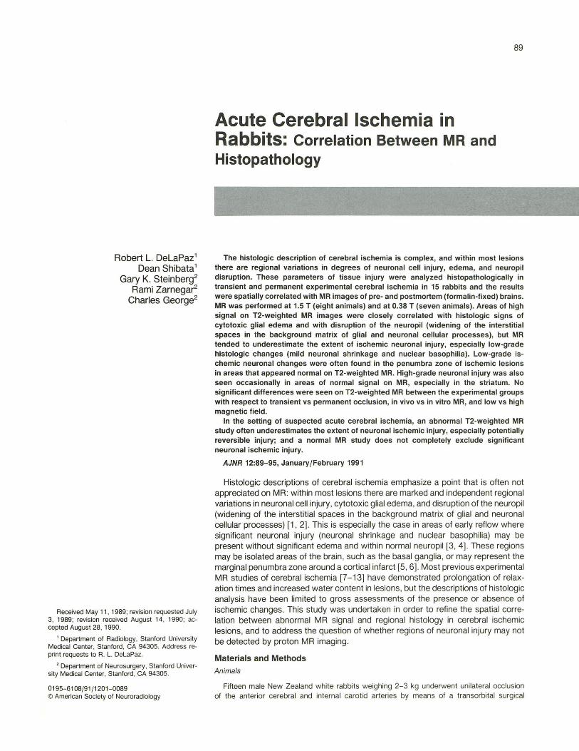
Fig. 2.-1.5-T MR and histology. A, In vivo coronal T2-weighted MR image shows abnormal high signal in left parietotemporal cortex (arrow) with periventricular medial extension. 8, In vitro coronal T2-weighted MR image shows abnormal high signal in left parietotemporal cortex (arrow) with periventricular medial extension. C, Coronal brain slice mapped with neuronal ischemic injury contours. D, Coronal brain slice mapped with neuropil disruption contours. A well-circumscribed region of palor is present in deep parietotemporal cortex, which
corresponds closely to high signal on MR.
This discrepancy tended to occur at lesion margins, and can be partially attributed to misregistration between MR and histology contour maps and to tissue changes in the ischemic penumbra, as illustrated in Figure 2. A likely physiologic explanation is the presence of increased tissue water due to cytotoxic cellular swelling, mostly glial , without significant interstitial vasogenic edema or neuropil disruption. While both cytotoxic and vasogenic edema occur in ischemic lesions (18] , the former is always present, at least in perivascular glia (5) , while vasogenic edema is widely variable, depending on local reflow [3, 4) and blood-brain barrier status (19, 20). Very mild interstitial edema, too subtle to be identified as neuropil loosening by light microscopic analysis, may also contribute
to this finding . A third mechanism for abnormal signal on MR may also be important; that is, a change in state rather than amount of intracellular water, producing longer relaxation times from alterations in electrolytes , pH , proteins , lactate, glucose, ketones , energy metabolites, mitochondria, etc. (6 , 8, 15-17, 20).
The relationship of high signal on MR to pallor is probably also explained in terms of changes in tissue water. However, the cause of pallor with H and E staining is not well understood. It is believed to reflect neuropil disruption in dense white matter regions, presumably by spreading out the stained myelin sheaths [2 , 21 ]. When pallor is seen in gray matter regions , it more likely reflects toxic effects to the
94 DELAPAZ ET AL. AJNR:12 , January/February 1991
myelin that produce reduced staining (not true demyelination acutely). MR signal change may reflect changes in myelin properties but the primary reason for the close association of pallor and MR abnormality is most likely the tissue edema usually associated with pallor. The false-positive discrepancy between MR and pallor at marginal zones of lesions likely has a similar explanation to that for neuropil disruption, reflecting cytotoxic edema and changes in MR properties of the tissue water.
There was a strong correlation between MR in vivo and in vitro in this study. Formalin fixation might be expected to change MR relaxation properties by causing membrane alterations, protein denaturation, and dehydration. Although we were not able to quantitatively compare relaxation times between pre- and postmortem scans, visual inspection demonstrated preservation of relative tissue signal intensities and lesion conspicuity. One possible explanation for preserved lesion identification is that formalin fixation was done with a saline/formalin perfusion technique rather than by immersion, so that all regions of the brain were exposed simultaneously and uniformly to the fixative. No gradients of osmolarity or protein denaturation should have been created as might occur with immersion fixation . Regardless of the explanation , the close correlation has important practical implications for animal research. The ability to image the fixed brain allows better control of the experimental treatment without the difficulties in handling and life support that accompany MR studies. In our laboratory we have begun to limit MR to the fixed brain after experimental treatment in studies of drug therapy for stroke [22].
As presented in the tabular data, there was no significant difference between transient and permanent occlusion ischemia models. The end point for the MR abnormality, increased tissue water, was achieved by both techniques. However, the two treatments probably produced different degrees of cytotoxic and vasogenic edema depending mostly on differences in rate and extent of reperfusion [3 , 23]. Early reperfusion in the transient ischemia model may have contributed to falsepositive MR vs neuronal correlations (i.e., high signal on MR without significant histologic neuronal ischemic injury). Reperfusion may produce early accumulation of interstitial free water due to blood-brain barrier injury but also provides sufficient oxygenation to prevent histologically observable neuronal changes [3, 4, 23].
The reason for the relatively low absolute degree of overlap among the data in Table 2 is both physiological and technical. The possible physiological explanations have been discussed. One of the most likely technical explanations is that multiple, small , irregular portions of histology contours were excluded by the smoother contours of the MR overlay. We felt that this was acceptable in order to preserve a conservative and blinded approach to the overlay spatial correlations. A conservative bias in the histologic mapping may have contributed to some MR-histology discrepancies by not identifying equivocal findings as areas of positive histologic change in regions of MR abnormality. Conversely, the same approach did not bias the data against MR by identifying equivocal findings as areas of histologic abnormality outside the region of MR
abnormality. Distortion between the MR and histologic sections due to differences in aspect ratio may also account for part of the discrepancy. Care was taken to match the histologic section to the same level and orientation as the MR, and minimal discrepancy appears to be attributable to that source of error. Another possible source of error is the difference in spatial resolution of the MR and histologic techniques, which is approximately an order of magnitude (0.5 to 1 mm in plane with 5-mm thickness for MR and micron resolution in plane with 40-~Lm slice thickness for the histology). This difference was nullified by the use of overall MR and histology lesion contours at a gross anatomic scale. Despite the low absolute degree of overlap, the percentage overlap values are useful for comparing relative performance between histologic categories and between experimental groups.
In conclusion, proton MR using T2-weighted spin-echo images is a sensitive technique for detecting subtle changes in cerebral water content andjor changes in state of cerebral water. As such it is useful in the setting of cerebral ischemia, which is usually accompanied by increases in tissue free water. However, this sensitivity does not necessarily imply accurate prediction of neuronal disease. High signal on T2-weighted proton MR images most reliably predicts the presence of histologic neuropil disruption and decreased staining (pallor) probably reflecting both cytotoxic intracellular free water accumulation (primarily in glia) and vasogenic interstitial edema. However, since these pathologic changes do not necessarily accompany neuronal injury, MR has proved to be a less reliable predictor of neuronal cell injury as determined by light microscopic histologic analysis. Individual examples of normal MR signal in regions of severe neuronal ischemic injury were seen. Overall statistical analysis demonstrated a tendency for MR to underestimate the extent of neuronal injury, especially low-grade changes (possibly representing reversible ischemic damage). The clinical implication of these findings is that, in the setting of suspected acute cerebral ischemia, a T2-weighted proton MR abnormality often underestimates the extent of neuronal ischemic injury, especially potentially reversible injury; and that a normal MR study does not completely exclude significant neuronal ischemic injury.
REFERENCES
1. Garcia JH, Lossinsky AS, Kauffman, Conger KA. Neuronal ischemic injury: light microscopy, ultrastructure and biochemistry. Acta Neuropathol 1978;43: 85-95
2. Little JR , Kerr FWL, Sundt TM. Microcirculatory obstruction in focal cerebral ischemia: relationship to neCJronal alterations. Mayo Clinic Proc 1975;50:264-270
3. Busto R, Yoshida S, Ginsberg MD. Postischemic brain edema-comparison of complete versus incomplete ischemia. Stroke 1985;16 :142
4. lanotti F, Hoff J. Ischemic brain edema with and without reperfusion: an experimental study in gerbils. Stroke 1983;14:562-567
5. Deitrich WD, Busto R, Yoshida S, Ginsberg MD. Histopathological and hemodynamic consequences of complete versus incomplete ischemia in the rat. J Cereb Blood FLow Metab 1987;7:300-308
6. Meyer FB , Anderson RE , Sundt TM , Yaksh TL. Intracellular pH, indicator tissue perfusion , electroencephalography, and histology in severe and moderate focal cortical ischemia in the rabbit. J Cereb Blood Flow Metab 1986;6:71-78
7. Brant-Zawadzki M, Pereira B, Weinstein P, et al. MR imaging of acute

AJNR:12, January/February 1991 ACUTE CEREBRAL ISCHEMIA IN RABBITS 95
experimental ischemia in cats. AJNR 1986;7 :7-11 8. Brant-Zawadzki M, Weinstein P, Bartkowski H, Moseley M. MR imaging
and spectroscopy in clinical and experimental cerebral ischemia: a review. AJNR 1987;8:39-48
9. Bryan NR, Willcott MR, Schneiders NJ , Rose JE. NMR evaluation of stroke in the rat . AJNR 1983;4:242-244
10. Buonanno FS, Pykett IL, Brady T J, et al. Proton NMR imaging in experimental ischemic infarction. Stroke 1983;14: 178-184
11. Levy RM, Mano I, Brito A, Hosobuchi Y. NMR imaging of acute experimental cerebral ischemia: time course and pharmacologic manipulations. AJNR 1983;4:238-241
12. Pykett IL, Buonanno FS, Brady T J, Kistler JP. True three-dimensional nuclear magnetic resonance neuro-imaging in ischemic stroke: correlation of NMR, X-ray CT and pathology. Stroke 1983;14:173-177
13. Spetzler RF, Zabramski JM, Kaufman B, Yeung HN. Acute NMR changes during MCA occlusion: a preliminary study in primates. Stroke 1983;14: 185-190
14. Yamamoto K, Yoshimine T, Yanagihara T. Cerebral ischemia in rabbits: a new experimental model with immunohistochemical investigation. J Cereb Blood Flow Metab 1985;5: 529-536
15. Ginsberg MD, Graham 01 , Welsh FA, Budd WW. DiHuse cerebral ischemia in the cat: Ill. Neuropathological sequelae of severe ischemia. Ann Neural
1979;5: 350-358 16. Klatzo I. Brain oedema following brain ischaemia and the influence of
therapy. Br J Anaesth 1985;57 : 18-22 17. Hassman KA, Schuier FJ. Experimental brain infarcts in cats . Stroke
1980;11 :583-592 18. Horikawa Y, Naruse S, Tanaka C, Hirakawa K, Nishikawa H. Proton NMR
relaxation times in ischemic brain edema. Stroke 1986;17 : 1149-1152 19. Kuroiwa T, Ting P, Martinez H, Klatzo I. The biphasic opening of the blood
brain barrier to proteins following temporary middle cerebral artery occlusion . Acta Neuropathol1985 ;68: 122-129
20. Gotch 0 , Asano T, Koide T, Takakura K. Ischemic brain edema following occlusion of the middle cerebral artery in the rat. 1: The time course of the brain water, sodium and potassium contents and blood-brain barrier permeability to 1251-albumin. Stroke 1985; 16 : 101-109
21 . Davis RL, Robertson OM. Textbook of neuropathology. Baltimore: Williams & Wilkins, 1985: 606-608
22. Steinberg GK, Goerge CP, DelaPaz RL, Shibata OK, Gross T. Dextromethorphan protects against cerebral injury following transient focal ischemia. Stroke 1988;19 : 1112-1118
23. Crowell RM , Olsson Y, Klatzo I, Ommaya A. Temporary occlusion of the middle cerebral artery in the monkey: clinical and pathological observations. Stroke 1970;1 :439-448
Related Documents