• actually 2 joints within the articular capsule – tibio-femoral – patello-femoral – the superior fibulo- tibial joint is also near – modified hinge joint • flexion and extension is primary motion • some rotation is possible when the knee is flexed Knee Joint

Actually 2 joints within the articular capsule –tibio-femoral –patello-femoral –the superior fibulo-tibial joint is also near –modified hinge joint flexion.
Dec 17, 2015
Welcome message from author
This document is posted to help you gain knowledge. Please leave a comment to let me know what you think about it! Share it to your friends and learn new things together.
Transcript

• actually 2 joints within the articular capsule– tibio-femoral– patello-femoral– the superior fibulo-tibial joint
is also near
– modified hinge joint• flexion and extension is
primary motion• some rotation is possible
when the knee is flexed
Knee Joint
Anterior Posterior AnteriorTransverse
condylesepicondyles
intercondylar notch
patella
tibial tuberosity
tibial plateaus
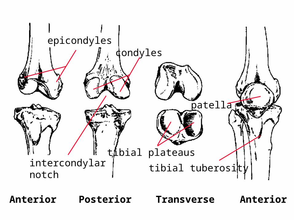
Ligamentous Support
Menisci
CollateralLigaments
CruciateLigaments
Other Ligaments

Menisci
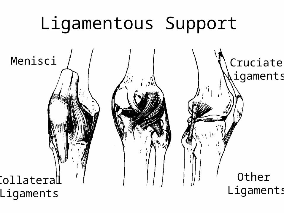
• The menisci are discs of fibrocartilage attached to tibial plateaus. They are thicker along the periphery.
The lateral meniscus is smaller and more mobile than the medial meniscus. The inner portion of the menisci are avascular. The outer portion has some blood supply, making healing of tears possible.
lateral medial

Menisci Function
• increases stability by deepening tibial plateaus
• decreases friction by 20%• increases contact area by 70%• absorbs shock
– removal of menisci does NOT preclude normal motion, but
• increase wear on articulating surfaces
• increase chance of developing degenerative joint disease
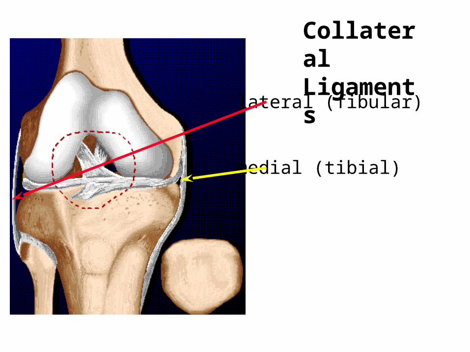
lateral (fibular)
medial (tibial)
Collateral Ligaments

prevents abduction and
adduction movement
of the knee
Collateral Ligaments
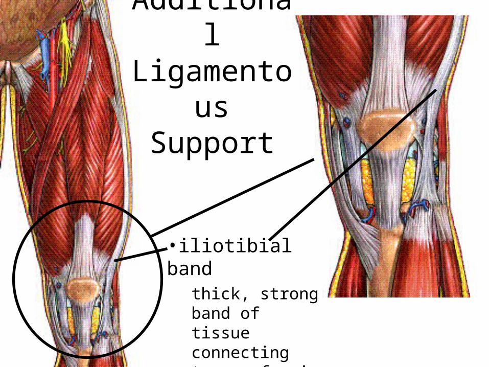
Additional Ligamentous
Support
•iliotibial bandthick, strong band of tissue connecting tensor fascia latae to femur and tibia
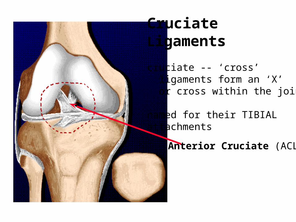
Anterior Cruciate (ACL)
Cruciate Ligaments
cruciate -- ‘cross’ ligaments form an ‘X’ or cross within the joint
named for their TIBIALattachments
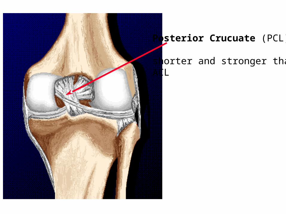
Posterior Crucuate (PCL)
shorter and stronger thanACL

FEMUR
TIBIA
PATELLA
The ACL prevents the femur from sliding posteriorly on the tibia or the tibia from sliding anteriorly on the femur.
The PCL prevents the femur from sliding anteriorly on the tibia or the tibia from sliding posteriorly on the femur.
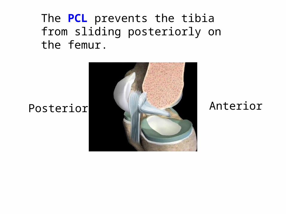
The PCL prevents the tibia from sliding posteriorly on the femur.
AnteriorPosterior

Cruciates During Flexion/ExtensionNote: the cruciate ligaments also limit rotation
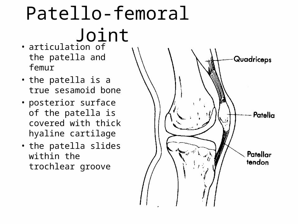
Patello-femoral Joint • articulation of the
patella and femur• the patella is a true
sesamoid bone• posterior surface of the
patella is covered with thick hyaline cartilage
• the patella slides within the trochlear groove

Functions of Patello-femoral Joint
(1) increases angle of pull of quads on tibia, improves the ratio of motive:resistive torque by 50%
(2) centralizes divergent tension of quads into a single line of action
(3) some protection of anterior aspect of knee
without patellawith patella
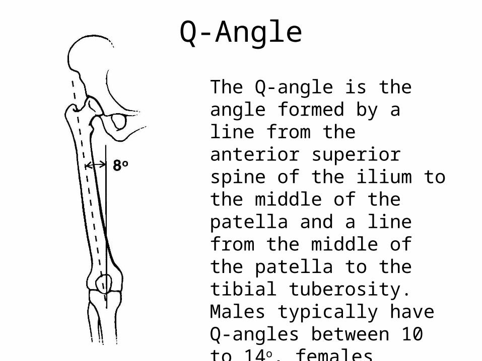
Q-Angle
The Q-angle is the angle formed by a line from the anterior superior spine of the ilium to the middle of the patella and a line from the middle of the patella to the tibial tuberosity. Males typically have Q-angles between 10 to 14o, females between 15-17o.
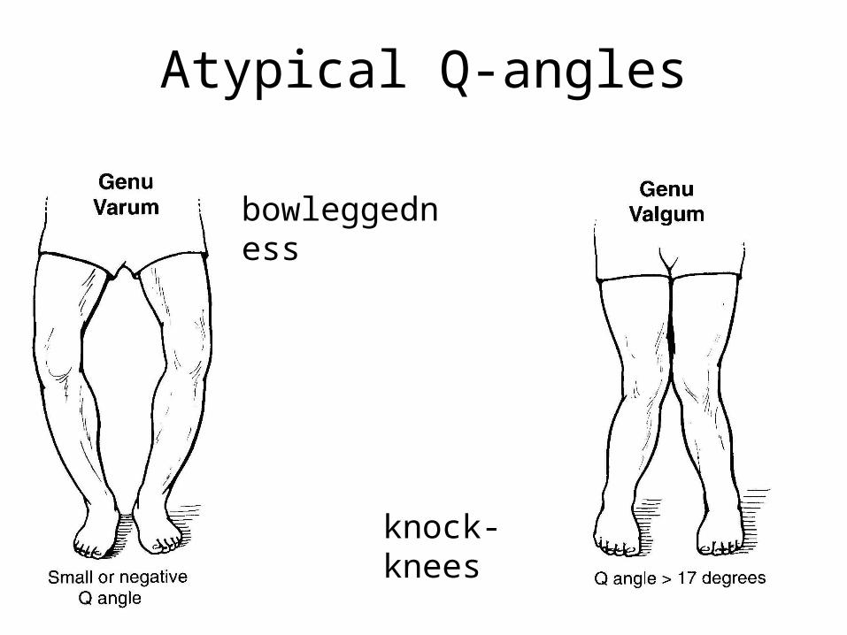
Atypical Q-angles
bowleggedness
knock-knees

Knee Rotation(Locking Your Knee)
• Six to 30 degrees of internal rotation of the tibia on the femur occurs through 90 degrees of knee flexion.
1 The femoral condyles do not have the same diameters, this helps cause internal rotation when the knee is flexed and external rotation when the knee is extended.
2 The lateral condyle slides more than medial condyle.3 The anterior cruciate ligament becomes taut just prior to the
rotation, this may help force a rotation of the femur on the tibia.
FlexionExternalRotation
InternalRotation
Extension
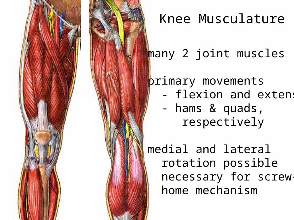
Knee Musculature
many 2 joint muscles
primary movements - flexion and extension - hams & quads, respectively
medial and lateral rotation possible necessary for screw- home mechanism

Knee FlexionHamstrings cross hip and knee
biceps femoris
semitendinosus
semimembranosus
gastrocnemius cross knee and ankle popliteus
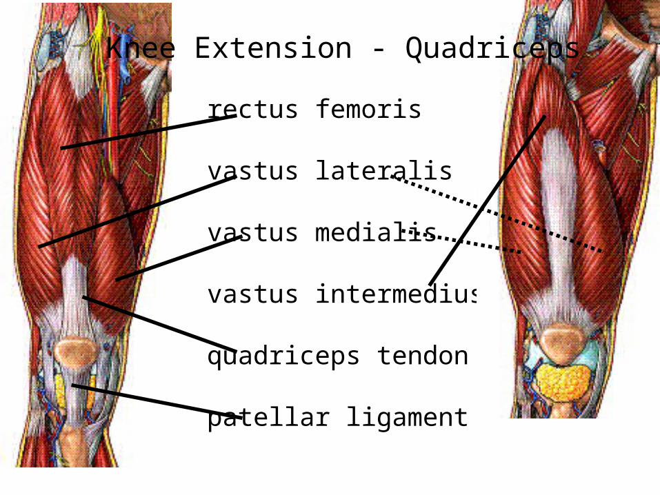
rectus femoris
vastus lateralis
vastus medialis
vastus intermedius
quadriceps tendon
patellar ligament
Knee Extension - Quadriceps
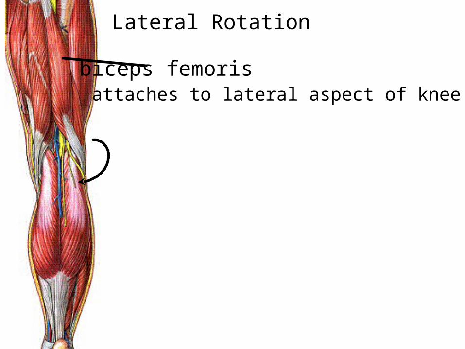
Lateral Rotation
biceps femoris attaches to lateral aspect of knee
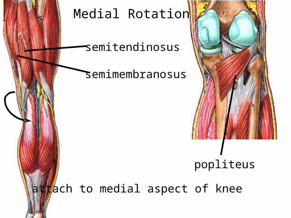
Medial Rotation
semitendinosus
semimembranosus
popliteus
attach to medial aspect of knee

Common Knee Injuries
• one of the most commonly injured joints– lack of bony and muscular support– positioned between the 2 longest bones– weight bearing and locomotion functions
• often tear or stretching of soft tissue

Ligament Injuries
• ACL– more prevalent than PCL injuries
– forces directed from posterior side of leg
• PCL– forces directed from anterior side of leg
– forced flexion of knee w/external rotation• wrestling and football

Intact Knee with ACL& PCL
Ruptured ACL Knee

Mechanisms of ACL injury
1) attempting a rapid cutting maneuver with foot in contact with the ground and knee flexed (problem exacerbated if an external force applied to knee during this movement)
2) knee hyperextension with internal tibial rotation
Examplebackward falling skier - boot
and skis accelerate forward creating an anterior drawer mechanism
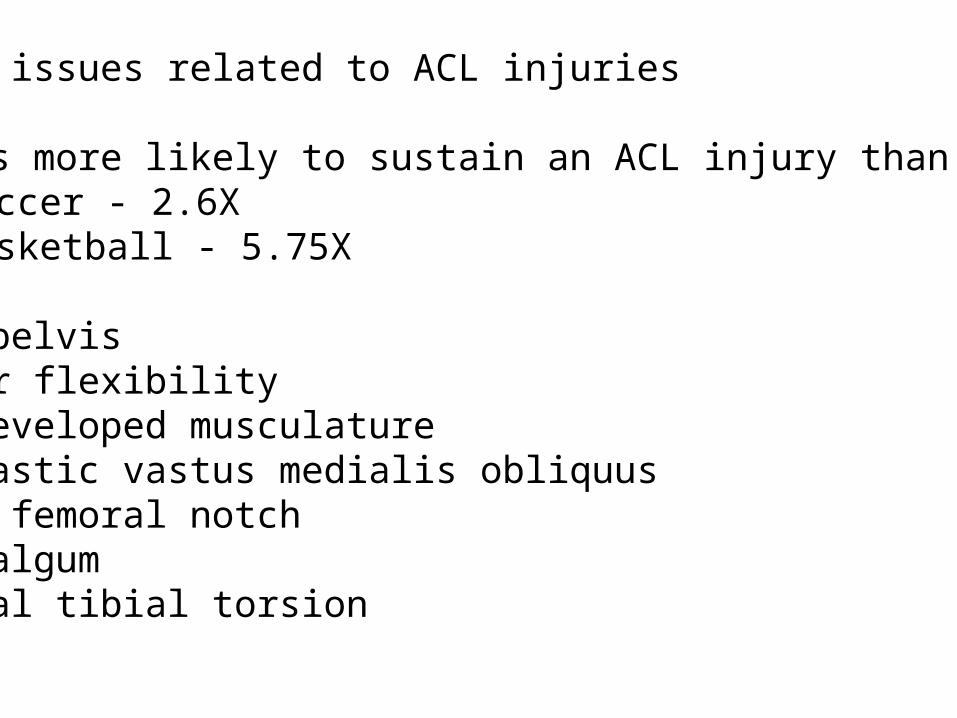
Gender issues related to ACL injuries
females more likely to sustain an ACL injury than malessoccer - 2.6Xbasketball - 5.75X
wider pelvisgreater flexibilityless-developed musculaturehypoplastic vastus medialis obliquusnarrow femoral notchgenu valgumexternal tibial torsion

PCL Injuries
When the knee is forcefully twisted or hyperextended BUT other ligaments are usually injured or torn, before the posterior cruciate ligament (PCL) is torn
Most common mechanism for PCL alone to be injured is from a direct blow to the front of the knee while the knee is bent.
Automobile accident1. Automobile strikes another and stops suddenly2. Front passenger or driver slides forward. 3. Bent knee hits the dashboard just below the knee cap forcing
tibia backwards on the femur tearing PCL.
The same force can occur during a fall on the bent knee, where the force of the fall on the tibia pushes it back against the femur and tears the posterior cruciate ligament (PCL).

When the tibia is displaced too much in the posterior direction the PCL may rupture.
Common mechanism ofPCL injury in football is being tackled while the knee is fully extended.
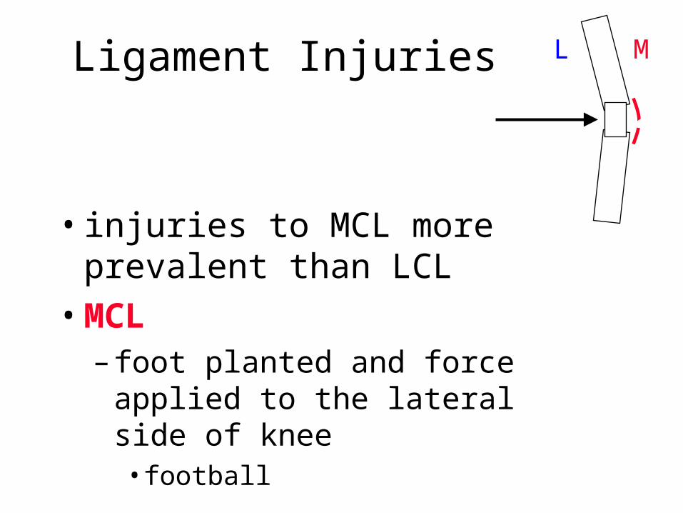
Ligament Injuries
• injuries to MCL more prevalent than LCL
• MCL– foot planted and force applied to the
lateral side of knee• football
ML
Meniscus Injuries
• most common injury in the knee
• tearing is most common
• medial side injured more often– medial meniscus more secured
– foot planted with excessive rotation
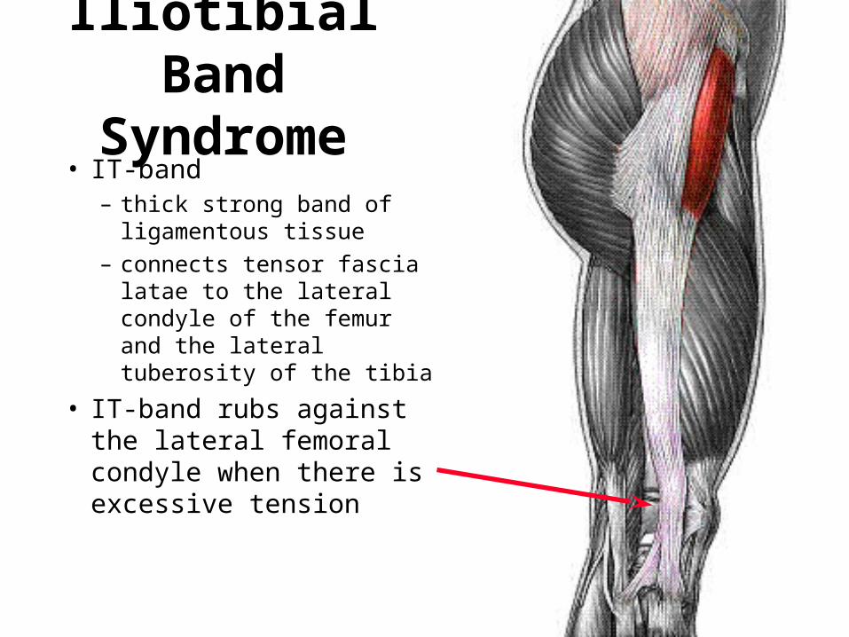
Iliotibial Band Syndrome
• IT-band– thick strong band of
ligamentous tissue– connects tensor fascia latae to
the lateral condyle of the femur and the lateral tuberosity of the tibia
• IT-band rubs against the lateral femoral condyle when there is excessive tension
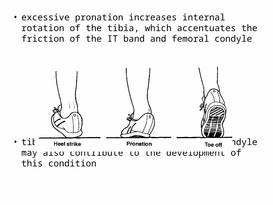
• excessive pronation increases internal rotation of the tibia, which accentuates the friction of the IT band and femoral condyle
• tibial alignment and size of femoral condyle may also contribute to the development of this condition
Related Documents