J. Clin. Endocrinol. Metab. 2011 96:755-765 originally published online Dec 22, 2010; , doi: 10.1210/jc.2010-0501 Filiberto Maria Severi, Wylie Vale, Mario Castellucci and Felice Petraglia Pasquapina Ciarmela, Enrrico Bloise, Peter C. Gray, Patrizia Carrarelli, Md. Soriful Islam, Flavio De Pascalis, Myometrium: Disruption of Their Signalling in Uterine Fibroid Activin-A and Myostatin Response and Steroid Regulation in Human Society please go to: http://jcem.endojournals.org//subscriptions/ or any of the other journals published by The Endocrine Journal of Clinical Endocrinology & Metabolism To subscribe to Copyright © The Endocrine Society. All rights reserved. Print ISSN: 0021-972X. Online

Welcome message from author
This document is posted to help you gain knowledge. Please leave a comment to let me know what you think about it! Share it to your friends and learn new things together.
Transcript

J. Clin. Endocrinol. Metab. 2011 96:755-765 originally published online Dec 22, 2010; , doi: 10.1210/jc.2010-0501
Filiberto Maria Severi, Wylie Vale, Mario Castellucci and Felice Petraglia Pasquapina Ciarmela, Enrrico Bloise, Peter C. Gray, Patrizia Carrarelli, Md. Soriful Islam, Flavio De Pascalis,
Myometrium: Disruption of Their Signalling in Uterine Fibroid
Activin-A and Myostatin Response and Steroid Regulation in Human
Society please go to: http://jcem.endojournals.org//subscriptions/ or any of the other journals published by The EndocrineJournal of Clinical Endocrinology & MetabolismTo subscribe to
Copyright © The Endocrine Society. All rights reserved. Print ISSN: 0021-972X. Online

Activin-A and Myostatin Response and SteroidRegulation in Human Myometrium: Disruption ofTheir Signalling in Uterine Fibroid
Pasquapina Ciarmela,* Enrrico Bloise,* Peter C. Gray, Patrizia Carrarelli,Md. Soriful Islam, Flavio De Pascalis, Filiberto Maria Severi, Wylie Vale,Mario Castellucci, and Felice Petraglia
Department of Molecular Pathology and Innovative Therapies (P.Ci., M.S.I., M.C.), Faculty of Medicine,Polytechnic University of Marche, 60020 Ancona, Italy; Division of Obstetrics and Gynecology (E.B.,P.Ca., F.D.P., F.M.S., F.P.), Department of Pediatrics, Obstetrics, and Reproductive Medicine; Universityof Siena, Siena 53100, Italy; and Clayton Foundation Laboratories for Peptide Biology (P.C.G., W.V.), TheSalk Institute for Biological Studies, La Jolla, California 92037
Context: Investigation of activin-A (A) and myostatin (M) in human myometrium (HM) and leio-myoma (HL) will explain their involvement in human myometrial pathophysiology.
Objective: We aimed to investigate A and M response and steroid regulation in HM. We alsoevaluated A and M expression and response in HL.
Design: Tissues were analyzed and cultured.
Patients: Patients included fertile (in proliferative phase) and menopausal women undergoinghysterectomy.
Interventions: HM explant cultures were treated with A and M (for Smad-7 mRNA quantification) orestrogen and progesterone (for A and M mRNA quantification). A and M expression levels were alsoevaluated in menopausal (physiological absence of steroids) HM specimens. A and M and their recep-tors were evaluated in HL (n � 8, diameter 5–8 cm) compared with their matched HM. HL explantscultures were treated with A and M (for Smad7 mRNA quantification), and, to explain the absence ofresponse, the levels of follistatin, follistatin-related gene (FLRG), and Cripto were evaluated.
Results: A and M increased Smad7 expression in HM explants. A and M mRNAs were both reducedafter estradiol treatment, unchanged after progesterone treatment, but were higher in meno-pausal than fertile (in proliferative phase) specimens. A, M, and FLRG were expressed at higherlevels in HL compared with adjacent HM, whereas the receptors, follistatin, and Smad7 mRNAsresulted unchanged. Cripto mRNA was expressed only in HL.
Conclusions: A and M act on human HM and are regulated by steroids. In HL there is an increaseof A, M, FLRG, and Cripto expression. (J Clin Endocrinol Metab 96: 755–765, 2011)
Activin and myostatin are important growth factors be-longing to the TGF-� family and represent key regu-
lators of cell growth and differentiation. Activins were firstidentified by virtue of their ability to regulate FSH secretionfrom the anterior pituitary. Activins are also powerful reg-
ulators of gonadal functions and are involved in fibrosis,inflammation, and neurogenesis (1). Myostatin, also calledgrowth differentiation factor (GDF)-8, is specifically ex-pressed in the skeletal muscle lineageand isacandidatemusclechalone negatively regulating the growth of myoblasts (2).
ISSN Print 0021-972X ISSN Online 1945-7197Printed in U.S.A.Copyright © 2011 by The Endocrine Societydoi: 10.1210/jc.2010-0501 Received March 1, 2010. Accepted November 8, 2010.First Published Online December 22, 2010* P.C. and E.B. contributed equally to this work.
Abbreviations: ActR, Activin receptor; ALK, activin receptor-like kinase; EGF, epidermalgrowth factor; FLRG, follistatin-related gene; FST, follistatin; GDF, growth differentiationfactor; RT, reverse transcription.
O R I G I N A L A R T I C L E
E n d o c r i n e R e s e a r c h
J Clin Endocrinol Metab, March 2011, 96(3):755–765 jcem.endojournals.org 755

Activin and myostatin share similar receptors and in-tracellular signaling. Activin initiates signaling by bindingtwo types of transmembrane serine/threonine receptor ki-nases classified as type II [activin receptor (ActR) type II orActRIIB] and type I [ActRIB; activin receptor-like kinase(ALK)-4]. ActRII binds activin with high affinity and fa-cilitates activin binding to ALK4 (3). In the receptor com-plex, the constitutively active type II receptor kinase phos-phorylates ALK4, and this phosphorylation leads toactivation of the ALK4 kinase (4). Once activated, ALK4phosphorylates Smad2 and Smad3, which form part of thepostreceptor signal transduction system (5). Myostatin,like activin, stimulates target cells by assembling a cellsurface receptor complex containing type I and II recep-tors. Myostatin binds the type II Ser/Thr kinase receptor,ActRIIB, and then partners with a type I receptor, eitherALK4 (or ActRIB) or ALK5 (TGF-� receptor I). Thesecomplexes induce phosphorylation of Smad2 and Smad3and activate an activin/TGF-�-like signaling pathway (6).Smad7 functions as an inhibitory downstream modulatorof activin/TGF-�-like signaling, and its expression is in-duced by both activin (7) and myostatin (8).
Extracellular activin-binding proteins control activinsignaling. Follistatin (FST) is a prototype of activin-bind-ing proteins that binds and inhibits both activins and myo-statin (9). The follistatin-related gene (FLRG) is a follista-tin domain-containing protein structurally similar to FST(10, 11). Whereas FST has three follistatin domains,FLRG has only two. Like FST, FLRG binds and neutral-izes activins and also myostatin (9, 11).
Cripto is an epidermal growth factor (EGF)-Cripto,FGF receptor-like-1, Cryptic protein that was first isolatedas a putative oncogene from human teratocarcinoma cellline (12). Cripto modulates the signaling of multipleTGF-� family members, and we have previously shownthat Cripto can antagonize TGF-� and activin signaling(13–15).
We recently demonstrated that activin-A and myo-statin act on uterus and on myometrial cells. Specificallywe found that myometrial cells are activin-A and myo-statin sensitive, that activin-A and myostatin can reg-ulate myometrial cell proliferation, and that their ex-pression levels are modulated in vivo in rats during theestrous cycle and in response to steroid deprivation andreplacement (16, 17).
Because no data are available on human tissues, in thepresent paper, we investigated whether human myometrialspecimens are responsive to activin and myostatin and testedthe hypothesis brought up recently in animal models thattheir expression is regulated by steroid hormones (16).
Myometrial mass is greatly modified in tumoral condi-tions including malignant leiomyosarcoma and benign leio-
myoma. Uterine leiomyoma, or fibroid, is the most commonbenign neoplasia in women, one of the most frequent causesof infertility in reproductive years, and the leading cause forhysterectomy. The pathophysiology of uterine leiomyomasis uncertain. Therefore, therapeutic approaches have beenprimarily empirical. However, recently the presence ofgrowthsubstances inuterine tissues suggested that the roleofsex steroid hormones in the pathophysiology of leiomyomasmay be mediated by substances influencing the proliferationof smooth muscle cells and fibroblasts (18, 19).
Using the Eker rat model, an animal model extensivelycharacterized for uterine leiomyoma studies that carries amutation in the Tsc2 gene (20), Laping et al. (21) demon-strated that the type I TGF-� receptor kinase inhibitor,SB-525334, is able to block TGF-� signaling in uterineleiomyoma cells. Specifically, SB-525334 significantly de-creased tumor incidence and multiplicity and reduced thesize of these mesenchymal tumors (leiomyoma).
Many microarray studies have analyzed differentialgene expression in uterine fibroids, and a very large num-ber of genes including TGF-� family components havebeen reported to either down- or up-regulated. Whereasthe microarray analyses remain to be confirmed/validatedusing real-time PCR and whereas there is considerablevariation and lack of overlap between studies, the appar-ent disparities in gene profiles cannot be fully explained bymethodological differences between studies and may beattributable in part to genetic variability between patients,differences in the hormonal milieu, or tumors size (22–24).
Our previous data indicate that activin and myostatinmay represent important factors regulating human myome-trialpathophysiology(16,17).Therefore,inthepresentpaper,we aimed also to evaluate their expression in paired myometri-um/leiomyoma and their activity in uterine fibroids.
Patients and Methods
Sample collectionAll patients gave their informed consent and the permission of
the Human Investigation Committee was granted. Samples of fi-broid and adjacent normal myometrium were excised from womenundergoing hysterectomy for fibroids. Fibroid tissue was definedbased on the well-established histopathological criteria. Consider-ing the high variability that could occur with different age, race,hormonal milieu, tumor size, and location of tumors, we includedin the study the most homogeneous sample possible. All patients(n � 8) were Caucasian (age range 41–49 yr) and in proliferativephase of the menstrual cycle. The location of the leiomyomas wasintramural, and their size range was 7–10 cm in diameter.
Histological evaluation of the endometrium and the patient’slast menstrual period were used to determine the phase of themenstrual cycle.
In Fig. 1, as well as in Fig. 2, A–D, the patients were in prolif-erative phase of the menstrual cycle. For the results shown in Fig. 2,
756 Ciarmela et al. Activin-A and Myostatin in Myometrium and Leiomyoma J Clin Endocrinol Metab, March 2011, 96(3):755–765

E and F the patients were in menopause (age range 60–70 yr), whilefor all the following results (Figs. 3–6), the patients were not inmenopause.Menopausal statuswasconfirmedbyserumFSHlevelsgreater than 40 mIU/ml and serum estradiol less than 30 pg/ml.
Tissue samples were taken only from women who had notreceived exogenous hormones for the previous 3 months.
Myometrial short-term tissue cultureAll myometrial samples were placed into Hanks’ balanced
salt solution (Euroclone, Milan, Italy) immediately after surgeryand then washed several times in Dulbecco’s PBS (Invitrogen,Paisley, UK) to remove the excess blood. The myometrium wasthen cut into small explants (10–15 mg). Myometrial explantswere placed in every well of a 24-well plate, each of which con-tained 1 ml of the culture medium (Dulbecco’s serum-free me-dium supplemented with 100 U/ml penicillin and 100 mg/mlstreptomycin; Invitrogen). After stabilization at 37 C for 45 minin a humidified atmosphere containing 95% O2-5% CO2, theculture medium was changed and tissue explants were incubatedwith activin (4 nM), myostatin (4 nM), estrogen (10�7 to 10�5 M),or progesterone (10�7 to 10�5 M).
MaterialsRecombinant human activin-A was generated using a stable ac-
tivin-expressing cell line generously provided by Dr. J. Mather (Ge-nentech, Inc., South San Francisco, CA) and was purified by Wolf-gang Fischer (Peptide Biology Laboratory, The Salk Institute, LaJolla, CA). Myostatin was produced as previously shown (16). Re-combinant Cripto, follistatin, and FLRG antibodies were pur-chased from R&D Systems (Minneapolis, MN), whereas estrogenand progesterone was purchased by Sigma-Aldrich (Steinheim,Germany). NuPAGE gels were purchased from Invitrogen.
RNA extractionFor reverse transcription (RT) experiments performed to in-
vestigate basal activin and myostatin related proteins, myome-trial tissue was snap frozen in liquid nitrogen at the operatingtheater immediately after washing in Dulbecco’s PBS and wasstored at �80 C. Additionally, myometrial explants from tissuecultures were snap frozen in liquid nitrogen at the end of incu-bation and stored at �80 C.
Total RNA was extracted using TRIZOL reagent (Invitrogen)according to the manufacturer’s instructions. Samples were di-gestedwitharibonuclease-freedeoxyribonuclease (PromegaCorp.,Madison,WI),andtheRNAwascleanedupandconcentratedusingan RNeasy microkit (QIAGEN, Milan, Italy).
Quantitative RT-PCRWe performed the RT using the high-capacity cDNA RT kit
(Applied Biosystems, Foster City, CA) with 1 �g RNA, and weperformed the TaqMan real-time PCR for all the genes analyzed.We used the TaqMan gene expression assays (Applied Biosys-tems) reported in Table 1, performing the following thermalcycle protocol (initial denaturation at 95 C for 20 sec, followedby 40 cycles of 95 C for 1 sec and 60 C for 20 sec) using 100 ngcDNA in a final reaction volume of 20 �l. The blank for eachreaction, consisting of amplifications performed in the absencesof RT enzyme, was performed. All results were normalizedagainst either �-actin and hypoxanthine-guanine phosphoribo-syl transferase expression to correct for differences in concen-tration of the starting template.
Western blottingTissues were flash frozen on dry ice, stored at �20 C, until
crushed while frozen, and suspended in ice-cold radioimmuno-precipitation assay buffer [50 mm Tris-HCl (pH 7.4), 150 mmNaCl, 1 mm EDTA, 1% Igepal CA-630, 0.25% Na-deoxy-cholate] supplemented with protease inhibitors (set III; Calbio-chem, San Diego, CA). Soluble protein was quantified using aBradford protein assay (Bio-Rad, Richmond, CA), and equalamounts of proteins were loaded onto 4–12% NuPAGE gels(Invitrogen) and resolved by SDS-PAGE. Proteins were trans-ferred to 0.2-�m nitrocellulose membranes in an X-cell II appa-ratus (Invitrogen) according to the manufacturer’s instructions.After blocking of the membrane with 5% (wt/vol) nonfat milkpowder in Tris-buffered saline with Tween 20 [50 mm Tris-HCl(pH 7.4), 150 mm NaCl, 0.05% Tween 20], membranes wereincubated overnight with 1:1000 dilutions of primary antibod-ies. Membranes were washed in Tris-buffered saline with Tween20 and incubated with 1:10,000 dilutions of horseradish perox-idase-conjugated antigoat IgG (Pierce, Rockford, IL) for 2 h.Immunoreactive proteins were visualized using Super SignalWest Pico chemiluminescent substrate (Pierce). Protein levelswere assessed by densitometric analysis using Chemidoc and theQuantity-One program (Bio-Rad Laboratories, Milan, Italy).
Luciferase assaysLuciferase assays were carried out using an A3-luciferase con-
struct containing three copies of the activin response elementfrom the Xenopus laevis Mix.2 promoter linked to a basic TATAbox and a luciferase reporter gene. Human embryonic kidney293T cells were plated on poly-L-lysine-coated 24-well plates at1 � 105 cells/well and transfected (Perfectin, Genlantis, San Di-ego, CA) in triplicate less than 24 h later with 500 ng of DNA/well: 400 ng of either mouse Cripto in pcDNA3.0 or emptyvector together with 50 ng FAST2 (FoxH1), 25 ng of A3-lu-ciferase, and 25 ng of cytomegalovirus-�-galactosidase. Cellswere treated less than 24 h after the transfection and then har-vested less than 16 h after treatment. Cells were incubated insolubilization buffer [1% Triton X-100, 25 mm glycylglycine(pH 7.8), 15 mm MgSO4, 4 mm EGTA, and 1 mm dithiothreitol]for 30 min on ice, and luciferase reporter activity was measuredand normalized relative to cytomegalovirus-�-galactosidaseactivities.
Data analysisData are presented as the mean � SEM, and a two-tailed t test
was used for data analysis. Differences were considered signifi-cant when P � 0.05. Linear correlations were calculated by non-parametric correlation (Spearman) performed using GraphPadPrism version 4.01 for Windows (GraphPad, San Diego, CA). Allexperiments were done in triplicate and repeated either two orthree times.
Results
Activin-A and myostatin responsiveness ofmyometrial tissues
To determine whether human myometrium is activinand myostatin responsive, we first tested whether myo-metrium explants exhibit detectable Smad signaling on
J Clin Endocrinol Metab, March 2011, 96(3):755–765 jcem.endojournals.org 757
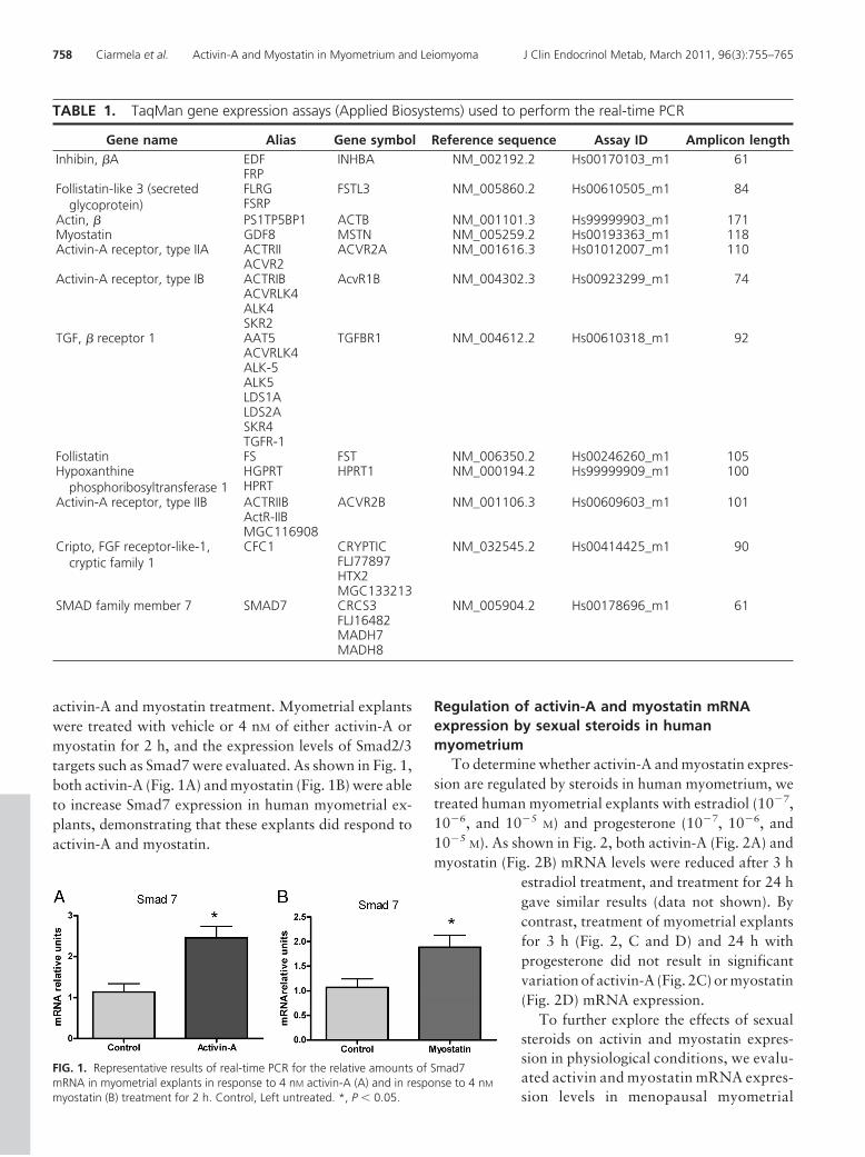
activin-A and myostatin treatment. Myometrial explantswere treated with vehicle or 4 nM of either activin-A ormyostatin for 2 h, and the expression levels of Smad2/3targets such as Smad7 were evaluated. As shown in Fig. 1,both activin-A (Fig. 1A) and myostatin (Fig. 1B) were ableto increase Smad7 expression in human myometrial ex-plants, demonstrating that these explants did respond toactivin-A and myostatin.
Regulation of activin-A and myostatin mRNAexpression by sexual steroids in humanmyometrium
To determine whether activin-A and myostatin expres-sion are regulated by steroids in human myometrium, wetreated human myometrial explants with estradiol (10�7,10�6, and 10�5
M) and progesterone (10�7, 10�6, and10�5
M). As shown in Fig. 2, both activin-A (Fig. 2A) andmyostatin (Fig. 2B) mRNA levels were reduced after 3 h
estradiol treatment, and treatment for 24 hgave similar results (data not shown). Bycontrast, treatment of myometrial explantsfor 3 h (Fig. 2, C and D) and 24 h withprogesterone did not result in significantvariation of activin-A (Fig. 2C) or myostatin(Fig. 2D) mRNA expression.
To further explore the effects of sexualsteroids on activin and myostatin expres-sion in physiological conditions, we evalu-ated activin and myostatin mRNA expres-sion levels in menopausal myometrial
FIG. 1. Representative results of real-time PCR for the relative amounts of Smad7mRNA in myometrial explants in response to 4 nM activin-A (A) and in response to 4 nM
myostatin (B) treatment for 2 h. Control, Left untreated. *, P � 0.05.
TABLE 1. TaqMan gene expression assays (Applied Biosystems) used to perform the real-time PCR
Gene name Alias Gene symbol Reference sequence Assay ID Amplicon lengthInhibin, �A EDF INHBA NM_002192.2 Hs00170103_m1 61
FRPFollistatin-like 3 (secreted
glycoprotein)FLRG FSTL3 NM_005860.2 Hs00610505_m1 84FSRP
Actin, � PS1TP5BP1 ACTB NM_001101.3 Hs99999903_m1 171Myostatin GDF8 MSTN NM_005259.2 Hs00193363_m1 118Activin-A receptor, type IIA ACTRII ACVR2A NM_001616.3 Hs01012007_m1 110
ACVR2Activin-A receptor, type IB ACTRIB AcvR1B NM_004302.3 Hs00923299_m1 74
ACVRLK4ALK4SKR2
TGF, � receptor 1 AAT5 TGFBR1 NM_004612.2 Hs00610318_m1 92ACVRLK4ALK-5ALK5LDS1ALDS2ASKR4TGFR-1
Follistatin FS FST NM_006350.2 Hs00246260_m1 105Hypoxanthine
phosphoribosyltransferase 1HGPRT HPRT1 NM_000194.2 Hs99999909_m1 100HPRT
Activin-A receptor, type IIB ACTRIIB ACVR2B NM_001106.3 Hs00609603_m1 101ActR-IIBMGC116908
Cripto, FGF receptor-like-1,cryptic family 1
CFC1 CRYPTIC NM_032545.2 Hs00414425_m1 90FLJ77897HTX2MGC133213
SMAD family member 7 SMAD7 CRCS3 NM_005904.2 Hs00178696_m1 61FLJ16482MADH7MADH8
758 Ciarmela et al. Activin-A and Myostatin in Myometrium and Leiomyoma J Clin Endocrinol Metab, March 2011, 96(3):755–765

specimens compared with myometrial specimens fromfertile (proliferative) women. As shown in Fig. 2, E and F,under physiological conditions in which sex steroids areabsent, the expression levels of activin-A (Fig. 2 E) andmyostatin (Fig. 2 F) are both higher.
Activin-A and myostatin expression in leiomyomaBecause our data show that activin-A and myostatin act
on myometrium and are regulated by sexual steroids, wehypothesized that they may represent gene candidates re-sponsible for pathologies that are very frequent in womensuch as leiomyoma. To test this, we evaluated the expres-sion levels of activin-A and myostatin in fibroid comparedwith matched myometrium specimens. We found that ac-tivin-A (Fig. 3A) and myostatin (Fig. 3B) mRNA levels arehigher expressed in fibroid specimens compared with ad-jacent myometrium.
Expression levels of activin-A and myostatinreceptors and Smad 7 in leiomyoma
Next we tested whether the higher levels of activin-A andmyostatin expression found in leiomyoma corresponded toa higher expression of the activin/myostatin signaling com-ponents. We evaluated the expression levels of all the recep-tors (Fig. 3, C–F) and the basal expression levels of Smad 7(Fig. 4A) and, as shown in Figs. 3, C–F (columns I and II) and4A, we found that all the receptors and Smad 7 mRNA levelswere not different in leiomyoma compared with matchedhealthy myometrium. Column III (Fig. 3) shows the cor-relation of activin-A levels with receptors levels, whereascolumn IV shows the correlation of myostatin levels withthose of its receptors. The statistical analysis (nonpara-metric correlation, Spearman) revealed no correlation ofboth activin and myostatin levels with receptor levels.
FIG. 2. Regulation of activin-A and myostatin mRNA expression by steroids in human myometrium. Activin-A (A) and myostatin (B) mRNA percentagerelative amount in explants treated with estradiol (E) were compared with untreated samples. Activin-A (C) and myostatin (D) mRNA percentage relativeamount in explants treated with progesterone (P) compared with untreated samples. Activin-A (E) and myostatin (F) mRNA expression levels inmyometrium from fertile were compared with menopausal women. n � 5. *, P � 0.05.
J Clin Endocrinol Metab, March 2011, 96(3):755–765 jcem.endojournals.org 759

Activin-A and myostatin responsiveness of fibrotictissues
Our result showing that Smad7 mRNA levels were nothigher in leiomyoma than in normal myometrium (Fig.4A) suggested to us that higher activin-A and myostatin
expression levels did not correspond a higher activin-Aand myostatin actions in leiomyoma. To test this, wetreated leiomyoma explants for 2 h with activin-A or myo-statin and then measured resulting effects on Smad7 ex-pression. As shown in Fig. 4B, neither activin-A (Fig. 4A)
FIG. 3. A and B, Activin and myostatin levels of mRNA expression in eight paired fibroid and myometrial specimens. C–F, Levels of mRNAexpression in eight paired fibroid and myometrial specimens for the four receptor genes ALK4, ALK5, ActRII, and ActRIIB. Graphs on column I areline graphs linking paired specimens, and those on column II are box-and-whisker plots. In column III is reported the correlation of activin-A withits receptors, whereas in column IV is reported the correlation of myostatin with its receptors. The nonparametric correlation (Spearman) revealedno correlation of both activin and myostatin levels with receptor levels. *, P � 0.05.
760 Ciarmela et al. Activin-A and Myostatin in Myometrium and Leiomyoma J Clin Endocrinol Metab, March 2011, 96(3):755–765

nor myostatin (Fig. 4B) were able to increase Smad7 ex-pression, demonstrating that, unlike normal myometrium(Fig. 1), leiomyoma explants do not respond to these li-gands. We had the same result with 4 and 10 nM of proteinconcentration.
Activin-A and myostatin binding proteinsfollistatin and FLRG expression levels inleiomyoma
To explain the previous finding, we hypothesized thatone or more inhibitors of activin and myostatin may dis-play an altered expression that could interfere with themby disrupting their signaling. We tested follistatin andFLRG mRNA expression, and we found that althoughfollistatin did not change, FLRG expression was higher inleiomyoma compared with matched healthy myometrium(Fig. 5A). As shown in Fig. 5B, activin and myostatin eval-uated as ratio with follistatin were still higher in leiomy-oma compared with matched healthy myometrium. Onthe other hand, activin and myostatin evaluated as ratiowith FLRG were not significantly higher in leiomyomacompared with matched healthy myometrium. To betterunderstand the role of follistatin and FLRG in blockingactivin and myostatin, we also evaluated their respectiveprotein levels. As shown in Fig. 5C, FLRG protein wasincreased in the leiomyoma compared with matchedhealthy myometrium, with a pattern similar to the RNA,whereas follistatin protein resulted unchanged.
Identification of Cripto expressionin leiomyoma
Because we previously identifiedCripto as factor that is able to antago-nize activin (13, 14), we also tested itsexpression in the leiomyoma. As shownin Fig. 6A, Cripto was expressed in al-most all the fibrotic specimens exam-ined but was not detected in healthymyometrium.
Cripto inhibits myostatinsignaling
We have previously shown thatCripto inhibits activin and TGF-� sig-naling (15). Because myostatin uses theactivin/TGF-� signaling pathway, wetested whether Cripto also functions asan antagonist of myostatin signaling.We transfected 293T cells with aSmad2-responsive luciferase reporterin the presence of empty vector orCripto and then treated cells with arange of doses of myostatin. As shownin Fig. 6A, myostatin induced Smad2-
dependent luciferase expression in a dose-dependent man-ner, and this myostatin response was inhibited by Cripto.The inhibitory effect of Cripto on myostatin signaling ap-peared to be similar to Cripto antagonism of activin sig-naling (15). This suggests that Cripto blocks both ligandsvia a similar mechanism and is consistent with the abilityof Cripto to bind the activin/myostatin type I receptorALK4.
Discussion
In the present paper, we provide the first evidence thathuman myometrial tissues are able to respond to activinand myostatin as measured by their induction of Smad7.This finding is consistent with what we have seen before ina myometrial cell line, PHM1, and in rat uterine explants(16, 17). In these previous studies, we also reported thatthe expression of activin and myostatin in rat uterine ex-plants (endometrium � myometrium) is regulated by ste-roid hormones. Similar to what was observed in rat, pro-gesterone did not change activin or myostatin expressionin human uterine myometrium. By contrast, although es-trogen treatment drastically decreased expression of ac-tivin and myostatin in human myometrium, it abrogatedmyostatin expression but increased expression of activinin rat uterus (endometrium � myometrium) (16). The rea-son for this discrepancy is not clear but may be due to
FIG. 4. A, Smad7 levels of mRNA expression in eight paired fibroid and myometrialspecimens. Graphs on the left are line graphs linking paired specimens, and those on the rightare box-and-whisker plots. B, Representative results of real-time PCR for the relative amountsof Smad7 mRNA in fibroid explants in response to 4 nM activin-A and in response to 4 nM
myostatin treatment for 2 h. Control, Left untreated.
J Clin Endocrinol Metab, March 2011, 96(3):755–765 jcem.endojournals.org 761

species-specific and/or tissue-specific regulatory mech-anisms and/or instead methods specific (in vivo vs. ex-plants culture). Of note, we found increased activin andmyostatin expression in menopausal tissues in the ab-
sence of steroids providing further ev-idence that estrogen acts to suppressthe levels of both these ligands inhumans.
After having established that activinand myostatin target human myome-trium explants and are regulated bysteroid hormones, we evaluated theirrelative expression levels in fibroidspecimens and adjacent healthy myo-metrium. Considering the high vari-ability that has been reported for manyfactors, including levels of estrogen andprogesterone receptors (25, 26) andgene profiles (23) between patients, wechose to use restricted inclusion criteriato keep group mean variation to a min-imum (same race, menstrual phase, sizeand location of the tumor). Our findingthat activin-A and myostatin expres-sion levels were higher in leiomyomathan in normal myometrium was un-expected because these growth factorshave cytostatic effects on the humanmyometrial PHM1 cell line (16, 17).On the other hand, basal Smad7 ex-pression levels and the expression lev-els of all the receptors did not changebetween normal and fibroid tissue. Wetested whether activin-A and myostatintarget the fibrotic tissue, and, not sur-prisingly, neither protein was able to in-crease Smad7 expression in leiomyomaspecimens. We hypothesized that the in-ability of fibroid to respond to activinand myostatin was due to the presenceof signaling inhibitors, i.e. higher ex-pression of FLRG in fibroid specimenscompared with adjacent healthy myo-metrium and the selective expression ofCripto in leiomyoma.
When activin and myostatin mRNAlevels were corrected relative to fol-listatin mRNA levels, we still observedsignificantly higher expression of theirmRNAs in leiomyoma compared withhealthy adjacent myometrium. By con-trast, FLRG expression differences be-tween normal tissue and leiomyoma
were sufficient to explain loss of signaling in leiomyoma.Furthermore, evaluating activin and myostatin mRNA ex-pression levels corrected to FLRG mRNA levels showed
FIG. 5. A, Levels of mRNA expression in paired fibroid and myometrial specimens for the two bindingproteins FST (n � 8) and FLRG (n � 6). Graphs on the left are line graphs linking paired specimens, andthose on the right are box-and-whisker plots. B, Activin and myostatin mRNA levels normalized withFST (on left) and FLRG (on right) in paired fibroid and myometrial specimens. C, Western blotting anddensitometric analisys of FST, FLRG, and proteins. M, Myometrium; F, fibroid. *, P � 0.05.
762 Ciarmela et al. Activin-A and Myostatin in Myometrium and Leiomyoma J Clin Endocrinol Metab, March 2011, 96(3):755–765

that their relative amounts were not different between thehealthy and pathological specimens.
Similar to previous studies, our experimental datasupport the concept that follistatin and FLRG are func-tionally redundant despite differences in their expres-sion and transcriptional regulation (27). Indeed, wehave observed this phenomenon in other reproductivesystems, such as placenta (28), endometrium (29, 30),and breast (31).
Activin and myostatin are members of the TGF-� su-perfamily and the presence of TGF-�s and their receptorsin human myometrium and leiomyoma was first shown in1994 by Chegini et al. (32). The same authors later alsoshowed that GnRH-� decreased expression of TFG-� re-ceptors and TGF-� receptor intracellular signaling mole-cules (33).
Cripto is a GPI-anchored signaling protein that con-trols the activity of multiple TGF-� superfamily members(34). Cripto binds directly to the activin/myostatin type Ireceptor ALK4 (35) and inhibits activin signaling (13, 14).This led us to hypothesize that Cripto may also function asan inhibitor of myostatin signaling. Indeed, we have nowshown that Cripto causes a reduction in myostatin signal-ing similar to that previously shown for activin. Cripto ispredominantly expressed during normal developmentalprocesses and in cancer. Cripto is expressed at high levels
in human breast, colon, stomach, pancreas, lung, ovaryendometrial, testis, bladder, prostate, and myometrial tu-mors but is absent or expressed at lower levels in theirnormal counterparts (34, 36).
Recently leiomyosarcoma of the uterus was observed inCripto-overexpressing transgenic mice (36). Regarding hu-manmyometrium,Criptohasbeen reported tobeexpressed,by immunohistochemistry, in leiomyosarcoma and only invery few samples of leiomyoma (36). Here, using real-timePCR, we were also able to show the presence of CriptomRNA in leiomyoma, suggesting that it may be a contrib-uting cause of benign tumor transformation.
The data presented in this paper support the hypothesisthat alterations in the activin- and myostatin-related pro-tein systems may produce loss of sensitivity to the anti-proliferative effects of activin and myostatin and that in-creased expression of FLRG and Cripto may contribute tothe growth of these tumors. Therefore, activin and myo-statin need to be considered as factors involved in myo-metrial functionality in addition to the other growth fac-tors already known to have this function such as EGF,heparin-binding EGF-like growth factor, platelet-derivedgrowth factor (37), IGF (38), vascular endothelial growthfactor, and TGF-� (39).
Regarding activin-A and myostatin, we evaluated theirexpression and tested their ability to act on fibrotic tissues.Although activin-A and myostatin are overexpressed inleiomyoma, they are unable to induce Smad7, likely due tooverexpression of inhibitors including FLRG and/orCripto.
Thus, it is possible that in fibrotic myometrium thereis an initial activation of Cripto expression and an in-creased FLRG expression that render activin and myo-statin less active. The reduction of activin and myostatinsignaling may then cause a compensatory increase intheir expression.
In conclusion, our findings suggest that activin-A andmyostatin act on human myometrium, that they are reg-ulated by steroid hormones, and that the disruption oftheir signaling may contribute to the fibroid growth.
Acknowledgments
Address all correspondence and requests for reprints to:Pasquapina Ciarmela, Ph.D., Department of Molecular Pathol-ogy and Innovative Therapies, Anatomy, and Cell Biology, Fac-ulty of Medicine, Polytechnic University of Marche, via Tronto10/a, 60020 Ancona, Italy. E-mail: [email protected].
This work was supported by the Grants4Targets initiativefrom Bayer Schering Pharma (to P.Ci.) and the “FondazioneCassa di Risparmio di Fabriano e Cupramontana” (to M.C.).This work was also supported by Award 2R56 CA107420-06from the National Cancer Institute (to P.C.G.). This work was
FIG. 6. A, Identification of mRNA Cripto expression in leiomyoma byreal-time PCR. B, Cripto disruption of myostatin (GDF8) signaling. Inthe x-axis is reported the treatment and the doses used for theluciferase assay. *, P � 0.05.
J Clin Endocrinol Metab, March 2011, 96(3):755–765 jcem.endojournals.org 763

also supported in part by the Clayton Medical Research Foun-dation, Inc. (to W.V. and P.C.G.). W.V. is a CMRF SeniorInvestigator.
The content of this work is solely the responsibility of theauthors and does not necessarily represent the official views ofthe National Cancer Institute or the National Institutes ofHealth.
Disclosure Summary: W.V. is a cofounder, consultant, equityholder, and member of the Board of Directors of NeurocrineBiosciences and Acceleron Pharma. All the other authors havenothing to declare.
References
1. Harrison CA, Gray PC, Vale WW, Robertson DM 2005 Antagonistsof activin signaling: mechanisms and potential biological applica-tions. Trends Endocrinol Metab 16:73–78
2. McPherron AC, Lawler AM, Lee SJ 1997 Regulation of skeletalmuscle mass in mice by a new TGF-� superfamily member. Nature387:83–90
3. Mathews LS, Vale WW 1991 Expression cloning of an activin re-ceptor, a predicted transmembrane serine kinase. Cell 65:973–982
4. Attisano L, Wrana JL, Montalvo E, Massague J 1996 Activation ofsignalling by the activin receptor complex. Mol Cell Biol 16:1066–1073
5. Derynck R, Zhang Y, Feng XH 1998 Smads: transcriptional acti-vators of TGF-� responses. Cell 95:737–740
6. Rebbapragada A, Benchabane H, Wrana JL, Celeste AJ, Attisano L2003 Myostatin signals through a transforming growth factor �-likesignaling pathway to block adipogenesis. Mol Cell Biol 23:7230–7242
7. Bilezikjian LM, Corrigan AZ, Blount AL, Chen Y, Vale WW 2001Regulation and actions of Smad7 in the modulation of activin, in-hibin, and transforming growth factor-� signaling in anterior pitu-itary cells. Endocrinology 142:1065–1072
8. Zhu X, Topouzis S, Liang LF, Stotish RL 2004 Myostatin signalingthrough Smad2, Smad3 and Smad4 is regulated by the inhibitorySmad7 by a negative feedback mechanism. Cytokine 26:262–272
9. Hill JJ, Davies MV, Pearson AA, Wang JH, Hewick RM, WolfmanNM, Qiu Y 2002 The myostatin propeptide and the follistatin-re-lated gene are inhibitory binding proteins of myostatin in normalserum. J Biol Chem 277:40735–40741
10. Hayette S, Gadoux M, Martel S, Bertrand S, Tigaud I, Magaud JP,Rimokh R 1998 FLRG (follistatin-related gene), a new target ofchromosomal rearrangement in malignant blood disorders. Onco-gene 16:2949–2954
11. Tsuchida K, Arai KY, Kuramoto Y, Yamakawa N, Hasegawa Y,Sugino H 2000 Identification and characterization of a novel fol-listatin-like protein as a binding protein for the TGF-� family. J BiolChem 275:40788–40796
12. Ciccodicola A, Dono R, Obici S, Simeone A, Zollo M, Persico MG1989 Molecular characterization of a gene of the ‘EGF family’ ex-pressed in undifferentiated human NTERA2 teratocarcinoma cells.EMBO J 8:1987–1991
13. Gray PC, Harrison CA, Vale W 2003 Cripto forms a complex withactivin and type II activin receptors and can block activin signaling.Proc Natl Acad Sci USA 100:5193–5198
14. Kelber JA, Shani G, Booker EC, Vale WW, Gray PC 2008 Criptois a noncompetitive activin antagonist that forms analogous sig-naling complexes with activin and nodal. J Biol Chem 283:4490 –4500
15. Gray PC, Shani G, Aung K, Kelber J, Vale W 2006 Cripto bindstransforming growth factor � (TGF-�) and inhibits TGF-� signal-ing. Mol Cell Biol 26:9268–9278
16. Ciarmela P, Wiater E, Smith SM, Vale W 2009 Presence, actions, and
regulation of myostatin in rat uterus and myometrial cells. Endo-crinology 150:906–914
17. Ciarmela P, Wiater E, Vale W 2008 Activin-A in myometrium: char-acterization of the actions on myometrial cells. Endocrinology 149:2506–2516
18. Flake GP, Andersen J, Dixon D 2003 Etiology and pathogenesis ofuterine leiomyomas: a review. Environ Health Perspect 111:1037–1054
19. Walker CL, Stewart EA 2005 Uterine fibroids: the elephant in theroom. Science 308:1589–1592
20. Everitt JI, Wolf DC, Howe SR, Goldsworthy TL, Walker C 1995Rodent model of reproductive tract leiomyomata. Clinical and path-ological features. Am J Pathol 146:1556–1567
21. Laping NJ, Everitt JI, Frazier KS, Burgert M, Portis MJ, Cadacio C,Gold LI, Walker CL 2007 Tumor-specific efficacy of transforminggrowth factor-� RI inhibition in Eker rats. Clin Cancer Res 13:3087–3099
22. Catherino WH, Prupas C, Tsibris JC, Leppert PC, Payson M,Nieman LK, Segars JH 2003 Strategy for elucidating differentiallyexpressed genes in leiomyomata identified by microarray technol-ogy. Fertil Steril 80:282–290
23. Catherino WH, Segars JH 2003 Microarray analysis in fibroids:which gene list is the correct list? Fertil Steril 80:293–294
24. Hodge JC, Morton CC 2007 Genetic heterogeneity among uterineleiomyomata: insights into malignant progression. Hum Mol Genet16(Spec No 1):R7–R13
25. Englund K, Blanck A, Gustavsson I, Lundkvist U, Sjoblom P,Norgren A, Lindblom B 1998 Sex steroid receptors in human myo-metrium and fibroids: changes during the menstrual cycle and go-nadotropin-releasing hormone treatment. J Clin Endocrinol Metab83:4092–4096
26. Sakaguchi H, Fujimoto J, Aoki I, Tamaya T 2003 Expression ofestrogen receptor � and � in myometrium of premenopausal andpostmenopausal women. Steroids 68:11–19
27. Saito S, Sidis Y, Mukherjee A, Xia Y, Schneyer A 2005 Differentialbiosynthesis and intracellular transport of follistatin isoforms andfollistatin-like-3. Endocrinology 146:5052–5062
28. Ciarmela P, Florio P, Toti P, Franchini A, Maguer-Satta V, Ginann-eschi C, Ottaviani E, Petraglia F 2003 Human placenta and fetalmembranes express follistatin-related gene mRNA and protein.J Endocrinol Invest 26:641–645
29. Florio P, Ciarmela P, Toti P, Maguer-Satta V, Rimokh R, BuonocoreG, Rossi M, Gioffre W, Petraglia F 2004 Human endometrium anddecidua express follistatin-related gene (FLRG) mRNA and peptide.Mol Cell Endocrinol 218:129–135
30. Ciarmela P, Florio P, Sigurdardottir M, Toti P, Maguer-Satta V,Rimokh R, Altomare A, Tosi P, Petraglia F 2004 Follistatin-related gene expression, but not follistatin expression, is de-creased in human endometrial adenocarcinoma. Eur J Endocrinol151:251–257
31. Bloise E, Couto HL, Massai L, Ciarmela P, Mencarelli M, BorgesLE, Muscettola M, Grasso G, Amaral VF, Cassali GD, Petraglia F,Reis FM 2009 Differential expression of follistatin and FLRG inhuman breast proliferative disorders. BMC Cancer 9:320
32. Chegini N, Zhao Y, Williams RS, Flanders KC 1994 Humanuterine tissue throughout the menstrual cycle expresses trans-forming growth factor-�1 (TGF �1), TGF �2, TGF �3, and TGF� type II receptor messenger ribonucleic acid and protein andcontains [125I]TGF �1-binding sites. Endocrinology 135:439 –449
33. Chegini N, Luo X, Ding L, Ripley D 2003 The expression of Smadsand transforming growth factor � receptors in leiomyoma and myo-metrium and the effect of gonadotropin releasing hormone analoguetherapy. Mol Cell Endocrinol 209:9–16
34. Saloman DS, Bianco C, Ebert AD, Khan NI, De Santis M, NormannoN, Wechselberger C, Seno M, Williams K, Sanicola M, Foley S,Gullick WJ, Persico G 2000 The EGF-CFC family: novel epidermal
764 Ciarmela et al. Activin-A and Myostatin in Myometrium and Leiomyoma J Clin Endocrinol Metab, March 2011, 96(3):755–765

growth factor-related proteins in development and cancer. EndocrRelat Cancer 7:199–226
35. Calvanese L, Saporito A, Oliva R, D’ Auria G, Pedone C, Paolillo L,Ruvo M, Marasco D, Falcigno L 2009 Structural insights into theinteraction between the Cripto CFC domain and the ALK4 receptor.J Pept Sci 15:175–183
36. Strizzi L, Bianco C, Hirota M, Watanabe K, Mancino M, HamadaS, Raafat A, Lawson S, Ebert AD, D’Antonio A, Losito S, NormannoN, Salomon DS 2007 Development of leiomyosarcoma of the uterusin MMTV-CR-1 transgenic mice. J Pathol 211:36–44
37. Rossi MJ, Chegini N, Masterson BJ 1992 Presence of epidermalgrowth factor, platelet-derived growth factor, and their receptors
in human myometrial tissue and smooth muscle cells: their actionin smooth muscle cells in vitro. Endocrinology 130:1716 –1727
38. Tang XM, Rossi MJ, Masterson BJ, Chegini N 1994 Insulin-likegrowth factor I (IGF-I), IGF-I receptors, and IGF binding proteins1–4 in human uterine tissue: tissue localization and IGF-I action inendometrial stromal and myometrial smooth muscle cells in vitro.Biol Reprod 50:1113–1125
39. Tang XM, Dou Q, Zhao Y, McLean F, Davis J, Chegini N 1997The expression of transforming growth factor-�s and TGF-� re-ceptor mRNA and protein and the effect of TGF-�s on humanmyometrial smooth muscle cells in vitro. Mol Hum Reprod3:233–240
The Endocrine Society journals for teaching purposes!
www.endo-society.org
J Clin Endocrinol Metab, March 2011, 96(3):755–765 jcem.endojournals.org 765
Related Documents