Neurobiology of Disease Activation of Neurotensin Receptor 1 Facilitates Neuronal Excitability and Spatial Learning and Memory in the Entorhinal Cortex: Beneficial Actions in an Alzheimer’s Disease Model Zhaoyang Xiao, 1 * Nicholas I. Cilz, 1 * Lalitha Kurada, 1 * Binqi Hu, 1 Chuanxiu Yang, 1 Etsuko Wada, 2 Colin K. Combs, 1 James E. Porter, 1 Florian Lesage, 3 and Saobo Lei 1 1 Department of Basic Sciences, School of Medicine and Health Sciences, University of North Dakota, Grand Forks, North Dakota 58203, 2 Department of Degenerative Neurological Diseases, National Institute of Neuroscience, Tokyo, Japan, and 3 Institut de Pharmacologie Mole ´culaire et Cellulaire, Centre National de la Recherche Scientifique, and Universite ´ de Nice Sophia-Antipolis, Sophia-Antipolis, 06560 Valbonne, France Neurotensin (NT) is a tridecapeptide distributed in the CNS, including the entorhinal cortex (EC), a structure that is crucial for learning and memory and undergoes the earliest pathological alterations in Alzheimer’s disease (AD). Whereas NT has been implicated in modulating cognition, the cellular and molecular mechanisms by which NT modifies cognitive processes and the potential therapeutic roles of NT in AD have not been determined. Here we examined the effects of NT on neuronal excitability and spatial learning in the EC, which expresses high density of NT receptors. Brief application of NT induced persistent increases in action potential firing frequency, which could last for at least 1 h. NT-induced facilitation of neuronal excitability was mediated by downregulation of TREK-2 K channels and required the functions of NTS1, phospholipase C, and protein kinase C. Microinjection of NT or NTS1 agonist, PD149163, into the EC increased spatial learning as assessed by the Barnes Maze Test. Activation of NTS1 receptors also induced persistent increases in action potential firing frequency and significantly improved the memory status in APP/PS1 mice, an animal model of AD. Our study identifies a cellular substrate underlying learning and memory and suggests that NTS1 agonists may exert beneficial actions in an animal model of AD. Key words: glutamate; ion channel; memory; peptide; synapse; synaptic transmission Introduction Progressive deterioration of cognitive performance is a critical characteristic of Alzheimer’s disease (AD), and improving cogni- tive function represents a major therapeutic approach for this disease. Current available therapies for AD, including the cholin- esterase inhibitors (Pepeu and Giovannini, 2009) and the partial NMDA receptor antagonist memantine (van Marum, 2008), provide only minimal benefit for a limited period to a subset of patients. Finding drugs that can improve memory is still a major method to relieve the symptoms of AD patients. Accumulating evidence demonstrates that neurotensin (NT), a tridecapeptide, and NT receptors are densely distributed in the entorhinal cortex (EC) (Wolf et al., 1994, 1995; Lahti et al., 1998; Hamid et al., 2002), a structure that is essential for learning and memory and undergoes the earliest pathological alteration in AD (Go ´ mez-Isla et al., 1996). However, the physiological functions of NT in the EC, especially in learning and memory, and the therapeutic po- tential of NT receptor modulators for AD have not been determined. NT-containing cell bodies and fibers have been detected in the EC and the structures that have inputs to the EC, such as olfactory tubercle, subiculum, amygdale, and claustrum (Chen et al., 1988; Atoji et al., 1995). NT interacts with three different types of NT receptors: NTS1, NTS2, and NTS3, of which NTS1 and NTS2 are G-protein-coupled (Vincent et al., 1999). NTS1 has higher affin- ity for NT and is blocked by the selective antagonist SR48692, whereas NTS2 has lower affinity for NT and is sensitive to levo- cabastine. NTS1 is functionally coupled to phospholipase C (PLC) pathway via Gq proteins (Hermans and Maloteaux, 1998; Vincent et al., 1999). The pharmacological and signaling proper- ties of NTS2 are still controversial. In particular, it is unclear whether NT acts as an agonist, inverse agonist, or antagonist for this type of receptor. The functions of NTS2 are unclear, although it may be involved in NT-induced internalization of receptor- ligand complexes (St-Gelais et al., 2006). NTS3 is a single trans- Received Jan. 29, 2014; revised March 31, 2014; accepted April 15, 2014. Author contributions: J.E.P., F.L., and S.L. designed research; Z.X., N.I.C., L.K., B.H., and C.Y. performed research; E.W., C.K.C., J.E.P., and F.L. contributed unpublished reagents/analytic tools; Z.X., N.I.C., L.K., B.H., and C.Y. analyzed data; S.L. wrote the paper. This work was supported by National Institute of Mental Health Grant MH082881. We thank Dr. Donghee Kim (Rosalind Franklin University of Medicine and Science, Chicago) for providing the mutant TREK-2 channel cDNA. The authors declare no competing financial interests. *Z.X., N.I.C., and L.K. contributed equally to this work. Correspondence should be addressed to Dr. Saobo Lei, Department of Basic Sciences, School of Medicine and Health Sciences, University of North Dakota, Grand Forks, ND 58203. E-mail: [email protected]. DOI:10.1523/JNEUROSCI.0408-14.2014 Copyright © 2014 the authors 0270-6474/14/347027-16$15.00/0 The Journal of Neuroscience, May 14, 2014 • 34(20):7027–7042 • 7027

Welcome message from author
This document is posted to help you gain knowledge. Please leave a comment to let me know what you think about it! Share it to your friends and learn new things together.
Transcript
Neurobiology of Disease
Activation of Neurotensin Receptor 1 Facilitates NeuronalExcitability and Spatial Learning and Memory in theEntorhinal Cortex: Beneficial Actions in an Alzheimer’sDisease Model
Zhaoyang Xiao,1* Nicholas I. Cilz,1* Lalitha Kurada,1* Binqi Hu,1 Chuanxiu Yang,1 Etsuko Wada,2 Colin K. Combs,1
James E. Porter,1 Florian Lesage,3 and Saobo Lei1
1Department of Basic Sciences, School of Medicine and Health Sciences, University of North Dakota, Grand Forks, North Dakota 58203, 2Department ofDegenerative Neurological Diseases, National Institute of Neuroscience, Tokyo, Japan, and 3Institut de Pharmacologie Moleculaire et Cellulaire, CentreNational de la Recherche Scientifique, and Universite de Nice Sophia-Antipolis, Sophia-Antipolis, 06560 Valbonne, France
Neurotensin (NT) is a tridecapeptide distributed in the CNS, including the entorhinal cortex (EC), a structure that is crucial for learningand memory and undergoes the earliest pathological alterations in Alzheimer’s disease (AD). Whereas NT has been implicated inmodulating cognition, the cellular and molecular mechanisms by which NT modifies cognitive processes and the potential therapeuticroles of NT in AD have not been determined. Here we examined the effects of NT on neuronal excitability and spatial learning in the EC,which expresses high density of NT receptors. Brief application of NT induced persistent increases in action potential firing frequency,which could last for at least 1 h. NT-induced facilitation of neuronal excitability was mediated by downregulation of TREK-2 K � channelsand required the functions of NTS1, phospholipase C, and protein kinase C. Microinjection of NT or NTS1 agonist, PD149163, into the ECincreased spatial learning as assessed by the Barnes Maze Test. Activation of NTS1 receptors also induced persistent increases in actionpotential firing frequency and significantly improved the memory status in APP/PS1 mice, an animal model of AD. Our studyidentifies a cellular substrate underlying learning and memory and suggests that NTS1 agonists may exert beneficial actions in ananimal model of AD.
Key words: glutamate; ion channel; memory; peptide; synapse; synaptic transmission
IntroductionProgressive deterioration of cognitive performance is a criticalcharacteristic of Alzheimer’s disease (AD), and improving cogni-tive function represents a major therapeutic approach for thisdisease. Current available therapies for AD, including the cholin-esterase inhibitors (Pepeu and Giovannini, 2009) and the partialNMDA receptor antagonist memantine (van Marum, 2008),provide only minimal benefit for a limited period to a subset ofpatients. Finding drugs that can improve memory is still a majormethod to relieve the symptoms of AD patients. Accumulatingevidence demonstrates that neurotensin (NT), a tridecapeptide,and NT receptors are densely distributed in the entorhinal cortex
(EC) (Wolf et al., 1994, 1995; Lahti et al., 1998; Hamid et al.,2002), a structure that is essential for learning and memory andundergoes the earliest pathological alteration in AD (Gomez-Islaet al., 1996). However, the physiological functions of NT in theEC, especially in learning and memory, and the therapeutic po-tential of NT receptor modulators for AD have not beendetermined.
NT-containing cell bodies and fibers have been detected in theEC and the structures that have inputs to the EC, such as olfactorytubercle, subiculum, amygdale, and claustrum (Chen et al., 1988;Atoji et al., 1995). NT interacts with three different types of NTreceptors: NTS1, NTS2, and NTS3, of which NTS1 and NTS2 areG-protein-coupled (Vincent et al., 1999). NTS1 has higher affin-ity for NT and is blocked by the selective antagonist SR48692,whereas NTS2 has lower affinity for NT and is sensitive to levo-cabastine. NTS1 is functionally coupled to phospholipase C(PLC) pathway via Gq proteins (Hermans and Maloteaux, 1998;Vincent et al., 1999). The pharmacological and signaling proper-ties of NTS2 are still controversial. In particular, it is unclearwhether NT acts as an agonist, inverse agonist, or antagonist forthis type of receptor. The functions of NTS2 are unclear, althoughit may be involved in NT-induced internalization of receptor-ligand complexes (St-Gelais et al., 2006). NTS3 is a single trans-
Received Jan. 29, 2014; revised March 31, 2014; accepted April 15, 2014.Author contributions: J.E.P., F.L., and S.L. designed research; Z.X., N.I.C., L.K., B.H., and C.Y. performed research;
E.W., C.K.C., J.E.P., and F.L. contributed unpublished reagents/analytic tools; Z.X., N.I.C., L.K., B.H., and C.Y. analyzeddata; S.L. wrote the paper.
This work was supported by National Institute of Mental Health Grant MH082881. We thank Dr. Donghee Kim(Rosalind Franklin University of Medicine and Science, Chicago) for providing the mutant TREK-2 channel cDNA.
The authors declare no competing financial interests.*Z.X., N.I.C., and L.K. contributed equally to this work.Correspondence should be addressed to Dr. Saobo Lei, Department of Basic Sciences, School of Medicine and
Health Sciences, University of North Dakota, Grand Forks, ND 58203. E-mail: [email protected]:10.1523/JNEUROSCI.0408-14.2014
Copyright © 2014 the authors 0270-6474/14/347027-16$15.00/0
The Journal of Neuroscience, May 14, 2014 • 34(20):7027–7042 • 7027

membrane domain receptor and binds NT with high affinity(Vincent et al., 1999).
The EC is closely related to consolidation and recall of mem-ories (Dolcos et al., 2005; Steffenach et al., 2005) and AD (Hymanet al., 1984; Kotzbauer et al., 2001). NT receptors are denselyexpressed in the EC, especially in the superficial layers (Wolf etal., 1994, 1995; Lahti et al., 1998; Hamid et al., 2002). However,the effects of NT in the EC, an indispensable structure in memoryprocessing, and the potential uses of NT receptor modulators inAD therapy have not been determined. In the present study, wedetermined the roles of NT in facilitation of neuronal excitabilityand spatial memory and tested the effects of NT and PD149163, asmall-molecule NTS1 agonist that can penetrate the blood– brainbarrier, on improving spatial memory ability in an AD mousemodel. Our research unraveled an unidentified cellular mecha-nism for memory and provides a potential approach for ADtherapy.
Materials and MethodsSlice preparation. The ages of the animals used for electrophysiologicalrecordings were postnatal 3–5 weeks for Sprague Dawley rats, individualknock-out (KO), and their corresponding wild-type (WT, C57BL/6J)mice. APP/PS1 mice (8 –10 months) were obtained from the The JacksonLaboratory: strain 005864 B6.Cg-Tg(APPswe,PSEN1dE9)85Dbo/J. Age-matched C57BL/6J WT mice provided the control as described previ-ously (Dhawan and Combs, 2012). Horizontal brain slices (400 �m)were prepared as described previously (Wang et al., 2012, 2013; Zhang etal., 2014), except with the following modifications. After being deeplyanesthetized with isoflurane, animals were decapitated and their brainswere dissected out in ice-cold saline solution that contained the following(in mM): 130 N-methyl-D-glucamine (NMDG)-Cl, 24 NaHCO3, 3.5 KCl,1.25 NaH2PO4, 0.5 CaCl2, 5.0 MgCl2, and 10 glucose, saturated with 95%O2 and 5% CO2 (pH 7.4, adjusted with HCl). Slices were then incubatedin the above solution, except that NMDG-Cl was replaced with NaCl at35°C for 1 h for recovery and then kept at room temperature (�24°C)until use. We found that these procedures yielded more viable slices fromold animals.
Whole-cell recordings from entorhinal neurons. Whole-cell patch-clamprecordings using 4 Multiclamp 700B amplifiers (Molecular Devices) incurrent- or voltage-clamp mode were made from the principal neuronsin the EC visually identified with infrared video microscopy (OlympusBX51WI) and differential interference contrast optics as described pre-viously (Deng et al., 2006, 2010; Wang et al., 2011). Recordings wereconducted at room temperature (�23°C). The recording electrodes werefilled with the following (in mM): 100 potassium gluconate, 0.6 EGTA, 5MgCl2, 8 NaCl, 2 ATPNa2, 0.3 GTPNa, phosphocreatine 7, and 33HEPES (pH 7.3 adjusted with KOH). The extracellular solution com-prised the following (in mM): 130 NaCl, 24 NaHCO3, 3.5 KCl, 1.25NaH2PO4, 2.5 CaCl2, 1.5 MgCl2, and 10 glucose, saturated with 95% O2
and 5% CO2, pH 7.4. Data were filtered at 2 kHz, digitized at 10 kHz,acquired on-line, and analyzed off-line using pCLAMP 9 or pCLAMP10software (Molecular Devices).
Action potential (AP) firing was induced by injecting a positive cur-rent (�30 –100 pA) to bring the resting membrane potential (RMP) to��50 mV to induce a basal AP firing frequency of 0.2–1 Hz unless statedotherwise. Drugs were applied after the AP firing had been stable for 5–10min. Frequency of APs was calculated by Mini Analysis 6.0.1 (Synap-tosoft). Holding currents (HCs) at �55 mV were recorded in the extra-cellular solution containing TTX (0.5 �M) every 3 s and then averaged perminute. We subtracted the average of the HCs recorded for the lastminute before the application of NT from those recorded at differenttime points to zero the basal level of HCs for better comparison unlessstated otherwise. I-V curves were constructed by using a ramp protocolfrom �140 to 0 mV at a speed of 0.1 mV/ms in the external solutionsupplemented with (in �M) 0.5 TTX, 100 CdCl2, 10 DNQX, 100 DL-APV,10 bicuculline, and 2 CGP55845 to block synaptic transmission and other
voltage-gated ion channels. We compared the I-V curves recorded beforeand after the application of NT for 5–10 min.
EPSPs were recorded from layer II stellate neurons by placing a stim-ulation electrode in layer I to stimulate glutamatergic inputs in the extra-cellular solution containing bicuculline (10 �M) and CGP55845 (2 �M)to block GABAergic transmission. To prevent the propagation of epilep-tic activity in the presence of GABAergic inhibitors, a cut was made alongthe middle line of layer III (Wang et al., 2012, 2013). The intracellularsolution was either the above K �-gluconate-containing intracellular so-lution or the intracellular solution in which K � was replaced by the sameconcentration of Cs �. The stimulation intensity was set to the level thatproduced �50% of the maximal EPSP amplitude. The stimulation fre-quency was set at 0.2 Hz. The numbers of APs were counted within 150ms after the stimulation artifact.
Transfection and electrophysiological recordings from transfected cells.Detailed procedures for transfection of HEK293 cells and electrophysio-logical recordings from the transfected cells were described previously(Deng et al., 2007, 2009; Xiao et al., 2009). Briefly, HEK293 cells weretransfected with NTS1 alone, or together with WT TREK-2 channels orTREK-2 mutant. GFP was cotransfected for each experiment to identifythe transfected cells. cDNA constructs coding for a human NT Type 1receptor (GenBank accession number AY429106, subcloned intopCDNA3.1 vector) was obtained from the Missouri S&T cDNA ResourceCenter (www.cdna.org). cDNA for the mutant TREK-2 channels waskindly provided by Dr. Donghee Kim (Rosalind Franklin University ofMedicine and Science, Chicago) (Kang et al., 2006). cDNA constructscoding for TREK-2 (NM_021161, subcloned into the pCMV6-XL4 vec-tor) were purchased from Origene. An empty pEGFP N-3 GFP fusionprotein vector (GenBank accession number U57609) was purchasedfrom Clontech. Transfected HEK293 cells were subsequently used forelectrophysiological recordings 24 – 48 h after transfection. HCs at �55mV were recorded from the HEK293 cells that showed fluorescence un-der a fluorescence microscope (Olympus 1X70) by whole-cell record-ings. The extracellular solution contained (in mM) 130 NaCl, 3 KCl, 2MgCl2, 2 CaCl2, 1.25 NaH2PO4, 10 HEPES, and 10 glucose (pH adjustedto 7.4 using NaOH and HCl). The above K �-gluconate internal solutionwas used for this experiment. A continuous gravity perfusion system(flow, 5–7 ml/min) was used to change solutions.
Immunocytochemistry. The methods for immunocytochemistry weredescribed previously (Deng and Lei, 2008; Deng et al., 2009; Xiao et al.,2009; Ramanathan et al., 2012). Briefly, horizontal sections (20 �m)from rat brain were washed in 0.1 M PBS and then treated with 0.3%hydrogen peroxide (H2O2) for 10 min to quench endogenous peroxidaseactivity. After being rinsed in 0.1 M PBS containing 1% Triton X-100 and5% normal donkey serum for 30 min, sections were incubated with theprimary goat antibody to NTS1 (Santa Cruz Biotechnology, sc-7598) at adilution of 1:200 at 4°C for 12 h. Sections were incubated at room tem-perature initially with biotinylated donkey anti-goat IgG (ImmunocruzStaining System, Santa Cruz Biotechnology) for 1 h and then with HRP-streptavidin complex (Immunocruz Staining System) for 1 h. After eachincubation, sections were washed three times for a total of 30 min. Di-aminobenzidine (Immunocruz Staining System) was used for a colorreaction to detect the positive signals. Finally, sections were mounted onslides, dehydrated through an alcohol range, cleared in xylene, and cov-ered with coverslips. We stained �10 nonadjacent sections for each rat,and each staining was repeated by using 3 rats. For a control, NTS1antibody was preabsorbed with the corresponding blocking peptide(Santa Cruz Biotechnology, sc-7598P) before being applied to the sec-tions, and the other experimental procedures were the same as describedabove.
Immunoprecipitation and Western blot. Brain slices containing the ECwere obtained from Sprague Dawley rats, WT (C57BL/6J) and TREK-2KO mice (n � 6). For each animal, horizontal brain slices were cutinitially, and the EC region was punched out from the slices under amicroscope. The isolated brain region was treated with 0.25 �M NT in theoxygenated extracellular solution for 5 min and then incubated in NT-free extracellular solution for varied times as described in Results. Tissuelysates from the EC were prepared as described previously (Deng et al.,2009; Xiao et al., 2009). The lysates were centrifuged at 14,000 rpm for 10
7028 • J. Neurosci., May 14, 2014 • 34(20):7027–7042 Xiao et al. • Neurotensin Facilitates Excitability and Memory
min to remove the insoluble materials, and protein concentrations in thesupernatant were determined (Bradford, 1976). Equivalent proteinswere added to Eppendorf tubes, and TREK-2 protein from these lysateswas immunoprecipitated using goat TREK-2 antibody (1 �g anti-body/mg protein; sc-11560, Santa Cruz Biotechnology) by overnightrocking at 4°C. Protein was then added to the agarose beads (40 �lbeads/IP, Protein A/G PLUS, Agarose, Santa Cruz Biotechnology) androcked at room temperature for 2 h. Beads were spun down and bufferwas aspirated. Beads were then rinsed with cold RIPA buffer for �3–5times. Equal amount of sample buffer was added to the beads and thenboiled for 5 min at 95°C. The immunoprecipitates were resolved bySDS-PAGE and Western blotted with anti-phosphoserine antibody(Zymed Laboratories) (Glogauer et al., 1998; Nishimura et al., 1998; Yagiet al., 1999). Detailed methods for Western blot were described previ-ously (Xiao et al., 2009; Ramanathan et al., 2012).
Barnes Maze Test. Detailed procedures for cannulation and microin-jection to the EC were described previously (Deng et al., 2009). For theexperiments with Sprague Dawley rats (male, 150 –200 g), the BarnesMaze Test consists of a rotatable circular platform (1.22 m in diameterand 1 m from the floor) with 18 holes (9.5 cm in diameter) evenly spacedaround the periphery. A removable box was placed underneath one of theholes for escape. The escape hole remained fixed in one location for eachanimal for all the trials. Visual cues were placed on the walls of the roomand on two stands located 50 cm from the platform for spatial references.An auditory buzzer producing 80 –100 dB was used as an aversive stim-ulus. On the first day of trials, each animal was placed on the platformwithout the escape box for 5 min allowing the animal to familiarize withthe maze. The escape box was then placed, and the animal was placed intothe escape box for 2 min. At the beginning of each trial, a closed startingchamber was used to place the animal in the center of the platform. Theauditory buzzer was then switched on. After �15 s, the starting chamberwas removed and the animal was allowed to explore the maze for 3 min.Once the animal entered the escape box, the auditory aversive stim-ulus was stopped. If the animal failed to enter the escape box in 3 min,it was guided to the escape box by the experimenter and the latencywas counted as 180 s. The animal was allowed to remain in the box for 1min before the next trial. The platform and the escape box were wipedwith 70% ethanol after each trial to eliminate the use of olfactory cues tolocate the target hole. Each animal was subject to a session of 3 trials perday. The performance of the animal was recorded by Videomex-Onemonitor system (Columbus Instruments). For the experiments withmice, the experimental methods were generally the same as describedabove, except for the following differences. First, the Barnes Maze Testfor mice consists of 40 holes. Second, the age of the mice was �2 months(30 –35 g) or 8 –10 months for the APP/PS1 mice or age-matched controlWT (C57BL/6J) mice, and the coordinates for the guide cannulae wereanteroposterior (�4 mm), mediolateral (�3.4 mm), and dorsoventral(�4.8 mm). Third, the training protocol was the same as that used forrats, except that we added another trial each day because our preliminarydata suggested that more trials are required for the training of mice.
Data analysis. Data are presented as the mean � SEM. NT concentra-tion–response curve was fit by the Hill equation as follows: I � Imax
� �1/�1 � EC50/[ligand�n}, where Imax is the maximum response,EC50 is the concentration of ligand producing a half-maximal response,and n is the Hill coefficient. To differentiate the involvement of TASK-1channels, we fit the net I-V curve induced by NT with the Goldman-Hodgkin-Katz (GHK) current equation:
Is � Pszs
EF2
RT
�S i � �So exp�zsFE/RT�
1 � exp�zsFE/RT�
where Ps is the permeability, [S]i and [S]o are the intracellular and extra-cellular concentrations, respectively, of potassium, zs is the valence, F isFaraday’s constant, R is the gas constant, E is the voltage, and T is theabsolute temperature. Paired or unpaired t test was used to compareelectrophysiological, quantified immunohistochemical, and Westernblot data as appropriate. The data obtained from the Barnes Maze Testexperiments were statistically analyzed by the repeated-measuresANOVA. p values are reported throughout the text, and significance was
set as p � 0.05. N (number) in the text represents the cells examined,unless stated otherwise.
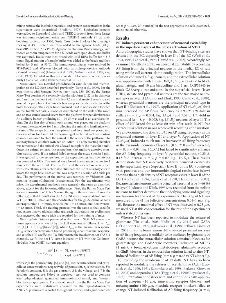
ResultsNT induces persistent enhancement of neuronal excitabilityin the superficial layers of the EC via activation of NTS1Autoradiographic studies have shown that NT-binding sites aredetected in the EC, especially in layer II of the EC (Wolf et al.,1994, 1995; Lahti et al., 1998; Hamid et al., 2002). Accordingly, weexamined the effects of NT on neuronal excitability by recordingAP firing from the principal neurons in the medial EC of ratsusing whole-cell current-clamp configuration. The intracellularsolution contained K�-gluconate, and the extracellular solutionwas supplemented with 10 �M DNQX, 50 �M DL-APV to blockglutamatergic, and 10 �M bicuculline and 2 �M CGP55845 toblock GABAergic transmission. In the superficial layers (layerII/III), stellate and pyramidal neurons are the two major neuro-nal types in layer II (Alonso and Klink, 1993; Canto et al., 2008),whereas pyramidal neurons are the principal neuronal type inlayer III (Dickson et al., 1997). Application of NT (0.25 �M) for 5min increased the AP firing frequency by 8.93 � 1.94-fold instellate (n � 7, p � 0.004; Fig. 1A1,A2) and 7.58 � 1.71-fold inpyramidal (n � 8, p � 0.003; Fig. 1B1,B2) neurons of layer II. Theeffect of NT lasted for at least 60 min after wash in NT-freeextracellular solution in our whole-cell recording configuration.We also examined the effects of NT on AP firing frequency in thepyramidal neurons of layer III and layer V. Application of NT(0.25 �M) induced a much smaller increase in AP firing frequencyin the pyramidal neurons of layer III (0.66 � 0.26-fold increase,n � 8, p � 0.04; Fig. 1C1,C2) but failed to significantly enhancethe AP firing frequency in layer V pyramidal neurons (0.25 �0.13-fold increase, n � 9, p � 0.09; Fig. 1D1,D2). These resultsdemonstrate that NT selectively facilitates neuronal excitabilityin the superficial layers (especially layer II) of the EC, consistentwith previous and our immunohistological results (see below)showing that a high density of NT receptors exists in layer II of theEC (Wolf et al., 1994; Lahti et al., 1998; Hamid et al., 2002).Because stellate neurons are the principal neuronal type (�69%)in layer II (Alonso and Klink, 1993), we recorded from the stellateneurons to further determine the underlying ionic and signalingmechanisms for the rest of the experiments. The EC50 of NT wasmeasured to be 41 nM (effective concentration: 0.01–1 �M; Fig.1E). Because the maximal effect of NT was observed at 0.25 �M,we used NT at this concentration for the rest of the experimentsunless stated otherwise.
Whereas NT has been reported to modulate the release ofglutamate (Yin et al., 2008; Kadiri et al., 2011) and GABA(O’Connor et al., 1992; Rakovska et al., 1998; Petkova-Kirova etal., 2008) in some brain regions, NT-induced persistent increasein AP firing frequency is unlikely to be mediated by glutamate orGABA because the extracellular solution contained blockers forglutamatergic and GABAergic receptors. Inclusion of MCPG(1 mM), a broad-spectrum metabotropic glutamate receptor(mGluR) blocker, in the extracellular solution failed to alter NT-induced facilitation of AP firing (n � 6, p � 0.48 vs NT alone; Fig.1F), excluding the involvement of mGluRs. NT has also beenreported to modulate the releases of acetylcholine (Ach) (Lap-chak et al., 1990, 1991; Rakovska et al., 1998; Petkova-Kirova etal., 2008) and dopamine (DA) (Faggin et al., 1990; Reyneke et al.,1992). Pretreatment of slices with and continuous bath applica-tion of atropine (10 �M, muscarinic receptor blocker) andmecamylamine (100 �M, nicotinic receptor blocker) failed tochange NT-induced facilitation of AP firing frequency (n � 6,
Xiao et al. • Neurotensin Facilitates Excitability and Memory J. Neurosci., May 14, 2014 • 34(20):7027–7042 • 7029
p � 0.44 vs NT alone; Fig. 1F), suggesting that it is unlikely thatNT facilitates neuronal excitability via releasing Ach. Further-more, application of SCH23390 (10 �M, D1-like receptor antag-onist) and sulpiride (100 �M, D2-like receptor antagonist) in thesame fashion did not alter NT-induced enhancement of AP firingfrequency (n � 5, p � 0.99 vs NT alone; Fig. 1F). These resultstogether indicate that it is unlikely that NT-induced facilitation ofAP firing is mediated indirectly by modulating the release of Achand DA. Finally, application of NT in the pure extracellular solu-tion without any blockers for any receptors still enhanced the APfiring frequency to the same level (8.4 � 3.1-fold increase, n � 6,p � 0.04; data not shown) supporting a physiological role forNT-induced facilitation of neuronal excitability in the EC. Bathapplication of the active NT fragment (NT8 –13, 0.25 �M) in-creased the AP firing frequency to the same level as NT (n � 6,p � 0.35 vs NT alone; Fig. 1F), whereas application of the inactivefragment (NT1– 8, 0.25 �M) failed to increase AP firing frequency(n � 7, p � 0.093 vs baseline; p � 0.0007 vs NT alone; Fig. 1F),suggesting that NT acts via activation of NT receptors.
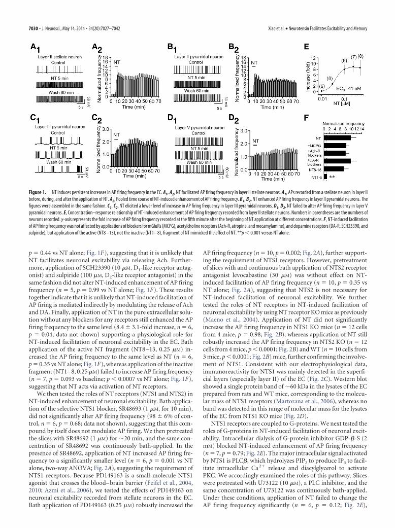
We then tested the roles of NT receptors (NTS1 and NTS2) inNT-induced enhancement of neuronal excitability. Bath applica-tion of the selective NTS1 blocker, SR48693 (1 �M, for 10 min),did not significantly alter AP firing frequency (98 � 6% of con-trol, n � 6, p � 0.68; data not shown), suggesting that this com-pound by itself does not modulate AP firing. We then pretreatedthe slices with SR48692 (1 �M) for �20 min, and the same con-centration of SR48692 was continuously bath-applied. In thepresence of SR48692, application of NT increased AP firing fre-quency to a significantly smaller level (n � 6, p � 0.001 vs NTalone, two-way ANOVA; Fig. 2A), suggesting the requirement ofNTS1 receptors. Because PD149163 is a small-molecule NTS1agonist that crosses the blood– brain barrier (Feifel et al., 2004,2010; Azmi et al., 2006), we tested the effects of PD149163 onneuronal excitability recorded from stellate neurons in the EC.Bath application of PD149163 (0.25 �M) robustly increased the
AP firing frequency (n � 10, p � 0.002; Fig. 2A), further support-ing the requirement of NTS1 receptors. However, pretreatmentof slices with and continuous bath application of NTS2 receptorantagonist levocabastine (30 �M) was without effect on NT-induced facilitation of AP firing frequency (n � 10, p � 0.35 vsNT alone; Fig. 2A), suggesting that NTS2 is not necessary forNT-induced facilitation of neuronal excitability. We furthertested the roles of NT receptors in NT-induced facilitation ofneuronal excitability by using NT receptor KO mice as previously(Maeno et al., 2004). Application of NT did not significantlyincrease the AP firing frequency in NTS1 KO mice (n � 12 cellsfrom 4 mice, p � 0.98; Fig. 2B), whereas application of NT stillrobustly increased the AP firing frequency in NTS2 KO (n � 12cells from 4 mice, p � 0.0001; Fig. 2B) and WT (n � 10 cells from3 mice, p � 0.0001; Fig. 2B) mice, further confirming the involve-ment of NTS1. Consistent with our electrophysiological data,immunoreactivity for NTS1 was mainly detected in the superfi-cial layers (especially layer II) of the EC (Fig. 2C). Western blotshowed a single protein band of �60 kDa in the lysates of the ECprepared from rats and WT mice, corresponding to the molecu-lar mass of NTS1 receptors (Martorana et al., 2006), whereas noband was detected in this range of molecular mass for the lysatesof the EC from NTS1 KO mice (Fig. 2D).
NTS1 receptors are coupled to G-proteins. We next tested theroles of G-proteins in NT-induced facilitation of neuronal excit-ability. Intracellular dialysis of G-protein inhibitor GDP-�-S (2mM) blocked NT-induced enhancement of AP firing frequency(n � 7, p � 0.79; Fig. 2E). The major intracellular signal activatedby NTS1 is PLC�, which hydrolyzes PIP2 to produce IP3 to facil-itate intracellular Ca 2� release and diacylglycerol to activatePKC. We accordingly examined the roles of this pathway. Sliceswere pretreated with U73122 (10 �M), a PLC inhibitor, and thesame concentration of U73122 was continuously bath-applied.Under these conditions, application of NT failed to change theAP firing frequency significantly (n � 6, p � 0.12; Fig. 2E),
Figure 1. NT induces persistent increases in AP firing frequency in the EC. A1, A2, NT facilitated AP firing frequency in layer II stellate neurons. A1, APs recorded from a stellate neuron in layer IIbefore, during, and after the application of NT. A2, Pooled time course of NT-induced enhancement of AP firing frequency. B1, B2, NT enhanced AP firing frequency in layer II pyramidal neurons. Thefigures were assembled in the same fashion. C1, C2, NT elicited a lower level of increase in AP firing frequency in layer III pyramidal neurons. D1, D2, NT failed to alter AP firing frequency in layer Vpyramidal neurons. E, Concentration–response relationship of NT-induced enhancement of AP firing frequency recorded from layer II stellate neurons. Numbers in parentheses are the numbers ofneurons recorded. y-axis represents the fold increase of AP firing frequency recorded at the fifth minute after the beginning of NT application at different concentrations. F, NT-induced facilitationof AP firing frequency was not affected by applications of blockers for mGluRs (MCPG), acetylcholine receptors (Ach-R, atropine, and mecamylamine), and dopamine receptors (DA-R, SCH23390, andsulpiride), but application of the active (NT8 –13), not the inactive (NT1– 8), fragment of NT mimicked the effect of NT. **p � 0.001 versus NT alone.
7030 • J. Neurosci., May 14, 2014 • 34(20):7027–7042 Xiao et al. • Neurotensin Facilitates Excitability and Memory

whereas application of U73343 (10 �M), the inactive analog, inthe same fashion did not significantly alter NT-induced enhance-ment of AP firing frequency (n � 6, p � 0.0001 vs baseline; Fig.2E). Similarly, application of edelfosine (10 �M), another PLCinhibitor, blocked NT-induced facilitation of AP firing frequency(n � 9, p � 0.09; Fig. 2E). We then used PLC� KO mice to furtherconfirm our results. Among the 4 isoforms of PLC� (PLC�1– 4),PLC�1 is expressed in the hippocampal formation (Watanabe etal., 1998). We therefore used PLC�1 KO mice as previously de-scribed (Deng et al., 2006; Lei et al., 2007) to examine the roles ofPLC in NT-induced facilitation of AP firing. Application of NTrobustly enhanced the AP firing frequency (n � 10 cells, p �0.0001) in slices cut from 3 WT mice, whereas it increased the APfiring frequency to a significantly smaller level (n � 13 cells) inslices cut from 4 PLC�1 KO mice (p � 0.0001 vs WT mice; Fig.2E). Together, these results demonstrate that NT-mediated en-hancement of neuronal excitability requires the function ofPLC�1. We then tested the roles of PKC and intracellular Ca 2�
release in NT-induced facilitation of neuronal excitability. Sliceswere pretreated with GF109203X (1 �M) and Ro318220 (1 �M)separately, and the same concentrations of the PKC inhibitorswere continuously bath-applied. Under these circumstances, ap-plication of NT failed to increase AP firing frequency significantlyin the presence of GF109203X (n � 6, p � 0.2; Fig. 2F) orRo318220 (n � 6, p � 0.83; Fig. 2F), demonstrating the involve-ment of PKC. Because PKC is an upstream activator of MEK/ERK
in some cell types (Hu and Gereau, 2003), we also tested whetherMEK/ERK is required for NT-induced increase in AP firing fre-quency. Slices were pretreated with the selective MEK/ERK in-hibitor PD98059 (50 �M) for 2 h and the extracellular solutioncontinued to contain the same concentration of PD98059. Underthese circumstances, bath application of NT still enhanced the APfiring frequency to a comparable level (n � 5, p � 0.02; Fig. 2F),suggesting that MEK/ERK activity is not required for NT-induced facilitation of neuronal excitability. To test the involve-ment of IP3 receptors, we included xestospongin C (5 �M, IP3
receptor inhibitor) in the recording pipettes and waited for �20min after the formation of whole-cell configuration. In this con-dition, application of NT still increased the AP firing frequency toa comparable level (n � 6, p � 0.83 vs NT alone; Fig. 2F). Simi-larly, depletion of intracellular Ca 2� stores by including thapsi-gargin (10 �M) in the pipettes for 20 min did not significantlyalter NT-induced facilitation of AP firing frequency (n � 6, p �0.41 vs NT alone; Fig. 2F). These results suggest that intracellularCa 2� release is not required for NT-induced increases in APfiring. We then included BAPTA (10 mM) in the recordingpipettes to chelate intracellular Ca 2�. In the presence ofBAPTA, bath application of NT increased the AP firing fre-quency to a significantly smaller level (n � 7, p � 0.007 vs NTalone; Fig. 2F ). Because NT still significantly increased the APfiring frequency in the extracellular solution without Ca 2�
(prepared by replacing extracellular Ca 2� with the same con-
Figure 2. NT-elicited facilitation of AP firing frequency requires the functions of NTS1, PLC�1, and PKC. A, NT-induced enhancement of AP firing frequency was remarkably reduced in thepresence of the selective NTS1 antagonist, SR48692, but insensitive to levocabastine, an NTS2 antagonist. Bath application of the specific NTS1 agonist PD149163 (0.25 �M) increased AP firingfrequency. Slices were pretreated with SR48692 (1 �M) or levocabastine (30 �M), and the bath was continuously perfused with the same concentration of the drugs. The horizontal bar representsthe period of bath application of NT or PD149163. B, NT-induced facilitation of AP firing frequency disappeared in slices cut from NTS1 KO mice (n � 12 cells from 4 mice) but was still observed inslices cut from WT mice (n � 10 cells from 3 mice) or NTS2 KO mice (n � 12 cells from 4 mice). C, Immunoreactivity to NTS1 receptors was mainly detected in the superficial layers of the EC. Right,Enlargement of the region marked in the left section. Preabsorption of the NTS1 antibody with the corresponding blocking peptide blocked the detection of the immunoreactivity. D, Western blotdemonstrated the expression of NTS1 receptors in the EC. Tissue lysates of the medial EC taken from rats, WT mice, and NTS1 KO mice (n � 6 animals) were loaded to the gel and blotted against theNTS1 antibody. A band of �60 kDa corresponding to the molecular mass of NTS1 was detected in the lysates from rats and WT mice but not in those from NTS1 KO mice. E, Roles of G-proteins andPLC�1 in NT-elicited facilitation of AP firing frequency. **p � 0.01 versus baseline. ##p � 0.01. F, Roles of intracellular Ca 2� and PKC in NT-mediated enhancement of AP firing frequency. *p �0.05 versus baseline. **p � 0.01 versus baseline. ##p � 0.01 versus NT alone.
Xiao et al. • Neurotensin Facilitates Excitability and Memory J. Neurosci., May 14, 2014 • 34(20):7027–7042 • 7031

centration of Mg 2� and adding 1 mM
EGTA, n � 9, p � 0.013; Fig. 2F ), oneexplanation for the effect of BAPTA isthat a certain basal level of intracellularCa 2� is required for the activation ofsignaling molecules, such as PLC� andPKC.
NT-induced facilitation of AP firing isgenerated by membrane depolarizationWe recorded RMPs in the presence ofTTX (0.5 �M) to block AP firing. Applica-tion of NT generated membrane depolar-ization (control: �57.7 � 1.2 mV, NT:�53.2 � 2.0 mV, n � 6, p � 0.006; Fig.3A) and significantly increased the inputresistance assessed by injection of a nega-tive current (�50 pA for 500 ms) before(115 � 6 M�) and after (133 � 6 M�) NTapplication (n � 6, p � 0.002; Fig. 3A,inset). We then recorded the HCs at �55mV, a potential close to the RMPs of theseneurons. Application of NT generated anet inward HC (�44.5 � 5.4 pA, n � 16,p � 0.001; Fig. 3B), indicating that NTincreases neuronal excitability by generat-ing membrane depolarization.
Cationic channels are not involved inNT-induced membrane depolarizationWe probed the ionic mechanismswhereby NT generates membrane depo-larization. NT could cause depolarizationby opening a cationic conductance and/orby inhibiting a background K� conduc-tance. We accordingly tested these possi-bilities by replacing the intracellular K�
with Cs�. Application of NT failed to in-duce an increase in inward HCs when in-tracellular K� was replaced with Cs�
(4.6 � 1.9 pA, n � 7, p � 0.06; Fig. 3B),suggesting the involvement of K � chan-nels. We further examined whetherthere is a role for cationic channels byperforming the following experiments.First, we replaced extracellular NaClwith NMDG-Cl and Ca 2� with Mg 2� be-cause, if cationic channels are involved,influx of extracellular Na� and Ca 2�
would be the ions to mediate the inwardcurrents. However, in this condition, ap-plication of NT still induced a comparableinward HC (n � 8, p � 0.45 vs the inwardHCs in control condition; Fig. 3B). Sec-ond, we examined the effects of other cat-ionic channel blockers. Pretreatment ofslices with and continuous bath applica-tion of SKF96365 (50 �M, an inhibitor ofreceptor-operated cationic channels, n �6, p � 0.85), Gd 3� (10 �M, n � 7, p � 0.37), La 3� (10 �M, n � 7,p � 0.65), or ZD7288 (20 �M, an inhibitor of Ih channels, n � 6,p � 0.57), failed to exert significant effects on NT-induced in-creases in inward HCs, demonstrating that NT-induced depolar-
ization is not mediated by activation of a cationic conductance(Fig. 3C).
NT has been reported to inhibit Na�/K� pump (Lopez Or-dieres and Rodríguez de Lores Arnaiz, 2000), and this pump
Figure 3. NT increases AP firing frequency by inhibition of TREK-2 channels. A, RMP recorded from a stellate neuron in current clampbefore, during, and after the application of NT. Inset, Voltage change in response to the injection of a hyperpolarizing current (�50 pA, 500ms) from the same cell before and after application of NT. NT induced membrane depolarization and increased input resistance. B, BathapplicationofNTinducedaninwardHCwhentherecordingpipettescontainedK �-gluconate,whereasreplacingtheintracellularK �withCs �blocked NT-induced increases in inward HCs. Replacement of extracellular NaCl with NMDG-Cl and Ca 2�with the same concentrationof Mg 2� failed to alter significantly NT-induced increases in inward HCs in intracellular solution containing K �-gluconate. C, NT-inducedincreases in inward HCs were insensitive to the extracellular application of blockers for nonselective cationic (SKF96365, Gd 3�, La 3�), Ih(ZD7288), and K � (TEA, Cs �, 4-AP, tertiapin-Q) channels and Zn 2�, but depletion of extracellular K � significantly enhanced NT-inducedHCs. *p � 0.05 versus NT alone. D, Voltage-current relationship induced by a ramp protocol from �140 to 0 mV at a speed of 0.1 mV/msbefore and during the application of NT. Subtraction of the current before the application of NT generated a net current. The traces wereaverages from 5 cells. The reversal potential was��87 mV, close to the calculated K � reversal potential (�85.4 mV). Inset, NT-inducednet current could not be fit by GHK equation, suggesting that the channels inhibited by NT were not TASK channels. E, Inclusion of Ba 2� inthe extracellular solution induced an inward HC but significantly reduced NT-induced increases in inward HCs. F, Bath application of NT failed toincreaseAPfiringfrequencyinslicescutfromTREK-2KOmicebutstill robustlyenhancedAPfiringfrequencyinslicecutfromWTmice.
7032 • J. Neurosci., May 14, 2014 • 34(20):7027–7042 Xiao et al. • Neurotensin Facilitates Excitability and Memory

normally creates a concentration gradient by moving 3 Na� outof the cell and 2 K� into the cells. NT-mediated inhibition of thispump should create a net accumulation of Na� inside the cellsresulting in depolarization. Because extracellular K� is necessaryfor the function of the Na�/K� pump, we replaced the extracel-lular K� (3.5 mM) with the same concentration of Na�. Insteadof reducing the inward HCs, removal of extracellular K� resultedin a significant increase in HCs (�83.5 � 18.8 pA, n � 6, p �0.012 vs NT alone, Fig. 3C), suggesting that NT-induced depo-larization is unlikely the result of the inhibition of Na�/K�
pump but may be relevant to K� movement across the mem-brane (see below).
NT generates membrane depolarization by inhibitingTREK-2 channelsWe further probed the involvement of K� conductance. We useda ramp protocol (from �140 to 0 mV) to construct the voltage-current relationship before and just after the application of NT.The intracellular solution contained K�-gluconate, and the ex-tracellular K� concentration was 3.5 mM. Under these circum-stances, application of NT induced a current with a reversalpotential of �86.7 � 1.5 mV (n � 5; Fig. 3D). The measuredreversal potential was close to the calculated K� reversal potential(�85.4 mV) in our recording condition further confirming thatNT increases neuronal excitability by inhibiting a backgroundK� conductance. We next characterized the properties of theinvolved K� channels. NT-induced increases in inward HCswere not significantly changed by including tetraethylammo-nium (TEA, 10 mM, n � 9, p � 0.66), Cs� (3 mM, n � 6, p �0.51), or 4-aminopyridine (4-AP, 2 mM, n � 6, p � 0.78) in theextracellular solution (Fig. 3C). Application of the selective in-ward rectifier K� channel inhibitor tertiapin-Q (100 nM) did notsignificantly alter NT-induced changes in HCs (n � 7, p � 0.97;Fig. 3C). Furthermore, NT-induced increases in inward HCswere not sensitive to extracellular application of Zn 2� (100 �M,n � 8, p � 0.63; Fig. 3C).
The above results prompted us to test the role of the two-poredomain K� (K2P) channels because K2P channels are involved incontrolling RMPs and usually insensitive to the classic K� chan-nel blockers (Lesage and Lazdunski, 2000; Bayliss et al., 2003;Lesage, 2003). Because some K2P channels are sensitive to Ba 2�,we tested the roles of Ba 2� in NT-induced increases in inwardHCs. Application of Ba 2� (3 mM) alone generated an inward HC(�78.1 � 18.2 pA, n � 9, p � 0.003; Fig. 3E), and it also signifi-cantly inhibited NT-induced inward HCs (�20.1 � 2.1 pA, n �9, p � 0.001 vs NT alone; Fig. 3E), suggesting the involvement ofBa 2�-sensitive K2P channels.
Among the K2P channels, TASK-1 (Han et al., 2002), TASK-3(Kim et al., 2000; Han et al., 2002), TREK-1 (Fink et al., 1996; Maet al., 2011), TREK-2 (Bang et al., 2000; Han et al., 2002), andTWIK-1 (Lesage et al., 1996) and TRESK (Sano et al., 2003; Kanget al., 2004) are sensitive to Ba 2�. TRESK channels are unlikely tobe involved because they are expressed in the spinal cord andother organs but not in the brain (Sano et al., 2003; Kang et al.,2004). Among the remaining 5 K2P channels, the EC expresseshigher densities of TASK-1 and TREK-2 (Deng et al., 2009). Be-cause TASK channel currents show GHK-type rectification (Les-age and Lazdunski, 2000; Bayliss et al., 2003) and fitting of theNT-induced net I-V curve by GHK equation demonstrated re-markable deviation (Fig. 3D, inset), suggesting that TASK-1channels are unlikely to be the target for NT, we focused onTREK-2 channels by using TREK-2 KO mice as described previ-ously (Guyon et al., 2009). Application of NT induced an inward
HC (�38.8 � 6.1 pA, n � 9 cells; data not shown) and robustlyincreased the firing frequency of APs (n � 9 cells, p � 0.001; Fig.3F) in slices cut from 3 WT mice, whereas it failed to induce aninward HC (�3.7 � 2.5 pA, n � 9 cells, p � 0.19; data not shown)and did not increase AP firing frequency (n � 9 cells, p � 0.32;Fig. 3F) in slices cut from 3 TREK-2 KO mice. However, theRMPs of stellate neurons in TREK-2 KO mice (�59.7 � 1.1 mV,n � 9 cells from 3 mice) were not significantly different (p �0.25) from those of WT mice (�61.4 � 0.9 mV, n � 9 cells from3 mice; data not shown). Together, these data indicate that NTfacilitates neuronal excitability by inhibiting TREK-2 channels.
Activation of NTS1 inhibits TREK-2 currents via PLC/PKCpathway in HEK293 cells cotransfected with NTS1 and TREK-2 channelsWe further probed the involvement of TREK-2 channels by usingwhole-cell recordings from the HEK293 cells expressing NTS1and TREK-2 channels (Deng et al., 2007, 2009; Xiao et al., 2009).Bath application of NT (0.25 �M) induced an inward HC re-corded at �55 mV in HEK293 cells cotransfected with NTS1 andTREK-2 channels (n � 8, p � 0.002; Fig. 4A,B). However, bathapplication of NT (0.25 �M) did not change the HCs recorded at�55 mV in HEK293 cells transfected with TREK-2 channelsalone (n � 5, p � 0.82; Fig. 4B) or NTS1 alone (n � 6, p � 0.86;Fig. 4B). We further determined the roles of PLC, PKC, andintracellular Ca 2� release in NT-mediated depression of TREK-2channels in HEK293 cells cotransfected with NTS1 and TREK-2channels. NT-induced inward HCs were significantly reduced inthe presence of PLC inhibitor (U73122, 10 �M, n � 8, p � 0.0001)compared with those in the presence of the inactive analog(U73343, 10 �M, n � 7; Fig. 4C). Similarly, treatment of the cellswith another PLC inhibitor (edelfosine, 10 �M, n � 8, p � 0.001)significantly reduced NT-induced inward HCs compared withcontrol (Fig. 4C). Furthermore, NT-induced inward HCs weresignificantly reduced by pretreatment of transfected cells with theselective PKC inhibitor (GF109203X, 1 �M, n � 5, p � 0.007) butwere unaffected by intracellular application of xestospongin C (1�M, IP3 receptor inhibitor, n � 8, p � 0.08; Fig. 4C) or thapsi-gargin (10 �M, n � 5, p � 0.051; Fig. 4C). However, intracellularapplication of BAPTA (10 mM) significantly reduced NT-induced inward HCs (n � 6, p � 0.008; Fig. 4C). These resultsdemonstrate that NT-induced depolarization requires the func-tions of PLC and PKC. One explanation for the effect of BAPTAis that intracellular Ca 2� may be required for the functions of thesignaling molecules, such as PLC and PKC.
Application of NT to entorhinal slices increases serinephosphorylation of TREK-2 channelsThe above experiments suggest that activation of NTS1 inhib-its TREK-2 channels via PKC-dependent phosphorylation ofTREK-2 channels. In accordance with our results, TREK-2 chan-nels are phosphorylated and inhibited by PKC on serine 326(S326) and serine 359 (S359) (Gu et al., 2002; Kang et al., 2006;Kreneisz et al., 2009). We next tested the roles of PKC-dependentphosphorylation of these two sites in NT-mediated depression ofTREK-2 channels. Because mutation of S326 and S359 to alanineblocked PKC-mediated inhibition of TREK-2 channels (Kang etal., 2006; Kreneisz et al., 2009), we cotransfected HEK293 cellswith NTS1 and the TREK-2 mutant in which both serine 326 andserine 359 were mutated to alanine (S326A/S359A) (Kang et al.,2006; Kreneisz et al., 2009). Application of NT to the HEK293cells cotransfected with NTS1 and TREK-2 mutant induced sig-
Xiao et al. • Neurotensin Facilitates Excitability and Memory J. Neurosci., May 14, 2014 • 34(20):7027–7042 • 7033

nificantly smaller HCs (�8.2 � 1.4 pA, n � 7, p � 0.002 vsNT-induced HCs in the cells transfected with WT TREK-2; Fig.4D,E). These data demonstrate that NT-induced depression ofTREK-2 channels is mediated by PKC-dependent phosphoryla-tion of TREK-2 channels on S326 and S359.
We further corroborated NT-induced phosphorylation ofTREK-2 channels in the EC. We treated entorhinal slices for 5min with or without NT in the absence or presence of the PKCinhibitor (GF109203X). Slices were then lysed, and the lysateswere immunoprecipitated with TREK-2 antibody. The immuno-precipitates were resolved by SDS-PAGE and Western blottedwith anti-phosphoserine antibody. As shown in Figure 4F, treat-ment of slices with NT for 5 min induced persistent increases inthe phosphorylation of TREK-2 channels when the slices werekept in NT-free solution for �1 h (n � 3 experiments, p � 0.01for each). Pretreatment of slices with GF109203X blocked NT-induced increases in the phosphorylation of TREK-2 channels(n � 3 experiments, p � 0.8). As a control, treatment of slices cutfrom WT mice with NT for 5 min induced phosphorylation ofTREK-2 channels, whereas no band was detected in the samplesprepared from the TREK-2 KO mice with the same experimentalprocedures (Fig. 4F). These results demonstrate that NT-induced depression of TREK-2 channels is mediated by PKC-dependent phosphorylation of TREK-2 channels.
NT enhances EPSP-AP coupling in layer II stellate neuronsAll the above experiments regarding the effects of NT on AP firingwere performed by injection of small positive current to elevate
the RMP to just above threshold to induce AP firing. We nexttested whether NT by itself could elevate the RMPs of stellateneurons to above the threshold to induce AP firing. In 10 stellateneurons, there was no spontaneous AP firing at rest. Applicationof NT induced AP firing in 6 of the 10 stellate neurons. The APfiring frequency reached maximal in �5 min after the beginningof NT application (141 � 26 APs/min, n � 6, p � 0.003; Fig.5A,B). Whereas the AP firing frequency was declined after washin NT-free extracellular solution, there was still AP firing afterwash for 1 h (34 � 7 APs/min, n � 6, p � 0.004; Fig. 5A,B).Application of NT to the remaining 4 stellate neurons failed toinduce AP firing but significantly elevated the RMPs (control:�58.7 � 1.3 mV, NT: �55.2 � 1.6 mV, n � 4, p � 0.012; data notshown). We then tested the effects of NT on EPSP-AP coupling inlayer II stellate neurons by placing a stimulation electrode in layerI to stimulate the glutamatergic inputs. Application of NT in-duced the depolarization of the baseline of the EPSPs (control:�59.5 � 1.3 mV, NT: �54.9 � 1.6 mV, n � 5, p � 0.0009; Fig.5C,D) and facilitated EPSP-AP coupling (n � 5, p � 0.0003; Fig.5C,D). To differentiate the presynaptic and postsynaptic effectsof NT, we replicated the experiment by using Cs�-gluconate-containing intracellular solution to block the depolarizing effectof NT on stellate neurons. Under these circumstances, NT failedto altered the slope (101 � 2% of control, n � 9, p � 0.55; Fig.5E,F) of the EPSPs, suggesting that NT-mediated facilitation ofEPSP-AP coupling was not mediated by modulating presynapticglutamate release, but because of its depolarizing effect on post-synaptic stellate neurons.
Figure 4. Activation of NTS1 receptors resulted in phosphorylation and inhibition of TREK-2 channels. A, I-V curves recorded by a ramp protocol from �140 mV to 0 mV in a HEK293 cellcotransfected with NTS1 and TREK-2 channels before (red) and during (green) the application of NT. B, Pooled time course of the HCs recorded at �55 mV from HEK293 cells transfected with NTS1alone, or TREK-2 channels alone or NTS1 together with TREK-2 channels in response to bath application of NT. Bath application of NT induced an inward HC only in HEK293 cells cotransfected withNTS1 and TREK-2 channels. C, NT-mediated inhibition of TREK-2 currents was mediated via activation of PLC and PKC in HEK293 cells cotransfected with NTS1 and TREK-2 channels. **p �0.01 versusNT alone. #p � 0.05. D, I-V curves in a HEK293 cell cotransfected with NTS1 and TREK-2 mutant before (red) and during (green) the application of NT. E, Pooled time course of the HCs recorded fromHEK293 cells cotransfected with NTS1 and TREK-2 mutant in response to NT. NT did not significantly alter the HCs. F, Persistent phosphorylation of TREK-2 channels on serine was detected in thelysate of the rat EC immunoprecipitated with antibody to TREK-2 channels. As a control, application of the same experimental procedures induced phosphorylation of TREK-2 channels in the samplesprepared from WT mice but not in those prepared from TREK-2 KO mice. The molecular mass of TREK-2 channels was �59 kDa.
7034 • J. Neurosci., May 14, 2014 • 34(20):7027–7042 Xiao et al. • Neurotensin Facilitates Excitability and Memory
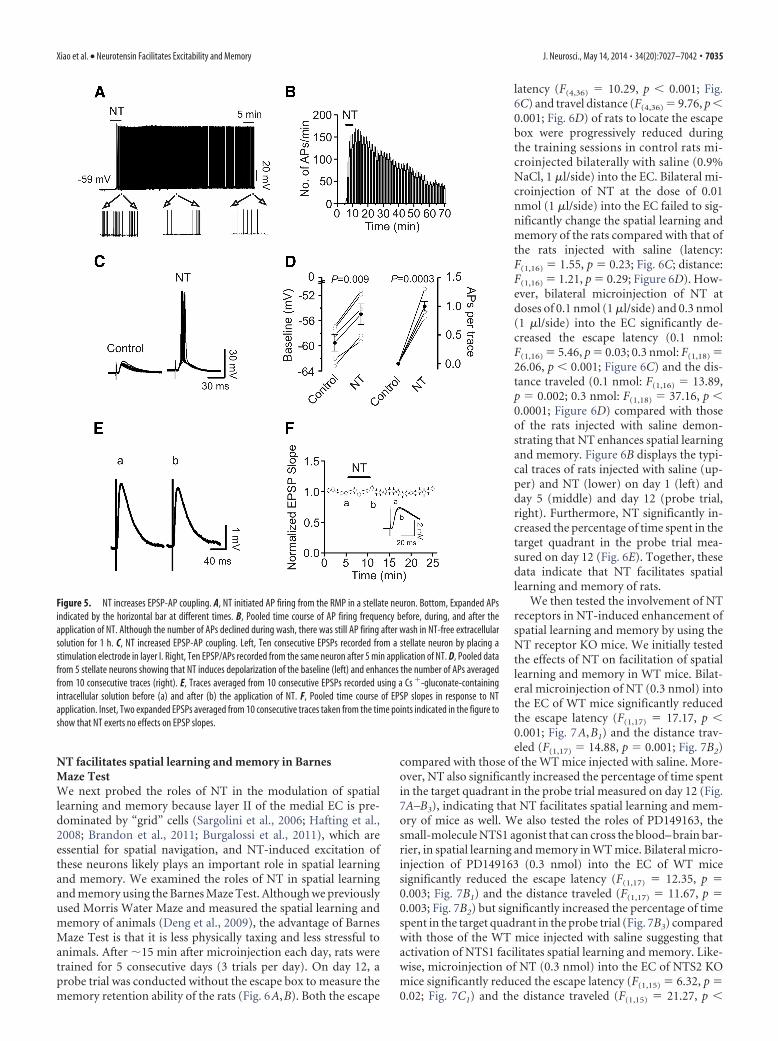
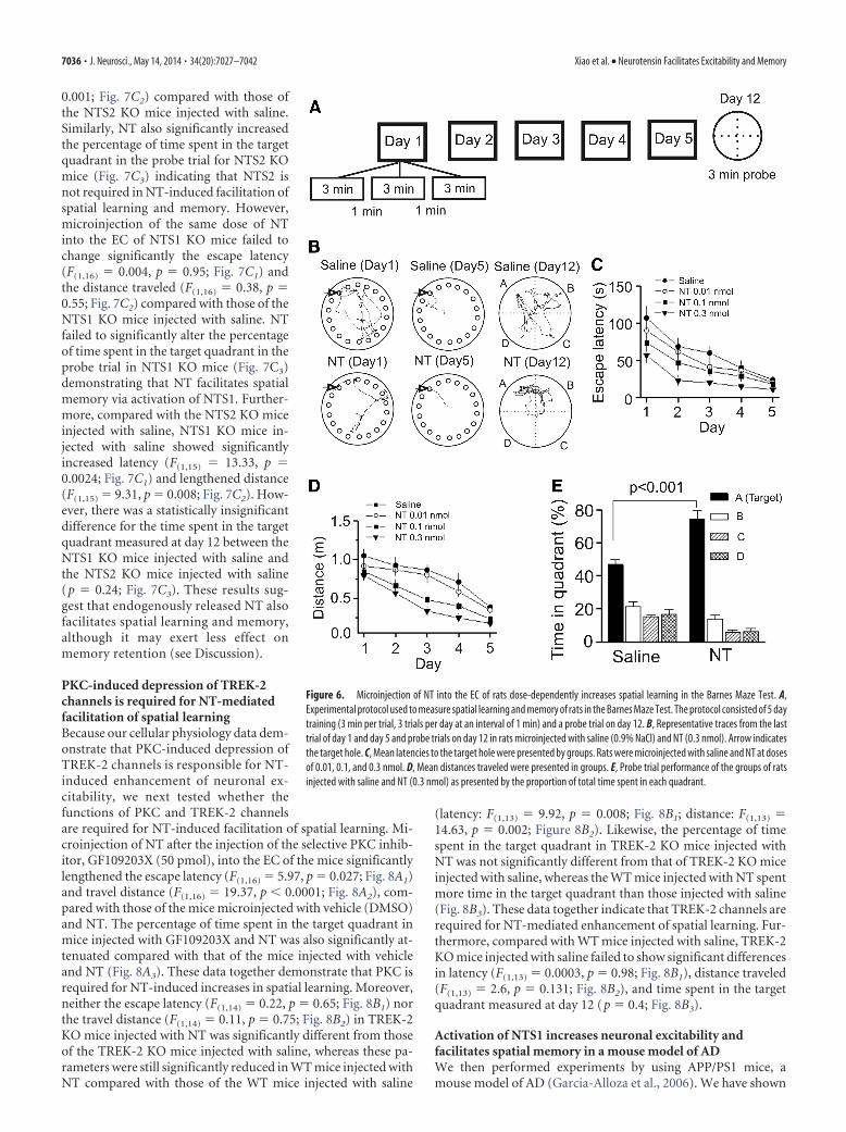
NT facilitates spatial learning and memory in BarnesMaze TestWe next probed the roles of NT in the modulation of spatiallearning and memory because layer II of the medial EC is pre-dominated by “grid” cells (Sargolini et al., 2006; Hafting et al.,2008; Brandon et al., 2011; Burgalossi et al., 2011), which areessential for spatial navigation, and NT-induced excitation ofthese neurons likely plays an important role in spatial learningand memory. We examined the roles of NT in spatial learningand memory using the Barnes Maze Test. Although we previouslyused Morris Water Maze and measured the spatial learning andmemory of animals (Deng et al., 2009), the advantage of BarnesMaze Test is that it is less physically taxing and less stressful toanimals. After �15 min after microinjection each day, rats weretrained for 5 consecutive days (3 trials per day). On day 12, aprobe trial was conducted without the escape box to measure thememory retention ability of the rats (Fig. 6A,B). Both the escape
latency (F(4,36) � 10.29, p � 0.001; Fig.6C) and travel distance (F(4,36) � 9.76, p �0.001; Fig. 6D) of rats to locate the escapebox were progressively reduced duringthe training sessions in control rats mi-croinjected bilaterally with saline (0.9%NaCl, 1 �l/side) into the EC. Bilateral mi-croinjection of NT at the dose of 0.01nmol (1 �l/side) into the EC failed to sig-nificantly change the spatial learning andmemory of the rats compared with that ofthe rats injected with saline (latency:F(1,16) � 1.55, p � 0.23; Fig. 6C; distance:F(1,16) � 1.21, p � 0.29; Figure 6D). How-ever, bilateral microinjection of NT atdoses of 0.1 nmol (1 �l/side) and 0.3 nmol(1 �l/side) into the EC significantly de-creased the escape latency (0.1 nmol:F(1,16) � 5.46, p � 0.03; 0.3 nmol: F(1,18) �26.06, p � 0.001; Figure 6C) and the dis-tance traveled (0.1 nmol: F(1,16) � 13.89,p � 0.002; 0.3 nmol: F(1,18) � 37.16, p �0.0001; Figure 6D) compared with thoseof the rats injected with saline demon-strating that NT enhances spatial learningand memory. Figure 6B displays the typi-cal traces of rats injected with saline (up-per) and NT (lower) on day 1 (left) andday 5 (middle) and day 12 (probe trial,right). Furthermore, NT significantly in-creased the percentage of time spent in thetarget quadrant in the probe trial mea-sured on day 12 (Fig. 6E). Together, thesedata indicate that NT facilitates spatiallearning and memory of rats.
We then tested the involvement of NTreceptors in NT-induced enhancement ofspatial learning and memory by using theNT receptor KO mice. We initially testedthe effects of NT on facilitation of spatiallearning and memory in WT mice. Bilat-eral microinjection of NT (0.3 nmol) intothe EC of WT mice significantly reducedthe escape latency (F(1,17) � 17.17, p �0.001; Fig. 7A,B1) and the distance trav-eled (F(1,17) � 14.88, p � 0.001; Fig. 7B2)
compared with those of the WT mice injected with saline. More-over, NT also significantly increased the percentage of time spentin the target quadrant in the probe trial measured on day 12 (Fig.7A–B3), indicating that NT facilitates spatial learning and mem-ory of mice as well. We also tested the roles of PD149163, thesmall-molecule NTS1 agonist that can cross the blood– brain bar-rier, in spatial learning and memory in WT mice. Bilateral micro-injection of PD149163 (0.3 nmol) into the EC of WT micesignificantly reduced the escape latency (F(1,17) � 12.35, p �0.003; Fig. 7B1) and the distance traveled (F(1,17) � 11.67, p �0.003; Fig. 7B2) but significantly increased the percentage of timespent in the target quadrant in the probe trial (Fig. 7B3) comparedwith those of the WT mice injected with saline suggesting thatactivation of NTS1 facilitates spatial learning and memory. Like-wise, microinjection of NT (0.3 nmol) into the EC of NTS2 KOmice significantly reduced the escape latency (F(1,15) � 6.32, p �0.02; Fig. 7C1) and the distance traveled (F(1,15) � 21.27, p �
Figure 5. NT increases EPSP-AP coupling. A, NT initiated AP firing from the RMP in a stellate neuron. Bottom, Expanded APsindicated by the horizontal bar at different times. B, Pooled time course of AP firing frequency before, during, and after theapplication of NT. Although the number of APs declined during wash, there was still AP firing after wash in NT-free extracellularsolution for 1 h. C, NT increased EPSP-AP coupling. Left, Ten consecutive EPSPs recorded from a stellate neuron by placing astimulation electrode in layer I. Right, Ten EPSP/APs recorded from the same neuron after 5 min application of NT. D, Pooled datafrom 5 stellate neurons showing that NT induces depolarization of the baseline (left) and enhances the number of APs averagedfrom 10 consecutive traces (right). E, Traces averaged from 10 consecutive EPSPs recorded using a Cs �-gluconate-containingintracellular solution before (a) and after (b) the application of NT. F, Pooled time course of EPSP slopes in response to NTapplication. Inset, Two expanded EPSPs averaged from 10 consecutive traces taken from the time points indicated in the figure toshow that NT exerts no effects on EPSP slopes.
Xiao et al. • Neurotensin Facilitates Excitability and Memory J. Neurosci., May 14, 2014 • 34(20):7027–7042 • 7035
0.001; Fig. 7C2) compared with those ofthe NTS2 KO mice injected with saline.Similarly, NT also significantly increasedthe percentage of time spent in the targetquadrant in the probe trial for NTS2 KOmice (Fig. 7C3) indicating that NTS2 isnot required in NT-induced facilitation ofspatial learning and memory. However,microinjection of the same dose of NTinto the EC of NTS1 KO mice failed tochange significantly the escape latency(F(1,16) � 0.004, p � 0.95; Fig. 7C1) andthe distance traveled (F(1,16) � 0.38, p �0.55; Fig. 7C2) compared with those of theNTS1 KO mice injected with saline. NTfailed to significantly alter the percentageof time spent in the target quadrant in theprobe trial in NTS1 KO mice (Fig. 7C3)demonstrating that NT facilitates spatialmemory via activation of NTS1. Further-more, compared with the NTS2 KO miceinjected with saline, NTS1 KO mice in-jected with saline showed significantlyincreased latency (F(1,15) � 13.33, p �0.0024; Fig. 7C1) and lengthened distance(F(1,15) � 9.31, p � 0.008; Fig. 7C2). How-ever, there was a statistically insignificantdifference for the time spent in the targetquadrant measured at day 12 between theNTS1 KO mice injected with saline andthe NTS2 KO mice injected with saline(p � 0.24; Fig. 7C3). These results sug-gest that endogenously released NT alsofacilitates spatial learning and memory,although it may exert less effect onmemory retention (see Discussion).
PKC-induced depression of TREK-2channels is required for NT-mediatedfacilitation of spatial learningBecause our cellular physiology data dem-onstrate that PKC-induced depression ofTREK-2 channels is responsible for NT-induced enhancement of neuronal ex-citability, we next tested whether thefunctions of PKC and TREK-2 channelsare required for NT-induced facilitation of spatial learning. Mi-croinjection of NT after the injection of the selective PKC inhib-itor, GF109203X (50 pmol), into the EC of the mice significantlylengthened the escape latency (F(1,16) � 5.97, p � 0.027; Fig. 8A1)and travel distance (F(1,16) � 19.37, p � 0.0001; Fig. 8A2), com-pared with those of the mice microinjected with vehicle (DMSO)and NT. The percentage of time spent in the target quadrant inmice injected with GF109203X and NT was also significantly at-tenuated compared with that of the mice injected with vehicleand NT (Fig. 8A3). These data together demonstrate that PKC isrequired for NT-induced increases in spatial learning. Moreover,neither the escape latency (F(1,14) � 0.22, p � 0.65; Fig. 8B1) northe travel distance (F(1,14) � 0.11, p � 0.75; Fig. 8B2) in TREK-2KO mice injected with NT was significantly different from thoseof the TREK-2 KO mice injected with saline, whereas these pa-rameters were still significantly reduced in WT mice injected withNT compared with those of the WT mice injected with saline
(latency: F(1,13) � 9.92, p � 0.008; Fig. 8B1; distance: F(1,13) �14.63, p � 0.002; Figure 8B2). Likewise, the percentage of timespent in the target quadrant in TREK-2 KO mice injected withNT was not significantly different from that of TREK-2 KO miceinjected with saline, whereas the WT mice injected with NT spentmore time in the target quadrant than those injected with saline(Fig. 8B3). These data together indicate that TREK-2 channels arerequired for NT-mediated enhancement of spatial learning. Fur-thermore, compared with WT mice injected with saline, TREK-2KO mice injected with saline failed to show significant differencesin latency (F(1,13) � 0.0003, p � 0.98; Fig. 8B1), distance traveled(F(1,13) � 2.6, p � 0.131; Fig. 8B2), and time spent in the targetquadrant measured at day 12 (p � 0.4; Fig. 8B3).
Activation of NTS1 increases neuronal excitability andfacilitates spatial memory in a mouse model of ADWe then performed experiments by using APP/PS1 mice, amouse model of AD (Garcia-Alloza et al., 2006). We have shown
Figure 6. Microinjection of NT into the EC of rats dose-dependently increases spatial learning in the Barnes Maze Test. A,Experimental protocol used to measure spatial learning and memory of rats in the Barnes Maze Test. The protocol consisted of 5 daytraining (3 min per trial, 3 trials per day at an interval of 1 min) and a probe trial on day 12. B, Representative traces from the lasttrial of day 1 and day 5 and probe trials on day 12 in rats microinjected with saline (0.9% NaCl) and NT (0.3 nmol). Arrow indicatesthe target hole. C, Mean latencies to the target hole were presented by groups. Rats were microinjected with saline and NT at dosesof 0.01, 0.1, and 0.3 nmol. D, Mean distances traveled were presented in groups. E, Probe trial performance of the groups of ratsinjected with saline and NT (0.3 nmol) as presented by the proportion of total time spent in each quadrant.
7036 • J. Neurosci., May 14, 2014 • 34(20):7027–7042 Xiao et al. • Neurotensin Facilitates Excitability and Memory

previously that these mice exhibited plaques when they wereolder than 6 months (Dhawan and Combs, 2012). We thereforeused the APP/PS1 mice at the age of 8 –10 months. We first testedthe effects of NT on AP firing recorded from layer II stellateneurons in the EC. The RMPs recorded from layer II stellateneurons in slices cut from APP/PS1 mice (�59.7 � 0.7 mV, n �11 cells from 4 mice) were not significantly different from thoseof WT mice (�60.2 � 0.5 mV, n � 12 cells from 4 mice; data notshown). Bath application of NT (0.25 �M) robustly increased thefiring frequency of APs recorded from stellate neurons in slicescut from APP/PS1 mice (4.7 � 0.8-fold increase, n � 11 cellsfrom 4 mice, p � 0.001; Fig. 9A). NT-induced facilitation of APfiring frequency recorded from APP/PS1 mice was not signifi-cantly different from that recorded from the age-matched WTmice (5.3 � 1.2-fold increase, n � 12 cells from 4 mice, p � 0.55;Fig. 9A). Furthermore, bath application of NTS1 agonistPD149163 (0.25 �M) also vigorously facilitated AP firing fre-quency in APP/PS1 mice (5.6 � 1.2-fold increase, n � 14 cellsfrom 4 mice, p � 0.001; Fig. 9A). These data together demon-strate that activation of NTS1 also increases neuronal excitabilityin APP/PS1 mice. We then compared the spatial learning andmemory ability of APP/PS1 mice with the WT mice using theBarnes Maze Test. Compared with the age-matched WT mice,APP/PS1 mice exhibited significantly enhanced latency (F(1,15) �
15.39, p � 0.001; Fig. 9B1), lengthened travel distance (F(1,15) �6.42, p � 0.023; Fig. 9B2), and decreased percentage of the timespent in the target quadrant (p � 0.02; Fig. 9B3). These resultsindicate that APP/PS1 mice showed impaired cognitive func-tions. We further tested the effects of PD149163 on spatial learn-ing and memory in APP/PS1 mice. Bilateral microinjection ofPD149163 (0.3 nmol) significantly reduced the escape latency(F(1,14) � 28.89, p � 0.0001; Fig. 9B1) and the distance traveled(F(1,14) � 23.47, p � 0.001; Fig. 9B2) but significantly increasedthe percentage of time spent in the target quadrant (Fig. 9B3)demonstrating that PD149163 improves spatial learning andmemory in APP/PS1 mice.
DiscussionOur results demonstrate that activation of NTS1 receptors in theEC generates persistent increases in neuronal excitability. NT-induced increases in neuronal excitability are mediated by phos-phorylation and inhibition of TREK-2 channels and require thefunctions of G-proteins, PLC and PKC. Activation of NTS1 facil-itates spatial learning and memory via PKC-dependent down-regulation of TREK-2 channels, suggesting that NT-inducedfacilitation of neuronal excitability is a novel cellular and molec-ular substrate for learning and memory. Furthermore, applica-tions of NT and NTS1 agonist, PD149163, also increase AP firing
Figure 7. NT facilitates spatial learning by activation of NTS1 receptors. A, B, Microinjection of NT and the small-molecule NTS1 agonist (PD149163) into the EC of WT mice facilitated spatiallearning and memory. A, Representative traces from the last trial of day 1 and day 5 and the probe trial in mice bilaterally microinjected with saline and NT (0.3 nmol, 0.5 �l/side). B1, Mean latenciesto the target hole. B2, Mean distances traveled. B3, Probe trial performance as presented by the proportion of total time spent in each quadrant. **p � 0.01. C1–C3, Microinjection of NT into the ECof NTS1 KO mice failed to facilitate spatial learning, whereas NT still increased spatial learning in NTS2 KO mice. C1, Mean latencies in NTS1 or NTS2 KO mice bilaterally injected with saline or NT (0.3nmol, 0.5 �l/side). C2, Mean distances traveled in NTS1 or NTS2 KO mice injected with saline or NT (0.3 nmol, 0.5 �l/side). C3, Percentage of the time spent in each quadrant in NTS1 or NTS2 KO miceinjected with saline or NT (0.3 nmol, 0.5 �l/side).
Xiao et al. • Neurotensin Facilitates Excitability and Memory J. Neurosci., May 14, 2014 • 34(20):7027–7042 • 7037

in slices cut from APP/PS1 mice and mi-croinjection of PD149163 into the EC ofAPP/PS1 mice significantly increased thespatial learning and memory of thesemice, suggesting that NTS1 agonists exertbeneficial effects at least on AD animalmodels.
Ionic mechanisms underlying NTS1-mediated increase in neuronalexcitability and facilitation of spatiallearningApplication of NT increases neuronal ex-citability by depolarizing entorhinal neu-rons suggesting that activation of NTS1receptors modifies the movement of ionsacross the membrane, which is usuallyconducted by ion channels or transport-ers. NT has been shown to inhibitNa�/K� pump (Lopez Ordieres and Ro-dríguez de Lores Arnaiz, 2000). TheNa�/K� pump creates a concentrationgradient by moving 3 Na� out and 2 K�
into cells and inhibition of this pump,thereby, should create a net accumulationof positive ions inside cells leading to de-polarization. Extracellular K� is a requi-site for the function of the Na�/K�
pump. Our result that removal of extra-cellular K� did not block but significantlyincreased NT-induced inward HCs dem-onstrates that NT increases neuronalexcitability unlikely by inhibition ofNa�/K� pump. One explanation for NT-induced higher level of inward HCs in theabsence of extracellular K� is that deple-tion of extracellular K� increases the driv-ing force for K� efflux via K� channelsleading to stronger inhibition of K� chan-nels by NT.
Our results demonstrate that NT facil-itates neuronal excitability by inhibitionof background K� channels based on thefollowing lines of evidence. First, NT sig-nificantly increased the input resistance,suggesting that NT inhibits a membraneconductance. Second, replacing intracel-lular K� with Cs� blocked NT-inducedinward HCs. Third, the reversal potentialof NT-induced currents had a reversal potential close to the K�
reversal potential. Nonetheless, the involved K� channels areunlikely to be the inwardly rectifier K� channels because the I-Vcurve of NT-sensitive currents did not show inward rectification,but a little outward rectification and application of the inwardrectifier K� channel blocker tertiapin-Q failed to significantlyalter NT-induced inward HCs. Moreover, SCH23390 is an antag-onist of D1-like receptors as well as a blocker for inward rectifierK� channels (Kuzhikandathil and Oxford, 2002). Our result thatapplication of SCH23390 failed to alter NT-induced facilitationof AP firing frequency (Fig. 1F) further excludes the participationof inward rectifier K� channels. The results that NT-inducedinward HCs were insensitive to the classical K� channel blockers,such as TEA, 4-AP, and extracellular Cs�, suggest that NT inhib-
its K2P channels. NT has been reported to inhibit a type of K2P
channels (possibly TRESK and/or TASK-3) in spinal cord neu-rons (Kadiri et al., 2011). However, the channels inhibited by NTin the spinal cord neurons are sensitive to both Ba 2� and Zn 2�
(Kadiri et al., 2011), which is in contrast to our results that NT-induced inward HCs in the EC were sensitive to Ba 2� but insen-sitive to Zn 2�. These results suggest that NT inhibits a differenttype of K2P channels in the EC. We provide further evidencedemonstrating that NT increases neuronal excitability by inhibi-tion of TREK-2 channels. Application of NT failed to increase theAP firing frequency in slices cut from TREK-2 KO mice, whereasapplication of NT still robustly increased AP firing frequency inslices cut from WT mice. Bath application of NT to HEK293 cellscoexpressing NTS1 and TREK-2 channels induced an inward
Figure 8. NT-mediated facilitation of spatial learning requires the functions of PKC and TREK-2 channels. A1–A3, Microinjectionof the PKC inhibitor GF109203X (50 pmol) into the EC significantly reduced the effects of NT on escape latency (A1), travel distance(A2), and the percentage of time spent in the target quadrant in probe trial (A3). B1–B3, Microinjection of NT into the EC of TREK-2KO mice failed to significantly alter escape latency (B1), travel distance (B2), and the percentage of time spent in the targetquadrant in probe trial (B3), whereas the effects of NT were still observed in WT mice.
7038 • J. Neurosci., May 14, 2014 • 34(20):7027–7042 Xiao et al. • Neurotensin Facilitates Excitability and Memory

HC, whereas NT failed to modify the HCs in HEK293 cells trans-fected with either NTS1 receptors or TREK-2 channels alone.These results together indicate that NT inhibits TREK-2 channelsto increase the excitability of entorhinal neurons.
Although we have demonstrated that NT-induced facilitationof neuronal excitability is unlikely because of its indirect effectson the releases of glutamate, GABA, DA, and Ach, the potentialindirect contributions of other factors, such as the serotoniner-gic, purinergic, glycinergic, and other peptidergic systems, havenot been examined. However, it seems less likely that the effects ofNT would be related to serotonin or glycine because both sero-tonin (Lei, 2012) and glycine (Breustedt et al., 2004) inhibit neu-ronal excitability in the EC. The potential interaction between NTand other neurotransmitters still needs further investigation.
Signaling mechanisms underlying NTS1-induced facilitationof neuronal excitability and augmentation of spatial learningin the ECWhereas NT interacts with NTS1, NTS2, and NTS3 receptors,our results demonstrate that NT increases neuronal excitabilityand facilitates spatial learning via activation of NTS1 receptorsbased on the results from both pharmacological experiments andKO mice. Activation of NTS1 increases the activity of PLC leadingto elevation of intracellular Ca 2� release and activation of PKC.Our results demonstrate that NT-induced facilitation of neuro-nal excitability is related to intracellular Ca 2� level because intra-
cellular application of BAPTA attenuatedthe effects of NT. However, blockade ofintracellular Ca 2� release or depletionof extracellular Ca 2� failed to alter sig-nificantly NT-mediated facilitation ofneuronal excitability. Different from NT-induced facilitation of neuronal excitabil-ity, Ca 2� influx via nonspecific cation andL-type Ca 2� channels is required for thepersistent firing induced by brief depolar-ization in layer V entorhinal neurons(Egorov et al., 2002). One plausible expla-nation for the role of Ca 2� in NT-inducedfacilitation of neuronal excitability is thatintracellular Ca 2� is required for the acti-vation of Ca 2�-dependent intracellularsignals. Indeed, our results demonstratethat PKC, possibly Ca 2�-dependent PKC,is necessary for NT-induced facilitation ofneuronal excitability. We further demon-strate that NT induces persistent phos-phorylation of TREK-2 channels. InHEK293 cells coexpressing NTS1 and theTREK-2 mutant in which serine 326 andserine 359 were mutated to alanine, NTfailed to induce an inward HC, suggestingthat PKC phosphorylates TREK-2 chan-nels on these two sites. Consistent withour results, PKC-dependent phosphory-lation of TREK-2 channels on these twoamino acids by activation of muscarinicreceptors (Kang et al., 2006) and AMP-activated protein kinase (Kreneisz et al.,2009) also results in inhibition of TREK-2channels. PKC-dependent phosphoryla-tion and inhibition of TREK-2 channelslikely contribute to NT-mediated facilita-
tion of spatial learning because inhibition of PKC or deletion ofTREK-2 gene nullified the facilitatory effects of NT on spatiallearning. However, the specific PKC isoforms involved in NT-mediated facilitation of neuronal excitability and spatial learningstill await further investigation. In line with our results, it is wellestablished that both PKC (Bonini et al., 2007; Dash et al., 2007;Sun and Alkon, 2010) and TREK-2 channels (Pan et al., 2003;Huang and Yu, 2008; Deng et al., 2009) are substrates of learningand memory.
Whereas our results demonstrate that both NTS1 and TREK-2channels are required for NT-induced facilitation of spatiallearning, the roles of NTS1 and TREK-2 channels in the modu-lation of spatial learning by endogenously released NT are stillelusive. NTS1 KO mice showed increased escape latency andtravel distance, suggesting that NTS1 activated by endogenouslyreleased NT is involved in the processes of spatial learning. How-ever, the percentage of time spent in the target quadrant in theprobe test measured on day 12 was not altered in NTS1 KO mice,suggesting that NTS1 may play a less important role in memoryretention. Moreover, TREK-2 KO mice showed no differences inspatial learning and memory, although acute microinjection ofNT into the EC of TREK-2 KO mice failed to facilitate spatiallearning and memory, demonstrating a role of TREK-2 channelsin exogenously applied NT. One explanation for the discrepancyof these results is that the roles of TREK-2 channels may be re-placed by other K2P channels in TREK-2 KO mice because the
Figure 9. Activation of NTS1 increases neuronal excitability and improves the memory status of APP/PS1 mice. A, Bath appli-cation of NT (red) and PD149163 (green) increased the firing frequency of APs recorded from layer II stellate neurons of the EC inslices cut from APP/PS1 mice and age-matched WT mice (black). B1–B3, Bilateral microinjection of PD149163 at 0.3 nmol into theEC of APP/PS1 mice significantly reduced the latency (B1) and the distance traveled (B2) but increased the percentage of time spentin the target quadrant (B3) compared with the APP/PS1 mice microinjected with saline (0.9% NaCl) in the same fashion. APP/PS1mice showed lengthened escape latency (B1) and travel distance (B2) but decreased percentage of time spent in the targetquadrant (B3) compared with the age-matched WT mice.
Xiao et al. • Neurotensin Facilitates Excitability and Memory J. Neurosci., May 14, 2014 • 34(20):7027–7042 • 7039
electrophysiological properties, such as the RMPs, were notchanged in these mice. Evidence supporting this possibilitycomes from the observations that the RMPs of neurons inTREK-1 (Heurteaux et al., 2004) or TASK-1 (Aller et al., 2005)KO mice were not changed either. Moreover, although our re-sults demonstrate that TREK-2 channel is the major target ofNTS1 receptors, the possibility that an alternative target for en-dogenously released NT has been developed in TREK-2 KO micecannot be excluded.
Significance of NTS1-induced enhancement of neuronalexcitability and spatial learning in ADThere is increasing evidence demonstrating that the functions ofNT are closely associated with the memory status of both animalsand humans. For example, in healthy adult humans, NTS1 poly-morphisms are closely associated with variance in the perfor-mance of working memory (Li et al., 2011). NT binding sites arevaried as a function of age and cognitive status with decreased NTbinding sites in the brains of aged and cognitively impaired rats(Rowe et al., 2006). Application of NT or NT receptor agonistsimproves memory (Azmi et al., 2006; Ohinata et al., 2007; Laszloet al., 2010), whereas administration of NT receptor antagonistsimpairs cognitive processes (Tirado-Santiago et al., 2006).Whereas the effects of NT on memory are usually considered tobe mediated by NTS1 (Azmi et al., 2006; Tirado-Santiago et al.,2006; Ohinata et al., 2007; Laszlo et al., 2010), reduced fear mem-ory has also been observed in NTS2 KO mice (Yamauchi et al.,2007). Although these data generally support a role for NT inlearning and memory, the cellular and molecular mechanismswhereby NT facilitates memory have not been determined. In thepresent study, we took the advantage that high density of NTreceptors is expressed in the superficial layers of the EC, an indis-pensable structure involved in learning and memory. We firstexamined the effects of NT on neuronal excitability, and ourresults demonstrated that brief (5 min) application of NT persis-tently increased the AP firing frequency. This phenomenonresembles the long-term potentiation. Whereas long-term po-tentiation is generally considered as a cellular and molecularsubstrate for learning and memory, the eventual outcome oflong-term potentiation is to increase the excitability of thepostsynaptic neurons. NT-induced persistent increase in neuro-nal excitability bypasses the processes of synaptic transmissionand directly leads to increases in neuronal excitability. Thus, it isreasonable to speculate that NT-induced persistent increase inneuronal excitability may represent a novel cellular mechanisminvolved in learning and memory.
Memory impairment is a characteristic symptom observed inmany diseases, such as AD. Up to date, there have been few effec-tive drugs that can relieve the memory loss of these diseases. Gridcell firing correlates to forming an internal spatial map and isdriven by the polymodal input to the EC that occurs as an animalexplores its environment (Sargolini et al., 2006; Hafting et al.,2008; Brandon et al., 2011; Burgalossi et al., 2011). We proposethat NT may increase the probability of grid cell firing in responseto that input, which promotes a more active circuit in the encod-ing of information about one’s environment. Our data clearlyshow that application of NT or NTS1 agonist PD149163 facili-tates spatial learning. We further showed that microinjection ofPD149163 into the EC of APP/PS1 mice also significantly im-proved the memory status of these mice, highlighting a therapeu-tic value of this type of drugs in AD. Therefore, one immediateimplication of our results is that NTS1 agonists may have the
potential to be developed as cognition-enhancing drugs to relievethe symptoms of AD.
In conclusion, we found that activation of NTS1 receptors inthe EC induces a persistent increase in AP firing and facilitatesspatial learning. NT-induced facilitation of neuronal excitabilityand spatial learning require the functions of PLC and PKC andare mediated by phosphorylation and inhibition of TREK-2channels. NT and NTS1 agonist PD149163 also induced persis-tent AP firing and facilitated spatial memory in APP/PS1 mice,suggesting that NT-mediated augmentation of learning andmemory may have therapeutic potentials to relieve memory lossexhibited in AD.
ReferencesAller MI, Veale EL, Linden AM, Sandu C, Schwaninger M, Evans LJ, Korpi
ER, Mathie A, Wisden W, Brickley SG (2005) Modifying the subunitcomposition of TASK channels alters the modulation of a leak conduc-tance in cerebellar granule neurons. J Neurosci 25:11455–11467. CrossRefMedline
Alonso A, Klink R (1993) Differential electroresponsiveness of stellate andpyramidal-like cells of medial entorhinal cortex layer II. J Neurophysiol70:128 –143. Medline
Atoji Y, Watanabe H, Yamamoto Y, Suzuki Y (1995) Distribution ofneurotensin-containing neurons in the central nervous system of the dog.J Comp Neurol 353:67– 88. CrossRef Medline
Azmi N, Norman C, Spicer CH, Bennett GW (2006) Effects of a neurotensinanalogue (PD149163) and antagonist (SR142948A) on the scopolamine-induced deficits in a novel object discrimination task. Behav Pharmacol17:357–362. CrossRef Medline
Bang H, Kim Y, Kim D (2000) TREK-2, a new member of the mechanosen-sitive tandem-pore K � channel family. J Biol Chem 275:17412–17419.Medline
Bayliss DA, Sirois JE, Talley EM (2003) The TASK family: two-pore domainbackground K � channels. Mol Interv 3:205–219. CrossRef Medline
Bonini JS, Da Silva WC, Bevilaqua LR, Medina JH, Izquierdo I, CammarotaM (2007) On the participation of hippocampal PKC in acquisition, con-solidation and reconsolidation of spatial memory. Neuroscience 147:37–45. CrossRef Medline
Bradford MM (1976) A rapid and sensitive method for the quantitation ofmicrogram quantities of protein utilizing the principle of protein-dyebinding. Anal Biochem 72:248 –254. CrossRef Medline
Brandon MP, Bogaard AR, Libby CP, Connerney MA, Gupta K, HasselmoME (2011) Reduction of theta rhythm dissociates grid cell spatial peri-odicity from directional tuning. Science 332:595–599. CrossRef Medline
Breustedt J, Schmitz D, Heinemann U, Schmieden V (2004) Characteriza-tion of the inhibitory glycine receptor on entorhinal cortex neurons. EurJ Neurosci 19:1987–1991. CrossRef Medline
Burgalossi A, Herfst L, von Heimendahl M, Forste H, Haskic K, Schmidt M,Brecht M (2011) Microcircuits of functionally identified neurons in therat medial entorhinal cortex. Neuron 70:773–786. CrossRef Medline
Canto CB, Wouterlood FG, Witter MP (2008) What does the anatomicalorganization of the entorhinal cortex tell us? Neural Plast 2008:381243.CrossRef Medline
Chen ST, Tsai MS, Shen CL (1988) Distribution of neurotensin-like immu-noreactivity in the central nervous system of the Formosan monkey. ProcNatl Sci Counc Repub China B 12:163–173. Medline
Dash PK, Moore AN, Kobori N, Runyan JD (2007) Molecular activity un-derlying working memory. Learn Mem 14:554 –563. CrossRef Medline
Deng PY, Lei S (2008) Serotonin increases GABA release in rat entorhinalcortex by inhibiting interneuron TASK-3 K � channels. Mol Cell Neuro-sci 39:273–284. CrossRef Medline
Deng PY, Porter JE, Shin HS, Lei S (2006) Thyrotropin-releasing hormoneincreases GABA release in rat hippocampus. J Physiol 577:497–511.CrossRef Medline
Deng PY, Poudel SK, Rojanathammanee L, Porter JE, Lei S (2007) Sero-tonin inhibits neuronal excitability by activating two-pore domain K �
channels in the entorhinal cortex. Mol Pharmacol 72:208 –218. CrossRefMedline
Deng PY, Xiao Z, Yang C, Rojanathammanee L, Grisanti L, Watt J, Geiger JD,Liu R, Porter JE, Lei S (2009) GABAB receptor activation inhibits neu-
7040 • J. Neurosci., May 14, 2014 • 34(20):7027–7042 Xiao et al. • Neurotensin Facilitates Excitability and Memory

ronal excitability and spatial learning in the entorhinal cortex by activat-ing TREK-2 K � channels. Neuron 63:230 –243. CrossRef Medline
Deng PY, Xiao Z, Jha A, Ramonet D, Matsui T, Leitges M, Shin HS, Porter JE,Geiger JD, Lei S (2010) Cholecystokinin facilitates glutamate release byincreasing the number of readily releasable vesicles and releasing proba-bility. J Neurosci 30:5136 –5148. CrossRef Medline
Dhawan G, Combs CK (2012) Inhibition of Src kinase activity attenuatesamyloid associated microgliosis in a murine model of Alzheimer’s disease.J Neuroinflammation 9:117. CrossRef Medline
Dickson CT, Mena AR, Alonso A (1997) Electroresponsiveness of medialentorhinal cortex layer III neurons in vitro. Neuroscience 81:937–950.CrossRef Medline
Dolcos F, LaBar KS, Cabeza R (2005) Remembering one year later: role ofthe amygdala and the medial temporal lobe memory system in retrievingemotional memories. Proc Natl Acad Sci U S A 102:2626 –2631. CrossRefMedline
Egorov AV, Hamam BN, Fransen E, Hasselmo ME, Alonso AA (2002)Graded persistent activity in entorhinal cortex neurons. Nature 420:173–178. CrossRef Medline
Faggin BM, Zubieta JK, Rezvani AH, Cubeddu LX (1990) Neurotensin-induced dopamine release in vivo and in vitro from substantia nigra andnucleus caudate. J Pharmacol Exp Ther 252:817– 825. Medline
Feifel D, Melendez G, Shilling PD (2004) Reversal of sensorimotor gatingdeficits in Brattleboro rats by acute administration of clozapine and aneurotensin agonist, but not haloperidol: a potential predictive model fornovel antipsychotic effects. Neuropsychopharmacology 29:731–738.CrossRef Medline
Feifel D, Goldenberg J, Melendez G, Shilling PD (2010) The acute and sub-chronic effects of a brain-penetrating, neurotensin-1 receptor agonist onfeeding, body weight and temperature. Neuropharmacology 58:195–198.CrossRef Medline
Fink M, Duprat F, Lesage F, Reyes R, Romey G, Heurteaux C, Lazdunski M(1996) Cloning, functional expression and brain localization of a novelunconventional outward rectifier K � channel. EMBO J 15:6854 – 6862.Medline
Garcia-Alloza M, Robbins EM, Zhang-Nunes SX, Purcell SM, Betensky RA,Raju S, Prada C, Greenberg SM, Bacskai BJ, Frosch MP (2006) Charac-terization of amyloid deposition in the APPswe/PS1dE9 mouse model ofAlzheimer disease. Neurobiol Dis 24:516 –524. CrossRef Medline
Glogauer M, Arora P, Chou D, Janmey PA, Downey GP, McCulloch CA(1998) The role of actin-binding protein 280 in integrin-dependentmechanoprotection. J Biol Chem 273:1689 –1698. Medline
Gomez-Isla T, Price JL, McKeel DW Jr, Morris JC, Growdon JH, Hyman BT(1996) Profound loss of layer II entorhinal cortex neurons occurs in verymild Alzheimer’s disease. J Neurosci 16:4491– 4500. Medline
Gu W, Schlichthorl G, Hirsch JR, Engels H, Karschin C, Karschin A, Derst C,Steinlein OK, Daut J (2002) Expression pattern and functional charac-teristics of two novel splice variants of the two-pore-domain potassiumchannel TREK-2. J Physiol 539:657– 668. CrossRef Medline
Guyon A, Tardy MP, Rovere C, Nahon JL, Barhanin J, Lesage F (2009) Glu-cose inhibition persists in hypothalamic neurons lacking tandem-poreK � channels. J Neurosci 29:2528 –2533. CrossRef Medline
Hafting T, Fyhn M, Bonnevie T, Moser MB, Moser EI (2008) Hippocampus-independent phase precession in entorhinal grid cells. Nature 453:1248–1252. CrossRef Medline
Hamid EH, Hyde TM, Egan MF, Wolf SS, Herman MM, Nemeroff CB, Klein-man JE (2002) Neurotensin receptor binding abnormalities in the ento-rhinal cortex in schizophrenia and affective disorders. Biol Psychiatry51:795– 800. CrossRef Medline
Han J, Truell J, Gnatenco C, Kim D (2002) Characterization of four types ofbackground potassium channels in rat cerebellar granule neurons.J Physiol 542:431– 444. CrossRef Medline
Hermans E, Maloteaux JM (1998) Mechanisms of regulation of neurotensinreceptors. Pharmacol Ther 79:89 –104. CrossRef Medline
Heurteaux C, Guy N, Laigle C, Blondeau N, Duprat F, Mazzuca M, Lang-Lazdunski L, Widmann C, Zanzouri M, Romey G, Lazdunski M (2004)TREK-1, a K � channel involved in neuroprotection and general anesthe-sia. EMBO J 23:2684 –2695. CrossRef Medline
Hu HJ, Gereau RW 4th (2003) ERK integrates PKA and PKC signaling insuperficial dorsal horn neurons: II. Modulation of neuronal excitability.J Neurophysiol 90:1680 –1688. CrossRef Medline
Huang D, Yu B (2008) Recent advance and possible future in TREK-2: a
two-pore potassium channel may involved in the process of NPP, brainischemia and memory impairment. Med Hypotheses 70:618 – 624.CrossRef Medline
Hyman BT, Van Hoesen GW, Damasio AR, Barnes CL (1984) Alzheimer’sdisease: cell-specific pathology isolates the hippocampal formation. Sci-ence 225:1168 –1170. CrossRef Medline
Kadiri N, Rodeau JL, Schlichter R, Hugel S (2011) Neurotensin inhibitsbackground K � channels and facilitates glutamatergic transmission in ratspinal cord dorsal horn. Eur J Neurosci 34:1230 –1240. CrossRef Medline
Kang D, Mariash E, Kim D (2004) Functional expression of TRESK-2, a newmember of the tandem-pore K � channel family. J Biol Chem 279:28063–28070. CrossRef Medline
Kang D, Han J, Kim D (2006) Mechanism of inhibition of TREK-2(K2P10.1) by the Gq-coupled M3 muscarinic receptor. Am J Physiol CellPhysiol 291:C649 –C656. CrossRef Medline
Kim Y, Bang H, Kim D (2000) TASK-3, a new member of the tandem poreK � channel family. J Biol Chem 275:9340 –9347. CrossRef Medline
Kotzbauer PT, Trojanowsk JQ, Lee VM (2001) Lewy body pathology in Alz-heimer’s disease. J Mol Neurosci 17:225–232. CrossRef Medline
Kreneisz O, Benoit JP, Bayliss DA, Mulkey DK (2009) AMP-activated pro-tein kinase inhibits TREK channels. J Physiol 587:5819 –5830. CrossRefMedline
Kuzhikandathil EV, Oxford GS (2002) Classic D1 dopamine receptorantagonist R-(�)-7-chloro-8-hydroxy-3-methyl-1-phenyl-2,3,4,5-tetra-hydro-1H-3-benzazepine hydrochloride (SCH23390) directly inhibits Gprotein-coupled inwardly rectifying potassium channels. Mol Pharmacol62:119 –126. CrossRef Medline
Lahti RA, Cochrane EV, Roberts RC, Conley RR, Tamminga CA (1998)[ 3H]Neurotensin receptor densities in human postmortem brain tissueobtained from normal and schizophrenic persons: an autoradiographicstudy. J Neural Transm 105:507–516. CrossRef Medline
Lapchak PA, Araujo DM, Quirion R, Beaudet A (1990) Neurotensin regu-lation of endogenous acetylcholine release from rat cerebral cortex: effectof quinolinic acid lesions of the basal forebrain. J Neurochem 55:1397–1403. CrossRef Medline
Lapchak PA, Araujo DM, Quirion R, Beaudet A (1991) Neurotensin regu-lation of endogenous acetylcholine release from rat striatal slices is inde-pendent of dopaminergic tone. J Neurochem 56:651– 657. CrossRefMedline
Laszlo K, Toth K, Kertes E, Peczely L, Ollmann T, Lenard L (2010) Effects ofneurotensin in amygdaloid spatial learning mechanisms. Behav Brain Res210:280 –283. CrossRef Medline
Lei S (2012) Serotonergic modulation of neural activities in the entorhinalcortex. Int J Physiol Pathophysiol Pharmacol 4:201–210. Medline
Lei S, Deng PY, Porter JE, Shin HS (2007) Adrenergic facilitation of GABAe-rgic transmission in rat entorhinal cortex. J Neurophysiol 98:2868 –2877.CrossRef Medline
Lesage F (2003) Pharmacology of neuronal background potassium chan-nels. Neuropharmacology 44:1–7. CrossRef Medline
Lesage F, Lazdunski M (2000) Molecular and functional properties of two-pore-domain potassium channels. Am J Physiol Renal Physiol 279:F793–F801. Medline
Lesage F, Guillemare E, Fink M, Duprat F, Lazdunski M, Romey G, BarhaninJ (1996) TWIK-1, a ubiquitous human weakly inward rectifying K �
channel with a novel structure. EMBO J 15:1004 –1011. MedlineLi J, Chen C, Chen C, He Q, Li H, Li J, Moyzis RK, Xue G, Dong Q (2011)
Neurotensin receptor 1 gene (NTSR1) polymorphism is associated withworking memory. PLoS One 6:e17365. CrossRef Medline
Lopez Ordieres MG, Rodríguez de Lores Arnaiz G (2000) Neurotensin in-hibits neuronal Na �, K �-ATPase activity through high affinity peptidereceptor. Peptides 21:571–576. CrossRef Medline
Ma XY, Yu JM, Zhang SZ, Liu XY, Wu BH, Wei XL, Yan JQ, Sun HL, Yan HT,Zheng JQ (2011) External Ba 2� block of the two-pore domain potas-sium channel TREK-1 defines conformational transition in its selectivityfilter. J Biol Chem 286:39813–39822. CrossRef Medline
Maeno H, Yamada K, Santo-Yamada Y, Aoki K, Sun YJ, Sato E, Fukushima T,Ogura H, Araki T, Kamichi S, Kimura I, Yamano M, Maeno-Hikichi Y,Watase K, Aoki S, Kiyama H, Wada E, Wada K (2004) Comparison ofmice deficient in the high- or low-affinity neurotensin receptors, Ntsr1 orNtsr2, reveals a novel function for Ntsr2 in thermal nociception. BrainRes 998:122–129. CrossRef Medline
Martorana A, Martella G, D’Angelo V, Fusco FR, Spadoni F, Bernardi G,
Xiao et al. • Neurotensin Facilitates Excitability and Memory J. Neurosci., May 14, 2014 • 34(20):7027–7042 • 7041

Stefani A (2006) Neurotensin effects on N-type calcium currents amongrat pallidal neurons: an electrophysiological and immunohistochemicalstudy. Synapse 60:371–383. CrossRef Medline
Nishimura R, Kato Y, Chen D, Harris SE, Mundy GR, Yoneda T (1998)Smad5 and DPC4 are key molecules in mediating BMP-2-induced osteo-blastic differentiation of the pluripotent mesenchymal precursor cell lineC2C12. J Biol Chem 273:1872–1879. CrossRef Medline
O’Connor WT, Tanganelli S, Ungerstedt U, Fuxe K (1992) The effects ofneurotensin on GABA and acetylcholine release in the dorsal striatumof the rat: an in vivo microdialysis study. Brain Res 573:209 –216.CrossRef Medline
Ohinata K, Sonoda S, Inoue N, Yamauchi R, Wada K, Yoshikawa M (2007)�-Lactotensin, a neurotensin agonist peptide derived from bovine�-lactoglobulin, enhances memory consolidation in mice. Peptides 28:1470 –1474. CrossRef Medline
Pan YP, Xu XH, Wang XL (2003) [mRNA expression alteration of two-porepotassium channels in the brain of beta-amyloid peptide25–35-inducedmemory impaired rats]. Yao Xue Xue Bao 38:721–724. Medline
Pepeu G, Giovannini MG (2009) Cholinesterase inhibitors and beyond.Curr Alzheimer Res 6:86 –96. CrossRef Medline
Petkova-Kirova P, Rakovska A, Della Corte L, Zaekova G, Radomirov R,Mayer A (2008) Neurotensin modulation of acetylcholine, GABA, andaspartate release from rat prefrontal cortex studied in vivo with microdi-alysis. Brain Res Bull 77:129 –135. CrossRef Medline
Rakovska A, Giovannini MG, Della Corte L, Kalfin R, Bianchi L, Pepeu G(1998) Neurotensin modulation of acetylcholine and GABA release fromthe rat hippocampus: an in vivo microdialysis study. Neurochem Int 33:335–340. CrossRef Medline
Ramanathan G, Cilz NI, Kurada L, Hu B, Wang X, Lei S (2012) Vasopressinfacilitates GABAergic transmission in rat hippocampus via activation ofV1A receptors. Neuropharmacology 63:1218 –1226. CrossRef Medline
Reyneke L, Russell VA, Taljaard JJ (1992) Regional effects of neurotensin onthe electrically stimulated release of [ 3H]dopamine and [ 14C]acetylcho-line in the rat nucleus accumbens. Neurochem Res 17:1143–1146.CrossRef Medline
Rowe WB, Kar S, Meaney MJ, Quirion R (2006) Neurotensin receptor levelsas a function of brain aging and cognitive performance in the Morriswater maze task in the rat. Peptides 27:2415–2423. CrossRef Medline
Sano Y, Inamura K, Miyake A, Mochizuki S, Kitada C, Yokoi H, Nozawa K,Okada H, Matsushime H, Furuichi K (2003) A novel two-pore domainK � channel, TRESK, is localized in the spinal cord. J Biol Chem 278:27406 –27412. CrossRef Medline
Sargolini F, Fyhn M, Hafting T, McNaughton BL, Witter MP, Moser MB,Moser EI (2006) Conjunctive representation of position, direction, andvelocity in entorhinal cortex. Science 312:758 –762. CrossRef Medline
Steffenach HA, Witter M, Moser MB, Moser EI (2005) Spatial memory inthe rat requires the dorsolateral band of the entorhinal cortex. Neuron45:301–313. CrossRef Medline
St-Gelais F, Jomphe C, Trudeau LE (2006) The role of neurotensin in cen-tral nervous system pathophysiology: what is the evidence? J PsychiatryNeurosci 31:229 –245. Medline
Sun MK, Alkon DL (2010) Pharmacology of protein kinase C activators:
cognition-enhancing and antidementic therapeutics. Pharmacol Ther127:66 –77. CrossRef Medline
Tirado-Santiago G, Lazaro-Munoz G, Rodríguez-Gonzalez V, Maldonado-Vlaar CS (2006) Microinfusions of neurotensin antagonist SR 48692within the nucleus accumbens core impair spatial learning in rats. BehavNeurosci 120:1093–1102. CrossRef Medline
van Marum RJ (2008) Current and future therapy in Alzheimer’s disease.Fundam Clin Pharmacol 22:265–274. CrossRef Medline
Vincent JP, Mazella J, Kitabgi P (1999) Neurotensin and neurotensin recep-tors. Trends Pharmacol Sci 20:302–309. CrossRef Medline
Wang S, Zhang AP, Kurada L, Matsui T, Lei S (2011) Cholecystokinin facil-itates neuronal excitability in the entorhinal cortex via activation ofTRPC-like channels. J Neurophysiol 106:1515–1524. CrossRef Medline
Wang S, Chen X, Kurada L, Huang Z, Lei S (2012) Activation of group IImetabotropic glutamate receptors inhibits glutamatergic transmission inthe rat entorhinal cortex via reduction of glutamate release probability.Cereb Cortex 22:584 –594. CrossRef Medline
Wang S, Kurada L, Cilz NI, Chen X, Xiao Z, Dong H, Lei S (2013) Adenos-inergic depression of glutamatergic transmission in the entorhinal cortexof juvenile rats via reduction of glutamate release probability and thenumber of releasable vesicles. PLoS One 8:e62185. CrossRef Medline
Watanabe M, Nakamura M, Sato K, Kano M, Simon MI, Inoue Y (1998)Patterns of expression for the mRNA corresponding to the four isoformsof phospholipase Cbeta in mouse brain. Eur J Neurosci 10:2016 –2025.CrossRef Medline
Wolf SS, Hyde TM, Moody TW, Saunders RC, Weinberger DR, Kleinman JE(1994) Autoradiographic characterization of 125I-neurotensin bindingsites in human entorhinal cortex. Brain Res Bull 35:353–358. CrossRefMedline
Wolf SS, Hyde TM, Saunders RC, Herman MM, Weinberger DR, KleinmanJE (1995) Autoradiographic characterization of neurotensin receptorsin the entorhinal cortex of schizophrenic patients and control subjects.J Neural Transm Gen Sect 102:55– 65. CrossRef Medline
Xiao Z, Deng PY, Rojanathammanee L, Yang C, Grisanti L, Permpoonput-tana K, Weinshenker D, Doze VA, Porter JE, Lei S (2009) Noradrenergicdepression of neuronal excitability in the entorhinal cortex via activationof TREK-2 K � channels. J Biol Chem 284:10980 –10991. CrossRefMedline
Yagi K, Goto D, Hamamoto T, Takenoshita S, Kato M, Miyazono K (1999)Alternatively spliced variant of Smad2 lacking exon 3: comparison withwild-type Smad2 and Smad3. J Biol Chem 274:703–709. CrossRefMedline
Yamauchi R, Wada E, Kamichi S, Yamada D, Maeno H, Delawary M, Naka-zawa T, Yamamoto T, Wada K (2007) Neurotensin type 2 receptor isinvolved in fear memory in mice. J Neurochem 102:1669 –1676. CrossRefMedline
Yin HH, Adermark L, Lovinger DM (2008) Neurotensin reduces glutama-tergic transmission in the dorsolateral striatum via retrograde endocan-nabinoid signaling. Neuropharmacology 54:79 – 86. CrossRef Medline
Zhang HP, Xiao Z, Cilz NI, Hu B, Dong H, Lei S (2014) Bombesin facilitatesGABAergic transmission and depresses epileptiform activity in the ento-rhinal cortex. Hippocampus 24:21–31. CrossRef Medline
7042 • J. Neurosci., May 14, 2014 • 34(20):7027–7042 Xiao et al. • Neurotensin Facilitates Excitability and Memory
Related Documents