Hindawi Publishing Corporation Advances in Orthopedics Volume 2013, Article ID 837167, 6 pages http://dx.doi.org/10.1155/2013/837167 Research Article Achieving Accurate Ligament Balancing Using Robotic-Assisted Unicompartmental Knee Arthroplasty Johannes F. Plate, 1 Ali Mofidi, 2 Sandeep Mannava, 1 Beth P. Smith, 1 Jason E. Lang, 1 Gary G. Poehling, 1 Michael A. Conditt, 3 and Riyaz H. Jinnah 1 1 Department of Orthopaedic Surgery, Wake Forest School of Medicine, Medical Center Boulevard, Winston-Salem, NC 27157-1070, USA 2 Morriston Hospital, Swansea SA6-6NL, UK 3 MAKO Surgical Corp., 2555 Davie Road, Fort Lauderdale, FL 33317, USA Correspondence should be addressed to Riyaz H. Jinnah; [email protected] Received 25 October 2012; Accepted 21 February 2013 Academic Editor: William L. Bargar Copyright © 2013 Johannes F. Plate et al. is is an open access article distributed under the Creative Commons Attribution License, which permits unrestricted use, distribution, and reproduction in any medium, provided the original work is properly cited. Unicompartmental knee arthroplasty (UKA) allows replacement of a single compartment in patients with limited disease. However, UKA is technically challenging and relies on accurate component positioning and restoration of natural knee kinematics. is study examined the accuracy of dynamic, real-time ligament balancing using a robotic-assisted UKA system. Surgical data obtained from the computer system were prospectively collected from 51 patients (52 knees) undergoing robotic-assisted medial UKA by a single surgeon. Dynamic ligament balancing of the knee was obtained under valgus stress prior to component implantation and then compared to final ligament balance with the components in place. Ligament balancing was accurate up to 0.53 mm compared to the preoperative plan, with 83% of cases within 1 mm at 0 ∘ , 30 ∘ , 60 ∘ , 90 ∘ , and 110 ∘ of flexion. Ligamentous laxity of 1.31±0.13 mm at 30 ∘ of flexion was corrected successfully to 0.78±0.17 mm ( < 0.05). Robotic-assisted UKA allows accurate and precise reproduction of a surgical balance plan using dynamic, real-time soſt-tissue balancing to help restore natural knee kinematics, potentially improving implant survival and functional outcomes. 1. Introduction Unicompartmental knee arthroplasty (UKA) has seen resur- gence in the past decade with approximately 51,300 cases performed in 2009 and an estimated growth of 32.5% annu- ally [1–3]. Benefits of UKA compared to total knee arthro- plasty include reduced blood loss, reduced perioperative morbidity, faster recovery, shorter rehabilitation, increased postoperative range of motion, and reduced surgical cost [4–9]. However, proper patient selection is vital and the procedure remains technically demanding as the minimally invasive procedure limits surgical exposure and impedes precise component alignment and fixation [3, 6, 10–14]. UKA failures have mainly been attributed to improper compo- nent alignment leading to altered knee biomechanics with accelerated polyethylene wear if deformity is undercorrected, disease progression in other compartments if overcorrected, and anterior knee pain [6, 8, 15–17]. UKA component posi- tion and alignment are intricately associated with soſt-tissue balancing during this procedure. UKA allows for minimal disruption of the patient’s native anatomy and is intended to restore the normal height of the affected compartment to produce normal ligament tension during the flexion-extension cycle. e success of UKA relies on proper soſt-tissue tensioning to obtain a balanced flexion- extension gap and varus-valgus stability [14]. While advances in surgical instrumentation with improved alignment guides and cutting blocks for minimally invasive surgery and naviga- tion systems have improved component positioning in UKA, soſt-tissue tensioning is still dependent on surgeon ability and experience. Achieving proper ligament balance throughout the flexion-extension cycle and avoiding tightness or laxity

Welcome message from author
This document is posted to help you gain knowledge. Please leave a comment to let me know what you think about it! Share it to your friends and learn new things together.
Transcript
Hindawi Publishing CorporationAdvances in OrthopedicsVolume 2013, Article ID 837167, 6 pageshttp://dx.doi.org/10.1155/2013/837167
Research ArticleAchieving Accurate Ligament Balancing Using Robotic-AssistedUnicompartmental Knee Arthroplasty
Johannes F. Plate,1 Ali Mofidi,2 Sandeep Mannava,1 Beth P. Smith,1 Jason E. Lang,1
Gary G. Poehling,1 Michael A. Conditt,3 and Riyaz H. Jinnah1
1 Department of Orthopaedic Surgery, Wake Forest School of Medicine, Medical Center Boulevard, Winston-Salem,NC 27157-1070, USA
2Morriston Hospital, Swansea SA6-6NL, UK3MAKO Surgical Corp., 2555 Davie Road, Fort Lauderdale, FL 33317, USA
Correspondence should be addressed to Riyaz H. Jinnah; [email protected]
Received 25 October 2012; Accepted 21 February 2013
Academic Editor: William L. Bargar
Copyright © 2013 Johannes F. Plate et al. This is an open access article distributed under the Creative Commons AttributionLicense, which permits unrestricted use, distribution, and reproduction in any medium, provided the original work is properlycited.
Unicompartmental knee arthroplasty (UKA) allows replacement of a single compartment in patients with limited disease. However,UKA is technically challenging and relies on accurate component positioning and restoration of natural knee kinematics.This studyexamined the accuracy of dynamic, real-time ligament balancing using a robotic-assisted UKA system. Surgical data obtained fromthe computer system were prospectively collected from 51 patients (52 knees) undergoing robotic-assisted medial UKA by a singlesurgeon. Dynamic ligament balancing of the knee was obtained under valgus stress prior to component implantation and thencompared to final ligament balance with the components in place. Ligament balancing was accurate up to 0.53mm compared to thepreoperative plan, with 83%of cases within 1mmat 0∘, 30∘, 60∘, 90∘, and 110∘ of flexion. Ligamentous laxity of 1.31±0.13mmat 30∘ offlexionwas corrected successfully to 0.78±0.17mm(𝑃 < 0.05). Robotic-assistedUKA allows accurate and precise reproduction of asurgical balance plan using dynamic, real-time soft-tissue balancing to help restore natural knee kinematics, potentially improvingimplant survival and functional outcomes.
1. Introduction
Unicompartmental knee arthroplasty (UKA) has seen resur-gence in the past decade with approximately 51,300 casesperformed in 2009 and an estimated growth of 32.5% annu-ally [1–3]. Benefits of UKA compared to total knee arthro-plasty include reduced blood loss, reduced perioperativemorbidity, faster recovery, shorter rehabilitation, increasedpostoperative range of motion, and reduced surgical cost[4–9]. However, proper patient selection is vital and theprocedure remains technically demanding as the minimallyinvasive procedure limits surgical exposure and impedesprecise component alignment and fixation [3, 6, 10–14]. UKAfailures have mainly been attributed to improper compo-nent alignment leading to altered knee biomechanics withaccelerated polyethylene wear if deformity is undercorrected,
disease progression in other compartments if overcorrected,and anterior knee pain [6, 8, 15–17]. UKA component posi-tion and alignment are intricately associated with soft-tissuebalancing during this procedure.
UKA allows for minimal disruption of the patient’s nativeanatomy and is intended to restore the normal height of theaffected compartment to produce normal ligament tensionduring the flexion-extension cycle.The success of UKA relieson proper soft-tissue tensioning to obtain a balanced flexion-extension gap and varus-valgus stability [14]. While advancesin surgical instrumentation with improved alignment guidesand cutting blocks forminimally invasive surgery and naviga-tion systems have improved component positioning in UKA,soft-tissue tensioning is still dependent on surgeon ability andexperience. Achieving proper ligament balance throughoutthe flexion-extension cycle and avoiding tightness or laxity

2 Advances in Orthopedics
(a) (b)
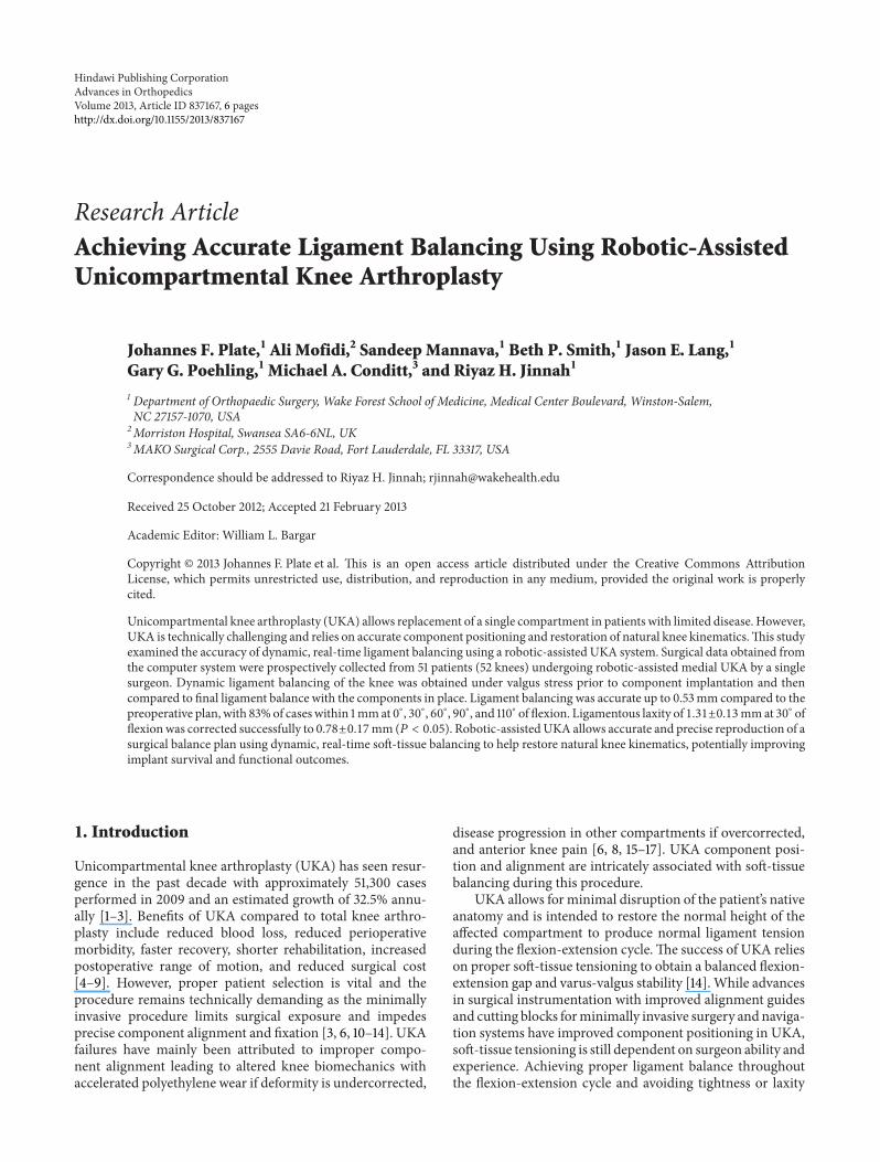
Figure 1: Ligament balancingwasmeasured throughout various angles during the flexion-extension cycle relative to tibia andmechanical axis.(a) The colored dots represent measurements during femoral range of motion. (b) Intraoperative screenshot of the robotic system showingligament balance at 0∘, 30∘, 60∘, 90∘, and 110∘ of flexion before resection, with the trial component in place, and after implantation.
are complex and partly rely on component size and position[14, 18]. Increased soft-tissue tightnessmay decrease the rangeof motion and increase wear while increased laxity may leadto joint instability and knee pain.
Robotic-assisted UKA allows for improved componentpositioning [2, 3, 19] with the ability of real-time, dynamicligament balancing intraoperatively. The robotic system usesoptical motion capture technology that dynamically tracksintracortical markers fixed to the tibia and femur. Thepurpose of the current study was to describe the techniqueof soft-tissue tensioning and assess the accuracy of robotic-assisted ligament balancing based on an intraoperative bal-ance plan during 52 consecutive medial robotic-assistedmedial UKAs. We hypothesized that robotic-assisted UKAaccurately produces ligament tension according to an intra-operative balance plan devised before component implanta-tion.
2. Material and Methods
2.1. Robotic-Assisted Ligament Balancing Technique for UKA.While the surgical technique using a robotic-assisted UKAsystem has been described elsewhere in detail [6], this paperwill focus on how to obtain accurate ligament balance forreplacement of the medial compartment. Preoperative CTscans are used by the computer system to render a three-dimensional model of patient anatomy. Intraoperatively,anatomic landmarks are used to register the patient tothe robot following intracortical placement of the femoraland tibial marker array. A minimally invasive medial jointincision is made, and medial osteophytes are resected. Theknee is then ranged through a number of flexion-extensioncycles. Valgus stress is then applied by the surgeon to open upthemedial compartment and bring the knee into its “natural”alignment. The ligament balance is then analyzed and dis-played by the computer system in real time as deviation from
the optimal tracking pattern of the prosthesis calculated bythe computer in millimeters (mm) during numerous flexion-extension cycles at 0∘, 30∘, 60∘, 90∘, and 110∘ of flexion(Figure 1). Negative deviation depicts ligamentous tightnessand positive values indicate ligamentous laxity.
The values obtained during the range of motion withvalgus stress serve as the intraoperative balance plan for liga-mentous tensioning. Using the computer system, componentposition or size can be altered, and the resulting changesin predicted ligament balance can be observed in real-time.If there is predicted laxity, component size and positioncan be changed to increase tightness, thereby programmingthe robot to alter bone cuts based on the preoperative CTscans and intraoperative findings. After the bone resectionshave been made using the robotic arm, the trial componentsare inserted and ligamentous tension is compared to theintraoperative balance plan. If proper balance is achievedwith the trial components in place, the final componentsare inserted and cemented, and final ligamentous balance isobtained during range of motion.
2.2. Assessment of Ligament Balancing Accuracy. The intra-operative data from 51 consecutive patients (52 knees) whounderwent robotic-assisted UKA (MAKOplasty,MAKO Sur-gical Corp.) of the medial compartment by a single surgeon(RHJ) were prospectively collected over a 6-month period.All patients received a fixed-bearing UKA with an onlaycemented tibial component and cemented femoral compo-nent. Following registration of the robotic system and prior toincision, the intraoperative balance plan for ligament tension-ing was obtained under valgus stress. After implantation ofthe final components, dynamic measurements were repeatedwithout valgus stress. Data was stored on the computersystem (Figure 1), and the actual ligament balancing wascompared to the intraoperative balance plan by subtractingthe planned measurements at 0∘, 30∘, 60∘, and 90∘ of flexion
Advances in Orthopedics 3
Table 1: Comparison of the intra-operative balance plan and ligament balance measurements following component implantation. Data isexpressed as mean ± standard error of the mean in millimeters.
Flexion angle Balance plan After implantation Change in balance 𝑃 value0∘ 0.34 ± 0.12 0.08 ± 0.18 −0.26 ± 0.17 𝑃 > 0.05
30∘ 1.31 ± 0.13 0.78 ± 0.17 −0.53 ± 0.18 𝑃 < 0.05∗
60∘ −0.28 ± 0.11 −0.33 ± 0.14 −0.04 ± 0.15 𝑃 > 0.05
90∘ −0.49 ± 0.12 −0.32 ± 0.13 0.16 ± 0.13 𝑃 > 0.05
110∘ 0.03 ± 0.16 −0.07 ± 0.19 −0.10 ± 0.14 𝑃 > 0.05
∗A 𝑃 value less than 0.05 was considered statistically significant.
from the actual postoperative measurements. Analysis ofvariance (ANOVA) was used to compare ligament balance at0∘, 30∘, 60∘, 90∘, and 110∘ of flexion with Bonferroni post-hoccomparison with alpha 0.05. All data are presented as mean ±standard error of the mean (SEM).
3. Results
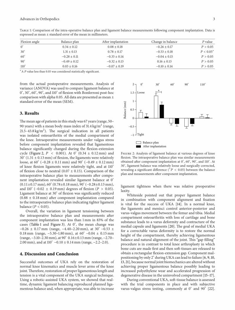
Themean age of patients in this studywas 67 years (range, 50–90 years) with a mean body mass index of 31.4 kg/m2 (range,21.5–43.8 kg/m2). The surgical indication in all patientswas isolated osteoarthritis of the medial compartment ofthe knee. Intraoperative measurements under valgus stressbefore component implantation revealed that ligamentousbalance significantly changed during the flexion-extensioncycle (Figure 2, 𝑃 < 0.001). At 0∘ (0.34 ± 0.12mm) and30∘ (1.31 ± 0.13mm) of flexion, the ligaments were relativelyloose, at 60∘ (−0.28 ± 0.11mm) and 90∘ (−0.49 ± 0.12mm)of knee flexion ligaments were relatively tight, and at 110∘of flexion close to neutral (0.07 ± 0.15). Comparison of theintraoperative balance plan to measurements after compo-nent implantation revealed similar ligament balance at 0∘(0.11±0.17mm), 60∘ (0.78±0.18mm), 90∘ (−0.28±0.13mm),and 110∘ (−0.02 ± 0.19mm) degrees of flexion (𝑃 > 0.05).Ligament balance at 30∘ of flexion was significantly reduced(0.88 ± 0.18mm) after component implantation comparedto the intraoperative balance plan indicating tighter ligamentbalance (𝑃 < 0.05).
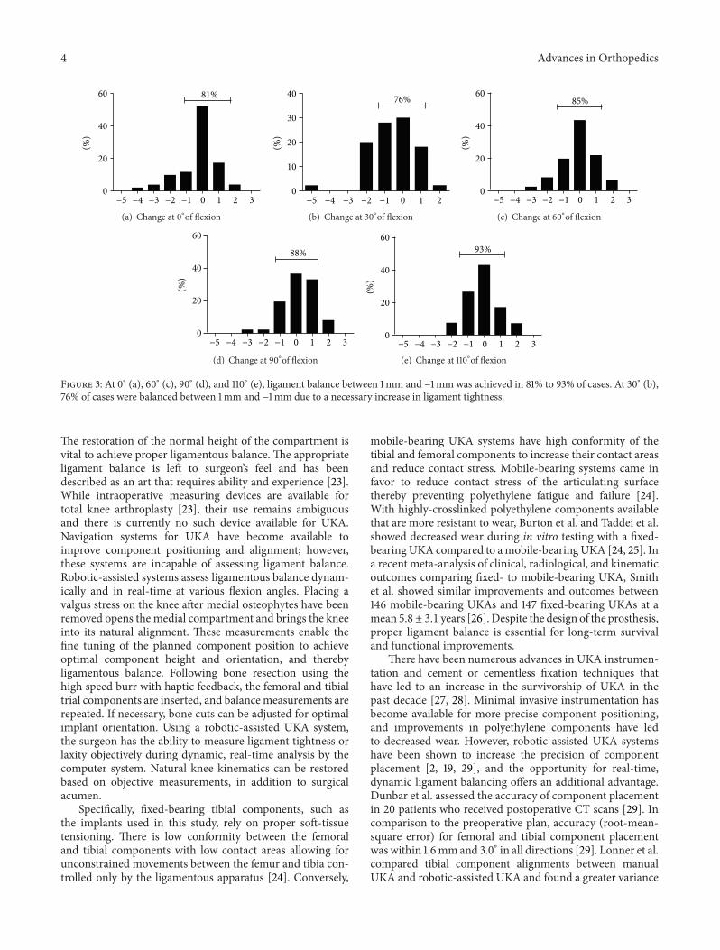
Overall, the variation in ligament tensioning betweenthe intraoperative balance plan and measurements aftercomponent implantation was less than 1mm in 83% of thecases (Table 1 and Figure 3). At 0∘, the mean change was−0.26 ± 0.17mm (range, −4.40–2.20mm), at 30∘ −0.53 ±0.18mm (range, −5.30–1.80mm), at 60∘ −0.04 ± 0.15mm(range, −3.10–2.30mm), at 90∘ 0.16±0.13mm (range, −2.70–2.00mm), and at 110∘ −0.10 ± 0.14mm (range, −2.2–2.0).
4. Discussion and Conclusion
Successful outcomes of UKA rely on the restoration ofnormal knee kinematics and muscle lever arms of the kneejoint.Therefore, restoration of proper ligamentous length andtension is a vital component of the UKA surgical technique.Using a robotic-assisted UKA system, we showed that real-time, dynamic ligament balancing reproduced planned liga-mentous balance and, when appropriate, was able to increase
2
1.5
1
0.5
0
−0.5
−1
Bala
nce (
mm
)
Loose
Tight
Balance planAfter implantation
0∘
30∘
60∘
90∘
110∘
∗
Figure 2: Analysis of ligament balance at various degrees of kneeflexion. The intraoperative balance plan was similar measurementsobtained after component implantation at 0∘, 60∘, 90∘, and 110∘. At30∘, ligament balance was relatively loose and surgically corrected,revealing a significant difference (∗𝑃 < 0.05) between the balanceplan and measurements after component implantation.
ligament tightness when there was relative preoperativelaxity.
Whiteside pointed out that proper ligament balancein combination with component alignment and fixationis vital for the success of UKA [14]. In a normal knee,the ligaments and menisci control anterior-posterior andvarus-valgus movement between the femur and tibia. Medialcompartment osteoarthritis with loss of cartilage and bonesubstance leads to a varus deformity and contracture of themedial capsule and ligaments [20]. The goal of medial UKAfor a correctable varus deformity is to restore the normalheight of the compartment, thereby achieving ligamentousbalance and natural alignment of the joint. This “gap filling”procedure is in contrast to total knee arthroplasty in whichbone cuts are made first and then soft-tissues are released toobtain a rectangular flexion-extension gap. Component mal-positioning by only 2∘ duringUKA can lead to failure [6, 9, 10,13, 21], because normal joint biomechanics are alteredwithoutachieving proper ligamentous balance possibly leading toincreased polyethylene wear and accelerated progression ofdegenerative disease in the uninvolved compartment [15–17].
During conventional UKA, soft-tissue balance is assessedwith the trial components in place and with subjectivevarus-valgus stress testing, commonly at 0∘ and 90∘ [22].
4 Advances in Orthopedics
60
40
20
0
(%)
81%
−5 −4 −3 −2 −1 0 1 2 3
(a) Change at 0∘of flexion
40
30
20
10
0
(%)
76%
−5 −4 −3 −2 −1 0 1 2
(b) Change at 30∘of flexion
60
40
20
0
(%)
85%
−5 −4 −3 −2 −1 0 1 2 3
(c) Change at 60∘of flexion
60
40
20
0
(%)
88%
−5 −4 −3 −2 −1 0 1 2 3
(d) Change at 90∘of flexion
60
40
20
0
(%)
93%
−5 −4 −3 −2 −1 0 1 2 3
(e) Change at 110∘of flexion
Figure 3: At 0∘ (a), 60∘ (c), 90∘ (d), and 110∘ (e), ligament balance between 1mm and −1mm was achieved in 81% to 93% of cases. At 30∘ (b),76% of cases were balanced between 1mm and −1mm due to a necessary increase in ligament tightness.
The restoration of the normal height of the compartment isvital to achieve proper ligamentous balance. The appropriateligament balance is left to surgeon’s feel and has beendescribed as an art that requires ability and experience [23].While intraoperative measuring devices are available fortotal knee arthroplasty [23], their use remains ambiguousand there is currently no such device available for UKA.Navigation systems for UKA have become available toimprove component positioning and alignment; however,these systems are incapable of assessing ligament balance.Robotic-assisted systems assess ligamentous balance dynam-ically and in real-time at various flexion angles. Placing avalgus stress on the knee after medial osteophytes have beenremoved opens the medial compartment and brings the kneeinto its natural alignment. These measurements enable thefine tuning of the planned component position to achieveoptimal component height and orientation, and therebyligamentous balance. Following bone resection using thehigh speed burr with haptic feedback, the femoral and tibialtrial components are inserted, and balancemeasurements arerepeated. If necessary, bone cuts can be adjusted for optimalimplant orientation. Using a robotic-assisted UKA system,the surgeon has the ability to measure ligament tightness orlaxity objectively during dynamic, real-time analysis by thecomputer system. Natural knee kinematics can be restoredbased on objective measurements, in addition to surgicalacumen.
Specifically, fixed-bearing tibial components, such asthe implants used in this study, rely on proper soft-tissuetensioning. There is low conformity between the femoraland tibial components with low contact areas allowing forunconstrained movements between the femur and tibia con-trolled only by the ligamentous apparatus [24]. Conversely,
mobile-bearing UKA systems have high conformity of thetibial and femoral components to increase their contact areasand reduce contact stress. Mobile-bearing systems came infavor to reduce contact stress of the articulating surfacethereby preventing polyethylene fatigue and failure [24].With highly-crosslinked polyethylene components availablethat are more resistant to wear, Burton et al. and Taddei et al.showed decreased wear during in vitro testing with a fixed-bearingUKA compared to amobile-bearingUKA [24, 25]. Ina recent meta-analysis of clinical, radiological, and kinematicoutcomes comparing fixed- to mobile-bearing UKA, Smithet al. showed similar improvements and outcomes between146 mobile-bearing UKAs and 147 fixed-bearing UKAs at amean 5.8 ± 3.1 years [26]. Despite the design of the prosthesis,proper ligament balance is essential for long-term survivaland functional improvements.
There have been numerous advances in UKA instrumen-tation and cement or cementless fixation techniques thathave led to an increase in the survivorship of UKA in thepast decade [27, 28]. Minimal invasive instrumentation hasbecome available for more precise component positioning,and improvements in polyethylene components have ledto decreased wear. However, robotic-assisted UKA systemshave been shown to increase the precision of componentplacement [2, 19, 29], and the opportunity for real-time,dynamic ligament balancing offers an additional advantage.Dunbar et al. assessed the accuracy of component placementin 20 patients who received postoperative CT scans [29]. Incomparison to the preoperative plan, accuracy (root-mean-square error) for femoral and tibial component placementwas within 1.6mm and 3.0∘ in all directions [29]. Lonner et al.compared tibial component alignments between manualUKA and robotic-assisted UKA and found a greater variance
Advances in Orthopedics 5
in component position, increased tibial slope, and increasedvarus alignment when the tibia was prepared manually [19].
A major limitation of this study is the lack of clinicalor functional outcomes in this patient cohort; the study wasintended to assess the accuracy of ligament tensioning onlybased upon the intraoperative balance plan. There are cur-rently no studies available on the clinical outcomes of robotic-assistedUKAdue to the novelty of the device. Certainly, long-term studies on the outcomes of the robotic-assisted devicecompared to manual UKA are needed to delineate a possibleadvantage of the robot in light of the financial investment.However, based on the technical demands ofUKA,we believethat improved component positioning and alignment incombination with dynamic, real-time assessment of ligamentbalance offered by the robotic-assisted system may improveoutcomes.
To our knowledge, this is the first study assessing real-time dynamic ligament balancing with a robotic-assistedsystem for UKA. We conclude from our findings thatrobotic-assisted UKA can accurately and precisely reproduceintraoperatively planned ligamentous balance using real-time, dynamic measurements. In combination with highaccuracy of component placement, robotic-assisted systemsmay improve functional outcomes and survivorship of UKApatients; however, further investigations into the benefits ofrobotic systems for UKA are needed.
Acknowledgments
The authors J. F. Plate, A. Mofidi, S. Mannava, B. P. Smith,and J. E. Lang report no conflict of interests. The authorsR. H. Jinnah, G. G. Poehling, and M. A. Conditt havereceived financial support from MAKO Surgical Corp., FortLauderdale, FL, USA. R. H. Jinnah and G. G. Poehling havereceived payment as consultants and are stock holders. M. A.Conditt receives compensation as Senior Director of ClinicalResearch. All authors certify that this investigation wasperformed in conformity with ethical principles of research.Institutional ReviewBoard approval was obtained prior to thestudy.
References
[1] M. Hoffman, “MAKO Robotics Yearly Numbers for Unicom-partmenal Knee Arthroplasty,” Fort Wayne, A. J. Floyd, E-mail,2010.
[2] A. D. Pearle, P. F. O’Loughlin, and D. O. Kendoff, “Robot-assisted unicompartmental knee arthroplasty,” Journal ofArthroplasty, vol. 25, no. 2, pp. 230–237, 2010.
[3] M. Roche, P. F. O’Loughlin, D. Kendoff, V. Musahl, andA. D. Pearle, “Robotic arm-assisted unicompartmental kneearthroplasty: preoperative planning and surgical technique,”American Journal of Orthopedics, vol. 38, no. 2, supplement, pp.10–15, 2009.
[4] J. M. Bert, “Unicompartmental knee replacement,” OrthopedicClinics of North America, vol. 36, no. 4, pp. 513–522, 2005.
[5] T. Borus and T. Thornhill, “Unicompartmental knee arthro-plasty,” Journal of the American Academy of Orthopaedic Sur-geons, vol. 16, no. 1, pp. 9–18, 2008.
[6] M. A. Conditt and M. W. Roche, “Minimally invasive robotic-arm-guided unicompartmental knee arthroplasty,” Journal ofBone and Joint Surgery. American, vol. 91, supplement 1, pp. 63–68, 2009.
[7] J. Y. Jenny, E. Ciobanu, and C. Boeri, “The rationale fornavigated minimally invasive unicompartmental knee replace-ment,” Clinical Orthopaedics and Related Research, no. 463, pp.58–62, 2007.
[8] J. H. Lonner, “Indications for unicompartmental knee arthro-plasty and rationale for robotic arm-assisted technology,”Amer-ican Journal of Orthopedics, vol. 38, no. 2, suplement, pp. 3–6,2009.
[9] A. D. Pearle, D. Kendoff, and V. Musahl, “Perspectives oncomputer-assisted orthopaedic surgery: movement towardquantitative orthopaedic surgery,” Journal of Bone and JointSurgery. American, vol. 91, no. 1, pp. 7–12, 2009.
[10] S. A. Banks, M. K. Harman, and W. A. Hodge, “Mechanismof anterior impingement damage in total knee arthroplasty,”Journal of Bone and Joint Surgery. American, vol. 84, no. 2, pp.37–42, 2002.
[11] G. Li, R. Papannagari, E. Most et al., “Anterior tibial postimpingement in a posterior stabilized total knee arthroplasty,”Journal of Orthopaedic Research, vol. 23, no. 3, pp. 536–541,2005.
[12] U. I. Maduekwe, M. G. Zywiel, P. M. Bonutti, A. J. Johnson, R.E. Delanois, and M. A. Mont, “Scientific evidence for the use ofmodern unicompartmental knee arthroplasty,”Expert Review ofMedical Devices, vol. 7, no. 2, pp. 219–239, 2010.
[13] E. M. Mariani, M. H. Bourne, R. T. Jackson, S. T. Jackson, andP. Jones, “Early failure of unicompartmental knee arthroplasty,”Journal of Arthroplasty, vol. 22, no. 6, supplement, pp. 81–84,2007.
[14] L. A. Whiteside, “Making your next unicompartmental kneearthroplasty last: three keys to success,” Journal of Arthroplasty,vol. 20, supplement 3, pp. 2–3, 2005.
[15] M. B. Collier, T. H. Eickmann, F. Sukezaki, J. P. McAuley,and G. A. Engh, “Patient, implant, and alignment factorsassociated with revision of medial compartment unicondylararthroplasty,” Journal of Arthroplasty, vol. 21, no. 6, supplement,pp. 108–115, 2006.
[16] P. Hernigou and G. Deschamps, “Alignment influences wear inthe knee after medial unicompartmental arthroplasty,” ClinicalOrthopaedics and Related Research, no. 423, pp. 161–165, 2004.
[17] S. R. Ridgeway, J. P. McAuley, D. J. Ammeen, and G. A.Engh, “The effect of alignment of the knee on the outcome ofunicompartmental knee replacement,” Journal of Bone and JointSurgery. American, vol. 84, no. 3, pp. 351–355, 2002.
[18] R. H. Emerson, W. C. Head, and P. C. Peters, “Soft tissuebalance and alignment in medical unicompartmental kneearthroplasty,” Journal of Bone and Joint Surgery. American, vol.74, no. 6, pp. 807–810, 1992.
[19] J. H. Lonner, T. K. John, and M. A. Conditt, “Robotic arm-assisted UKA improves tibial component alignment: a pilotstudy,” Clinical Orthopaedics and Related Research, vol. 468, no.1, pp. 141–146, 2010.
[20] A. Claus and H. P. Scharf, “Ligament balancing and varusdeformity in total knee arthroplasty,” Orthopade, vol. 36, no. 7,pp. 643–649, 2007.
[21] S. E. Park and C. T. Lee, “Comparison of robotic-assisted andconventional manual implantation of a primary total kneearthroplasty,” Journal of Arthroplasty, vol. 22, no. 7, pp. 1054–1059, 2007.

6 Advances in Orthopedics
[22] W. M. Mihalko, K. J. Saleh, K. A. Krackow, and L. A. Whiteside,“Soft-tissue balancing during total knee arthroplasty in thevarus knee,” Journal of the American Academy of OrthopaedicSurgeons, vol. 17, no. 12, pp. 766–774, 2009.
[23] D. D. D’Lima, S. Patil, N. Steklov, and C. W. Colwell, “An ABJSbest paper: dynamic intraoperative ligament balancing for totalknee arthroplasty,” Clinical Orthopaedics and Related Research,no. 463, pp. 208–212, 2007.
[24] A. Burton, S. Williams, C. L. Brockett, and J. Fisher, “In vitrocomparison of fixed- andmobile meniscal-bearing unicondylarknee arthroplasties. Effect of design, kinematics, and condylarliftoff,” Journal of Arthroplasty, vol. 27, no. 8, pp. 1452–1459, 2012.
[25] P. Taddei, E. Modena, T. M. Grupp, and S. Affatato, “Mobileor fixed unicompartmental knee prostheses? In-vitro wearassessments to solve this dilemma,” Journal of the MechanicalBehavior of Biomedical Materials, vol. 4, no. 8, pp. 1936–1946.
[26] T. O. Smith, C. B. Hing, L. Davies, and S. T. Donell, “Fixedversus mobile bearing unicompartmental knee replacement: ameta-analysis,” Orthopaedics and Traumatology, vol. 95, no. 8,pp. 599–605, 2009.
[27] G. Labek, K. Sekyra, W. Pawelka, W. Janda, and B. Stockl,“Outcome and reproducibility of data concerning the Oxfordunicompartmental knee arthroplasty: a structured literaturereview including arthroplasty registry data,”Acta Orthopaedica,vol. 82, no. 2, pp. 131–135, 2011.
[28] A. V. Lombardi, K. R. Berend, M. E. Berend et al., “Currentcontroversies in partial knee arthroplasty,” Instructional CourseLectures, vol. 61, pp. 347–381, 2012.
[29] N. J. Dunbar, M. W. Roche, B. H. Park, S. H. Branch, M. A.Conditt, and S. A. Banks, “Accuracy of dynamic tactile-guidedunicompartmental knee arthroplasty,” Journal of Arthroplasty,vol. 27, no. 5, pp. 803–808.e1, 2012.
Related Documents