KNEE Variable bone mineral density reductions post-unicompartmental knee arthroplasty Mahmut Tuncer • Rajesh Patel • Justin P. Cobb • Ulrich N. Hansen • Andrew A. Amis Received: 22 August 2013 / Accepted: 13 April 2014 / Published online: 27 April 2014 Ó The Author(s) 2014. This article is published with open access at Springerlink.com Abstract Purpose Radiolucencies are commonly observed in uni- compartmental knee arthroplasty (UKA) patients within 1 year of arthroplasty. The objective of the study was to identify how the bone mineral density (BMD) changes up to 1 year post-arthroplasty. Methods Dual X-ray absorptiometry scans were obtained from 11 UKA patients at 10 days and 3, 6, and 12 months post-surgery. Patients were scanned in both anteroposterior and lateral knee orientations. Results Most subjects saw a large decline in BMD in the first 6 months following surgery, followed by some recovery in bone mass. The biggest change occurred under the tibial intercondylar eminence, which decreased signif- icantly by an average of 18 % at 6 months and was 15 % at 1 year. The average bone loss under the tibial tray was low; however, the bone loss at the anterior portion was higher with a significant average decrease of 14 %. There was no change in BMD under the tibial keel. There was significant bone loss of 13 % under the femoral component; the regions anterior and posterior to the central femoral implant peg both had significant bone loss of 14 %. The bone response between patients was very variable, with some patients losing bone steadily, and others gaining it rapidly after an early fall. Conclusions While the overall reduction in BMD under both components was low, it was significant and there was substantial individual variation superimposed on this. Improving our understanding of this response to surgery may impact on prosthesis survival. Level of evidence Therapeutic study: case series with no comparison group, Level IV. Keywords Unicompartmental knee arthroplasty UKA Bone density changes BMD DXA Introduction There is increasing evidence that unicompartmental knee arthroplasty (UKA) can have reliable long-term perfor- mance [7, 14, 17, 18]. Early loosening is the most com- mon reason for revision surgery [1, 2, 12, 17, 23], and stress shielding followed by bone resorption may con- tribute to the process. Radiolucencies are very commonly seen beneath mobile-bearing UKA, starting to occur within one year post-arthroplasty [19]. While most of these radiolucencies are claimed to be ‘physiological’, those that are thick with undefined borders have been linked to loosened implants [5]. There is a need to understand the bone density changes that occur beneath UKA components post-arthroplasty, to aid further devel- opment of their fixation. Dual X-ray absorptiometry (DXA) scanning is com- monly used to measure bone mineral density (BMD) and changes in BMD over time. Although numerous studies have been conducted on total knee arthroplasty (TKA) patients [3, 10, 13, 16, 25], only one DXA study has been conducted on UKA patients [24], examining fixed-bearing UKA up to 7 years post-surgery; they did not find signifi- cant changes in BMD beyond 1 year post-UKA. A further study [20] used CT slices to find almost no changes in M. Tuncer U. N. Hansen A. A. Amis (&) Department of Mechanical Engineering, Imperial College London, Exhibition Road, London SW7 2AZ, UK e-mail: [email protected] R. Patel J. P. Cobb A. A. Amis Department of Musculoskeletal Surgery, Charing Cross Hospital, Imperial College London, London W6 8RF, UK 123 Knee Surg Sports Traumatol Arthrosc (2015) 23:2230–2236 DOI 10.1007/s00167-014-3014-5

Welcome message from author
This document is posted to help you gain knowledge. Please leave a comment to let me know what you think about it! Share it to your friends and learn new things together.
Transcript
KNEE
Variable bone mineral density reductions post-unicompartmentalknee arthroplasty
Mahmut Tuncer • Rajesh Patel • Justin P. Cobb •
Ulrich N. Hansen • Andrew A. Amis
Received: 22 August 2013 / Accepted: 13 April 2014 / Published online: 27 April 2014
� The Author(s) 2014. This article is published with open access at Springerlink.com
Abstract
Purpose Radiolucencies are commonly observed in uni-
compartmental knee arthroplasty (UKA) patients within 1
year of arthroplasty. The objective of the study was to
identify how the bone mineral density (BMD) changes up
to 1 year post-arthroplasty.
Methods Dual X-ray absorptiometry scans were obtained
from 11 UKA patients at 10 days and 3, 6, and 12 months
post-surgery. Patients were scanned in both anteroposterior
and lateral knee orientations.
Results Most subjects saw a large decline in BMD in the
first 6 months following surgery, followed by some
recovery in bone mass. The biggest change occurred under
the tibial intercondylar eminence, which decreased signif-
icantly by an average of 18 % at 6 months and was 15 % at
1 year. The average bone loss under the tibial tray was low;
however, the bone loss at the anterior portion was higher
with a significant average decrease of 14 %. There was no
change in BMD under the tibial keel. There was significant
bone loss of 13 % under the femoral component; the
regions anterior and posterior to the central femoral
implant peg both had significant bone loss of 14 %. The
bone response between patients was very variable, with
some patients losing bone steadily, and others gaining it
rapidly after an early fall.
Conclusions While the overall reduction in BMD under
both components was low, it was significant and there was
substantial individual variation superimposed on this.
Improving our understanding of this response to surgery
may impact on prosthesis survival.
Level of evidence Therapeutic study: case series with no
comparison group, Level IV.
Keywords Unicompartmental knee arthroplasty � UKA �Bone density changes � BMD � DXA
Introduction
There is increasing evidence that unicompartmental knee
arthroplasty (UKA) can have reliable long-term perfor-
mance [7, 14, 17, 18]. Early loosening is the most com-
mon reason for revision surgery [1, 2, 12, 17, 23], and
stress shielding followed by bone resorption may con-
tribute to the process. Radiolucencies are very commonly
seen beneath mobile-bearing UKA, starting to occur
within one year post-arthroplasty [19]. While most of
these radiolucencies are claimed to be ‘physiological’,
those that are thick with undefined borders have been
linked to loosened implants [5]. There is a need to
understand the bone density changes that occur beneath
UKA components post-arthroplasty, to aid further devel-
opment of their fixation.
Dual X-ray absorptiometry (DXA) scanning is com-
monly used to measure bone mineral density (BMD) and
changes in BMD over time. Although numerous studies
have been conducted on total knee arthroplasty (TKA)
patients [3, 10, 13, 16, 25], only one DXA study has been
conducted on UKA patients [24], examining fixed-bearing
UKA up to 7 years post-surgery; they did not find signifi-
cant changes in BMD beyond 1 year post-UKA. A further
study [20] used CT slices to find almost no changes in
M. Tuncer � U. N. Hansen � A. A. Amis (&)
Department of Mechanical Engineering, Imperial College
London, Exhibition Road, London SW7 2AZ, UK
e-mail: [email protected]
R. Patel � J. P. Cobb � A. A. Amis
Department of Musculoskeletal Surgery, Charing Cross
Hospital, Imperial College London, London W6 8RF, UK
123
Knee Surg Sports Traumatol Arthrosc (2015) 23:2230–2236
DOI 10.1007/s00167-014-3014-5
overall tibial BMD post-UKA. There remains a need for
more detailed information on bone changes in UKA, par-
ticularly for mobile-bearing prostheses.
Noting that most bone remodelling occurs within 1 year
[4, 22, 24], it was hypothesised that there would be a loss
of BMD within 1 year post-UKA; knowledge of such
changes would aid work to improve the fixation design.
Materials and methods
Following approval by the Charing Cross Hospital
Research Ethics Committee (Ref 09/H0711/51), thirteen
UKA patients were recruited over the course of 1 year. All
surgeries were performed by a single consultant surgeon
and his registrar.
Patients were selected upon satisfying three conditions: (1)
they had a pre-operative knee computed tomography (CT)
scan; (2) they would have the Oxford UKA (Biomet Ltd,
Swindon, UK) on their medial condyle; (3) they lived within
10miles of the hospital. The patientswere recruited regardless
of whether cemented or cementless fixation would be used.
The first DXA scan was performed within 10 days from
the date of surgery, with the remaining scans carried out at
3, 6, and 12 months. Patients were scanned in both AP and
lateral knee orientations.
All DXA scans were performed using a GE Lunar
Prodigy Scanner (GE Healthcare, Chalfont St Giles, UK).
AP and lateral scans were performed using wooden limb-
positioning jigs specially made for the study. The AP
scan was taken with the tibia inclined at 7� to the
scanner bed so that a vertical X-ray beam would be
approximately parallel to the tibial plateau, while the
lateral scan was taken at 30� knee flexion. Since the
scanner did not have a pre-defined setting for knee scans,
the ‘AP Spine’ mode was selected with ‘Smart Scan’
mode setting deactivated. As is commonly used for knee
scans [25], two rice bags were also used as a soft tissue
substitute.
The reproducibility of the BMD measurements was
calculated in each subject by carrying out two consecutive
scans at 6 months in both AP and lateral projections, with
the subject being repositioned between scans. The test–
retest error was calculated as follows:
Error ð%Þ ¼ 1
N
XN
n¼1
ffiffiffiffiffiffiffiffiffiffiffiffiffiffiffiffiffiffiffiffiffiffiffiffiffiffiffiffiffiffiffiffiffiffiffiffiffiffiffiffiffiffiBMDin � BMDiinð Þ2
q
12BMDin þ BMDiinð Þ
where BMDi is the first BMD reading of patient n, BMDii
is the second reading of patient n, and N is the total number
of patients. A standard Lunar calibration block was used
for daily quality assurance of the scanner, and at regular
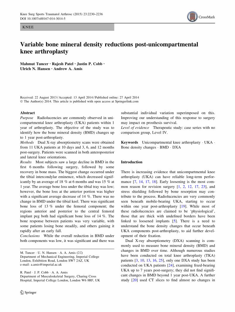
Fig. 1 Average test–retest error
of the DXA BMD
measurements for each ROI.
ROIs F1–10 are AP scan and
L1–10 are lateral scan ROIs
(mean ? SD, n = 11)
Fig. 2 BMD changes post-
UKA at ROI F6. A significant
drop of BMD was observed at 6
months and 1 year
Knee Surg Sports Traumatol Arthrosc (2015) 23:2230–2236 2231
123
intervals, a secondary calibration check was completed
using an aluminium spine phantom.
Figure 1 shows the errors associated with patient repo-
sitioning. The errors for ROI F7 and F8 were larger
because they were sensitive to the medial position of the
patella: BMD was higher when the patella was medial and
overlapping ROI F7 and F8. The high error of ROI L6
occurred because the BMD was sensitive to the position of
the fibula. In addition, baseline data for one knee were
unavailable in the lateral view.
The patient data were anonymised and analysed using
EnCore 2008 (GE Healthcare, Chalfont St Giles, UK). The
scans were converted to ‘knee’ mode, and ten regions-of-
interest (ROI) were defined for the AP (ROI F1–10) and
lateral (ROI L1–10) scans. All the 1-year data were analysed
at the same time by a single user to ensure consistency.
Statistical analysis
The Kolmogorov–Smirnov test was used to test all vari-
ables for normality using SPSS software (IBM Software
Group, New York, USA). The test confirmed that all BMD
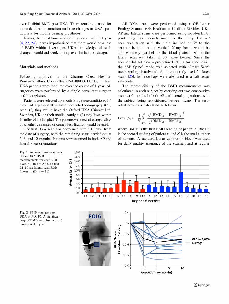
Fig. 3 Anteroposterior scan BMD changes under the tibial tray of each patient
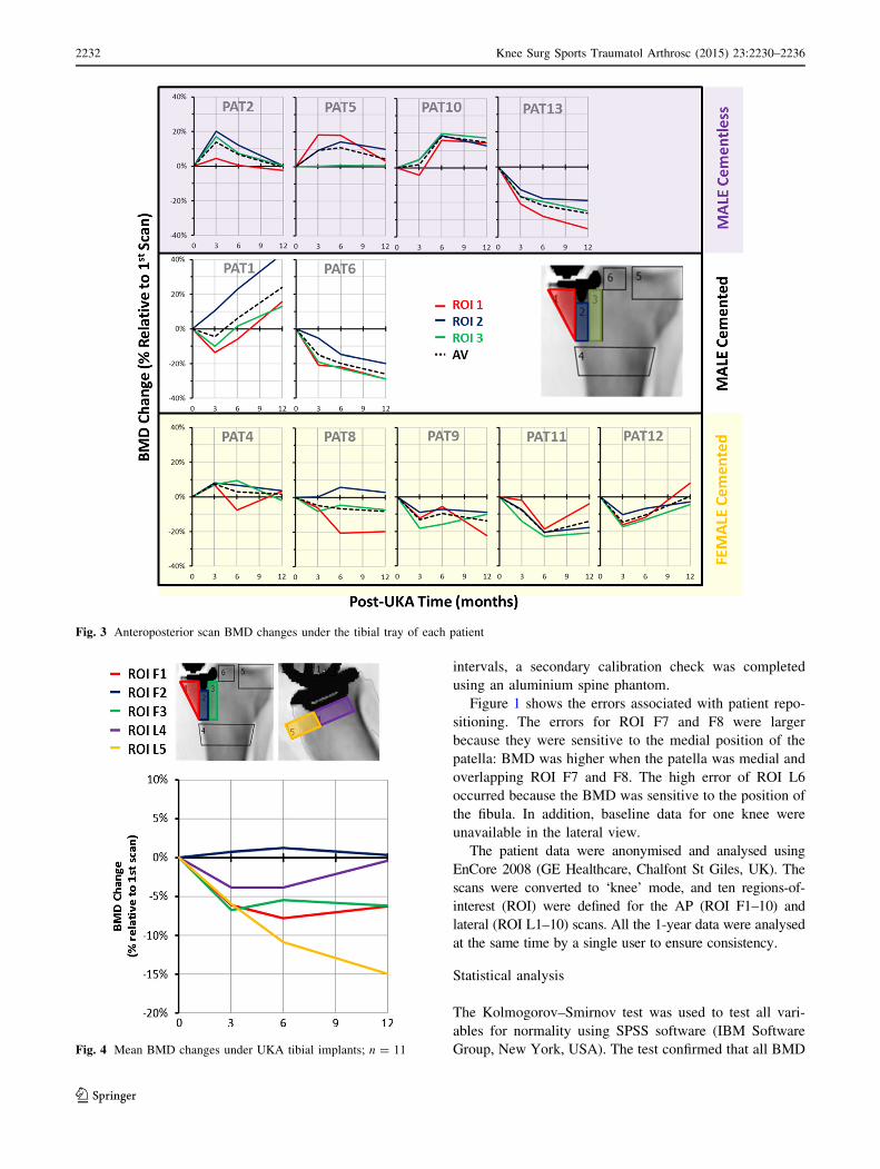
Fig. 4 Mean BMD changes under UKA tibial implants; n = 11
2232 Knee Surg Sports Traumatol Arthrosc (2015) 23:2230–2236
123
variables were normally distributed and that a paired Stu-
dent t test was suitable for testing statistical difference. A
power analysis was not done in view of the ethics permit
only being for the small number of cases.
Results
Thirteen patients consented to join this study, but one
patient dropped out immediately after the first scan, due to
discomfort in other joints during the scanning procedure,
and another was lost prior to the scan at 12 months, leaving
11 patients. The data relate to seven UKA patients with
cement fixation (two male and five female, aged
59 ± 12 years (mean ± SD), range 42–79 years) and four
patients with cementless fixation (four male, aged
69 ± 8 years, range 61–79 years).
Tibia
Figure 2 presents the BMD changes beneath the tibial in-
tercondylar eminence (ROI F6) up to 1 year post-arthro-
plasty; the BMD drop was significant at 6 months
(P = 0.0001) and at 1 year (P = 0.0022).
Figure 3 displays the BMD changes at three ROI located
beneath the UKA tibial tray: there was a considerable
variation between the subjects. The total average change in
BMD under the tibial tray at 1 year was -4 ± 17 %.
Figure 4 shows the average BMD changes in the proximal
tibia, with no mean change under the keel (ROI F2) and
small mean losses (6 %) in the regions medial (ROI F1)
and lateral to the keel (ROI F3).
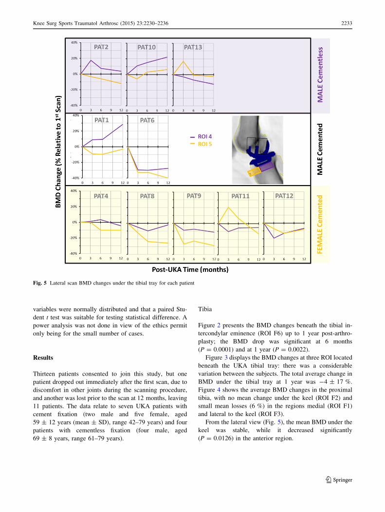
From the lateral view (Fig. 5), the mean BMD under the
keel was stable, while it decreased significantly
(P = 0.0126) in the anterior region.
Fig. 5 Lateral scan BMD changes under the tibial tray for each patient
Knee Surg Sports Traumatol Arthrosc (2015) 23:2230–2236 2233
123
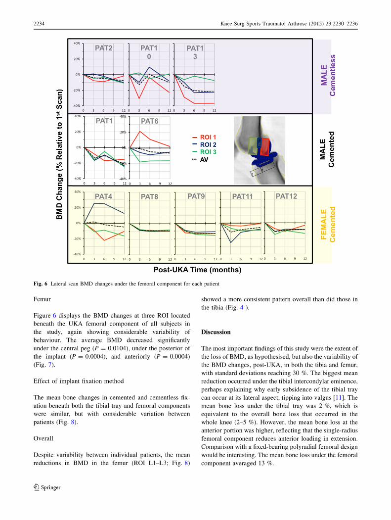
Femur
Figure 6 displays the BMD changes at three ROI located
beneath the UKA femoral component of all subjects in
the study, again showing considerable variability of
behaviour. The average BMD decreased significantly
under the central peg (P = 0.0104), under the posterior of
the implant (P = 0.0004), and anteriorly (P = 0.0004)
(Fig. 7).
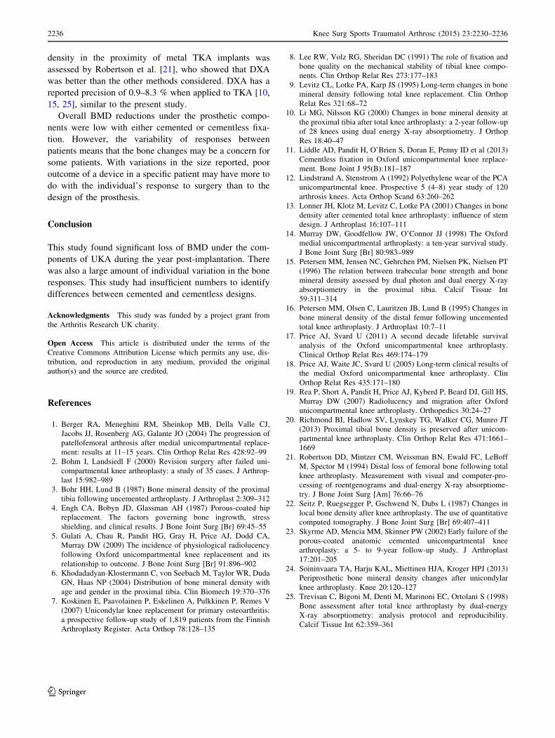
Effect of implant fixation method
The mean bone changes in cemented and cementless fix-
ation beneath both the tibial tray and femoral components
were similar, but with considerable variation between
patients (Fig. 8).
Overall
Despite variability between individual patients, the mean
reductions in BMD in the femur (ROI L1–L3; Fig. 8)
showed a more consistent pattern overall than did those in
the tibia (Fig. 4 ).
Discussion
The most important findings of this study were the extent of
the loss of BMD, as hypothesised, but also the variability of
the BMD changes, post-UKA, in both the tibia and femur,
with standard deviations reaching 30 %. The biggest mean
reduction occurred under the tibial intercondylar eminence,
perhaps explaining why early subsidence of the tibial tray
can occur at its lateral aspect, tipping into valgus [11]. The
mean bone loss under the tibial tray was 2 %, which is
equivalent to the overall bone loss that occurred in the
whole knee (2–5 %). However, the mean bone loss at the
anterior portion was higher, reflecting that the single-radius
femoral component reduces anterior loading in extension.
Comparison with a fixed-bearing polyradial femoral design
would be interesting. The mean bone loss under the femoral
component averaged 13 %.
Fig. 6 Lateral scan BMD changes under the femoral component for each patient
2234 Knee Surg Sports Traumatol Arthrosc (2015) 23:2230–2236
123
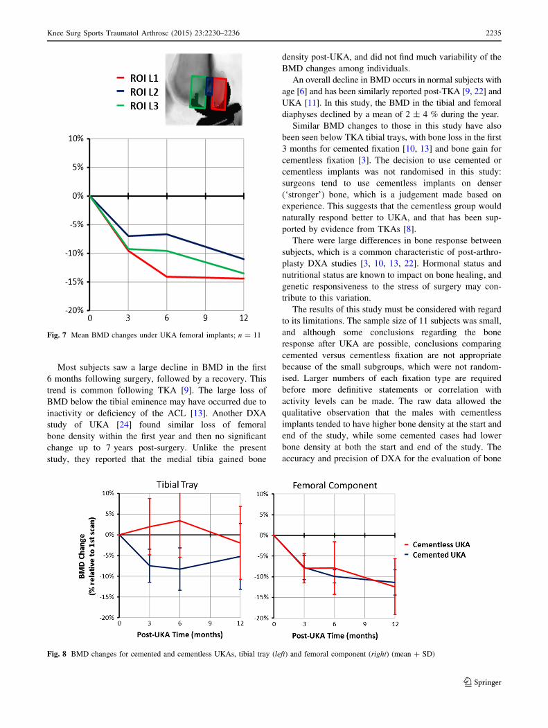
Most subjects saw a large decline in BMD in the first
6 months following surgery, followed by a recovery. This
trend is common following TKA [9]. The large loss of
BMD below the tibial eminence may have occurred due to
inactivity or deficiency of the ACL [13]. Another DXA
study of UKA [24] found similar loss of femoral
bone density within the first year and then no significant
change up to 7 years post-surgery. Unlike the present
study, they reported that the medial tibia gained bone
density post-UKA, and did not find much variability of the
BMD changes among individuals.
An overall decline in BMD occurs in normal subjects with
age [6] and has been similarly reported post-TKA [9, 22] and
UKA [11]. In this study, the BMD in the tibial and femoral
diaphyses declined by a mean of 2 ± 4 % during the year.
Similar BMD changes to those in this study have also
been seen below TKA tibial trays, with bone loss in the first
3 months for cemented fixation [10, 13] and bone gain for
cementless fixation [3]. The decision to use cemented or
cementless implants was not randomised in this study:
surgeons tend to use cementless implants on denser
(‘stronger’) bone, which is a judgement made based on
experience. This suggests that the cementless group would
naturally respond better to UKA, and that has been sup-
ported by evidence from TKAs [8].
There were large differences in bone response between
subjects, which is a common characteristic of post-arthro-
plasty DXA studies [3, 10, 13, 22]. Hormonal status and
nutritional status are known to impact on bone healing, and
genetic responsiveness to the stress of surgery may con-
tribute to this variation.
The results of this study must be considered with regard
to its limitations. The sample size of 11 subjects was small,
and although some conclusions regarding the bone
response after UKA are possible, conclusions comparing
cemented versus cementless fixation are not appropriate
because of the small subgroups, which were not random-
ised. Larger numbers of each fixation type are required
before more definitive statements or correlation with
activity levels can be made. The raw data allowed the
qualitative observation that the males with cementless
implants tended to have higher bone density at the start and
end of the study, while some cemented cases had lower
bone density at both the start and end of the study. The
accuracy and precision of DXA for the evaluation of bone
Fig. 7 Mean BMD changes under UKA femoral implants; n = 11
Fig. 8 BMD changes for cemented and cementless UKAs, tibial tray (left) and femoral component (right) (mean ? SD)
Knee Surg Sports Traumatol Arthrosc (2015) 23:2230–2236 2235
123
density in the proximity of metal TKA implants was
assessed by Robertson et al. [21], who showed that DXA
was better than the other methods considered. DXA has a
reported precision of 0.9–8.3 % when applied to TKA [10,
15, 25], similar to the present study.
Overall BMD reductions under the prosthetic compo-
nents were low with either cemented or cementless fixa-
tion. However, the variability of responses between
patients means that the bone changes may be a concern for
some patients. With variations in the size reported, poor
outcome of a device in a specific patient may have more to
do with the individual’s response to surgery than to the
design of the prosthesis.
Conclusion
This study found significant loss of BMD under the com-
ponents of UKA during the year post-implantation. There
was also a large amount of individual variation in the bone
responses. This study had insufficient numbers to identify
differences between cemented and cementless designs.
Acknowledgments This study was funded by a project grant from
the Arthritis Research UK charity.
Open Access This article is distributed under the terms of the
Creative Commons Attribution License which permits any use, dis-
tribution, and reproduction in any medium, provided the original
author(s) and the source are credited.
References
1. Berger RA, Meneghini RM, Sheinkop MB, Della Valle CJ,
Jacobs JJ, Rosenberg AG, Galante JO (2004) The progression of
patellofemoral arthrosis after medial unicompartmental replace-
ment: results at 11–15 years. Clin Orthop Relat Res 428:92–99
2. Bohm I, Landsiedl F (2000) Revision surgery after failed uni-
compartmental knee arthroplasty: a study of 35 cases. J Arthrop-
last 15:982–989
3. Bohr HH, Lund B (1987) Bone mineral density of the proximal
tibia following uncemented arthroplasty. J Arthroplast 2:309–312
4. Engh CA, Bobyn JD, Glassman AH (1987) Porous-coated hip
replacement. The factors governing bone ingrowth, stress
shielding, and clinical results. J Bone Joint Surg [Br] 69:45–55
5. Gulati A, Chau R, Pandit HG, Gray H, Price AJ, Dodd CA,
Murray DW (2009) The incidence of physiological radiolucency
following Oxford unicompartmental knee replacement and its
relationship to outcome. J Bone Joint Surg [Br] 91:896–902
6. Khodadadyan-Klostermann C, von Seebach M, Taylor WR, Duda
GN, Haas NP (2004) Distribution of bone mineral density with
age and gender in the proximal tibia. Clin Biomech 19:370–376
7. Koskinen E, Paavolainen P, Eskelinen A, Pulkkinen P, Remes V
(2007) Unicondylar knee replacement for primary osteoarthritis:
a prospective follow-up study of 1,819 patients from the Finnish
Arthroplasty Register. Acta Orthop 78:128–135
8. Lee RW, Volz RG, Sheridan DC (1991) The role of fixation and
bone quality on the mechanical stability of tibial knee compo-
nents. Clin Orthop Relat Res 273:177–183
9. Levitz CL, Lotke PA, Karp JS (1995) Long-term changes in bone
mineral density following total knee replacement. Clin Orthop
Relat Res 321:68–72
10. Li MG, Nilsson KG (2000) Changes in bone mineral density at
the proximal tibia after total knee arthroplasty: a 2-year follow-up
of 28 knees using dual energy X-ray absorptiometry. J Orthop
Res 18:40–47
11. Liddle AD, Pandit H, O’Brien S, Doran E, Penny ID et al (2013)
Cementless fixation in Oxford unicompartmental knee replace-
ment. Bone Joint J 95(B):181–187
12. Lindstrand A, Stenstrom A (1992) Polyethylene wear of the PCA
unicompartmental knee. Prospective 5 (4–8) year study of 120
arthrosis knees. Acta Orthop Scand 63:260–262
13. Lonner JH, Klotz M, Levitz C, Lotke PA (2001) Changes in bone
density after cemented total knee arthroplasty: influence of stem
design. J Arthroplast 16:107–111
14. Murray DW, Goodfellow JW, O’Connor JJ (1998) The Oxford
medial unicompartmental arthroplasty: a ten-year survival study.
J Bone Joint Surg [Br] 80:983–989
15. Petersen MM, Jensen NC, Gehrchen PM, Nielsen PK, Nielsen PT
(1996) The relation between trabecular bone strength and bone
mineral density assessed by dual photon and dual energy X-ray
absorptiometry in the proximal tibia. Calcif Tissue Int
59:311–314
16. Petersen MM, Olsen C, Lauritzen JB, Lund B (1995) Changes in
bone mineral density of the distal femur following uncemented
total knee arthroplasty. J Arthroplast 10:7–11
17. Price AJ, Svard U (2011) A second decade lifetable survival
analysis of the Oxford unicompartmental knee arthroplasty.
Clinical Orthop Relat Res 469:174–179
18. Price AJ, Waite JC, Svard U (2005) Long-term clinical results of
the medial Oxford unicompartmental knee arthroplasty. Clin
Orthop Relat Res 435:171–180
19. Rea P, Short A, Pandit H, Price AJ, Kyberd P, Beard DJ, Gill HS,
Murray DW (2007) Radiolucency and migration after Oxford
unicompartmental knee arthroplasty. Orthopedics 30:24–27
20. Richmond BI, Hadlow SV, Lynskey TG, Walker CG, Munro JT
(2013) Proximal tibial bone density is preserved after unicom-
partmental knee arthroplasty. Clin Orthop Relat Res 471:1661–
1669
21. Robertson DD, Mintzer CM, Weissman BN, Ewald FC, LeBoff
M, Spector M (1994) Distal loss of femoral bone following total
knee arthroplasty. Measurement with visual and computer-pro-
cessing of roentgenograms and dual-energy X-ray absorptiome-
try. J Bone Joint Surg [Am] 76:66–76
22. Seitz P, Ruegsegger P, Gschwend N, Dubs L (1987) Changes in
local bone density after knee arthroplasty. The use of quantitative
computed tomography. J Bone Joint Surg [Br] 69:407–411
23. Skyrme AD, Mencia MM, Skinner PW (2002) Early failure of the
porous-coated anatomic cemented unicompartmental knee
arthroplasty: a 5- to 9-year follow-up study. J Arthroplast
17:201–205
24. Soininvaara TA, Harju KAL, Miettinen HJA, Kroger HPJ (2013)
Periprosthetic bone mineral density changes after unicondylar
knee arthroplasty. Knee 20:120–127
25. Trevisan C, Bigoni M, Denti M, Marinoni EC, Ortolani S (1998)
Bone assessment after total knee arthroplasty by dual-energy
X-ray absorptiometry: analysis protocol and reproducibility.
Calcif Tissue Int 62:359–361
2236 Knee Surg Sports Traumatol Arthrosc (2015) 23:2230–2236
123
Related Documents