Accuracy of real time noninvasive temperature measurements using magnetic resonance thermal imaging in patients treated for high grade extremity soft tissue sarcomas Oana I. Craciunescu a and Paul R. Stauffer Department of Radiation Oncology, Duke University Medical Center, Durham, North Carolina 27710 Brian J. Soher Department of Radiology, Duke University Medical Center, Durham, North Carolina 27710 Cory R. Wyatt Department of Biomedical Engineering, Duke University Medical Center, Durham, North Carolina 27710 Omar Arabe, Paolo Maccarini, Shiva K. Das, and Kung-Shan Cheng Department of Radiation Oncology, Duke University Medical Center, Durham, North Carolina 27710 Terence Z. Wong Department of Radiology, Duke University Medical Center, Durham, North Carolina 27710 Ellen L. Jones, Mark W. Dewhirst, and Zeljko Vujaskovic Department of Radiation Oncology, Duke University Medical Center, Durham, North Carolina 27710 James R. MacFall Department of Radiology, Duke University Medical Center, Durham, North Carolina 27710 and Department of Biomedical Engineering, Duke University Medical Center, Durham, North Carolina 27710 Received 26 April 2009; revised 7 August 2009; accepted for publication 21 August 2009; published 2 October 2009 Purpose: To establish accuracy of real time noninvasive temperature measurements using magnetic resonance thermal imaging in patients treated for high grade extremity soft tissue sarcomas. Methods: Protocol patients with advanced extremity sarcomas were treated with external beam radiation therapy and hyperthermia. Invasive temperature measures were compared to noninvasive magnetic resonance thermal imaging MRTI at 1.5 T performed during hyperthermia. Volumetric temperature rise images were obtained using the proton resonance frequency shift PRFS technique during heating in a 140 MHz miniannular phased array applicator. MRTI temperature changes were compared to invasive measurements of temperature with a multisensor fiber optic probe inside a #15 g catheter in the tumor. Since the PRFS technique is sensitive to drifts in the primary imaging magnetic field, temperature change distributions were corrected automatically during treatment using temperature-stable reference materials to characterize field changes in 3D. The authors ana- lyzed MRT images and compared, in evaluable treatments, MR-derived temperatures to invasive temperatures measured in extremity sarcomas. Small regions of interest ROIs were specified near each invasive sensor identified on MR images. Temperature changes in the interstitial sensors were compared to the corresponding ROI PRFS-based temperature changes over the entire treatment and over the steady-state period. Nonevaluable treatments motion/imaging artifacts, noncorrectable drifts were not included in the analysis. Results: The mean difference between MRTI and interstitial probe measurements was 0.91 °C for the entire heating time and 0.85 ° C for the time at steady state. These values were obtained from both tumor and normal tissue ROIs. When the analysis is done on just the tumor ROIs, the mean difference for the whole power on time was 0.74 °C and during the period of steady state was 0.62 °C. Conclusions: The data show that for evaluable treatments, excellent correlation T 1 °C of MRTI-ROI and invasive measurements can be achieved, but that motion and other artifacts are still serious challenges that must be overcome in future work. © 2009 American Association of Physi- cists in Medicine. DOI: 10.1118/1.3227506 Key words: noninvasive temperature measurement, soft tissue sarcoma, proton resonance fre- quency shift method I. INTRODUCTION Soft tissue sarcomas STS comprise of approximately 1% of all malignant tumors. In the United States, the incidence of STS is approximately 9000 new cases per year. The histol- ogy and presentation are quite heterogeneous. The overall 5 yr survival remains approximately 50%–60% for all stages and sites combined. 1 Multidisciplinary management is essen- 4848 4848 Med. Phys. 36 „11…, November 2009 0094-2405/2009/36„11…/4848/11/$25.00 © 2009 Am. Assoc. Phys. Med.

Welcome message from author
This document is posted to help you gain knowledge. Please leave a comment to let me know what you think about it! Share it to your friends and learn new things together.
Transcript
Accuracy of real time noninvasive temperature measurementsusing magnetic resonance thermal imaging in patientstreated for high grade extremity soft tissue sarcomas
Oana I. Craciunescua� and Paul R. StaufferDepartment of Radiation Oncology, Duke University Medical Center, Durham, North Carolina 27710
Brian J. SoherDepartment of Radiology, Duke University Medical Center, Durham, North Carolina 27710
Cory R. WyattDepartment of Biomedical Engineering, Duke University Medical Center, Durham, North Carolina 27710
Omar Arabe, Paolo Maccarini, Shiva K. Das, and Kung-Shan ChengDepartment of Radiation Oncology, Duke University Medical Center, Durham, North Carolina 27710
Terence Z. WongDepartment of Radiology, Duke University Medical Center, Durham, North Carolina 27710
Ellen L. Jones, Mark W. Dewhirst, and Zeljko VujaskovicDepartment of Radiation Oncology, Duke University Medical Center, Durham, North Carolina 27710
James R. MacFallDepartment of Radiology, Duke University Medical Center, Durham, North Carolina 27710 andDepartment of Biomedical Engineering, Duke University Medical Center, Durham, North Carolina 27710
�Received 26 April 2009; revised 7 August 2009; accepted for publication 21 August 2009;published 2 October 2009�
Purpose: To establish accuracy of real time noninvasive temperature measurements using magneticresonance thermal imaging in patients treated for high grade extremity soft tissue sarcomas.Methods: Protocol patients with advanced extremity sarcomas were treated with external beamradiation therapy and hyperthermia. Invasive temperature measures were compared to noninvasivemagnetic resonance thermal imaging �MRTI� at 1.5 T performed during hyperthermia. Volumetrictemperature rise images were obtained using the proton resonance frequency shift �PRFS� techniqueduring heating in a 140 MHz miniannular phased array applicator. MRTI temperature changes werecompared to invasive measurements of temperature with a multisensor fiber optic probe inside a#15 g catheter in the tumor. Since the PRFS technique is sensitive to drifts in the primary imagingmagnetic field, temperature change distributions were corrected automatically during treatmentusing temperature-stable reference materials to characterize field changes in 3D. The authors ana-lyzed MRT images and compared, in evaluable treatments, MR-derived temperatures to invasivetemperatures measured in extremity sarcomas. Small regions of interest �ROIs� were specified neareach invasive sensor identified on MR images. Temperature changes in the interstitial sensors werecompared to the corresponding ROI PRFS-based temperature changes over the entire treatment andover the steady-state period. Nonevaluable treatments �motion/imaging artifacts, noncorrectabledrifts� were not included in the analysis.Results: The mean difference between MRTI and interstitial probe measurements was 0.91 °C forthe entire heating time and 0.85 °C for the time at steady state. These values were obtained fromboth tumor and normal tissue ROIs. When the analysis is done on just the tumor ROIs, the meandifference for the whole power on time was 0.74 °C and during the period of steady state was0.62 °C.Conclusions: The data show that for evaluable treatments, excellent correlation ��T�1 °C� ofMRTI-ROI and invasive measurements can be achieved, but that motion and other artifacts are stillserious challenges that must be overcome in future work. © 2009 American Association of Physi-cists in Medicine. �DOI: 10.1118/1.3227506�
Key words: noninvasive temperature measurement, soft tissue sarcoma, proton resonance fre-
quency shift methodI. INTRODUCTION
Soft tissue sarcomas �STS� comprise of approximately 1% of
all malignant tumors. In the United States, the incidence of4848 Med. Phys. 36 „11…, November 2009 0094-2405/2009/36„
STS is approximately 9000 new cases per year. The histol-ogy and presentation are quite heterogeneous. The overall5 yr survival remains approximately 50%–60% for all stages
1
and sites combined. Multidisciplinary management is essen-484811…/4848/11/$25.00 © 2009 Am. Assoc. Phys. Med.
4849 Craciunescu et al.: Noninvasive temperature monitoring using magnetic resonance thermal imaging 4849
tial for optimal treatment. Novel approaches incorporatingchemotherapy agents with more intensive dose schedulesmay improve response rates and survival.
For extremity STS, local control rates of 70%–90% havebeen reported with combinations of radiation and surgeryincluding preoperative or postoperative external beam radia-tion therapy2 or brachytherapy in the immediate postopera-tive setting.3 At Duke University Medical Center, local con-trol of 94% has been achieved with preoperative regionalhyperthermia �HT� combined with concurrent external beamradiation, but with an approximate 50% frequency of distantmetastases.4 More recently, Issels et al.5 reported on the re-sults of the European Organization for Research and Treat-ment of Cancer �EORTC� Phase III trial on neoadjuvant che-motherapy alone versus chemotherapy in conjunction withregional hyperthermia for patients with locally advancedhigh grade STS. They showed that regional hyperthermiaimproves response and survival when combined with sys-temic chemotherapy vs. chemotherapy alone.
The efficacy of hyperthermia for cancer therapy is depen-dent on the delivery of well-controlled moderate heating�41–45 °C� to the entire tumor volumes without overheatingthe surrounding critical normal tissues. Traditionally, per in-ternational guidelines,6–9 monitoring of hyperthermia treat-ments is done with invasive temperature probes. However,such a thermometry system has many disadvantages: Lowacceptance in patients, time-consuming invasive placementwith risk of hemorrhage, infection, low density of tempera-ture samples, and potentially increased risk of metastasis.Moreover, invasive thermometry lacks the ability to providevolumetric information that is needed to control power de-livery from newer more adjustable heating systems and toprovide adequate thermal dosimetry for subsequent correla-tion with clinical response. To address this issue, at least twostrategies have been identified:10 Recording temperatures at alimited number of “tumor-related reference points,” whichare reachable intraluminally �rectum, vagina, bladder, andurethra�, and the use of noninvasive MR thermometry for 3Ddosimetry that has significant potential to automate and im-prove the quality of HT treatments.
Noninvasive MR thermometry has been investigated formore than a decade to assist in the delivery of the HT treat-ments and can offer better characterization of the treatmentefficacy by allowing the calculation of the required thermalmetrics from the volumetric data. Moreover, MR imagingoffers opportunities for validation of treatment planningsystems,11,12 dynamic control of treatment delivery,13–15 andpost-treatment assessment of tissue damage.16 Magneticresonance thermal imaging �MRTI� has been developed andapplied mainly to assist in delivering thermal therapies likelaser, radiofrequency, and high intensity focusedultrasound.17 The International Journal of Hyperthermiasummarized in a special issue the current status of MRthermometry.18–21 Additional background can be found in thereview article of Rieke and Pauly22 and in the RSNA Re-fresher Course “MR imaging in hyperthermia.”23
The use of noninvasive MR thermometry in STS has been
Medical Physics, Vol. 36, No. 11, November 2009
described in phantoms and human patients at DukeUniversity,24–26 at Klinikum Grosshadern �Munich,Germany�,27 and most recently and comprehensively at theUniversity Hospital Charite �Berlin, Germany�.28 TheCharite group performed a detailed study to evaluate nonin-vasive MR thermometry for monitoring HT treatments inpatients with STS of the lower extremities and pelvis. Theyfound an excellent correlation between pathohistologic re-sponse and CEM43T90 �cumulative equivalent minutes at43 °C to 90% of the target volume� and also an excellentcorrelation between the invasive and the noninvasive MRtemperatures �R2=0.96�. The treatments were done in a hy-brid system that integrates a multiantenna applicator, SigmaEye �BSD 2000/3D, BSD Corporation, Salt Lake City, UT�into a 1.5 T MR scanner �Siemens Magnetom Symphony,Erlagen, Germany�. They have previously addressed the fea-sibility of performing noninvasive MR thermometry in thishybrid system12 and more recently the issue of imagingartifacts.29
The current system at Duke University for performing HTtreatments of extremity STS patients aided by MRTI is adedicated 1.5 T GE scanner �Signa Excite, GE Healthcare,Milwaukee, WI� together with a miniannular phased array�MAPA� applicator operated at 140 MHz. This GE Excitescanner/MAPA applicator combination was not characterizedpreviously with respect to the acuity of MR noninvasive tem-perature measurements. The purpose of this study is to char-acterize the accuracy of temperatures measured noninva-sively with MRTI when compared to the existing goldstandard of invasive thermometry with fiber optic probes inpatients with high grade STS as established with the scanner/applicator combination in use currently at our institution.
II. MATERIALS AND METHODS
II.A. Clinical protocol
Selected patients with high grade extremity STS, i.e., pa-tients that could be heated in the MAPA applicator, weretreated at our institution on an IRB approved protocol usingstandard radiotherapy and hyperthermia with or without che-motherapy. The treatment consisted of 45 Gy over 25 frac-tions and once a week HT for 5 weeks, followed by surgicalresection after 3–5 weeks in a setting that allows the use of a1.5 T MR scanner for noninvasive MRTI. MR perfusion im-aging studies were performed on heated patients before thestart of treatment �baseline� and 24 h after the first HT treat-ment using dynamic contrast-enhanced magnetic resonanceimaging �DCE-MRI�.
The primary goal of this clinical investigation was to de-termine the feasibility and accuracy of real time noninvasivemagnetic resonance image based thermometry in the moni-toring and control of hyperthermia treatments of extremitySTS. A secondary goal of the study, not presented here, wasto determine if measurements of tumor physiology as definedby DCE-MRI studies are predictive of clinical and/or patho-logic response, and/or metastatic potential.
Initially, the protocol was written for the first HT treat-
ment to be delivered with conventional thermometry and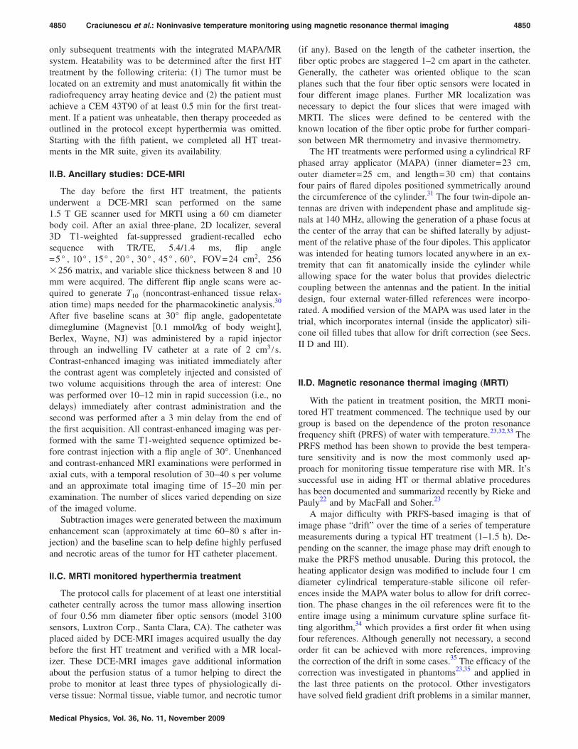
4850 Craciunescu et al.: Noninvasive temperature monitoring using magnetic resonance thermal imaging 4850
only subsequent treatments with the integrated MAPA/MRsystem. Heatability was to be determined after the first HTtreatment by the following criteria: �1� The tumor must belocated on an extremity and must anatomically fit within theradiofrequency array heating device and �2� the patient mustachieve a CEM 43T90 of at least 0.5 min for the first treat-ment. If a patient was unheatable, then therapy proceeded asoutlined in the protocol except hyperthermia was omitted.Starting with the fifth patient, we completed all HT treat-ments in the MR suite, given its availability.
II.B. Ancillary studies: DCE-MRI
The day before the first HT treatment, the patientsunderwent a DCE-MRI scan performed on the same1.5 T GE scanner used for MRTI using a 60 cm diameterbody coil. After an axial three-plane, 2D localizer, several3D T1-weighted fat-suppressed gradient-recalled echosequence with TR/TE, 5.4/1.4 ms, flip angle=5° , 10° , 15° , 20° , 30° , 45° , 60°, FOV=24 cm2, 256�256 matrix, and variable slice thickness between 8 and 10mm were acquired. The different flip angle scans were ac-quired to generate T10 �noncontrast-enhanced tissue relax-ation time� maps needed for the pharmacokinetic analysis.30
After five baseline scans at 30° flip angle, gadopentetatedimeglumine �Magnevist �0.1 mmol/kg of body weight�,Berlex, Wayne, NJ� was administered by a rapid injectorthrough an indwelling IV catheter at a rate of 2 cm3 /s.Contrast-enhanced imaging was initiated immediately afterthe contrast agent was completely injected and consisted oftwo volume acquisitions through the area of interest: Onewas performed over 10–12 min in rapid succession �i.e., nodelays� immediately after contrast administration and thesecond was performed after a 3 min delay from the end ofthe first acquisition. All contrast-enhanced imaging was per-formed with the same T1-weighted sequence optimized be-fore contrast injection with a flip angle of 30°. Unenhancedand contrast-enhanced MRI examinations were performed inaxial cuts, with a temporal resolution of 30–40 s per volumeand an approximate total imaging time of 15–20 min perexamination. The number of slices varied depending on sizeof the imaged volume.
Subtraction images were generated between the maximumenhancement scan �approximately at time 60–80 s after in-jection� and the baseline scan to help define highly perfusedand necrotic areas of the tumor for HT catheter placement.
II.C. MRTI monitored hyperthermia treatment
The protocol calls for placement of at least one interstitialcatheter centrally across the tumor mass allowing insertionof four 0.56 mm diameter fiber optic sensors �model 3100sensors, Luxtron Corp., Santa Clara, CA�. The catheter wasplaced aided by DCE-MRI images acquired usually the daybefore the first HT treatment and verified with a MR local-izer. These DCE-MRI images gave additional informationabout the perfusion status of a tumor helping to direct theprobe to monitor at least three types of physiologically di-
verse tissue: Normal tissue, viable tumor, and necrotic tumorMedical Physics, Vol. 36, No. 11, November 2009
�if any�. Based on the length of the catheter insertion, thefiber optic probes are staggered 1–2 cm apart in the catheter.Generally, the catheter was oriented oblique to the scanplanes such that the four fiber optic sensors were located infour different image planes. Further MR localization wasnecessary to depict the four slices that were imaged withMRTI. The slices were defined to be centered with theknown location of the fiber optic probe for further compari-son between MR thermometry and invasive thermometry.
The HT treatments were performed using a cylindrical RFphased array applicator �MAPA� �inner diameter=23 cm,outer diameter=25 cm, and length=30 cm� that containsfour pairs of flared dipoles positioned symmetrically aroundthe circumference of the cylinder.31 The four twin-dipole an-tennas are driven with independent phase and amplitude sig-nals at 140 MHz, allowing the generation of a phase focus atthe center of the array that can be shifted laterally by adjust-ment of the relative phase of the four dipoles. This applicatorwas intended for heating tumors located anywhere in an ex-tremity that can fit anatomically inside the cylinder whileallowing space for the water bolus that provides dielectriccoupling between the antennas and the patient. In the initialdesign, four external water-filled references were incorpo-rated. A modified version of the MAPA was used later in thetrial, which incorporates internal �inside the applicator� sili-cone oil filled tubes that allow for drift correction �see Secs.II D and III�.
II.D. Magnetic resonance thermal imaging „MRTI…
With the patient in treatment position, the MRTI moni-tored HT treatment commenced. The technique used by ourgroup is based on the dependence of the proton resonancefrequency shift �PRFS� of water with temperature.23,32,33 ThePRFS method has been shown to provide the best tempera-ture sensitivity and is now the most commonly used ap-proach for monitoring tissue temperature rise with MR. It’ssuccessful use in aiding HT or thermal ablative procedureshas been documented and summarized recently by Rieke andPauly22 and by MacFall and Soher.23
A major difficulty with PRFS-based imaging is that ofimage phase “drift” over the time of a series of temperaturemeasurements during a typical HT treatment �1–1.5 h�. De-pending on the scanner, the image phase may drift enough tomake the PRFS method unusable. During this protocol, theheating applicator design was modified to include four 1 cmdiameter cylindrical temperature-stable silicone oil refer-ences inside the MAPA water bolus to allow for drift correc-tion. The phase changes in the oil references were fit to theentire image using a minimum curvature spline surface fit-ting algorithm,34 which provides a first order fit when usingfour references. Although generally not necessary, a secondorder fit can be achieved with more references, improvingthe correction of the drift in some cases.35 The efficacy of thecorrection was investigated in phantoms23,35 and applied inthe last three patients on the protocol. Other investigators
have solved field gradient drift problems in a similar manner,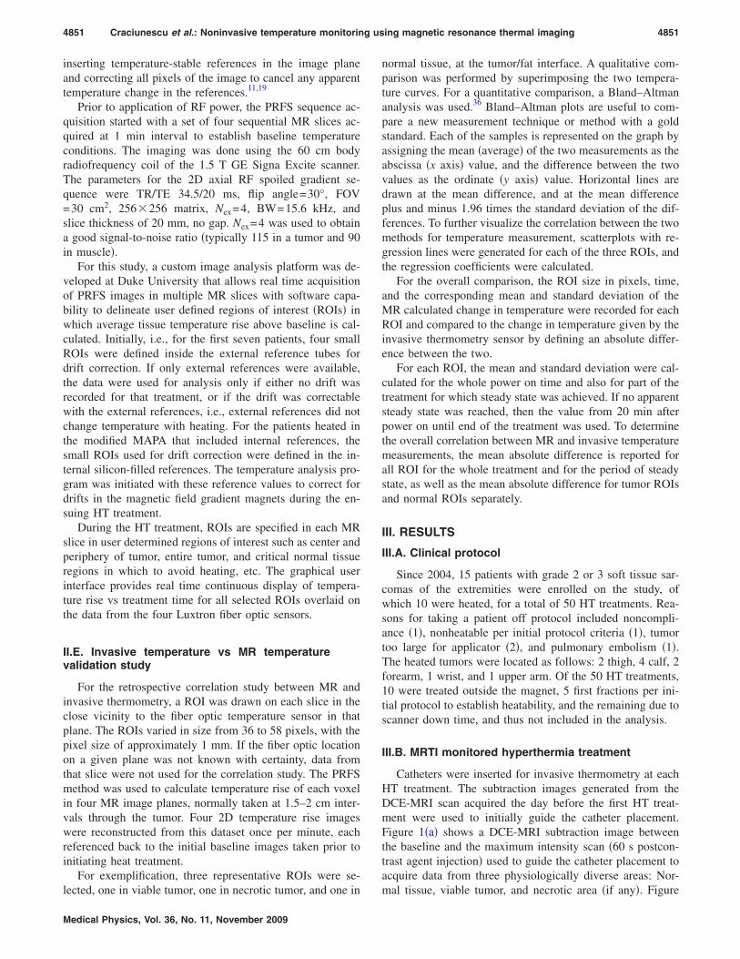
4851 Craciunescu et al.: Noninvasive temperature monitoring using magnetic resonance thermal imaging 4851
inserting temperature-stable references in the image planeand correcting all pixels of the image to cancel any apparenttemperature change in the references.11,19
Prior to application of RF power, the PRFS sequence ac-quisition started with a set of four sequential MR slices ac-quired at 1 min interval to establish baseline temperatureconditions. The imaging was done using the 60 cm bodyradiofrequency coil of the 1.5 T GE Signa Excite scanner.The parameters for the 2D axial RF spoiled gradient se-quence were TR/TE 34.5/20 ms, flip angle=30°, FOV=30 cm2, 256�256 matrix, Nex=4, BW=15.6 kHz, andslice thickness of 20 mm, no gap. Nex=4 was used to obtaina good signal-to-noise ratio �typically 115 in a tumor and 90in muscle�.
For this study, a custom image analysis platform was de-veloped at Duke University that allows real time acquisitionof PRFS images in multiple MR slices with software capa-bility to delineate user defined regions of interest �ROIs� inwhich average tissue temperature rise above baseline is cal-culated. Initially, i.e., for the first seven patients, four smallROIs were defined inside the external reference tubes fordrift correction. If only external references were available,the data were used for analysis only if either no drift wasrecorded for that treatment, or if the drift was correctablewith the external references, i.e., external references did notchange temperature with heating. For the patients heated inthe modified MAPA that included internal references, thesmall ROIs used for drift correction were defined in the in-ternal silicon-filled references. The temperature analysis pro-gram was initiated with these reference values to correct fordrifts in the magnetic field gradient magnets during the en-suing HT treatment.
During the HT treatment, ROIs are specified in each MRslice in user determined regions of interest such as center andperiphery of tumor, entire tumor, and critical normal tissueregions in which to avoid heating, etc. The graphical userinterface provides real time continuous display of tempera-ture rise vs treatment time for all selected ROIs overlaid onthe data from the four Luxtron fiber optic sensors.
II.E. Invasive temperature vs MR temperaturevalidation study
For the retrospective correlation study between MR andinvasive thermometry, a ROI was drawn on each slice in theclose vicinity to the fiber optic temperature sensor in thatplane. The ROIs varied in size from 36 to 58 pixels, with thepixel size of approximately 1 mm. If the fiber optic locationon a given plane was not known with certainty, data fromthat slice were not used for the correlation study. The PRFSmethod was used to calculate temperature rise of each voxelin four MR image planes, normally taken at 1.5–2 cm inter-vals through the tumor. Four 2D temperature rise imageswere reconstructed from this dataset once per minute, eachreferenced back to the initial baseline images taken prior toinitiating heat treatment.
For exemplification, three representative ROIs were se-
lected, one in viable tumor, one in necrotic tumor, and one inMedical Physics, Vol. 36, No. 11, November 2009
normal tissue, at the tumor/fat interface. A qualitative com-parison was performed by superimposing the two tempera-ture curves. For a quantitative comparison, a Bland–Altmananalysis was used.36 Bland–Altman plots are useful to com-pare a new measurement technique or method with a goldstandard. Each of the samples is represented on the graph byassigning the mean �average� of the two measurements as theabscissa �x axis� value, and the difference between the twovalues as the ordinate �y axis� value. Horizontal lines aredrawn at the mean difference, and at the mean differenceplus and minus 1.96 times the standard deviation of the dif-ferences. To further visualize the correlation between the twomethods for temperature measurement, scatterplots with re-gression lines were generated for each of the three ROIs, andthe regression coefficients were calculated.
For the overall comparison, the ROI size in pixels, time,and the corresponding mean and standard deviation of theMR calculated change in temperature were recorded for eachROI and compared to the change in temperature given by theinvasive thermometry sensor by defining an absolute differ-ence between the two.
For each ROI, the mean and standard deviation were cal-culated for the whole power on time and also for part of thetreatment for which steady state was achieved. If no apparentsteady state was reached, then the value from 20 min afterpower on until end of the treatment was used. To determinethe overall correlation between MR and invasive temperaturemeasurements, the mean absolute difference is reported forall ROI for the whole treatment and for the period of steadystate, as well as the mean absolute difference for tumor ROIsand normal ROIs separately.
III. RESULTS
III.A. Clinical protocol
Since 2004, 15 patients with grade 2 or 3 soft tissue sar-comas of the extremities were enrolled on the study, ofwhich 10 were heated, for a total of 50 HT treatments. Rea-sons for taking a patient off protocol included noncompli-ance �1�, nonheatable per initial protocol criteria �1�, tumortoo large for applicator �2�, and pulmonary embolism �1�.The heated tumors were located as follows: 2 thigh, 4 calf, 2forearm, 1 wrist, and 1 upper arm. Of the 50 HT treatments,10 were treated outside the magnet, 5 first fractions per ini-tial protocol to establish heatability, and the remaining due toscanner down time, and thus not included in the analysis.
III.B. MRTI monitored hyperthermia treatment
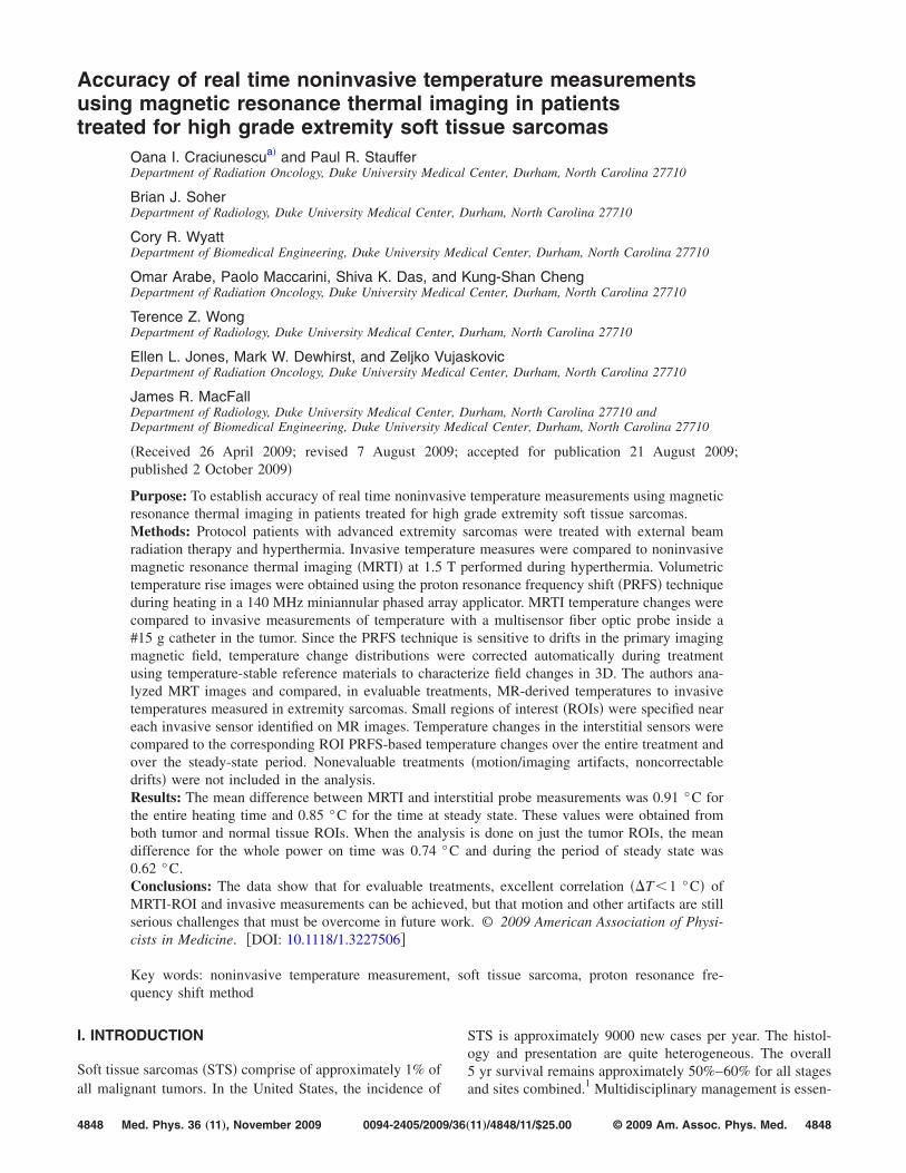
Catheters were inserted for invasive thermometry at eachHT treatment. The subtraction images generated from theDCE-MRI scan acquired the day before the first HT treat-ment were used to initially guide the catheter placement.Figure 1�a� shows a DCE-MRI subtraction image betweenthe baseline and the maximum intensity scan �60 s postcon-trast agent injection� used to guide the catheter placement toacquire data from three physiologically diverse areas: Nor-
mal tissue, viable tumor, and necrotic area �if any�. Figure
4852 Craciunescu et al.: Noninvasive temperature monitoring using magnetic resonance thermal imaging 4852
1�b� shows an image of a catheter placement in one of thepatients with a RT thigh high grade STS. A postcatheter in-sertion MR verification is shown in Fig. 1�c�, depicting thecatheter spanning the three areas to be monitored: Normaltissue, viable tumor, and necrotic core.
The MR localization scans performed in the early patientshad variable success in identifying exact locations of thecatheter relative to tumor as the air filled catheter did notcontrast well against many tissues. An adjustment to the scanprocedure in later patients provided significantly improvedvisualization of the catheter by inserting a smaller diametercatheter filled with gadolinium for the preocalization scan.This facilitated precise localization of the catheter in the tu-mor and subsequent calculation of the position of the fourstaggered Luxtron sensing points.
From the first ten patients that underwent HT treatmentsunder this protocol, seven were treated using the originalMAPA design and three treated with the drift compensation-ready MAPA.
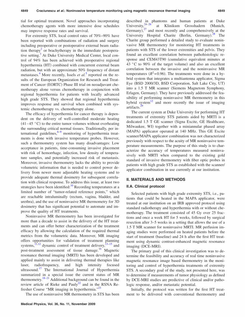
Examples of patients with extremity STS are shown inFigs. 2�a�–2�c�, showing the tumor bearing extremity in-serted into the cylindrical MAPA applicator with the tumorcentered within the four twin-dipole antennas. For the firstcohort of patients, the bolus was filled completely around theextremity with de-ionized water at 42 °C. The patient wasthen moved into the MR magnet, RF power cables attached,and input impedance parameters measured for the four an-tennas. Figure 2�c� shows the integration of the patient andMAPA applicator into a 1.5 T GE scanner for temperaturemonitoring using MRTI.
Following the initial baseline scans, approximately 20–50W of power at 140 MHz was applied to each of the four
FIG. 1. �a� DCE-MRI subtraction image between the baseline and the maxi-mum intensity scan �60 s postcontrast agent injection� used to guide thecatheter placement to acquire data from three physiologically diverse areas:Normal tissue, viable tumor, and necrotic tumor �if any�. �b� Image showingcatheter placement in one of the patients with a right thigh high grade STS.�c� Postcatheter insertion MR verification, showing the catheter spanning thethree areas to be monitored: Normal tissue, viable tumor, and necrotic core.
FIG. 2. �a� MAPA positioning for a patient with a lower extremity STS. Tapeshown on patient’s leg holds a single invasive #15 g catheter in place whichextends diagonally across the tumor volume and is filled with four stationaryLuxtron 3100 fiber optic sensors spaced 1.5 cm apart along the catheter�seen better in Fig. 1�b��;. �b� MAPA positioning for a patient with upperextremity STS. �c� Patient inside MAPA and 1.5 T MR scanner ready for
treatment.Medical Physics, Vol. 36, No. 11, November 2009
antennas, initially with equal phases and amplitudes to pro-duce a central zone of heating deep in the extremity. As thetissue is heated, the MR imaging of four slices through thetumor continued at 1 min interval. A drift correction mecha-nism was implemented in our MRTI treatment console. Anexample of a treatment where images were corrected usingthis method is shown elsewhere.23
The potential of using the real time MRTI data to guidethe treatment is demonstrated in Fig. 3. During treatment, thetumor temperature as recorded by both the invasive probe,and tumor ROI was rising but the MR image showed signifi-cant temperature rise in the normal muscle tissue as well. At18 min into the treatment, the relative phases of the fourantennas were adjusted to refocus heating away from thenormal tissue and into the tumor. The net effect of thesephase adjustments is seen by rising temperatures in the inva-sive tumor sensors as well as enhanced differential tempera-ture change in the tumor vs normal tissue ROIs on the MRTIimage. Figure 3 shows the initial baseline image �c�, fol-lowed by a diffuse heating pattern developing after 18 min ofcentral focus with 0,0,0,0 phases �d�, and finally the effectiveshifting of heat focus into the tumor by 25 min into treatmentdue to the change in relative antenna phases to 0,30,60,30.37
The steering of power is possible in our setting as the imagetransfer from the MR console to the analysis software is fast�within a minute of acquisition�. A few other minutes areneeded to analyze the images and determine the need forsteering. Once a sufficient number of scans have been ac-quired to decide the need for steering, changes in phase andamplitude can be implemented and the results displayedwithin a few minutes, giving the whole procedure a real time
FIG. 3. Summary of HT treatment: �a� Axial MRI image of the patient’slower extremity inside the MAPA applicator as seen in Fig. 2�a�. �b� Tem-perature data as a function of time for two regions of interest: Muscle�normal tissue� ROI and tumor ROI. �c� Baseline temperature change mapafter 4 min of imaging without application of heat, temperatures in the legare stable at about 36 °C. �d� Temperature rise image at approximately 18min into treatment heating of tumor is visible but is less than undesirableheating of normal tissue on the upper left side of leg. �e� 25 min intotreatment, after adjusting the antenna phases, heat is focused primarily in thetumor region creating a large differential heating of tumor.
characteristic.
4853 Craciunescu et al.: Noninvasive temperature monitoring using magnetic resonance thermal imaging 4853
III.C. Invasive temperature vs MR temperaturevalidation study
We analyzed MRTI images and compared to invasivetemperatures measured in extremity sarcomas over 40 heattreatments. The validation study was performed with datafrom 6/10 heated patients. Studies were discarded from thisanalysis due to lack of MR information in HT treatments
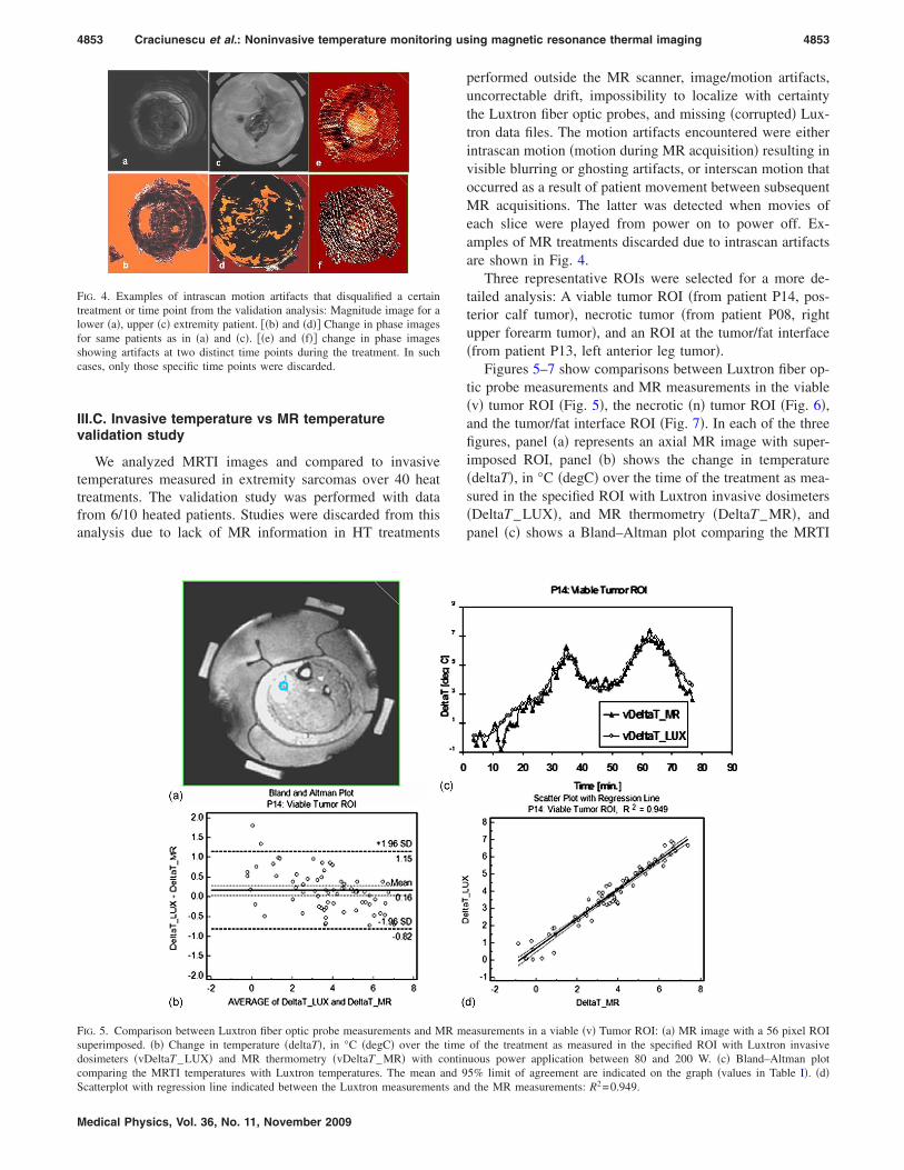
FIG. 4. Examples of intrascan motion artifacts that disqualified a certaintreatment or time point from the validation analysis: Magnitude image for alower �a�, upper �c� extremity patient. ��b� and �d�� Change in phase imagesfor same patients as in �a� and �c�. ��e� and �f�� change in phase imagesshowing artifacts at two distinct time points during the treatment. In suchcases, only those specific time points were discarded.
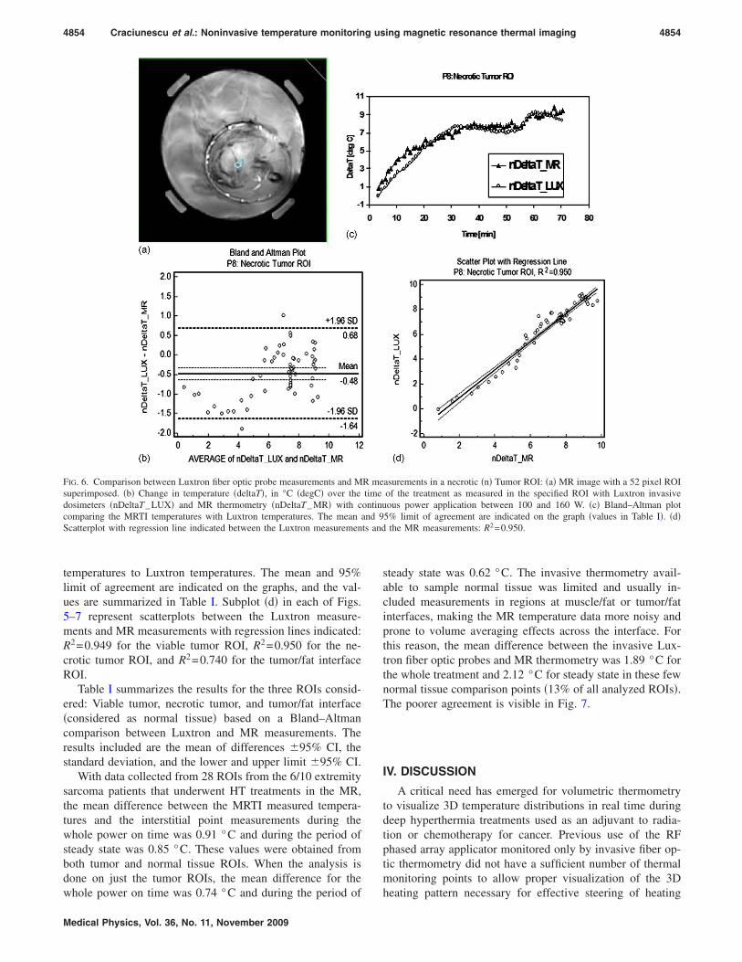
FIG. 5. Comparison between Luxtron fiber optic probe measurements and Msuperimposed. �b� Change in temperature �deltaT�, in °C �degC� over thedosimeters �vDeltaT_LUX� and MR thermometry �vDeltaT_MR� with ccomparing the MRTI temperatures with Luxtron temperatures. The mean a
Scatterplot with regression line indicated between the Luxtron measurements andMedical Physics, Vol. 36, No. 11, November 2009
performed outside the MR scanner, image/motion artifacts,uncorrectable drift, impossibility to localize with certaintythe Luxtron fiber optic probes, and missing �corrupted� Lux-tron data files. The motion artifacts encountered were eitherintrascan motion �motion during MR acquisition� resulting invisible blurring or ghosting artifacts, or interscan motion thatoccurred as a result of patient movement between subsequentMR acquisitions. The latter was detected when movies ofeach slice were played from power on to power off. Ex-amples of MR treatments discarded due to intrascan artifactsare shown in Fig. 4.
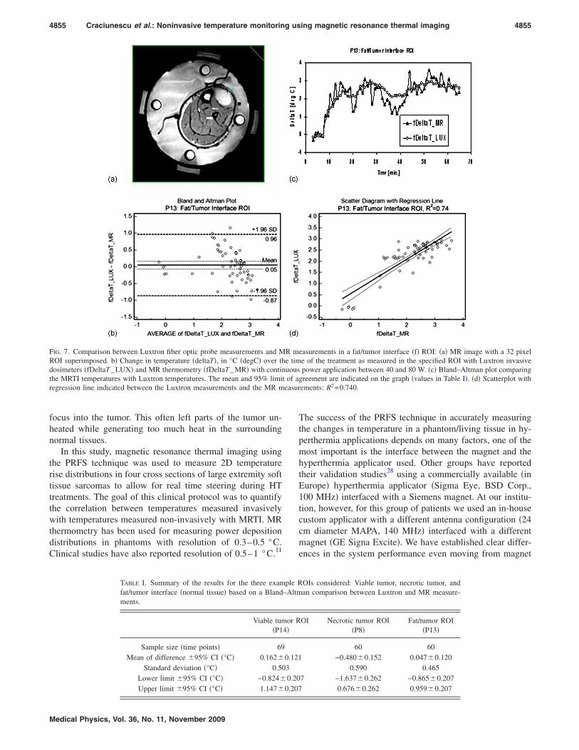
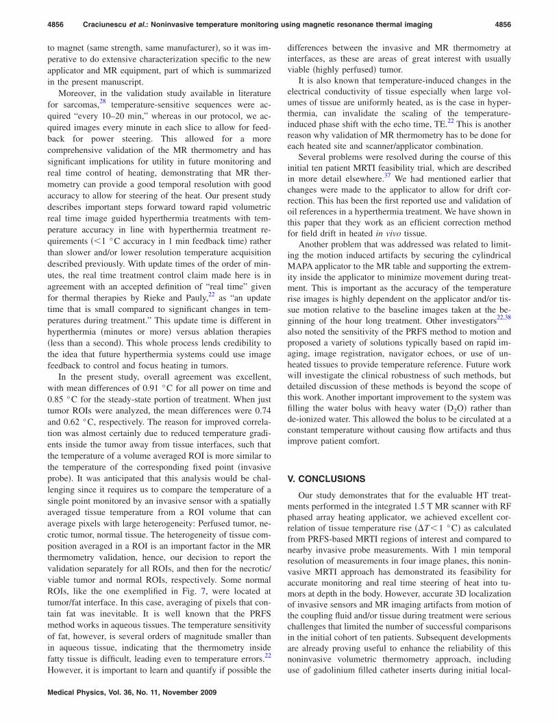
Three representative ROIs were selected for a more de-tailed analysis: A viable tumor ROI �from patient P14, pos-terior calf tumor�, necrotic tumor �from patient P08, rightupper forearm tumor�, and an ROI at the tumor/fat interface�from patient P13, left anterior leg tumor�.
Figures 5–7 show comparisons between Luxtron fiber op-tic probe measurements and MR measurements in the viable�v� tumor ROI �Fig. 5�, the necrotic �n� tumor ROI �Fig. 6�,and the tumor/fat interface ROI �Fig. 7�. In each of the threefigures, panel �a� represents an axial MR image with super-imposed ROI, panel �b� shows the change in temperature�deltaT�, in °C �degC� over the time of the treatment as mea-sured in the specified ROI with Luxtron invasive dosimeters�DeltaT_LUX�, and MR thermometry �DeltaT_MR�, andpanel �c� shows a Bland–Altman plot comparing the MRTI
easurements in a viable �v� Tumor ROI: �a� MR image with a 56 pixel ROIof the treatment as measured in the specified ROI with Luxtron invasiveuous power application between 80 and 200 W. �c� Bland–Altman plot5% limit of agreement are indicated on the graph �values in Table I�. �d�
2
R mtimeontinnd 9
the MR measurements: R =0.949.
ts and
4854 Craciunescu et al.: Noninvasive temperature monitoring using magnetic resonance thermal imaging 4854
temperatures to Luxtron temperatures. The mean and 95%limit of agreement are indicated on the graphs, and the val-ues are summarized in Table I. Subplot �d� in each of Figs.5–7 represent scatterplots between the Luxtron measure-ments and MR measurements with regression lines indicated:R2=0.949 for the viable tumor ROI, R2=0.950 for the ne-crotic tumor ROI, and R2=0.740 for the tumor/fat interfaceROI.
Table I summarizes the results for the three ROIs consid-ered: Viable tumor, necrotic tumor, and tumor/fat interface�considered as normal tissue� based on a Bland–Altmancomparison between Luxtron and MR measurements. Theresults included are the mean of differences �95% CI, thestandard deviation, and the lower and upper limit �95% CI.
With data collected from 28 ROIs from the 6/10 extremitysarcoma patients that underwent HT treatments in the MR,the mean difference between the MRTI measured tempera-tures and the interstitial point measurements during thewhole power on time was 0.91 °C and during the period ofsteady state was 0.85 °C. These values were obtained fromboth tumor and normal tissue ROIs. When the analysis isdone on just the tumor ROIs, the mean difference for the
FIG. 6. Comparison between Luxtron fiber optic probe measurements and Msuperimposed. �b� Change in temperature �deltaT�, in °C �degC� over thedosimeters �nDeltaT_LUX� and MR thermometry �nDeltaT_MR� with ccomparing the MRTI temperatures with Luxtron temperatures. The mean aScatterplot with regression line indicated between the Luxtron measuremen
whole power on time was 0.74 °C and during the period of
Medical Physics, Vol. 36, No. 11, November 2009
steady state was 0.62 °C. The invasive thermometry avail-able to sample normal tissue was limited and usually in-cluded measurements in regions at muscle/fat or tumor/fatinterfaces, making the MR temperature data more noisy andprone to volume averaging effects across the interface. Forthis reason, the mean difference between the invasive Lux-tron fiber optic probes and MR thermometry was 1.89 °C forthe whole treatment and 2.12 °C for steady state in these fewnormal tissue comparison points �13% of all analyzed ROIs�.The poorer agreement is visible in Fig. 7.
IV. DISCUSSION
A critical need has emerged for volumetric thermometryto visualize 3D temperature distributions in real time duringdeep hyperthermia treatments used as an adjuvant to radia-tion or chemotherapy for cancer. Previous use of the RFphased array applicator monitored only by invasive fiber op-tic thermometry did not have a sufficient number of thermalmonitoring points to allow proper visualization of the 3D
asurements in a necrotic �n� Tumor ROI: �a� MR image with a 52 pixel ROIof the treatment as measured in the specified ROI with Luxtron invasiveous power application between 100 and 160 W. �c� Bland–Altman plot
5% limit of agreement are indicated on the graph �values in Table I�. �d�the MR measurements: R2=0.950.
R metimeontinund 9
heating pattern necessary for effective steering of heating
4855 Craciunescu et al.: Noninvasive temperature monitoring using magnetic resonance thermal imaging 4855
focus into the tumor. This often left parts of the tumor un-heated while generating too much heat in the surroundingnormal tissues.
In this study, magnetic resonance thermal imaging usingthe PRFS technique was used to measure 2D temperaturerise distributions in four cross sections of large extremity softtissue sarcomas to allow for real time steering during HTtreatments. The goal of this clinical protocol was to quantifythe correlation between temperatures measured invasivelywith temperatures measured non-invasively with MRTI. MRthermometry has been used for measuring power depositiondistributions in phantoms with resolution of 0.3–0.5 °C.Clinical studies have also reported resolution of 0.5–1 °C.11
FIG. 7. Comparison between Luxtron fiber optic probe measurements and MROI superimposed. b� Change in temperature �deltaT�, in °C �degC� over thdosimeters �fDeltaT_LUX� and MR thermometry �fDeltaT_MR� with continthe MRTI temperatures with Luxtron temperatures. The mean and 95% limiregression line indicated between the Luxtron measurements and the MR m
TABLE I. Summary of the results for the three examfat/tumor interface �normal tissue� based on a Blandments.
Viable tum�P1
Sample size �time points� 69Mean of difference �95% CI �°C� 0.162�
Standard deviation �°C� 0.50Lower limit �95% CI �°C� −0.824�
Upper limit �95% CI �°C� 1.147�
Medical Physics, Vol. 36, No. 11, November 2009
The success of the PRFS technique in accurately measuringthe changes in temperature in a phantom/living tissue in hy-perthermia applications depends on many factors, one of themost important is the interface between the magnet and thehyperthermia applicator used. Other groups have reportedtheir validation studies28 using a commercially available �inEurope� hyperthermia applicator �Sigma Eye, BSD Corp.,100 MHz� interfaced with a Siemens magnet. At our institu-tion, however, for this group of patients we used an in-housecustom applicator with a different antenna configuration �24cm diameter MAPA, 140 MHz� interfaced with a differentmagnet �GE Signa Excite�. We have established clear differ-ences in the system performance even moving from magnet
easurements in a fat/tumor interface �f� ROI: �a� MR image with a 32 pixele of the treatment as measured in the specified ROI with Luxtron invasivepower application between 40 and 80 W. �c� Bland–Altman plot comparing
greement are indicated on the graph �values in Table I�. �d� Scatterplot withements: R2=0.740.
OIs considered: Viable tumor, necrotic tumor, andan comparison between Luxtron and MR measure-
OI Necrotic tumor ROI�P8�
Fat/tumor ROI�P13�
60 60−0.480�0.152 0.047�0.120
0.590 0.4657 −1.637�0.262 −0.865�0.207
0.676�0.262 0.959�0.207
R me timuous
t of aeasur
ple R–Altm
or R4�
0.12130.20
0.207
4856 Craciunescu et al.: Noninvasive temperature monitoring using magnetic resonance thermal imaging 4856
to magnet �same strength, same manufacturer�, so it was im-perative to do extensive characterization specific to the newapplicator and MR equipment, part of which is summarizedin the present manuscript.
Moreover, in the validation study available in literaturefor sarcomas,28 temperature-sensitive sequences were ac-quired “every 10–20 min,” whereas in our protocol, we ac-quired images every minute in each slice to allow for feed-back for power steering. This allowed for a morecomprehensive validation of the MR thermometry and hassignificant implications for utility in future monitoring andreal time control of heating, demonstrating that MR ther-mometry can provide a good temporal resolution with goodaccuracy to allow for steering of the heat. Our present studydescribes important steps forward toward rapid volumetricreal time image guided hyperthermia treatments with tem-perature accuracy in line with hyperthermia treatment re-quirements ��1 °C accuracy in 1 min feedback time� ratherthan slower and/or lower resolution temperature acquisitiondescribed previously. With update times of the order of min-utes, the real time treatment control claim made here is inagreement with an accepted definition of “real time” givenfor thermal therapies by Rieke and Pauly,22 as “an updatetime that is small compared to significant changes in tem-peratures during treatment.” This update time is different inhyperthermia �minutes or more� versus ablation therapies�less than a second�. This whole process lends credibility tothe idea that future hyperthermia systems could use imagefeedback to control and focus heating in tumors.
In the present study, overall agreement was excellent,with mean differences of 0.91 °C for all power on time and0.85 °C for the steady-state portion of treatment. When justtumor ROIs were analyzed, the mean differences were 0.74and 0.62 °C, respectively. The reason for improved correla-tion was almost certainly due to reduced temperature gradi-ents inside the tumor away from tissue interfaces, such thatthe temperature of a volume averaged ROI is more similar tothe temperature of the corresponding fixed point �invasiveprobe�. It was anticipated that this analysis would be chal-lenging since it requires us to compare the temperature of asingle point monitored by an invasive sensor with a spatiallyaveraged tissue temperature from a ROI volume that canaverage pixels with large heterogeneity: Perfused tumor, ne-crotic tumor, normal tissue. The heterogeneity of tissue com-position averaged in a ROI is an important factor in the MRthermometry validation, hence, our decision to report thevalidation separately for all ROIs, and then for the necrotic/viable tumor and normal ROIs, respectively. Some normalROIs, like the one exemplified in Fig. 7, were located attumor/fat interface. In this case, averaging of pixels that con-tain fat was inevitable. It is well known that the PRFSmethod works in aqueous tissues. The temperature sensitivityof fat, however, is several orders of magnitude smaller thanin aqueous tissue, indicating that the thermometry insidefatty tissue is difficult, leading even to temperature errors.22
However, it is important to learn and quantify if possible the
Medical Physics, Vol. 36, No. 11, November 2009
differences between the invasive and MR thermometry atinterfaces, as these are areas of great interest with usuallyviable �highly perfused� tumor.
It is also known that temperature-induced changes in theelectrical conductivity of tissue especially when large vol-umes of tissue are uniformly heated, as is the case in hyper-thermia, can invalidate the scaling of the temperature-induced phase shift with the echo time, TE.22 This is anotherreason why validation of MR thermometry has to be done foreach heated site and scanner/applicator combination.
Several problems were resolved during the course of thisinitial ten patient MRTI feasibility trial, which are describedin more detail elsewhere.37 We had mentioned earlier thatchanges were made to the applicator to allow for drift cor-rection. This has been the first reported use and validation ofoil references in a hyperthermia treatment. We have shown inthis paper that they work as an efficient correction methodfor field drift in heated in vivo tissue.
Another problem that was addressed was related to limit-ing the motion induced artifacts by securing the cylindricalMAPA applicator to the MR table and supporting the extrem-ity inside the applicator to minimize movement during treat-ment. This is important as the accuracy of the temperaturerise images is highly dependent on the applicator and/or tis-sue motion relative to the baseline images taken at the be-ginning of the hour long treatment. Other investigators22,38
also noted the sensitivity of the PRFS method to motion andproposed a variety of solutions typically based on rapid im-aging, image registration, navigator echoes, or use of un-heated tissues to provide temperature reference. Future workwill investigate the clinical robustness of such methods, butdetailed discussion of these methods is beyond the scope ofthis work. Another important improvement to the system wasfilling the water bolus with heavy water �D2O� rather thande-ionized water. This allowed the bolus to be circulated at aconstant temperature without causing flow artifacts and thusimprove patient comfort.
V. CONCLUSIONS
Our study demonstrates that for the evaluable HT treat-ments performed in the integrated 1.5 T MR scanner with RFphased array heating applicator, we achieved excellent cor-relation of tissue temperature rise ��T�1 °C� as calculatedfrom PRFS-based MRTI regions of interest and compared tonearby invasive probe measurements. With 1 min temporalresolution of measurements in four image planes, this nonin-vasive MRTI approach has demonstrated its feasibility foraccurate monitoring and real time steering of heat into tu-mors at depth in the body. However, accurate 3D localizationof invasive sensors and MR imaging artifacts from motion ofthe coupling fluid and/or tissue during treatment were seriouschallenges that limited the number of successful comparisonsin the initial cohort of ten patients. Subsequent developmentsare already proving useful to enhance the reliability of thisnoninvasive volumetric thermometry approach, including
use of gadolinium filled catheter inserts during initial local-
4857 Craciunescu et al.: Noninvasive temperature monitoring using magnetic resonance thermal imaging 4857
ization scans for more precise probe localization, D2O as azero signal coupling fluid, and applicator stabilizationmounts to restrict patient movement.
As MRTI technology continues to mature, it offers oppor-tunities for more than just temperature measurement. It canprovide volumetric thermal data that can be correlated withvolumetric physiological parameters as established with im-aging, like perfusion, cell kill, and hypoxia. By monitoringreal time changes in temperature during heating, MRTI canprovide critical feedback for dynamic control of treatmentdelivery.
This type of high precision thermometry demonstratedhere is a very useful result to the hyperthermia community tosupport continued development of hyperthermia systems thatwill rely on monitoring and control noninvasively rather thanfrom a restricted set of implanted sensors.
ACKNOWLEDGMENT
This work was supported by NCI Grant No. CA42745.
a�Author to whom correspondence should be addressed. Electronic mail:[email protected]; telephone: �919� 660-2192.
1B. C. P. O’Sullivan, C. Euler, and C. Catton, “Soft tissue sarcoma,” inClinical Radiation Oncology, edited by G. a. Tepper �Churchill Living-stone Elsevier, Philadelphia, PA, 2007�, Vol. 1, pp. 1519–1550.
2H. D. Suit, H. J. Mankin, W. C. Wood, M. C. Gebhardt, D. C. Harmon, A.Rosenberg, J. E. Tepper, and D. Rosenthal, “Treatment of the patient withstage M0 soft tissue sarcoma,” J. Clin. Oncol. 6, 854–862 �1988�.
3L. B. Harrison, F. Franzese, J. J. Gaynor, and M. F. Brennan, “Long-termresults of a prospective randomized trial of adjuvant brachytherapy in themanagement of completely resected soft tissue sarcomas of the extremityand superficial trunk,” Int. J. Radiat. Oncol., Biol., Phys. 27, 259–265�1993�.
4L. R. Prosnitz, P. Maguire, J. M. Anderson, S. P. Scully, J. M. Harrelson,E. L. Jones, M. Dewhirst, T. V. Samulski, B. E. Powers, G. L. Rosner, R.K. Dodge, L. Layfield, R. Clough, and D. M. Brizel, “The treatment ofhigh-grade soft tissue sarcomas with preoperative thermoradiotherapy,”Int. J. Radiat. Oncol., Biol., Phys. 45, 941–949 �1999�.
5R. D. Issels, L. H. Lindner, P. Wust, P. Hohenberger, K. Jauch, S.Daugaard, U. Mansmann, W. Hiddemann, J. Blay, and J. Verweij, “Re-gional hyperthermia �RHT� improves response and survival when com-bined with systemic chemotherapy in the management of locally ad-vanced, high grade soft tissue sarcomas �STS� of the extremities, the bodywall and the abdomen: A phase II randomised pros,” J. Clin. Oncol. 25,10009 �2007�.
6J. J. Lagendijk, G. C. Van Rhoon, S. N. Hornsleth, P. Wust, A. C. DeLeeuw, C. J. Schneider, J. D. Van Dijk, J. Van Der Zee, R. Van Heek-Romanowski, S. A. Rahman, and C. Gromoll, “ESHO quality assuranceguidelines for regional hyperthermia,” Int. J. Hyperthermia 14, 125–133�1998�.
7M. W. Dewhirst et al., “RTOG quality assurance guidelines for clinicaltrials using hyperthermia,” Int. J. Radiat. Oncol., Biol., Phys. 18, 1249–1259 �1990�.
8B. Emami et al., “RTOG quality assurance guidelines for interstitial hy-perthermia,” Int. J. Radiat. Oncol., Biol., Phys. 20, 1117–1124 �1991�.
9M. D. Sapozink et al., “RTOG quality assurance guidelines for clinicaltrials using hyperthermia for deep-seated malignancy,” Int. J. Radiat. On-col., Biol., Phys. 20, 1109–1115 �1991�.
10P. Wust, C. H. Cho, B. Hildebrandt, and J. Gellermann, “Thermal moni-toring: Invasive, minimal-invasive and non-invasive approaches,” Int. J.Hyperthermia 22, 255–262 �2006�.
11J. Gellermann, M. Weihrauch, C. H. Cho, W. Wlodarczyk, H. Fahling, R.Felix, V. Budach, M. Weiser, J. Nadobny, and P. Wust, “Comparison ofMR-thermography and planning calculations in phantoms,” Med. Phys.33, 3912–3920 �2006�.
12J. Gellermann, W. Wlodarczyk, H. Ganter, J. Nadobny, H. Fahling, M.
Seebass, R. Felix, and P. Wust, “A practical approach to thermography inMedical Physics, Vol. 36, No. 11, November 2009
a hyperthermia/magnetic resonance hybrid system: Validation in a hetero-geneous phantom,” Int. J. Radiat. Oncol., Biol., Phys. 61, 267–277�2005�.
13E. Hutchinson, M. Dahleh, and K. Hynynen, “The feasibility of MRIfeedback control for intracavitary phased array hyperthermia treatments,”Int. J. Hyperthermia 14, 39–56 �1998�.
14K. S. Cheng, V. Stakhursky, O. I. Craciunescu, P. Stauffer, M. Dewhirst,and S. K. Das, “Fast temperature optimization of multi-source hyperther-mia applicators with reduced-order modeling of ‘virtual sources’,” Phys.Med. Biol. 53, 1619–1635 �2008�.
15V. L. Stakhursky, O. Arabe, K. S. Cheng, J. Macfall, P. Maccarini, O.Craciunescu, M. Dewhirst, P. Stauffer, and S. K. Das, “Real-time MRI-guided hyperthermia treatment using a fast adaptive algorithm,” Phys.Med. Biol. 54, 2131–2145 �2009�.
16H. E. Cline, K. Hynynen, E. Schneider, C. J. Hardy, S. E. Maier, R. D.Watkins, and F. A. Jolesz, “Simultaneous magnetic resonance phase andmagnitude temperature maps in muscle,” Magn. Reson. Med. 35, 309–315 �1996�.
17G. C. van Rhoon and P. Wust, “Introduction: Non-invasive thermometryfor thermotherapy,” Int. J. Hyperthermia 21, 489–495 �2005�.
18B. Denis de Senneville, B. Quesson, and C. T. Moonen, “Magnetic reso-nance temperature imaging,” Int. J. Hyperthermia 21, 515–531 �2005�.
19J. Gellermann, W. Wlodarczyk, A. Feussner, H. Fahling, J. Nadobny, B.Hildebrandt, R. Felix, and P. Wust, “Methods and potentials of magneticresonance imaging for monitoring radiofrequency hyperthermia in a hy-brid system,” Int. J. Hyperthermia 21, 497–513 �2005�.
20K. Kuroda, “Non-invasive MR thermography using the water protonchemical shift,” Int. J. Hyperthermia 21, 547–560 �2005�.
21N. McDannold, “Quantitative MRI-based temperature mapping based onthe proton resonant frequency shift: Review of validation studies,” Int. J.Hyperthermia 21, 533–546 �2005�.
22V. Rieke and K. B. Pauly, “MR thermometry,” J. Magn. Reson Imaging27, 376–390 �2008�.
23J. R. MacFall and B. J. Soher, “From the RSNA refresher courses: MRimaging in hyperthermia,” Radiographics 27, 1809–1818 �2007�.
24D. L. Carter, J. R. MacFall, S. T. Clegg, X. Wan, D. M. Prescott, H. C.Charles, and T. V. Samulski, “Magnetic resonance thermometry duringhyperthermia for human high-grade sarcoma,” Int. J. Radiat. Oncol.,Biol., Phys. 40, 815–822 �1998�.
25O. I. Craciunescu, S. K. Das, R. L. McCauley, J. R. MacFall, and T. V.Samulski, “3D numerical reconstruction of the hyperthermia inducedtemperature distribution in human sarcomas using DE-MRI measured tis-sue perfusion: Validation against non-invasive MR temperature measure-ments,” Int. J. Hyperthermia 17, 221–239 �2001�.
26O. I. Craciunescu, T. V. Samulski, J. R. MacFall, and S. T. Clegg, “Per-turbations in hyperthermia temperature distributions associated withcounter-current flow: Numerical simulations and empirical verification,”IEEE Trans. Biomed. Eng. 47, 435–443 �2000�.
27M. Peller, H. M. Reinl, A. Weigel, M. Meininger, R. D. Issels, and M.Reiser, “T1 relaxation time at 0.2 Tesla for monitoring regional hyper-thermia: Feasibility study in muscle and adipose tissue,” Magn. Reson.Med. 47, 1194–1201 �2002�.
28J. Gellermann, B. Hildebrandt, R. Issels, H. Ganter, W. Wlodarczyk, V.Budach, R. Felix, P. U. Tunn, P. Reichardt, and P. Wust, “Noninvasivemagnetic resonance thermography of soft tissue sarcomas during regionalhyperthermia: Correlation with response and direct thermometry,” Cancer107, 1373–1382 �2006�.
29J. Gellermann, H. Faehling, M. Mielec, C. H. Cho, V. Budach, and P.Wust, “Image artifacts during MRT hybrid hyperthermia—Causes andelimination,” Int. J. Hyperthermia 24, 327–335 �2008�.
30B. M. Dale, J. A. Jesberger, J. S. Lewin, C. M. Hillenbrand, and J. L.Duerk, “Determining and optimizing the precision of quantitative mea-surements of perfusion from dynamic contrast enhanced MRI,” J. Magn.Reson Imaging 18, 575–584 �2003�.
31Y. Zhang, W. T. Joines, R. L. Jirtle, and T. V. Samulski, “Theoretical andmeasured electric field distributions within an annular phased array: Con-sideration of source antennas,” IEEE Trans. Biomed. Eng. 40, 780–787�1993�.
32Y. Ishihara, A. Calderon, H. Watanabe, K. Okamoto, Y. Suzuki, and K.Kuroda, “A precise and fast temperature mapping using water proton
chemical shift,” Magn. Reson. Med. 34, 814–823 �1995�.
4858 Craciunescu et al.: Noninvasive temperature monitoring using magnetic resonance thermal imaging 4858
33J. R. MacFall, D. M. Prescott, H. C. Charles, and T. V. Samulski, “1HMRI phase thermometry in vivo in canine brain, muscle, and tumor tis-sue,” Med. Phys. 23, 1775–1782 �1996�.
34R. Franke, “Smooth interpolation of scattered data by local thin platesplines,” Comput. Math. Appl. 8, 273–281 �1982�.
35C. Wyatt, B. Soher, V. Stakhursky, P. Maccarini, C. Charles, P. Stauffer,and J. MacFall, “Hyperthermia MRI temperature measurement: Evalua-tion of measurement stabilization strategies for extremity and breast tu-mors,” Int. J. Hyperthermia 25�6�, 422–433 �2009�.
36J. M. Bland and D. G. Altman, “Statistical methods for assessing agree-ment between two methods of clinical measurement,” Lancet 1, 307–310
Medical Physics, Vol. 36, No. 11, November 2009
�1986�.37P. Stauffer, O. Craciunescu, P. Maccarini, C. Wyatt, K. Arunachalam, O.
Arabe, V. Stakhursky, Z. Li, B. Soher, J. MacFall, S. Rangarao, K. S.Cheng, S. Das, M. W. Dewhirst, T. Wong, E. Jones, and Z. Vujaskovic,“Clinical utility of magnetic resonance thermal imaging �MRTI� for realtime guidance of deep hyperthermia,” Proc. SPIE 7181, 718101-1–718101-12 �2009�.
38B. D. de Senneville, C. Mougenot, and C. T. Moonen, “Real-time adap-tive methods for treatment of mobile organs by MRI-controlled high-intensity focused ultrasound,” Magn. Reson. Med. 57, 319–330 �2007�.
Related Documents