Copyright 1997 by The Journal of Bone and Joint Surgery, Incorporated Accelerated Healing of Distal Radial Fractures with the Use of Specific, Low-Intensity Ultrasound A MULTICENTER, PROSPECTIVE, RANDOMIZED, DOUBLE-BUND, PLACEBO-CONTROLLED STUDY* BY THOMAS K. KRISTIANSEN, M.D.f, BURLINGTON, JOHN P. RYABY*, JOAN McCABE, R.N.t, PISCATAWAY, NEW JERSEY, JOHN J. FREY, PH.D.§, LANCASTER, PENNSYLVANIA, AND LINDA R. ROE, M.D.I BURLINGTON, VERMONT Investigation performed at Community Health Care Plan, New Haven, Connecticut; Conway Hospital, Conway, South Carolina; Emory University, Atlanta, Georgia; Florida Medical Center, Plantation, Florida; Greenville Hospital System, Greenville, South Carolina; Haifa City Medical Center, Haifa, Israel; Mount Sinai Medical Center, New York, N Y; St. Luke's Hospital, New Bedford, Massachusetts; University of Vermont, Burlington, Vermont; and Community Medical Center, Scranton, Pennsylvania ABSTRACT: A multicenter, prospective, randomized, double-blind, placebo-controlled clinical trial was con- ducted to test the efficacy of a specifically programmed, low-intensity, non-thermal, pulsed ultrasound medical device for shortening the time to radiographic healing of dorsally angulated fractures (negative volar angula- tion) of the distal aspect of the radius that had been treated with manipulation and a cast. Sixty patients (sixty-one fractures) were enrolled in the study within seven days after the fracture. The patients used either an active ultrasound device (thirty fractures) or a pla- cebo device (thirty-one fractures) daily for twenty min- utes at home for ten weeks. The two types of devices were identical except that the placebo devices emit- ted no ultrasound energy. Clinical examination was per- formed and radiographs were made at one, two, three, four, five, six, eight, ten, twelve, and sixteen weeks after the fracture by each site investigator. The time to union was significantly shorter for the fractures that were treated with ultrasound than it was for those that were treated with the placebo (mean [and standard error], 61 ± 3 days compared with 98 ± 5 days; p < 0.0001). Each radiographic stage of healing also was significantly accelerated in the group that was treated with ultrasound as compared with that treated with the placebo. Compared with treatment with the placebo, treatment with ultrasound was associated with *One or more of the authors have received or will receive ben- efits for personal or professional use from a commercial party re- lated directly or indirectly to the subject of this article. Funds were received in total or partial support of the research or clinical study presented in this article. The funding source was Exogen, Incorpo- rated, Piscataway, New Jersey. tDepartment of Orthopaedics and Rehabilitation, College of Medicine, University of Vermont, Robert T. Stafford Hall, Room 426B, Burlington, Vermont 05405-0084. E-mail address for Dr. Kris- liansen: [email protected]. tExogen, Incorporated, 10 Constitution Avenue, Piscataway, New jersey 08855. E-mail address for Mr. Ryaby and Ms. McCabe: [email protected]. §Health Products Development, Incorporated, 12 Lark Lane, Lancaster, Pennsylvania 17603. 1 Department of Radiology, Fletcher Allen Health Care, Uni- versity of Vermont, Burlington, Vermont 05401. a significantly smaller loss of reduction (20 ± 6 per cent compared with 43 ± 8 per cent; p < 0.01), as determined by the degree of volar angulation, as well as with a significant decrease in the mean time until the loss of reduction ceased (12 ± 4 days compared with 25 ± 4 days; p < 0.04). We concluded that this specific ultra- sound signal accelerates the healing of fractures of the distal radial metaphysis and decreases the loss of re- duction during fracture-healing. Ultrasound is acoustic radiation at frequencies above the limit of human hearing. It is a form of mechanical energy that can be transmitted into the body as high- frequency acoustical pressure waves. The micromechani- cal strains produced by these pressure waves in body tissue can result in biochemical events at the cellular level 3529 and may promote bone formation in a man- ner comparable with the bone responses to mechanical stress postulated by Wolffs law 41 . Ultrasound currently is used both operatively and therapeutically; it achieves its biological results by considerably increasing the tem- perature of the tissue, with intensities ranging from 0.2 to 100 watts per square centimeter 1 '• 24 - 4(M7 . In contrast, safe intensities for diagnostic imaging are much lower (0.5 to fifty milliwatts per square centimeter) and are con- sidered non-thermal stimuli 35 . The ultrasound device used in the current study applied thirty milliwatts per square centimeter, in the range of diagnostic imaging, to the skin at the site of the fracture. Duarte 10 reported an acceleration of healing, as as- sessed radiographically and histologically, with use of a low-intensity ultrasound device, both at the site of a controlled fibular osteotomy and in a femoral drill-hole defect in a rabbit model. Studies of other animal models have demonstrated a bone response to low-intensity ultrasound, including the promotion of cartilage-related gene expression and increased rates of endochondral ossification' 9 - 2526363945 '"''. In vitro studies have suggested that low-intensity pulsed ultrasound produces signifi- cant multifunctional effects (p < 0.05) that are directly relevant to bone formation and resorption 303242 . VOL. 79-A, NO. 7, JULY 1997 961

Welcome message from author
This document is posted to help you gain knowledge. Please leave a comment to let me know what you think about it! Share it to your friends and learn new things together.
Transcript
Copyright 1997 by The Journal of Bone and Joint Surgery, Incorporated
Accelerated Healing of Distal Radial Fractures with the Use of Specific, Low-Intensity Ultrasound
A MULTICENTER, PROSPECTIVE, RANDOMIZED, D O U B L E - B U N D , PLACEBO-CONTROLLED STUDY*
BY THOMAS K. KRISTIANSEN, M.D.f, BURLINGTON, JOHN P. RYABY*, JOAN McCABE, R.N.t, PISCATAWAY, NEW JERSEY,
JOHN J. FREY, PH.D.§, LANCASTER, PENNSYLVANIA, AND LINDA R. ROE, M.D. I BURLINGTON, VERMONT
Investigation performed at Community Health Care Plan, New Haven, Connecticut; Conway Hospital, Conway, South Carolina; Emory University, Atlanta, Georgia; Florida Medical Center, Plantation, Florida; Greenville Hospital System, Greenville, South Carolina; Haifa City Medical Center, Haifa, Israel; Mount Sinai Medical Center, New York, N Y; St. Luke's Hospital, New Bedford, Massachusetts;
University of Vermont, Burlington, Vermont; and Community Medical Center, Scranton, Pennsylvania
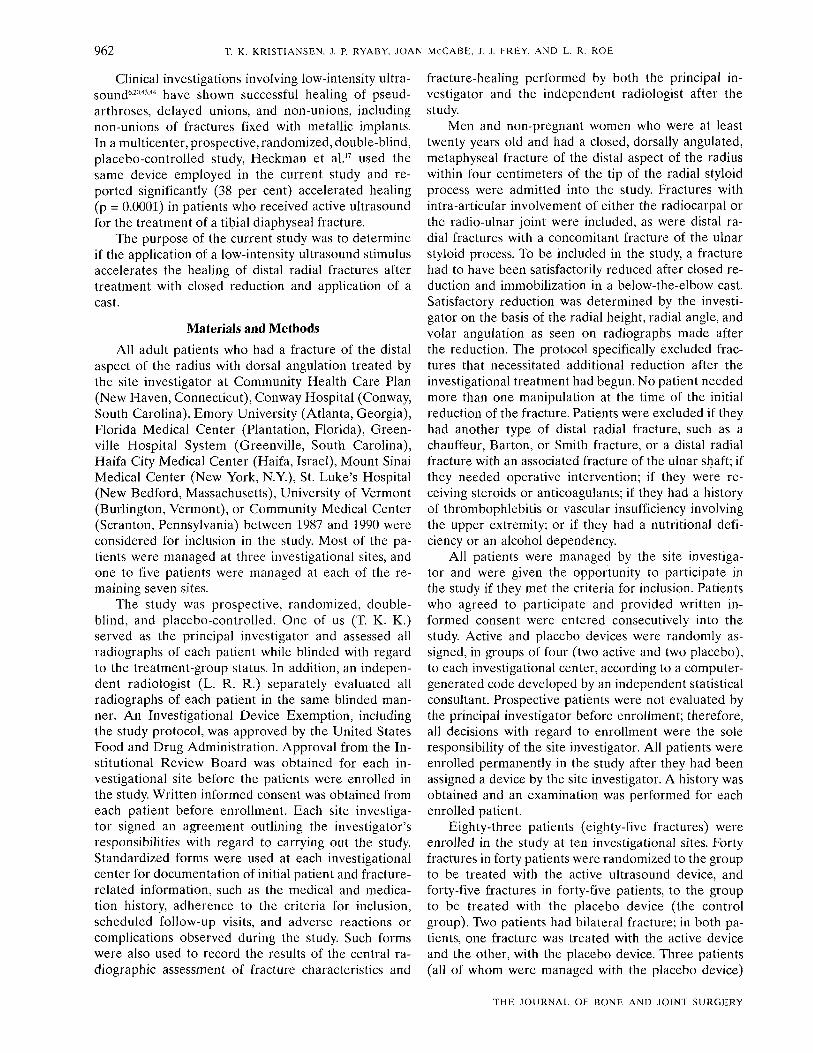
ABSTRACT: A multicenter, prospective, randomized, double-blind, placebo-controlled clinical trial was conducted to test the efficacy of a specifically programmed, low-intensity, non-thermal, pulsed ultrasound medical device for shortening the time to radiographic healing of dorsally angulated fractures (negative volar angulation) of the distal aspect of the radius that had been treated with manipulation and a cast. Sixty patients (sixty-one fractures) were enrolled in the study within seven days after the fracture. The patients used either an active ultrasound device (thirty fractures) or a placebo device (thirty-one fractures) daily for twenty minutes at home for ten weeks. The two types of devices were identical except that the placebo devices emitted no ultrasound energy. Clinical examination was performed and radiographs were made at one, two, three, four, five, six, eight, ten, twelve, and sixteen weeks after the fracture by each site investigator.
The time to union was significantly shorter for the fractures that were treated with ultrasound than it was for those that were treated with the placebo (mean [and standard error], 61 ± 3 days compared with 98 ± 5 days; p < 0.0001). Each radiographic stage of healing also was significantly accelerated in the group that was treated with ultrasound as compared with that treated with the placebo. Compared with treatment with the placebo, treatment with ultrasound was associated with
*One or more of the authors have received or will receive benefits for personal or professional use from a commercial party related directly or indirectly to the subject of this article. Funds were received in total or partial support of the research or clinical study presented in this article. The funding source was Exogen, Incorporated, Piscataway, New Jersey.
tDepartment of Orthopaedics and Rehabilitation, College of Medicine, University of Vermont, Robert T. Stafford Hall, Room 426B, Burlington, Vermont 05405-0084. E-mail address for Dr. Kris-liansen: [email protected].
tExogen, Incorporated, 10 Constitution Avenue, Piscataway, New jersey 08855. E-mail address for Mr. Ryaby and Ms. McCabe: [email protected].
§Health Products Development, Incorporated, 12 Lark Lane, Lancaster, Pennsylvania 17603.
1 Department of Radiology, Fletcher Allen Health Care, University of Vermont, Burlington, Vermont 05401.
a significantly smaller loss of reduction (20 ± 6 per cent compared with 43 ± 8 per cent; p < 0.01), as determined by the degree of volar angulation, as well as with a significant decrease in the mean time until the loss of reduction ceased (12 ± 4 days compared with 25 ± 4 days; p < 0.04). We concluded that this specific ultrasound signal accelerates the healing of fractures of the distal radial metaphysis and decreases the loss of reduction during fracture-healing.
Ultrasound is acoustic radiation at frequencies above the limit of human hearing. It is a form of mechanical energy that can be transmitted into the body as high-frequency acoustical pressure waves. The micromechani-cal strains produced by these pressure waves in body tissue can result in biochemical events at the cellular level3529 and may promote bone formation in a manner comparable with the bone responses to mechanical stress postulated by Wolffs law41. Ultrasound currently is used both operatively and therapeutically; it achieves its biological results by considerably increasing the temperature of the tissue, with intensities ranging from 0.2 to 100 watts per square centimeter1 '•24-4(M7. In contrast, safe intensities for diagnostic imaging are much lower (0.5 to fifty milliwatts per square centimeter) and are considered non-thermal stimuli35. The ultrasound device used in the current study applied thirty milliwatts per square centimeter, in the range of diagnostic imaging, to the skin at the site of the fracture.
Duarte10 reported an acceleration of healing, as assessed radiographically and histologically, with use of a low-intensity ultrasound device, both at the site of a controlled fibular osteotomy and in a femoral drill-hole defect in a rabbit model. Studies of other animal models have demonstrated a bone response to low-intensity ultrasound, including the promotion of cartilage-related gene expression and increased rates of endochondral ossification'9-2526363945'"''. In vitro studies have suggested that low-intensity pulsed ultrasound produces significant multifunctional effects (p < 0.05) that are directly relevant to bone formation and resorption303242.
VOL. 79-A, NO. 7, JULY 1997 961
962 T. K. KRISTIANSEN, J. P. RYABY, JOAN McCABE, J. J. FREY, AND L. R. ROE
Clinical investigations involving low-intensity ultra-sound621"41-44 have shown successful healing of pseud-arthroses, delayed unions, and non-unions, including non-unions of fractures fixed with metallic implants. In a multicenter, prospective, randomized, double-blind, placebo-controlled study, Heckman et al." used the same device employed in the current study and reported significantly (38 per cent) accelerated healing (p = 0.0001) in patients who received active ultrasound for the treatment of a tibial diaphyseal fracture.
The purpose of the current study was to determine if the application of a low-intensity ultrasound stimulus accelerates the healing of distal radial fractures after treatment with closed reduction and application of a cast.
Materials and Methods
All adult patients who had a fracture of the distal aspect of the radius with dorsal angulation treated by the site investigator at Community Health Care Plan (New Haven, Connecticut), Conway Hospital (Conway, South Carolina), Emory University (Atlanta, Georgia), Florida Medical Center (Plantation, Florida), Greenville Hospital System (Greenville, South Carolina), Haifa City Medical Center (Haifa, Israel), Mount Sinai Medical Center (New York, N.Y.), St. Luke's Hospital (New Bedford, Massachusetts), University of Vermont (Burlington, Vermont), or Community Medical Center (Scranton, Pennsylvania) between 1987 and 1990 were considered for inclusion in the study. Most of the patients were managed at three investigational sites, and one to five patients were managed at each of the remaining seven sites.
The study was prospective, randomized, double-blind, and placebo-controlled. One of us (T. K. K.) served as the principal investigator and assessed all radiographs of each patient while blinded with regard to the treatment-group status. In addition, an independent radiologist (L. R. R.) separately evaluated all radiographs of each patient in the same blinded manner. An Investigational Device Exemption, including the study protocol, was approved by the United States Food and Drug Administration. Approval from the Institutional Review Board was obtained for each investigational site before the patients were enrolled in the study. Written informed consent was obtained from each patient before enrollment. Each site investigator signed an agreement outlining the investigator's responsibilities with regard to carrying out the study. Standardized forms were used at each investigational center for documentation of initial patient and fracture-related information, such as the medical and medication history, adherence to the criteria for inclusion, scheduled follow-up visits, and adverse reactions or complications observed during the study. Such forms were also used to record the results of the central radiographic assessment of fracture characteristics and
fracture-healing performed by both the principal investigator and the independent radiologist after the study.
Men and non-pregnant women who were at least twenty years old and had a closed, dorsally angulated, metaphyseal fracture of the distal aspect of the radius within four centimeters of the tip of the radial styloid process were admitted into the study. Fractures with intra-articular involvement of either the radiocarpal or the radio-ulnar joint were included, as were distal radial fractures with a concomitant fracture of the ulnar styloid process. To be included in the study, a fracture had to have been satisfactorily reduced after closed reduction and immobilization in a below-the-elbow cast. Satisfactory reduction was determined by the investigator on the basis of the radial height, radial angle, and volar angulation as seen on radiographs made after the reduction. The protocol specifically excluded fractures that necessitated additional reduction after the investigational treatment had begun. No patient needed more than one manipulation at the time of the initial reduction of the fracture. Patients were excluded if they had another type of distal radial fracture, such as a chauffeur, Barton, or Smith fracture, or a distal radial fracture with an associated fracture of the ulnar shaft; if they needed operative intervention; if they were receiving steroids or anticoagulants; if they had a history of thrombophlebitis or vascular insufficiency involving the upper extremity; or if they had a nutritional deficiency or an alcohol dependency.
All patients were managed by the site investigator and were given the opportunity to participate in the study if they met the criteria for inclusion. Patients who agreed to participate and provided written informed consent were entered consecutively into the study. Active and placebo devices were randomly assigned, in groups of four (two active and two placebo), to each investigational center, according to a computer-generated code developed by an independent statistical consultant. Prospective patients were not evaluated by the principal investigator before enrollment; therefore, all decisions with regard to enrollment were the sole responsibility of the site investigator. All patients were enrolled permanently in the study after they had been assigned a device by the site investigator. A history was obtained and an examination was performed for each enrolled patient.
Eighty-three patients (eighty-five fractures) were enrolled in the study at ten investigational sites. Forty fractures in forty patients were randomized to the group to be treated with the active ultrasound device, and forty-five fractures in forty-five patients, to the group to be treated with the placebo device (the control group). Two patients had bilateral fracture; in both patients, one fracture was treated with the active device and the other, with the placebo device. Three patients (all of whom were managed with the placebo device)
THE JOURNAL OF BONE AND JOINT SURGERY
ACCELERATED HEALING OF DISTAL RADIAL FRACTURES 963
withdrew from the study within the first three weeks and were lost to follow-up. In addition, twenty-one fractures (ten that were treated with the active ultrasound device and eleven that were treated with the placebo) were excluded from the analyses: four, because the principal investigator determined that they were not dorsally angulated distal radial fractures (two were chauffeur fractures, one was a Smith fracture, and one was a vertical fracture); eight, because the patient withdrew from the study (one each during the first, second, fourth, and eighth weeks, and two each, during the sixth and twelfth weeks); two, because of a decision by the site investigator (one patient had open reduction and internal fixation during the first week because of instability, and the other fell and sustained a refracture during the eighth week); two, because the patient had used the device improperly (one did not use coupling gel, and the other did not operate the device correctly); three, because the reduction was poor; one, because a repeat reduction was performed at two weeks; and one, because it had only two evaluable cortices and therefore could not be assessed with regard to the time necessary to attain bridging of four cortices. Documentation that the fracture had healed eventually was obtained for all twenty-one excluded fractures.
The remaining sixty patients (sixty-one fractures) made up the core group that adhered to the study protocol and were the basis for inferences regarding the efficacy of the ultrasound device. Thirty fractures were treated with an active ultrasound device, and thirty-one fractures were treated with a placebo device. The two patients who had bilateral fracture were in the core group; one was managed with both an active and a placebo device, and the other was managed with an active device only (the contralateral fracture was excluded because of a poor reduction).
The low-intensity ultrasound device consisted of three components: a plastic retaining and alignment fixture; a battery-operated treatment head module, which supplied the ultrasound signal to the skin at the site of the fracture; and a main operating unit (Sonic Accelerated Fracture Healing System [SAFHS 2A]; Exogen, Piscataway, New Jersey) operated by 110-volt alternating current. The retaining and alignment fixture initially was incorporated into the cast and held the treatment head module in place during treatment. After the cast had been removed, a Velcro-strap arrangement was used to hold the treatment head module on the skin over the site of the fracture. The treatment head module and the main operating unit were connected by fiberoptic cables that allowed for communication of control and sensing signals by means of light pulses and, in addition, provided the patient with electrical isolation from the electrical line-operated main operating unit. The ultrasound acoustic pressure wave was not adjustable by either the patient or the
investigator. The ultrasound pressure-wave signal was composed of a pulse burst width of 200 microseconds containing approximately 300 sine-wave pressure pulses, each approximately 0.67 nanosecond in duration (frequency, 1.5 megahertz). The 200-microsecond burst of pressure pulses was followed by an off-time of 800 microseconds and, therefore, repeated every millisecond (repetition rate, one kilohertz). The intensity of the pressure wave applied to the skin at the site of the fracture was thirty milliwatts per square centimeter (spatial average-temporal average). The placebo device had a disconnected ultrasound transducer and emanated no ultrasound pressure wave; however, it was identical to the active unit with regard to all of its operations and its visual and audible characteristics. Neither the patient nor the investigator could ascertain whether ultrasound was being emitted; therefore, both were blinded with regard to the treatment-group status. In addition, both the principal investigator and the independent radiologist were blinded when they performed their separate assessments of all radiographs.
After adequate reduction had been attained in accordance with the study protocol, the extremity was immobilized in a below-the-elbow cast with the wrist in slight volar flexion and ulnar deviation. With use of a template and a standardized technique, a cast window was created in the dorsal aspect of the cast over the site of the fracture, and the retaining and alignment fixture was secured in the cast window with plaster. The fixture held the treatment head module in place during the daily twenty-minute treatment sessions, and the cap of the retaining and alignment fixture, used in conjunction with a felt plug, then was inserted into the fixture and was kept in place for the remainder of the day. This system was designed to prevent window edema by providing even pressure on the skin at the site of the window; there were no instances of window edema in this study. A small portion of the body of the retaining and alignment fixture was radiopaque, permitting radiographic confirmation of correct positioning over the fracture. The remaining portion of the body was invisible radiographically so as not to interfere with the radiographic visualization of fracture-healing.
After the cap and the felt plug had been removed, the patient applied approximately one teaspoon (five milliliters) of ultrasonic coupling gel to the transducer surface of the treatment head module. The treatment head module was connected through the fiberoptic cables to the main operating unit, which contained the circuitry to control the module and to monitor the proper attachment of the module in the retaining and alignment fixture. If the device was not operated correctly, no ultrasound signal was generated and an audible warning signal alerted the patient. The main operating unit contained an integral timer that monitored the duration of treatment and automatically turned off the unit after twenty minutes. A visible and audible sig-
VOL. 79-A, NO. 7, JULY 1997
964 T. K. KRISTIANSEN, J. P. RYABY, JOAN McCABE, J. J. FREY, AND L. R. ROE
nal alerted the patient that the treatment was complete. The patient's compliance with use of the device was measured by an elapsed-time recorder inside the main operating unit and by a daily logbook maintained by the patient. The elapsed-time recorder readings and the logbook entries were assessed by the sponsor of the study on return of the device after the completion of treatment. All patients started ultrasound treatment within seven days after having sustained the fracture, and they were instructed to use the device for one continuous twenty-minute period each day until the ten-week follow-up visit.
Posteroanterior and lateral radiographs were made for all patients at the time of the injury, after the reduction, and at all follow-up visits. At each follow-up visit, clinical assessments were performed by the site investigator by palpation through the cast window or by manual application of stress after removal of the cast. Immobilization in the cast was discontinued at the discretion of the investigator. The protocol specified that the first change of the cast was to be at three weeks so that radiographs could be made and the fracture site could be manually stressed and palpated for indications of pain, tenderness, and instability while the extremity was out of the cast.
Difficulties, which had been predicted during the development of the protocol, were encountered in the subjective evaluation of clinical healing, mainly because some investigators were unwilling to change the cast at three weeks or at the next two follow-up visits and also because of individual patients' intolerance to pain. As a result, clinical examinations were performed neither in a uniform manner nor at uniform time-points. Therefore, the data pertaining to clinical healing alone are invalid and are not reported.
The duration of immobilization in the cast was determined by the site investigator and averaged twenty-six days for the group that was managed with the active device and twenty-eight days for the group that was managed with the placebo. A removable splint was used for thirty-nine (64 per cent) of the sixty-one fractures after the cast had been removed: a plastic splint was used for thirty-five fractures, and a bivalved cast was used for the remaining four. The splint was used for a mean of eighteen days in the group that was managed with the active device and a mean of twenty-two days in the group that was managed with the placebo device. Daily ultrasound therapy with a wristband applicator was continued after the cast had been removed, until the tenth week. The patients were scheduled to return for follow-up at one, two, three, four, five, six, eight, ten, twelve, and sixteen weeks.
The treatment groups were comparable with regard to the mechanism of the injury: fifty-nine fractures (97 per cent) -— twenty-nine that were treated with the active device and thirty, with the placebo device — had been sustained in a low-energy accident. Comorbidity
factors also were similar between the treatment groups: four patients who were managed with the active device had comorbidity factors (two had diabetes and two had a thyroid condition), compared with five patients (one each who had osteoporosis, allergies, Parkinson disease, a gastric ulcer, and ischemic heart disease) who were managed with the placebo device. The comparability of the two treatment groups was assessed with use of statistical analyses of patient characteristics, such as age and gender, and analysis of the following predefined fracture characteristics as assessed on radiographs made before and after the reduction1-2'314'38.
Displacement: This was defined as translation with or without angulation of the distal fragment relative to the proximal fragment on either the posteroanterior radiograph (medial or lateral translation) or the lateral radiograph (dorsal or volar translation) made before the reduction. A fracture with so-called hinged angulation without translation of the fragment was not considered displaced.
Fracture of the ulnar styloid process: This was defined as fracture of any section of the ulnar styloid process (including a tip avulsion).
Frykman score'3: The score was used as an index of the severity of the injury on the basis of the presence or absence of a fracture of the ulnar styloid process and whether the fracture line extended through the radioulnar or radiocarpal joint.
Involvement of the radio-ulnar joint: The radio-ulnar joint was considered to be involved when the fracture line at the medial radial cortex was distal to the point of convergence of the radial and ulnar cortices, as seen on the posteroanterior radiograph made after the reduction.
Involvement of the radiocarpal joint: Such involvement was defined as evidence of fracture along the articular surface of the distal aspect of the radius.
Comminution: The fracture was considered to be comminuted when it involved more than two fragments.
Impaction: This was recorded when the distal radial segment was driven into the proximal segment.
Radial deviation: As seen on posteroanterior radiographs made before and after the reduction, radial deviation was defined as the angle of the articular surface of the radius in relation to the long axis of the radius in the frontal plane (normal range, 16 to 28 degrees).
Volar angulation: This was indicated by the angulation of the distal fragment as measured by the inclination of the articular surface of the radius in relation to the long axis of the radius in the sagittal plane on lateral radiographs made before and after the reduction. (All angulations were designated as volar according to the criteria of Altissimi et al.1; a negative value was assigned when the angulation was dorsal in relation to the long axis, and a positive value, when it was volar [normal range, 0 to 18 degrees].)
THE JOURNAL OF BONE AND JOINT SURGERY
ACCELERATED HEALING OF DISTAL RADIAL FRACTURES 965
Radio-ulnar index: This was measured, on postero-anterior radiographs made before and after the reduction, as the distance between the most proximal point of the articular surface of the radius and the ulnar head. (A positive value was assigned when the most proximal point of the ulna was more distal than that of the radius, and a negative value, when it was more proximal [normal range,-2.5 to 3.1 millimeters].)
Radial height: Radial height was measured, on pos-teroanterior radiographs made before and after the reduction, as the distance that the radial styloid process projected distal to a line drawn perpendicular to the center axis of the radius at the level of the ulnar head (normal range, eleven to twelve millimeters).
The patient and fracture characteristics were summarized either by counts and percentages by category or by mean and standard error of the mean for each non-categorical parameter. A statistical test of the null hypothesis of equality of the two treatment groups versus the two-sided alternative hypothesis of inequality was performed, as no directional response was hypothesized in the study protocol. The Fisher exact test12 was used for frequencies, and analysis of variance was used for means.
The principal investigator (T. K. K.) and the independent radiologist (L. R. R.), who were blinded with regard to the treatment group, performed independent, central assessments of the radiographic parameters of healing on the basis of radiographs made at each follow-up visit for each fracture and with use of standard evaluation forms. The defined end point of the study was a healed fracture. The following specific definitions of each parameter of healing were developed before the assessments.
Time to a healed, fracture: This was defined as the interval, in days, between the occurrence of the fracture and the time when it was healed both clinically (so that it was solid and not causing tenderness or pain) and radiographically (as evidenced by complete bridging of the dorsal, volar, radial, and ulnar cortices). All fractures were assessed as clinically healed by the site investigator, during examinations of the limb out of the cast, before the date of radiographic healing.
Time to early trabecular (endosteal) healing: This was defined as the interval between the occurrence of the fracture and the first radiographically observable changes, as evidenced by the gradual appearance of a narrow, irregular band of increased radiodensity along each side of the primary fracture line37. This increase in bone density is due to the addition of new trabeculae without the previous removal of existing bone and is accompanied and followed by the removal of bone debris from the fracture gap18. These early healing changes were the first that were observed at every fracture site and were assessed on each follow-up radiograph. The changes were defined as none when there was no evidence of increased radiodensity along the fracture line
compared with the appearance on the radiograph made immediately after the reduction; as initial when any area of increased radiodensity, except for that due to overlap of the fragments, was noted along the fracture line; as intermediate when areas of radiodensity had increased in size compared with those seen initially; and as complete when the increased radiodensity along the fracture line had reached its maximum and the fracture line was most visible, usually before bridging was noted across the fracture line.
Time to cortical bridging: This was defined as the interval between the occurrence of the fracture and the re-establishment of cortical continuity across the interruption of each cortex. A determination was made, at each follow-up evaluation, for the two cortices (medial and lateral) that were visible on the posteroanterior radiograph and the two (dorsal and volar) that were seen on the lateral radiograph. Bridging was categorized as none when there was no change at the cortical interruption compared with the appearance on the radiograph made immediately after the reduction, as initial when the change first was noted at the cortical interruption, as intermediate when the change had progressed compared with the initial appearance, and as complete when a bridge was observed across the cortical interruption.
Percentage of organized, trabecular bridging: The return to the normal trabecular bone architecture progresses in the later stage of healing as the irregular density or disorganized trabeculae at the fracture line are replaced with organized trabeculae extending across the former fracture line33. The percentage of the fracture line that had been replaced by organized trabecular bridging was assessed, on the posteroanterior radiograph at each follow-up visit, by measuring (in millimeters) the total length of the fracture line and the length bridged by the organized trabeculae.
Loss of reduction: This was assessed only when there had been at least 10 degrees of negative volar angulation on the initial assessment of the fracture. The change between the pre-reduction and post-reduction values for volar angulation was greater than the changes between those values for radial deviation and radial height; therefore, changes in volar angulation could be measured with greater accuracy. The total amount of reduction, in degrees, was the difference between the measured volar angulation on the radiographs made before and after the reduction. At every visit, volar angulation was measured, in degrees, and compared with the measurement made at the previous visit to determine the extent of the loss of reduction (the return toward the value for volar angulation before the reduction), in degrees, and to ascertain when the loss of reduction stopped. The number of days after the fracture until the loss of reduction stopped was recorded, as was the final degree of volar angulation. Loss of reduction was calculated as the percentage of the initial
VOL. 79-A, NO. 7, JULY 1997
966 T. K. KRIST1ANSEN, J. P. RYABY, JOAN McCABE, J. J. FREY, AND L. R. ROE
reduction lost during healing: ([post-reduction volar angulation - volar angulation at the time when loss of reduction ceased]/[post-reduction volar angulation -pre-reduction volar angulation]) x 100. Loss of reduction was calculated as a percentage for analysis of the data because doing so normalized the variation between fractures with regard to the number of degrees of prereduction, post-reduction, and final volar angulation.
All data were entered into a computer file, and the computer printout was proofread against the case-record forms. A second, independent verification of the computer file, by comparison with the case-record forms, was performed before any statistical analyses were done. All statistical analyses were performed with Statistical Analysis System software (SAS Institute, Cary, North Carolina).
The efficacy of the two treatment devices was compared by statistical analysis of the principal investigator's assessments of radiographic healing, and the independent radiologist's assessments were used as a check. The time to the first occurrence of the defined end point for each stage of healing was calculated as the number of days after the fracture. As specified in the Investigation Device Exemption study protocol approved by the Food and Drug Administration, which hypothesized a priori the directional response of a superior treatment effect for the active device, the null hypothesis that the time to response for the active device was the same as or worse than that for the placebo device was tested against the one-sided alternative hypothesis that the time to response was superior for the active device, with superior defined as a shorter time to attain a specific healing response, such as a healed fracture. Therefore, p values were calculated in order to assess the superiority of treatment with the active device as compared with treatment with the placebo device. The per cent acceleration (also referred to as the per cent accelerated healing or decreased time to healing) for the two treatment groups was a descriptive statistic calculated as: ([mean for placebo device - mean for active device]/mean for placebo device) x 100. The Fisher exact test'2 was used to compare categorical parameters (for example, the percentage of fractures that healed, according to follow-up week, and the percentage that lost no reduction) between the two treatment groups.
For the patients who had complied with the study protocol and had had complete follow-up (the core group), time to healing was assessed with use of three complementary statistical methods to test the stated null hypothesis792'-233'': analysis of variance, to compare the mean times to healing for the two treatment groups; Kruskal-Wallis analysis of variance by ranks, because it does not make the statistical assumptions of a Gaussian distribution or homogeneity of variances; and log-rank life-table analysis (the log-rank statistic was used to test the equality of the time to healing response distribution
between the two treatment groups), because it properly analyzes right censored observations with use of days to the latest follow-up examination as the time-to-event value.
All fractures that were randomized into the study (those in the core group, those that were lost to follow-up, and those that were excluded from the study) were analyzed with regard to the time to healing with use of an intention-to-treat log-rank life-table analysis based on the number of days to the most recent complete follow-up visit for fractures with right censored observations because they had not healed by the time of the latest complete follow-up visit. This analysis was used to assess whether the exclusion of patients who were lost to follow-up and patients who had not complied with the protocol biased the determination of time to healing for the core group. Five fractures that had been treated with the placebo device had not healed at the latest (sixteen-week) follow-up visit and, for the analysis of variance and analysis of variance by ranks, right censored estimated values were derived by the addition of twenty-eight days (the time to the next scheduled follow-up visit) to the time of the latest follow-up, whereas all fractures that had been treated with the active device healed during the sixteen-week follow-up period. Because of the unequal enrollment of patients at the various investigational sites, the effects of investigational site and investigational site by treatment interaction were analyzed with Cox regression analysis8
to determine whether these covariates significantly influenced the time to healing. Analysis of variance was used to compare the mean percentage of organized trabecular bridging for the two treatment groups according to the follow-up interval and the mean percentage of the reduction that was lost.
The time after the fracture at which the loss of reduction had stopped was assessed with both analysis of variance and log-rank life-table analysis. Analysis of variance was used to compare the mean within stratum for the two types of treatment in the core group, with use of the least-squares standard error of the mean from an analysis-of-variance model that included stratum effect, treatment effect, and stratum by treatment interaction effect. The strata consisted of the patient characteristics of age and gender and the fracture characteristics of volar angulation before the reduction, Frykman score13, involvement of the radio-ulnar and radiocarpal joints, displacement before the reduction, and fracture of the ulnar styloid process.
Results
No adverse reactions or complications attributable to the device, and no contraindications to use of the device, were reported during the study. Sudeck atrophy (reflex sympathetic dystrophy) was reported in one patient in each treatment group; however, the diagnosis was probably incorrect as the condition had resolved by
THE JOURNAL OF BONE AND JOINT SURGERY
ACCELERATED HEALING OF DISTAL RADIAL FRACTURES 967
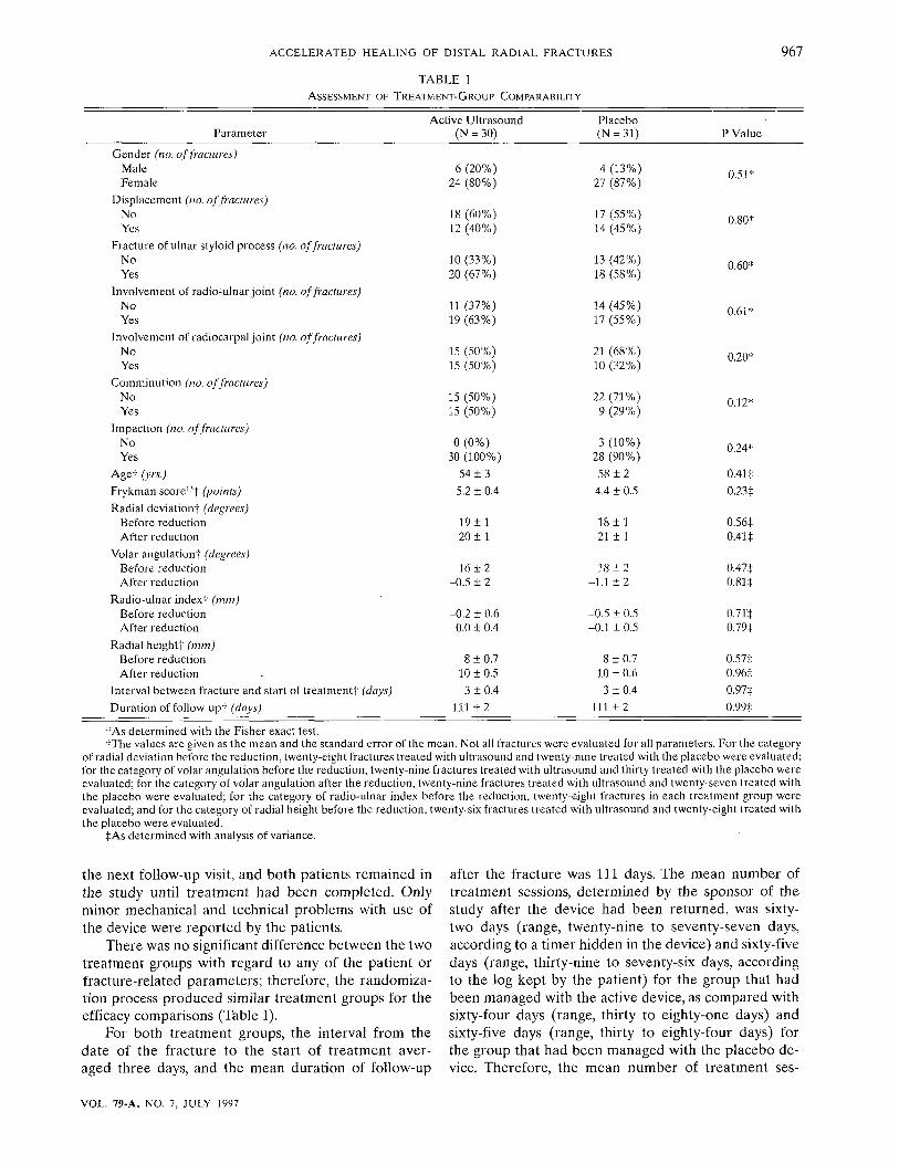
TABLE 1 ASSESSMENT OF TREATMENT-GROUP COMPARABILITY
Parameter
Gender (no. of fractures) Male Female
Displacement (no. of fractures) No Yes
Fracture of ulnar styloid process (no. of fractures) No Yes
Involvement of radio-ulnar joint (no. of fractures) No Yes
Involvement of radiocarpal joint (no. of fractures) No Yes
Comminution (no. of fractures) No Yes
Impaction (no. of fractures) No Yes
Agef (yrs.)
Frykman score ,3t (points)
Radial deviation* (degrees) Before reduction After reduction
Volar angulation* (degrees) Before reduction After reduction
Radio-ulnar indexf (mm) Before reduction After reduction
Radial height* (mm) Before reduction After reduction
Interval between fracture and start of treatment* (days)
Duration of follow-upt (days)
Active Ultrasound (N = 30)
6 (20%) 24 (80%)
18 (60%) 12 (40%)
10 (33%) 20 (67%)
11 (37%) 19 (63%)
15 (50%) 15 (50%)
15 (50%) 15 (50%)
0 (0%) 30 (100%)
54 ± 3
5.2 ± 0.4
19 ± 1 20 ± 1
16 ± 2 -0.5 ± 2
-0.2 ± 0.6 0.0 ± 0.4
8 + 0.7 10 ±0.5
3 ±0.4
111 ± 2
Placebo (N = 31)
4 (13%) 27 (87%)
17 (55%) 14 (45%)
13 (42%) 18 (58%)
14 (45%) 17 (55%)
21 (68%) 10 (32%)
22 (71%) 9 (29%)
3 (10%) 28 (90%)
58 ± 2
4.4 ± 0.5
18 ±1 21 ± 1
18 ± 2 -1.1 ± 2
-0.5 ± 0.5 -0.1 ± 0.5
8 ±0.7 10 + 0.6 3 ±0.4
111 ± 2
P Value
0.51*
0.80*
0.60*
0.61*
0.20*
0.12*
0.24*
0.41*
0.23*
0.56* 0.414
0.47* 0.81 +
OJT* 0.79*
0.57+ 0.96* 0.97*
0.99*
*As determined with the Fisher exact test. fThe values are given as the mean and the standard error of the mean. Not all fractures were evaluated for all parameters. For the category
of radial deviation before the reduction, twenty-eight fractures treated with ultrasound and twenty-nine treated with the placebo were evaluated; for the category of volar angulation before the reduction, twenty-nine fractures treated with ultrasound and thirty treated with the placebo were evaluated; for the category of volar angulation after the reduction, twenty-nine fractures treated with ultrasound and twenty-seven treated with the placebo were evaluated; for the category of radio-ulnar index before the reduction, twenty-eight fractures in each treatment group were evaluated; and for the category of radial height before the reduction, twenty-six fractures treated with ultrasound and twenty-eight treated with the placebo were evaluated.
*As determined with analysis of variance.
the next follow-up visit, and both patients remained in the study until treatment had been completed. Only minor mechanical and technical problems with use of the device were reported by the patients.
There was no significant difference between the two treatment groups with regard to any of the patient or fracture-related parameters; therefore, the randomization process produced similar treatment groups for the efficacy comparisons (Table I).
For both treatment groups, the interval from the date of the fracture to the start of treatment averaged three days, and the mean duration of follow-up
after the fracture was 111 days. The mean number of treatment sessions, determined by the sponsor of the study after the device had been returned, was sixty-two days (range, twenty-nine to seventy-seven days, according to a timer hidden in the device) and sixty-five days (range, thirty-nine to seventy-six days, according to the log kept by the patient) for the group that had been managed with the active device, as compared with sixty-four days (range, thirty to eighty-one days) and sixty-five days (range, thirty to eighty-four days) for the group that had been managed with the placebo device. Therefore, the mean number of treatment ses-
VOL. 79-A, NO. 7, JULY 1997
968 T. K. KRISTIANSEN, J. P. RYABY, JOAN McCABE, J. J. FREY, AND L. R. ROE
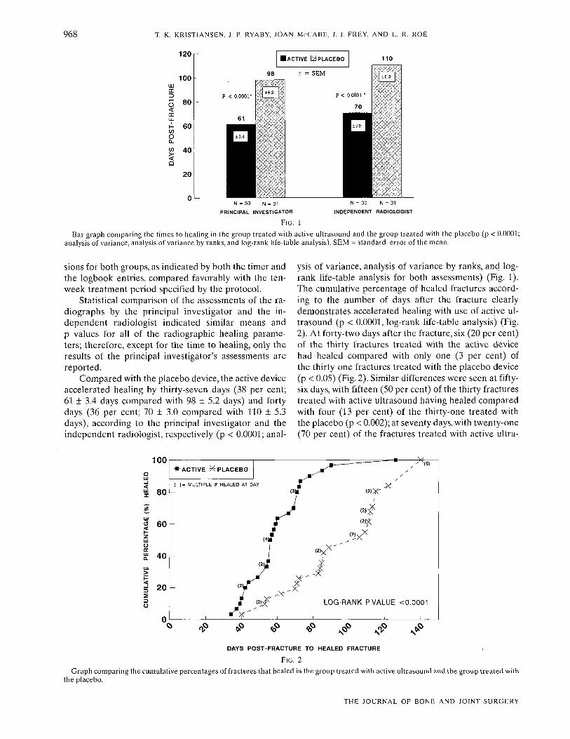
120
100 UJ cc •D I -O < ir u.
i i -co O Q_
CO
Q
PRINCIPAL INVESTIGATOR INDEPENDENT RADIOLOGIST
FIG. 1
Bar graph comparing the times to healing in the group treated with active ultrasound and the group treated with the placebo (p < 0.0001; analysis of variance, analysis of variance by ranks, and log-rank life-table analysis). SEM = standard error of the mean.
sions for both groups, as indicated by both the timer and the logbook entries, compared favorably with the ten-week treatment period specified by the protocol.
Statistical comparison of the assessments of the radiographs by the principal investigator and the independent radiologist indicated similar means and p values for all of the radiographic healing parameters; therefore, except for the time to healing, only the results of the principal investigator's assessments are reported.
Compared with the placebo device, the active device accelerated healing by thirty-seven days (38 per cent; 61 ± 3.4 days compared with 98 ± 5.2 days) and forty days (36 per cent; 70 ± 3.0 compared with 110 ± 5.3 days), according to the principal investigator and the independent radiologist, respectively (p < 0.0001; anal
ysis of variance, analysis of variance by ranks, and log-rank life-table analysis for both assessments) (Fig. 1). The cumulative percentage of healed fractures according to the number of days after the fracture clearly demonstrates accelerated healing with use of active ultrasound (p < 0.0001, log-rank life-table analysis) (Fig. 2). At forty-two days after the fracture, six (20 per cent) of the thirty fractures treated with the active device had healed compared with only one (3 per cent) of the thirty-one fractures treated with the placebo device (p < 0.05) (Fig. 2). Similar differences were seen at fifty-six days, with fifteen (50 per cent) of the thirty fractures treated with active ultrasound having healed compared with four (13 per cent) of the thirty-one treated with the placebo (p < 0.002); at seventy days, with twenty-one (70 per cent) of the fractures treated with active ultra-
Ul
a
2 u
uu
80
60
40
20
•
(
—
-
ACTIVE X- PLACEBO
= MULTIPLE # HEALED AT D AY
'"•
(2)J
1 (2ix^' ^X
(3|T
> • • , * m Jm / -^ /
/ <3>X-
w£
LOG-RANK P VALUE <0.0001
i ; i i
T? tf> «? <£> N * N * > *
DAYS POST-FRACTURE TO HEALED FRACTURE
FIG. 2
Graph comparing the cumulative percentages of fractures that healed in the group treated with active ultrasound and the group treated with the placebo.
THE JOURNAL OF BONE AND JOINT SURGERY
ACCELERATED HEALING OF DISTAL RADIAL FRACTURES 969
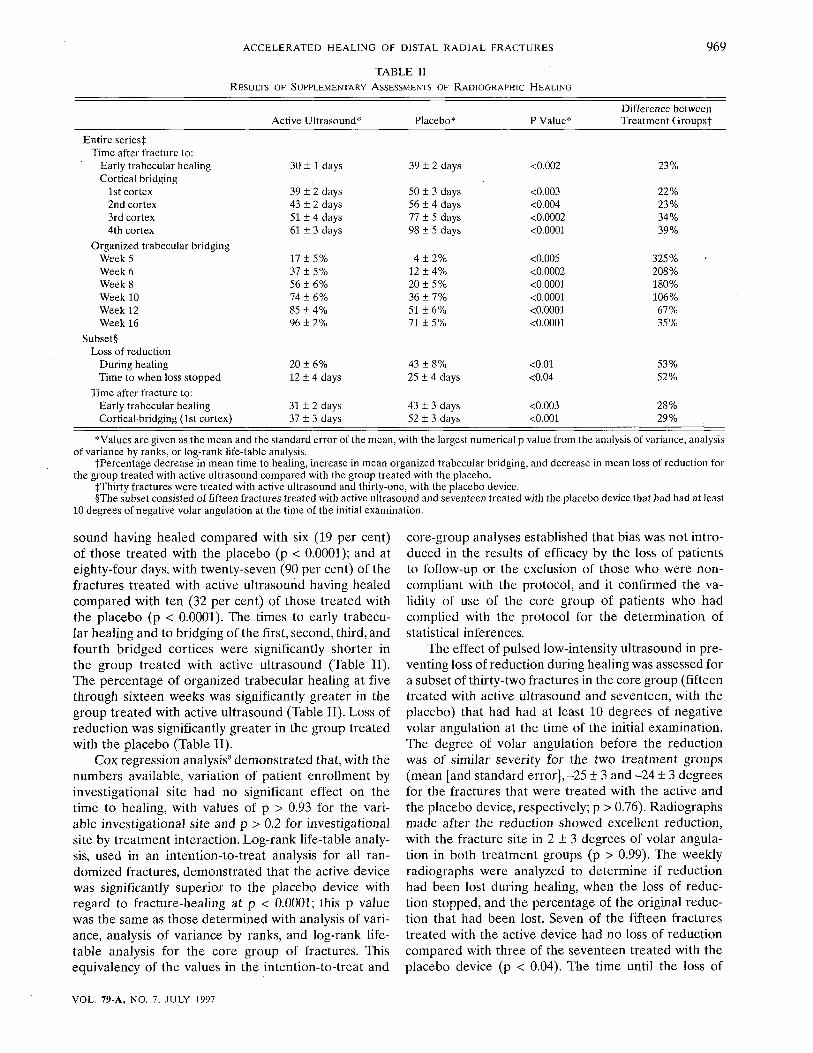
TABLE II
RESULTS OF SUPPLEMENTARY ASSESSMENTS OF RADIOGRAPHIC HEALING
Entire series^ Time after fracture to:
Early trabecular healing Cortical bridging
1st cortex 2nd cortex 3rd cortex 4th cortex
Organized trabecular bridging Week 5 Week 6 Week 8 Week 10 Week 12 Week 16
Subset§ Loss of reduction
During healing Time to when loss stopped
Time after fracture to: Early trabecular healing Corticalbridging (1st cortex)
Active Ultrasound*
30 ± 1 days
39 ± 2 days 43 ± 2 days 51 ± 4 days 61 ± 3 days
17 ± 5% 37 ± 5% 56 ± 6% 74 ± 6% 85 ± 4% 96 + 2%
20 ± 6% 12 ± 4 days
31 ± 2 days 37 + 3 days
Placebo*
39 ± 2 days
50 ± 3 days 56 ± 4 days 77 ± 5 days 98 + 5 days
4 ± 2% 12 + 4% 20 ± 5 % 36 ± 7% 51 ± 6% 71 ± 5%
43 ± 8% 25 ± 4 days
43 ± 3 days 52 ± 3 days
P Value*
<0.002
<0.003 <0.004 <0.0002 •cO.0001
<0.005 <0.0002 <0.0001 <0.0001 <0.0001 <0.0001
<0.01 <0.04
<0.003 <0.001
Difference between Treatment Groupst
23%
22% 23% 34% 39%
325% 208% 180% 106% 67% 35%
53% 52%
28% 29%
*Values are given as the mean and the standard error of the mean, with the largest numerical p value from the analysis of variance, analysis of variance by ranks, or log-rank life-table analysis.
tPercentage decrease in mean time to healing, increase in mean organized trabecular bridging, and decrease in mean loss of reduction for the group treated with active ultrasound compared with the group treated with the placebo.
IThirty fractures were treated with active ultrasound and thirty-one, with the placebo device. §The subset consisted of fifteen fractures treated with active ultrasound and seventeen treated with the placebo device that had had at least
10 degrees of negative volar angulation at the time of the initial examination.
sound having healed compared with six (19 per cent) of those treated with the placebo (p < 0.0001); and at eighty-four days, with twenty-seven (90 per cent) of the fractures treated with active ultrasound having healed compared with ten (32 per cent) of those treated with the placebo (p < 0.0001). The times to early trabecular healing and to bridging of the first, second, third, and fourth bridged cortices were significantly shorter in the group treated with active ultrasound (Table II). The percentage of organized trabecular healing at five through sixteen weeks was significantly greater in the group treated with active ultrasound (Table II). Loss of reduction was significantly greater in the group treated with the placebo (Table II).
Cox regression analysis8 demonstrated that, with the numbers available, variation of patient enrollment by investigational site had no significant effect on the time to healing, with values of p > 0.93 for the variable investigational site and p > 0.2 for investigational site by treatment interaction. Log-rank life-table analysis, used in an intention-to-treat analysis for all randomized fractures, demonstrated that the active device was significantly superior to the placebo device with regard to fracture-healing at p < 0.0001; this p value was the same as those determined with analysis of variance, analysis of variance by ranks, and log-rank life-table analysis for the core group of fractures. This equivalency of the values in the intention-to-treat and
core-group analyses established that bias was not introduced in the results of efficacy by the loss of patients to follow-up or the exclusion of those who were non-compliant with the protocol, and it confirmed the validity of use of the core group of patients who had complied with the protocol for the determination of statistical inferences.
The effect of pulsed low-intensity ultrasound in preventing loss of reduction during healing was assessed for a subset of thirty-two fractures in the core group (fifteen treated with active ultrasound and seventeen, with the placebo) that had had at least 10 degrees of negative volar angulation at the time of the initial examination. The degree of volar angulation before the reduction was of similar severity for the two treatment groups (mean [and standard error], -25 ± 3 and -24 ± 3 degrees for the fractures that were treated with the active and the placebo device, respectively; p > 0.76). Radiographs made after the reduction showed excellent reduction, with the fracture site in 2 ± 3 degrees of volar angulation in both treatment groups (p > 0.99). The weekly radiographs were analyzed to determine if reduction had been lost during healing, when the loss of reduction stopped, and the percentage of the original reduction that had been lost. Seven of the fifteen fractures treated with the active device had no loss of reduction compared with three of the seventeen treated with the placebo device (p < 0.04). The time until the loss of
VOL. 79-A, NO. 7, JULY 1997
970 T. K. KRISTIANSEN, J. P. RYABY, JOAN McCABE, J. J. FREY, AND L. R. ROE
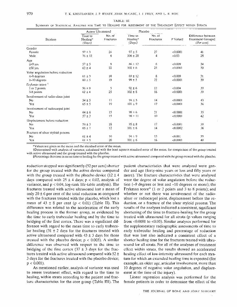
TABLE III
SUMMARY OF STATISTICAL ANALYSES FOR TIME TO HEALING FOR ASSESSMENT OF THE TREATMENT EFFECT WITHIN STRATA
Stratum
Gender Female Male
Age <49 yrs. >50 yrs.
Volar angulation before reduction <-'9 degrees >-10 degrees
Frykman score13
1 or 2 points 3-8 points
Involvement of radio-ulnar joint No Yes
Involvement of radiocarpal joint No Yes
Displacement before reduction No Yes
Fracture of ulnar styloid process No Yes
Active Time to Healing*
(Days)
57 ± 3 76 ±12
57 ± 5 62 + 4
61 ± 3 60 ± 5
56 ± 9 62 ± 4
54 ± 5 65 ± 5
64 ± 6 57 ± 3
58 ± 3 65 ± 7
61 ± 4 61 ± 5
Ultrasound No. of
Fractures
24 6
8 22
10 19
5 25
11 19
15 15
18 12
10 20
Placebo Time to Healing*
(Days)
97 ± 5 106 ± 20
86 ± 12 102 ± 6
89 ±12 99 ± 5
92 ± 6 102 ± 8
94 ± 5 101 ± 9
98 + 6 98 ±11
95 ± 8 101 + 6
94 ± 9 101 ± 6
No. of Fractures
27 4
8 23
8 22
13 18
14 17
21 10
17 14
13 18
P Valuet
<0.0001 <0.03
<0.009 <0.0001
<0.009 <0.0001
<0.004 <0.0001
<0.0001 <0.0001
<0.0001 <0.0001
<o.ooo:i <0.0002
<0.001 <0.0001
Difference between Treatment Groupst
(Per cent)
41 28
34 39
31 39
39 39
43 36
35 42
39 36
35 40
*Values are given as the mean and the standard error of the mean. tDetermined with analysis of variance, calculated with the least-squares standard error of the mean, for comparison of the group treated
with active ultrasound and the group treated with the placebo. ^Percentage decrease in mean time to healing for the group treated with active ultrasound compared with the group treated with the placebo.
reduction stopped was significantly (52 per cent) shorter for the group treated with the active device compared with the group treated with the placebo device (1.2 ± 4 days compared with 25 ± 4 days; p < 0.02, analysis of variance, and p < 0.04, log-rank life-table analysis). The fractures treated with active ultrasound lost a mean of only 20 ± 6 per cent of the total reduction as compared with the fractures treated with the placebo, which lost a mean of 43 ± 8 per cent (p < 0.01) (Table II). This difference was related to the acceleration of the early healing process in the former group, as evidenced by the time to early trabecular healing and by the time to bridging of the first cortex. There was a significant difference with regard to the mean time to early trabecular healing (31 ± 2 days for the fractures treated with active ultrasound compared with 43 ± 3 days for those treated with the placebo device; p < 0.003). A similar difference was observed with respect to the time to bridging of the first cortex (37 + 3 days for the fractures treated with active ultrasound compared with 52 ± 3 days for the fractures treated with the placebo device; p < 0.001).
As mentioned earlier, analysis of variance was used to assess treatment effect, with regard to the time to healing, within strata according to the patient and fracture characteristics for the core group (Table III). The
patient characteristics that were analyzed were gender and age (forty-nine years or less and fifty years or more). The fracture characteristics that were analyzed were the degree of volar angulation before the reduction (-9 degrees or less and -1.0 degrees or more); the Frykman score" (1. or 2 points and 3 to 8 points); and whether or not there was involvement of the radioulnar or radiocarpal joint, displacement before the reduction, or a fracture of the ulnar styloid process. The results of the analyses indicated a consistent, significant shortening of the time to fracture-healing for the group treated with ultrasound for all strata (p values ranging from <0.0001 to <0.03). Similar analyses performed for the supplementary radiographic assessments of time to early trabecular healing and percentage of reduction that was lost also indicated a consistent effect of a shorter healing time for the fractures treated with ultrasound for all strata. For all of the analyses of treatment effect within strata, the results showed an accelerated healing effect of low-intensity ultrasound for each stratum for which an extended healing time is expected (for example, an older age, articular involvement, more than 10 degrees of negative volar angulation, and displacement at the time of the injury).
Linear regression analysis was performed for the female patients in order to determine the effect of the
THE JOURNAL OF BONE AND JOINT SURGERY
ACCELERATED HEALING OF DISTAL RADIAL FRACTURES 971
device on the time to healing as a consequence of age. The linear regression coefficient (slope) for the group treated with the placebo (a group essentially equivalent to a population that has normal healing) was 0.8, representing a significant (p < 0.04) increase in healing time of approximately 0.8 day for each additional year of age. In contrast, the linear regression coefficient for the group treated with ultrasound was 0.1, representing a slope that was essentially flat, with only a 0.1-day increase in healing time for each additional year of age; this value was not significantly different from zero (p > 0.57). Analysis of variance to test the hypothesis that the regression coefficient for the women managed with ultrasound was equal to that for the women managed with the placebo revealed that the regression coefficients were significantly different (p < 0.03). This regression analysis established that low-intensity ultrasound accelerates fracture-healing regardless of the age of the patient and mitigates the extended healing-time effect of aging on the healing process.
A Premarket Approval application for the device used in this study was submitted to the Food and Drug Administration in early 1990, and the device was approved for commercial marketing in October 1994. The Food and Drug Administration requested that all patients enrolled in the study be contacted at, a minimum of two years after the fracture had healed, to determine whether the fracture was still healed. Seventy-seven (93 per cent) of the eighty-three patients who had been randomized into the study were located in 1994, and healing was verified for each patient at a mean of seventy-two months.
The smoking status of the core group of sixty patients before and after the study was documented retrospectively in 1995. Ten of the patients had died or could not be located. Of the remaining fifty patients, nine had smoked and forty-one had not smoked during the study. Use of the active ultrasound device significantly reduced the time to fracture-healing for the patients who had smoked during the study (mean, 48 ± 5 days for the patients managed with ultrasound and 98 ± 30 days for those managed with the placebo; p < 0.003) and for those who had not smoked (mean, 66 + 5 days for the patients managed with ultrasound and 100 ± 6 days for those managed with the placebo; p < 0.0001). In addition, the nine patients who had smoked during the study were included with the three who had stopped smoking within ten years before enrollment, and the patients who had never smoked were included with those who had stopped smoking more than ten years before enrollment. Use of the active ultrasound device significantly reduced the time to healing for the combined group of smokers (mean, 51 ± 4 days for the patients managed with ultrasound and 109 ± 24 days for those managed with the placebo; p < 0.0002) and for the combined group of non-smokers (mean, 67 ± 5 days for the patients managed with ultra
sound and 98 ± 5 days for those managed with the placebo; p < 0.0001).
Discussion
We conducted this study because of several reports indicating that non-thermal, pulsed, low-intensity ultrasound stimulates and accelerates the normal fracture-repair process. Dorsally angulated fractures (negative volar angulation) of the distal aspect of the radius were selected as the model for this study for several reasons. First, the bone is primarily cancellous, with a thin layer of cortical bone; therefore, the effect on the healing of both cancellous and cortical bone could be assessed. Second, these fractures involve the upper extremity, and they usually are treated non-operatively; therefore, weight-bearing and operative intervention, two variables that influence fracture-healing, could be eliminated from the study. Third, the investigational therapy could easily be incorporated into conventional treatment.
The patient and fracture characteristics in the two treatment groups were very similar before treatment, as shown with the comparability analysis. The patients found the portable treatment unit easy to use, including application of the coupling gel to the transducer, securing of the treatment head module, and reinsertion of the felt plug and the cap when the device was not in use. There were no complications or adverse reactions attributable to any aspect of the treatment. No contraindications to use of the device and only minor mechanical and technical problems were encountered during the study.
Comparison of the results for the two treatment groups demonstrated a significant acceleration of the fracture-healing process, which led to a significant decrease in the time needed to achieve each radiographic stage of healing as well as a significant decrease in the loss of reduction during healing in the group that was treated with active, pulsed low-intensity ultrasound. The analysis of treatment effect within the patient and fracture-characteristics strata indicated consistent, accelerated healing for all strata, including those for which extended healing times are expected.
The specific mechanisms by which ultrasound accelerates bone-healing are unknown; however, in terms of the physical mechanism, ultrasound provides a mechanical force to the cellular system. Several investigators have demonstrated that mechanical force modulates bone formation both in vitro and in vivo3-5-27"25. Recent research with use of low-intensity ultrasound demonstrated increased blood flow in an animal fracture model15, and real-time in vitro experiments with use of fluorescent microscopy suggested that ultrasound promotes calcium flux changes within seconds after application4. Several investigators have postulated a mul-tifaceted biological mechanism. Wang et al." suggested that the primary effect of low-intensity pulsed ultra-
VOL. 79-A, NO. 7, JULY 1997
9 7 2 T. K. KRISTIANSEN, J. P. RYABY, JOAN McCABE, J. J. FREY, AND L. R. ROE
sound is on the chondrocyte population in the healing was used in our study. Heckman and Sarasohn-Kahn16
fracture; they found evidence of increased soft-callus demonstrated substantial cost-savings as a result of formation, advanced endochondral ossification of the reduced costs of Workers' Compensation and of fewer callus, and increased stiffness and strength of the frac- secondary procedures when low-intensity ultrasound ture site. Animal and in vitro studies by Yang et al.45-46, was used in conjunction with non-operative or operand in vitro studies by Ryaby et al.3032 and Wu et al.42, ative treatment of tibial diaphyseal fractures. The cur-have shown that low-intensity pulsed ultrasound modi- rent multicenter, prospective, randomized, double-blind, fies cell processes such as proteoglycan and transform- placebo-controlled study demonstrated that fracture-ing growth factor-|3 synthesis, type-II collagen content, healing also is accelerated in the distal radial meta-messenger-RNA aggrecan production, calcium uptake, physis, a primarily cancellous bone model. This modality and reduced parathyroid-hormone response. All of may be useful clinically, as its application continues these observations are related directly to bone forma- to be explored for other types of fractures and for tion or resorption. In uncontrolled in vivo studies, those with characteristics that lead to slow or delayed Choffie and Duarte6, Duarte10, Knoch and Klug20, and healing. Xavier and Duarte4344 reported an increased rate of h e a l i n g Of a n i m a l a n d h u m a n f r a c t u r e s t h a t h a d b e e n NoTF;, T h e authors thank the following site investigators for their participation and trpatpH With llltrn^niinH cooperation: David Caborn, M.D.; James Capozzi, M.D.; Richard Christian, M.D.; Joseph E.
Cronkey, M.D.; Robin DeAndrade, M.D.; M. E. Hale, M.D.; J. Stewart Haskin, M.D.; M. R e c e n t l y , H e C k m a n e t a l ." , in a p r o s p e c t i v e , r a n d o m - Iusim. M.D.; David C. Mendes, M.D.; Michael Mont, M.D.; Arthur A. Pilla, Ph.D.; Roger S,
, , I'ocze, M.D.; C.Dayton Riddle, M.D.; Robert S. Siffert, M.D.; Elton E.Strauss. M.D.;Thomas lZed, dOUble -b l ind , placebO-COntrOlled StUdy, r e p o r t e d Trumble, M.D.; and James Yates, M.D. The authors also thank J.Stewart Haskin. M.D., and n r inn i f l^onfK; flom-oQeorl tirvio t/-\ U&rtM-nrr tr\ — C\ flftHI ^ i-if David G. Mendes, M.D., for their special dedication and efforts; Richard Russo. Michael a SlgmriCdniiy d e c r e a s e d l imC lO n e d i m g (J) - U.UUUlj Ol Hausman,M.D.,EltonStrauss,M.D.,andRobertS.Sifferl,M.D.,fortheirinvaluablecounsel fraCtUreS Of t h e t ibial Shaft that h a d b e e n t r e a t e d With a n d a d v i c e o n development of the protocol and other aspects of the study; and Roger Talish,
Art Lifshey, Tom Wilson, and Raphael Dickerson for their design of the device and their
the same low-intensity, pulsed ultrasound device that engineering expertise.
References 1. Altissiini, M.; Antenucci, R.; Fiacca, C; and Mancini, G. B.: Long-term results of conservative treatment of fractures of the distal radius.
Clin. Orthop., 206:202-210,1986. 2. Altissimi, M.; Mancini, G. B.; Azzara, A.; and Ciaffoloni, E.: Early and late displacement of fractures of the distal radius. The prediction
of instability. Internal Orthop., 18:61-65,1994. 3. Binderman, I.; Zor, U.; Kaye, A. M.; Shimshoni, Z.; Harell, A.; and Somjen, D.: The transduction of mechanical force into biochemical
events in bone cells may involve activation of phospholipase A2. Calcif. Tissue Internat., 42:261-266,1988. 4. Bolander, M. E.: Personal communication, 1997. 5. Buckley, M. J.; Banes, A. J.; Levin, L. G.; Sumpio, B. E.; Sato, M.; Jordan, R.; Gilbert, J.; Link, G. W.; and Tran Soy Tay, R.: Osteoblasts
increase their rate of division and align in response to cyclic, mechanical tension in vitro. Bone and Mm.., 4: 225-236,1988. 6. Choffie, M., and Duarte, L. R.: Low-intensity pulsed ultrasound and effects on ununited fractures. Read at the Orthopaedic Health
Conference, University Hospital, University of Sao Paulo, Brazil, June 15,1994. 7. Conover, W. J.: Practical Nonparametric Statistics. Ed. 2. New York, Wiley, 1980. 8. Cox, D. R.: Regression models and life-tables./ Roy. Statist. Soc, Series B, 34:187-220,1972. 9. Cox, D. R., and Oakes, D.: Analysis of survival data. London, Chapman and Hall, 1984.
10. Duarte, L. R.: The stimulation of bone growth by ultrasound. Arch. Orthop. and Traumat. Surg., 101:153-159,1983. 11. Dyson, M.: Therapeutic applications of ultrasound. In Biological Effects of Ultrasound, pp. 121-133. Edited by W. L. Nyborg and M. C.
Ziskin. New York, Churchill Livingstone, 1985. 12. Fleiss, J. L.: Statistical Methods for Rates and Proportions. Ed. 2, pp. 24-26. New York, Wiley, 1981. 13. Frykman, G.: Fracture of the distal radius including sequelae — shoulder-hand-finger syndrome, disturbance in the distal radio-ulnar
joint and impairment of nerve function. A clinical and experimental study. Acta Orthop. Scandinavica, Supplement 108,1967. 14. Gartland, J. J., Jr., and Werley, C. W.: Evaluation of healed Colles' fractures. J. Bone and Joint Surg., 33-A: 895-907, Oct. 1951. 15. Goldberg, B. B.: Personal communication, 1997. 16. Heckman, J. D., and Sarasohn-Kahn, J.: The economics of treating tibia fractures. The cost of delayed unions. Bull. Hosp. Joint Dis., 56:
63-72,1997. 17. Heckman, J. D.; Ryaby, J. P.; McCabe, J.; Frey, J. J.; and Kilcoyne, R. F.: Acceleration of tibial fracture-healing by non-invasive,
low-intensity pulsed ultrasound./ Bone and Joint Surg., 16-A: 26-34, Jan. 1994. 18. Jarry, L., and Uhthoff, H. K.: Differences in healing of metaphyseal and diaphyseal fractures. Canadian J. Surg., 14:127-135,1971. 19. Klug, W.; Franke, W. G.; and Knoch, H. G.: Scintigraphic control of bone-fracture healing under ultrasonic stimulation: an animal
experimental study. European J. Nucl. Med., 11: 494-497,1986. 20. Knoch, G. H., and Klug, W.: Stimulation of Fracture Healing with Ultrasound, translated by T. C. Telger. New York, Springer, 1991. 21. Lehmann, E. L.: Nonparametrics: Statistical Methods Based on Ranks. San Francisco, Holden-Day, 1975. 22. Mantel, N.: Evaluation of survival data and two new rank order statistics arising in its consideration. Cancer Chemother. Rep., 50:
163-170,1966. 23. Mantel, N.: Ranking procedures for arbitrarily restricted observation. Biometrics, 23: 65-78,1967. 24. Nyborg, W. L.: Mechanisms. In Biological Effects of Ultrasound, pp. 23-33. Edited by W. L. Nyborg and M. C. Ziskin. New York,
Churchill Livingstone, 1985. 25. Pilla, A. A.; Mont, M. A.; Nasser, P. R.; Khan, S. A.; Figueiredo, M.; Kaufman, J. J.; and Siffert, R. S.: Non-invasive low-intensity pulsed
ultrasound accelerates bone healing in the rabbit. J. Orthop. Trauma, 4:246-253,1990. 26. Pilla, A. A.; Figueiredo, M.; Nasser, P.; Alves, J. M.; Ryaby, J. T.; Klein, M.; Kaufman, J. J.; and Siffert, R. S.: Acceleration of bone repair
THE JOURNAL OF BONE AND JOINT SURGERY
ACCELERATED HEALING OF DISTAL RADIAL FRACTURES 973
by pulsed sine wave ultrasound: animal, clinical, and mechanistic studies. In Electromagnetics in Medicine and Biology, pp. 331-341. Edited by C. T. Brighton and S. R. Pollack. San Francisco, San Francisco Press, 1991.
27. Rubin, C. T., and McLeod, K. J.: Inhibition of osteopenia by biophysical intervention. In Osteoporosis, pp. 351-371. Edited by R. Marcus, D. Feldman, and J. Kelsey. San Diego, Academic Press, 1996.
28. Rubin, C; Gross, T.; Qin, Y.-X.; Fritton, S.; Guilak, E; and McLeod, K.: Differentiation of the bone-tissue remodeling response to axial and torsional loading in the turkey ulna../. Bone and Joint Surg., 78-A: 1523-1533, Oct. 1996.
29. Rubin, J.; Biskobing, D.; Fan, X.; Rubin, C; McLeod, K.; and Taylor, W. R.: Pressure regulates osteoclast formation and MCSF expression in marrow culture./ Cell Physiol., 170:81-87,1997.
30. Ryaby, J. T.; Mathew, J.; and Duarte-Alves, P.: Low intensity pulsed ultrasound affects adenylate cyclase and TGF-(3 synthesis in osteoblastic cells. Trans. Orthop. Res. Soc, 17: 590,1992.
31. Ryaby, J. T.; Mathew, J.; Pilla, A. A.; and Duarte-Alves, P.: Low-intensity pulsed ultrasound modulates adenylate cyclase activity and transforming growth factor beta synthesis. In Electromagnetics in Medicine and Biology, pp. 95-100. Edited by C. T. Brighton and S. R. Pollack. San Francisco, San Francisco Press, 1991.
32. Ryaby, J. T.; Bachner, E. J.; Bendo, J. A.; Dalton, P. F.; Tannenbaum, S.; and Pilla, A. A.: Low intensity pulsed ultrasound increases calcium incorporation in both differentiating cartilage and bone cell cultures. Trans. Orthop. Res. Soc, 14:15,1989.
33. Salter, R. B.: Textbook of Disorders and Injuries of the Musculoskeletal System: An Introduction to Orthopaedics, Rheumatology, Metabolic Bone Disease, Rehabilitation and Fractures, pp. 340-369. Baltimore, Williams and Wilkins, 1970.
34. Scheffe, H.: The Analysis of Variance. New York, Wiley, 1959. 35. St. John Brown, B.: How safe is diagnostic ultrasonography? Canadian Med. Assn..!., 131: 307-311,1984. 36. Tanzer, M.; Harvey, E.; Kay, A.; Morton, P.; and Bobyn, J. D.: Effect of noninvasive low intensity ultrasound on bone growth into
porous-coated implants. 7. Orthop. Res., 14:901-906,1996. 37. Uhthoff, H. K., and Rahn, B. A.: Healing patterns of metaphyseal fractures. Clin, Orthop., 160: 295-303,1981. 38. Van der Linden, W., and Ericson, R.: Colles' fracture. How should its displacement be measured and how should it be immobilized?
J. Bone and Joint Surg., 63-A: 1285-1288, Oct. 1981. 39. Wang, S.-J.; Lew alien, D. G.; Bolander, M. E.; Chao, E. Y. S.; Ilstrup, D. M.; and Greenleaf, J. F.: Low intensity ultrasound treatment
increases strength in a rat femoral fracture model../. Orthop. Res., 12: 40-47,1994. 40. Wells, P. N. T.: Surgical applications of ultrasound. In Biological Effects of Ultrasound, pp. 157-167. Edited by W. L. Nyborg and M. C.
Ziskin. New York, Churchill Livingstone, 1985. 41. Wolff, J.: The Law of Bone Remodeling, translated by P. Maquet and R. Furlong. New York, Springer, 1986. 42. Wu, C.-C; Lewallen, D. G.; Bolander, M. E.; Bronk, J.; Kinnick, R.; and Greenleaf, J. F.: Exposure to low intensity ultrasound stimulates
aggrecan gene expression by cultured chondrocytes. Trans. Orthop. Res. Soc, 21: 622,1996. 43. Xavier, C. A. M., and Duarte, L. R.: Estimulaca ultra-sonica de calo osseo: applicaca clinica. Rev. Brasileira Orlop., 18: 73-80,1983. 44. Xavier, C. A. M., and Duarte, L. R.: Treatment of nonunions by ultrasound stimulation: first clinical applications. Read at the Meeting of
the Latin-American Orthopedic Association, at the Annual Meeting of The American Academy of Orthopaedic Surgeons, San Francisco, California, Jan. 25,1987.
45. Yang, K. H.; Parvizi, J.; Wang, S. J.; Lewallen, D. G.; Kinnick, R. R.; Greenleaf, .1. E; and Bolander, M. E.: Exposure to low intensity ultrasound increases aggrecan gene expression in a rat femur fracture model. / Orthop. Res., 14: 802-809,1996.
46. Yang, K. H.; Wang, S.-J.; Lewallen, D. G.; Greenleaf, J.; Oles, K.; Bronk, J.; and Bolander, M. E.: Low intensity ultrasound stimulates fracture healing in rat model: biomechanical and gene expression analysis. Trans. Orthop. Res. Soc, 19: 519,1994.
47. Ziskin, M. C: Applications of ultrasound in medicine — comparison with other modalities. In Ultrasound: Medical Applications, Biological Effects, and Hazard Potential, pp. 49-59. Edited by M. H. Repacholi, M. Grandolfo, and A. Rindi. New York, Plenum Press, 1987.
VOL. 79-A, NO. 7, JULY 1997
Related Documents