The PDF of the article you requested follows this cover page. This is an enhanced PDF from The Journal of Bone and Joint Surgery 2007;89:44-58. doi:10.2106/JBJS.G.00648 J Bone Joint Surg Am. Shane J. Nho, Robert H. Brophy, Joseph U. Barker, Charles N. Cornell and John D. MacGillivray Literature Management of Proximal Humeral Fractures Based on Current This information is current as of July 22, 2010 Reprints and Permissions Permissions] link. and click on the [Reprints and jbjs.org article, or locate the article citation on to use material from this order reprints or request permission Click here to Publisher Information www.jbjs.org 20 Pickering Street, Needham, MA 02492-3157 The Journal of Bone and Joint Surgery

Welcome message from author
This document is posted to help you gain knowledge. Please leave a comment to let me know what you think about it! Share it to your friends and learn new things together.
Transcript

The PDF of the article you requested follows this cover page.
This is an enhanced PDF from The Journal of Bone and Joint Surgery
2007;89:44-58. doi:10.2106/JBJS.G.00648 J Bone Joint Surg Am.Shane J. Nho, Robert H. Brophy, Joseph U. Barker, Charles N. Cornell and John D. MacGillivray
LiteratureManagement of Proximal Humeral Fractures Based on Current
This information is current as of July 22, 2010
Reprints and Permissions
Permissions] link. and click on the [Reprints andjbjs.orgarticle, or locate the article citation on
to use material from thisorder reprints or request permissionClick here to
Publisher Information
www.jbjs.org20 Pickering Street, Needham, MA 02492-3157The Journal of Bone and Joint Surgery
COPYRIGHT © 2007 BY THE JOURNAL OF BONE AND JOINT SURGERY, INCORPORATED
44
Management of Proximal Humeral Fractures
Based on Current LiteratureBy Shane J. Nho, MD, MS, Robert H. Brophy, MD,
Joseph U. Barker, MD, Charles N. Cornell, MD, and John D. MacGillivray, MD
Introductionroximal humeral fractures are the second most com-mon upper-extremity fracture and the third most com-mon fracture, after hip fractures and distal radial
fractures, in patients who are older than sixty-five years ofage1. Although the overwhelming majority of proximal hu-meral fractures are either nondisplaced or minimally dis-placed and can be treated with sling immobilization andphysical therapy, approximately 20% of displaced proximalhumeral fractures may benefit from operative treatment.Many surgical techniques have been described, but no singleapproach is considered to be the standard of care. Surgeonswho treat proximal humeral fractures should be able to iden-tify the fracture pattern and select an appropriate treatmenton the basis of this pattern and the underlying quality of thebone. Orthopaedic surgeons should have experience with abroad range of techniques, including transosseous suture fixa-tion, closed reduction and percutaneous fixation, open reduc-tion and internal fixation with conventional and locked-platefixation, and hemiarthroplasty. In the future, locked-platetechnology and the use of osteobiologics may play an increas-ingly important role in the treatment of displaced proximalhumeral fractures, facilitating preservation of the humeralhead in appropriately selected patients.
The goals of this article are to enable the reader to: (1)become familiar with the recent literature on the classificationof and treatment options for proximal humeral fractures, and(2) better identify fracture characteristics and devise an ap-propriate treatment plan.
Treatment OptionsTransosseous Suture FixationSurgical Technique
ark et al.2 described different operative approaches foreach fracture pattern described by Neer3. For two-part
greater tuberosity fractures, an anterosuperior approachalong the Langer lines extending from the lateral aspect ofthe acromion toward the lateral tip of the coracoid is used.The split occurs in the anterolateral raphe and allows expo-sure of the displaced greater tuberosity fracture. When a sur-gical neck fracture exists, Park et al.2 prefer a standarddeltopectoral approach. Nonabsorbable suture is used tocapture rotator cuff tissue anteriorly, laterally, and posteri-orly to the fragment. The displaced humeral head is reducedand fixed to the shaft through drill holes or suture anchors.Three-part fractures involving the greater tuberosity and thesurgical neck can be repaired by initially bringing the head tothe shaft, followed by reduction and fixation of the greatertuberosity. Flatow et al.4 described an anterosuperior ap-proach and the use of heavy nonabsorbable sutures forgreater tuberosity fractures (Fig. 1).
Indications
Transosseous suture fixation has been described as a treat-ment option for proximal humeral fractures that have at least1 cm of displacement between the head and the shaft frag-ments or 5 mm of displacement of the tuberosity fragment.The proponents of this technique emphasize the advantage ofavoiding the risks associated with hardware, which includepain, neurovascular compromise, migration, failure, and theneed for removal. The underlying rotator cuff musculaturecan be used as a means to realign the fractures and enhancestability. Furthermore, the long-term functional recovery ofthe rotator cuff is a key component of overall patient outcome.
Contraindications
Contraindications to this approach include previous at-tempt(s) at internal fixation or fractures older than six weeks.Also, the use of this technique for highly comminuted four-part fractures is not recommended.
P
PDisclosure: The authors did not receive any outside funding or grants in support of their research for or preparation of this work. Neither they nor a member of their immediate families received payments or other benefits or a commitment or agreement to provide such benefits from a commercial entity. No commercial entity paid or directed, or agreed to pay or direct, any benefits to any research fund, foundation, division, center, clinical prac-tice, or other charitable or nonprofit organization with which the authors, or a member of their immediate families, are affiliated or associated.
J Bone Joint Surg Am. 2007;89(Suppl 3):44-58 • doi:10.2106/JBJS.G.00648
Nho_00648.fm Page 44 Monday, September 10, 2007 2:01 PM

45
THE JOU R N A L OF BO N E & JO I N T SU RG ER Y · JB JS .ORG
VO LUM E 89-A · SU P P L E M E N T 3 · 2007MA N AG E M EN T OF PROX I M A L HU M E R A L FR A C T URE S BA S E D ON CUR REN T LI TER AT U RE
Results
Flatow et al.4 reported that all twelve patients who had tran-sosseous suture fixation of an isolated greater tuberosity frac-ture had good or excellent results with osseous union. Parket al.2, in a review of twenty-eight shoulders with two-partgreater tuberosity, two-part surgical neck, and three-partgreater tuberosity and surgical neck fractures that were treatedwith transosseous suture fixation, reported that 78% of thepatients had an excellent result according to the criteria ofNeer et al.5 and that there was no difference between the re-sults obtained with two-part greater tuberosity fractures andthose obtained with two-part surgical neck or three-part frac-tures. Panagopoulos et al.6 used transosseous suture fixationfor four-part valgus-impacted proximal humeral fractures,and the mean Constant-Murley score7 for the operative shoul-der was 87 compared with 94 for the contralateral shoulder.Partial osteonecrosis of the humeral head developed in onepatient.
Closed Reduction and Percutaneous FixationSurgical Technique
With the aid of an image intensifier, the fracture is reduced byclosed means into anatomic alignment. For surgical neck frac-tures, two to three threaded Kirschner wires (0.045 to 0.0625mm) are inserted into the lateral cortex distal to the deltoidinsertion and advanced into the subchondral bone of the hu-meral head without penetrating the articular surface. Forgreater tuberosity fractures in isolation or in conjunction witha surgical neck fracture, the two wires should purchase themedial cortex >20 mm from the inferior border of the head.
Resch et al.8 described a technique for closed reductionand percutaneous fixation of three and four-part proximalhumeral fractures. For three-part fractures, the subcapitalfracture is reduced with adduction, internal rotation, and axialtraction on the arm. A pointed hook retractor is inserted intothe subacromial space to manipulate the greater tuberosityfragment anteriorly and inferiorly into anatomic position.Under image intensification, the shoulder is brought throughinternal and external rotation to confirm reduction of thegreater tuberosity and two cannulated self-tapping 2.7-mmscrews are used to fix the fragments.
For four-part valgus impacted fractures or true four-part fractures, a periosteal elevator is used to elevate and la-terally translate the articular fragment9. When the head iselevated, the periosteum on the medial side acts as a hinge andthe greater tuberosity is reduced into anatomic position. Ablunt trocar and a 2.7-mm cannulated screw is advanced to-ward the superior aspect of the greater tuberosity, and a screwis directed toward the humeral head. Another screw is posi-tioned at the inferior portion of the greater tuberosity and di-rected into the shaft to provide fixation between the head andshaft. The lesser tuberosity is provisionally fixed with a Kirsch-ner wire, and a screw is placed from anterior to posterior.
Patients are generally immobilized for three to fourweeks in most protocols. After this initial period, passive mo-tion is started, consisting of pendulum exercises, forward ele-vation, and external rotation with the arm at the side10. Activemotion starts at six weeks, provided that radiographs demon-strate evidence of healing.
Indications
Percutaneous fixation of proximal humeral fractures requiresless dissection and therefore less disruption of the vascular sup-ply than traditional open approaches do. Advocates cite thehigh risk of osteonecrosis in these fractures as an important rea-son to avoid extensive exposure of the individual fragments.Percutaneous fixation also has the advantage of decreased scar-ring in the scapulohumeral interface and subsequent easierrehabilitation10.
Contraindications
Contraindications include the presence of severe osteopeniaor osteoporosis. Comminution of the medial portion of thecalcar or proximal part of the humeral shaft is also a relativecontraindication. Patients who are noncompliant or nonco-
Fig. 1
Tension-band construct with transosseous suture fixation can be
used for minimally displaced (AO/ASIF type-A) proximal humeral
fracture involving the surgical neck, greater tuberosity, or lesser tuber-
osity. (Reproduced with modification, with permission from: Nho SJ,
Brophy RH, Barker JU, Cornell CN, MacGillvray JD. Innovations in the
management of displaced proximal humerus fractures. J Am Acad Or-
thop Surg. 2007;15:17.)
Nho_00648.fm Page 45 Monday, September 10, 2007 2:01 PM

46
THE JOU R N A L OF BO N E & JO I N T SU RG ER Y · JB JS .ORG
VO LUM E 89-A · SU P P L E M E N T 3 · 2007MA N AG E M EN T OF PROX I M A L HU M E R A L FR A C T URE S BA S E D ON CUR REN T LI TER AT U RE
operative should not be treated with this technique. Tuberos-ity comminution that prevents screw or pin fixation precludesuse of this technique. Finally, if a stable closed reduction can-not be obtained, an open reduction with internal fixationshould be performed.
Pearls and Pitfalls
This procedure is technically demanding and has a substan-tial learning curve. Many anatomical studies have evaluatedthe relationship of the neurovascular structures to the pins.Rowles and McGrory11 evaluated ten cadaver shoulders inwhich percutaneous pin fixation (two lateral, one anterior,and two greater tuberosity pins) was performed. They foundthat the proximal lateral pins were a mean distance of 3 mmfrom the anterior branch of the axillary nerve. The anteriorpins were a mean distance of 2 mm from the long head of thebiceps tendon and 11 mm from the cephalic vein11. The prox-imal tuberosity pins were a mean distance of 6 mm from theaxillary nerve and 7 mm from the posterior humeral circum-flex artery. The pins were found to tent these structureswhen the shoulder was placed in internal rotation (Figs. 2-Aand 2-B)11.
Kamineni et al.12 also performed a cadaver study in whichthe results were evaluated after inserting one anteroposterior
and two lateral Kirschner wires, as described by Jaberg et al.13,into forty shoulders. The axillary nerve was injured by the lat-eral wire in three specimens, and the damage included two di-rect penetrations. The anterior wire caused a perineural injuryof a terminal branch of the axial nerve. They concluded thatfixation should be performed through a limited open ap-proach to prevent these injuries12.
Results
Resch et al.8 reviewed the cases of twenty-seven patients withthree-part or four-part fractures treated with closed reduc-tion and percutaneous screw fixation. For the three-part frac-tures, the mean Constant score was 85.4 without evidence ofpostoperative osteonecrosis. Thirteen of the eighteen patientswith four-part fractures had valgus impacted fractures, andpartial osteonecrosis developed in only one patient. Of the fivelaterally displaced four-part fractures, two required revision toa hemiarthroplasty.
Keener et al.10, with use of closed reduction and percuta-neous fixation, treated thirty-five patients with two-part,three-part, and four-part fractures. The mean follow-up wasthirty-five months. All fractures healed, and the mean painscore on a visual analog scale was 1.4. The mean AmericanShoulder and Elbow Surgeons score14 was 83.4, and the mean
Fig. 2-A
Fig. 2-A Anteroposterior radiograph showing the proximal part of the humerus with percutaneous pin placement as described by Jaberg et
al.113. (Reproduced from: Rowles DJ, McGrory JE. Percutaneous pinning of the proximal part of the humerus. An anatomic study. J Bone
Joint Surg Am. 2001;83:1696.) Fig. 2-B The safe starting point for the proximal lateral pins and the end point for the greater tuberosity
pins. X = distance from the superiormost aspect of the humeral head to the inferiormost aspect of the humeral head. 2X = the starting
point for the proximal lateral pin. The end point for the greater tuberosity pin should be >2 cm from the inferiormost margin of the hu-
meral head. (Reproduced from: Rowles DJ, McGrory JE. Percutaneous pinning of the proximal part of the humerus. An anatomic study. J
Bone Joint Surg Am. 2001;83:1697.)
Fig. 2-B
Nho_00648.fm Page 46 Monday, September 10, 2007 2:01 PM
47
THE JOU R N A L OF BO N E & JO I N T SU RG ER Y · JB JS .ORG
VO LUM E 89-A · SU P P L E M E N T 3 · 2007MA N AG E M EN T OF PROX I M A L HU M E R A L FR A C T URE S BA S E D ON CUR REN T LI TER AT U RE
Constant score was 73.9. Osteoarthritis developed in four pa-tients, and four fractures healed in malunion. There were noinfections and no neurovascular injuries.
Fenichel et al.15 retrospectively reviewed (mean follow-up, 2.5 years postoperatively) the cases of fifty patients whohad unstable two or three-part fractures that were treatedwith percutaneous pin fixation with use of threaded pins.Excellent or good results were obtained in thirty-five pa-
tients; fair results, in eight; and poor results, in seven. Theaverage Constant score was 81. Fractures of the surgical oranatomic neck were associated with better scores (averagescore, 86) than those with tuberosity fragments (averagescore, 78). There were no occurrences of osteonecrosis, neu-rovascular complications, or deep infections. Seven patientshad a severe loss of reduction, however, and three of theseven needed revision surgery.
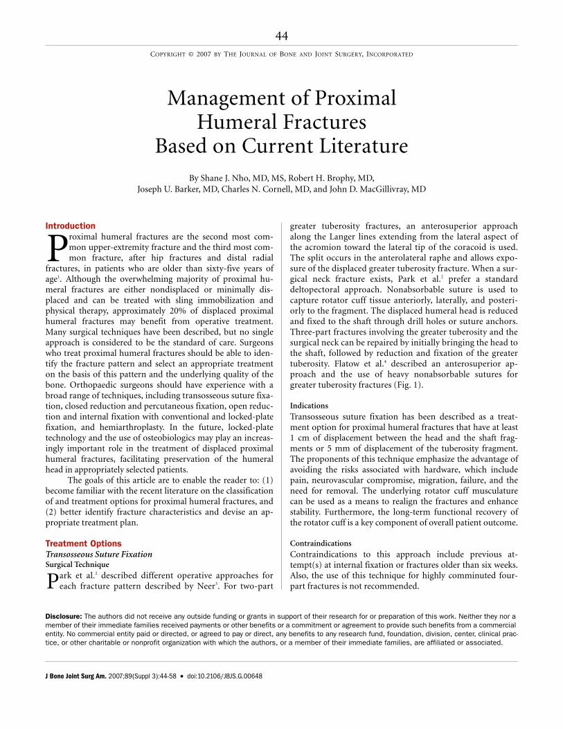
Fig. 3-A
Intraoperative photo of a patient in the beach-chair position with the c-arm over the operative shoulder.
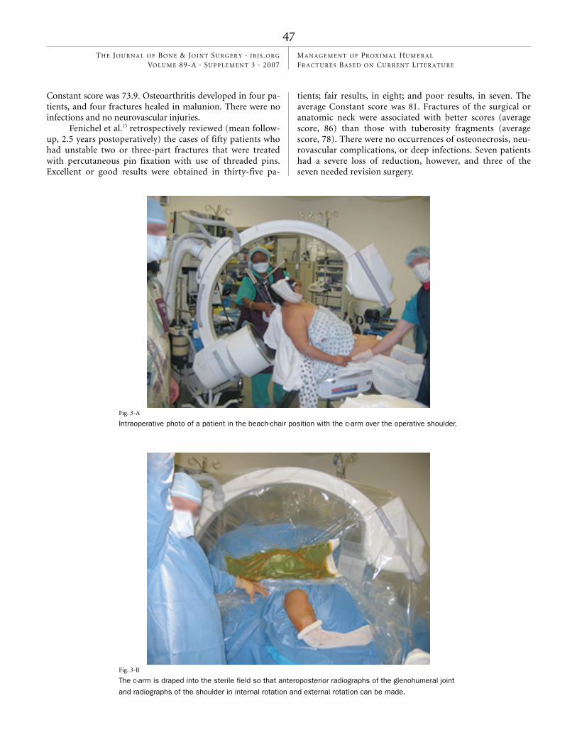
Fig. 3-B
The c-arm is draped into the sterile field so that anteroposterior radiographs of the glenohumeral joint
and radiographs of the shoulder in internal rotation and external rotation can be made.
Nho_00648.fm Page 47 Monday, September 10, 2007 2:01 PM
48
THE JOU R N A L OF BO N E & JO I N T SU RG ER Y · JB JS .ORG
VO LUM E 89-A · SU P P L E M E N T 3 · 2007MA N AG E M EN T OF PROX I M A L HU M E R A L FR A C T URE S BA S E D ON CUR REN T LI TER AT U RE
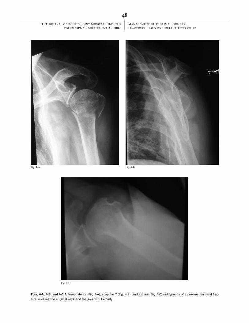
Fig. 4-A
Figs. 4-A, 4-B, and 4-C Anteroposterior (Fig. 4-A), scapular Y (Fig. 4-B), and axillary (Fig. 4-C) radiographs of a proximal humeral frac-
ture involving the surgical neck and the greater tuberosity.
Fig. 4-B
Fig. 4-C
Nho_00648.fm Page 48 Monday, September 10, 2007 2:01 PM

49
THE JOU R N A L OF BO N E & JO I N T SU RG ER Y · JB JS .ORG
VO LUM E 89-A · SU P P L E M E N T 3 · 2007MA N AG E M EN T OF PROX I M A L HU M E R A L FR A C T URE S BA S E D ON CUR REN T LI TER AT U RE
Open Reduction and Internal Fixation—Conventional PlateSurgical Technique—Double-Plate Fixation
Wanner et al.16 used two one-third tubular plates to treat pa-tients with two, three, or four-part proximal humeral frac-tures. A standard deltopectoral approach was used to gainaccess to the fracture. An emphasis was placed on anatomicalreduction, with particular attention given to reducing thegreater and lesser tuberosities and achieving correct length ofthe humeral shaft and retroversion of the head. Lateral platefixation to reduce the greater tuberosity was achieved first,typically with a five or six-hole one-third tubular plate. Thiswas followed by fixation of a ventral plate at a 90° angle to thelateral plate. A four-hole one-third tubular plate with oneproximal and one distal screw was usually used. Bone cementwas injected into the screw holes when bone quality wasdeemed to be poor.
Indications
Prior to the use of locking-plate technology, conventionalplate fixation was used for the majority of patients who hadopen reduction and internal fixation of proximal humeralfractures. Many different plates have been used in a variety ofsupplementation techniques16-21. The poor bone quality in thisregion of the proximal part of the humerus results in a de-creased ability to secure these conventional plates withscrews22. The loosening and pull-out of screws are commonreasons for failure23. Traditional plate techniques can still pro-vide satisfactory outcome when anatomic reduction can beachieved18. Furthermore, double-plating techniques as de-scribed by Wanner et al. can provide satisfactory outcome16.
Contraindications
Traditional plate constructs are usually reserved for young pa-tients with an intact medial hinge, an adequate diaphyseal cor-tex (>4 mm), and no metaphyseal comminution. Patients whohave osteoporosis or whose fracture lacks any of the abovecharacteristics would likely benefit from locking-plate tech-nology. The disadvantage of traditional plating systems is thehigh rate of osteonecrosis due to extensive soft-tissue dissec-tion. The rate of osteonecrosis has been reported to be as highas 35% in some case series18,21.
Results
Wanner et al.16 treated sixty shoulders with one-third tubularplates fixed orthogonally on the anterior and lateral cortices.Sixty-three percent of the patients had good or very good re-sults. Seven patients (12%) had complications that includedfracture displacement, osteonecrosis, adhesive capsulitis, sub-acromial impingement, and hardware loosening. Conven-tional plate fixation may produce satisfactory clinical resultseven in the setting of osteonecrosis18,21; however, anatomic re-duction with conventional plate fixation in the absence of os-teonecrosis produces superior clinical results18.
Open Reduction and Internal Fixation—Locked PlateSurgical TechniqueWe prefer to place the patient in the beach-chair positionwith the c-arm placed over the shoulder and draped into thesterile field. The c-arm fluoroscopic image intensifier pro-vides an anteroposterior view of the glenohumeral joint, andthe humerus can be rotated to obtain radiographs of the
Fig. 4-D
Tagging sutures are used to obtain reduction of the tuberosity fracture and then are
passed through the suture holes in the proximal humeral locking plate.
Nho_00648.fm Page 49 Monday, September 10, 2007 2:01 PM
50
THE JOU R N A L OF BO N E & JO I N T SU RG ER Y · JB JS .ORG
VO LUM E 89-A · SU P P L E M E N T 3 · 2007MA N AG E M EN T OF PROX I M A L HU M E R A L FR A C T URE S BA S E D ON CUR REN T LI TER AT U RE
shoulder in internal and external rotation (Figs. 3-A and 3-B).We use a standard deltopectoral approach to the shoulder.The anterior third of the deltoid may be reflected to allowgreater exposure of the proximal part of the humerus. Therotator cuff tendons are tagged with multiple number-2braided nonabsorbable sutures, whether as a part of a tuber-
osity fragment or in continuity with the head fragment. Thetagging sutures are used to bring the tuberosity fragments incontinuity with the lateral cortex of the shaft fragment,which may indirectly reduce the head fragment to the shaft.If the head fragment is impacted onto the shaft, a periostealelevator can be inserted into the fracture site to disimpact
Fig. 4-F
Fig. 4-E
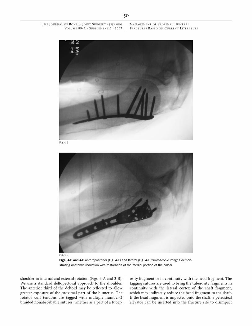
Figs. 4-E and 4-F Anteroposterior (Fig. 4-E) and lateral (Fig. 4-F) fluoroscopic images demon-
strating anatomic reduction with restoration of the medial portion of the calcar.
Nho_00648.fm Page 50 Monday, September 10, 2007 2:01 PM

51
THE JOU R N A L OF BO N E & JO I N T SU RG ER Y · JB JS .ORG
VO LUM E 89-A · SU P P L E M E N T 3 · 2007MA N AG E M EN T OF PROX I M A L HU M E R A L FR A C T URE S BA S E D ON CUR REN T LI TER AT U RE
the head and thus restore the medial portion of the calcar.When the fracture is anatomically reduced, the tagging su-tures are passed through the suture holes of the proximal hu-meral locking plate. The plate should be positioned directlyon the middle of the lateral cortex and approximately 8 mmdistal to the superior aspect of the greater tuberosity. Someplates have an oblong hole at the level of the humeral shaftinto which a cortical screw can be partially advanced to allowthe height of the plate to be adjusted; once the plate is in thecorrect position, the cortical screw can be completely ad-vanced to secure the plate. With use of the insertion guideand sleeve assembly, the locked screws can be placed in thehumeral head. After achieving fixation in the humeral head,at least three diaphyseal screws are inserted. The final fluoro-scopic images should demonstrate anatomic reduction of theproximal humeral fracture (Figs. 4-A through 4-F).
Indications
For AO/ASIF type-B (bifocal) and type-C (anatomic neck)proximal humeral fractures24, humeral head preservation maybe possible with locked-plate fixation supplemented with localbone graft or bone-graft substitute. Because of the fixed-anglerelationship between the plate and screws, locked-plate fixa-tion provides a mechanical advantage in fractures with meta-physeal comminution, particularly when there is insufficientosseous contact opposite the plate25. The indications for locked-plate fixation continue to evolve as long-term outcomes afterlocked-plate fixation for proximal humeral fractures becomeavailable.
Contraindications
Open reduction and internal fixation with locked-plate fixa-tion is contraindicated in some fracture-dislocations, head-splitting fractures, and impression fractures that involve>40% of the articular surface26-28.
Pearls and Pitfalls
As in other applications of locked-plate fixation, the proximalhumeral fracture must be reduced prior to placement of thehardware. The precontoured proximal humeral locking platemust be placed at the appropriate height, as an excessivelysuperior position may cause impingement of the plate on theacromion. Restoration of the medial hinge is critical to suc-cessful anatomic healing of the proximal humeral fracture. Ifthe medial hinge is disrupted, it must be reduced. Some advo-cate the use of a 2.0-mm intramedullary plate to maintain thereduction (Figs. 5-A and 5-B)29. In cases of comminution ormalreduction of the medial hinge, the placement of calcar-specific screws is critical to support the medial column andtherefore maintain fracture reduction30. If calcar screws arenecessary, the plate must be positioned to ensure that thescrew will purchase the inferior part of the calcar30.
ResultsFrom 2002 to 2004, the senior authors (C.N.C. and J.D.MacG.)managed patients with AO/ASIF type-B proximal humeralfractures with open reduction and locked-plate fixation. Eightpatients (six women and two men) with an average age ofsixty-nine years and a mean follow-up of fifteen months were
Fig. 5-A
Figs. 5-A and 5-B Drawing showing a proximal humeral fracture with a disrupted medial hinge
(Fig. 5-A) that has been reconstituted with a 2.0-mm intramedullary plate as a preliminary re-
duction (Fig. 5-B). (Reproduced, with permission, from: Sperling JW, Cuomo F, Hill JD, Hertel R,
Chuinard C, Boileau P. The difficult proximal humerus fracture: tips and techniques to avoid
complications and improve results. Instr Course Lect. 2007;56:52.)
Fig. 5-B
Nho_00648.fm Page 51 Monday, September 10, 2007 2:01 PM

52
THE JOU R N A L OF BO N E & JO I N T SU RG ER Y · JB JS .ORG
VO LUM E 89-A · SU P P L E M E N T 3 · 2007MA N AG E M EN T OF PROX I M A L HU M E R A L FR A C T URE S BA S E D ON CUR REN T LI TER AT U RE
evaluated with subjective questionnaires, physical examina-tion, and plain radiographs. The injured shoulders demon-strated a mean forward flexion of 110° and a mean abductionof 99°. The contralateral shoulders exhibited a mean forwardflexion of 168° and a mean abduction of 159°. Shoulderstrength was slightly lower in the injured shoulders (mean,13.2 lb [5.9 kg]) than in the contralateral shoulders (mean,15.9 lb [7.2 kg]). The mean Constant score for the injuredshoulders was 70.4 compared with 88.8 for the contralateralshoulders. The mean Neer score was 76.3 for the injured and95.4 for the contralateral shoulders. All radiographs demon-strated evidence of excellent healing and well-positioned im-plants. There was no evidence of hardware loosening, failure,or nonunion.
HemiarthroplastySurgical TechniqueThe technique for shoulder hemiarthroplasty is well de-scribed in the literature and follows the principles originallyoutlined by Neer31-35. Typically, a standard deltopectoral ap-
Fig. 6-A
Figs. 6-A through 6-D Anatomical suture technique38. Fig. 6-A Sutures
are placed in the greater tuberosity fragment and the humeral shaft.
Fig. 6-B
Figs. 6-B and 6-C Sutures are passed medial to the humeral neck and subsequently tied around the greater tuberosity fragment while the involved
arm is held in neutral position. The lesser tuberosity fragment is fixed with the remaining two sutures.
Fig. 6-C
Nho_00648.fm Page 52 Monday, September 10, 2007 2:01 PM
53
THE JOU R N A L OF BO N E & JO I N T SU RG ER Y · JB JS .ORG
VO LUM E 89-A · SU P P L E M E N T 3 · 2007MA N AG E M EN T OF PROX I M A L HU M E R A L FR A C T URE S BA S E D ON CUR REN T LI TER AT U RE
proach is used. Deep soft-tissue dissection may be minimal,depending on the fracture pattern and soft-tissue injury. It isessential to identify and tag the tuberosities with use of suturesat the bone-tendon junction to gain control of the rotator cuffand its insertion. The humeral head and shaft fragments arethen exposed, and sutures are passed through drill holes in theshaft. The glenoid should be carefully inspected to determineif a glenoid component is warranted.
Once the joint has been adequately exposed andcleaned, preparation of the humeral shaft begins. It is criti-cal to place the humeral component so that it has the cor-rect amount of height and retroversion (typically 30° to40°); to accomplish this, the bicipital groove can be used asa landmark36. Different trial modular heads can be used toidentify the optimal configuration. For most joints, the stemshould be cemented to ensure rotational control of the pros-thesis. Once the humeral component is secure, bone graftmay be placed to promote healing between the tuberositiesand the shaft.
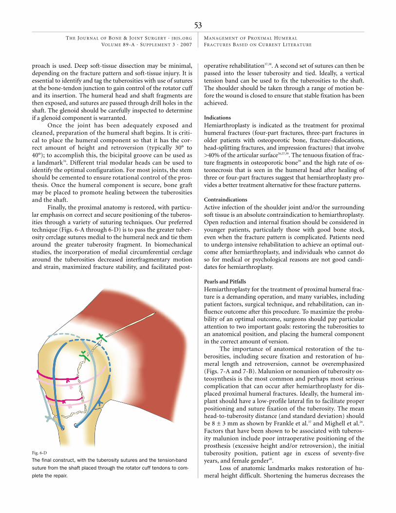
Finally, the proximal anatomy is restored, with particu-lar emphasis on correct and secure positioning of the tuberos-ities through a variety of suturing techniques. Our preferredtechnique (Figs. 6-A through 6-D) is to pass the greater tuber-osity cerclage sutures medial to the humeral neck and tie themaround the greater tuberosity fragment. In biomechanicalstudies, the incorporation of medial circumferential cerclagearound the tuberosities decreased interfragmentary motionand strain, maximized fracture stability, and facilitated post-
operative rehabilitation37,38. A second set of sutures can then bepassed into the lesser tuberosity and tied. Ideally, a verticaltension band can be used to fix the tuberosities to the shaft.The shoulder should be taken through a range of motion be-fore the wound is closed to ensure that stable fixation has beenachieved.
IndicationsHemiarthroplasty is indicated as the treatment for proximalhumeral fractures (four-part fractures, three-part fractures inolder patients with osteoporotic bone, fracture-dislocations,head-splitting fractures, and impression fractures) that involve>40% of the articular surface26,27,39. The tenuous fixation of frac-ture fragments in osteoporotic bone39 and the high rate of os-teonecrosis that is seen in the humeral head after healing ofthree or four-part fractures suggest that hemiarthroplasty pro-vides a better treatment alternative for these fracture patterns.
Contraindications
Active infection of the shoulder joint and/or the surroundingsoft tissue is an absolute contraindication to hemiarthroplasty.Open reduction and internal fixation should be considered inyounger patients, particularly those with good bone stock,even when the fracture pattern is complicated. Patients needto undergo intensive rehabilitation to achieve an optimal out-come after hemiarthroplasty, and individuals who cannot doso for medical or psychological reasons are not good candi-dates for hemiarthroplasty.
Pearls and Pitfalls
Hemiarthroplasty for the treatment of proximal humeral frac-ture is a demanding operation, and many variables, includingpatient factors, surgical technique, and rehabilitation, can in-fluence outcome after this procedure. To maximize the proba-bility of an optimal outcome, surgeons should pay particularattention to two important goals: restoring the tuberosities toan anatomical position, and placing the humeral componentin the correct amount of version.
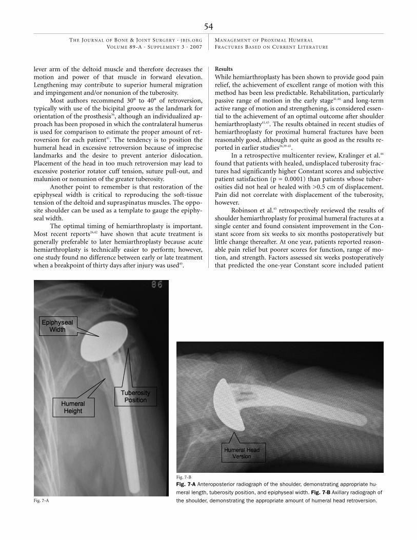
The importance of anatomical restoration of the tu-berosities, including secure fixation and restoration of hu-meral length and retroversion, cannot be overemphasized(Figs. 7-A and 7-B). Malunion or nonunion of tuberosity os-teosynthesis is the most common and perhaps most seriouscomplication that can occur after hemiarthroplasty for dis-placed proximal humeral fractures. Ideally, the humeral im-plant should have a low-profile lateral fin to facilitate properpositioning and suture fixation of the tuberosity. The meanhead-to-tuberosity distance (and standard deviation) shouldbe 8 ± 3 mm as shown by Frankle et al.37 and Mighell et al.26.Factors that have been shown to be associated with tuberos-ity malunion include poor intraoperative positioning of theprosthesis (excessive height and/or retroversion), the initialtuberosity position, patient age in excess of seventy-fiveyears, and female gender40.
Loss of anatomic landmarks makes restoration of hu-meral height difficult. Shortening the humerus decreases the
Fig. 6-D
The final construct, with the tuberosity sutures and the tension-band
suture from the shaft placed through the rotator cuff tendons to com-
plete the repair.
Nho_00648.fm Page 53 Monday, September 10, 2007 2:01 PM
54
THE JOU R N A L OF BO N E & JO I N T SU RG ER Y · JB JS .ORG
VO LUM E 89-A · SU P P L E M E N T 3 · 2007MA N AG E M EN T OF PROX I M A L HU M E R A L FR A C T URE S BA S E D ON CUR REN T LI TER AT U RE
lever arm of the deltoid muscle and therefore decreases themotion and power of that muscle in forward elevation.Lengthening may contribute to superior humeral migrationand impingement and/or nonunion of the tuberosity.
Most authors recommend 30° to 40° of retroversion,typically with use of the bicipital groove as the landmark fororientation of the prosthesis36, although an individualized ap-proach has been proposed in which the contralateral humerusis used for comparison to estimate the proper amount of ret-roversion for each patient41. The tendency is to position thehumeral head in excessive retroversion because of impreciselandmarks and the desire to prevent anterior dislocation.Placement of the head in too much retroversion may lead toexcessive posterior rotator cuff tension, suture pull-out, andmalunion or nonunion of the greater tuberosity.
Another point to remember is that restoration of theepiphyseal width is critical to reproducing the soft-tissuetension of the deltoid and supraspinatus muscles. The oppo-site shoulder can be used as a template to gauge the epiphy-seal width.
The optimal timing of hemiarthroplasty is important.Most recent reports26,42 have shown that acute treatment isgenerally preferable to later hemiarthroplasty because acutehemiarthroplasty is technically easier to perform; however,one study found no difference between early or late treatmentwhen a breakpoint of thirty days after injury was used43.
Results
While hemiarthroplasty has been shown to provide good painrelief, the achievement of excellent range of motion with thismethod has been less predictable. Rehabilitation, particularlypassive range of motion in the early stage41,44 and long-termactive range of motion and strengthening, is considered essen-tial to the achievement of an optimal outcome after shoulderhemiarthroplasty41,43. The results obtained in recent studies ofhemiarthroplasty for proximal humeral fractures have beenreasonably good, although not quite as good as the results re-ported in earlier studies26,39-45.
In a retrospective multicenter review, Kralinger et al.44
found that patients with healed, undisplaced tuberosity frac-tures had significantly higher Constant scores and subjectivepatient satisfaction (p = 0.0001) than patients whose tuber-osities did not heal or healed with >0.5 cm of displacement.Pain did not correlate with displacement of the tuberosity,however.
Robinson et al.45 retrospectively reviewed the results ofshoulder hemiarthroplasty for proximal humeral fractures at asingle center and found consistent improvement in the Con-stant score from six weeks to six months postoperatively butlittle change thereafter. At one year, patients reported reason-able pain relief but poorer scores for function, range of mo-tion, and strength. Factors assessed six weeks postoperativelythat predicted the one-year Constant score included patient
Fig. 7-A
Fig. 7-B
Fig. 7-A Anteroposterior radiograph of the shoulder, demonstrating appropriate hu-
meral length, tuberosity position, and epiphyseal width. Fig. 7-B Axillary radiograph of
the shoulder, demonstrating the appropriate amount of humeral head retroversion.
Nho_00648.fm Page 54 Monday, September 10, 2007 2:01 PM

55
THE JOU R N A L OF BO N E & JO I N T SU RG ER Y · JB JS .ORG
VO LUM E 89-A · SU P P L E M E N T 3 · 2007MA N AG E M EN T OF PROX I M A L HU M E R A L FR A C T URE S BA S E D ON CUR REN T LI TER AT U RE
Fig. 8-C
Fig. 8-C Undisplaced medial hinge. Fig. 8-D Medial hinge with >2 mm of displacement. (Reprinted, with modification, with permission from: Hertel
R, Hempfing A, Stiehler M, Leunig M. Predictors of humeral head ischemia after intracapsular fracture of the proximal humerus. J Shoulder Elbow
Surg. 2004;13:427-33.)
Fig. 8-D
Fig. 8-A
Figs. 8-A through 8-D Hertel radiographic criteria. Fig. 8-A Metaphyseal extension of the humeral head of >9 mm. Fig. 8-B Metaphyseal extension
of the humeral head of <8 mm.
Fig. 8-B
Nho_00648.fm Page 55 Monday, September 10, 2007 2:01 PM
56
THE JOU R N A L OF BO N E & JO I N T SU RG ER Y · JB JS .ORG
VO LUM E 89-A · SU P P L E M E N T 3 · 2007MA N AG E M EN T OF PROX I M A L HU M E R A L FR A C T URE S BA S E D ON CUR REN T LI TER AT U RE
age, a persistent neurological deficit, the need for early reoper-ation, and the degree of displacement of both the prosthetichead (<5 mm displacement of the prosthetic head from thecentral axis of the glenoid) and the tuberosities.
Discussionhe treatment of displaced proximal humeral fractures iscomplex and requires careful assessment of patient factors
(such as age and activity level) and fracture-related factors(such as bone quality, fracture pattern, degree of comminu-tion, and vascular status). The goal of treatment is a pain-freeshoulder with restoration of pre-injury function.
The first step is to assess the vascular status of the hu-meral head with use of the Hertel radiographic criteria forperfusion of the humeral head46 and the AO/ASIF classifica-tion of fractures of the proximal part of the humerus24. In theHertel criteria, metaphyseal extension of the humeral head of<8 mm and medial hinge disruption of >2 mm were de-termined to be good predictors of ischemia (Figs. 8-A through8-D)46. The combination of metaphyseal extension of the hu-meral head, medial hinge disruption of >2 mm, and an ana-tomic neck fracture pattern had a 97% positive predictivevalue for humeral head ischemia46. The AO/ASIF classifica-tion for proximal humeral fractures provides information
about the energy and severity of the fracture and the likeli-hood of vascular injury. Type A is a unifocal, extra-articularfracture with an intact vascular supply. Type B is a bifocal,extra-articular fracture with possible injury to the vascularsupply. Type C is an articular fracture of the anatomic neckwith a high probability of osteonecrosis24.
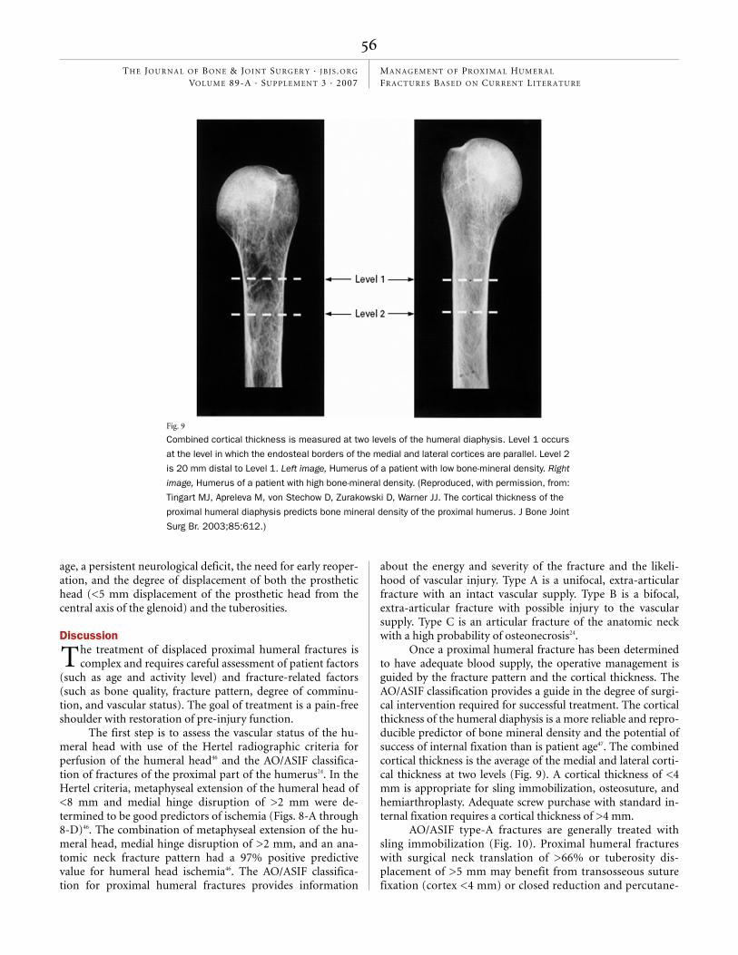
Once a proximal humeral fracture has been determinedto have adequate blood supply, the operative management isguided by the fracture pattern and the cortical thickness. TheAO/ASIF classification provides a guide in the degree of surgi-cal intervention required for successful treatment. The corticalthickness of the humeral diaphysis is a more reliable and repro-ducible predictor of bone mineral density and the potential ofsuccess of internal fixation than is patient age47. The combinedcortical thickness is the average of the medial and lateral corti-cal thickness at two levels (Fig. 9). A cortical thickness of <4mm is appropriate for sling immobilization, osteosuture, andhemiarthroplasty. Adequate screw purchase with standard in-ternal fixation requires a cortical thickness of >4 mm.
AO/ASIF type-A fractures are generally treated withsling immobilization (Fig. 10). Proximal humeral fractureswith surgical neck translation of >66% or tuberosity dis-placement of >5 mm may benefit from transosseous suturefixation (cortex <4 mm) or closed reduction and percutane-
T
Fig. 9
Combined cortical thickness is measured at two levels of the humeral diaphysis. Level 1 occurs
at the level in which the endosteal borders of the medial and lateral cortices are parallel. Level 2
is 20 mm distal to Level 1. Left image, Humerus of a patient with low bone-mineral density. Right
image, Humerus of a patient with high bone-mineral density. (Reproduced, with permission, from:
Tingart MJ, Apreleva M, von Stechow D, Zurakowski D, Warner JJ. The cortical thickness of the
proximal humeral diaphysis predicts bone mineral density of the proximal humerus. J Bone Joint
Surg Br. 2003;85:612.)
Nho_00648.fm Page 56 Monday, September 10, 2007 2:01 PM

57
THE JOU R N A L OF BO N E & JO I N T SU RG ER Y · JB JS .ORG
VO LUM E 89-A · SU P P L E M E N T 3 · 2007MA N AG E M EN T OF PROX I M A L HU M E R A L FR A C T URE S BA S E D ON CUR REN T LI TER AT U RE
ous fixation (cortex >4 mm). For multifragment fractures,open reduction with locked-plate fixation allows preserva-tion of the periosteal blood supply and is appropriate forcortices less than or greater than 4 mm, regardless of thethickness of the cortex. Recent studies have emphasized theimportance of stabilizing the medial column to preventvarus malunion, plate failure, screw cutout, and impinge-ment. In cases with an adequate medial cortex, the medialhinge should be reduced; in cases with medial comminution,calcar-specific screws should be used to stabilize the medialcolumn. Local adjuvants, such as calcium phosphate cement,demineralized bone matrix, or allografts, may improve therate of union and minimize malunion. Hemiarthroplasty is
the treatment of choice for fractures with vascular compro-mise, certain fracture-dislocations, head-splitting fractures,and impression fractures with >40% of articular surface in-volvement, although locking-plate technology is allowingsurgeons to attempt fixation in some of these fractures, par-ticularly in younger patients.
Corresponding author:Shane J. Nho, MD, MSThe Hospital for Special Surgery, 535 East 70th Street, New York, NY 10021. E-mail address: [email protected]
References
1. Baron JA, Barrett JA, Karagas MR. The epidemiology of peripheral fractures. Bone. 1996;18(3 Suppl):209S-213S.
2. Park MC, Murthi AM, Roth NS, Blaine TA, Levine WN, Bigliani LU. Two-part and three-part fractures of the proximal humerus treated with suture fixation. J Orthop Trauma. 2003;17:319-25.
3. Neer CSI. Displaced proximal humeral fractures. Part I: classification and eval-uation. J Bone Joint Surg Am. 1970;52:1077-89.
4. Flatow EL, Cuomo F, Maday MG, Miller SR, McIlveen SJ, Bigliani LU. Open reduc-tion and internal fixation of two-part displaced fractures of the greater tuberosity of the proximal part of the humerus. J Bone Joint Surg Am. 1991;73:1213-8.
Fig. 10
Treatment algorithm for displaced proximal humeral fractures. 2-SN (<66%) = two-part surgical neck fracture with <66%
translation. 2-SN (>66%) = two-part surgical neck fracture with >66% translation. BG = bone graft or bone-graft substi-
tute. CRPF = closed reduction and percutaneous fixation. Hemi = hemiarthroplasty. LCP = locked compression plate.
ORIF = open reduction and internal fixation. TOSF = transosseous suture fixation. (Reproduced in modified form, with
permission, from: Nho SJ, Brophy RH, Barker JU, Cornell CN, MacGillivray JD. Innovations in the management of dis-
placed proximal humerus fractures. J Am Acad Orthop Surg. 2007;15:15.)
Nho_00648.fm Page 57 Monday, September 10, 2007 2:01 PM
58
THE JOU R N A L OF BO N E & JO I N T SU RG ER Y · JB JS .ORG
VO LUM E 89-A · SU P P L E M E N T 3 · 2007MA N AG E M EN T OF PROX I M A L HU M E R A L FR A C T URE S BA S E D ON CUR REN T LI TER AT U RE
5. Neer CS 2nd, Watson KC, Stanton FJ. Recent experience in total shoulder re-placement. J Bone Joint Surg Am. 1982;64:319-37
6. Panagopoulos AM, Dimakopoulos P, Tyllianakis M, Karnabatidis D, Siablis D, Papadopoulos AX, Lambiris E, Kraniotis P, Sakellaropoulos G. Valgus impacted proximal humeral fractures and their blood supply after transosseous suturing. Int Orthop. 2004;28:333-7.
7. Constant CR, Murley AH. A clinical method of functional assessment of the shoulder. Clin Orthop Relat Res. 1987;214:160-4.
8. Resch H, Povacz P, Frohlich R, Wambacher M. Percutaneous fixation of three- and four-part fractures of the proximal humerus. J Bone Joint Surg Br. 1997;79:295-300.
9. Resch H, Beck E, Bayley I. Reconstruction of the valgus-impacted humeral head fracture. J Shoulder Elbow Surg. 1995;4:73-80.
10. Keener JD, Parsons BO, Flatow EL, Rogers K, Williams GR, Galatz LM. Out-comes after percutaneous reduction and fixation of proximal humeral fractures. J Shoulder Elbow Surg. 2007;16:330-8.
11. Rowles DJ, McGrory JE. Percutaneous pinning of the proximal part of the humerus. An anatomic study. J Bone Joint Surg Am. 2001;83:1695-9.
12. Kamineni S, Ankem H, Sanghavi S. Anatomical considerations for percuta-neous proximal humeral fracture fixation. Injury. 2004;35:1133-6.
13. Jaberg H, Warner JJ, Jakob RP. Percutaneous stabilization of unstable frac-tures of the humerus. J Bone Joint Surg Am. 1992;74:508-15.
14. Richards RR, An KN, Bigliani LU, Friedman RJ, Gartsman GM, Gristina AG, Iannotti JP, Mow VC, Sidles JA, Zuckerman JD. A standardized method for the assessment of shoulder function. J Shoulder Elbow Surg. 1994;3:347-52.
15. Fenichel I, Oran A, Burstein G, Perry Pritsch M. Percutaneous pinning using threaded pins as a treatment option for unstable two- and three-part fractures of the proximal humerus: a retrospective study. Int Orthop. 2006;30:153-7.
16. Wanner GA, Wanner-Schmid E, Romero J, Hersche O, von Smekal A, Trentz O, Ertel W. Internal fixation of displaced proximal humeral fractures with two one-third tubular plates. J Trauma. 2003;54:536-44.
17. Hessmann M, Baumgaertel F, Gehling H, Klingelhoeffer I, Gotzen L. Plate fixa-tion of proximal humeral fractures with indirect reduction: surgical technique and results utilizing three shoulder scores. Injury. 1999;30:453-62.
18. Gerber C, Werner CM, Vienne P. Internal fixation of complex fractures of the proximal humerus. J Bone Joint Surg Br. 2004;86:848-55.
19. Robinson CM, Page RS. Severely impacted valgus proximal humeral frac-tures. Results of operative treatment. J Bone Joint Surg Am. 2003;85:1647-55.
20. Robinson CM, Page RS. Severely impacted valgus proximal humeral frac-tures. J Bone Joint Surg Am. 2004;86 Suppl 1(Pt 2):143-55.
21. Wijgman AJ, Roolker W, Patt TW, Raaymakers EL, Marti RK. Open reduction and internal fixation of three and four-part fractures of the proximal part of the humerus. J Bone Joint Surg Am. 2002;84:1919-25.
22. Cornell CN, Levine D, Pagnani MJ. Internal fixation of proximal humerus frac-tures using the screw-tension band technique. J Orthop Trauma. 1994;8:23-7.
23. Cornell CN. Internal fracture fixation in patients with osteoporosis. J Am Acad Orthop Surg. 2003;11:109-19.
24. Nho SJ, Brophy RH, Barker JU, Cornell CN, MacGillivray JD. Innovations in the management of displaced proximal humerus fractures. J Am Acad Orthop Surg. 2007;15:12-26.
25. Haidukewych GJ. Innovations in locking plate technology. J Am Acad Orthop Surg. 2004;12:205-12.
26. Mighell MA, Kolm GP, Collinge CA, Frankle MA. Outcomes of hemiarthroplasty for fractures of the proximal humerus. J Shoulder Elbow Surg. 2003;12:569-77.
27. Bosch U, Skutek M, Fremerey RW, Tscherne H. Outcome after primary and secondary hemiarthroplasty in elderly patients with fractures of the proximal humerus. J Shoulder Elbow Surg. 1998;7:479-84.
28. Zyto K, Ahrengart L, Sperber A, Törnkvist H. Treatment of displaced proximal humeral fractures in elderly patients. J Bone Joint Surg Br. 1997;79:412-7.
29. Sperling JW, Cuomo F, Hill JD, Hertel R, Chuinard C, Boileau P. The difficult proximal humerus fracture: tips and techniques to avoid complications and im-prove results. Instr Course Lect. 2007;56:45-57.
30. Gardner MJ, Weil Y, Barker JU, Kelly BT, Helfet DL, Lorich DG. The importance of medial support in locked plating of proximal humerus fractures. J Orthop Trauma. 2007;21:185-91.
31. Neer CS, Brown TH, McLaughlin HL. Fracture of the neck of the humerus with dislocation of the head fragment. Am J Surg. 1953;85:252-8.
32. Neer CS 2nd. Displaced proximal humeral fractures. II. Treatment of three-part and four-part displacement. J Bone Joint Surg Am. 1970;52:1090-103.
33. Compito CA, Self EB, Bigliani LU. Arthroplasty and acute shoulder trauma. Reasons for success and failure. Clin Orthop Relat Res. 1994;307:27-36.
34. Zuckerman JD, Cuomo F, Koval KJ. Proximal humeral replacement for complex fractures: indications and surgical technique. Instr Course Lect. 1997;46:7-14.
35. Dines DM, Warren RF. Modular shoulder hemiarthroplasty for acute frac-tures. Surgical considerations. Clin Orthop Relat Res. 1994;307:18-26.
36. Kontakis GM, Damilakis J, Christoforakis J, Papadakis A, Katonis P, Prassopoulos P. The bicipital groove as a landmark for orientation of the humeral prosthesis in cases of fracture. J Shoulder Elbow Surg. 2001;10:136-9.
37. Frankle MA, Ondrovic LE, Markee BA, Harris ML, Lee WE 3rd. Stability of tu-berosity reattachment in proximal humeral hemiarthroplasty. J Shoulder Elbow Surg. 2002;11:413-20.
38. Boileau P, Walch G, Krishnan SG. Tuberosity osteosynthesis and hemiarthro-plasty for four-part fractures of the proximal humerus. Tech Shoulder Elbow Surg. 2000;1:96-109.
39. Zyto K, Wallace WA, Frostick SP, Preston BJ. Outcome after hemiarthroplasty for three- and four-part fractures of the proximal humerus. J Shoulder Elbow Surg. 1998;7:85-9.
40. Boileau P, Krishnan SG, Tinsi L, Walch G, Coste JS, Mole D. Tuberosity malposition and migration: reasons for poor outcomes after hemiarthroplasty for displaced fractures of the proximal humerus. J Shoulder Elbow Surg. 2002;11:401-12.
41. Christoforakis JJ, Kontakis GM, Katonis PG, Stergiopoulos K, Hadjipavlou AG. Shoulder hemiarthroplasty in the management of humeral head fractures. Acta Orthop Belg. 2004;70:214-8.
42. Becker R, Pap G, Machner A, Neumann WH. Strength and motion after hemi-arthroplasty in displaced four-fragment fracture of the proximal humerus: 27 pa-tients followed for 1-6 years. Acta Orthop Scand. 2002;73:44-9.
43. Prakash U, McGurty DW, Dent JA. Hemiarthroplasty for severe fractures of the proximal humerus. J Shoulder Elbow Surg. 2002;11:428-30.
44. Kralinger F, Schwaiger R, Wambacher M, Farrell E, Menth-Chiari W, Lajtai G, Hübner C, Resch H. Outcome after primary hemiarthroplasty for fracture of the head of the humerus. A retrospective multicentre study of 167 patients. J Bone Joint Surg Br. 2004;86:217-9.
45. Robinson CM, Page RS, Hill RM, Sanders DL, Court-Brown CM, Wakefield AE. Primary hemiarthroplasty for treatment of proximal humeral fractures. J Bone Joint Surg Am. 2003;85:1215-23.
46. Hertel R, Hempfing A, Stiehler M, Leunig M. Predictors of humeral head is-chemia after intracapsular fracture of the proximal humerus. J Shoulder Elbow Surg. 2004;13:427-33.
47. Tingart MJ, Apreleva M, von Stechow D, Zurakowski D, Warner JJ. The cortical thickness of the proximal humeral diaphysis predicts bone mineral density of the proximal humerus. J Bone Joint Surg Br. 2003;85:611-7.
Nho_00648.fm Page 58 Monday, September 10, 2007 2:01 PM
Related Documents