1 ACADEMIE UNIVERSITAIRE WALLONIE-EUROPE UNIVERSITE DE LIEGE FACULTE DE MEDECINE VETERINAIRE DEPARTEMENT DES SCIENCES CLINIQUES DES ANIMAUX DE COMPAGNIE ET DES EQUIDES PATHOLOGIE MEDICALE DES ANIMAUX DE COMPAGNIE EVALUATION DE MARQUEURS D’INFLAMMATION, DE BIOMARQUEURS CARDIAQUES ET DE LA FONCTION CARDIAQUE DANS LE SYNDROME DE REPONSE D’INFLAMMATION SYSTEMIQUE CHEZ LE CHIEN EVALUATION OF INFLAMMATORY MARKERS, CARDIAC BIOMARKERS AND CARDIAC FUNCTION IN THE SYSTEMIC INFLAMMATORY RESPONSE SYNDROME IN THE DOG Kris GOMMEREN THESE PRESENTEE EN VUE DE L’OBTENTION DU GRADE DE DOCTEUR EN SCIENCE VETERINAIRE ANNEE ACADEMIQUE 2016-2017

Welcome message from author
This document is posted to help you gain knowledge. Please leave a comment to let me know what you think about it! Share it to your friends and learn new things together.
Transcript
1
ACADEMIE UNIVERSITAIRE WALLONIE-EUROPE
UNIVERSITE DE LIEGE
FACULTE DE MEDECINE VETERINAIRE
DEPARTEMENT DES SCIENCES CLINIQUES DES ANIMAUX DE COMPAGNIE ET DES
EQUIDES
PATHOLOGIE MEDICALE DES ANIMAUX DE COMPAGNIE
EVALUATION DE MARQUEURS D’INFLAMMATION, DE BIOMARQUEURS
CARDIAQUES ET DE LA FONCTION CARDIAQUE DANS LE SYNDROME DE REPONSE
D’INFLAMMATION SYSTEMIQUE CHEZ LE CHIEN
EVALUATION OF INFLAMMATORY MARKERS, CARDIAC BIOMARKERS AND
CARDIAC FUNCTION IN THE SYSTEMIC INFLAMMATORY RESPONSE SYNDROME
IN THE DOG
Kris GOMMEREN
THESE PRESENTEE EN VUE DE L’OBTENTION DU GRADE DE
DOCTEUR EN SCIENCE VETERINAIRE
ANNEE ACADEMIQUE 2016-2017

2

3
WORDS OF GRATITUDE
At the end of this journey I want to take the time to thank the people that helped launching this project,
and made sure it came to an end. First of all, none of this would have been possible, without the staff of
the small animal university clinic understanding the need for better emergency and critical care at their
institution. Without their support, I would never have decided to invest myself in this “development
project” which François would describe as a “North-South” transfer .
Secondly, Dominique Peeters deserves to be praised (a lot). When I arrived at this university, I soon
found out that Dominique shares many flaws with me, such as being overly direct and stubborn.
However, Dominique also had the patience to let me undertake a research project that was situated miles
away from his comfort zone, and has taken the time to familiarize himself with my weird thinking
patterns. If he wouldn’t have been there to calm me down and get me back on track at the appropriate
times, this PhD surely would only have ended somewhere around May 2045.
Dr. Natali Bauer, Prof. Joachim Roth, Prof. Andreas Moritz, Prof. Kathleen McEntee and Prof. Soren
Boysen also merit special credit. Dr. Bauer and Professor Moritz made the measurements of the
inflammatory markers possible, and without Professor Roth I would never have been able to interpret
our findings and compare them with the available literature. Professor McEntee and Professor Boysen
introduced me into the world of cardiac ultrasonography, FAST ultrasound, and cardiac biomarkers.
You have widened my horizon and the knowledge I have acquired thanks to you will hopefully one day
help me to help ECC patients. Soren, looking forward to continuing this research with you whilst having
a couple of beers and watching some soccer!
I would also like to thank the members of the jury. Undoubtedly I owe you an apology for the extensive
literature review that I wrote in the initial document, and I hope you will find these revised manuscript
easier to digest. All of your comments improved the scientific value of this document tremendously.
The flow diagrams, reports on correlation, improvement of figures and correction of silly typos with
massive implications were all very much appreciated. Many people don’t know that you all do this on a
voluntary basis, drive by nothing but passion.
As I did spend a couple of years working on the findings of this project, in between clinics, lectures and
work for the European Veterinary Emergency and Critical Care Society, it would be easy to forget how
it all started. Most of the practical work that has been performed was performed by two extremely
motivated (or perhaps naïve?) interns: Isabelle Desmas and Alexandra Garcia. Besides being awesome
veterinarians, they are both lovely human beings, and wonderful colleagues and I have nothing but
gratitude for how they devoted their time to this project. It was an honor to get to know you girls.
The past eight years I also shared my office space with special people such as Elise Mercier and Kiki
Merveille, and the last years with a bunch of ‘visiting’ clinicians. These people were extremely helpful

4
not only for being able to stand my loud music and smelly socks, but I also want to thank them for their
kind friendship, and their intellectual support whenever I was struggling.
Performing this PhD also made it obvious to me that I’m much more a clinician than a researcher,
although clinical research will always remain a passion. Working on this project often meant less time
in clinics. Clinics that are ran by our residents, interns, ‘oriented interns’, and our support staff. Finishing
this project also would never have been possible if it weren’t for the arrival of Liz-Valerie, who gave
me the feeling I could turn my back on our ECC patients knowing they were in extremely well trained
hands. Hopefully they all know how much I appreciate their efforts, how guilty I’ve felt whenever I was
unavailable. Hopefully the little time I had to spare could still be appreciated. I’m already looking
forward to being on the floor again, being able to prepare the transition of our ECC department into the
new clinic, and to be able to continue performing clinical research with future colleagues, residents
interns, and students.
Although they’ll probably never read this, I also want to thank my non-veterinary friends. Thank God
you exist and allow me to talk about something else than dogs or cats for a change. Without the fun you
all bring to my life, I would not be able to find the energy to do what I do, including this project. I’ll
also take the opportunity to thank my parents and my sisters. I know they are always struggling to
understand what I do, or what I don’t do (No I don’t operate…! No I don’t do vaccines…! Yes, I do
have a job dad, you can stop worrying!). The past 15 years I’ve been away or absent a lot, and I might
not have been the son, brother or uncle you wished for. Well, don’t get your hopes up to high, finishing
this probably won’t change anything on that level, as I’ll undoubtedly keep all of the other irritating
habits I have. I will however do the best I can to no longer miss any festivities, and I’ll be the best uncle
and godfather I can be.
Finally, Liesbeth, I need to thank you for making me the happy man I am. Falling in love with you
undoubtedly is the best thing that ever happened to me, marrying you the best decision I ever took
(moving to Leut remains debatable however ). Thanks for being there, and being you, coping with me,
being me… Thanks for being an amazing, passionate general practitioner, a brilliant mother for Hanne
and Jeff, and a lovely companion for me on this road that’s called life. Looking forward to build our
house together and grow old there with you .
THANKS
Krisje G.

5
SUMMARY
The systemic inflammatory response syndrome (SIRS) accounts for a significant part of the clinical
syndrome of sepsis. SIRS is not limited to infectious causes, but can also be caused by non-infectious
inflammatory conditions such as, for example, pancreatitis1. SIRS is mediated by the release of pro-
inflammatory cytokines, such as TNF-α, IL-1β and IL-6, from activated macrophages and other sentinel
cells2. TNF-α and IL-1β are both produced early in inflammation, with rapidly declining concentrations3
that often are undetectable within 24 hours4-8, rendering both cytokines poor tools for diagnostic and
prognostic purposes in critical care patients. TNF-α and IL-1β induce the release of IL-69, which readily
circulates10. Moreover, IL-6 has a longer half-life than TNF-α and IL-1β11. IL-6 seems to be an
interesting marker of systemic inflammation and could potentially be an interesting prognostic marker
(increased mortality above 1000ng/L in humans)12. Concentrations however overlap too much to
distinguish infectious from non-infectious causes, although septic patients tend to demonstrate higher
levels. In canine medicine, evidence regarding the prognostic utility of IL-6 in SIRS and sepsis is
unequivocal11,13,14.
The main pro-inflammatory cytokines IL-6, IL-1β and TNF-α also initiate the acute phase response
(APR)9, characterized by increased concentration of acute phase proteins (APPs) leading to different
systemic effects such as fever, leukocytosis or metabolic changes15-17. APPs such as C-reactive protein
(CRP) allow for diagnosing systemic inflammation, evaluate the extent of ongoing lesions and the
severity of the disease, and may give prognostic information and evaluate the response to treatment17-25.
CRP concentrations usually are less than 5mg/L in healthy dogs and reference ranges vary from 0.22 to
16.4mg/L24. The late-coming peak of CRP at 36 to 48 hours after the start of the inflammatory process
may reduce the sensitivity of the marker to identify patients in SIRS in an emergency setting26. CRP
appears very useful to detect systemic inflammation in dogs27-29 while it does not seem useful to
distinguish septic and non-septic disease in dogs30 and is a poor marker of disease severity. This is easily
explained as CRP not only is influenced by the type of underlying disease and the timing of sampling,
but also by the definition of ‘disease severity’. According to literature, a single CRP concentration at
presentation probably does not add valuable prognostic information in SIRS patients, yet CRP-kinetics
might predict prognosis in dogs with SIRS30. Moreover, CRP-kinetics could be used to monitor disease
progression and the response to treatment27,30.
Currently the clinical diagnosis of SIRS in canine patients is based on finding two or more abnormalities
in clinical and basic laboratory parameters31,32, a clinical diagnosis which is highly sensitive, but poorly
specific33. We therefore wanted to evaluate whether dogs presented to the emergency department with
SIRS had measurable concentrations of the main inflammatory cytokines and CRP. In a cohort of 69
dogs, CRP was increased in 73.1% (49/67) of dogs at presentation, and remained within the reference
interval (0-14.9 mg/L) throughout hospitalization in only 6% (4/67) of cases. CRP decreased
significantly over time during treatment and hospitalization. At the time of the follow-up visit, CRP

6
measurements (2.4±4.5 mg/L) were within reference interval (0-14.9 mg/L) in 95% (18/19) of dogs.
CRP concentrations at presentation tended to be higher in dogs with SIRS due to an infectious cause,
but the difference was not statistically significant. The utility of CRP as a monitoring tool for treatment
evaluation in the acute phase appears limited based on the findings of this study. CRP concentrations
remained elevated during the initial 24 hours and were only mildly decreased by day 3 in survivors, and
therefore do not appear to be very informative to evaluate treatment efficacy.
As expected based on the available literature, TNF-α was detected in only a small percentile of patients
(29.0%), and this for a limited period. TNF-α concentrations still changed significantly over time and
values observed at T6, T12 and T24 were significantly different from observed concentrations at T72
and during the control visit. TNF-α shows a very early peak activity (within 2 hours), typically vanishes
within 6 hours after induction and rarely remains present for longer than 24 hours7,34-38. Therefore TNF-
α was expected to only be detectable in dogs presented with hyperacute disease such as gastric dilation
and volvulus (GDV) and trauma, while it probably would have been detectable at time points prior to
presentation in other dogs. IL-6 on the other hand is even detectable in the plasma of healthy dogs, but
reference ranges have not been described37. Concentrations of IL-6 changed significantly during
hospitalization, with concentrations at T0, T6 and T12 higher than at T72, T120 and the control visit.
Therefore IL-6 concentrations did indicate systemic inflammation in our population of dogs with a
clinical diagnosis of SIRS.
Additionally, CRP and IL-6 were significantly correlated (p <0.001 with r 0.605). Unfortunately, based
on our findings, neither CRP, IL-6 or TNF-α can predict underlying disease or outcome in dogs with
SIRS, and these biomarkers seem to be of limited value to evaluate treatment efficacy in canine
emergencies with a clinical diagnosis of SIRS.
In human medicine, it is generally accepted that SIRS and sepsis influence cardiac function in a large
percentile of these patients39. As an example, a quarter of hemodynamically unstable human critically
ill patients display significant LV systolic dysfunction40-42. TNF-α, IL-1β and IL-6 induce myocardial
depression in humans and in experimental studies in dogs43, and normalization of cardiac function is
associated with decreases in TNF-α and IL-6 concentrations44,45. This myocardial
depression/dysfunction/hibernation during SIRS is characterized by a variation of left and right
ventricular systolic and diastolic dysfunction, with potential ventricular dilation despite adequate
resuscitation. Modifications can resolve completely within 10 days to 4 weeks, and might serve as a
protective mechanism for the patient42,46. Unfortunately, little clinical information is however available
about the impact of SIRS and sepsis on cardiac function in dogs.
Although cardiac function was initially evaluated via invasive procedures, practical knowledge in
echocardiography has developed while the value of central venous and pulmonary arterial pressures has
been questioned40. This lead to an increased interest in the use of echocardiography for the evaluation

7
of cardiovascular function47,48. Echocardiography offers the benefits of direct visualization, allowing for
real-time assessment of cardiovascular structure and function48. Non-cardiologists can adequately
answer a limited amount of clinical questions via focused goal-oriented echocardiography, allowing to
titrate fluid therapy and hemodynamic care40,49. Left atrial size and the left ventricular diameter in
diastole estimate preload50,51, while fractional shortening (FS) evaluates ventricular systolic function52.
In veterinary medicine, left atrial size is typically assessed using LA/Ao-ratios51. Left ventricular
diameter in diastole is normalized according to bodyweight (nLVIDd) and is easily assessed in dogs,
just like FS53,54.
Despite experimental evidence of myocardial hibernation in dogs43,55,56, only few clinical studies
evaluated myocardial dysfunction via echocardiography in dogs in SIRS. A retrospective study in dogs
with critical (both septic- and non-septic) illness reports 16 dogs with poor cardiovascular function and
prognosis57. To the authors knowledge, no single study did ever prospectively evaluate cardiac effects
of SIRS in dogs. Although our study only included a limited number of dogs without severe hypotension,
it did identify a few interesting changes. In our study, dogs with SIRS did not display clear evidence of
cardiac dysfunction on echocardiography. Ventricular function (evaluated via FS) did not change during
hospitalization, however left atrial size (evaluated via the LA/Ao ratio) and left ventricular diameter
(expressed as nLVIDd) significantly increased during hospitalization. Heart rate was significantly
associated with prognosis. Despite not reaching significance, LA/Ao and nLVIDd were higher, while
FS was lower in survivors compared to non survivors during the initial 24 hours. Heart rate was
negatively correlated with LA/Ao and nLVIDd and positively correlated with FS. nLVIDd was
positively correlated with LA/Ao but negatively correlated with FS. The increase of nLVIDd and LA/Ao
during hospitalization could either be explained by the decreasing heart rate (mediated by decreasing
stress, pain relief, anti-inflammatory treatment or any other factor than hypovolemia). But might also
indicate a mild degree of hypovolemia in these patients. Whether the trend towards lower FS and higher
nLVIDd in survivors observed in this study are consequences of changing heart rates and sympathetic
tone, explained by changes in volume status, or early signs of myocardial hibernation, can unfortunately
not be determined in this study. The major limitation of this paper was the reluctance of clinicians in
charge to allow for rapid evaluation of cardiac function via ultrasonography. Consequently, findings of
this study are influenced by the inclusion of fewer dogs with in general less severe disease. Inclusion of
all presented canine emergencies in SIRS should allow to demonstrate more significant changes, and
needs to be the objective of future studies.
To avoid the necessity of a time-consuming and technique-requiring cardiac ultrasonography, cardiac
biomarkers might allow for indirect evaluation of the effects of systemic inflammation on cardiac
function. Cardiac troponins (cTnI and cTnT) are leakage markers, as increased myocyte permeability
secondary to irreversible or reversible injury causes the release of cTn into circulation58-61. Cardiac
8
troponin I is elevated in 43 to 85% of human critical patients62,63, while incidence varies from 36 to 69%
for cTnT64,65.
Increased cTnI concentration have been associated with increased pro-inflammatory cytokine levels (IL-
1β, IL-6 and TNF-α) in experimental and clinical studies in human critical patients62,66. cTn
concentrations are correlated with the severity of myocardial hibernation67, the severity of lesions68 and
with poor outcome62,65,68,69. However, as concentrations remain increased for over 50 hours in humans,
they are less useful to evaluate the response to therapeutic interventions70-73. Several studies looked into
cTn concentrations in canine SIRS populations presented to the ICU at the same time as when our
research was performed74-76. These papers demonstrated that increased cTnI and cTnT concentrations
are associated with short term and long term prognosis74-76. Moreover, cTnI concentrations were
demonstrated to be correlated with CRP concentrations at presentation77.
Natriuretic peptides form an important endocrine system of cardiovascular and renal origin that
participates in the integrative control of cardiovascular and renal function. Elevated ventricular filling
pressures secondary to chronic or acute fluid or pressure overload lead to increased cardiac wall stress,
inducing secretion of brain natriuretic peptide (BNP) from cardiomyocytes78,79. The N-terminal portion
of proBNP (NT-proBNP) circulates at higher levels, has a longer half-life, is less likely to be perturbed
by acute stimuli, and rise more steeply for a given degree of cardiac impairment, compared with
BNP80,81. NT-proBNP concentrations should be interpreted carefully without proper understanding of
renal function. Increased NT-proBNP concentrations in critical human patients indicates myocardial
depression82-85. NT-proBNP levels are poor markers to distinguish SIRS from sepsis, but are correlated
with hemodynamic and echocardiographic parameters, indicating the severity of cardiac dysfunction86-
88. Finally, several studies identified that NT-proBNP is a valuable prognostic marker in human SIRS89-
92. Only very limited studies have been performed in domestic animals and until now increased NT-
proBNP and/or BNP concentrations have been described in pulmonary disease, renal disease, and some
other systemic illnesses such as canine babesiosis93-98, but their role as a potential marker for diagnosis,
severity, prognosis or treatment evaluation in SIRS has not been studied in the dog.
Our study detected significant changes in concentrations of cTnT and NT-proBNP during
hospitalization. cTnT concentrations were higher at T12, T24 and T72 and were always below the lower
limit of detection at the control visit. NT-proBNP was significantly higher at T24, T72 and T120.
Moreover this paper confirmed that serum cTnT concentrations are correlated with survival in SIRS
patients, but did not find a significant correlation with increased NT-proBNP concentrations.
Besides significant correlations demonstrated between parameters within each paper, we also identified
several correlations between inflammatory markers, cardiac biomarkers and echocardiographic
parameters which are interesting to note. nLVIDd appeared to be mildly positively correlated with cTnT
and NT-proBNP concentrations, suggesting a link between echocardiographic findings and cardiac

9
biomarkers in this population of SIRS patients. Moreover cTnT concentrations were positively
correlated with TNF-α, suggesting a direct link between inflammation and cardiac biomarkers. However
inversely NT-proBNP was negatively correlated with IL-6 concentrations. The small study population
and the bias towards the selection of less severely affected patients (especially in the echocardiographic
study) should caution us to interpret all of these findings very carefully.
The research performed in this PhD demonstrated that biochemical confirmation of inflammation can
indeed be identified in the majority of dogs presented to an emergency department with a clinical
diagnosis of SIRS. Unfortunately, CRP did not appear to be an independent predictor of prognosis in
this cohort of patients. The third study confirmed the prognostic value of cardiac troponins in canine
emergencies presented with SIRS. However, cardiac biomarkers offer interesting but only indirect
information, as an increase can be the result of a primary cardiac dysfunction, myocardial hibernation
or inflammatory and/or ischemic effects on the cardiorespiratory system. Only when critical care in
companion animals will be more frequently confronted with ‘chronically critical cases’, such as
ventilator patients, where markers might offer an additional method to evaluate treatment efficacy, will
the application of cardiac biomarkers to evaluate the response to therapy gain interest. The value of
cardiac biomarkers until then in canine SIRS appears to be rather limited, as the obtained information is
rather unspecific, and for NT-proBNP appears to be obtained only late in the process.
It therefore seems much more interesting to investigate the possibility to adequately train veterinarians
in the rapid assessment of cardiac function and fluid status via ultrasound. These real-time images could
potentially allow to detect cardiac dysfunction, hypo- or hypervolemia, and allow the monitoring of the
response to therapeutic interventions. This option can only be valid when veterinarians feel competent
in the performance of this complementary examination. If veterinarians are faced with an emergency,
the step towards the use of ultrasonography is bigger than one might expect. Almost half of our patients
did not receive a cardiac ultrasound as the attending clinician thought this would be too stressful or time
consuming. As the attending clinician remained blinded to the obtained results, one must also consider
that the cardiac ultrasound would not provide any useful information to the clinician. We are currently
investigating the possibility to perform an adequate basic cardiac ultrasound after a minimal training
programme. These findings have been quite encouraging, and it seems that a 6 hour theoretical course
allows for ‘naïve’ veterinarians to perform repeatable echocardiographic studies in healthy research
beagles. Whether findings are also repeatable in ill clinical patients however remains to be determined.
If we manage to design minimal training programmes for basic cardiac ultrasonography, we hope this
will allow for easier implementation of echocardiography techniques in a clinical setting. Larger, ideally
multicentre studies including all SIRS or emergency patients will allow us to confirm the findings of
these papers. Furthermore, with these basic tools, we should also be capable to investigate these
parameters in experimental and clinical research projects.

10

11
RESUME
Le syndrome de réponse inflammatoire systémique (SIRS) joue un rôle significatif dans le syndrome de
sepsis. Le SIRS peut être causé par des agents infectieux mais aussi par diverses affections
inflammatoires non infectieuses comme, par exemple, une pancréatite aiguë1. Ce syndrome est induit
par le relargage de cytokines pro-inflammatoires, comme TNF-α, IL-1β et IL-6, par les macrophages
activés et d’autres cellules sentinelles2. TNF-α et IL-1β sont produites tôt dans le processus
inflammatoire, avec des concentrations qui chutent rapidement3 et qui sont souvent non détectables dans
les 24 heures4,7,8,35,99, ce qui rend le dosage de ces cytokines peu utile pour préciser le diagnostic et le
pronostic chez des patients en état critique. TNF-α et IL-1β induisent le relargage rapide d’IL-69,10. Cette
cytokine, qui a une demi-vie plus longue que TNF-α et IL-1β11, semble constituer un marqueur
d’inflammation systémique intéressant et pourrait aussi être un marqueur pronostique intéressant
(augmentation du risque de mortalité si concentration sérique en IL-6 > 1000ng/L chez l’homme12).
Cependant, les concentrations en IL-6 se chevauchent de trop pour permettre la distinction entre une
cause infectieuse et une cause non-infectieuse de SIRS, même si les patients septiques tendent à avoir
des concentrations supérieures. En médecine canine, l’utilité du dosage de l’IL-6 lors de SIRS ou de
sepsis est non équivoque11,13,14.
Les cytokines pro-inflammatoires majeures que sont IL-6, IL-1β et TNF-α initient également la réponse
de la phase aiguë de l’inflammation (APR)9, caractérisée par l’augmentation de la concentration en acute
phase proteins (APPs) qui déclenchent divers effets systémiques comme la fièvre, une leucocytose ou
certaines modifications métaboliques15-17. La protéine C-réactive (CRP) est une APP dont le dosage est
utile en médecine vétérinaire pour diagnostiquer une inflammation systémique et en évaluer la sévérité,
donner des informations pronostiques et évaluer la réponse au traitement17-25. Cependant, chez l’homme,
le pic de concentration en CRP est tardif et survient 36 à 48 heures après le début de l’inflammation, ce
qui peut réduire la sensibilité de ce marqueur pour la détection de patients en SIRS reçus en urgence26.
La concentration en CRP est habituellement < 5mg/L chez le chien sain, les valeurs de référence variant
de 0.22 à 16.4 mg/L24. Le dosage de la CRP semble très utile pour détecter une inflammation systémique
chez le chien27-29 alors qu’il ne semble pas utile pour distinguer un patient septique d’un patient non-
septique dans cette espèce30 et c’est un mauvais marqueur de la sévérité de la maladie. Ceci est expliqué
par le fait que la valeur de CRP est influencée par le type de maladie sous-jacente, le timing du
prélèvement, mais aussi par la définition de ‘la sévérité de la maladie’. Un seul dosage de la
concentration en CRP lors de la présentation n’apporte probablement pas d’information pronostique
pour les chiens en SIRS ; cependant, la cinétique de la CRP pourrait prédire le pronostic des chiens en
SIRS30. De plus, cette cinétique pourrait aussi permettre le monitoring de l’évolution de la maladie et de
la réponse au traitement27,30.

12
Actuellement, le diagnostic clinique de SIRS chez le chien est basé sur la présence de deux ou plusieurs
anomalies à l’examen clinique et dans un bilan sanguin de base31,32. Cette méthode de diagnostic est très
sensible mais très peu spécifique33. C’est pourquoi nous avons voulu évaluer si les chiens présentés en
urgence avec un SIRS avaient des concentrations mesurables en CRP, TNF-α et IL-6. Dans une cohorte
de 69 chiens, la concentration sérique en CRP était augmentée chez 73.1% (49/67) des chiens lors de la
présentation, et elle est restée dans l’intervalle de référence (0-14.9 mg/L) au cours de l’hospitalisation
dans seulement 6% (4/67) des cas. La concentration en CRP a diminué en cours de traitement et
d’hospitalisation. Lors de la visite de contrôle/suivi, la concentration en CRP était dans l’intervalle de
référence (2.4±4.5 mg/L) chez 95% (18/19) des chiens. La concentration en CRP lors de la présentation
tendait à être plus haute chez les chiens souffrant d’une maladie infectieuse que chez les autres, mais la
différence n’était pas statistiquement significative. L’utilité du dosage de la CRP comme moyen de
monitoring de l’efficacité du traitement lors de la phase aiguë de SIRS apparait limitée sur base des
résultats de cette étude. En effet, la concentration en CRP est restée élevée au cours des 24 premières
heures d’hospitalisation et elle était seulement légèrement diminuée à J3 chez les chiens survivants.
Comme attendu sur base des données de la littérature, du TNF-α n’a été détecté dans le sérum que d’un
faible pourcentage des patients (29.0%), et cela pendant une période de temps limitée. La concentration
en TNF-α montre un pic très précoce après le début de l’inflammation (endéans les 2 heures), elle
disparait le plus souvent endéans les 6 heures après l’induction et elle reste rarement détectable pendant
plus de 24 heures7,34-38. C’est pourquoi, nous nous attendions, dans cette étude, à ne détecter du TNF-α
que chez les chiens souffrant d’une maladie suraiguë comme une torsion d’estomac ou un trauma, alors
que cette cytokine aurait probablement été détectée avant la présentation en urgence chez les autres
chiens. L’IL-6 par contre est détectable même dans le plasma des chiens sains, mais aucun intervalle de
référence n’est rapporté pour l’instant chez le chien37. Dans ce travail, les concentrations en IL-6 n’ont
pas changé significativement en cours d’hospitalisation, mais elles étaient significativement supérieures
en cours d’hospitalisation par rapport à celles obtenues lors de la visite de contrôle. C’est pourquoi, la
concentration en IL-6 indique bien la présence d’une inflammation systémique dans notre population de
chiens avec un diagnostic clinique de SIRS.
De plus, les concentrations logarithmiques en CRP et IL-6 étaient significativement corrélées (p <0.001
avec r = 0.479). Malheureusement, sur base de nos résultats, ni la CRP, ni l’IL-6 ou le TNF-α ne peuvent
prédire la maladie sous-jacente ou l’issue chez les chiens en SIRS, et ces biomarqueurs semble avoir
une valeur limitée pour évaluer l’efficacité du traitement chez les chiens présentés en urgence avec un
diagnostic clinique de SIRS.
En médecine humaine, il est généralement accepté que le SIRS et le sepsis influencent la fonction
cardiaque chez un grand pourcentage de patients39. Par exemple, un quart des patients en phase critique
qui sont instables du point de vue hémodynamique présentent une dysfonction systolique significative

13
du ventricule gauche (LV)40-42. TNF-α, IL-1β et IL-6 induisent une dépression myocardique chez
l’homme et chez le chien en conditions expérimentales43, et la normalisation de la fonction cardiaque
est associée à la diminution des concentrations en TNF-α et IL-644,45. Cette
dépression/dysfonction/hibernation myocardique lors de SIRS est caractérisée par une variété de
dysfonction systolique et diastolique ventriculaire gauche et droite, avec une dilatation ventriculaire
potentielle malgré une ressuscitation adéquate. Ces modifications peuvent se résoudre complètement
endéans 10 jours à 4 semaines, et elles peuvent constituer un mécanisme de protection du patient42,46.
Malheureusement, il y a très peu d’information clinique à propos de l’impact du SIRS et du sepsis sur
la fonction cardiaque chez le chien.
Même si la fonction cardiaque a d’abord été évaluée par des procédures invasives, les connaissances en
échocardiographie se sont développées alors que la valeur clinique des pressions veineuse centrale et
artérielle pulmonaire a été remise en question40. Ceci a augmenté l’intérêt pour l’utilisation de
l’échocardiographie pour l’évaluation de la fonction cardiovasculaire47,48. L’échocardiographie permet
l’évaluation en temps réel des structures et de la fonction cardiovasculaire48. Des médecins non
cardiologues peuvent ainsi répondre de manière adéquate à un nombre limité de questions cliniques via
une échocardiographie ciblée, ce qui permet le suivi étroit de la fluidothérapie et du traitement
hémodynamique40,49. La taille de l’oreillette gauche et le diamètre du ventricule gauche en diastole
estiment la précharge50,51, alors que la fraction de raccourcissement (FS) évalue la fonction ventriculaire
systolique52. En médecine vétérinaire, la taille de l’oreillette gauche est évaluée à l’aide de rapports
entre les tailles de l’oreillette gauche et de l’aorte (LA/Ao-ratios)51, le diamètre de la ventricule gauche
est exprimé par rapport au poids (nLVIDd) et ces paramètres, ainsi que le FS sont facilement évaluables
chez le chien53,54.
Malgré les preuves expérimentales de l’existence de l’hibernation myocardique chez le chien43,55,56, peu
d’études cliniques ont évalué la dysfonction myocardique par échocardiographie chez les chiens avec
SIRS. Une étude rétrospective a rapporté 16 chiens en état critique (souffrant de maladie septique ou
non) avec une dysfonction cardiovasculaire et un mauvais pronostic57. A la connaissance de l’auteur,
aucune n’a pour l’heure évalué de façon prospective les effets cardiaques du SIRS chez le chien. Bien
que notre étude ne comporte qu’un nombre limité de chiens, elle a permis d’identifier quelques
modifications intéressantes. Ainsi, il n’y avait pas de signes évidents de dysfonction cardiaque à
l’échocardiographie chez nos chiens en SIRS. La fonction ventriculaire (évaluée par FS) n’a pas changé
en cours d’hospitalisation ; cependant, la taille de l’oreillette gauche (évaluée par le rapport LA/Ao) et
le diamètre du ventricule gauche (nLVIDd) a significativement augmenté, et les rapports observés à J3
étaient similaires à ceux observés lors de la visite de contrôle. De plus, une FS plus petite et un LA/Ao
plus grand étaient associés à un meilleur pronostic. Un LA/Ao plus grand et une rapide augmentation
du LA/Ao chez les survivants (en comparaison avec les non-survivants) illustrent probablement
l’importance de la volémie et de sa restauration chez les chiens en SIRS. La FS plus petite chez les

14
survivants pourrait indiquer une hibernation myocardique, comme décrit chez l’homme en SIRS. Le
principal problème dans le design de cette étude réside dans le refus de certains cliniciens de permettre
l’évaluation rapide de la fonction cardiaque de leur patient par échocardiographie. Par conséquent, les
données de cette étude sont influencées par l’inclusion de moins de chiens en général souffrant de
maladie moins sévère. L’inclusion de tous les chiens présentés en urgence en SIRS aurait probablement
permis de démontrer des modifications plus importantes dans les indices étudiés. Ceci sera étudié dans
des études futures.
Pour éviter la nécessité d’une échocardiographie, l’utilisation de biomarqueurs cardiaques pourrait
permettre l’évaluation indirecte des effets de l’inflammation systémique sur la fonction cardiaque. Les
troponines cardiaques (cTn) sont des marqueurs de fuite cellulaire liée à l’augmentation de la
perméabilité des cardiomyocytes secondaire à un dommage réversible ou irréversible causant la
libération de cTn dans la circulation58-61. La concentration en troponine cardiaque I (cTnI) et T (cTnT)
est élevée chez, respectivement, 43 à 85% et 36 à 69% des patients humains en phase critique62,63,64,65.
Une augmentation de la concentration en cTnI a été associée à une augmentation de la concentration en
cytokines pro-inflammatoires (IL-1β, IL-6 et TNF-α) chez des patients humains en phase critique
(études expérimentales et cliniques)62,66. Les concentrations en cTn sont corrélées à la sévérité de
l’hibernation myocardique67, la sévérité des lésions68 et le caractère grave du pronostic62,65,68,69.
Cependant, comme les concentrations demeurent augmentées pendant plus de 50 heures chez l’homme,
elles sont moins utiles pour évaluer la réponse au traitement70-73. Quelques études concomitantes à la
nôtre ont investigué les concentrations en cTn dans des populations de chiens en SIRS74-76. Ces études
ont démontré qu’une augmentation des concentrations en cTnI et cTnT est corrélée au pronostic à court
et à long terme74-76. De plus, la concentration en cTnI est corrélée à la concentration en CRP à la
présentation77.
Les peptides natriurétiques forment un important système endocrine d’origine cardiovasculaire et rénale
qui participe au contrôle intégré des fonctions cardiovasculaire et rénale. Des pressions de remplissage
ventriculaires élevées secondairement à une surcharge volumique ou en pression, aiguë ou chronique,
entrainent une augmentation du stress de la paroi cardiaque, ce qui induit la sécrétion du peptide
natriurétique cérébral (BNP) à partir des cardiomyocytes78,79. La portion N-terminale du proBNP (NT-
proBNP) se retrouve à de plus fortes concentrations dans la circulation, a une plus longue demi-vie, et
est moins influencée par les stimuli aigus. De plus, la concentration plasmatique en NT-proBNP
augmente plus fortement que celle en BNP pour un degré donné de dysfonctionnement cardiaque80,81.
Une valeur de concentration en NT-proBNP doit être interprétée avec prudence sans connaissance de la
fonction rénale du patient. Une augmentation de la concentration en NT-proBNP chez un patient humain
en phase critique indique la présence d’une dépression myocardique82-85. La valeur de NT-proBNP est
un mauvais marqueur de distinction entre SIRS et sepsis, mais cette valeur est corrélée avec certains
15
paramètres hémodynamiques et échocardiographiques, et elle constitue donc un indicateur de la sévérité
de la dysfonction cardiaque86-88. Enfin, plusieurs études rapportent que le NT-proBNP est un marqueur
fiable de pronostic lors de SIRS chez l’homme89-92. Seulement un nombre limité d’études sont rapportées
chez les animaux domestiques. Ainsi, une augmentation des concentrations en NT-proBNP et/ou BNP
ont été décrites dans des maladies pulmonaires, rénales ou systémiques (comme la babésiose canine)93-
98, mais leur valeur comme marqueur de diagnostic, sévérité, pronostic ou pour l’évaluation de la réponse
au traitement lors de SIRS n’a pas été étudiée chez le chien.
Notre étude, réalisée sur des chiens présentés en urgence chez qui un diagnostic clinique de SIRS a été
posé, a confirmé que la concentration sérique en cTnT est corrélée avec la survie chez ces chiens. De
plus, notre étude a aussi détecté des concentrations en NT-proBNP augmentées (avec les concentrations
les plus hautes observées après 72 heures d’hospitalisation), et démontré que ces concentrations
augmentées sont négativement corrélées avec la probabilité de survie, quelle que soit la catégorie de
maladie à l’origine du SIRS.
La recherche réalisée dans cette thèse de doctorat a démontré qu’une inflammation peut être
biochimiquement confirmée (via le dosage sérique de cytokines pro-inflammatoires) chez la majorité
des chiens présentés en urgence avec un diagnostic clinique de SIRS. Malheureusement, la CRP ne
semble pas être un marqueur indépendant fiable de prédiction du pronostic dans cette cohorte de chiens.
La troisième étude a confirmé la valeur pronostique des troponines cardiaques, et elle a démontré que le
NT-proBNP apporte de l’information supplémentaire sur le pronostic des chiens présentés en SIRS.
Cependant, les biomarqueurs cardiaques offrent une information intéressante mais seulement indirecte
car une augmentation peut indiquer une maladie cardiaque primaire, de l’hibernation cardiaque ou des
répercussions d’une inflammation ou d’une ischémie sur le système cardiorespiratoire. L’utilisation des
biomarqueurs cardiaques pour évaluer la réponse au traitement gagnera de l’intérêt lorsque la médecine
de soins intensifs sera plus fréquemment confrontée à des cas critiques ‘plus chroniques’, comme des
patients sous ventilation, où les marqueurs peuvent offrir une source d’information supplémentaire à
propos de l’efficacité du traitement. Actuellement, l’utilité des biomarqueurs cardiaques lors de SIRS
chez le chien apparait assez limité puisque l’information obtenue est peu spécifique, et, pour ce qui
concerne NT-proBNP, apparait être obtenue seulement tard dans l’évolution.
C’est pourquoi, il apparait beaucoup plus intéressant d’investiguer la possibilité d’entrainer de façon
adéquate les vétérinaires à l’évaluation rapide de la fonction cardiaque et le statut volumique. Ces images
en temps réel pourraient permettre de détecter une dysfonction cardiaque, une hypo- ou une
hypervolémie, et ainsi permettre le monitoring de la réponse au traitement. Cette option n’est valable
que si les vétérinaires se sentent compétents pour la réalisation de cet examen complémentaire.
Lorsqu’un vétérinaire est confronté à une urgence, le pas à franchir pour utiliser l’échographie est plus
grand qu’il n’y parait. Presque la moitié des patients inclus dans nos études n’ont pas subi

16
d’échocardiographie car le clinicien en charge du cas pensait que cet examen serait trop stressant ou
prendrait trop de temps. Nous investiguons pour le moment la possibilité de réaliser un examen
échocardiographique de base après un programme d’entrainement minimal. Nos données sont
encourageantes et il semble qu’un cours de 6 heures permet à des vétérinaires ‘novices’ de réaliser des
examens échocardiographiques répétables chez des chiens expérimentaux de race beagle. Il nous reste
à déterminer si les données obtenues sont aussi répétables chez des chiens cliniquement malades. Si
nous parvenons à mettre au point des programmes d’entrainement minimaux pour l’échocardiographie
de base, nous espérons que cela facilitera l’implantation de ces techniques dans le contexte clinique
général de la profession vétérinaire.

17
LIST OF ABBREVIATIONS
ANP Atrial natriuretic peptide
Ao Aorta
APACHE II Acute physiologic assessment and chronic health evaluation II
APP Acute phase proteins
APR Acute phase response
ARDS Acute respiratory disease syndrome
ATP Adenosine triphosphate
ATPase Adenosine triphosphatase
A-wave Atrial wave
BNP Brain natriuretic peptide
cAMP Cyclic adenosine monophosphate
cGMP Guanosine 3’:5’-cyclic monophosphatase
CHF Congestive heart failure
CI Cardiac index
CIBDAI Canine inflammatory bowel disease activity index
CNP C-type natriuretic peptide
CO Cardiac output
COPD Chronic obstructive pulmonary disease
COX-2 Cyclooxygenase 2
CRP C-reactive protein
CSF Cerebrospinal fluid
cTn Cardiac troponin
cTnC Cardiac troponin C
cTnI Cardiac troponin I

18
cTnT Cardiac troponin T
CVP Central venous pressure
cTNFR Cell surface TNF receptor
DAMP Damage associated molecular pattern
DCM Dilated cardiomyopathy
DNA Deoxyribonucleic acid
ECC Emergency and critical care
ECG Electrocardiogram
Ea Peak velocity of mitral annulus displacement
E/A ratio Relation of early to late transmitral diastolic filling
EDA End diastolic area
EDTA Ethylenediaminetetraacetic acid
EDV End diastolic volume
EF Ejection fraction
ELISA Enzyme-linked immunosorbent assay
ESA End systolic area
ESV End systolic volume
ESVI End systolic volume index
ET-1 Endothelin-1
E-wave Early wave
FAC Fractional area change
FAST Focused assessment with sonography for trauma
FS Fractional shortening
GDV Gastric dilation and volvulus

19
GLUT Glucose transporter
ICU Intensive care unit
IL Interleukin
IL-1β Interleukin 1β
IL-1RA IL-1 receptor antagonist
IL-6 Interleukin 6
IL-6R IL-6 receptor
i-NOS Inducible nitric oxide synthase
ISACHC International small animal cardiac health council
IVC Inferior vena cava
IVRT Isovolumetric relaxation time
LA Left atrium
LA/Ao Left atrium to aortic ratio
LAX Long axis movement
LBP LPS binding protein
L-NMMA NG-monomethyl-L-arginine
LPS Lipopolysaccharide
LV Left ventricle
LVEDV LV end diastolic volume
LVOT Left ventricular outflow tract
LVSWI Left ventricular stroke work index
MDP Muramyl dipeptide
MI Myocardial infarction
MIF Migration inhibitory factor

20
MOF Multiple organ failure
mRNA Messenger RNA
MVD Mitral valve disease
NK Natural killer
NO Nitric oxide
NOS-2 Nitric oxide synthase 2
NPR-A Natriuretic peptide A receptor
NPR-B Natriuretic peptide B receptor
NSAID Non-steroidal anti-inflammatory drug
NT-proBNP N-terminal fragment of proBNP
(NT-pro)BNP BNP and NT-proBNP
NT-proANP N-terminal fragment of proANP
NYHA New York heart association
PAC Pulmonary artery catheter
PAI-1 Plasminogen activator inhibitor 1
PAMP Pathogen associated molecular pattern
PAOP Pulmonary artery occlusive pressure
PAP Pulmonary artery pressure
PCT Procalcitonin
PCWP Pulmonary capillary wedge pressure
PEEP Positive end expiratory pressure
PG Prostaglandin
PIRO Predisposition, Insult, Response, Organ dysfunction
preproANP Prepro-atrial natriuretic peptide

21
preproBNP Prepro-brain natriuretic peptide
PRR Pattern-recognition receptor
PTE Pulmonary thromboembolism
Q Flow
RA Right atrium
RAAS Renin angiotensin aldosterone system
RAP Right atrium pressure
RNA Ribonucleic acid
RV Right ventricle
SAA Serum amyloid A
SIRS Systemic inflammatory response syndrome
SOFA Sepsis-related organ function assessment
SRMA Steroid responsive meningitis-arteritis
sTNFR Soluble TNF receptor
SV Stroke volume
SVC Superior vena cava
SVR Systemic vascular resistance
TDI Tissue Doppler imaging
TEE Transesophageal echocardiography
TF Tissue factor
TFAST Thoracic FAST
TNF-α Tumor necrosis factor α
TNF-bp TNF binding protein
TNFR:Fc TNF receptor antibodies

22
TTE Transthoracic echocardiography
Ved Ventricular end-diastolic volume
VO2 Maximal oxygen uptake volume
Vp Flow propagation velocity of early mitral inflow
VTI(a) Flow velocity variation across the aortic valve
WBC White blood cell
23
TABLE OF CONTENTS
Words of gratitude .................................................................................................................................. 3
Summary ................................................................................................................................................. 5
RESUME ................................................................................................................................................. 11
List of Abbreviations .............................................................................................................................. 17
1. Preface ........................................................................................................................................... 29
2. Literature review ........................................................................................................................... 31
2.1 INTRODUCTION ..................................................................................................................... 31
2.2 SIRS AND SEPSIS .................................................................................................................... 32
2.3 INFLAMMATORY CYTOKINES ................................................................................................. 34
2.3.1 Tumor necrosis factor α ................................................................................................ 36
2.3.1.1 Experimental studies and human experience ........................................................... 36
2.3.1.1.1 Molecular properties and analysis ...................................................................... 36
2.3.1.1.2 Role in sepsis and SIRS ........................................................................................ 36
2.3.1.1.3 Clinical application .............................................................................................. 38
2.3.1.2 Canine experience ......................................................................................................... 39
2.3.1.2.1 Role in sepsis and SIRS ........................................................................................... 39
2.3.1.2.2 Clinical application .............................................................................................. 40
2.3.2 Interleukin-1 .................................................................................................................. 41
2.3.2.1 Experimental studies and human experience ........................................................... 41
2.3.2.1.1 Molecular properties and analysis ...................................................................... 41
2.3.2.1.2 Role in sepsis and SIRS ........................................................................................ 41
2.3.2.1.3 Clinical application .............................................................................................. 42
2.3.2.1.4 Canine experience ............................................................................................... 43
2.3.3 Interleukin-6 .................................................................................................................. 43
2.3.3.1 Experimental studies and human medicine .............................................................. 43
2.3.3.1.1 Molecular properties and analysis ...................................................................... 43
2.3.3.1.2 Role in sepsis and SIRS ........................................................................................ 44
24
2.3.3.1.3 Clinical application .............................................................................................. 45
2.3.3.2 Canine experience ..................................................................................................... 46
2.3.3.2.1 Molecular properties and analysis ...................................................................... 46
2.3.3.2.2 Role in sepsis and SIRS ........................................................................................ 46
2.3.3.2.3 Clinical application .............................................................................................. 47
2.3.4 Conclusion ..................................................................................................................... 47
2.4 ACUTE PHASE PROTEINS ....................................................................................................... 48
2.4.1 Acute phase response ................................................................................................... 48
2.4.2 C-reactive protein .......................................................................................................... 49
2.4.2.1 Experimental studies and human experience ........................................................... 49
2.4.2.1.1 Molecular properties and analysis ...................................................................... 49
2.4.2.1.2 Role in sepsis and SIRS ........................................................................................ 50
2.4.2.1.3 Clinical application .............................................................................................. 50
2.4.2.2 Canine experience ..................................................................................................... 52
2.4.2.2.1 Molecular properties and analysis ...................................................................... 53
2.4.2.2.2 Clinical application .............................................................................................. 54
2.5 CARDIAC (and cardiOVASCULAR) function ............................................................................ 57
2.5.1 Evaluation of cardiovascular (dys-)function .................................................................. 57
2.5.1.1 Invasive techniques ................................................................................................... 57
2.5.1.2 Transthoracic and transoesophageal echocardiography .......................................... 59
2.5.1.2.1 Human experience .............................................................................................. 59
2.5.1.2.2 Training in ECC ultrasonography ......................................................................... 61
2.5.1.3 Volume status or preload and volume responsiveness ............................................ 63
2.5.1.3.1 Ventilated patients .............................................................................................. 64
2.5.1.3.2 Spontaneously breathing patients ...................................................................... 65
2.5.1.4 Left Ventricular Systolic dysfunction ......................................................................... 65
2.5.1.5 Left Ventricular Diastolic dysfunction ....................................................................... 67
2.5.1.6 Ventricular dilation .................................................................................................... 67

25
2.5.1.7 Right ventricular dysfunction and dilation ................................................................ 68
2.5.1.8 Assessment of cardiac output ................................................................................... 68
2.5.1.9 Conclusion ................................................................................................................. 68
2.5.1.10 Canine experience ................................................................................................. 69
2.5.1.11 Left atrial size ......................................................................................................... 70
2.5.2 Cardiac function in human critical care ......................................................................... 71
2.5.2.1 Myocardial infarction ................................................................................................ 71
2.5.2.2 Myocardial dysfunction in SIRS and sepsis ................................................................ 71
2.5.2.2.1 Systolic left ventricular dysfunction .................................................................... 72
2.5.2.2.2 Diastolic left ventricular dysfunction .................................................................. 72
2.5.2.2.3 Increased left ventricular volume ....................................................................... 72
2.5.2.2.4 Right ventricular dysfunction ................................................................................ 73
2.5.2.2.5 Cardiovascular consequences of myocardial dysfunction .................................. 73
2.5.2.3 Pathophysiology of myocardial dysfunction ............................................................. 73
2.5.2.3.1 Myocardial ischemia and myocardial injury ........................................................ 73
2.5.2.3.2 The role of pro-inflammatory cytokines ............................................................. 74
2.5.2.3.3 Molecular basis of myocardial systolic dysfunction ............................................ 74
2.5.2.3.4 Pathophysiology of diastolic dysfunction ............................................................ 76
2.5.2.3.5 Myocardial dysfunction and prognosis ............................................................... 76
2.5.3 Cardiac function in canine critical care ......................................................................... 76
2.5.3.1 Experimental evidence .............................................................................................. 76
2.5.3.2 Clinical evidence ........................................................................................................ 77
2.5.4 Conclusion ..................................................................................................................... 77
2.6 CARDIOVASCULAR BIOMARKERS .......................................................................................... 78
2.6.1 Cardiac Troponins .......................................................................................................... 78
2.6.1.1 Human experience .................................................................................................... 79
2.6.1.1.1 Molecular properties and analysis ...................................................................... 79
2.6.1.1.2 Myocardial Infarction .......................................................................................... 80

26
2.6.1.1.3 Other cardiac conditions ..................................................................................... 81
2.6.1.1.4 Non-cardiac conditions ....................................................................................... 81
2.6.1.1.5 SIRS, sepsis and myocardial dysfunction ............................................................. 83
2.6.1.2 Canine experience ..................................................................................................... 85
2.6.1.2.1 Molecular properties and analysis ...................................................................... 85
2.6.1.2.2 Experimental myocardial infarction .................................................................... 86
2.6.1.2.3 Other cardiac conditions ..................................................................................... 86
2.6.1.2.4 Non-cardiac conditions ....................................................................................... 86
2.6.1.2.5 SIRS, sepsis and myocardial dysfunction ............................................................. 87
2.6.2 Brain Natriuretic Peptides ............................................................................................. 88
2.6.2.1 Human experience .................................................................................................... 89
2.6.2.1.1 Molecular properties and analysis ...................................................................... 89
2.6.2.1.2 Clinical application .............................................................................................. 91
2.6.2.2 Canine experience ..................................................................................................... 96
2.6.2.2.1 Molecular properties and analysis ...................................................................... 96
2.6.2.2.2 Clinical application .............................................................................................. 97
2.6.2.2.3 SIRS, sepsis and myocardial dysfunction ............................................................. 98
3. OBJECTIVES .................................................................................................................................... 99
3.1 General objective .................................................................................................................. 99
3.2 Specific objectives and hypotheses ..................................................................................... 101
3.2.1 Inflammatory cytokines and C-reactive protein .......................................................... 101
3.2.2 Cardiac ultrasound ...................................................................................................... 101
3.2.3 Cardiac biomarkers ...................................................................................................... 102
4. SCIENTIFIC SYNOPSIS ................................................................................................................... 103
4.1 General design of the studies .............................................................................................. 103
4.2 Inflammatory cytokines and c-reactive protein in canine SIRS ........................................... 105
4.3 Cardiac findings in canine emergencies with a clinical diagnosis of systemic inflammatory
response syndrome without hypotension ...................................................................................... 133
27
4.4 Cardiac biomarkers in canine emergencies with a clinical diagnosis of systemic
inflammatory response syndrome .................................................................................................. 165
5. DISCUSSION ................................................................................................................................. 191
5.1 Inflammatory cytokines and c-reactive protein in canine SIRS ........................................... 191
5.2 Cardiac findings in canine emergencies with a clinical diagnosis of systemic inflammatory
response syndrome without hypotension ...................................................................................... 194
5.3 Cardiac biomarkers in canine emergencies with a clinical diagnosis of systemic
inflammatory response syndrome .................................................................................................. 195
5.4 Correlation of studied markers in canine emergencies with a clinical diagnosis of systemic
inflammatory response syndrome .................................................................................................. 197
6. LIMITATIONS OF THE PERFORMED RESEARCH ............................................................................ 201
6.1 General limitations of the studies ....................................................................................... 201
6.2 Inflammatory cytokines and c-reactive protein in canine SIRS ........................................... 202
6.3 Cardiac findings in canine emergencies with a clinical diagnosis of systemic inflammatory
response syndrome without hypotension ...................................................................................... 203
6.4 Cardiac biomarkers in canine emergencies with a clinical diagnosis of systemic
inflammatory response syndrome .................................................................................................. 204
7. CONCLUSIONS ............................................................................................................................. 205
8. FUTURE PERSPECTIVES ................................................................................................................ 207
9. BIBLIOGRAPHY ............................................................................................................................. 211
Appendix 1: summary of assessment of normal distribution ............................................................. 269
Appendix 2: CVC diameter and basic echocardiography by non-cardiologist veterinarians following a
6-hour training course ......................................................................................................................... 273
Appendix 3: EVECC – SCIL Research Grant 2016 ................................................................................. 275
Specific Aims ................................................................................................................................ 276
Background .................................................................................................................................. 276
How are the results likely to benefit pets? ................................................................................. 277
Experimental Design .................................................................................................................... 277

28
29
1. PREFACE
Contrary to most PhD projects, this manuscript will contain a vast and broad literature review. In fact
the starting point of this project for me was to orient the future clinical research that I would like to
perform during the years to come. Several years in emergency and critical care have taught me the
obvious: despite all energy and commitment, a portion of our patients will in the end not survive. Some
of the most frustrating scenarios are those patients presented in hypotensive shock that you do not
manage to resuscitate, or possibly even more devastating those that you tried to resuscitate but
apparently overcharged, and subsequently die due to volume overload. I still clearly remember an
American Staffordshire terrier with an acute cholangiohepatitis on which we ‘diagnosed’ a decreased
systolic function at presentation, and whom after appropriate therapy for his cholangiohepatitis was
found to have a restored systolic function. This dog, and the large amount of feline emergencies
presenting with bradycardia and hypotension, spiked my interest in the cardiac function in emergency
patients. Trying to read up on the available literature on cardiac function in canine emergencies, I soon
figured out that very little had been studied. In contrast, in human emergency and critical care literature,
I discovered a huge amount of interesting information.
My aim for the performed research was to find new ways to help general practitioners providing better
care for canine emergencies. I therefore invite the reader to look at this thesis as my personal
investigation on some easily available tools to evaluate cardiac function in canine emergencies presented
with systemic inflammation. This journey resulted in three distinct chapters/studies, and these required
a vast literature review comparing the available human and canine literature to allow the reader to
understand the goals of the research.

30

31
2. LITERATURE REVIEW
2.1 INTRODUCTION
Current veterinary guidelines advise to provide cardiovascular support to patients presented to the
emergency department with signs of hypoperfusion in a step-wise fashion100. Canine emergencies
presented with shock secondary to a systemic inflammatory response syndrome (SIRS), receive large
volumes of isotonic crystalloids for initial cardiovascular support. Insufficient response is answered by
the administration of hypertonic saline solutions and/or (although controversy surrounds this topic)
colloid solutions. Most textbooks recommend the administration of vasopressors or inotrope therapy
only after these procedures fail to result in an improved circulation.
In human medicine, it has been generally accepted that SIRS leads to major implications on cardiac
function in a large percentile of patients39. Little clinical information is available about the impact of
SIRS on cardiac function in dogs. If a similar proportion of dogs experiences cardiac consequences of
SIRS, then our current veterinary concept of a stepwise approach to the cardiovascular support of these
patients may require rethinking. Obviously, the first hurdle in this thought process would be to
investigate whether dogs indeed display cardiac consequences of SIRS.
SIRS is a syndrome, which can be provoked by a multitude of diseases. Therefore, although an
experimental design would have allowed to limit the amount of unknown factors, and would allow for
a more controlled evaluation of cardiac consequences, it would also only represent a single inciting
factor, and findings would be hard to extrapolate to a clinical setting. Moreover, such studies are not
without possible harm to the studied dogs. Therefore, we decided to perform prospective clinical studies,
accepting the typical difficulties this decision would imply.
In the following chapters we will try to give an overview of the current evidence on the effects of SIRS
on cardiac function. Throughout the literature overview, we will first present the current evidence in
human medicine, and compare this with the available literature in canine veterinary medicine.
The first chapter will discuss the definition and clinical diagnosis of SIRS and sepsis, as well as its’
clinical importance. Subsequent chapters will discuss the role of three key pro-inflammatory cytokines
(interleukin-1β (IL-1β), interleukin 6 (IL-6) and tumor necrosis factor-alpha (TNF-α)) in the
development of SIRS. Afterwards the focus shifts to the acute phase response (APR) and how acute
phase proteins (APP) such as C-reactive protein (CRP) could help to more rapidly obtain a clinical
diagnosis of SIRS, provide additional information regarding disease severity, prognosis for survival,
and guide and monitor therapy.
32
The next chapter is dedicated to the historical evolution of cardiac evaluation in human emergency and
critical care (ECC) settings. The current standards in human ECC are afterwards compared with the
current state in small animal veterinary care.
The past decade also saw the development of cardiac biomarkers such as troponins and natriuretic
peptides, allowing for point-of-care evaluation of cardiac function and integrity. Although biomarkers
do not replace gold standard diagnostics, they might be useful in directing care in the early phase after
presentation to an emergency or intensive care unit. A literature overview of the information available
for cardiac troponins (cTn) and brain natriuretic peptide (BNP), with an emphasis on their use in SIRS
and sepsis is therefore provided.
When reading this literature overview, it will hopefully become clear to the reader that very little is
currently known about the effect of SIRS and sepsis on cardiac function in dogs… and this lack of
information constituted the basis of this PhD.
2.2 SIRS AND SEPSIS
Sepsis was until recently defined as SIRS secondary to an infectious cause101. The third international
consensus definition for sepsis and septic shock however changed this definition as they considered it
overemphasized inflammation. Sepsis was therefore redefined as a life-threatening organ dysfunction
caused by a dysregulated host response to infection102. Such an infection is usually caused by Gram
negative and positive bacteria, whose membrane substances (such as lipopolysaccharides, lipoteichoic
acid and peptidoglycanes) stimulate the cellular immune system103. Endotoxin (a heat stable toxin from
the outer membrane of gram-negative bacteria) binds to receptors on cell-membranes and induces
inflammation and cytokine production104. A cascade of events leads to maldistributed blood flow,
disturbed oxygen delivery, nitric oxide production, increased catecholamine concentrations lead to
organ dysfunction and multiple organ failure (MOF)105. Mortality rates of sepsis are as high as 50%106
in human medicine and range from 20% to 68%101 in veterinary medicine. The organ dysfunction seen
during sepsis is however associated with less cellular death than commonly assumed107.
The third international consensus meeting has also recommended to eliminate the terms sepsis syndrome
and septicemia to improve future clarity and content validity of publications108. Moreover, severe sepsis
which was defined as a subset of sepsis with organ failure, has been considered redundant102. Septic
shock is now redefined as a subset of sepsis in which underlying circulatory and cellular metabolism
abnormalities are profound enough to substantially increase mortality102. This again is in contrast to the
‘narrower’ old definition in which septic shock was defined as a state of sepsis which required
vasopressor therapy despite adequate resuscitation109. However, the new consensus definitions have not
yet been adopted by all medical councils. Moreover, as most publications in this review applied the old
nomenclature, we still refer to severe sepsis and septic shock as they are defined in the previous
surviving sepsis campaigns for ease of understanding109.

33
Although infection results in direct tissue injury, the inflammatory response accounts for a significant
part of the clinical syndrome1. As an example, many complications described in leptospirosis are caused
by the cytokine network of the host’s inflammatory response, activating coagulation and
fibrinolysis110,111. In 1992, the term SIRS was introduced1 to describe the effects of systemic activation
of inflammation on organ function33. SIRS is not limited to infectious causes, but can also be caused by
several non-infectious inflammatory conditions such as pancreatitis (Figure 1)1. Meeting the clinical
diagnostic criteria of SIRS is associated with lower survival rates and longer hospitalization, and if not
successfully addressed, can lead to multiple organ failure, shock or death in humans and dogs31.
Currently, the clinical diagnosis of canine SIRS is based on finding two or more abnormalities in five
clinical and basic laboratory parameters31,32. Different guidelines have been published, with mild
differences regarding the cut-offs for body temperature, respiratory rate and white blood cell count:
Hauptman et al32 Brady and Otto31
- Hypothermia or hyperthermia <38.0°C or >39.2°C <38°C or >40°C
- Tachycardia >120 beats/min >120 beats/min
- Tachypnea >20 breaths/min >40 breaths/min
- Leukocytosis or leukopenia <6000 or > 16.000 x 10³/µL <5000 or >18000 x 10³/µL
- Band neutrophils >3% >3%
Figure 1 : The relationship of infection, SIRS, sepsis, severe sepsis and septic shock. From Brunicardi F.C.,
Andersen D.K., Billiar T.R., Dunn D.L., Hunter J.G., Matthews J.B. and Pollock R.E., Schwartz’s Principles
of Surgery, 9th Edition

34
The high sensitivity (97%) of this clinical screening for SIRS is very much appreciated as delayed
diagnosis of SIRS can have severe consequences33. This high sensitivity has however recently been
questioned in human medicine112 and several studies indicated that classical inflammatory markers such
as hyperthermia, leukocytosis, leukopenia and a left shift, as well as this clinical diagnosis are unspecific
markers of SIRS in dogs33,113. This places the emergency veterinarian in a difficult position, proposing
time-consuming and costly diagnostics and procedures without the guarantee of finding an underlying
cause. Moreover, the clinical diagnosis of SIRS lacks prognostic information and does not allow to guide
treatment114.
In conclusion, SIRS remains a theoretical concept based on a clinical diagnosis rather than a practical
tool in human and veterinary medicine, as it is unspecific, has questionable sensitivity, and fails to guide
treatment decisions or contribute significantly to prognosis. Therefore alternative, ideally inexpensive,
easily available, and practical tests correctly identifying dogs in SIRS, guide therapy and offer
prognostic information are needed in veterinary emergency and critical care.
2.3 INFLAMMATORY CYTOKINES
Tissue damage and microbial invasion lead to the circulation of substances that are often referred to as
DAMPs (damage-associated molecular patterns such as high-mobility group box 1) and PAMPs
(pathogen-associated molecular patterns)115, which are alarmins, recognized by pattern-recognition
receptors (PRR) on sentinel cells116. These sentinel cells (such as macrophages, dendritic cells and mast
cells) are the key components of the inflammatory response of the innate immune system. Toll-like
receptors are the most important PRR and activate the genes for the three major pro-inflammatory
cytokines: interleukin-1β (IL-1β), interleukin-6 (IL-6) and tumor necrosis factor-alpha (TNF-α) in the
sentinel cells116. The release of pro-inflammatory cytokines from sentinel cells leads to the systemic
effects of the inflammatory response in addition to local tissue inflammation2. Excessive production of
pro-inflammatory cytokines secreted by macrophages activates neutrophils, the coagulation system and
other mediator cascades potentially leading to organ dysfunction117. IL-1β, IL-6 and TNF-α also induce
the hepatic APR, fever, activate T-, B- and NK-cells and induce IL-2 production in T-cells, inducing
further organ dysfunction118-120.
Cytokines are protein mediators with local effects on surrounding cells via cell-to-cell communication,
and systemic (endocrine) effects, via transport via the blood stream, playing an important part in the
APR15,121-123. Cytokines affect many different cell types, and cells rarely secrete a single cytokine at a
time. Moreover, cytokines are redundant in their biological activities in that many different cytokines
have similar effects118,124,125. TNF-α, IL-1β and IL-6 indeed exert a broad range of biological effects with
substantial cross-species activity126-130. Finally, cytokine-mediated signals are transient, and the
delivered message may vary over time125. This complexity results in a cytokine network - whereby a
series of cytokines has a concerted effect, or antagonize each other - with different signals by complex
35
mixtures of cytokines9,123. IL-1β and TNF-α are IL-1β type cytokines acting through different receptors
than IL-6 type cytokines. IL-1β type cytokines elicit a primary auto-stimulatory signal131-133, stimulating
the release of secondary cytokine signal (IL-6 type cytokines)134, while IL-6 type cytokines exert a
negative feed-back on the production of IL-1β type cytokines135-138.
Lipopolysaccharide (LPS) endotoxins and different bacterial components from Gram positive bacteria
produce similar inflammatory and hemodynamic changes, suggesting a common pathway of injury106,139.
Endotoxin is cleared from the circulation within minutes, making it a poor disease marker in a clinical
setting140, and suggesting that secondary mediators provoking the sustained systemic effects141,142, such
as the inflammatory cytokines may be more interesting markers of disease.
Variable host reactivity mirrored in the cytokine response, plays a major role in defining the prognosis
of septic patients143,144. Gene polymorphism of cytokines such as TNF-α and interleukins IL-1β and IL-
6 contributes influences prognosis and survival scores2,145-149. Sensitivity to LPS also differs between
species, with rabbits being much more sensitive than mice3 and humans more sensitive than
dogs141,142,150-152.
Cytokines also play a role in myocardial depression in sepsis. The presence of negatively inotropic
factors in septic plasma was suspected in the early 70s5, and was confirmed in experimental studies
demonstrating in vitro depression of rodent cardiac myocyte contractile function by serum from human
acute septic shock patients153,154. Other evidence confirmed the suspicion of a circulating myocardial
depressant substance45,106,155,156. It was demonstrated that cytokine containing supernatants of activated
macrophages exhibit myocardial depressant activity157,158. Subsequent studies demonstrated that these
myocardial depressant substances were heat-labile and proteinase-sensitive and had a molecular mass
of 10 to 30kD, consistent with cytokines, yet excluding electrolytes, catecholamines, pharmacological
agents and prostaglandins and leukotrienes45,153,155.
IL-1β and TNF-α were subsequently found to be responsible for myocardial depression126,159-161. TNF-
α and IL-1β demonstrate a dual or biphasic mechanism for myocardial depression126. A rapid (<10min)
depressing effect126, and a later onset myocardial depression. This depression is unrelated to direct
cytotoxicity126. As TNF-α is produced very early in inflammation, followed by waves of IL-1β and IL-
69, these pivotal pro-inflammatory cytokines will be discussed in this chronological order in the present
manuscript.

36
2.3.1 Tumor necrosis factor α
2.3.1.1 Experimental studies and human experience
2.3.1.1.1 Molecular properties and analysis
Tumor necrosis factor alfa (TNF-α) also previously described as cachectin, is a polypeptide that is
induced by endotoxins, muramyl dipeptide (MDP) and macrophage migration inhibitory factor (MIF)162
and is primarily produced by monocytes3,163.
It is a 17kDa monomer, shaped as an elongated
antiparallel β-pleated sheet, circulating as a trimer164, and
produced as a soluble and membrane-bound form. The
membrane bound form of TNF-α is cleaved from the cell
surface by TNF-α convertase9. The biological function of
TNF-α is largely influenced by two receptors: soluble
TNF receptors (sTNFR) and cell surface TNF receptors
(cTNFR)165. At least two different forms of soluble
neutralizing receptors exist, with different properties166.
TNF-α is not re-released after binding the soluble 55kDa
receptor, but is released again from the 75kDa receptor166.
The soluble TNF-α receptor type 1, often referred to as
TNF binding protein (TNF-bp) is even present in the
blood of septic patients in the absence of circulating TNF-α167 and soluble TNF-α receptors have a longer
half-life in plasma than TNF-α itself168.
The used anticoagulant affects assayed levels with comparable results for TNF-α in serum and EDTA,
but lower levels in lithium heparin and sodium citrate10. If blood samples are separated immediately
after sampling, TNF-α remains stable at 4°C for up to 6 hours and remains stable at -70°C for prolonged
(although undefined) periods10. Minor losses in these circumstances are due to plasma proteases. Freeze-
thaw cycles induces increases in TNF-α levels, due to the β-pleated sheet structure10.
2.3.1.1.2 Role in sepsis and SIRS
TNF-α is a crucial triggering cytokine for the cytokine cascade together with IL-1β in sepsis and
SIRS117,169,170. TNF-α predominantly functions in an autocrine/paracrine fashion, as opposed to IL-1β
and IL-6, which are primarily circulating cytokines10.
Heat-killed staphylococci, LPS, endotoxin, toxic shock syndrome toxins, lipoteichoic acid, viruses,
fungi, parasites and non-microbial products such as C5a can induce TNF-α synthesis by
macrophages4,7,36,171,172, yet elective surgery or accidental injury is not typically associated with TNF-α
Figure 2: Molecular structure of TNF-α
Source: Wikipedia.com

37
in the acute phase in humans173. TNF-α is produced within minutes after stimulation and peaks after 1.5
to 3 hours in horses, rabbits, dogs and humans3,7,174-176. The peak is very short-lived, lasting only 1 to 4
hours174, and concentrations usually non-detectable within 24 hours3,4,7,8. The disappearance of TNF-α
is biphasic, suggesting two forms being cleared from plasma, perhaps depending on the degree of
aggregation177,178. Following maximal stimulation, macrophages do not release additional TNF-α upon
repeated stimulation179. In early studies in human clinical septic patients, TNF-α levels were reported to
remain elevated for more than 24 hours34, but these studies used immunoradiometric assays and ELISA
techniques, detecting both biologically active TNF-α and inactive TNF-α-TNF-receptor complexes180.
High dose steroids prior to endotoxin administration prevent TNF-α release, and endotoxin mediated
toxicity181,182. Similarly, anti-TNF-α antibodies protect against lethal intravenous bacterial and
endotoxin infusion when administered prior to or shortly after the septic insult in baboons, rabbits, rats
and mice3,177,183-186. TNF-bp also neutralizes TNF-α rapidly in guinea-pigs injected with LPS or MDP166.
Administration of TNF-α induces a SIRS-like clinical picture within hours43. TNF-α has many biological
functions shared with IL-1β, and both cytokines are largely synergetic in the development of
hemodynamic changes in rabbits and rats187,188. Additionally TNF-α increases IL-1β and TNF-α
synthesis9,132,188.
TNF-α has a pyrogenic action, activates neutrophils and osteoclasts, induces IL-6 and APP synthesis,
provokes hypotension, metabolic acidosis, hemoconcentration, capillary leak and pathophysiologic
changes similar to septic shock which may even lead to death43,121,169,176,183,188-193.
A more detailed list of actions of TNF-α is detailed hereunder. This cytokine
- induces the release of chemokines and cytokines (such as IL-6) from nearby cells9,194
- induces change in vascular endothelial cells, leading to heat, swelling, pain and redness at a
local level9
- promotes adherence, migration, attraction, and activation of leukocytes9,195-198
- participates in cell destruction by suppressing protein synthesis with resulting cachexia3
- provokes endothelial damage leading to capillary leakage177,199
- downregulates the normal anticoagulant properties of the endothelial surface and express pro-
coagulant activity that may induce aggregation of platelets leading to microvascular
thrombosis200-202
- increases catecholamine concentrations, in association with an increased urine output and
falling arterial blood pressure and lack of increased vascular resistance163
- increases lactate concentrations163,203
- facilitates the transition from innate to adaptive immunity via T-cells in a later phase9

38
TNF-α has important cardiac and hemodynamic effects in humans, dogs, cats, hamsters and other lab
animals9,159,204-207, for which many have been demonstrated to occur synergistically with IL-
1β126,188,208,209. TNF-α induces depressed contractility and velocity of shortening cardiomyocytes126, left
ventricular (LV) dysfunction, pulmonary edema and cardiomyopathy204. In human septic shock patients
increased TNF-α plasma levels are associated with early impaired LV relaxation, either isolated, or in
combination with LV dysfunction, while normalization of LV systolic and diastolic function was
associated with significant decreases of TNF-α44,45.
The main pathway for the myocardial depression induced by TNF-α is nitric oxide (NO)-mediated
blunting of α-adrenergic receptor signaling157,158. Maximal depression is observed after 72 hours, and
can persist up to 1 week, and occurs in the absence of altered α-adrenergic receptor density or ligand
binding affinity157. NOS-independent pathways such as degradation of the fibrillary collagen matrix,
altering the spatial arrangement of myocytes also contributes to the myocardial depression210.
Removal of TNF-α via immunoabsorption from serum of humans acute septic shock patients eliminates
the myocardial depressant activity of this serum126. Blockade of TNF-α with soluble TNF-α receptor or
antibodies in murine heart failure models also improves ventricular dysfunction6,211. TNF receptor
antibodies (TNFR:Fc), a specific TNF-α antagonist, reverses the negative inotropic effects of TNF-α in
cardiomyocytes in vitro and partially reverses myocardial depression in rats210,212. Unfortunately, clinical
trials of TNF-α blockade in human heart failure patients demonstrated little to no benefit, even
harm213,214.
TNF-α is only one of the responsible mediators of myocardial depression, and its effects are potentiated
by endotoxins215. Anti-TNF-α antibodies temper clinical and cardiac signs, but do not block the
development of an appreciable response by other substances such as IL-1β3. TNF-α appears to be an
important early step in the process, inducing production of other mediators such as IL-1β132,177,216.
2.3.1.1.3 Clinical application
TNF-α blood levels in severely septic human patients have been related to the severity of disease217. In
humans, serum concentrations of pro-inflammatory cytokines such as TNF-α correlate with morbidity
and mortality in severe inflammatory diseases such as meningococcemia34,218-221. TNF-α was detected
more commonly in patients with septic shock than in humans with non-septic shock34. TNF-α levels
remain higher throughout time in human septic shock non survivors222. The log transformed
concentration of TNF-soluble receptors at presentation also seems predictive of 28-day mortality in
human severe sepsis patients220, although this was not confirmed in a multivariate analysis220.
Indeed, most publications find TNF-α to be a rather poor diagnostic and prognostic tool in critical care
patients. Liability of serum dynamics of TNF-α explains its poor capacity to evaluate changes in patient
status over time223,224. Plasma TNF-α levels depend on the net balance between production and

39
disappearance, and this disappearance is influenced by receptor-binding, metabolism, degradation, and
neutralization by inhibitors4. TNF-α is an early mediator of the APR, with rapid downregulation and
rapid neutralization and degradation, explaining its limited clinical use4,34,35,37,38,170. Due to the transient
elevation of TNF-α, levels often returned to normal before admission in clinical cases177,225.
Consequently, TNF-α levels do not reflect the precise cytokine activation225. To add to the confusion,
circulating inhibitors, such as the TNF-bp or α2-macroglobulin, interfere with TNF-α assays4. The
difference in assays also explains why, although most studies display rapid decreases of circulating
TNF-α177,218,226, some studies found levels to remain stable over time227,228.
The pivotal role of TNF-α in the development of the APR make it an interesting target for the treatment
of these patients. Although anti-TNF-α immunotherapy with monoclonal antibodies has marked efficacy
in experimental studies on mice and primates, results in human studies are very disappointing3,184,229,230.
Positive results in baboons might be explained by the fact that monoclonal antibodies were administered
2 hours prior to the administration of a lethal dose of E. coli184. Similarly, although glucocorticoids
decreased TNF-α response when administered prior to endotoxin, administration of prednisolone after
endotoxin does not affect TNF-α levels182, illustrating the importance of timing of events. The lack of
efficacy of anti-TNF-α treatments could also be due to immunosuppression caused by the interruption
of the normal host defense mechanism117,231. Chronic anti-TNF-α therapies does however decrease the
risk heart failure in rheumatoid arthritis patients214, and anti-TNF-α antibodies can result in transiently
increased left ventricular stroke work index (LVSWI) and reduced heart rate in human septic shock
patients180.
2.3.1.2 Canine experience
2.3.1.2.1 Role in sepsis and SIRS
Dogs are particularly sensitive to TNF-α displaying severe hypotension at doses 50 times less than
required in rabbits183. Mature dogs express higher TNF-α production than puppies232. TNF-α levels
increase within 30 minutes after stimulation and peak after 2 to 3 hours in dogs, with concentrations
becoming hardly detectable from 6 to 24 hours35,175. TNF-α does not typically rise following elective
surgery or accidental injury, although some mild changes can be observed 3 to 24 hours after the
injury233. Increases are relatively mild in localized inflammation in dogs173,233. TNF-α concentrations
rise in certain non-infectious conditions such as experimental intestinal ischemia, despite the absence of
detectable concentrations of endotoxin234. TNF-α concentrations decrease rapidly due to inhibitory
effects of IL-6 on TNF-α production135.
Administration of TNF-α to dogs results in findings over a seven to ten day period similar to those in
humans, notably fever, hypotension, metabolic acidosis, hemoconcentration, capillary leak and even
death43,176,190,191, strongly resembling the clinical signs of septic shock43,176. TNF-α induces myocardial

40
depression in dogs characterized by LV depression and reduced LVEF43 and a reduced maximal early
velocity of ejection180. TNF-α reversibly impairs LV diastolic and systolic function and increases LV
unstressed dimension in dogs, suggesting diastolic myocardial creep and increased diastolic elastic
stiffness235. TNF-α induced elongation of the external diameter of the LV is explained by elongation of
myocardial fibers, rearrangement of interstitial matrix and formation of myocardial edema235. The
prolonged duration indicates the involvement of secondary mediators or a change in gene expression235.
Despite functional changes, cardiac index (CI) is maintained via compensatory tachycardia235. This
hyperdynamic state with concomitant myocardial depression due to TNF-α is similar to observations in
sepsis and septic shock3,42,55,56,139,236,237. LVEF decreases 2 hours after and peaks 8 hours after TNF-α
administration238. Although sympathetic system compensation via changing heart rate could explain
these findings239, later work on dogs without interference by the endogenous sympathetic tone and with
a single paced heart confirmed a biphasic effect of TNF-α on myocardial contractility240. After a short-
lasting increase in contractile performance during the initial 60 minutes, TNF-α induces systolic
dysfunction between 2 to 7 hours after exposure, persisting for 25 hours240. TNF-α also affected diastolic
LV performance, and induced LV dilation, suggesting myocardial creep240.
Myocardial injury induced by TNF-α may depend upon the recruitment and activation of neutrophils235.
TNF-α enhances margination and infiltration of neutrophils through endothelium195,241 and promotes
adhesion of neutrophils to cardiac myocytes196. Neutrophils are known to participate in ischemic
myocardial injuries resulting in both cell death and reversible contractile dysfunction242-246. TNF-α
promotes systemic and local release of secondary mediators from white blood cells197,216,247-249 that may
further compromise myocardial contractile function250-253.
2.3.1.2.2 Clinical application
The rapidly evolving kinetics of TNF-α render this cytokine of limited value in a clinical setting and
techniques to measure TNF-α are labor-intensive and expensive10,254. Canine TNF-α remains stable at
temperatures below -70°C, allowing for pooling of samples to measure TNF-α in batches for research
purposes. Two clinical studies demonstrated detectable TNF-α concentrations in a high proportion of
dogs with SIRS and sepsis14,255. One study however used an enzyme-linked immunosorbent assay
(ELISA) to measure TNF-α, which also measures clinically inactivated TNF-α by TNF-α soluble
receptors. Nevertheless, the other paper, used a (different) bioassay, detecting biologically active TNF-
α in 39/42 dogs with SIRS or sepsis14,135. In a group of dogs with pyometra only a low proportion of
dogs had detectable TNF-α concentrations, and TNF-α concentrations were not related to SIRS255. Most
clinical studies failed to detect significant differences in TNF-α related to outcome13,14, although one
study found TNF-α to be predictive of mortality in puppies with parvoviral enteritis256.
Anti-TNF-α antibodies protect against lethal intravenous bacterial and endotoxin infusion176.
Pretreatment with cyclooxygenase inhibitors such as ibuprofen abolishes most of the hemodynamic

41
changes and attenuates other responses to TNF-α infusion in dogs163. Ibuprofen reverses many of the
deleterious hemodynamic and metabolic effects in canine septic shock, but simultaneously failed to
demonstrate an effect on TNF-α or IL-6 levels257. This supports the hypothesis that TNF-α and IL-6
mediate proximal events in the sepsis cascade, while ibuprofen exerts inhibitory effects distal to this
point257.
2.3.2 Interleukin-1
2.3.2.1 Experimental studies and human experience
2.3.2.1.1 Molecular properties and analysis
There are two distinct genes coding for IL-1, IL-1α and IL-1β with the latter the predominant form and
a major product of human monocytes, accounting for 1-2% of ribonucleic acid (RNA) after
stimulation258. IL-1α remains attached to the cell, IL-1β is produced as a large precursor protein that is
cleaved by caspase-1 to form a 17.5kDa molecule9.
IL-1β was previously called endogenous pyrogen, leukocyte
endogenous mediator, and leukocyte-activating factor259,
and is a polypeptide constituted of long-chain β-sheet
structures, similar to TNF-α125. IL-1β and TNF-α are
important triggers of the cytokine cascade117.
Despite the similarities in molecular structure between TNF-
α and IL-1β, their biological activity is regulated differently,
and both substances bind different receptors. IL-1β activity
is regulated by CD121b and IL-1RA, an antagonist and
blocker of the antagonist, respectively9. IL-1β type II
receptors act as decoys as they are biologically inactive125. Finally IL-1β binds to glycosaminoglycans
such as heparin allowing it to form a reservoir of readily available molecules125.
2.3.2.1.2 Role in sepsis and SIRS
Endo- and exotoxins such as LPS from bacteria and tissue injury initiate the synthesis and release of IL-
1β from macrophages188. IL-1β levels peak after 3-4 hours, and decline after several hours4,9. The
transient release of IL-1β illustrates its role in the first response of the host to bacterial infection35. IL-
1β predominantly induces local effects and only small amounts spill over into circulation and frequently
escape detection166,260. Elevated IL-1β plasma levels are inconsistently demonstrated after LPS
administration or in sepsis in humans217,261.
Figure 3: Molecular structure of IL-1β
Source: Wikipedia.com

42
Biologic activities of IL-1β largely overlap with TNF-α121, and often IL-1β and TNF-α act
synergistically, as in the production of hypotension188 and pro-coagulant activity in endothelial cells201.
Moreover, IL-1β potentiates the lethal effects of TNF-α in mice209.
IL-1β is responsible for
- sickness such as fever, lethargy, malaise, lack of appetite, pain and fatigue9
- systemic changes such as hypoferremia, and elevated corticosteroid levels188,262,263
- stimulation of synthesis of nitric oxide synthase (NOS)-2 and cyclooxygenase (COX)-2 by
macrophages9
- mobilization of mature neutrophils from the bone marrow into the peripheral blood, resulting in
neutrophilia121
- tissue infiltration by leukocytes via IL-8 synthesis264
- increased adherence and activation of neutrophils121,132,189,201,241,265
- induction of IL-6 synthesis125
- induction of synthesis of some APPs121,189
- non-specific vascular smooth muscle relaxation which can induce systemic vasodilation and
decreased systemic vascular resistance (SVR)188,266
- downregulation of anticoagulant properties of the endothelium and induction of pro-coagulatory
factors that may induce aggregation of platelets200,201
- osteoclast and osteoblast activation with bone and cartilage degradation267
- activation of stroma, chondrocytes and epithelium268
- regulation of B-lymphopoiesis in bone marrow269
Many effects of IL-1β are mediated through the induction of prostaglandins (PG) such as PGE2, PGI2,
thromboxane B2 and other secondary substances such as platelet activating factor270-273. Administration
of a cyclooxygenase inhibitor prior to the administration of IL-1β prevents many of the effects of IL-
1β188. In summary, IL-1β plays a key role in the development of a SIRS like symptomatology188,209,274.
IL-1β induces myocardial depression (cardiomyocyte contractility, velocity of shortening, inhibition of
α-adrenergic increases in cardiomyocyte contractility), and this synergistically with TNF-α126,157.
Maximal suppression occurs after 72 hours, and persists up to 1 week126,157, suggesting that IL-1β’s
effects are mediated by secondary effector factors157. NOS, induced by IL-1β, produces NO which is
possibly the key-involved secondary mediator275.
2.3.2.1.3 Clinical application
The elevation of IL-1β is transient and levels often returned to normal on hospital admission in
humans225. Consequently, blood levels do not reflect the exact inflammatory situation of patients225, and
few studies evaluated this biomarker in a clinical setting. IL-1β levels have been correlated with the

43
severity of sepsis in two studies on infectious purpura and meningococcal meningitis in humans219,226.
IL-1β correlated with survival in human sepsis patients, although it was only detected in 14% of patients
in one paper4,222. IL-1β was inferior to TNF-α to assess disease severity in human volunteers with
injected endotoxin217. A recent study demonstrated decreasing IL-1β plasma levels in human septic
patients during normalization of LV systolic and diastolic function44. IL-1β could be a target for
treatment, IL-1β-receptor antagonists improved survival in septic rabbits, and similar treatment
modalities might become available for human and canine septic patients276,277.
2.3.2.1.4 Canine experience
LPS administration induces increased IL-1β levels after 30 to 60 minutes with peak levels at 1.5 to 3
hours in dogs35,99. Turpentine oil injection or intestinal ischemia does not result in detectable rises in IL-
1β concentrations in dogs233,234. Increased concentrations of IL-1β in septic conditions are short-lived,
returning to normal within 6 to 24 hours35,99, rendering IL-1β less interesting in a clinical setting. BNP
and prepro-atrial natriuretic peptide (preproANP) gene expression is enhanced by IL-1β278,279, and these
cardiac biomarkers might therefore be correlated with IL-1β levels in SIRS.
2.3.3 Interleukin-6
2.3.3.1 Experimental studies and human medicine
2.3.3.1.1 Molecular properties and analysis
IL-6 was previously called B-cell/hybridoma growth factor,
interferon β2, B-cell stimulatory factor 2, and hepatocyte
stimulating factor. It is one of the major pro-inflammatory
cytokines, mainly produced by macrophages, although many
cell types such have the potential to produce IL-6 after
stimulation by LPS, PAMPs, DAMPS, TNF-α or IL-1β232,280-
282. IL-6 exerts its function via IL-6 receptors, heterodimers
consisting of two proteins, gp130 and IL-6R, found on T-cells,
neutrophils, macrophages, hepatocytes and neurons9. In
contrast to TNF-α and IL-1β, IL-6 primarily is a circulating
cytokine with a longer half-life10,11. Subsequently, plasma
concentrations of IL-6 are elevated in various diseases
associated with systemic inflammation35,117,122,283-286 and
measurable baseline values of IL-6 can be detected in healthy
guinea-pigs, while TNF-α and IL-1β are undetectable in healthy individuals166.
Figure 4: Molecular structure of IL-6
Source: Wikipedia.com

44
IL-6 is a 30kDa molecule with a four α-helical bundle fold and intramolecular disulphide bonds287. If
blood samples are separated immediately, IL-6 remains stable at 4°C for up to 6 hours, and at -70°C for
prolonged (although unspecified) periods10,254. Minor losses in refrigerated samples occur due to
proteases in the plasma. Freeze-thaw cycles do not affect IL-6 concentrations due to its stable α-helical
structure, whereas TNF-α levels increase due to the unstable β-pleated sheet structure 10. Anticoagulants
impact IL-6 levels, with comparable levels in serum and EDTA, yet lower levels in lithium heparin and
sodium citrate10.
2.3.3.1.2 Role in sepsis and SIRS
IL-6 is a major mediator of the APR and septic shock and circulates in large quantities9,220. Increases in
IL-1β and TNF-α in endotoxemic shock precede increases in IL-6 activity7,34,36,37. Administration of
TNF-bp or IL-1β receptor antagonists blunts the rise in IL-6 concentrations upon stimulation with LPS
or MDP in guinea-pigs and rats166,288,289. IL-6 secretion via IL-1β has been demonstrated in a variety of
cells290,291. IL-6 concentrations increase >4 hours and peak 24 hours after stimultation4. Biological
activities of IL-6 partially overlap those of IL-1β and both cytokines act synergistically284. IL-6 is
however less toxic than IL-1β and TNF-α121,122. The most important functions of Il-6 are listed below.
- IL-6 is an important endogenous pyrogen122,290,292-294.
- IL-6 acts as a messenger between damaged tissues and the liver281, where it is the main inducer
of the APR122,281,292-296. The rise in IL-6 typically precedes the rise of APPs173,233,297.
- IL-6 stimulates production of LPS binding protein (LBP), an APP capturing and presenting
bacterial endotoxin, lipoteichoic acid and peptidoglycan fragments to CD14 receptors on
monocytes and endothelial cells, thereby inducing the secretion of TNF-α IL-1β and IL-62,298.
- IL-6 regulates the transition of a neutrophil-dominated process to a macrophage-dominated
process9. IL-6 increases the membrane expression of tissue factor (TF) on circulating
monocytes. Destruction of these monocytes releases TF causing massive activation of the
coagulation cascade leading to thrombin production and clotting2,299,300.
- IL-6 stimulates the adaptive immune system via several actions. It induces B cell
differentiation122 and immunoglobulin secretion292-294, induces cytotoxic T lymphocyte
activation and differentiation284,301 and activates thymocytes122,293.
- IL-6 stimulates hematopoiesis and differentiation of hematopoietic stem cells122,284,302 and
stimulates neutrophil mobilization from bone marrow292-294.
- IL-6 is considered a growth factor for plasmocytomas and hybridomas284 and induces
adrenocorticotropic hormone122.
- IL-6 has immunomodulatory roles by inhibiting some actions of TNF-α and IL-1β, promoting
the production of IL-1β receptor antagonist (IL-1RA) and IL-109, and modulating IL-1β and
TNF-α production122,135,138.

45
Besides these functions, IL-6 possibly affects myocardial function. IL-6 depresses papillary muscle
contraction159, and has negative inotropic effects in chick and guinea-pig ventricular myocytes in
vitro160,303. IL-6 would have a prominent role in myocardial dysfunction in meningococcal septic shock,
although TNF-α might have synergistic activity304 as IL-6 correlates with sTNFR-p55168. IL-6 induces
increased secretion of natriuretic peptides such as ANP and BNP in cultured cardiomyocytes305 and co-
secretion of peptides of the IL-6 family (cardiotrophin-1) has been described with BNP secretion306. In
human septic patients, LV systolic dysfunction and normalization is associated by increasing and
decreasing concentrations of IL-644,168. However, IL-6 fails to depress cardiac myocyte contractility over
a wide range of concentrations in an experimental study in rats126, and evidence is lacking that IL-6
induces myocardial depression via the NO-cyclic GMP pathway307.
2.3.3.1.3 Clinical application
When discussing the clinical application of disease markers, one needs to describe the context in which
it was evaluated. Markers can help to respond to different questions, and the value of a biomarker
depends on the question asked. For this literature review 5 questions were evaluated.
- Value to diagnose SIRS patients
- Value to differentiate non-infectious SIRS from sepsis
- Value to evaluate disease severity
- Value to give prognostic information
- Value to evaluate therapeutic response
2.3.3.1.3.1 Value to diagnose SIRS patients
As discussed, IL-6 concentrations are more sustained, making IL-6 more interesting than TNF-α in a
clinical setting38,220. IL-6 rises before APPs such as procalcitonin (PCT), making it an interesting marker
in a hyperacute setting308. Clinical utility of IL-6 measurement has been confirmed in various
publications309-313 and IL-6 concentrations above 1000pg/mL are considered indicative of SIRS in
humans4,220. Unfortunately, inter- and intra-individual heterogeneity in reaction patterns make it difficult
to establish valid decision cut-offs within a population143,144 and extremely high IL-6 concentrations in
humans with septic shock are associated with cytokine-related gene polymorphism314. Consequently,
although IL-6 is an interesting marker of inflammation, it is inferior to procalcitonin in humans2.
2.3.3.1.3.2 Value to differentiate non-infectious SIRS from sepsis
High IL-6 levels are indicative of septic disease in human medical critical care patients315. Unfortunately,
concentrations of non-infectious and infectious disease patients overlap significantly. A paper
demonstrated higher IL-6 concentrations in septic shock patients compared to SIRS and septic patients,
yet mean IL-6 levels were above 1000pg/mL in all groups and significant overlap was identified117.

46
2.3.3.1.3.3 Value to evaluate disease severity
Peak IL-6 concentrations are correlated with maximum sepsis-related organ function assessment
(SOFA) scores in SIRS and cardiogenic shock patients, suggesting IL-6 accurately reflects disease
severity117,316. IL-6 blood levels are useful in severity assessment in human trauma, severe acute
pancreatitis, cardiogenic and septic shock patients316-319. IL-6 concentrations correlate with plasma
lactate concentrations and heart rate and are inversely correlated with arterial blood pressure and platelet
counts in shock patients284. IL-6 concentrations are correlated with complement factors C1-inhibitor and
C3a, which play a key role in sepsis mediated vasodilatation ad increased vasopermeability284.
2.3.3.1.3.4 Value to give prognostic information
IL-6 blood levels are useful in outcome prediction in humans with a number of inflammatory conditions,
such as SIRS, septic shock, trauma, severe acute pancreatitis and cardiogenic
shock2,4,12,38,117,220,221,226,284,312,313,316-328. In humans, IL-6 concentrations are correlated with mortality in
severe sepsis patients220,221,329, similarly levels 6 hours after experimentally induced sepsis are predictive
of mortality in mice330. Of all cytokines, IL-6 levels correlate best with mortality in septic patients4,226,284.
Studies demonstrated that IL-6 kinetics are more important for prognosis prediction, with survivors
displaying a rapid decrease in plasma IL-6, and non-survivors displaying persistently high IL-6
levels117,221,331-333. Consequently, prognosis prediction of SIRS patients should not be based on a single
IL-6 measurement, but the kinetics of IL-6 should be monitored117.
2.3.3.2 Canine experience
2.3.3.2.1 Molecular properties and analysis
IL-6 is highly conserved across species and IL-6 kinetics are similar in dogs and other species, with IL-
6 levels increasing after 60 minutes to 2 hours, peaking at 1,5 hours to 12 hours, and concentrations
remaining high for 24 hours to 6 days after stimulation35,37,233. IL-6 kinetics depend on the origin of the
inflammation (the administration of an intravenous toxin, compared to the provocation of an
inflammatory response after injection of an inflammatory substance). IL-6 is detectable in the plasma
of healthy dogs37, but reference ranges for IL-6 have not been established. Regarding sample
conservation, canine IL-6 remains stable at temperatures below -70°C10,254.
2.3.3.2.2 Role in sepsis and SIRS
Induction of inflammation, whether via infusion of LPS or artificial inflammation by turpentine oil, will
invariably result in high IL-6 levels in dogs35,37,233,234,257,334. Dogs with pyometra failed to demonstrate
increased IL-6 concentrations compared to healthy control dogs255. However, the healthy dogs in this
study had remarkably high IL-6 concentrations, which does place some questions about the validity of
the used assay255. Ibuprofen reverses many of the deleterious hemodynamic and metabolic effects seen
in canine E. coli septic shock, despite unchanged TNF-α and IL-6 levels257. Therefore, although TNF-α

47
and IL-6 are mediators of proximal events in the sepsis cascade, ibuprofen exerts its inhibitory effects
distal to this point257. IL-6 does not appear to result in adverse hemodynamic changes or cause acute
toxic effect on the cardiovascular system, as it does not create hypotension and does not induce a
decreased CI in dogs335,336.
2.3.3.2.3 Clinical application
IL-6 appears to be a good diagnostic marker of SIRS and IL-6 concentrations are markedly increased in
dogs with an APR11,233,293. High IL-6 levels have been identified in serum and CSF of dogs with juvenile
polyarteritis syndrome and steroid responsive meningitis-arteritis (SRMA)293,337,338. However, IL-6
concentrations were unchanged in idiopathic epilepsy, non-inflammatory central nervous disease and
healthy dogs293.
IL-6 concentrations are correlated with disease severity, and the likelihood of septic disease11. IL-6
appeared to be a good prognostic marker in canine SIRS and sepsis11. This paper measured biologically
active IL-6 using a bioassay11. However, the mortality rate was rather high (48%), and 71% of deceased
cases were euthanized, creating a serious bias to these findings. The association of IL-6 with prognosis
was not confirmed in two later clinical studies13,14. The findings of one of these studies are difficult to
interpret as an ELISA technique was used, poorly reflecting biologically active IL-6 and no other
inflammatory cytokines or acute phase proteins were assessed for comparison13. A single paper
suggested that IL-6 can be useful to monitor SRMA patients, as increased IL-6 concentrations were
detected in relapsing patients338.
In summary, based on these findings, IL-6 seems to be an interesting marker of systemic inflammation
and could potentially be an interesting prognostic marker.
2.3.4 Conclusion
Elevated IL-6, IL-1β and TNF-α concentrations occur in a variety of inflammatory diseases220,339-341, can
assist in the diagnosis of inflammatory disease, could indicate the severity of disease and the prognosis
of a patient320,342,343, but do not give additional information regarding the etiology of the inflammation.
Several studies in human medicine evaluated complex scoring systems of several cytokine
concentrations and cells associated with circulating proteins with variable results329,344-346. IL-6 and
TNF-α levels and clinical scores are correlated in some research rather than clinical papers343. Ratios
evaluating markers of a hyperinflammatory response (IL-6) together with increased anti-inflammatory
molecules (TNF-bp) have a high potential to predict complications in septic shock220. However, most
classical pro-inflammatory cytokines (TNF-α, and IL-1β in particular) are only briefly or intermittently
increased, if at all347. Although cytokines are closely linked with inflammation, the major pro-
inflammatory cytokines TNF-α, IL-6 and IL-1β will probably never be regularly used as markers of

48
sepsis in general practice, as the assays to measure their biologically active fraction are time-consuming
and not developed for routine use348.
2.4 ACUTE PHASE PROTEINS
2.4.1 Acute phase response
Erythrocyte sedimentation rate is increased in blood from patients with infectious disease18 and reflects
elevated concentrations of plasma proteins such as fibrinogen349, CRP and serum amyloid-A (SAA) in
dogs350. The term acute phase was introduced in 1941 to describe serum in which acute phase proteins
(APP), such as CRP are present351,352. The APR is characterized by different systemic effects as fever,
leukocytosis, increased blood cortisol and decreased thyroxine, metabolic changes (ie, lipolysis,
gluconeogenesis, muscle catabolism), decreased serum iron
and zinc concentrations and dramatic changes in the
concentration of APPs15-17. The APR is a phylogenetically
old component of the nonspecific innate host defense
syndrome124,353-355. The APR aims to repair host tissue
damage and is initiated in a reaction to traumatic,
infectious, immunologic or neoplastic “injury”, as all these
processes provoke increases in pro-inflammatory
cytokines2,17. The initiation of the APR (Figure 5) is
induced by IL-6, IL-1β and TNF-α, acting as messengers
between the site of injury and the hepatocytes that
synthesize most of the APPs17,18, after which these APPs
are released into the bloodstream2.
Positive APPs are blood proteins or glycoproteins that are synthesized by hepatocytes17, leading to
concentrations rising over 25%, as opposed to negative APPs such as albumin which are produced less
during the APR. APPs can have both pro- and anti-inflammatory effects356, regulate the immune
response, or protect and repair tissue17. Some of the APPs down-regulate pro-inflammatory cytokine
production and activity in monocytic cells, providing a negative feedback mechanism357.
Although increased levels of IL-6 can be demonstrated during the APR in dogs233, APPs are easier to
measure than IL-6, and are preferred to diagnose a systemic response to infection or
inflammation17,18,358,359. The response pattern of APPs is species specific17, although serum albumin
concentration decreases 10-30% in all studied mammalian species134. For example, CRP is a major APP
in dogs, but not in cats360.
APPs are divided into major, moderate and minor APPs, reflecting the magnitude of the increase in
serum concentrations and the speed at which this increase occurs (major: 100-1000 fold increased
Figure 5: The acute phase response
Source: www.medscape.com
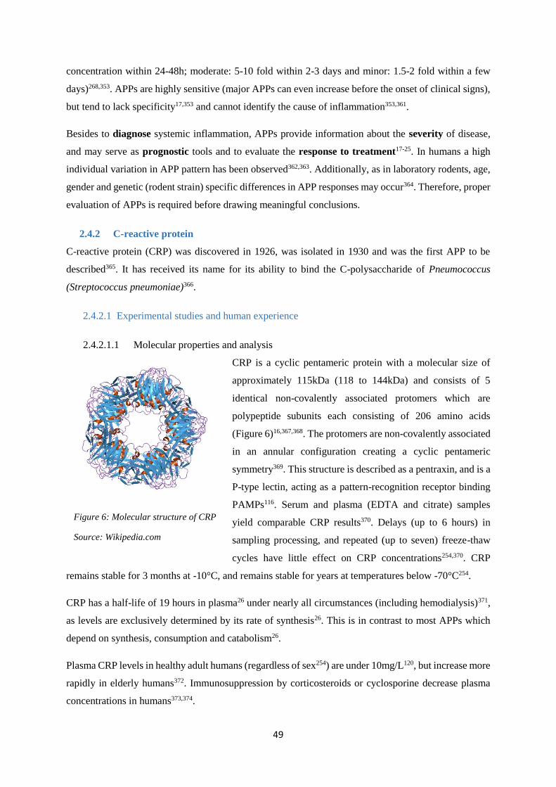
49
concentration within 24-48h; moderate: 5-10 fold within 2-3 days and minor: 1.5-2 fold within a few
days)268,353. APPs are highly sensitive (major APPs can even increase before the onset of clinical signs),
but tend to lack specificity17,353 and cannot identify the cause of inflammation353,361.
Besides to diagnose systemic inflammation, APPs provide information about the severity of disease,
and may serve as prognostic tools and to evaluate the response to treatment17-25. In humans a high
individual variation in APP pattern has been observed362,363. Additionally, as in laboratory rodents, age,
gender and genetic (rodent strain) specific differences in APP responses may occur364. Therefore, proper
evaluation of APPs is required before drawing meaningful conclusions.
2.4.2 C-reactive protein
C-reactive protein (CRP) was discovered in 1926, was isolated in 1930 and was the first APP to be
described365. It has received its name for its ability to bind the C-polysaccharide of Pneumococcus
(Streptococcus pneumoniae)366.
2.4.2.1 Experimental studies and human experience
2.4.2.1.1 Molecular properties and analysis
CRP is a cyclic pentameric protein with a molecular size of
approximately 115kDa (118 to 144kDa) and consists of 5
identical non-covalently associated protomers which are
polypeptide subunits each consisting of 206 amino acids
(Figure 6)16,367,368. The protomers are non-covalently associated
in an annular configuration creating a cyclic pentameric
symmetry369. This structure is described as a pentraxin, and is a
P-type lectin, acting as a pattern-recognition receptor binding
PAMPs116. Serum and plasma (EDTA and citrate) samples
yield comparable CRP results370. Delays (up to 6 hours) in
sampling processing, and repeated (up to seven) freeze-thaw
cycles have little effect on CRP concentrations254,370. CRP
remains stable for 3 months at -10°C, and remains stable for years at temperatures below -70°C254.
CRP has a half-life of 19 hours in plasma26 under nearly all circumstances (including hemodialysis)371,
as levels are exclusively determined by its rate of synthesis26. This is in contrast to most APPs which
depend on synthesis, consumption and catabolism26.
Plasma CRP levels in healthy adult humans (regardless of sex254) are under 10mg/L120, but increase more
rapidly in elderly humans372. Immunosuppression by corticosteroids or cyclosporine decrease plasma
concentrations in humans373,374.
Figure 6: Molecular structure of CRP
Source: Wikipedia.com

50
2.4.2.1.2 Role in sepsis and SIRS
CRP production in hepatocytes is under transcriptional control of IL-6 in combination with either IL-1β
or TNF-α375-378, and CRP has several functions:
- general scavenger protein,
o recognition and binding of microorganisms, LPS, PAMPs and DAMPs116,359,379-386
- opsonization of material387-390
- activation of the classical complement pathway116,379,387-390 (although not in rats!)391
- facilitating phagocytosis359,380,392-394
- modulating neutrophil, monocyte, macrophage and natural killer (NK) cell function116,359,380,392-
394
- inducing cytokine production359,380,392-394
- inhibiting chemotactic effects359,380
o preventing tissue migration386
o modulating neutrophil function and chromatin binding359,380,395
- anti-inflammatory role379
o inhibiting neutrophil superoxide production379
o inhibiting degranulation379
o blocking platelet aggregation379
- induction of vascular endothelial dysfunction396,397
o decreasing vasodilatory molecule release from endothelial cells396-399
o procoagulatory396-399
o pro-inflammatory effects396-399
CRP binds phosphocholine which is found in many bacteria and protozoa, forming a crystal structure369.
The other side binds to antibody receptors on neutrophils, promoting phagocytosis379. Besides this main
role in phagocytosis, there is accumulating evidence that CRP induces vascular endothelial
dysfunction396,397. PGI2 production, a major arachidonic acid product in macrovascular endothelium with
potent vasodilatory and antiplatelet effects, is decreased by CRP, inducing increased platelet
aggregability398,400,401. CRP increases plasminogen activator inhibitor (PAI)-1 in aortic endothelial cells
and promotes tissue factor expression in monocytes393,396-398,402. CRP should be considered a pro-
inflammatory, vasoconstrictive381,383,384, procoagulant403 and prothrombotic substance393,396-398,402.
2.4.2.1.3 Clinical application
Comparison of studies is severely hampered as the etiology, the severity of illness and the risk of
infection in between populations differs; different papers use different cut-off values for decision
making; timing of sampling varies, or often even is not specified404. As CRP secretion starts only 4 to
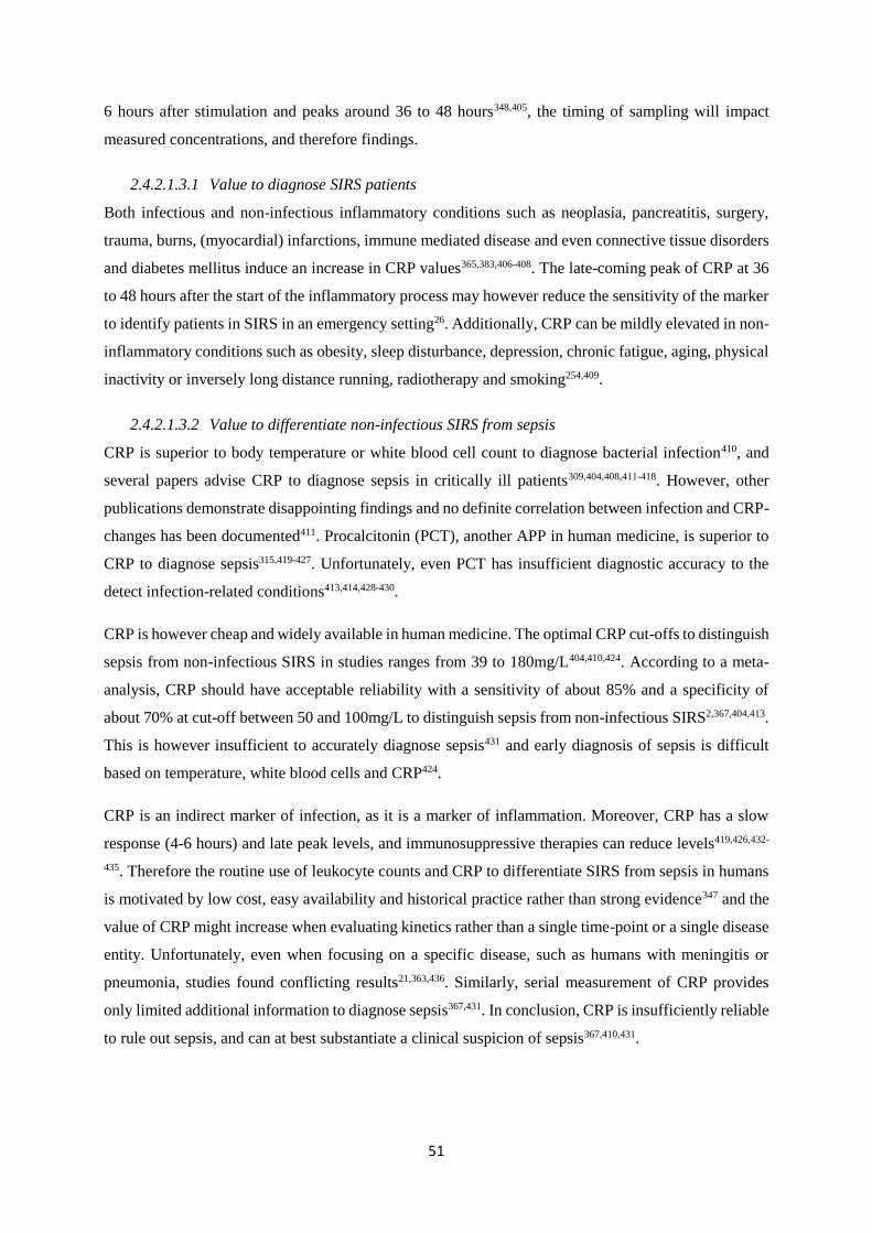
51
6 hours after stimulation and peaks around 36 to 48 hours348,405, the timing of sampling will impact
measured concentrations, and therefore findings.
2.4.2.1.3.1 Value to diagnose SIRS patients
Both infectious and non-infectious inflammatory conditions such as neoplasia, pancreatitis, surgery,
trauma, burns, (myocardial) infarctions, immune mediated disease and even connective tissue disorders
and diabetes mellitus induce an increase in CRP values365,383,406-408. The late-coming peak of CRP at 36
to 48 hours after the start of the inflammatory process may however reduce the sensitivity of the marker
to identify patients in SIRS in an emergency setting26. Additionally, CRP can be mildly elevated in non-
inflammatory conditions such as obesity, sleep disturbance, depression, chronic fatigue, aging, physical
inactivity or inversely long distance running, radiotherapy and smoking254,409.
2.4.2.1.3.2 Value to differentiate non-infectious SIRS from sepsis
CRP is superior to body temperature or white blood cell count to diagnose bacterial infection410, and
several papers advise CRP to diagnose sepsis in critically ill patients309,404,408,411-418. However, other
publications demonstrate disappointing findings and no definite correlation between infection and CRP-
changes has been documented411. Procalcitonin (PCT), another APP in human medicine, is superior to
CRP to diagnose sepsis315,419-427. Unfortunately, even PCT has insufficient diagnostic accuracy to the
detect infection-related conditions413,414,428-430.
CRP is however cheap and widely available in human medicine. The optimal CRP cut-offs to distinguish
sepsis from non-infectious SIRS in studies ranges from 39 to 180mg/L404,410,424. According to a meta-
analysis, CRP should have acceptable reliability with a sensitivity of about 85% and a specificity of
about 70% at cut-off between 50 and 100mg/L to distinguish sepsis from non-infectious SIRS2,367,404,413.
This is however insufficient to accurately diagnose sepsis431 and early diagnosis of sepsis is difficult
based on temperature, white blood cells and CRP424.
CRP is an indirect marker of infection, as it is a marker of inflammation. Moreover, CRP has a slow
response (4-6 hours) and late peak levels, and immunosuppressive therapies can reduce levels419,426,432-
435. Therefore the routine use of leukocyte counts and CRP to differentiate SIRS from sepsis in humans
is motivated by low cost, easy availability and historical practice rather than strong evidence347 and the
value of CRP might increase when evaluating kinetics rather than a single time-point or a single disease
entity. Unfortunately, even when focusing on a specific disease, such as humans with meningitis or
pneumonia, studies found conflicting results21,363,436. Similarly, serial measurement of CRP provides
only limited additional information to diagnose sepsis367,431. In conclusion, CRP is insufficiently reliable
to rule out sepsis, and can at best substantiate a clinical suspicion of sepsis367,410,431.
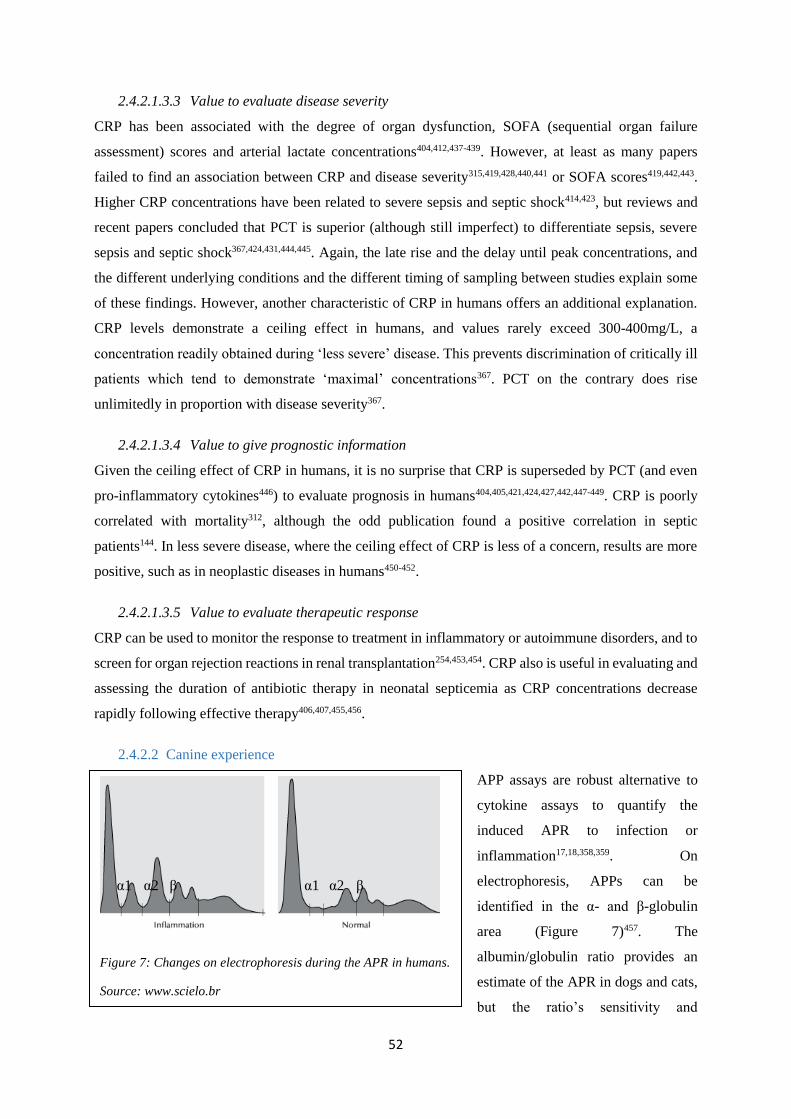
52
2.4.2.1.3.3 Value to evaluate disease severity
CRP has been associated with the degree of organ dysfunction, SOFA (sequential organ failure
assessment) scores and arterial lactate concentrations404,412,437-439. However, at least as many papers
failed to find an association between CRP and disease severity315,419,428,440,441 or SOFA scores419,442,443.
Higher CRP concentrations have been related to severe sepsis and septic shock414,423, but reviews and
recent papers concluded that PCT is superior (although still imperfect) to differentiate sepsis, severe
sepsis and septic shock367,424,431,444,445. Again, the late rise and the delay until peak concentrations, and
the different underlying conditions and the different timing of sampling between studies explain some
of these findings. However, another characteristic of CRP in humans offers an additional explanation.
CRP levels demonstrate a ceiling effect in humans, and values rarely exceed 300-400mg/L, a
concentration readily obtained during ‘less severe’ disease. This prevents discrimination of critically ill
patients which tend to demonstrate ‘maximal’ concentrations367. PCT on the contrary does rise
unlimitedly in proportion with disease severity367.
2.4.2.1.3.4 Value to give prognostic information
Given the ceiling effect of CRP in humans, it is no surprise that CRP is superseded by PCT (and even
pro-inflammatory cytokines446) to evaluate prognosis in humans404,405,421,424,427,442,447-449. CRP is poorly
correlated with mortality312, although the odd publication found a positive correlation in septic
patients144. In less severe disease, where the ceiling effect of CRP is less of a concern, results are more
positive, such as in neoplastic diseases in humans450-452.
2.4.2.1.3.5 Value to evaluate therapeutic response
CRP can be used to monitor the response to treatment in inflammatory or autoimmune disorders, and to
screen for organ rejection reactions in renal transplantation254,453,454. CRP also is useful in evaluating and
assessing the duration of antibiotic therapy in neonatal septicemia as CRP concentrations decrease
rapidly following effective therapy406,407,455,456.
2.4.2.2 Canine experience
APP assays are robust alternative to
cytokine assays to quantify the
induced APR to infection or
inflammation17,18,358,359. On
electrophoresis, APPs can be
identified in the α- and β-globulin
area (Figure 7)457. The
albumin/globulin ratio provides an
estimate of the APR in dogs and cats,
but the ratio’s sensitivity and
Figure 7: Changes on electrophoresis during the APR in humans.
Source: www.scielo.br
α1 α1 α2 α2 β β

53
specificity to detect clinical or subclinical disease is inferior to individual positive APPs assays458,459,
which are recommended to evaluate the systemic response secondary to infection or
inflammation17,18,358,359.
2.4.2.2.1 Molecular properties and analysis
CRP is a highly conserved protein weighing 100kDa in dogs17. However, CRP is not identical in dogs
and humans, as canine CRP has 2 out of 5 subunits that are glycosylated460,461. These small molecular
differences account for some of the encountered difficulties when measuring canine CRP concentrations
with human CRP-antibodies17,462. Automated turbidimetric human CRP immunoassays463 and a semi-
quantitative near-patient slide reversed passive latex agglutination test (randox®) have been validated
to measure canine CRP462-464, but a canine commercial ELISA kit27, a rapid nephelometric assay465,466
canine specific immunoturbidimetric and467,468 even canine automated immunoturbidimetric assay469,470
have also been validated.
CRP concentrations do not have a circadian rhythm in dogs and are not affected by sex, age, breed, or
repeated venous blood sampling. Nevertheless, pregnancy induces increased CRP concentrations471,472,
one month old puppies might have lower peak concentrations24,472-475, and long-distance exercise induces
severe increases in dogs476 (contrary to moderate exercise)477. The administration of short term
administration of non-steroidal anti-inflammatory drugs (NSAIDs) does not alter CRP concentrations
in dogs478,479 as NSAIDs do not suppress IL-6 production, the major stimulus for CRP production478,480.
CRP concentrations are not affected by glucocorticoid administration in dogs, in contrast to other APPs
such as haptoglobin481.
CRP can be measured in canine blood, saliva, effusions (abdominal, thoracic and pericardial; transudate,
modified transudate and exudate) and cerebrospinal fluid353,482-485. CRP is stable for 14 days at room
temperature or 4°C, for 3 months at -10°C, and remains stable at temperatures below -70°C for
prolonged, undefined periods in canine studies (for years in human studies)254,470,486. Hemolysis,
lipaemia and hyperbilirubinaemia can falsely modify CRP measurement487. Changes are especially
expected in lipemic or hemolytic samples using the ELISA test, and with hemolytic samples using the
canine-species specific immunoturbidimetric method that was first described. Interference would only
be expected at very high concentrations of intralipid (10g/L), bilirubin (800mg/L) and hemoglobin
(5g/L) with the latest canine specific automated immunoturbidimetric technique467,469,470,487.
CRP concentration is usually less than 5mg/L in healthy dogs and reference ranges vary from 0.22 to
16.4mg/L24,233,474,481,488-490. When looking at the kinetics of CRP, it has been suggested not to compare
to the reference range, but rather to screen for a critical difference of 4,85mg/L in CRP concentrations480.
This may be more interesting when one considers the high individual variability in CRP491.

54
Although CRP is not considered to be an APP in cats and cattle360,492, it is in dogs, pigs and
horses233,460,488,490,493-498. In comparison with other APPs such as haptoglobin, CRP concentration
increases more rapidly in dogs with SIRS, resulting in an earlier serum peak (1-2 days versus 3-7 days
for haptoglobin) which can rise up to 800-fold the starting concentrations17,350.
2.4.2.2.2 Clinical application
2.4.2.2.2.1 Value to diagnose SIRS patients
CRP is very useful to detect systemic inflammation in dogs17,27-29,461,473,499 secondary to a myriad of
conditions such as infectious disease (e.g. babesiosis, leishmaniosis, ehrlichiosis, trypanosomiasis,
leptospirosis, Bordetella bronchiseptica infection, parvovirosis, E. coli endotoxinaemia, pyometra,
cystitis and pneumonia)17,23,24,255,350,353,458,490,491,500-502, immune mediated disease (e.g. arthritis,
inflammatory bowel disease, immune mediated hemolytic anemia, steroid-responsive meningitis
arteritis)353,484,490,503-506, neoplasia (e.g. lymphoma, hemangiosarcoma)490,503,507-509, tissue trauma (e.g.
surgery or experimental gastric lesions)510-512, but also intestinal obstruction and acute pancreatitis or
sterile pericarditis465,473,510,513, and CRP is increased in a general population of critically ill patients514.
CRP is more sensitive to detect inflammatory disease than white blood cell counts17,350,515 or erythrocyte
sedimentation rates17,350. CRP is a promising marker for dogs suspected to be in SIRS491, as it
discriminates canine pyometra patients with and without SIRS491, healthy dogs and dogs with focal
inflammation from SIRS patients516, and asymptomatic Leishmania carriers from dogs with
symptomatic leishmaniasis458. CRP can indicate concurrent inflammation in dogs suffering from
hyperadrenocorticism, although hyperadrenocorticism did seem to blunt the response517.
Canine CRP concentrations were first thought to be unaffected by glucocorticoid administration481,
however administration of methylprednisolone acetate does moderately (not statistically significant)
decrease CRP518. The CRP-suppressive effect of steroids could be explained by glucocorticoid mediated
suppression of the NF-kB pathway which activates genes involved in cytokine production519,520.
The timing of peak values depends on the insult, with surgery511 and bacterial pneumonia causing peak
concentrations within 1 day, and rickettsial disease only resulting in peak concentrations 4 to 10 days
after infection 23. Furthermore, the half-life of canine CRP is relatively short511 and concentrations return
to normal range within 14 to 21 days of experimentally induced inflammation17,233,481. These factors may
limit the use of CRP to detect systemic inflammation.
In conclusion, identifying clinical SIRS-criteria in canine emergency cases justifies CRP
measurement491. CRP concentrations are correlated with WBC, segmented and banded neutrophil
counts521,522, but have superior sensitivity than WBC count, longer analyte stability and exhibit a faster
response17.

55
2.4.2.2.2.2 Value to differentiate non-infectious SIRS from sepsis
The only paper specifically evaluating the value of CRP to distinguish SIRS from sepsis did not find
significant differences between both groups on day 0, 1 and 230.
2.4.2.2.2.3 Value to evaluate disease severity
CRP could be a valuable parameter to evaluate the severity of any ongoing inflammatory
disease500,501,510, reflecting the clinical situation at the time of
sampling27,30,460,503,523,52427,30,460,503,523,52427,30,460,503,523,52427,30,460,503,523,52427,30,460,503,523,52427,30,460,503,523,52427,30,46
0,503,523,52427,30,460,503,523,52427,30,474,517,538,539. However, as the magnitude of the increase in CRP depends on
multiple factors besides the severity of disease such as the initiating cause, and the extent of tissue
damage, mixed findings have been published17,23,24.
CRP is correlated with disease severity in canine pyometra255,491, babesiosis350,525, mammary
tumors490,526,527, immune-mediated polyarthritis528, and in critically ill patients514. In multicentric
lymphoma, CRP is not different between substage a versus substage b patients507, but mean CRP
concentrations are higher in more aggressive lymphoid neoplasia (acute lymphoblastic leukemia and
lymphoma)508. CRP concentrations in acute pancreatitis are not correlated with a clinical severity index
score529, but higher CRP concentrations are correlated with pancreatic necrosis530. In idiopathic
inflammatory bowel disease, CRP is not correlated with the canine inflammatory bowel disease activity
index (CIBDAI)504,531, but a correlation between CIBDAI and the combination of CRP and
histopathology has been demonstrated504. CRP failed to assess disease severity in dogs with immune
mediated hemolytic anemia, defined as the amount of blood transfusions or duration of
hospitalization532.
In conclusion, the link between CRP levels and disease severity is poorly convincing, and depends on
the type of disease, timing of sampling, and the definition of ‘disease severity’.
2.4.2.2.2.4 Value to give prognostic information
CRP could be considered a marker of prognosis in dogs27,30,460,503,523,524, but no correlation between CRP
and prognosis is identified in conditions such as leptospirosis114, Babesia rossi infection533, immune
mediated hemolytic anemia506,532, acute pancreatitis529, and acute abdomen syndrome515. In three
populations of dogs with SIRS, no correlation was found between the initial CRP concentration and
survival30,514,516. However CRP is higher in parvovirosis non survivors534, but as the survival rate in this
study was particularly low (46,5%) these results should be interpreted with great caution.
APP-kinetics appear to be more suited to assess prognosis in canine SIRS30,503,524. The 2- or 3-day change
in CRP predicts survival, survivors experiencing a bigger drop in CRP concentrations30,516. Similarly,
persistently elevated CRP concentrations in acute pancreatitis529 and acute abdomen syndrome515 are

56
correlated with poor outcome. Contrarily, CRP changes during the first 24 hours do not distinguish
survivors and non-survivors in Babesia rossi infection533.
In conclusion, although CRP concentrations at presentation does not add prognostic information of SIRS
patients, CRP-kinetics are promising to predict prognosis in dogs with SIRS30,503,524.
2.4.2.2.2.5 Value to evaluate therapeutic response
Successful treatment results in decreased APP concentrations, while increasing or persistently elevated
concentrations are associated with poor response to treatment or relapse503. Therefore CRP may allow
to monitor disease progression and treatment response27,30,460,500,503,523,524.
CRP is useful to monitor treatment response in dogs with pancreatitis513, Babesia canis infection350,
leishmaniosis535, trypanosomiasis501, inflammatory bowel disease504, immune mediated hemolytic
anemia532, steroid responsive meningitis-arteritis484,523,536, and polyarthritis505,528. CRP can be used to
screen for postoperative complications524,537,538. CRP concentrations are also lower in dogs in complete
remission of lymphoma compared to other remission states507.
In conclusion, CRP is very useful to monitor treatment response in dogs, and persistently elevated serum
CRP concentrations in patients receiving appropriate therapy warrants further clinical investigation.
2.4.2.2.2.6 Value in canine heart disease
CRP increases during natural canine heart disease539-541. In dogs with mitral valve disease (MVD), CRP
is inconsistently increased541,542 and CRP is not correlated with the severity of MVD543. In human
medicine, high-sensitivity CRP assays evaluate for the risk of cardiovascular disease544. The lower limit
of detection of most canine assays is however higher470, and several assays demonstrate slight
proportional discrepancy at low concentrations463. These characteristics do not allow to screen for such
small differences in concentrations545,546. Moreover, dogs do not suffer from the same cardiac diseases
as humans.
In conclusion, despite positive results regarding CRP measurement in dogs with SIRS for the clinical
diagnosis, evaluation of disease severity, prognosis and monitoring treatment response, its use in the
emergency setting is limited, mainly due to practical concerns. However, the availability of canine
specific kits and user-friendly immunoassays at an economic price make CRP-analysis in clinical
practice a reality. Findings relating CRP to cardiac disease in humans should not be extrapolated to
canine medicine.

57
2.5 CARDIAC (AND CARDIOVASCULAR) FUNCTION
2.5.1 Evaluation of cardiovascular (dys-)function
A patient’s history, physical examination, electrocardiogram and radiologic data are often sufficient to
explain cardiac problems. Unfortunately, these tools are not specific and can lead to misinterpretation
of findings547. Simple obtainable information such as blood pressure, pulse quality, urine output and
central/peripheral temperature gradients provide a lot of information and are often sufficient to guide
therapy in non-critically ill patients548. Although clinical observation and these simple monitoring tools
might indicate tissue- and organ- hypoperfusion, they also are insensitive and non-specific548.
To better understand cardiac function, in vitro models and invasive or advanced techniques such as
radionuclide assessment and thermodilution were developed549. Although such methods have been
applied in human critical care for decades, these methods are now replaced by echocardiography. The
next chapters will give an overview of the benefits and drawbacks of these techniques.
2.5.1.1 Invasive techniques
The pulmonary artery catheter (PAC) has
been the standard hemodynamic monitoring
technique for patients in the ICU since the
70s550,551. The PAC is placed through an
introducer in any of the central venous
cannulation sites (the internal jugular veins,
the subclavian veins, and the femoral veins).
The right internal jugular vein is often
preferred as it is situated closest to the heart
and provides a direct route to the right atrium
(RA). PACs have a flow-directed balloon-
tipped end. Inflating the balloon when the
catheter reaches the heart allows to ‘go with
the flow’ through the RA and RV into the
pulmonary artery. The placement of PACs
allows to (Figure 8) measure central venous pressure (CVP) evaluating preload, pulmonary artery
pressure (PAP), right ventricular ejection fraction (RVEF), estimate LV filling pressures (evaluating
preload), sample for continuous mixed venous oxygen saturation (SVO2), and calculate cardiac output
(CO)548.
CVP and PAP unfortunately are unreliable parameters to predict fluid responsiveness (i.e. the benefit of
an additional fluid bolus to CO) in critical care patients552. Volume administration in hypotensive critical
Figure 8: The pulmonary artery catheter
Source: www.studypk.com
58
care patients has been guided by PAC measurements of pulmonary capillary wedge pressure (PCWP).
PCWP is measured after placement of the PAC into a pulmonary artery and inflation of the balloon,
occluding the pulmonary artery. Obstruction of the artery causes pressure in the part distal to the
occlusion to drop rapidly and within a couple of seconds reach a steady state where the remnant pressure
will be equal to the pressure in the left atrium (LA) (mean pressure around 8-10mmHg in healthy
humans). During volume loading to increase blood pressure and improve ventricular function the
objective is to keep PCWP at 12-14mmHg. The upper endpoint of LA pressure is set at 18 to 20mmHg
as higher values are associated with an increased risk of pulmonary edema553,554. These values
correspond to the pressures in the healthy heart at which the plateau in the relationship between cardiac
function and filling pressures is reached555,556.
Thermodilution applies a special thermistor-tipped catheter (Swan-Ganz catheter) inserted from a
peripheral vein into the pulmonary artery. A cold saline solution of known temperature and volume is
injected into the RA from a proximal catheter port. The injected solution mixes with blood as it passes
through the right ventricle (RV) into the pulmonary artery, cooling the blood. The temperature of the
blood is measured at the catheter tip by a thermistor situated in the pulmonary artery, and a computer is
used to acquire the thermodilution profile (quantifying the change in blood temperature as it flows over
the thermistor surface). The CO computer calculates flow (CO from the RV) using the blood temperature
information, and the temperature and volume of the injected solution. This scenario is repeated several
times and results are averaged to obtain CO. Because CO changes with respiration, saline must be
injected at a consistent time point during the respiratory cycle, usually the end of expiration. Since its
first description, fully automated systems have been developed to assess CO continuously, avoiding
variations due to operator technique557.
Radionuclide-gated blood pool scanning can also be performed using PACs. The technique uses a
radioactive tracer such as Technetium-99m-pertechnetate that labels the patient’s red blood pool and
radioactivity is measured with a gamma camera over an area of interest. The acquisition can be ‘gated’
to coincide with the cardiac cycle, allowing for the measurement of ejection fractions and calculations
of the peak systolic pressure/end-systolic volume index ratio, considered to be a load-independent
marker of ventricular function558. PACs also allow for the assessment of venous oxygen saturation
(SVO2)559. Although whole body maximal oxygen uptake volume (VO2) offers interesting information,
gastric intramucosal pH and subcutaneous oxygen tension might be more sensitive indicators of
circulatory impairment than this conventional measurements560,561, which by any means is outside the
scope of this literature review.
Although technical progress has been made, PACs remain impractical, require substantial amounts of
material and equipment for easy monitoring, and are associated with severe complications562,563. Some
observational studies suggest an association of PAC with increased mortality563-565. Catheter associated

59
morbidity includes local trauma at the insertion site, generalized infections, an increased incidence of
pulmonary thromboembolism and even pulmonary artery rupture548,562. Very pessimistic speculations
suggested that PACs might be accountable for up to 15000 excess deaths per year in the United States566.
Besides the safety-aspect, the diagnostic yield of pulmonary artery catheters has also been
questioned563,567. Several studies have demonstrated a poor correlation between PCWP, CO and the
systolic function as evaluated by echocardiography548,568,569. Measurements of PCWP might be
influenced by underlying lung disease such as chronic obstructive pulmonary disease (COPD) and acute
respiratory disease syndrome (ARDS), alterations in the filling of the pulmonary circulation, increases
in pulmonary vascular resistance, mitral stenosis or incompetence, aortic incompetence, changes in LV
compliance, changes in thoracic muscle tone, changes in lung compliance, changes in intrathoracic
pressure and bad coordination of spontaneous breathing during mechanical ventilation, and finally the
timing of measurements within the ventilator cycle42,548,570. As all these factors can influence PCWP,
correctly interpreting changes in PCWP is virtually impossible if no complementary information is
available. Currently baseline PCWP is generally considered an inaccurate predictor of preload568,570,571,
that also fails to predict fluid responsiveness in the individual patient572,573. The information obtained
from PACs does not allow the detection of early changes, or discern systolic from diastolic changes574.
It is therefore not surprising that a randomized controlled clinical trial did not find any benefit of PAC
directed therapy over standard care in intensive care patients562.
2.5.1.2 Transthoracic and transoesophageal echocardiography
2.5.1.2.1 Human experience
Echocardiography has been used to assess cardiac function as early as the 50s575. Echocardiography was
first limited to M-mode studies, but two-dimensional (2D) imaging and Doppler systems were rapidly
developed576. The 80s and 90s saw the development of phased array scanners with M-mode
visualization, the assessment of intra-cardiac pressures and flow velocities, the birth of color Doppler
and contrast ultrasonography577,578. The technical progress allowed for the manufacturing of
transoesophageal echocardiography probes, real-time three-dimensional (3D) imaging and even intra-
cardiac echocardiography579-581. Intracardiac and 3D echocardiography have not yet gained access to
general clinical practice, and are outside of the scope of this literature review, although 3D-
measurements correlate well with conventional 2D-measurements581.
While technical and practical knowledge in echocardiography developed, negative results from studies
evaluating the value of CVP and PAP accelerated the interest in the use of echocardiography for the
evaluation of cardiovascular function47,48,547,562,563,582-584. Echocardiography does indeed offer the
benefits of direct visualization of the heart, allowing for real-time assessment of cardiovascular structure
and function, as well as providing information on hemodynamics via Doppler measurements of blood

60
flow velocity48. This combination offers enough information to determine the cause of hypotension that
is refractory to the use of vasopressors or inotropic support585.
When physical examination and echocardiography on cardiac patients performed by a general physician
were compared, cardiac examination missed 59% of all abnormalities and failed to correctly detect 43%
of major cardiovascular findings, while echocardiography missed 29% of all and failed to correctly
detect 21% of major abnormalities586. As physicians were not even trained in echocardiography and
received only 15 minutes to complete the echocardiography, this study illustrates the potential of
echocardiography to complement physical findings586.
Several studies comparing PACs and echocardiography demonstrate that transthoracic (TTE) and
transesophageal (TEE) echocardiography add valuable information to patients already monitored by
PACs41,587-589. Echocardiography provides a better index of LV preload than invasive monitoring571. In
human patients with uncomplicated acute MI, PAC estimates of LA pressure agree with
echocardiographic measurements in up to 85% of patients, however in patients with multisystem failure
this level can be as low as 30%590,591. PACs give unreliable information on preload, LV end-diastolic
volume, and LV systolic function in septic patients compared to TTE568.
If fluid loading to correct hypovolemia in patients with preserved LV systolic function is guided by
echocardiography, some patients display PCWP values of 13 to 19 mmHg, while others demonstrate
‘supranormal’ PCWP pressures (20 to 25mmHg). These second groups of patients also demonstrate
increased wall thickness and decreased ventricular compliance. Although PCWP values are increased,
fluid administration based on echocardiography is beneficial in these cases, demonstrating the benefit
of echocardiography over PCWP592.
Currently, several human ICUs have more than 15 years of experience guiding the initial management
of acute circulatory failure solely based on echocardiography, and no longer use PACs49,593.
Echocardiography however remains confronted with several challenges. Echocardiography findings are
besides cardiac disease, influenced by variations in loading conditions, positive pressure ventilation,
sedation, changes in arterial carbon dioxide pressures, vasopressors or positive inotropes and cardiac
pacing; common circumstances in critically ill patients549. Moreover, ‘normal’ values are not necessarily
applicable to an ICU population, and separate references might be applicable549.
For TTE a dedicated machine should be available 24 hours a day with a probe working at a variety of
frequencies to obtain optimal resolution at different depths, and a high frame rate to smoothly display
cardiac movement594. In immobile ventilated patients, TTE can usually be performed using a sub-costal
view, and poor image quality can usually be improved by contrast ultrasonography549. The main reasons
TTE may result in inadequate image quality are interposed structures such as thoracic wall edema,
surgical dressings, air (mechanical ventilation), bone, calcium or foreign bodies (chest tubes), and
61
restricted patient positioning547,595. Failure rates of TTE were first reported to be 30-40%596,597, hindering
its clinical efficacy, but technological improvements have decreased this number to 5-15% in
ambulatory patients595, with even lower percentiles in ventilated patients548. Some authors describe
adequate image quality for bedside TTE in 99% of cases598.
Echocardiography has found a place in human critical care, and the most common motivations to request
an echocardiography are assessment of volume status and left and right ventricular function549. TTE is
recommended over TEE in most cases, unless when superior resolution is required or views are
impossible to acquire using TTE549. Although many urgent management decisions can be based on
echocardiographic interpretations of ventricular filling and function, many clinicians turn to PAC data
for objective quantification of data, as echocardiography remains operator dependent and interpretation
can be subjective592,599,600. Indeed, the availability of an echography machine does not omit the need of
a trained physician, and the development of echocardiographic training for critically ill patients by
criticalists will be discussed in the following section88.
2.5.1.2.2 Training in ECC ultrasonography
Over the last decade, interest of application of echocardiography in the ICU has greatly increased in
human medicine, leading to an increased availability48,595,601. However, only 20% of European
intensivists have certification to perform echocardiography48. So despite greater availability of
machines, the clinical application diffuses slowly into ICUs48. When implementing echocardiography
for monitoring critically ill patients, a decision needs to be made as to whom will be the trained physician
performing these exams: an internist, intensivist, anesthesiologist, cardiologist or imager. Early official
guidelines to practice echocardiography did not include trained intensivists for evaluation of ICU
patients602, and even discouraged their participation603. If however one wants to use echocardiography
24h/24h, training of intensivists seems logical, and training programs for intensivists should be
provided48. Although several papers warn intensivists to anticipate resistance from cardiology
colleagues when suggesting such an approach594, echocardiographic training is already being
incorporated into some human ICU fellowships48.
Performance characteristics of echocardiography by non-specialists is determined by the hours of
training, the quality of the device, the patient characteristics and the definition of a ‘successful
examination’559. Two different training approaches are proposed: Courses aimed to perform a complete
ultrasound, and focused goal directed training courses trying to answer specific questions. Focused goal-
oriented TTE training courses of 3 hours of theoretical training and 5 hours of hands-on training have
been described with positive results604. Trainees could adequately answer clinical questions such as
- LV systolic dysfunction (subjectively assessed as <50% volume change)
- presence of LV dilation

62
- presence of right ventricular dilatation (cor pulmonale)
- presence of pericardial effusion
- presence of pleural effusion
A slightly longer program (10 one-hour tutorials) allowed candidates to successfully perform limited
TTE exams evaluating 4 views (the parasternal long- and short-axis and apical 2- and 4- chamber views
(Figure 9)) to assess LV volume and function605. An extended 20 hour training program aiming to
complete an 18-point check-list resulted in 23% of missed significant findings compared to 14% of
missed findings by experts606. Such studies illustrate how short training programs are feasible, and that
broadening the objectives can be associated with poorer results607,608. Non-cardiologists will not achieve
better results than trained echocardiographers606, and will easily miss regional wall motion
abnormalities, intra-cardiac thrombi, right ventricular dysfunction and non-trivial pericardial effusion606.
A French ECHO-in-ICU group offering a complete 2-year training program had accredited 16% of
French ICUs by 2008. The program consists of a 20 hour specific course and 120 supervised TTEs
during the first year, and hands-on practice in ICUs or in cardiac surgery rooms with at least 50 TEEs
Figure 9: The 4 basic cardiac views in human ECC training
Source: www.criticalecho.com

63
during the second year. Despite the success of the course, this and other groups express the need for 2-
levels of training48,594.
Advanced training would allow for comprehensive evaluation of cardiac anatomy and function with
two-dimensional echocardiography and Doppler echocardiography. The trainee would be able to
identify segmental wall dysfunctions, mild ventricular dysfunction or abnormal interventricular septal
motion. However, this in-depth training would require a lot of time (as in the ECHO-in-ICU protocol),
and this level probably only needs to be obtained by a minority of intensivists594.
The basic level training would aim for goal-directed examination via TTE or TEE, allowing the
evaluation of simple but useful parameters and techniques to evaluate volume status, systolic function
and diastolic function594. These assessments should allow the clinician to categorize the cause of shock
and decide on the treatment strategy594. Furthermore basic training should also allow the clinician to
adequately assess the presence of pericardial fluid, presence of pleural effusion, valvular function, and
assist in the placement of central lines and performing thoracocentesis48,594.
The next chapters will give a short overview of the most important techniques to assess volume status,
systolic function and diastolic function.
2.5.1.3 Volume status or preload and volume responsiveness
Critical patients often suffer from hypovolemia, requiring volume expansion to prevent or treat
hemodynamic collapse. Echocardiography can aid hemodynamic care by diagnosing hypovolemia
and/or decreased preload40,49. When preload is optimized further fluid loading will not increase oxygen
delivery further, and may cause harm through the development of pulmonary edema and increased
intestinal bacterial translocation609.
As previously described, data from PACs may be misleading as ventricular compliance is influenced by
numerous factors570,610. Differences in diastolic compliance explain the weak correlation in between
pressure and volume, limiting the use of pressure measurements alone to predict LV preload611.
Subjective assessment of LV volume evaluating cavity size in the short- and long-axis
echocardiographic views is often adequate to guide fluid therapy at the extreme ends of cardiac filling
and function595. Finding a small, hyperdynamic LV is strongly indicative of a severely hypovolemic
patient with normal underlying cardiac function595. An extreme form of severe hypovolemia can be
recognized as systolic obliteration, characterized by dynamic obstruction of the LV cavity and a
decreased end diastolic volume595. Unfortunately, a large end diastolic volume can also indicate LV
dysfunction595. Detecting changes in LV volume will therefore be more difficult in patients with dilated
or poorly contractile ventricles592. Quantitative values are therefore preferable. Transient changes in
blood volume due to de- or over-hydration alter atrial geometry51. Therefore, LA dimensions are
interesting for the assessment of volume status. Evaluation of ventricular dimensions using TTE is useful

64
to assess preload and optimize therapy of ICU patients40,612. Atrial and ventricular dimensions however
dependent on other factors such as systolic and diastolic properties, and do not allow to assess the benefit
of additional fluid loading613. Fluid responsiveness is the potential to increase CO in response to a fluid
challenge, and assessing fluid responsiveness is important in the initial treatment of emergency
patients552. Different techniques assessing fluid responsiveness have been described in ventilated and
spontaneously breathing patients.
2.5.1.3.1 Ventilated patients
The heart-lung interactions produced by controlled positive-pressure ventilation creates a well-
controlled situation that allows fluid responsiveness to be easily assessed614-617. Under controlled
ventilation with tidal volumes above 8ml/kg, and with
patients in sinus rhythm, respiratory changes in vena
cava diameter or in stroke volume are considered the
most useful echocardiographic parameters to assess
fluid responsiveness618. Mechanical ventilation causes
an increase in intrathoracic pressure, impeding venous
blood returning to the heart. A small vena cava in
ventilated patients reliably excludes the presence of
elevated right atrial pressure (RAP)619,620.The changes
in intrathoracic pressure throughout the respiratory
cycle impact the amount of blood in the vena cava
(Figure 10). Changes in inferior vena cava (IVC)
diameter during respiration are correlated with volume responsiveness615,616. The variation of IVC
diameter during respiration after a fluid bolus is significantly correlated with an increase in CO615,616.
The superior vena cava (SVC) suffers more from intrathoracic pressure changes, especially during
positive pressure ventilation, but is less influenced by intraabdominal pressure changes and can also be
used to assess fluid responsiveness621,622. The collapsibility index is based on beat-to-beat changes in the
diameter of the superior vena cava. High SVC collapsibility (>30%) predicts a positive response to
volume expansion49,614,618.
Several techniques using Doppler flow measurement (aortic velocity-time integral617,621,623,624,
transmitral and pulmonary vein Doppler patterns595,625-627) have also been described to assess fluid
responsiveness. These are however technically more difficult to perform compared to evaluation of the
vena cava size and collapsibility. In addition to respiratory pressure variations, systolic pressure
variations and pulse pressure variations can also be used to evaluate flow variations secondary to
pressure changes and to develop an index of volume loading617. The relation of early to late transmitral
diastolic filling (E/A ratio), isovolumetric relaxation time and rate of deceleration of early diastolic
inflow (deceleration time) all provide additional information regarding preload585. Finally, a small LV
Figure 10: IVC diameter in M-mode
Source: www.acep.org

65
cavity, with end-systolic obliteration628 or the presence of interatrial septal deviation to the left during
positive-pressure ventilation629 are strongly indicative of hypovolemia and can be assessed subjectively.
2.5.1.3.2 Spontaneously breathing patients
Spontaneously breathing patients are harder to evaluate than ventilated patients, as the respiratory
pressure changes are not controlled, and cannot be used to evaluate volume or flow changes. However,
several static and dynamic parameters have been suggested to evaluate volume responsiveness. An
inferior vena cava diameter <1cm is indicative of a low preload and volume responsiveness in
hypotensive human patients630,631. A small hyperdynamic LV with end-systolic cavity effacement
indicates hypovolemia594. An IVC >20mm without a >50% decrease in diameter with gentle sniffing
indicates elevated RAP (>10mmHg)632. The effect of passive leg raising on stroke volume has been
validated in human patients and an increase in CO of >10% predicts a positive response to volume
loading in a hypotensive individual633,634.
2.5.1.4 Left Ventricular Systolic dysfunction
Approximately a quarter of hemodynamically unstable critically ill patients, including septic patients,
suffer from significant LV systolic dysfunction40-42,568,635. Evaluation of LV systolic function can usually
be performed using TTE595, but most echocardiographic parameters evaluating systolic function are
affected by loading conditions49,54988. Systolic dysfunction can become apparent after restoration of
afterload, and revaluation is important in critically ill patients636,637. LV size and function in critically ill
patients might not be similar to outpatients, and separate ‘reference ranges’ might be applicable549. A
large number of parameters have been described. Subjective visual inspection is very reliable when used
by criticalist experienced in echocardiography or echocardiographers601,638. The basic-level
echocardiographer should be able to distinguish global hypokinesis from regional abnormalities559.
Regional wall motion is evaluated using semi-quantitative scoring systems (1= normal; 2=hypokinesia;
3=akinesia; 4=dyskinesia)52. Regional hypo- or dyskinesis is typical of MI and chronic ischemic
conditions600. Besides such qualitative and semi-quantitative visual evaluation, several indices have been
described to quantitatively assess systolic function568,639. Fractional shortening (FS) is assessed via M-
mode echocardiography (Figure 12), and is the most commonly described parameter evaluating
ventricular systolic function52-54. FS is the degree of systolic reduction in the minor axis expressed as a
percentage of end-diastolic dimension548. It is an estimate of global systolic function, but depends on
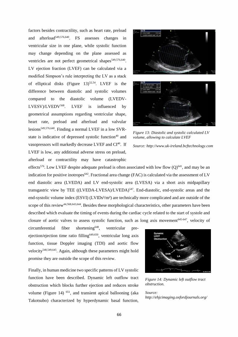
66
factors besides contractility, such as heart rate, preload
and afterload549,576,640. FS assesses changes in
ventricular size in one plane, while systolic function
may change depending on the plane assessed as
ventricles are not perfect geometrical shapes549,576,640.
LV ejection fraction (LVEF) can be calculated via a
modified Simpson’s rule interpreting the LV as a stack
of elliptical disks (Figure 13)52,54. LVEF is the
difference between diastolic and systolic volumes
compared to the diastolic volume (LVEDV-
LVESV)/LVEDV568. LVEF is influenced by
geometrical assumptions regarding ventricular shape,
heart rate, preload and afterload and valvular
lesions549,576,640. Finding a normal LVEF in a low SVR-
state is indicative of depressed systolic function40 and
vasopressors will markedly decrease LVEF and CI40. If
LVEF is low, any additional adverse stress on preload,
afterload or contractility may have catastrophic
effects576. Low LVEF despite adequate preload is often associated with low flow (Q)641, and may be an
indication for positive inotropes642. Fractional area change (FAC) is calculated via the assessment of LV
end diastolic area (LVEDA) and LV end-systolic area (LVESA) via a short axis midpapillary
transgastric view by TEE ((LVEDA-LVESA)/LVEDA)547. End-diastolic, end-systolic areas and the
end-systolic volume index (ESVI) (LVIDs³/m²) are technically more complicated and are outside of the
scope of this review44,568,643,644. Besides these morphological characteristics, other parameters have been
described which evaluate the timing of events during the cardiac cycle related to the start of systole and
closure of aortic valves to assess systolic function, such as long axis movement645-647, velocity of
circumferential fiber shortening648, ventricular pre-
ejection/ejection time ratio filling649,650, ventricular long axis
function, tissue Doppler imaging (TDI) and aortic flow
velocity548,549,645. Again, although these parameters might hold
promise they are outside the scope of this review.
Finally, in human medicine two specific patterns of LV systolic
function have been described. Dynamic left outflow tract
obstruction which blocks further ejection and reduces stroke
volume (Figure 14) 651, and transient apical ballooning (aka
Takotsubo) characterized by hyperdynamic basal function,
Figure 12: M-mode of the left ventricle,
allowing to calculate FS
Source: http://www.uk-
ireland.bcftechnology.com
Figure 13: Diastolic and systolic calculated LV
volume, allowing to calculate LVEF
Source: http://www.uk-ireland.bcftechnology.com
Figure 14: Dynamic left outflow tract
obstruction.
Source:
http://ehjcimaging.oxfordjournals.org/
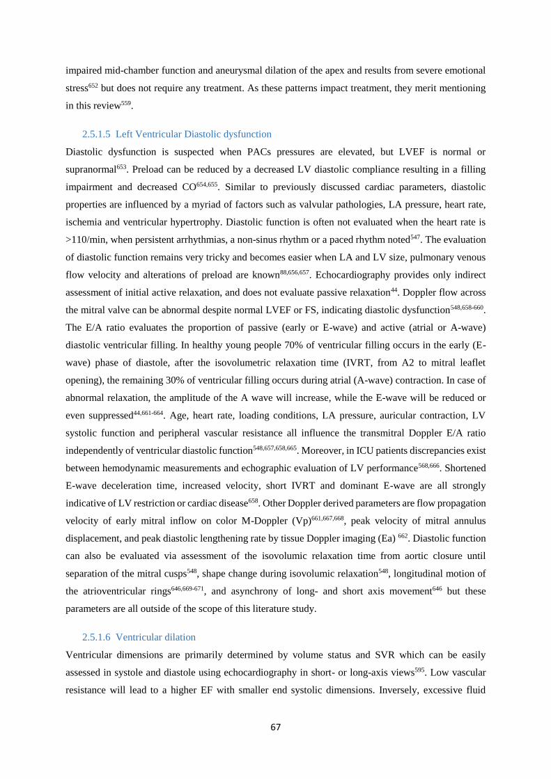
67
impaired mid-chamber function and aneurysmal dilation of the apex and results from severe emotional
stress652 but does not require any treatment. As these patterns impact treatment, they merit mentioning
in this review559.
2.5.1.5 Left Ventricular Diastolic dysfunction
Diastolic dysfunction is suspected when PACs pressures are elevated, but LVEF is normal or
supranormal653. Preload can be reduced by a decreased LV diastolic compliance resulting in a filling
impairment and decreased CO654,655. Similar to previously discussed cardiac parameters, diastolic
properties are influenced by a myriad of factors such as valvular pathologies, LA pressure, heart rate,
ischemia and ventricular hypertrophy. Diastolic function is often not evaluated when the heart rate is
>110/min, when persistent arrhythmias, a non-sinus rhythm or a paced rhythm noted547. The evaluation
of diastolic function remains very tricky and becomes easier when LA and LV size, pulmonary venous
flow velocity and alterations of preload are known88,656,657. Echocardiography provides only indirect
assessment of initial active relaxation, and does not evaluate passive relaxation44. Doppler flow across
the mitral valve can be abnormal despite normal LVEF or FS, indicating diastolic dysfunction548,658-660.
The E/A ratio evaluates the proportion of passive (early or E-wave) and active (atrial or A-wave)
diastolic ventricular filling. In healthy young people 70% of ventricular filling occurs in the early (E-
wave) phase of diastole, after the isovolumetric relaxation time (IVRT, from A2 to mitral leaflet
opening), the remaining 30% of ventricular filling occurs during atrial (A-wave) contraction. In case of
abnormal relaxation, the amplitude of the A wave will increase, while the E-wave will be reduced or
even suppressed44,661-664. Age, heart rate, loading conditions, LA pressure, auricular contraction, LV
systolic function and peripheral vascular resistance all influence the transmitral Doppler E/A ratio
independently of ventricular diastolic function548,657,658,665. Moreover, in ICU patients discrepancies exist
between hemodynamic measurements and echographic evaluation of LV performance568,666. Shortened
E-wave deceleration time, increased velocity, short IVRT and dominant E-wave are all strongly
indicative of LV restriction or cardiac disease658. Other Doppler derived parameters are flow propagation
velocity of early mitral inflow on color M-Doppler (Vp)661,667,668, peak velocity of mitral annulus
displacement, and peak diastolic lengthening rate by tissue Doppler imaging (Ea) 662. Diastolic function
can also be evaluated via assessment of the isovolumic relaxation time from aortic closure until
separation of the mitral cusps548, shape change during isovolumic relaxation548, longitudinal motion of
the atrioventricular rings646,669-671, and asynchrony of long- and short axis movement646 but these
parameters are all outside of the scope of this literature study.
2.5.1.6 Ventricular dilation
Ventricular dimensions are primarily determined by volume status and SVR which can be easily
assessed in systole and diastole using echocardiography in short- or long-axis views595. Low vascular
resistance will lead to a higher EF with smaller end systolic dimensions. Inversely, excessive fluid

68
loading will lead to LV dilatation672. Heart rate affects ventricular volume, with diastolic volumes being
inversely correlated with heart rate673. In general, echocardiography tends to underestimate LV
volume674. Different parameters (many of which have previously been described as ratios used to assess
ventricular function) are described to evaluate ventricular volume. LVEDV can be assessed by TTE673.
LV end diastolic area (LVEDA) is measured via TTE in a left parasternal short-axis view or via
TEE40,569,612. LV end diastolic and end systolic long axis can also be measured as the distance from the
apex to the midpoint of the mitral valve ring on the long axis view568.
2.5.1.7 Right ventricular dysfunction and dilation
In the critical care setting acute cor pulmonale can occur secondary to massive pulmonary embolism or
acute respiratory distress syndrome675-678. Right ventricular function can be affected by right ventricular
infarction, increased pulmonary vascular resistance or ventilation with positive end expiratory
pressure595. RV failure is always a combination of pressure and volume overload due to its inherent
muscle fiber characteristics compared to the more sturdy LV. RV failure may become apparent after
starting mechanical ventilation and periodic bedside evaluation is therefore warranted559. The primary
focus of the assessment of right ventricular function is the evaluation of the size and kinetics of the
cavity and the septum593,679. RV pressure and volume overload will distort LV geometry, and induce
abnormal motion of the interventricular septum (paradoxic septum motion), flattening out and giving a
D-shape to the LV593,679. In case of acute pulmonary thromboembolism regional RV dysfunction can
also be observed, characterized by akinesia of the mid-free wall, but normal motion at the apex (observed
via TTE)680. Although TTE is useful for the evaluation of RV function, TEE is often preferred to detect
emboli in the main and right pulmonary arteries681. Many parameters, such as septal flattening and
paradoxic septum motion, RV-LV ratio, right ventricular long axis function, the eccentricity index,
tissue Doppler indices and RV filling patterns have been described for the assessment of right ventricular
function and size, but are outside of the scope of this literature review.
2.5.1.8 Assessment of cardiac output
When assessing hemodynamics in critically ill patients, measurement of CO is invaluable595. Early
reports assessed ventricular dimensions on the short axis and calculated the volume of the ventricle
based on a modified ellipsoid model equation which correlated cardiac indices with thermodilution
values571. Several methods using two-dimensional and Doppler echocardiography have since been
described to determine CO682-685. The rationale is to use Doppler to assess instantaneous blood flow
velocity, while two-dimensional echocardiography gives information about the cross-sectional area of
the conduit548,685. Again an in depth discussion is outside the scope of this review.
2.5.1.9 Conclusion
The key benefits of echocardiography are speed, noninvasiveness, possibility to assess pericardial and
valvular disease simultaneously and intuitiveness559. Additionally echocardiography can detect diastolic

69
dysfunction, hyperdynamic obstruction and acute right heart failure which are difficult to diagnose with
invasive techniques547,592. Ultrasound decreases the number of missed cardiac findings on physical
examination586, helps to explain unexplained hypotension686, and changes treatment (fluids, inotropic
agents, anticoagulants and antibiotics)41,588,635,687,688. The use of bedside echocardiography is well
demonstrated in human emergency and ICU patients with acute hemodynamic disturbances549,595, and
undoubtedly deserves consideration in veterinary medicine.
2.5.1.10 Canine experience
Canine echocardiography continues to evolve alongside human echocardiography581,662689. The biggest
practical differences between human and canine echocardiography, besides financial restraints, is the
wide variation in breed characteristics. This variation makes the evaluation of cavity sizes based on
ratios and indices more practical. The general application of ultrasound in canine emergency and critical
care is still in its infancy. Focused assessment with sonography for trauma (FAST) has become common
place in human medicine, but these techniques are just starting to find their way into general veterinary
ECC, with the first FAST studies only recently being published in dogs690.
FAST techniques allow veterinary clinicians to
take the first steps in sonographic evaluation of
cardiovascular function of the emergency
canine patient. The use of thoracic FAST
(TFAST) describes a right pericardial site view,
which allows for evaluation of LA to aortic
ratio (LA/Ao) and the assessment of volume
status and contractility on a LV short axis
view690. Additionally the diaphragmatico-
hepatic view allows screening of the size of the
caudal vena cava and to look for hepatic
venous distension, allowing assessment of
preload and the detection of volume
overload691. The described VetBlue technique
(Figure 15) identifies signs of ‘wet lungs’ (B-
lines or lung rockets) in the perihilar region
and enables to detect cardiogenic pulmonary
edema at an early stage, but more experience
needs to be gained with these techniques690,692-
695. Recently a 6-hour training program for
non-cardiologists demonstrated successful
recording of correct views (97%), pleural (90%) and pericardial (95%) effusion, and identification of
Figure 15: VetBlue protocol
Source: Greg Lisciandro
Figure 16: B-lines on thoracic ultrasound
Source: http://www.scielo.br
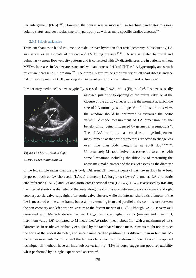
70
LA enlargement (86%) 696. However, the course was unsuccessful in teaching candidates to assess
volume status, and ventricular size or hypertrophy as well as more specific cardiac diseases696.
2.5.1.11 Left atrial size
Transient changes in blood volume due to de- or over-hydration alter atrial geometry. Subsequently, LA
size serves as an estimate of preload and LV filling pressure50,51. LA size is related to mitral and
pulmonary venous flow velocity patterns and is correlated with LV diastolic pressure in patients without
MVD50. Increases in LA size are associated with an increased risk of CHF as LA hypertrophy and stretch
reflect an increase in LA pressure697. Therefore LA size reflects the severity of left heart disease and the
risk of development of CHF, making it an inherent part of the evaluation of cardiac function51.
In veterinary medicine LA size is typically assessed using LA/Ao-ratios (Figure 12)51. LA size is usually
assessed just prior to opening of the mitral valve or at the
closure of the aortic valve, as this is the moment at which the
size of LA normally is at its peak51. In the short-axis view,
the window should be optimized to visualize the aortic
valve51. M-mode measurement of LA dimension has the
benefit of not being influenced by geometric assumptions50.
The LA/Ao-ratio is a consistent, age-independent
measurement, as the aortic diameter is expected to change less
over time than body weight in an adult dog51,698-700.
Unfortunately M-mode derived assessment also comes with
some limitations including the difficulty of measuring the
aortic maximal diameter and the risk of assessing the diameter
of the left auricle rather than the LA body. Different 2D measurements of LA size in dogs have been
proposed, such as LA short axis (LASAX) diameter, LA long axis (LALAX) diameter, LA and aortic
circumference (LACIRC) and LA and aortic cross-sectional area (LAAREA). LASAX is assessed by tracking
the internal short-axis diameter of the aorta along the commissure between the non-coronary and right
coronary aortic valve cups right after aortic valve closure, while the internal short-axis diameter of the
LA is measured on the same frame, but as a line extending from and parallel to the commissure between
the non-coronary and left aortic valve cups to the distant margin of LA51. Although LASAX is very well
correlated with M-mode derived values, LASAX results in higher results (median and mean 1.3,
maximum value 1.6) compared to M-mode LA/Ao-ratios (mean about 1.0, with a maximum of 1.3).
Differences in results are probably explained by the fact that M-mode measurements might not transect
the aorta at the widest diameter, and since canine cardiac positioning is different than in humans, M-
mode measurements could transect the left auricle rather than the atrium51. Regardless of the applied
technique, all methods have an intra subject variability ≤12% in dogs, suggesting good repeatability
when performed by a single experienced observer51.
Figure 11 : LA/Ao-ratio in dogs
Source : www.vettimes.co.uk

71
2.5.2 Cardiac function in human critical care
2.5.2.1 Myocardial infarction
Myocardial infarction (MI) is a common disease in human medicine and is associated with a 5 to 17%
incidence of cardiogenic shock701. Although MI in itself is not the scope of this literature review, it
merits mentioning, as the monitoring of these patients was the trigger for echocardiographic monitoring
of critical patients and we will come back to myocardial infarction when discussing cardiac biomarkers.
2.5.2.2 Myocardial dysfunction in SIRS and sepsis
In human medicine, several infectious diseases such as Q-fever702, Chagas disease703, bartonellosis704
and Rocky Mountain spotted fever705 are associated with infectious myocarditis resulting in LV
enlargement. In addition to these infectious pathogens with specific cardiac tropisms, SIRS and sepsis
have been reported to cause a cardiovascular and hemodynamic impairment in humans42,49,205,706-711.
Septic shock was first described as a “hyperdynamic” state of low SVR due to an abnormal vascular
tone712. It was further characterized by a hyperkinetic LV on echocardiography and high forward
Doppler flow, resulting in high CO or CI639,713,714. Patients displaying low CO were believed to have
absolute or relative hypovolemia, as fluid resuscitation normalized preload and increased CO in the
presence of low SVR236,706,713,715-718. The typical symptoms of poor peripheral perfusion, thready pulses
and cool extremities, also referred to as “cold shock”, was considered a reflection of inadequate
resuscitation and relative hypovolemia714,719,720.
Nearly half (35% to 50%) of septic human patients have a low CO at admission42. Despite the
encouraging reports of increasing CO after fluid resuscitation, low CO often remains unresponsive to
fluid resuscitation. These patients, unresponsive to fluid challenges, often suffer from decreased
ventricular contractility or LV hypokinesis, a phenomenon first described as a “hypodynamic” state, and
later redefined as myocardial dysfunction40,49,568,641. Myocardial dysfunction in SIRS patients is referred
to as myocardial depression describing a state of poor myocardial contractility, decreased peripheral
vascular tone (SVR), changed afterload and a loss of microvascular control548. The increased awareness
of the repercussions of sepsis on cardiac function lead to the incorporation of proof of cardiac
dysfunction (low CI or echocardiographic evidence) in the diagnostic criteria for severe sepsis,
highlighting its important role in sepsis309,721.
Myocardial dysfunction or depression has also been described in situations other than sepsis, such as
secondary to ischemia, hypoxemia, respiratory or metabolic (lactic) acidosis, low ionized serum
calcium, hypothermia, hyperthermia, advanced neoplasia and immune mediated disease214,722-724.
Myocardial dysfunction is currently reported in up to 44% of normotensive septic
patients42,162,641,710,711,725-727. Other contributing factors such as diastolic dysfunction, ventricular dilation
and decreased adrenergic response have been reported in sepsis and SIRS. Myocardial dysfunction or
72
depression is also called myocardial hibernation, indicating the supposedly “physiologic” and
“reversible” nature728. Myocardial depression could be an adaptive response to decrease energy
requirements and oxygen and adenosine triphosphate (ATP) demands, preventing initiation of cell death
pathways39,729 and preserving cell viability39,730. The next sections describes current evidence of
myocardial dysfunction and will discuss the pathophysiological mechanisms that may explain these
observations.
2.5.2.2.1 Systolic left ventricular dysfunction
The first report of myocardial depression dating back to 1984 identified decreased systolic LV function
in adequately resuscitated severe sepsis patients, with low SVR and low PCWP42. These findings have
been confirmed by many other publications731-735, and indicate that survivors can display more severe
systolic dysfunction than non-survivors40,42,49,65,88. Despite severely depressed LVEF, survivors have an
adequate LV stroke volume thanks to simultaneous acute LV dilation42,639. This confirms that systolic
function can be impaired in sepsis despite normal or increased CO42. Later work illustrates that normal
or supranormal LVEF does not exclude systolic dysfunction, as it can be masked by a decreased
afterload following inappropriately decreased SVR595.
2.5.2.2.2 Diastolic left ventricular dysfunction
Cardiac dysfunction in SIRS and sepsis is most often systolic, but can also be systolic and diastolic, or
solely diastolic55,56,654,655,736,737. Reduced LV compliance with increased end-diastolic LV pressures have
been demonstrated738. About 20% of septic shock patients suffer from isolated diastolic dysfunction,
with abnormal cardiac filling and relaxation, and preserved systolic function44,672.
2.5.2.2.3 Increased left ventricular volume
Septic patients often display ventricular dilation, with dilation more pronounced in survivors40,42,46,672,739.
However, LV dilation is not a consistent finding40,654 and acute LV dilatation in septic shock is not
supported by all authors549,569,573,641. Based on the physiological properties of the pericardial sac,
pericardial stiffness may preclude acute dilation, regardless of the administration of fluids for
resuscitation. Furthermore, a normal LV should not have a preload reserve, as it operates on the steep
portion of its pressure-volume relation beyond its optimal filling pressure740. Methodological differences
might account for these discrepancies: differences in treatment strategies, inherent differences in studies
using TTE or TEE and differences between ventilated and non-ventilated patients make it difficult to
compare findings. Less severely dilated LV were found in patients receiving less fluids and more
vasopressors672. TTE underestimates LV volumes and mechanical ventilation decreases image quality40.
Finally, as the incidence of impaired LV relaxation is estimated around 50%, LV dilatation might be
missed in smaller studies44,672.

73
2.5.2.2.4 Right ventricular dysfunction
Although less, myocardial dysfunction can affect the RV569,641,741-744. Right ventricular dysfunction
explains poor response to volume resuscitation, demonstrated by a lack of increase in CI despite a
decreased LV preload744. Right ventricular dysfunction may be due to intrinsic depressed contractility,
acute cor pulmonale675, or an acute increase in pulmonary vascular resistance (PVR). Besides intrinsic
pulmonary or vascular disease increasing PVR, mechanical ventilation can increase PVR, and raise RV
afterload745. Mechanical ventilation will reduce venous return secondary to the provoked increase in
intrathoracic pressure. Therefore, volume-controlled positive end expiratory pressure (PEEP) will lead
to respiration-phase-specific reductions in RV output, which are most pronounced during inspiration746.
Demonstrating acute right ventricular dilation and RV failure indicates RV volume overload and
excludes hypovolemia, but increases the likelihood of acute cor pulmonale (e.g. pulmonary
thromboembolism)559,679.
2.5.2.2.5 Cardiovascular consequences of myocardial dysfunction
Myocardial depression during SIRS is characterized by a variation of left and right ventricular systolic
and diastolic dysfunction, with potential ventricular dilation and potentially resolves within 10 days to
4 weeks40,42,46,49,153,558,639. To add to the confusion, these patients often display peripheral vasodilatation
resulting and reduced SVR40,42,44,672,739. The combination of myocardial dysfunction and decreased SVR
results in an unexpected maintained CO and CI42. In cases of normal LVEDV SV will however tend to
be severely reduced40. Therefore an increased end-diastolic volume might compensate for a decreased
systolic function and be a pathophysiological adaptation to keep CO in the normal
range42,43,55,56,237,725,732,733,744.
2.5.2.3 Pathophysiology of myocardial dysfunction
2.5.2.3.1 Myocardial ischemia and myocardial injury
Although myocardial hibernation was first hypothesized to occur secondary to myocardial ischemia45,747,
echocardiography, electrocardiography and experimental models evaluating “coronary blood flow”- and
“myocardial metabolism”-studies demonstrate preserved myocardial blood flow and lactate extraction,
refuting the hypothesis of global myocardial ischemia64,235,748,749. Microcirculation is however severely
altered in sepsis, and regional ischemia could still potentially contribute to myocardial depression750.
Cytopathic hypoxia occurs in other organs during sepsis and could be another potential explanation of
myocardial dysfunction751. However, finding preserved ATP in dysfunctional myocardium contradicts
this hypothesis752-755. During myocardial hibernation in ischemia and hypoxia, cardiomyocytes remain
viable by down-regulation of oxygen consumption, energy requirements and ATP demands756,757.
Histopathological studies do not demonstrate myocardial injury as a prerequisite for clinical myocardial
dysfunction57,758,759. Disruption of the actin/myosin contractile apparatus could also contribute to

74
myocardial depression in septic shock760, but the major explanation for the depressed myocardial
contractility during systemic inflammation appears to be endotoxin, interleukins and tumour necrosis
factor548.
2.5.2.3.2 The role of pro-inflammatory cytokines
Injection of bacteria or endotoxins into lab animals and humans results in myocardial depression55,205.
Although endotoxin triggers the inflammatory cascade, endotoxin itself does not cause myocardial
dysfunction45,153 however endotoxin elicits the release of inflammatory mediators761. Serum from human
septic patients induces similar cardiac effects in rats, and heat-labile, proteinase-sensitive substances
with a molecular mass of 10 to 30kD appear to be responsible45,153. These properties are consistent with
cytokines, but exclude prostaglandins and leukotrienes45,155. Studies investigating different cytokines
demonstrated that TNF-α and IL-1β have a synergistic depressant effect on myocardial contractility at
concentrations similar to clinical conditions4,126,159,219,762 and explain most of the symptoms observed in
circulatory shock43,55,235,304,763-765, while publications on IL-6 are inconclusive on its role126. In vitro
studies and observations in living lab-animals confirm these hemodynamic effects126,204,766188,208,209.
Removal of TNF-α and IL-1β from serum of septic humans (by washing or using immunoabsorption)
rapidly eliminates myocardial depressor effects126,204. Exposure of cardiomyocytes to TNF-α and IL-1
does not increase lactate dehydrogenase concentrations, neither does supravital staining demonstrate
any loss of cell viability126, underlining the concept of depression rather than injury.
TNF-α induces reversible (systolic and diastolic) myocardial depression in dogs and other
animals43,235,767. Myocardial depression in dogs appears 24 hours after administration of TNF-α and
disappears after 72 hours235. This late onset of action supports the theory that secondary mediators are
involved in the process235, and explains why early studies focusing on the first day after injection did
not see myocardial depression with TNF-α or IL-1β304. TNF-α causes myocardial depression via
secondary mediators released by recruited and activated neutrophils235. Neutrophils participate in
myocardial ischemic injury and lead to myocardial dysfunction242-246, and neutrophil margination and
diapedesis through the endothelium as well as adhesion to cardiomyocytes is enhanced by TNF-α and
IL-1β195,196,241. TNF-α also stimulates liberation of IL-1β from neutrophils which will further aggravate
myocardial depression216,247,768.
2.5.2.3.3 Molecular basis of myocardial systolic dysfunction
2.5.2.3.3.1 Nitric oxide
Nitric oxide (NO) is an important intracellular mediator, which regulates myocardial energy production,
coronary vessel tone, thrombogenecity and has direct effects on cardiac contractility769-773. NO is
produced via NO synthases (NOS), of which NOS2 (or inducible NOS (i-NOS)) is induced in the
myocardium in response to pro-inflammatory cytokines, endotoxin and CRP398,774-777. Low doses of NO

75
improve LV function, yet high concentrations decrease myocardial contractility778-780, and can induce
apoptosis781. Several experimental studies demonstrate the key-importance of NO and NOS2 in the
development of myocardial depression158,159,275,782-784. NO additionally has direct cytotoxic effects via
generation of the oxidant peroxynitrite, which is harmful to DNA, proteins and lipids785. Studies
demonstrating a near immediate effect of endotoxin are explained by effects of endotoxin on endothelial
NOS, rather than the effects of cytokines on inducible NOS159.
2.5.2.3.3.2 Altered metabolism and cytopathic hypoxia
Myocardial hypoperfusion does not participate in myocardial dysfunction (myocardial blood flow even
seems elevated)748,749, but altered myocardial metabolism is suspected, although research does not agree
on the occurring alterations748. During endotoxinaemic shock myocardial cells switch to glucose as a
primary energy substrate and undergo anaerobic glycolysis for ATP production786,787. Myocardial
specific glucose transporters (GLUT1 and GLUT4) facilitate the required increased glucose uptake787-
789 and hibernation leads to increased glycogen deposition in the cardiomyocytes790. Sepsis in mice leads
to increased glucose uptake, up-regulation of the GLUT-4 myocardial specific glucose transporter and
increased glycogen deposits in the cardiomyocytes, indicating altered metabolism728. Other studies in
human septic patients and canine endotoxemic models describe an increased uptake of lactate, with
decreased use of glucose, free fatty acids and ketone bodies748,791, indicating that lactate becomes the
main energy source786. On the other hand, nearly half of myocardial oxygen consumption cannot be
explained by substrate extraction, indicating a significant amount of endogenous substrate utilization748.
As carbohydrate reserves are low in cardiomyocytes, consumption of those reserves might add to
progressive cardiac depression, as described in experimental models792-794.
Cytopathic hypoxia leads to uncoupling of oxidative phosphorylation and disruption of adenosine 5’-
triphosphate production, resulting in decreased contractile forces644. Sepsis-induced irreversible
inhibition of myocardial cytochrome oxidase was previously demonstrated795, rendering the
cardiomyocyte “functionally hypoxic” despite the presence of oxygen, and inducing myocardial
hibernation728,795.
As previously stated, coronary hypoperfusion has not been identified in sepsis748,749 and sepsis is
accompanied by increased oxygen availability749. Moreover, there is no evidence of net myocardial
lactate production749. This indicates that coronary blood flow is probably not determined by myocardial
oxygen needs in sepsis, similar to systemic peripheral shunting in septic shock42,713. However, sepsis
decreases oxygen extraction in other tissues749 and increases lactate uptake748. Myocardial blood flow
redistribution occurs in canine septic shock796 and global myocardial lactate extraction can mask
regional lactate production797. Therefore the hypothesis of microcirculatory derangements in sepsis can
not be excluded.

76
2.5.2.3.3.3 Other mechanisms
Although the primary method of action of TNF-α is probably centered on changes in nitric oxide (NO)
production, calcium homeostasis and other mechanisms have a role in the development of myocardial
dysfunction. Sepsis and TNF-α decrease sarcolemmal calcium concentrations, leading to reduced
cardiac contractility39,204,798. As previously stated, neutrophils adhere to the endothelium during
myocardial depression799 and contribute to ventricular hypocontractility by generation of oxygen free-
radicals causing oxidative injury210,799,800.
Cardiac dysfunction is associated with a strong attenuation of sympathetically and vagally mediated
heart rate variability707. Endotoxins reduce β-adrenergic receptors, leading to a decreased response to
catecholamines801. TNF-α (similarly to IL-1β) decreases the sensitivity of the septic heart to β-adrenergic
catecholamines157. This impaired adrenergic responsiveness802 improves during the course of disease802-
804. The effect is induced by TNF-α and IL-1β which inhibit cyclic adenosine monophosphate (cAMP)
accumulation in response to catecholamines and increases inhibitory G-proteins which inhibit cAMP,
leading to decreased β-adrenergic responsiveness without a decrease in β-adrenergic receptor
density157,805,806.
2.5.2.3.4 Pathophysiology of diastolic dysfunction
Few studies looked into the pathophysiology of diastolic dysfunction. Impaired ventricular relaxation is
associated with increased concentrations of TNF-α, IL-8, IL-10 and cTnI807. The initial part of relaxation
depends on an activation-inactivation process involving β-adrenergic stimulation, calcium homeostasis,
functions of sarcoplasmic reticulum, contractile proteins of myofilaments, and their interaction with
calcium, all of which can be impeded during septic shock as previously explained44.
2.5.2.3.5 Myocardial dysfunction and prognosis
Although a general consensus exists that myocardial depression or dysfunction has a high prevalence in
sepsis and SIRS, there is a lot of contradicting information regarding its effect on prognosis. Early
reports indicated a better prognosis for patients displaying signs of cardiac depression42, but recent
papers indicate a worse prognosis for patients with decreased systolic function88. Increased heart rate808
and severely decreased SVR are other cardiovascular parameters that are associated with poor prognosis
in SIRS808-810.
2.5.3 Cardiac function in canine critical care
2.5.3.1 Experimental evidence
Despite the vast amount of scientific evidence in the human field, little is known about the clinical
prevalence of myocardial dysfunction in veterinary medicine. Most of our current knowledge is based
on animal experiments and extrapolations from human medicine. Experimental studies of septic
peritonitis, bacteremia or infusion of TNF-α and endotoxin in dogs43,55,56,139,235,237,240,811 following

77
adequate fluid resuscitation results in hyperdynamic patients with a high CO, low SVR, and myocardial
(systolic and diastolic) dysfunction similar to human patients, including ventricular
dilatation42,43,55,56,139,237,240,732,811.
Early experimental canine models did not identify myocardial depression811, or suspected a principal
role for coronary hypoperfusion812-816 but these studies displayed major differences from the human
clinical setting. First of all, many experiments were performed on denervated hearts using bypass models
on anesthetized animals not receiving fluid resuscitation, resulting in low CO. Moreover, these studies
only recorded the initial 3 to 6 hours, after which animals died due to poor hemodynamic support812,814.
As myocardial depression typically takes 24 hours to appear, it was probably missed43,235. The major
contribution of canine experimental studies is undoubtedly that myocardial depression was observed in
any therapy besides fluid resuscitation, confirming it is a result of pathology secondary to a disease
process and not a result of therapeutic interventions55.
2.5.3.2 Clinical evidence
As echocardiography in dogs is complicated by breed differences, ratios and indices are used to evaluate
cardiac function817. Unfortunately very limited literature on clinical myocardial dysfunction during SIRS
in dogs is available. One publication systematically evaluating cardiac function in an emergency setting,
focused on the ability of the veterinarian to perform the examination rather than on the results696. In
ICU-settings published canine studies focused on a single disease. A non-blinded retrospective study in
dogs with critical illness identified 16 dogs with cardiovascular dysfunction57. Prognosis of dogs with
myocardial dysfunction appeared poor, but was not compared with a control group of critical patients
without myocardial dysfunction57. In canine ehrlichiosis one third of animals demonstrated
echocardiographic abnormalities, a prevalence similar to a group with systemic inflammation due to
other causes818. One abstract describes reversible myocardial dysfunction, demonstrated by decreased
systolic function and ventricular dilation (non-detailed echocardiographic ratio indices) in a canine
septic patient644.
In summary clinical evidence of myocardial dysfunction in dogs with SIRS/sepsis is available, but little
is known about its prevalence, echocardiographic parameters have not been studied and nothing is
known about the effect of myocardial dysfunction on prognosis.
2.5.4 Conclusion
Because of the variable nature of the peripheral and central cardiovascular effects of sepsis, any rational
treatment regimen requires monitoring of blood pressure, CO, circulating volume, myocardial function
and vascular tone548. Point of care echocardiography is valuable for the identification of the cause of
hemodynamic instability (hypovolemic, cardiogenic or distributive) and for the subsequent optimization
of therapy (fluid administration, inotropic or vasopressor therapy), and the possibility to repeat this

78
examination allows to assess response to treatment819. Unfortunately, despite the convincing knowledge
gathered in human medicine, the information in canine medicine is very limited.
2.6 CARDIOVASCULAR BIOMARKERS
2.6.1 Cardiac Troponins
Myocytes contain abundant contractile proteins organized in sarcomeres with overlapping thick and thin
filaments, sliding past each other to produce muscle contraction820. Thick filaments are primarily
composed of myosin, having adenosine triphosphatase (ATPase) activity and forming cross-bridges
with actin. Thin filaments consist of actin, tropomyosin and
the troponin complex (Figure 17). Within the troponin
complex, there are 3 interacting and functionally distinct
proteins (I, T and C). Tissue-specific isoforms exist for each
type of troponin, and hence these troponins differ between
cardiac and skeletal muscle tissue821. The cardiac troponins
(cTn) are important for excitation-contraction coupling822,823
via regulation of the calcium-mediated interaction between
actin and myosin73,824,825. Tropomyosin dimers form a continuous chain along the groove of the actin
helix within the thin filament and block the myosin binding sites on actin. At regular intervals along the
filament lies a troponin complex, and each troponin protein (Figure 18) has specific functions regulating
muscle contraction823.
- Troponin C binds calcium to initiate muscle
contraction. Troponin C is not used as a marker, as the
cardiac and one skeletal isoform are homologous and
therefore does not have cardiac specificity821,826.
- Troponin T attaches the troponin complex to
tropomyosin and actin, and has a molecular weight of
37kDa827. Cardiac troponin T (cTnT) isoforms share
more than 50% homology with skeletal isoforms, but
can be separately identified828. Of 4 existing cardiac
isoforms of troponin T, only 1 is characteristic of the normal adult heart, while the 3 others are
normally expressed in fetal tissue. These isoforms can however also be re-expressed in damaged
skeletal muscles or in heart disease821,829-836, leading to increased cTnT values in non-cardiac
muscle disease such as polymyositis837. Early cTnT tests cross-reacted with the skeletal
isoforms, but this has been resolved in current kits using more specific antibodies838,839.
- Troponin I inhibits actomyosin ATPase and prevents the structural interaction of myosin with
actin-binding sites. The cAMP-dependent phosphorylation of troponin I at two adjacent serine
Figure 17: The troponin complex
Source: www.emdocs.net
Figure 18: Troponin C (Blue), I (Green)
and T (Magenta)
Source: Wikipedia.org

79
residues in the amino-terminal of the molecule causes a decrease in the affinity of calcium for
the calcium-binding troponin C and inhibition of actin-myosin interactions840. The binding of
calcium to troponin C displaces troponin I and causes a conformational change in tropomyosin
so it no longer interferes with myosin/actin binding and muscle contraction can occur. Cardiac
troponin I (cTnI) has three existing isoforms, two of which are present in skeletal muscle and a
third is specific to cardiac tissue as it shares <50% homology with skeletal isoforms828,841. This
cardiac isoform has a molecular weight of 24kDa, is larger than the two other isoforms and
contains an additional post-transitional 32 amino acid N-terminal peptide842-846. The rest of the
protein has more than 40% dissimilarity in its amino-acid sequence compared with skeletal
muscle TnI, allowing for the development of highly specific monoclonal antibodies without
cross-reactivity with non-cardiac forms821,827,847,848. Moreover, cTnI is not expressed in fetal or
damaged skeletal muscle846,849-851.
2.6.1.1 Human experience
2.6.1.1.1 Molecular properties and analysis
Cardiac ultrasound is very insensitive to diagnose myocardial injury as injuries must involve over 20%
of myocardial wall thickness to identify abnormal segmental wall motion, explaining the interest in
cardiac biomarkers852. The unique amino acid sequence of cTnI and cTnT allow for the development of
immunoassays for use in clinical laboratories853. Troponins are more specific for cardiac damage than
other cardiac biomarkers such as lactate dehydrogenase and creatine kinase isoenzymes in humans,
common laboratory animals and dogs59,854-856. Moreover, MB-creatine kinase, the creatinine kinase
expressing specific cardiac iso-enzymes, persists only for 18 to 36 hours and cannot determine whether
patients sustained an acute MI a couple of days before presentation857. Cardiac troponins become
detectable at a similar time-interval after acute infarction, but display more sustained values858,859.
Troponins are leakage markers as increased myocyte permeability causes the release of cTn into
circulation58-61. Apoptosis on the other hand should not decrease membrane integrity, and subsequently
no significant leakage of troponins should occur860. cTnI is more sensitive and specific than cTnT at
detecting myocardial injury829,830,849,861-863. The smaller size of cTnI (with a molecular weight of 22kDa)
versus cTnT (40kDa) could explain the higher sensitivity of cTnI864.
The majority of troponin is structurally bound within the thin filaments, and only a small percentage
remains free in the cytosol (2-4% for cTnI and 6-8% for cTnT). The free part can be released without
histological evidence of myocardial cell injury854,865,866. Structurally bound cardiac troponin is only
released after major injury with cell disruption and necrosis58,867,868. cTnI is mostly released in complexes
(Figure 19) of Troponin T-I-C and Troponin I-C, while cTnT is released as free Troponin T, or a
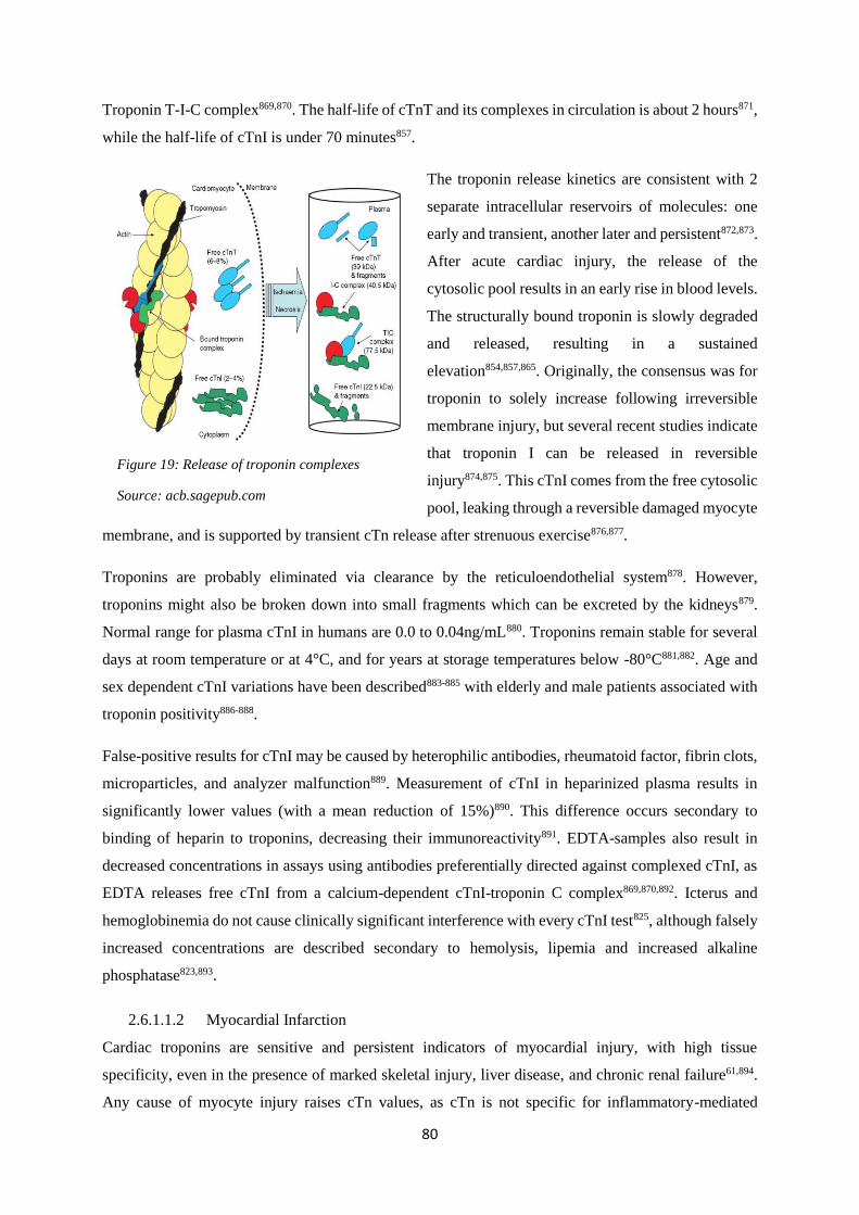
80
Troponin T-I-C complex869,870. The half-life of cTnT and its complexes in circulation is about 2 hours871,
while the half-life of cTnI is under 70 minutes857.
The troponin release kinetics are consistent with 2
separate intracellular reservoirs of molecules: one
early and transient, another later and persistent872,873.
After acute cardiac injury, the release of the
cytosolic pool results in an early rise in blood levels.
The structurally bound troponin is slowly degraded
and released, resulting in a sustained
elevation854,857,865. Originally, the consensus was for
troponin to solely increase following irreversible
membrane injury, but several recent studies indicate
that troponin I can be released in reversible
injury874,875. This cTnI comes from the free cytosolic
pool, leaking through a reversible damaged myocyte
membrane, and is supported by transient cTn release after strenuous exercise876,877.
Troponins are probably eliminated via clearance by the reticuloendothelial system878. However,
troponins might also be broken down into small fragments which can be excreted by the kidneys879.
Normal range for plasma cTnI in humans are 0.0 to 0.04ng/mL880. Troponins remain stable for several
days at room temperature or at 4°C, and for years at storage temperatures below -80°C881,882. Age and
sex dependent cTnI variations have been described883-885 with elderly and male patients associated with
troponin positivity886-888.
False-positive results for cTnI may be caused by heterophilic antibodies, rheumatoid factor, fibrin clots,
microparticles, and analyzer malfunction889. Measurement of cTnI in heparinized plasma results in
significantly lower values (with a mean reduction of 15%)890. This difference occurs secondary to
binding of heparin to troponins, decreasing their immunoreactivity891. EDTA-samples also result in
decreased concentrations in assays using antibodies preferentially directed against complexed cTnI, as
EDTA releases free cTnI from a calcium-dependent cTnI-troponin C complex869,870,892. Icterus and
hemoglobinemia do not cause clinically significant interference with every cTnI test825, although falsely
increased concentrations are described secondary to hemolysis, lipemia and increased alkaline
phosphatase823,893.
2.6.1.1.2 Myocardial Infarction
Cardiac troponins are sensitive and persistent indicators of myocardial injury, with high tissue
specificity, even in the presence of marked skeletal injury, liver disease, and chronic renal failure61,894.
Any cause of myocyte injury raises cTn values, as cTn is not specific for inflammatory-mediated
Figure 19: Release of troponin complexes
Source: acb.sagepub.com

81
myocyte injury895-898. cTnI contributes to the early diagnosis, prognosis and treatment of MI in human
medicine, but increases can be explained by any type of cardiac injury or increased permeability899-902.
Test-improvement and continuously increased sensitivity result in lower diagnostic cutoffs, leading to
an increased detection of cTnI elevations, which are sometimes difficult to explain885,903 or attributable
to chronic conditions904.
In myocardial infarction, cTnI increases 2 hours and peak after 48 (12 to 72) hours60,823,827,847,884,905-908.
cTnI levels can remain increased for up to 8 days, suggesting irreversible and active cardiomyocyte
damage823,827,847,884,906,909. cTn tests have lower sensitivity within six hours after symptom onset 910-913
due to the slow release kinetics of troponins. Therefore, samples are ideally taken at admission, after 6-
9 hours and again 12-24 hours after presentation896 when cTnT and cTnI have 100% sensitivity for MI900.
2.6.1.1.3 Other cardiac conditions
Although troponins were first applied to detect MI, many other cardiac conditions increase serum
troponins in humans. Severe CHF902,914-920, aortic stenosis, LV hypertrophy, myocardial bridging,
coronary spasms, cardiac rhabdomyolysis, subendocardial ischemia847, cardioversion (albeit very
mildly)921,922, prolonged resuscitation923, atrial fibrillation924, supraventricular tachycardia925,
pericarditis926, myocarditis61,895 and blunt cardiac trauma927-929 are associated with increased troponin
concentrations930.
In human medicine, 75% of patients with acute heart failure have detectable cTnI or cTnT
concentrations915, and increased cTnI concentrations are predictive of mortality and recurrence of
readmission61,895,916,917,931-935. cTn levels can be used of to monitor the clinical course of the disease917,
with persisting elevations of cTnT associated with poorer outcome916,936. cTnI and cTnT are elevated in
myocarditis895,937, but cTnI levels are not correlated with histological lesion severity895. Moreover,
increased cTnI concentrations in patients with myocarditis are independent predictors of mortality, and
recurrence of hospital readmission61,895,916,917,931-935. cTnI is more accurate and sensitive than
transthoracic echocardiography and ECG to detect cardiac contusions855,938. Subsequently, cTnI testing
is valuable in ruling out significant blunt chest trauma as a cause of cardiogenic shock, severe
arrhythmias, or structural damage898,928. Troponin concentrations improve risk prediction for serious
cardiac complications as myocardial ischemia60,907 and inflammation or necrosis937, regardless of the
cause.
2.6.1.1.4 Non-cardiac conditions
Many non-cardiac conditions are also associated with increased troponin concentrations in humans,
most notably in pulmonary embolism939,940, chronic pulmonary obstructive disease exacerbation and
respiratory failure941 ; renal failure942,943; sepsis65,68,944,945 and non-specific critical illness which are

82
discussed in the next paragraphs. The exact mechanism by which this occurs remains however
controversial69,847,878,889,930,946-950.
In humans, pulmonary embolism is the most common non-acute coronary syndrome to cause increased
troponin levels, and results in increased cTn concentrations in up to 50% of patients944,951, which are
correlated with prognosis952.
Increased troponin concentrations are often associated with renal disease. The original hypotheses in
human medicine were that increased cTnI were caused by independent and unassociated comorbidity of
coronary artery disease in these patients, or by uremic cardiomyopathy878,953-959. A third explanation was
that cTnI increases occurred without any concurrent myocardial cell damage960.
cTnT concentrations are more often increased compared to cTnI in renal failure patients961-964, and
several hypotheses can explain this. cTnT fragments might accumulate in the bloodstream due to poor
renal clearance879, while the half-life and clearance of cTnI is similar regardless of renal function956. If
the free cytosolic protein fraction is responsible for these increased concentrations, the fraction of free
cTnT is higher than that of cTnI, and there is twice as much cTnT than cTnI per gram of myocardium948.
Furthermore, cTnT is released both as a free fragment and in a complex, but cTnI is only released as a
complex. Additionally the concurrent uremia can modify cTnI via phosphorylation, oxidation or
proteolysis, while cTnT only undergoes limited proteolysis869,878,965,966. With only one standardized assay
available for cTnT, and different monoclonal antibodies exist for cTnI, such modified molecules might
influence measurements of cTnI more than cTnT948.
Severity of renal failure does not correlate with cTnI concentrations (not evaluated for cTnT)948,967, and
cTnI and cTnT analysis remains useful in identifying myocardial injury in renal patients954,955,968.
Similarly, cTnI and cTnT can identify patients with worse prognosis despite concurrent renal failure
and/or hemodialysis952,954,962,969-972.
Troponin elevations also increase following high dose chemotherapy (doxorubicin,
cyclophosphamide, carboplatin, ifosfamide, methotrexate and taxotere)973,974 or thoracic radiation
therapy975. In patients receiving chemotherapy, troponins are independent predictors of the development
of cardiac toxicity and decreased LV function, recurrence of readmission to hospital, and
mortality973,976,977.
cTnI apparently increases secondary to physiologically mediated cardiac remodeling as seen in
marathon runners978,979. Contrary, detectable concentrations of troponin have a prognostic value for
future cardiac events and all-cause mortality in a healthy population885-887,904,938,980-983. Even small rises
in apparently healthy older people appear indicative of future death to cardiac disease984 and
perioperative cTnI elevations are associated with major cardiac complications up to 1 year after

83
surgery985. In summary it is likely that mild to moderate changes associated with non-cardiac disease
reflect subclinical myocardial damage905,986-990.
2.6.1.1.5 SIRS, sepsis and myocardial dysfunction
Cardiac troponin I is elevated in critical illness62,945,991 with incidence rates of 43% across all patient
groups, 60% in septic patients, 53% in medical ICUs, 43% in mixed medical and surgical ICUs, and
32% in mixed surgical and trauma ICUs63,992. cTn positivity is seen in heterogeneous populations of
critically ill patients in medical993,994, surgical64, and pediatric995 ICUs. The cause of a septic process
does not play a role in the incidence and extent of cTn elevations64. Several studies found no sign of
coronary artery disease upon testing in these critically ill patients62,945,996, but these increased
concentrations might indicate myocardial injury. As critical patients often require anesthesia and
surgery, and these procedures may cause further damage to the myocytes because of potential
perioperative myocardial hypoxia997-999, further research into the cause and meaning of these elevated
troponin concentrations has been performed and several hypotheses regarding the etiology of these so
called ‘shock related troponin elevations’1000 have been proposed.
- the first hypothesis is that increased cTn concentrations might be provoked by ischemia,
subsequent cellular hypoxia and lesions following a mismatch between oxygen supply and
demand69,748,749,945. Besides the decreased supply, demands might indeed increase in shock
patients65,69,126,847,945,952. The tachycardia of shock patients has been implicated as a cause of
increased oxygen consumption69,750,952. This hypothesis has been largely refuted by observations
that coronary blood flow actually increases and regional lactate concentrations do not increase
in SIRS and septic patients748,749.
- focal microvascular dysfunction could provoke regional myocardial
ischemia65,126,728,750,847,945,952,1001,1002. Hypoxia can not only cause cellular lesions but also induce
increased cell membrane permeability to macromolecules as large as albumin or cTn1003,1004.
cTnI may be degraded into smaller fragments after brief periods of myocardial ischemia, and
these molecules might more easily pass the (perhaps) more permeable membrane965.
- hypercoagulability and (septic) microthrombi can provoke regional
hypoperfusion65,126,750,847,945,952, but this theory was not supported in a recent study996.
- toxic effects from bacterial endotoxins65,126,847,945,952.
- ventricular wall stress, hypertension and left wall hypertrophy can activate intracellular
signaling leading to apoptosis, damage and micronecrosis, leading to troponin elevation69,931,1005.
- therapy with vasopressors and positive inotropes could impact cTn levels65,126,847,945,952,1006
via decreased perfusion secondary to vasoconstriction and cellular injury750,952,1007.
- free radicals and leucocyte-derived superoxide radicals can cause myocardial cell damage and
apoptosis leading to troponin elevations728.

84
- paracrine mediated (e.g. natriuretic peptides) increases in cell membrane permeability1007.
- stress mediated effects could induce leakage of troponins without further explanation1008.
- pro-inflammatory cytokines (TNF-α and IL-6) possess direct cardiac depressant effects and
increase membrane permeability65,71,126,847,945,952,1005,1007. TNF-α also exert effects via the
activation of neutral sphingomyelinase, suppression of nitric oxide and calcium transient
pathways, modulation of intracellular proteases, effects on arachinodate metabolism, protein
kinases, oxygen-free radicals, transcription of cytotoxic genes, nuclear regulatory factors and
ADP ribosylation, which all contribute to myocardial depression and troponin elevation66,1006.
In line with this hypothesis, troponins would be potential biomarkers of myocardial depression.
In summary, the two main hypotheses are increases secondary to irreversible (necrosis, apoptosis and
cell turn over), or reversible injury (increased permeability, intracellular proteolysis and formation of
vesicles)828. As myocardial dysfunction during sepsis is reversible, troponin release probably occurs
following reversible rather than irreversible injury42,1009.
In human medicine, cTn concentrations are correlated with the severity of cardiac dysfunction in
severe sepsis168,710,1010,1011 and human critically ill patients67,728. Cardiac dysfunction and cTn elevations
are often simultaneously identified in sepsis65,889,1012, and cTn concentrations are correlated with
biventricular increased ventricular volumes739, LV dysfunction62,65,71, LVSWI1008, regional wall motion
abnormalities68, lower EF65,945,993,1008,1012-1014, and isolated and reversible impairment of LV relaxation44.
Normalization of LV systolic and diastolic function is associated with normalization of cTnI44. Shock
associated troponin elevations are associated with need of vasopressors or inotropes63,68,71,1008, although
this was not confirmed in a later study65. Troponin concentrations in sepsis and critical disease are
correlated with the clinical condition1010,1011, critical illness scores65,1011, degree of hypotension1014 and
APACHE II score65,68.
In non-cardiac disease, cTnI levels correlate with prognosis, regardless of the underlying primary
disease process62,945,957,961. In septic (shock), surgical ICU and critically ill patients, cTnT and cTnI are
associated with length of ICU stay63,68,1015, and outcome44,62,64,65,68,69,71,994,1008,1012,1015.
cTnI concentrations in septic patients might be useful to monitor treatment response as concentrations
correlate with severity of lesions68, but as troponin concentrations remain increased for over 50 hours,
they are probably less useful for this purpose70-73.
In conclusion, the mechanisms of myocardial injury remains poorly understood and is likely
multifactorial, but the increased cTn concentrations are universally predictive of poor outcome in critical
human patients991,993,1016.

85
2.6.1.2 Canine experience
2.6.1.2.1 Molecular properties and analysis
The amino acid sequence of humans and canine cardiac troponins is nearly identical, as troponins are
phylogenetically highly conserved proteins in mammals863,894,1017. Homology between canine and human
genes for cTnI is 95%1018. Canine cTnI has 1 additional amino acid (Ala25), yet this does not affect
functionality1018. The region commonly targeted for antibody production for most assays has a high
degree of homology, with only one amino acid that differs in the 83 amino acid region853,1018. In total,
there are 4 amino acid changes in the 23 amino acid N-terminal region between human and canine
cTnI1018. Because of this high homology in amino-acid sequences, and because myocardial
concentrations of cardiac troponins in humans and dogs are very similar, several human commercial
cTnI analyzers can be used to measure canine cTnI1018. Homology between canine and human cTnT
amino acid sequence is >91%58,863,868,1019. It is generally believed that human assays can be used to
measure blood levels of cardiac troponin I and T in most species59,894,1018. Serum, heparinized plasma
and whole blood can be used to measure cTnI, depending on the assay used, although serum
concentrations tend to be slightly lower824,1020. cTnI remains stable at -70°C, several months at -20°C or
at -4°C for 1 to 18 months864,881,953, but variable results are described after repeated freeze-thaw
cycles824,884,1020.
Most healthy animals have cTn levels below the threshold of detection861,880,1021-1024. The range of plasma
cTnI concentrations in dogs is <0.03 to 0.07ng/mL with a mean of 0.02ng/mL880. High sensitivity cTnI
tests are not yet convincingly described in companion animals828. Healthy dogs have cTnT levels below
the threshold of detection861,1021,1023,1025. cTnI assays are more sensitive than cTnT to detect cardiac
involvement, and cTnI received more attention in veterinary medicine824,825. However, alongside its
superior sensitivity, cTnI has decreased specificity for myocardial damage compared to cTnT1025.
Age dependent cTnI variations are described, yet sex effects are not confirmed543,828,884,1024. The strong
association between age and cTnI concentrations suggests that age causes myocardial changes leading
to cTnI leakage543. Loss of myocytes with increasing age may occur because of defects in the
oxygenation potential in the myocardium. Associations between arteriosclerosis and age in dogs have
been described543. Azotemia and hyperbilirubinemia may affect cTnI assay results1026 and Greyhounds
and Boxers have higher cTnI concentrations than other breeds1027,1028. Exercise can influence canine
cTnI concentrations, with transient release of cTnI after exercise in Alaskan sled dogs477,823,1029, with
dramatically increased concentrations after prolonged and heavy exercise1029.
The following sections discuss the clinical use of cTn. Different studies measured cTnI and/or cTnT,
making these paragraphs sometimes hard to read. However, bear in mind that both troponins have
similar release patterns and indicate myocardial lesions, with the major difference in between both the
fact that cTnI is more sensitive than cTnT.
86
2.6.1.2.2 Experimental myocardial infarction
Dogs have often been used as an experimental model for induced MI. cTnT and cTnI are sensitive and
specific biomarkers of cardiac injury in dogs, as in humans58,867,868,1030,1031. cTnI concentrations increase
earlier than cTnT58,861,867, and both can remain increased up to 7 or 10 days after the insult58,865. The half-
life of cTnI in dogs is 120 minutes, compared to a half-life under 70 minutes in humans1032,1033.
2.6.1.2.3 Other cardiac conditions
cTn levels can be elevated in primary heart disease1021-1024,1034 such as MVD, sub-aortic stenosis, dilated
cardiomyopathy, and in arrhythmic dogs1021-1024,1035. cTnI is more sensitive than cTnT for the diagnosis
of acquired heart disease1021,1023, although mild subclinical heart disease does not result in marked
elevations in cTnI1024. cTnI is increased in English springer spaniels with bradyarrhythmias73,1036, Boxers
with arrhythmogenic right ventricular cardiomyopathy1037, pericardial effusion1022,1038,1039, experimental
infarction867, cardiac pacing-induced injury, positive inotropic and cardiotoxic drugs884 and
neoplasia1040. One publication found a correlation between cTnI and CRP concentrations in dogs with
MVD, suggesting a link with inflammation543.
cTnI is associated with severity of cardiac disease in MVD, sub-aortic stenosis, dilated cardiomyopathy
and Boxers with right ventricular cardiomyopathy73,543,1021-1024,1028. Progression of cardiac disease and
response to treatment can be assessed by repeated sampling73,1036,1041. cTn levels may be correlated
with prognosis in dogs with MVD, sub-aortic stenosis, and dilated cardiomyopathy73,864,1021-1024. On the
contrary, cTnI was not correlated with median survival time in Brady arrhythmic dogs1036.
Finally, dogs with pericardial effusion have increased cTnI levels, and concentrations might be even
higher if caused by a hemangiosarcoma1038, although these findings were not confirmed in a subsequent
study1039.
2.6.1.2.4 Non-cardiac conditions
In canine medicine, increased concentrations of cardiac troponins are observed in several non-cardiac
conditions such as gastric dilation and volvulus (GDV), trauma, infectious processes and non-cardiac
systemic disease93,818,824,825,861,953,1032,1042-1044. cTnI concentrations are often increased in azotemic
dogs953,1045, although concentrations are not correlated with the degree of azotemia953. As coronary artery
disease is not a feature of companion animals, this hypothesis from human medicine cannot explain this
finding953. As one paper demonstrated cardiac lesions in 3 out of 4 autopsied cases, concurrent cardiac
disease might be the cause of increased cTn levels1045.
cTnI does not reliably distinguish cardiac from non-cardiac causes of dyspnea93,1046, as increased
concentrations are seen in dogs with and without cardiac disease93. cTnI release in non-cardiac dyspnea
could be explained by pulmonary vasculature endothelial damage resulting in activation of angiotensin-
converting enzyme, and subsequent cardiac injury and cTnI release1047,1048.

87
2.6.1.2.5 SIRS, sepsis and myocardial dysfunction
cTnI concentrations are increased in 70% of dogs with a variety of systemic non-cardiac diseases953,
regardless of the presence of cardiac murmurs, hypertension or the type of non-cardiac illness953. This
high incidence is not due to increased sensitivity of high-sensitivity cTnI assays, as concentrations are
significantly elevated in infectious disease98,818,825,1042,1049, blunt thoracic trauma861,863, GDV824,861,
pulmonary hypertension1050, patients receiving doxorubicin1034,1051, snake bites77,1052-1054, and in canine
critically ill ICU patients74,75. Peak concentrations of cTnT and cTnI are observed at similar time-points
than in humans or dogs with cardiac disease77,824,861,868,1030.
The incidence of elevated cTnI concentrations in dogs with a pyometra is 43%, but similar amounts of
dogs had elevated concentrations before and after surgery1032,1043, and no association was observed
between a clinical diagnosis of SIRS and increased cTnI concentrations1032,1043. Other infectious diseases
such as E. canis and trypanosomiasis1038,1055,1056 have been associated with more significant elevations
in cTnI. In ehrlichiosis, babesiosis and trypanosomiasis, cTnI levels are significantly correlated with
lesion scores, severity of anemia, severity of ECG abnormalities, disease severity, clinical diagnosis of
SIRS, and in case of Babesia infection, with the presence of ventricular premature complexes825,1056,1057.
Doxorubicin induces elevated troponin concentrations prior to the diagnosis of cardiac dysfunction1051,
but troponin elevations do not predict development of cardiac dysfunction1051.
Increased cardiac troponin concentrations correlate with poor prognosis in several studies on non-
cardiac disease74-76,114,824,825,861,1058. In GDV patients and after blunt thoracic trauma with secondary
myocardial contusions, increased troponin concentrations are associated with poor
prognosis824,861,863,1024,1054. In parvovirosis, higher cTnI levels are associated with non survival1058. In
canine pyometra a non-significant trend was observed between increased troponin concentrations and
mortality, but this may be explained by the low mortality rate of pyometra patients1043. cTnI is associated
with prognosis in dogs with leptospirosis or babesiosis114,825.
Increased cTnI concentrations have been demonstrated retrospectively in a group of non-cardiac
critically ill patients953 and several studies looked into cTn concentrations in canine SIRS populations
presented to the ICU74-76. These papers demonstrated that cTnI and cTnT are associated with short and
long term prognosis74-76. The greater sensitivity of cTnI over cTnT was demonstrated, yet this was not
reflected in a clear superiority to evaluate prognosis75,76. As summarized by the author, both markers are
highly correlated sources of similar information, and clinically it is considered sufficient to measure one
or the other828,884. Moreover, two of these studies failed to demonstrate an additional value of serial
measurement of cTn concentrations, although in one study peak cTnI concentrations were more
informative than concentrations at presentation74,76. cTnI concentrations are also correlated with CRP
concentrations at presentation77, suggesting a link with inflammation. Finally, two papers identified
myocardial dysfunction in dogs with severe systemic illness, providing an additional explanation for

88
some of the cTnI elevations observed in dogs with systemic disease, which once more might be linked
with the inflammation57,953.
In summary, cardiac troponins increase in many non-cardiac diseases, and are of prognostic significance,
but much more work is required in canine medicine to better understand the added value of this
biomarker.
2.6.2 Brain Natriuretic Peptides
Activation of neurohumoral systems in heart failure is beneficial in the short term to maintain cardiac
output and organ perfusion, but is detrimental in the long term and contributes to progressive myocardial
dysfunction and clinical deterioration in CHF patients1059. Natriuretic peptides form an important
endocrine system of cardiovascular and renal origin that participates in the integrative control of
cardiovascular and renal function by counteracting the initial neurohumoral responses1060.
The group of natriuretic peptides consists of
three distinct hormones: atrial natriuretic
peptide (ANP) and brain natriuretic peptide
(BNP) which are primarily of myocardial cell
origin (ANP predominantly atrial1061, and BNP
primarily from ventricular origin1062-1065), and
C-type natriuretic peptide (CNP) which is of
endothelial and renal epithelial cell origin1060.
Besides these 3 peptides, dendroaspis
natriuretic peptide, identified in the human and
canine heart has natriuretic effects, but its role
in mammals is undetermined1046,1066. Finally,
in primitive fishes ventricular natriuretic peptide is demonstrated, but is not found in mammals1067. ANP
and BNP mainly act as circulating hormones, whereas CNP is a paracrine/autocrine hormone1068.
Although ANP and BNP are derived from different genes, they share a highly homologous 17-amino
acid ring structure1069. Cardiomyocytes synthesize preprohormones such as preprobrain natriuretic
peptide (preproBNP) which is first cleaved into a signal peptide and probrain natriuretic peptide
(proBNP or proBNP1-108) which is then cleaved into a physiologically active C-terminal peptide (BNP)
and an inactive N-terminal fragment (NT-proBNP or NT-proBNP1-76) (Figure 20). These peptides
possess natriuretic, diuretic, renin-inhibiting and vasodilatory properties, mediated via second
messengers guanosine 3’:5’-cyclic monophosphatase (cGMP) after peptide binding to the respective
guanylate cyclase receptor. ANP and BNP bind to the natriuretic peptide receptor A (NPR-A) while
CNP binds to NPR-B, which are located in the kidneys, vasculature and heart1070. BNP has similarities
to ANP both in structure and in peripheral and central actions1061,1069,1071-1075. However, atrial stretch and
Figure 20: ProBNP secretion and cleavage by corin
Source: www.medscape.com

89
atrial tachycardia is the major stimuli for ANP secretion, whereas ventricular stimuli are more important
for BNP. We focused less on ANP but will highlight some of its properties when these are of (historical)
interest1076.
2.6.2.1 Human experience
2.6.2.1.1 Molecular properties and analysis
BNP was first discovered in the porcine brain1069, but was subsequently found in mammalian organs
such as the human heart. Where ANP (28 amino acids)1061 and CNP (22 amino acids)1077 are rather
homologous between different species, BNP is less well conserved, and contains 26, 45 and 32 amino
acid residues in swine, rats and humans, respectively1073,1078. The biological actions of BNP are in part
species specific, unlike those of ANP1079. Cardiomyocytes synthesize proBNP, which is almost entirely
immediately secreted into circulation, and is proteolytically cleaved by corin97 into BNP and NT-
proBNP81,87,1080. As little BNP is stored in granules1081-1083, BNP de novo synthesis and messenger RNA
(mRNA) degradation occurs rapidly, as a result of a rapid-turnover nucleic acid sequence (TATTTAT)
in the 3’ untranslated region of the mRNA1081,1084. Larger quantities of ANP are stored in granules,
suggesting a distinctive regulation in the production and secretion of these different peptides1085.
Ventricular BNP gene expression is rapidly induced, while ANP is not1081,1086,1087. Increased BNP
concentrations do not occur within minutes1088, but within 1 hour BNP gene expression is activated and
plasma concentrations increase rapidly thereafter1089. BNP is degraded after secretion by neutral
endopeptidases in the myocardium, lungs and kidneys1090.
Although the metabolism and tissue uptake of NT-proBNP and BNP appears similar81, the half-life for
BNP is approximately 22 minutes in humans1091 while that of NT-proBNP is 1-2 hours1092-1095. Plasma
concentrations of BNP and ANP are low and rapidly fluctuating1096, but the half-life of their N-terminal
fragments are considerably longer1097,1098, resulting in 5- to 15- times greater plasma levels1099,1100.
Neurologic disorders do not consistently result in increased BNP concentrations1101,1102 despite its
expression in the brain1103, and in subarachnoid hemorrhage most of the BNP appears to be of cardiac
origin1104. In the absence of cardiac disease, the tissue concentration of BNP is greatest in the atria1105,
but 50 to 60% of circulating BNP is synthesized in the ventricles1105.
In cardiac impairment, the proportional and absolute increment of NT-proBNP exceeds that of BNP.
This suggests that NT-proBNP may be a more discerning marker for early cardiac dysfunction than
BNP81. The disproportionate rise of NT-proBNP compared to BNP results either from differential
clearance rates from the peptides across tissues and organs or from differential changes in cardiac
secretion81. Renal clearance of NT-proBNP and BNP is however rather similar81,86,1106. Although NT-
proBNP has several theoretical benefits, both BNP and NT-proBNP assays are used in human medicine,
and are considered equally valid97.

90
Regulation of BNP synthesis differs between atria and ventricles1105,1107-1109. In the atria, the small
portion that is stored in granules is released by a regulatory pathway, while in the ventricles BNP is
rapidly secreted from myocytes after synthesis via a constitutive pathway1081,1083,1110,1111. Ventricular
overload or mechanical stretch increases BNP gene expression and secretion78,79,1085,1112-1114. Moreover,
increased ventricular afterload via neurohumoral activation or pharmacologic vasopressors also results
in increased production of BNP1115 1116. Finally, angiotensin II and endothelin-1 (ET-I) induce
ventricular synthesis of BNP in rats1108,1109,1115,1117. This implies that, while ET-I directly favors LV
remodeling, it simultaneously stimulates BNP synthesis, creating a negative feedback mechanism1118.
In summary, BNP secretion is mainly regulated by LV wall tension1062, and this is largely related to the
degree of ventricular volume expansion and pressure overload168,1062,1081,1082,1107,1119,1120.
BNP binds the NPA-receptor, resulting in a multitude of systemic effects
- inhibition of
o the renin-angiotensin-aldosterone system via inhibition of renin secretion, causing1137
Natriuresis in the proximal and distal renal tubules1075
Diuresis1075
Vasodilation and vasorelaxation, subsequently decreasing CO1121
o ET-I secretion1122,1123
o the thirst center1124
o salt appetite1124,1125
o vasopressin1122,1123
o adrenocorticotropic hormone synthesis1122,1123
o myocardial hypertrophy1126,1127
o smooth muscle proliferation1128
o collagen synthesis (possibly via ET-I inhibition)1129
o bronchoconstriction1130
- alteration of the vago-sympathetic balance1137
- anti-proliferative properties in cardiac fibroblasts and cardiomyocytes1129
In summary, the major functions of BNP are to protect against the deleterious effects of prolonged
activation of the renin-angiotensin-aldosterone system1131. BNP has inhibitory actions on renin and
aldosterone release1132, and is involved in the regulation of blood pressure and fluid volume168. The
release of BNP is associated with improved cardiovascular hemodynamics, including reductions in
cardiac preload and SVR, without reflex tachycardia1133. As an important counter-regulatory hormone
on cardiac cell growth and proliferation1134-1136 with sympathico-inhibitory effects1137, BNP protects
against cardiac hypertrophy1134,1135,1138-1140. Finally, BNP has cytoprotective effects, as BNP opens the
adenosine triphosphate-sensitive potassium channels of myocardial mitochondria via the NPR-A

91
signaling pathway1141. The actions of BNP are probably impaired in advanced cardiac injury1142 as BNP
receptors coupled to guanylate cyclase are downregulated in advanced LV dysfunction1143,1144.
BNP is very stable under in vitro laboratory conditions, and is less affected by postural change or
exercise than ANP1145-1147. Storage in glass is associated with decreased stability and recovery of
BNP1148,1149. BNP is liable when stored at room temperature or at 4°C, and even at -20°C1150. NT-
proBNP is more stable during storage1150 and laboratory handling and processing of NT-proBNP can be
undertaken without special procedures1151,1152. NT-proBNP analysis is free from common interferences
and does not cross-react with BNP. EDTA or heparinized plasma samples in glass or plastic tubes can
be used, and samples can be stored at room temperature for 3 days or at 4°C for up to 6 days and for at
least 10 days at -20°C1150.
BNP and NT-proBNP concentrations are higher in healthy and critically ill elderly people, independent
of the presence of underlying heart-disease86,1113,1153,1154. BNP and NT-proBNP concentrations are higher
in healthy and critically ill women compared to men86,1155-1158. BNP is not affected by a circadian rhythm
in humans1159,1160. Obesity does not significantly affect NT-proBNP1161-1163 although decreased
concentrations have been reported1164. As gender and age have a significant effect on BNP
concentrations, it is probably inappropriate to apply a single cutoff point for BNP across the
population86, and the same probably applies for NT-proBNP.
As stated previously, NT-proBNP should probably be preferred over BNP as NT-proBNP circulates at
higher levels than BNP, has a longer half-life, is less likely to be perturbed by acute stimuli, and plasma
NT-proBNP levels rise more steeply for a given degree of cardiac improvement80,81. The following
chapters will describe studies evaluating both markers, when (NT-pro)BNP is written, this indicates that
the statement is valid for both markers, and the specific term BNP or NT-proBNP will be used when
referring to a specific parameter.
2.6.2.1.2 Clinical application
2.6.2.1.2.1 Myocardial infarction
After acute MI, BNP levels rise rapidly during the first 24 hours and are typically increased upon arrival
helping in the diagnosis1087. Levels stabilize thereafter1087,1165-1169, before a second peak is observed after
5 days, after which concentrations remain above normal for four weeks1087. (NT-pro)BNP increases
because of impaired cardiac function despite normal global hemodynamic parameters via release of
BNP from the necrotic cardiomyocytes1170 and increased mechanical stress on the area adjacent to the
infarcted region1085,1086,1170,1171. (NT-pro)BNP helps to evaluate severity of MI1086,1087,1172-11751165,1176-1179,
is superior to ANP as prognostic marker after MI1180, and is associated with risk of heart failure and
death1106,1167,1179-1182. NT-proBNP concentrations later during hospitalization are better predictors1167,1182,
but levels at admission remain independent markers of prognosis1183 and additional sampling 6 hours

92
later does not contribute significantly1183. A single measurement in the first few days after the onset of
symptoms provides prognostic information superior to LVEF and other baseline variables80,1167,1172,1178-
1182. (NT-proBNP) is an interesting marker to guide therapeutic decisions in MI80,1173. In conclusion,
(NT-pro)BNP is an acute-phase reactant in response to acute tissue injury in the early phase after acute
MI1081,1085,1087, appearing useful for diagnosis, estimating severity, and establishing prognosis, perhaps
even to guide therapy1086,1087,1106,1165,1176,1177,1180,1184.
2.6.2.1.2.2 Other cardiac conditions
Natriuretic peptides have clinical interest for human patients with congestive heart failure (CHF). BNP
is the more interesting diagnostic marker of cardiac dysfunction1185, although ANP and BNP increase
in response to atrial and ventricular overload respectively1082. Normal NT-proBNP concentrations rule
out heart failure with a very high likelihood1152. Studies in (a) symptomatic human DCM patients
identified increased BNP concentrations1186-1188 and BNP accurately distinguishes cardiac from other
dyspneic patients in the emergency department1189. However, overlap of NT-proBNP values between
patients with and without relevant heart disease exists1190 and a positive result does not equal cardiac
disease, but rather identifies patients requiring cardiac imaging1152.
Levels of ANP and BNP increase proportionally with heart failure severity1065,1082,1191-1198. (NT-pro)BNP
is correlated with NYHA class81,1119,1143,1182,1190,1199-1202 and hemodynamic parameters such as PCWP in
chronic CHF1062,1065,1082,1203. Although ventricular dilation is a major independent predictor of
progressive cardiac disease1204, LV size assessed by ultrasound is not a good approximation of LV wall
stress1205. (NT-pro)BNP is however inversely related to LVEF81,1185 and could be a useful tool to rule
out severe systolic LV dysfunction in high risk patients because of its good negative predictive value1190.
(NT-pro)BNP-levels predict poor prognosis in CHF1119,1143,1180,1182,1190,1199-1202,1206-1208, even after NYHA
classification and independently of cTn983,1144,1209-1211. Kinetic (NT-pro)BNP studies might even be better
to evaluate prognosis1187. (NT-pro)BNP assays can be used to monitor therapy: BNP concentrations
can be titrated to normal levels using vasodilators1212. Similarly, NT-proBNP is reduced by
intensification of drug therapy in heart failure patients1213 and the guidance of treatment of heart failure
by NT-proBNP results in better outcome than treatment guided by clinical assessement1213.
Increased (NT-pro)BNP levels may also reflect diastolic dysfunction1201,1202,1214-1219. End-diastolic wall
stress is one of the strongest predictors of BNP concentrations in humans1220 and BNP is a marker of
ventricular preload. (NT-pro)BNP therefore has a role in diagnosing patients with diastolic dysfunction,
especially in those having a restrictive filling pattern or pseudo-normalized mitral flow patterns and in
those who are asymptomatic1221. A low level in the setting of normal systolic function rules out clinically
important diastolic dysfunction1222, and elevated BNP in patients with clinically evident heart failure yet
normal systolic function substantiates the diagnosis of diastolic dysfunction1222.
93
In summary, (NT-pro)BNP concentrations are an excellent indicator of global myocardial function1223.
Concentrations are elevated in all major causes of heart failure1214, whether secondary to LV
dysfunction, atrial fibrillation, valvular disease1152, LV hypertrophy, systemic hypertension1224,
cardiomyopathy1225, or acute coronary syndromes1183. (NT-pro)BNP is a more powerful indicator of LV
systolic and diastolic dysfunction and LV hypertrophy than N- and C-terminal ANP1106,1180,1226. (NT-
pro)BNP are good indicators of the severity and prognosis of human acute MI, CHF1106,1226 or systemic
hypertension1227,1228. If LV systolic dysfunction is excluded, elevated (NT-pro)BNP may indicate
diastolic dysfunction1214,1229. All these findings lead to common use of (NT-pro)BNP assays in human
medicine for the diagnosis, stratification, and prognosis of CHF1120.
2.6.2.1.2.3 Non-cardiac conditions
BNP is a marker in patients with pulmonary rather than cardiac disease as (NT-pro)BNP concentrations
increase with the degree of hypoxia in patients with chronic respiratory disease and pulmonary
hypertension1145,1152,1230,1231. (NT-pro)BNP concentrations rise secondary to RV pressure overload as
mRNA for BNP can be detected in the RV of normal human cardiac tissues obtained at autopsy1232 and
hypoxia can increase the RV content and mRNA levels of BNP1233. As BNP is synthesized in the
ventricles, whereas ANP originates from the atria, BNP is more useful to evaluate RV overload and end-
stage chronic respiratory disease1234. BNP is an index of severity as it correlates with PAP, PCWP and
PVR, mean RAP, RV myocardial mass, RVEDP, RVEF and CO1145,1234 in chronic respiratory
disease1065,1232,1234,1235. (NT-pro)BNP may be an independent prognostic marker of end stage chronic
respiratory disease death1234. Similarly, (NT-pro)BNP concentrations are elevated in pulmonary
thromboembolism (PTE)1236and NT-proBNP levels reflected severity of RV overload (as evidenced by
the RV to LV ratio and inferior vena cava dimensions)1237 and were prognostic of complicated clinical
outcome1237-1239. Although NT-proBNP is an interesting marker of RV overload and prognosis in
pulmonary disease1237, it is important to consider comorbidities to improve the accuracy of BNP in the
diagnosis of dyspnea1240.
Renal dysfunction is an often cited cause of increased (NT-pro)BNP concentrations secondary to
decreased clearance1241,1242. Kidney disease is the most common cause of increased (NT-pro)BNP in
people without CHF83,1243-1245. However in some publications elevated BNP concentrations in renal
patients were only observed with concurrent ventricular hypertrophy or cardiac dysfunction1246,1247, and
in critically ill patients renal function contributed insignificantly to BNP concentrations86. In chronic
renal failure, high NT-proBNP concentrations are associated with higher CRP concentrations,
suggesting a possible link with systemic inflammation (which could have an effect on cardiac function
and cardiac biomarkers)1248. Additionally, serum sodium and potassium concentrations are not
correlated with BNP in human critically ill patients86. The severity of renal disease might have an
important effect on the prognostic value of NT-proBNP concentrations1249. Therefore, without proper
understanding of renal function, (NT-pro)BNP concentrations should be interpreted carefully.

94
NT-proBNP is significantly higher in anemic patients1161. This is explained by the hemodynamic
changes associated with anemia, increasing proBNP synthesis by cardiac myocytes. Evidence is
accumulating that anemia contributes to the functional impairment and poor outcome of patients with
heart failure1161. Inversely, heart failure can lead to anemia via a suppression of hematopoiesis via TNF-
α and/or other cytokines1250,1251, or simply via hemodilution due to volume overload or impaired
production of erythropoietin secondary to renal disease, a common comorbidity of heart failure1161. In
patients with diastolic heart failure and normal EF, an inverse relationship exists between hemoglobin
and BNP1252. The rise in (NT-pro)BNP is probably not caused by the anemia in se, yet rather by the
adaptive mechanisms. Increased BNP concentrations are also associated with neurologic stroke, via
direct brain-derived BNP or similarly secondary to cardiovascular adaptive changes leading to increase
in cardiac-derived BNP83.
Finally, (NT-pro)BNP concentrations rise secondary to prolonged strenuous exercise1170,1253. Similarly
to cardiac disease, BNP rises more markedly than ANP during exercise1170. Although BNP and cTnT
correlated in one paper1170, (NT-pro)BNP concentrations are more related to duration of exercise than
cTn concentrations1253. The release of BNP in this setting may have cytoprotective and growth-
regulating effects, rather than reflecting myocardial damage and may occur secondary to different
mechanisms1253.The resting (NT-pro)BNP concentrations in the physiologically hypertrophied hearts of
these endurance athletes are not elevated1224,1254. BNP values before and after exercise are however
negatively correlated with LVEF1255. Exercise induced (NT-pro)BNP increases may result from the
increase in myocardial stress (measured as CO or ventricular pressure) during exercise in a time-
dependent manner, as reflected by the positive correlation between exercise time and NT-proBNP
concentrations1253. However, exercise intensity (related to heart rate, and ventricular and arterial
pressure) might also influence exercise-induced NT-proBNP release1253,1256.
2.6.2.1.2.4 SIRS, sepsis and myocardial dysfunction
(NT-pro)BNP concentrations are useful markers in SIRS and sepsis82,87,88,1092,1243-1245,1257,1258. Increased
BNP concentrations in sepsis and septic shock represent myocardial depression secondary to systemic
disease82-85, although (NT-pro)BNP concentrations can also be elevated despite a lack of obvious cardiac
depression1259. BNP may actually also be an acute-phase reactant, released in response to acute tissue
injury1260. The DNA for BNP, in contrast to that for ANP, has an adenosine adenine thymine-rich
sequence in the ‘3-untranslated region which is known to destabilize mRNA and is known to be
associated with acute-phase reactants1081,1261,1262. Furthermore, MI induces both IL-1β and BNP, and IL-
1β is a transcriptional activator of the BNP promoter279,1263,1264. LPS and up-regulation of cytokines and
other inflammatory mediators can increase BNP gene transcription91,1265-1267, reflecting the role of BNP
in the inflammatory cascade279.

95
Increased concentrations of cardiac troponins62,72 and natriuretic peptides89,91,1268 help in the diagnosis
of human SIRS patients88,1092 as (NT-pro)BNP increase in human intensive care, septic and septic shock
patients82,87-89,91,92,168,1092,1269-1272. BNP is significantly more often and more severely increased in SIRS
patients than in non-SIRS patients in the emergency department91,1273, and this occurs regardless of the
presence of cardiac dysfunction1269. Increases in NT-proBNP in patients with severe sepsis rise to levels
comparable to those found in heart failure82.
BNP levels are poor markers to distinguish SIRS from sepsis, severe sepsis or septic shock. BNP tends
to be higher in patients with sepsis and septic shock compared to other intensive care patients1158, but
concentrations do not differ significantly between patients with severe sepsis and those with septic
shock1269. BNP levels correlate with hemodynamic and echocardiographic parameters indicating the
severity of cardiac dysfunction86-88. BNP increases inversely in proportion to CI168 and NT-proBNP is
associated with LVSWI in septic shock1274. NT-proBNP correlates with cTnI in septic patients87,1092,1274.
BNP is inconsistently correlated with pulmonary artery occlusive pressure (PAOP) in critical care
patients1268,1270,1275, but this probably reflects the imprecision of PAOP in a critical care setting. (NT-
pro)BNP levels are related to disease severity expressed via APACHE II and SOFA scores in SIRS,
non-SIRS and septic emergencies91,1273,1274,1276, and with concentrations of inflammatory cytokines, CRP
and leukocyte counts in SIRS and sepsis168,710,1268,1277.
(NT-pro)BNP are valuable prognostic markers in SIRS, sepsis, severe sepsis, and septic shock80,87-
92,168,710,1092,1190,1268-1271,1273,1278-1280. (NT-pro)BNP is significantly associated with risk of mortality
according to a recent meta-analysis1259. Septic patients with NT-proBNP values >1400pmol/L are 3.9
times more likely to die from sepsis1281. Two studies suggest that BNP levels are superior to clinical
scores to predict mortality84,91. However, not all studies support these findings168,1158,1269,1282, and one
paper identified BNP as a predictor of mortality, but this association was lost after adjustment for
LVEF1283.
Increased wall stress and subsequent myocardial dilation are the major stimuli of BNP excretion in septic
cardiomyopathy82,87,88,1008,1012,1272. This wall stress and dilation occurs secondary to myocardial
hibernation induced by TNF-α, IL-1β and possibly IL-667,126,153,168,707,728,739, which reduce LV
function1284. Besides the direct myocardial effects, patients also receive acute fluid loading with
subsequent RV dilation1285, and frequently suffer from lung insults or require mechanical ventilation,
which all increase RV pre- or afterload and subsequently increase BNP1234. Therefore, (NT-pro)BNP
concentrations in sepsis can be explained by sepsis-induced biventricular dilatation726, direct stimulation
by LPS1265 and pro-inflammatory cytokines279,305, volume resuscitation1244, sepsis-associated lung injury
or acute respiratory distress syndrome67. Clinicians can be misled to believe that a patient has an acute
cardiologic pathologic condition, when they actually have cardiovascular dysfunction related to septic
shock1274, and this potential cause of elevated BNP levels should be kept in mind1286.

96
The optimal timing of (NT-pro)BNP measurement varies across studies, from the day of admission to
day 2 and day 5 after admission1259,1274, due to the difficulty in determining the time of onset of sepsis.
Peak concentrations are usually found two days after hospitalization168,710,1092. Concentrations on day 1,
2 and 3 are all associated with survival88,1092. In summary, (NT-pro)BNP concentrations are useful to
diagnose disease, identify patients with asymptomatic LV dysfunction, predict mortality and is a tool to
monitor therapy in humans with SIRS and sepsis1287,1288. Elevated (NT-pro)BNP levels in the presence
of sepsis do not equal cardiac dysfunction due to low specificity, but normal (NT-pro)BNP levels could
be used to rule out the need for further cardiac investigation1259.
2.6.2.2 Canine experience
2.6.2.2.1 Molecular properties and analysis
Most commercial kits used in canine medicine are designed for human use, but there is good reason to
anticipate cross-reactivity based on the highly conserved nature of some of the peptides among
mammalian species1289. However, the amino acid sequence of BNP is not homologous1290 and the lack
of homology of preproBNP is also more marked than for preproANP1291. Canine preproBNP shares
approximately 45% homology with human pre-proBNP1292, whereas canine NT-proANP is 87%
homologous with human NT-proANP1289.
In the dog, BNP has a very short half-life of 90 seconds1293-1295, and is technically difficult to
measure93,1205,1296. Initial methodologies for measurement of canine BNP involved a radioimmunoassay
requiring an extraction procedure from plasma1296. Therefore, species-specific immunoassays were
designed for detection and measurement of canine (NT-pro)BNP1292,1297,1298. As in human medicine, the
half-life of canine NT-proBNP is 15 times longer than that of BNP1299. Consequently, NT-proBNP is
preferable to BNP in dogs for diagnostic purposes1291.
In contrast to humans, BNP is mainly synthesized in the atrium rather than the ventricle in healthy
dogs1297,1300. In dogs, BNP has the same primary actions as in humans, counteracting the renin-
angiotensin system and protecting the heart by lowering cardiac preload (venous return) and afterload
(arterial pressure) while maintaining blood flow to extrasplanchnic regions1294. BNP decreases renin
activity and arterial pressure, independently of reflex sympathetic activation1294 and causes a dose-
related increase in mesenteric vascular resistance, urine flow, natriuresis and hematocrit in dogs1294.
Experimental studies in dogs and cats demonstrated that BNP not only uses the kidneys for controlling
body fluid homeostasis, but also uses the intestine, decreasing jejunal fluid and electrolyte absorption1301-
1303. BNP inhibits ET-1 release1110, while local and circulating ET-1 inductions increase BNP
synthesis1205. Most of the biological activities of BNP disappear 20 minutes after discontinuing BNP
infusion, demonstrating the rapid effects of this molecule1294.
97
Concentrations of NT-proBNP are unaffected by sample type between serum and plasma1291. NT-
proBNP is very stable in the dog1304 and lipids, hemoglobin and bilirubin do not have a significant
influence on the NT-proBNP concentrations1291. Age, gender and bodyweight do not influence (NT-
pro)BNP concentrations in healthy dogs97,1291,1305,1306. However, a moderate correlation between
natriuretic peptide and creatinine concentrations has been demonstrated in (cardiac) dogs1291. Creatinine
tended to be increased in patients with heart failure rather than cardiac disease, and some of the elevation
of NT-proBNP may be caused by decreased glomerular filtration rather than increased release from the
myocardium1291. Thus, natriuretic peptides should be interpreted with caution in dogs with advanced
renal disease1046.
2.6.2.2.2 Clinical application
Initial studies on natriuretic peptides in dogs focused on cardiac disease and studied (NT-pro)ANP
rather than (NT-pro)BNP. NT-proANP is highly correlated with heart rate, echocardiographic
dimensions of the LA and LV, and fractional shortening in dilated cardiomyopathy and decompensated
chronic valvular disease1076,1307,1308. Several studies failed to identify significantly increased ANP
concentrations in the occult phase of canine DCM1076,1309,1310. BNP, secreted from the ventricles in
response to volume or pressure overload, is likely to be more sensitive and specific for identification of
subclinical LV dysfunction1310,1311. Subsequently, the focus shifted to research on (NT-pro)BNP in
cardiac disease1311-1313.
Plasma BNP is elevated in dogs with naturally occurring or experimentally induced MVD1296,1314 and
BNP concentrations are correlated with PCWP1296. In early studies, BNP concentrations in dogs with
MVD were lower than values observed in humans with myocardial disease1315, but assay methodology
is probably to blame for these discrepant findings. In later studies, plasma BNP concentrations and
severity were positively correlated1205,1296,1314, and BNP concentrations were associated with mortality
in MVD patients1205.
Early studies on canine CHF secondary to ventricular rapid pacing demonstrated increased BNP
concentrations, yet to a lesser extent than ANP1107,1316,1317. Similarly, doxorubicin failed to induce
significant increases in plasma BNP concentrations1318, despite decreases in fractional shortening and
LVEF. However assay methodology of these studies may explain the disappointing findings in these
paper. Several papers described high ANP and BNP concentrations in dogs with naturally occurring
CHF secondary to DCM, with BNP significantly increased in the occult stage1076,1309-1311.
Although the utility of (NT-pro)BNP for the individual diagnosis of cardiac disease is variable
depending on the type of underlying disease, with more positive results described for MVD than for
DCM, (NT-pro)BNP may still be valuable to assess disease severity. ANP and BNP are significantly
different in canine patients with CHF among the heart failure classes according to the NYHA functional
classification, and concentrations are significantly higher in decompensated heart failure patients1296.
98
Finally, (NT-pro)BNP concentrations may help to distinguish dyspneic patients with cardiac disease
from those with respiratory disease, and this with greater accuracy than NT-proANP plasma
concentrations and with similar accuracy than serum NT-proANP concentrations93,1291.
2.6.2.2.3 SIRS, sepsis and myocardial dysfunction
In parallel to human literature, recent papers demonstrated that BNP can be markedly increased in dogs
with systemic diseases without dyspnea97. A clinically relevant proportion of non-dyspneic and non-
cardiac dogs has increased BNP concentrations exceeding previously identified diagnostic thresholds.
This may limit the ability of BNP to identify CHF when non-cardiac comorbidities exist97, but
simultaneously opens a new use of this marker. Currently increased (NT-pro)BNP concentrations have
been described in pulmonary disease, renal disease, and some other systemic illnesses such as canine
babesiosis, trauma, neurological and gastrointestinal disease93-98,1319, but their role as a potential marker
for diagnosis, severity, and prognosis or treatment evaluation has not been studied.

99
3. OBJECTIVES
3.1 GENERAL OBJECTIVE
In 1992, the term “systemic inflammatory response syndrome” (SIRS) was introduced1 to describe
the effects of systemic activation of inflammation on organ function33. Sepsis has been defined as a
systemic inflammatory response secondary to an infection101. Although in septic patients the infection
itself can result in direct tissue injury, the subsequent inflammatory response accounts for a significant
part of the clinical syndrome of sepsis1. SIRS itself is not limited to infectious causes, and can occur
independently, as a consequence of trauma, major surgery or burns, and several non-infectious
inflammatory conditions such as pancreatitis1. The clinical diagnosis of SIRS has been linked with lower
survival rates and longer hospitalization, and if not successfully treated, may lead to multiple organ
failure, shock or death in humans and dogs31.
SIRS and sepsis cause cardiovascular and hemodynamic impairment in human critical care patients, also
referred to as myocardial hibernation. This myocardial hibernation during SIRS is characterized by a
variation of left and right ventricular systolic and diastolic dysfunction, with potential ventricular
dilation despite adequate resuscitation. These cardiac effects might have a great impact on prognosis.
Rapid identification of these consequences could therefore greatly modify the initial stabilization and
benefit the intensive care of these patients.
This PhD focused to find out whether cardiac effects of SIRS and sepsis could be identified in canine
emergencies in a clinical setting. From the start, we decided not to work with experimental designs, as
we did not want to induce SIRS in experimental dogs or subject them to invasive procedures.
Additionally, any experimental design would fail to mimic the questions asked in a clinical setting, and
this work specifically aimed to evaluate whether cardiac effects can be observed in a clinical setting of
dogs presented in SIRS to the emergency department, regardless of the underlying etiology.
The present thesis should therefore be regarded as a sentinel to evaluate whether such effects can be
observed, and by which means, in a clinical setting. If cardiac effects similar to those observed in humans
are identified in dogs in a clinical context, in an easy, cost-effective point-of-care fashion, this would
open many perspectives for future research and pet care. Little was known at the start of this work (in
2010). We therefore decided to focus on: confirming that canine emergencies with a clinical diagnosis
of SIRS display objective signs of systemic inflammation; observing whether cardiac effects can be
identified in these patients during hospitalization; evaluating whether canine SIRS patients demonstrate
alterations in cardiac biomarkers; and whether changes in inflammatory cytokines, acute phase proteins,
cardiac biomarkers or on cardiac ultrasound can be linked with the prognosis of such patients.

100

101
3.2 SPECIFIC OBJECTIVES AND HYPOTHESES
3.2.1 Inflammatory cytokines and C-reactive protein
The current clinical diagnostic criteria for SIRS are considered to be oversensitive and unspecific.
Although these characteristics are exactly what you would expect for a clinical screening for an
important syndrome, their lack of specificity has often been criticized. Our first objective was therefore
to evaluate whether concentrations of major inflammatory cytokines (IL-6 and TNF-α) and CRP
would confirm systemic inflammation in dogs with a clinical diagnosis of SIRS presented to an
emergency department, and whether the concentrations of these proteins would have any prognostic
information. Our hypothesis was that dogs presented to an emergency department with a clinical
diagnosis of SIRS would have increased concentrations of inflammatory cytokines and CRP. The second
hypothesis was that changes in these inflammatory cytokines and CRP would be associated with
prognosis in these patients.
3.2.2 Cardiac ultrasound
The evaluation of the effect of SIRS on cardiac function has experienced a tremendous evolution in
human medicine thanks to the development of echocardiography. The use of echocardiography was
demonstrated to give more reliable information than invasive procedures such as the direct measurement
of pulmonary capillary wedge and central venous pressures. Preload can be evaluated by evaluating the
size of the left atrium (e.g. left atrium to aortic (LA/Ao) ratio in dogs) and of the left ventricular diameter
in diastole (expressed as the normalized diameter to adjust for variation in body weight (nLVIDd), and
systolic function can be evaluated via measurement of the fractional shortening (FS). Despite the huge
possibilities that echocardiography offers, this technique is very much operator dependent, and the
required equipment is expensive (although often available) and often is reserved for the use by
cardiologists or medical imagers. In human medicine, training programs have slowly been developed
allowing intensivists to perform bedside echocardiography on a 24 hour service to their patients. These
training courses permit the intensivist to reply to a specific set of questions. Echocardiography in canine
medicine is further complicated by the huge variety in body shapes, sizes and conformations, requiring
ratios (such as LA/Ao and nLVIDd) to evaluate cardiac size and function. Based on findings in human
medicine and the available information from studies on dogs, we designed a protocol for non-
cardiologist veterinarians to perform a basic cardiac ultrasound, recording LA/Ao, nLVIDd and FS in
canine SIRS patients. The second objective of this thesis was to evaluate whether dogs presented to
the emergency department with a clinical diagnosis of SIRS display changes in LA/Ao, nLVIDd
and FS at presentation or during hospitalization, and whether changes would be associated with
prognosis. Our hypothesis was that dogs presented to an emergency department with a clinical diagnosis
of SIRS would have lower FS compared to their values at a control visit, and that LA/Ao and nLVIDd

102
values would increase during the initial hours of hospitalization to levels above values observed at a
control visit. Moreover, we hypothesized that FS, nLVIDd and LA/Ao would be associated with
prognosis in these patients.
3.2.3 Cardiac biomarkers
Cardiac biomarkers such as cardiac troponins (cTn) and natriuretic peptides, such as brain natriuretic
peptide (BNP) and the N-terminal portion of the prohormone (NT-proBNP), can help to evaluate cardiac
effects of SIRS. Measuring cardiac biomarker concentrations does not require appropriate training of
the veterinarian and point-of-care tests are becoming readily available at an affordable price. Any
information offered by point-of-care tests of cTn or NT-proBNP would be immediately available to any
general practitioner. Both cTn and NT-proBNP are important markers of myocardial hibernation in
humans, and concentrations of these biomarkers give prognostic information. The kinetics of these
cardiac biomarkers however appears to be different depending on the underlying cause of SIRS and/or
sepsis. The third objective of this PhD was to evaluate the concentrations of cardiac troponin T
(cTnT) and NT-proBNP in dogs presented to the emergency department with a clinical diagnosis of
SIRS. We measured concentrations at presentation and at several time-points thereafter to obtain a better
insight into the kinetics of these biomarkers and identify interesting time-points for further research.
Additionally, we evaluated whether changes in concentrations of cTnT or NT-proBNP are
associated with prognosis. Our hypothesis was that dogs presented to an emergency department with
a clinical diagnosis of SIRS would develop detectable concentrations of cTnT and increased
concentrations of NT-proBNP. Moreover, we hypothesized that cTnT and NT-proBNP would be
associated with prognosis of these patients.
Based on these three studies, we want to demonstrate that biochemical evidence of systemic
inflammation and initiation of the acute phase reaction can be identified in canine emergencies with a
clinical diagnosis of SIRS. In these dogs, we hope to demonstrate cardiac effects of SIRS either by
echocardiography performed at the bedside by non-cardiologists, or by the measurement of cardiac
biomarkers which are available to any general practitioner. Finally we also wish to investigate whether
the changes in the evaluated parameters are associated with the prognosis of these patients.

103
4. SCIENTIFIC SYNOPSIS
4.1 GENERAL DESIGN OF THE STUDIES
All dogs presented to the university veterinary emergency room between January and August 2010 were
eligible for inclusion. Dogs were considered eligible to enter the study if a clinical diagnosis of SIRS
was made based on the suspicion of an underlying disease process known to trigger the systemic
inflammatory response and finding 2 or more abnormalities of the following clinical (temperature, heart
rate and respiratory rate) and basic laboratory parameters (abnormal leukocyte counts). The cut-off
values for white blood cell counts were modified from the original paper to adhere with the reference
ranges of our own clinical laboratory (Table 1) and the limits of normal body temperature were set at 38
to 39°C. Owners signed an informed
consent form prior to inclusion of the
patient. The study protocol received
approval (letter #1709) of the
institutional ethical committee.
Dogs weighing less than 5 kilograms
were excluded to avoid negative
consequences of blood sampling. The
case veterinarian could exclude the dog
if they considered the dog too unstable
to sustain additional stress or the
procedure to be too time-consuming for
the patient.
Dogs underwent further investigations and received treatment according to their underlying condition,
at the discretion of the attending veterinarian. Final diagnoses were classified into 7 disease categories
for statistical comparison. These disease categories were infection (I), neoplasia (N), trauma (T), gastric-
dilation and volvulus (GDV), other gastrointestinal (GI), renal (R) and miscellaneous (M) diseases.
Baseline parameters (T0) were assessed prior to beginning treatment and according to the study blood
sampling and echocardiography were repeated after 6 hours (T6), 12 hours (T12), 24 hours (T24) and
then every other day (T72, T120, …) until the dog was discharged or died. Owners of discharged dogs
were asked to return for a follow up assessment one month after discharge to collect additional samples
(T1m). The flow diagrams of patients in both studies are presented hereunder in Table 2 and 3.
Parameter Limit Unit
Heart rate > 120 bpm
Respiratory rate > 20 rpm
Temperature < 38 or > 39 °C
Leucocytosis/leucopenia > 16000 or < 5000 /µL
Left shift on blood smear > 3% bands %
Table 1: Applied SIRS Criteria

104
Table 2. Flow diagram of patients entering the study on inflammatory and cardiac biomarkers
D=deceased; P=Euthanized for prognostic reasons; F=Euthanized for Financial reasons; U=Euthanized
for unspecified reasons; R=Died more than one month after discharge but prior to the control visit;
L=Lost to follow-up.
Table 3. Flow diagram of patients entering the echocardiography study
D=deceased; P=Euthanized for prognostic reasons; F=Euthanized for Financial reasons; R=Died more
than one month after discharge but prior to the control visit; L=Lost to follow-up.

105
4.2 INFLAMMATORY CYTOKINES AND C-REACTIVE PROTEIN IN CANINE SIRS
Concentrations of inflammatory cytokines increase dramatically during inflammation, and therefore
statistical evaluation is usually performed using logarithmic concentrations. Logarithmic transformation
of the residues of the data resulted in a distribution (near to) normal for the tested inflammatory cytokines
and CRP, allowing the use of a logistic procedure to evaluate the effect of biomarker concentrations on
survival to discharge. Logarithmic IL-6 concentrations changed significantly over time (p <0.0001), and
were significantly higher at T0, T6, T12 and T24 compared to values observed at T72, T120 and the
control visit (p <0.05). Median concentrations and range of IL-6 decreased from 780 (53 - 90225)
IU/mL at T0 to 453 (56-1901) IU/mL at T72). At the follow-up visit, median IL-6 concentrations and
range was 287 (46-574) IU/mL.
Only 29.0% of dogs had detectable plasma TNF-α at any time point during hospitalization, and although
the chance of detecting TNF-α was higher for dogs with acute conditions such as GDV (45%) and trauma
(50%), there was no significant effect of disease group on concentrations (p 0.232). TNF-α
concentrations changed significantly over time (p 0.032) throughout hospitalization with concentrations
observed at T6, T12 and T24 being significantly different from values at T72 and the control visit (p
<0.05). None of the dogs had plasma concentrations of TNF-α above the lower detection limit of the
assay at the time of their follow-up visit.
CRP was increased in the majority of dogs (73.1%) at presentation, and only 6% never displayed an
increase in CRP (reference interval 0-14.9 mg/L) throughout hospitalization. Concentrations of CRP
changed significantly over time (p <0.001) and CRP concentrations at the control visit were significantly
lower than concentrations at all time points during hospitalization (p <0.001). Median concentrations
and range of CRP at presentation were 58.3 (0.1 – 665) mg/L, 76.1 (1.6 - 481) mg/L at T24, and 47.9
(4.4 - 402) mg/L at T72. In contrast, at the moment of their follow-up visit median and range of CRP
concentrations were 0.7 (0.1 – 18.2) mg/L and 95% of dogs had within reference interval (0 – 14.9
mg/L).
In conclusion, the vast majority of dogs presented with a clinical diagnosis of SIRS to a university
emergency department demonstrate evidence of activation of pro-inflammatory cytokines by increased
levels of IL-6, and TNF-α. Moreover, nearly all patients displayed increased CRP concentrations during
hospitalization. Logarithmic concentrations of CRP and IL-6 were significantly correlated (p <0.001 with
r 0.605). As IL-6 is the main cytokine involved in the production of CRP, such a correlation was
expected. Unfortunately, none of the inflammatory cytokines, neither CRP was associated with disease
category or outcome. Therefore, the performance of cumbersome bioassays to measure the biologically
active concentrations of pro-inflammatory cytokines does not appear to offer large benefits in a clinical
context. Whether CRP should be considered as an additional criterion for the clinical diagnosis of SIRS
would require a larger study including all dogs presented to an emergency department and was not the

106
scope of this study. Based on this study, CRP does not seem to add prognostic information, when
evaluating a heterogenous population of dogs with a clinical diagnosis of SIRS. Further studies in
specific populations suffering from a single disease might demonstrate CRP can be useful in such a
context.

107
INFLAMMATORY CYTOKINES AND C-REACTIVE PROTEIN IN CANINE SYSTEMIC
INFLAMMATORY RESPONSE SYNDROME
K. Gommeren*, I. Desmas*, A. Garcia*, N. Bauer**, A. Moritz**, J. Roth***, D. Peeters*
*Department of Clinical Sciences, School of Veterinary Medicine, University of Liège, Liège, Belgium,
**Department of Veterinary Clinical Sciences, Clinical Pathology, and Clinical Pathophysiology,
Justus-Liebig-University Giessen, Giessen, Germany,
***Institute for Veterinary Physiology, Justus-Liebig-University Giessen, Giessen, Germany
Manuscript ID JVECC-15-03-0006.R2
Accepted in the
Journal of Veterinary Emergency and Critical Care
108
Objective – To evaluate C- reactive protein (CRP), interleukin 6 (IL-6) and tumor necrosis factor alpha
(TNF-α) kinetics in emergency dogs with a systemic inflammatory response syndrome (SIRS). We
hypothesized CRP concentrations would (1) increase, and vary during hospitalization, (2) correlate with
IL-6 and TNF-α concentrations, (3) vary in magnitude according to the underlying etiology, and (4)
serve as a prognostic marker.
Design – Prospective, observational, clinical study.
Setting – University emergency department.
Animals – Canine emergencies weighing over 5kg with SIRS. Dogs were not sampled if blood
collection was deemed unduly stressful.
Interventions – Serum and plasma were collected (and stored at -80°C) at presentation (T0), after 6
(T6), 12 (T12), 24 (T24) and 72 (T72) hours, and at a follow-up visit (T1m) at least one month after
discharge. Disease categories were infection (I), neoplasia (N), trauma (T), gastric-dilation and volvulus
(GDV), other gastrointestinal (GI), renal (R) and miscellaneous (M) disease.
Measurements and Main Results – Serum CRP was measured using a dog-specific
immunoturbidimetric assay. Biologically active plasma IL-6 and TNF-α concentrations were assessed
using bioassays. Sixty-nine dogs were included. Forty-four dogs survived, eight died and seventeen
were euthanized. Nineteen dogs had follow-up visits. CRP was increased in 73.1% (49/67) of dogs at
presentation, and remained within the reference interval (0-14.9 mg/L) throughout hospitalization in 6%
(4/67). CRP concentrations were significantly higher at all time points between T0 (92.7±113.7 mg/L)
and T24 (95.2±90.2 mg/L) before decreasing at T72 (71.1±76.6 mg/L). At follow-up CRP measurements
were within reference interval (2.4±4.5 mg/L) in 95% (18/19) of dogs. Logarithmic concentrations of
CRP and IL-6 were significantly correlated (p <0.001 with r 0.479). None of the parameters were
associated with disease category or outcome.
Conclusions – CRP is elevated in canine emergencies with SIRS, and decreases during treatment and
hospitalization. CRP, IL-6 and TNF-α cannot predict underlying disease or outcome in dogs with SIRS.

109
Introduction
The systemic inflammatory response syndrome (SIRS) describes the systemic repercussions seen with
a generalized state of inflammation that can occur secondary to an infectious or non-infectious
inflammatory challenge. A diverse range of underlying conditions including infection, trauma and sterile
inflammatory conditions such as pancreatitis may provoke SIRS.1 The clinical diagnosis of SIRS, is
based on defined changes in clinical (body temperature, heart rate and respiratory rate) and hematologic
(leucocyte counts, presence of a left shift) variables (Table 1). Diagnosing a patient with SIRS
recognizes the presence of clinical signs compatible with systemic inflammation, but the previously
described SIRS criteria are considered overly sensitive and poorly specific.2,3
SIRS is largely mediated by proinflammatory cytokines, and a SIRS-like clinical picture can be induced
within a couple of hours by injection of proinflammatory cytokines such as TNF-α.4 Infusion of
lipopolysaccharide in dogs causes a rapid increase in TNF-α concentrations that peaks within three hours
of the start of administration.5 In man, serum concentrations of proinflammatory cytokines such as TNF-
α have been shown to correlate with morbidity and mortality in certain inflammatory diseases.6
Interleukin (IL)-6 is another major proinflammatory cytokine, with upregulated transcription and
production by monocytes, macrophages and fibroblasts in response to TNF-α, pathogen- and damage-
associated molecular pattern molecules.7,8 IL-6 concentrations display a greater and more sustained
increase compared to TNF-α, making it a more interesting parameter for the diagnosis and follow-up of
human SIRS patients.9 Indeed, some human studies have identified IL-6 as a good diagnostic and
prognostic marker of SIRS10,11, and this has also been confirmed in a canine study on SIRS.12
Systemic inflammation due to any immune-mediated, neoplastic, infectious or traumatic challenge can
prompt a reaction from the host’s innate immune system: the APR.13 The APR has been shown to be
associated with markedly increased serum concentrations of IL-6 in dogs.14 Serum concentrations of
Acute Phase Proteins (APP)s can be used to assess the systemic APR.13,15,16 APPs are usually
glycoproteins, predominantly synthesized by hepatocytes in response to IL-6, TNF-α, and other pro-
inflammatory cytokines.13 APPs can have both pro- and anti-inflammatory effects, and, therefore, act to
regulate the immune response, control inflammation or protect and repair tissues.13
APPs are divided into major, moderate and minor APPs, (major: 100-1000 fold increased concentration
within 24-48h; moderate: 5-10 fold within 2-3 days and minor: 1.5-2 fold within a few days) reflecting
the magnitude of the increase in serum concentrations and the speed at which this increase occurs.17 C-
reactive protein (CRP) is a major APP in dogs that received its name for its ability to bind the C-
polysaccharide of Pneumococcus (Streptococcus pneumoniae). The main functions of CRP are thought
to be promotion of complement binding to facilitate phagocytosis of bacteria, induction of cytokines,
inhibitory effects on chemotaxis, and modulation of neutrophil function.15,18 CRP concentration is

110
usually less than 5mg/L in healthy dogs and reference ranges vary from 0.22 to 16.4mg/L.19-22 CRP
displays a rapid increase in serum concentration from <1mg/L to >100mg/L in response to tissue
destruction or inflammatory stimulation23 secondary to a variety of infectious13,17, neoplastic24, immune
mediated17,24,25 and other inflammatory26 conditions.
In comparison with other APPs such as haptoglobin, CRP concentration increases more rapidly resulting
in an earlier serum peak (1-2 days versus 3-7 days for haptoglobin).13,22 CRP has a short half-life in the
dog26 and serum CRP concentrations are not affected by glucocorticoid administration.22 The
administration of placebo or short term administration of non-steroidal anti-inflammatory drugs
(NSAIDs) does not alter CRP concentrations likely because NSAIDs do not suppress IL-6 production -
the major stimulus for CRP production.27,28 Moreover, CRP concentrations do not have a circadian
rhythm in dogs and are not affected by sex, age, or repeated venous blood sampling.19,21,29,30
Taking all of these facts into consideration, CRP could serve as a useful clinical marker for systemic
inflammatory activity in various diseases in dogs.31 CRP may represent a perfect parameter to evaluate
the severity of any ongoing inflammatory disease and to monitor disease progression and the response
to treatment. These characteristics have already been confirmed in several human medicine studies32,33
and some evidence exists in veterinary studies evaluating canine pancreatitis, Ehrlichiosis,
Leishmaniosis and steroid responsive meningitis-arteritis.17,34-38 The kinetics and prognostic value of
CRP in a cohort of dogs presenting to a veterinary emergency room with clinical SIRS has however
never been evaluated.
We measured CRP, IL-6 and TNF-α kinetics in dogs presenting to the emergency room with a clinical
diagnosis of SIRS. We hypothesized that CRP (1) would increase in dogs with a clinical SIRS-diagnosis
and would vary throughout hospitalization (2) would correlate with IL-6 and TNF-α concentrations, (3)
would be influenced by the underlying etiology, and (4) would serve as a reliable prognostic marker for
patient survival.
Materials and Methods
Dog selection
All dogs presented to the university veterinary emergency room between January and August 2010 were
eligible for inclusion in the study. A clinical diagnosis of SIRS was based on clinical criteria and white
blood cell total and differential counts. As previously established, a dog with at least two of the
diagnostic criteria (Table 1) was considered to have SIRS2 and was included in the study once informed
consent was obtained from the owner. The study protocol received approval (letter #1709) of the
institutional ethical committee.

111
Dogs weighing less than 5 kilograms were excluded to avoid negative consequences of iatrogenic blood
removal. Finally, the case veterinarian could exclude the dog from the study if they considered the dog
too unstable to sustain the additional stress provoked by blood collection.
Dogs underwent further investigations and received treatment according to their underlying condition,
at the discretion of the attending veterinarian. Final diagnoses were classified into 7 disease categories
for statistical comparison. These disease categories were infection (I), neoplasia (N), trauma (T), gastric-
dilation and volvulus (GDV), other gastrointestinal (GI), renal (R) and miscellaneous (M) diseases.
Data collection
Baseline parameters (T0) were assessed prior to beginning treatment. Blood was taken again after 6
hours (T6), 12 hours (T12), 24 hours (T24) and then every other day (T72, T120, …) until the dog was
discharged or died. Owners of discharged dogs were asked to return for a follow up assessment one
month to one year after discharge to collect additional samples (T1m).
At each time point, 6 mL of blood were taken and divided into EDTA (4mL) and serum tubes (2mL).
Blood was taken from the jugular vein unless a coagulopathy was suspected (in which case cephalic
vein collection was preferred). Samples were centrifuged within 15 minutes at 1500g for 10 minutes and
plasma and serum were immediately separated and stored at -80°C. Transportation to the laboratory was
performed by one of the authors on dry ice in cooled containers after which all analyses were
immediately performed simultaneously.
Analyses
Serum CRP was measured according to the manufacturer’s instructions with a previously validateda
dog-specific immunoturbidimetric CRP assayb on a fully automated clinical chemistry analyzerc. In
summary, the main reagent is a polyclonal chicken anti-canine CRP antibody, resulting in increased
turbidity upon reaction with canine CRP, which is eventually measured spectrophotometrically.39
Calibration of the assay was performed with canine CRPd. Serum samples were semiquantitavely
assessed for presence of hemolysis, hyperbilirubinemia and hyperlipemia. Samples with CRP
concentrations >300mg/L were diluted 1:5 with 0.9% NaCl and re-analyzed. Two canine control
samplese,f were analyzed each day when analysis was performed. CRP samples were not run in duplicate.
Previously described bioassays were used to measure biologically active plasma IL-6 and TNF-α
cytokine concentrations.40 Both assays have repeatedly been used and reported in dogs, in experimental
as well as in clinical studies.41,42 Determination of TNF-α concentration was achieved by a cell-kill
bioassay based on the cytotoxic effect of TNF-α on the mouse fibrosarcoma cell line, WEHI 164
subclone 13.43 This assay detects only bioactive TNF-α , in contrast to immunoassays depending on
antibodies to recognize an epitope of the TNF-α molecule, which may not be biologically active.44

112
Another advantage of this assay is its very high sensitivity.45 The assay was performed in sterile, 96-
well microtiter plates. Serial dilutions of biological samples, which were run in duplicate, or different
concentrations of a murine TNF-α standardg were incubated for 24 hours in wells that had been seeded
with 50 000 actinomycin D-treated WEHI cells. The number of surviving cells after 24 hours was
measured by use of the dimethylthiazol-diphenyl tetrazolium bromide (MTT) colorimetric assay.46
Plasma samples were prediluted in order to obtain parallel serial dilutions of samples and standard
dilution curves. The detection limit of the assay, after considering the dilution of samples into the assays,
was 6ng/L.
IL-6 concentrations were determined by a bioassay based on the dose-dependent growth stimulation of
IL-6 on the B9 hybridoma cell line.47 This cell line requires IL-6 for survival and proliferation. The
advantages of the B9 assay are its extreme sensitivity and its feature that only bioactive molecules are
measured48. The assay was performed in sterile, 96-well microtiter plates. In each well, 5000 B9 cells
were incubated for 72 h with serial dilutions of biological samples which were run in duplicate, or with
different concentrations of a human IL-6 standardh. Plasma samples were prediluted so that serial
dilutions of samples and standard dilution curves were parallel. The number of cells in each well was
measured by use of the MTT assay (see above). The detection limit of the assay, after considering the
dilution of samples into the assays, was set at 3 international units (I.U.) /mL.
Statistical analysis
Statistical analysis was performed using SASi. Unmeasurable samples were attributed the value of the
lower detection limit. A Shapiro-Wilk and Kolmogorov-Smirnov test (univariate procedure) and
normality QQplots were performed to assess for normal distribution of the data. As the distribution of
CRP and cytokines was skewed, a logarithmic transformation of these parameters was performed prior
to statistical analysis. A mixed procedure on a generalized linear model was used to assess the effect of
time, age, sex, reproductive status and disease category on the logarithmic concentrations of CRP, IL-6
and TNF-α simultaneously. A mixed procedure was applied to evaluate whether hemolysis,
hyperbilirubinemia or hyperlipemia had an effect on logarithmic CRP concentrations. As the data were
taken repeatedly over time on the same animals, there is a correlation between successive data. This
correlation structure is reflected in the linear mixed model used (MIXED procedure, repeated by time
which was treated as a categorical variable). Correlation between different biomarkers was performed
(CORR procedure). A logistic analysis (LOGISTIC procedure) was performed in order to evaluate the
effect of biomarker concentrations on survival to discharge. Only dogs that survived, died of natural
causes or were euthanized for prognostic reasons were included for the assessment of prognostic value
of the evaluated parameters. Statistical significance was reached at a p value < 0.05.
113
Results
Dogs
Fifty-eight pure bred and 11 mixed-breed dogs (69 dogs in total) were included in the study. The most
commonly represented breeds were Bernese mountain dog (n=8), German shepherd (n=6), Great Dane
(n=4), Jack Russell terrier (n=4) and Belgian shepherd (n=3). There were 38 male (29 intact and 9
castrated) and 31 female (17 intact and 14 neutered) dogs with a median age of 6.5 years (ranged between
7 months and 15.2 years) and with a median weight of 30.3kg (ranging from 5.5 to 75kg). Dogs were
grouped into disease category (N=13; I=12; GDV=11; GI=5; T=6; R=3; and M=19). Forty-four of 69
dogs were discharged (63.8%), 8/69 (11.6%) died during hospitalization while 17/69 (24.6%) dogs were
euthanized (8/17 for prognostic, 7/17 for financial reasons and 2 for reasons not specified). Thirty-four
of 69 dogs were confirmed to be alive and considered healthy at least one month after discharge (5 died
from related causes such as continued GI signs in 2 dogs, aspiration pneumonia secondary to a
megaesophagus, worsening hepatocutaneous syndrome and tumor recurrence with secondary
hemoabdomen and 5 were lost to follow-up). Nineteen of 34 returned for a follow-up visit. Sex,
reproductive status and age did not have a significant effect on CRP, IL-6, TNF-α and prognosis
(p>0.05).
CRP analysis
CRP concentrations were significantly increased (p <0.0001) during hospitalization compared to values
from the follow-up visit (Figure 1a). CRP concentrations were above the upper limit of the reference
range (14.9mg/L) in 73.1% (49/67) of dogs at presentation with a mean concentration of 92.7±113.7
mg/L. CRP was only assessed in 67/69 patients at presentation as two samples contained insufficient
amounts of serum for analysis. CRP concentrations remained similarly elevated during the first 24 hours
(T24: 95.2±90.2 mg/L), only decreasing at T72 (71.1±76.6 mg/L). A similar pattern was detected when
only dogs that survived to discharge were considered (Figure 1b). Of 11 dogs with GDV, CRP was
within reference range in 6, 2, 1 and 0 dogs at T0, T6, T12 and T24, respectively, while in dogs with
trauma (n=6), CRP was within reference range in 4, 1 and 0 dogs at T0, T6 and T12, respectively. CRP
concentrations remained within reference range (0-14.9 mg/L) throughout hospitalization in 4/67 (6.0%)
dogs. Diagnoses for these four dogs included wound dehiscence following elective ovariohysterectomy,
acute paralysis due to suspected fibrocartilaginous embolism, status epilepticus, and GDV.
Concentrations of CRP were within reference range at T1m (2.4±4.5 mg/L) in 18/19 dogs. The only
increased value (18.2 mg/L), was the dog with normal CRP concentrations during hospitalization with
wound dehiscence after ovariohysterectomy. This dog had no other abnormalities detected and remained
clinically fine after the visit. Hemolysis, hyperlipemia and hyperbilirubinemia was detected in 85, 50
and 18 serum samples respectively, and was considered severe in 22, 3 and 0 of these samples,

114
respectively. Presence of hemoglobinemia, hyperlipemia and hyperbilirubinemia did not significantly
influence CRP concentrations (p>0.05).
IL-6 analysis
Although there were no significant differences in IL-6 concentrations in between different time points
during the study (Figure 2), logarithmic IL-6 concentrations decreased significantly between initial
presentation and the follow-up visit (p <0.0001). Mean IL-6 concentrations at T0 were 3758 (±11395)
IU/mL and decreased to 508 (±430) IU/mL at T72. At the follow-up visit, mean IL-6 concentrations had
dropped to 274 (±135) IU/mL.
TNF-α analysis
Logarithmic TNF-α concentrations did not change significantly (p 0.167) throughout hospitalization
(Figure 3) with a mean TNF-α concentration of 64 (±188) ng/L. Only 20 out of 69 dogs (29.0%) had
detectable plasma TNF-α at any time point. TNF-α was detectable in 5/11 (45%) dogs with GDV and
3/6 (50%) dogs presented for trauma. TNF-α was detectable in 2 dogs on day 3 after presentation and 1
dog on day 5 (T72 384 and 478ng/L, T120 422ng/L). None of the dogs that presented for their follow-
up visit had detectable plasma concentrations of TNF-α. Twelve of the 20 (60%) dogs that had detectable
TNF-α concentrations survived to discharge, 5 were euthanized for prognostic reasons and 3 died during
hospitalization.
Correlation of biomarkers
Logarithmic concentrations of CRP and IL-6 were weakly but significantly correlated (p <0.001 with r
0.479). Logarithmic TNF-α were however not correlated with the logarithmic concentrations of CRP (p
0.126 with r -0.093) or IL-6 (p 0.739 with r -0.020). Statistical analysis failed to identify any significant
influence of the underlying disease category on CRP, IL-6 and TNF-α. Although not significantly
different, CRP concentrations at presentation were highest in dogs with SIRS due to an infectious cause
(mean 196.4±188.0 mg/L) compared to the other disease groups combined (mean 69.7±76.0 mg/L) (p
0.120).
Prognostic value
Finally, CRP, IL-6 and TNF-α concentrations in survivors did not significantly differ from
concentrations in non survivors at any time during hospitalization (Figure 4), and hence were not
associated with outcome.
115
Discussion
CRP analysis
The systemic inflammatory response syndrome frequently accompanies diseases requiring emergency
treatment in dogs. The present study demonstrates that the majority of dogs presenting on an emergency
basis with SIRS have, or will soon develop, increased CRP concentrations. Since high CRP denotes an
APR, this study indicates that the clinical diagnosis of SIRS in an emergency service might not be as
unspecific as commonly assumed.2 Similarly, the majority of this cohort of dogs presented to the
emergency room with a clinical diagnosis of SIRS also displayed additional indicators of an active
inflammatory process (i.e. increased concentrations of pro-inflammatory cytokines).
The focus of our study was to evaluate the prognostic value and kinetics of CRP and major
proinflammatory cytokines. Emergency cases that did not fulfil the SIRS criteria were excluded and we
can therefore not comment on the sensitivity of the SIRS criteria to identify dogs with increased CRP,
nor on the agreement between a clinical diagnosis of SIRS and CRP testing. Previous studies in dogs
with pyometra do however demonstrate that CRP appears to be associated with SIRS.49 Identification
of SIRS in a dog presenting as an emergency suggests the presence of systemic inflammation, and
justifies CRP measurement.50
Only 71.2% of dogs had increased CRP at presentation. The absence of an increased CRP at presentation
may, at least in part, be explained by the kinetics of APPs. Typically major APPs increase before the
onset of clinical signs;13 in experimental studies CRP has been found to increase within 4 to 6 hours of
stimulation and peak after 36 hours.51 In this study some dogs were presented for hyperacute conditions
such as GDV and trauma. In both groups CRP tended to rise during the initial hours of hospitalization
but was often within normal limits at initial presentation. It is interesting to note that presence of SIRS
may precede changes in APPs in dogs presenting to the emergency room.
IL-6 and TNF-α analysis
Reference ranges for IL-6 have not been established in dogs. IL-6 is one of the few cytokines that is
detectable in the plasma of healthy dogs, unlike TNF-α.42 A previous study did not detect significant
differences between healthy control dogs and dogs suffering from pyometra.49 Experimental models
demonstrated that plasma IL-6 and TNF-α concentrations can rapidly rise exponentially in dogs with
sepsis.42 Therefore, similar to previous studies12, absolute values of cytokines were not normally
distributed and logarithmic values were used for statistical analysis. In our study we did detect
significantly higher logarithmic IL-6 concentrations at presentation compared to the follow-up visit.
In this study TNF-α was only detected in 20/69 dogs (29.0%) throughout hospitalization. It has
previously been established in a homogenous group of dogs with pyometra that TNF-α concentrations

116
are not related to SIRS.49 Several factors can explain this low proportion of detectable TNF-α
concentrations. In experimental models, TNF-α peaks within 2 hours but often becomes unmeasurable
within 6 hours and rarely remains present for longer than 24 hours, although sustained increases have
been described in sepsis.42,52 Most of the dogs presenting with detectable TNF-α concentrations indeed
suffered from hyperacute disease such as GDV and trauma. TNF-α concentrations decrease rapidly due
to inhibitory effects of IL-6 on TNF-α production via negative feedback.53 In agreement with these
findings, only 2/69 dogs in the present study had measurable TNF-α concentrations for longer than 24
hours. It is possible that a rise in TNF-α occurred in some of the other dogs prior to presentation and
thus was missed. Furthermore, TNF-α does not typically rise following elective surgery or accidental
injury, and increases in TNF-α may be relatively mild in localized inflammation in man and dogs.14,54 It
is therefore possible that some of the dogs had disease conditions that failed to provoke an increase in
TNF-α despite signs of SIRS and increases in IL-6 and CRP. Other studies in dogs with SIRS and sepsis
identified a higher proportion of dogs with detectable TNF-α concentrations.49,55 These difference can
be explained by differences in assay methodologies and variations in the enrolled cohort of dogs. ELISA
techniques measure all the TNF-α present in the sample including the portion that is clinically
inactivated by TNF-α soluble receptors, while bioassays only measure the active TNF-α.53 Besides
variations between assays, differences in studied population probably play an important role. Another
study using a (different) bioassay found measurable TNF-α concentrations in 39/42 dogs with SIRS or
sepsis.55 This study however studied dogs at admission to an intensive care unit, regardless of the
presenting signs and previous history, and can therefore not be easily compared with this cohort of
emergency patients. Additionally, this latter study did not perform kinetic studies of TNF-α and we can
therefore not evaluate the speed at which TNF-α became undetectable again.
Correlation and Prognostic value of biomarkers
Although logarithmic concentrations of CRP, IL-6 and TNF-α were correlated, none of the evaluated
parameters in this study were associated with the underlying disease category, or with prognosis. The
large proportion of enrolled dogs with non-detectable concentrations of biologically active TNF-α
concentrations probably explains the lack of correlation between logarithmic concentrations of TNF-α
and IL-6 and CRP. APPs are highly sensitive markers of inflammation but lack specificity regarding the
underlying disease process.17 The magnitude of the increase in CRP depends on multiple factors
including the initiating cause, disease severity and the extent of tissue damage.13,19,36 Highest CRP values
may occur at different time points depending on the type of insult26,36. The inclusion criteria of this study
imply that dogs may have suffered from a great variety of initiating causes of SIRS, and may have been
presented at different points in the disease process.
CRP concentrations at presentation tended to be higher in dogs with SIRS due to an infectious cause,
but the difference was not significant. The use of CRP to discriminate septic from non-septic SIRS

117
patients in human medicine has met with variable results, and has generally been superseded by
procalcitonin which also confers prognostic value.56-61
Our finding that CRP was not predictive of prognosis contradicts with several previous studies on APP-
kinetics and prognosis in canine SIRS.24,62,63 When evaluating a single disease entity such as pyometra,
CRP may predict disease severity.50 However, for conditions such as canine leptospirosis, with a more
variable clinical presentation, CRP was not found to be useful to predict prognosis.64 A previous study
on CRP in canine SIRS found that while initial CRP concentrations were unhelpful, the 3-day change
in CRP predicted survival with survivors experiencing a bigger drop in CRP concentrations.62 The utility
of CRP as a monitoring tool for treatment evaluation in the acute phase appears limited based on the
findings of this study. CRP concentrations remained elevated during the initial 24 hours and were only
mildly decreased by day 3 in survivors, and therefore do not appear to be very informative to evaluate
treatment efficacy.
In humans with SIRS, concentrations of some proinflammatory cytokines have been demonstrated to
correlate better with prognosis than CRP concentrations.65 The role of TNF-α as an early mediator of
the APR with rapid downregulation makes it a poor diagnostic and prognostic tool in critical care
patients.42,52,66-68 In the present study, IL-6 was not related to outcome either. Mean IL-6 values for
survivors were not significantly higher at presentation compared to non survivors, and were not
significantly lower from T6 onwards. These findings are in agreement with two other clinical studies on
dogs that also failed to detect significant differences in IL-6 and TNF-α related to outcome.55,69 Research
in human medicine and a canine clinical study in SIRS and sepsis, does however suggest prognostic
value of IL-6 concentrations.9,12,70 The clinical study on dogs did however include dogs that were
hospitalized and dogs with chronic conditions (mean sign of illness 6.7 days, range 1 to 65 days) and
lacked trauma cases or dogs with GDV. Population characteristics therefore differed significantly from
the emergency SIRS-population evaluated here.12
Study limitations
The inherent characteristics of a veterinary clinical study, may account for some of the differences in
our findings. Allowing the case veterinarian to exclude dogs considered too unstable for blood sampling
was an important ethical consideration. However this could introduce an important bias to this study, as
the most severely affected animals may be more likely to be excluded. In order to avoid an effect of
financial considerations on the evaluation of prognostic information, all dogs that were euthanized for
financial or unspecified reasons were removed for prognostic analysis. Including dogs that were
euthanized for prognostic reasons might still have had an influence our findings, however all these dogs
had a deteriorating clinical condition that did not respond to appropriate treatment or suffered life-
threatening complications. Placing clinical cases in specified disease categories is sometimes
complicated, resulting in a high proportion of animals in the miscellaneous group, and low numbers of

118
dogs in each separate disease category. Therefore findings of this study should ideally be tested in larger,
multicenter studies, in order to confirm our findings.
A previous study demonstrated that age can have a significant effect on the immune response7, with
mature animals expressing a higher TNF-α production. We did not identify any effect of age on our
findings, but such an effect could have been missed as we had few immature dogs and did not attempt
to arbitrarily subdivide patients into immature, mature and geriatric dogs based on body weight.
Samples were stored at -80°C for 1 year prior to analysis. It has been shown that CRP, IL-6 and TNF-α
remain stable at temperatures below -70°C.71,72 Hemolysis, lipemia and hyperbilirubinemia can
influence CRP measurements73 but we did not find a significant effect of these factors on CRP
measurements. According to previously reported data, interference would indeed only be expecteda at
very high concentrations of hemoglobin (5g/L), intralipid (10g/L), and bilirubin (800mg/L).
Conclusion
CRP is often increased in dogs presenting to the emergency room with SIRS, and is positively correlated
with increased concentrations of proinflammatory biomarkers IL- 6 and TNF-α. However, neither CRP,
IL-6 nor TNF-α concentrations helped identify the underlying disease or predict outcome in this cohort
of dogs .
Footnotes
a Klenner S, Zielinsky S, Kneier N, et al. Validation of a new canine species-specific C-reactive protein
assay on the Pentra 400. Abstract at the European Society of Veterinary Clinical Pathology (ESVCP)/
European College of Veterinary Clinical Pathology (ECVCP) 15th Annual Congress. Veterinary
Clinical Pathology 2013:29.
b Gentian cCRP; Gentian AS, Moss, Norway.
c ABX Pentra 400; Horiba ABX SAS, Montpellier, France.
d cCRP calibrator; Gentian AS, Moss, Norway.
e cCRP low control; Gentian AS, Moss, Norway.
f cCRP high control; Gentian AS, Moss, Norway.
g code 88/532; National Institute for Biological Standards and Control, South Mimms, UK.

119
h code 89-548; National Institute for Biological Standards and Control, South Mimms, UK.
i SAS; Statistical Analysis Software, Cary, United States.
References
1. de Laforcade AM. Systemic Inflammatory Response Syndrome. In: Silverstein DC, Hopper K,
editors. Small Animal Critical Care Medicine, 2nd ed. St. Louis, Missouri, United States: Saunders
Elsevier; 2015, pp. 31-34.
2. Hauptman JG, Walshaw R, Olivier NB. Evaluation of the sensitivity and specificity of diagnostic
criteria for sepsis in dogs. Vet Surg 1997;26:393-397.
3. Lavrentieva A, Kontakiotis T, Lazaridis L, et al. Inflammatory markers in patients with severe burn
injury. What is the best indicator of sepsis? Burns 2007;33:189-194.
4. Natanson C, Eichenholz PW, Danner RL, et al. Endotoxin and tumor necrosis factor challenges in
dogs simulate the cardiovascular profile of human septic shock. J Exp Med 1989;169:823-832.
5. Yu DH, Kim B, Park J. Pathophysiologic and immunologic changes in a canine endotoxemia over a
period of 24 hours. J Vet Med Sci 2012;74:537-544.
6. Waage A, Halstensen A, Espevik T. Association between tumour necrosis factor in serum and fatal
outcome in patients with meningococcal disease. Lancet 1987;1:355-357.
7. Deitschel SJ, Kerl ME, Chang CH, et al. Age-associated changes to pathogen-associated molecular
pattern-induced inflammatory mediator production in dogs. J Vet Emerg Crit Care (San Antonio)
2010;20:494-502.
8. Diebel LN, Liberati DM, Ledgerwood AM, et al. Changes in lymph proteome induced by
hemorrhagic shock: the appearance of damage-associated molecular patterns. J Trauma Acute Care Surg
2012;73:41-50; discussion 51.
9. Oberholzer A, Souza SM, Tschoeke SK, et al. Plasma cytokine measurements augment prognostic
scores as indicators of outcome in patients with severe sepsis. Shock 2005;23:488-493.
10. Pettila V, Hynninen M, Takkunen O, et al. Predictive value of procalcitonin and interleukin 6 in
critically ill patients with suspected sepsis. Intensive Care Med 2002;28:1220-1225.
11. Reinhart K, Karzai W, Meisner M. Procalcitonin as a marker of the systemic inflammatory response
to infection. Intensive Care Med 2000;26:1193-1200.
12. Rau S, Kohn B, Richter C, et al. Plasma interleukin-6 response is predictive for severity and
mortality in canine systemic inflammatory response syndrome and sepsis. Veterinary clinical pathology
/ American Society for Veterinary Clinical Pathology 2007;36:253-260.
13. Ceron JJ, Eckersall PD, Martynez-Subiela S. Acute phase proteins in dogs and cats: current
knowledge and future perspectives. Vet Clin Pathol 2005;34:85-99.

120
14. Yamashita K, Fujinaga T, Miyamoto T, et al. Canine acute phase response: relationship between
serum cytokine activity and acute phase protein in dogs. J Vet Med Sci 1994;56:487-492.
15. Murata H, Shimada N, Yoshioka M. Current research on acute phase proteins in veterinary
diagnosis: an overview. Vet J 2004;168:28-40.
16. Petersen HH, Nielsen JP, Heegaard PM. Application of acute phase protein measurements in
veterinary clinical chemistry. Vet Res 2004;35:163-187.
17. Eckersall PD, Bell R. Acute phase proteins: Biomarkers of infection and inflammation in veterinary
medicine. Vet J 2010;185:23-27.
18. Ebersole JL, Cappelli D. Acute-phase reactants in infections and inflammatory diseases. Periodontol
2000 2000;23:19-49.
19. Yamamoto S, Shida T, Okimura T, et al. Determination of C-reactive protein in serum and plasma
from healthy dogs and dogs with pneumonia by ELISA and slide reversed passive latex agglutination
test. Vet Q 1994;16:74-77.
20. Yamamoto S, Tagata K, Nagahata H, et al. Isolation of canine C-reactive protein and
characterization of its properties. Vet Immunol Immunopathol 1992;30:329-339.
21. Otabe K, Sugimoto T, Jinbo T, et al. Physiological levels of C-reactive protein in normal canine
sera. Vet Res Commun 1998;22:77-85.
22. Martinez-Subiela S, Ginel PJ, Ceron JJ. Effects of different glucocorticoid treatments on serum
acute phase proteins in dogs. Vet Rec 2004;154:814-817.
23. Eckersall PD, Conner JG. Bovine and canine acute phase proteins. Vet Res Commun 1988;12:169-
178.
24. Tecles F, Spiranelli E, Bonfanti U, et al. Preliminary studies of serum acute-phase protein
concentrations in hematologic and neoplastic diseases of the dog. J Vet Intern Med 2005;19:865-870.
25. Jergens AE, Schreiner CA, Frank DE, et al. A scoring index for disease activity in canine
inflammatory bowel disease. J Vet Intern Med 2003;17:291-297.
26. Conner JG, Eckersall PD, Ferguson J, et al. Acute phase response in the dog following surgical
trauma. Res Vet Sci 1988;45:107-110.
27. Borer LR, Peel JE, Seewald W, et al. Effect of carprofen, etodolac, meloxicam, or butorphanol in
dogs with induced acute synovitis. Am J Vet Res 2003;64:1429-1437.
28. Martinez-Subiela S, Tecles F, Ceron JJ. Critical differences of acute phase proteins in canine serum
samples. Vet J 2003;166:233-237.
29. Hayashi S, Jinbo T, Iguchi K, et al. A comparison of the concentrations of C-reactive protein and
alpha1-acid glycoprotein in the serum of young and adult dogs with acute inflammation. Vet Res
Commun 2001;25:117-126.
30. Kuribayashi T, Shimada T, Matsumoto M, et al. Determination of serum C-reactive protein (CRP)
in healthy beagle dogs of various ages and pregnant beagle dogs. Exp Anim 2003;52:387-390.

121
31. Kjelgaard-Hansen M, Kristensen AT, Jensen AL. Evaluation of a commercially available enzyme-
linked immunosorbent assay (ELISA) for the determination of C-reactive protein in canine serum. J Vet
Med A Physiol Pathol Clin Med 2003;50:164-168.
32. Ehl S, Gering B, Bartmann P, et al. C-reactive protein is a useful marker for guiding duration of
antibiotic therapy in suspected neonatal bacterial infection. Pediatrics 1997;99:216-221.
33. Jaswal RS, Kaushal RK, Goel A, et al. Role of C-reactive protein in deciding duration of antibiotic
therapy in neonatal septicemia. Indian Pediatr 2003;40:880-883.
34. Lowrie M, Penderis J, Eckersall PD, et al. The role of acute phase proteins in diagnosis and
management of steroid-responsive meningitis arteritis in dogs. Vet J 2009;182:125-130.
35. Lowrie M, Penderis J, McLaughlin M, et al. Steroid responsive meningitis-arteritis: a prospective
study of potential disease markers, prednisolone treatment, and long-term outcome in 20 dogs (2006-
2008). J Vet Intern Med 2009;23:862-870.
36. Rikihisa Y, Yamamoto S, Kwak I, et al. C-reactive protein and alpha 1-acid glycoprotein levels in
dogs infected with Ehrlichia canis. J Clin Microbiol 1994;32:912-917.
37. Martinez-Subiela S, Bernal LJ, Ceron JJ. Serum concentrations of acute-phase proteins in dogs with
leishmaniosis during short-term treatment. Am J Vet Res 2003;64:1021-1026.
38. Holm JL, Rozanski EA, Freeman LM, et al. C-reactive protein concentrations in canine acute
pancreatitis. J Vet Emerg Crit Care (San Antonio) 2004;14:183-186.
39. Hillstrom A, Hagman R, Tvedten H, et al. Validation of a commercially available automated canine-
specific immunoturbidimetric method for measuring canine C-reactive protein. Vet Clin Pathol
2014;43:235-243.
40. Roth J, Martin D, Storr B, et al. Neutralization of pyrogen-induced tumour necrosis factor by its
type 1 soluble receptor in guinea-pigs: effects on fever and interleukin-6 release. J Physiol
1998;509(Pt1):267-275.
41. Otto CM, Drobatz KJ, Soter C. Endotoxemia and tumor necrosis factor activity in dogs with
naturally occurring parvoviral enteritis. J Vet Intern Med 1997;11:65-70.
42. LeMay DR, LeMay LG, Kluger MJ, et al. Plasma profiles of IL-6 and TNF with fever-inducing
doses of lipopolysaccharide in dogs. Am J Physiol 1990;259:R126-132.
43. Espevik T, Nissen-Meyer J. A highly sensitive cell line, WEHI 164 clone 13, for measuring
cytotoxic factor/tumor necrosis factor from human monocytes. J Immunol Methods 1986;95:99-105.
44. Figenschau Y, Sveinbjornsson B, Bertheussen K. Improvement of a cytokine (TNF-alpha) bioassay
by serum-free target cell (WEHI 164) cultivation. Cytotechnology 1999;29:121-134.
45. Eskandari MK, Nguyen DT, Kunkel SL, et al. WEHI 164 subclone 13 assay for TNF: sensitivity,
specificity, and reliability. Immunol Invest 1990;19:69-79.
46. Holt I, Cooper RG, Hopkins SJ. Relationships between local inflammation, interleukin-6
concentration and the acute phase protein response in arthritis patients. Eur J Clin Invest 1991;21:479-
484.

122
47. Aarden LA, De Groot ER, Schaap OL, et al. Production of hybridoma growth factor by human
monocytes. Eur J Immunol 1987;17:1411-1416.
48. Nordan RP, Richards CD, Gauldie J. Measurement of interleukin 6. Curr Protoc Immunol
2001;Chapter 6:Unit 6 6.
49. Fransson BA, Lagerstedt A-S, Bergstrom A, et al. C-reactive protein, tumor necrosis factor α, and
interleukin-6 in dogs with pyometra and SIRS. J Vet Emerg Crit Care (San Antonio) 2007;17:373-381.
50. Fransson BA, Karlstam E, Bergstrom A, et al. C-reactive protein in the differentiation of pyometra
from cystic endometrial hyperplasia/mucometra in dogs. J Am Anim Hosp Assoc 2004;40:391-399.
51. Spapen HD, Hachimi-Idrissi S, Corne L, et al. Diagnostic markers of sepsis in the emergency
department. Acta Clin Belg 2006;61:138-142.
52. Miyamoto T, Fujinaga T, Yamashita K, et al. Changes of serum cytokine activities and other
parameters in dogs with experimentally induced endotoxic shock. Jpn J Vet Res 1996;44:107-118.
53. Aderka D, Le JM, Vilcek J. IL-6 inhibits lipopolysaccharide-induced tumor necrosis factor
production in cultured human monocytes, U937 cells, and in mice. J Immunol 1989;143:3517-3523.
54. Pullicino EA, Carli F, Poole S, et al. The relationship between the circulating concentrations of
interleukin 6 (IL-6), tumor necrosis factor (TNF) and the acute phase response to elective surgery and
accidental injury. Lymphokine Res 1990;9:231-238.
55. DeClue AE, Sharp CR, Harmon M. Plasma inflammatory mediator concentrations at ICU admission
in dogs with naturally developing sepsis. J Vet Intern Med 2012;26:624-630.
56. Falcoz PE, Laluc F, Toubin MM, et al. Usefulness of procalcitonin in the early detection of infection
after thoracic surgery. Eur J Cardiothorac Surg 2005;27:1074-1078.
57. Bell K, Wattie M, Byth K, et al. Procalcitonin: a marker of bacteraemia in SIRS. Anaesth Intensive
Care 2003;31:629-636.
58. Sierra R, Rello J, Bailen MA, et al. C-reactive protein used as an early indicator of infection in
patients with systemic inflammatory response syndrome. Intensive Care Med 2004;30:2038-2045.
59. Uzzan B, Cohen R, Nicolas P, et al. Procalcitonin as a diagnostic test for sepsis in critically ill adults
and after surgery or trauma: a systematic review and meta-analysis. Crit Care Med 2006;34:1996-2003.
60. Silvestre J, Povoa P, Coelho L, et al. Is C-reactive protein a good prognostic marker in septic
patients? Intensive Care Med 2009;35:909-913.
61. Silvestre J, Coelho L, Povoa P. Should C-reactive protein concentration at ICU discharge be used
as a prognostic marker? BMC Anesthesiol 2010;10:17.
62. Gebhardt C, Hirschberger J, Rau S, et al. Use of C-reactive protein to predict outcome in dogs with
systemic inflammatory response syndrome or sepsis. J Vet Emerg Crit Care (San Antonio) 2009;19:450-
458.
63. Dabrowski R, Kostro K, Lisiecka U, et al. Usefulness of C-reactive protein, serum amyloid A
component, and haptoglobin determinations in bitches with pyometra for monitoring early post-
ovariohysterectomy complications. Theriogenology 2009;72:471-476.
123
64. Mastrorilli C, Dondi F, Agnoli C, et al. Clinicopathologic features and outcome predictors of
Leptospira interrogans Australis serogroup infection in dogs: a retrospective study of 20 cases (2001-
2004). J Vet Intern Med 2007;21:3-10.
65. Marti L, Cervera C, Filella X, et al. Cytokine-release patterns in elderly patients with systemic
inflammatory response syndrome. Gerontology 2007;53:239-244.
66. Marks JD, Marks CB, Luce JM, et al. Plasma tumor necrosis factor in patients with septic shock.
Mortality rate, incidence of adult respiratory distress syndrome, and effects of methylprednisolone
administration. Am Rev Resp Dis 1990;141:94-97.
67. Moscovitz H, Shofer F, Mignott H, et al. Plasma cytokine determinations in emergency department
patients as a predictor of bacteremia and infectious disease severity. Crit Care Med 1994;22:1102-1107.
68. Yousef AA, Suliman GA. The predictive prognostic values of serum TNF-alpha in comparison to
SOFA score monitoring in critically ill patients. Biomed Res Int 2013;2013:258029.
69. Yu DH, Nho DH, Song RH, et al. High-mobility group box 1 as a surrogate prognostic marker in
dogs with systemic inflammatory response syndrome. J Vet Emerg Crit Care (San Antonio)
2010;20:298-302.
70. Oda S, Hirasawa H, Shiga H, et al. Sequential measurement of IL-6 blood levels in patients with
systemic inflammatory response syndrome (SIRS)/sepsis. Cytokine 2005;29:169-175.
71. Aziz N, Fahey JL, Detels R, et al. Analytical performance of a highly sensitive C-reactive protein-
based immunoassay and the effects of laboratory variables on levels of protein in blood. Clin Diagn Lab
Immunol 2003;10:652-657.
72. Flower L, Ahuja RH, Humphries SE, et al. Effects of sample handling on the stability of interleukin
6, tumour necrosis factor-alpha and leptin. Cytokine 2000;12:1712-1716.
73. Martinez-Subiela S, Ceron JJ. Effects of hemolysis, lipemia, hyperbilirrubinemia, and
anticoagulants in canine C-reactive protein, serum amyloid A, and ceruloplasmin assays. Can Vet J
2005;46:625-629.

124

125
Table 1: Clinical criteria for the diagnosis of SIRS
Parameter Limit Unit
Heart rate > 120 bpm
Respiratory rate > 20 rpm
Temperature < 38 or > 39 °C
Leucocytosis/leucopenia > 16000 or < 5000 /µL
Left shift on blood smear > 3% bands %

126
Figure 1a: Box plots displaying the median and range of CRP concentrations (mg/L) in canine
SIRS patients at different time points during hospitalization.
The central line of the box plot indicates the median value, the upper and lower line of the box plot
illustrate the range of the 25% and 75% of the values, the outer lines at the end of the vertical lines
indicate the 95% and 5% range of the recorded values.

127
Figure 1b: Box plots displaying the median, and range of CRP concentrations (mg/L) in canine
SIRS patients that survived until discharge at different time points during hospitalization.
The central line of the box plot indicates the median value, the upper and lower line of the box plot
illustrate the range of the 25% and 75% of the values, the outer lines at the end of the vertical lines
indicate the 95% and 5% range of the recorded values.

128
Figure 2: Box plots displaying the median and range of IL-6 concentrations (IU/mL) in canine
SIRS patients at different time points during hospitalization.
The central line of the box plot indicates the median value, the upper and lower line of the box plot
illustrate the range of the 25% and 75% of the values, the outer lines at the end of the vertical lines
indicate the 95% and 5% range of the recorded values.

129
Figure 3: Scatterplot displaying the median and range of TNF-α concentrations (ng/L) in canine
SIRS patients at different time points during hospitalization.
Black dots represent obtained values for TNF-α. The red line indicates the median value (which always
equals 0ng/L). The three red dots at the top of the scale represent extreme values that fell of the presented
scale (respectively 2438 for T6, and 6368 and 1601ng/L for T12).

130
Figure 4a: Scatter plots displaying the median value and range of CRP concentrations (mg/L) in
survivors and non survivors (deceased or euthanized for prognostic reasons) at different time
points during hospitalization.
The red line indicates the median value of the recorded values. S indicates survivors, NS non survivors

131
Figure 4b: Scatter plots displaying the median value and range of IL-6 concentrations (IU/mL) in
survivors and non survivors (deceased or euthanized for prognostic reasons) at different time
points during hospitalization.
The red line indicates the median value of the recorded values. S indicates survivors, NS non survivors.

132
Figure 4c: Scatter plots displaying the median value, and distribution of TNF-α concentrations
(ng/L) in survivors and non survivors (deceased or euthanized for prognostic reasons) at different
time points during hospitalization.
Black dots represent obtained values for TNF-α. The red line indicates the median value (which always
equals 0ng/L). S indicates survivors, NS non survivors. The three red dots at the top of the scale represent
extreme values that fell of the presented scale (respectively 2438 for T6 NS, 1601ng/L for T12S and
6368 for T12 NS)

133
4.3 CARDIAC FINDINGS IN CANINE EMERGENCIES WITH A CLINICAL DIAGNOSIS
OF SYSTEMIC INFLAMMATORY RESPONSE SYNDROME WITHOUT
HYPOTENSION
In this cohort of dogs with a clinical diagnosis of SIRS, heart rate changed significantly over time (p
0.002). During hospitalization heart rate decreased significantly, with values at presentation [147 (66 -
193] significantly higher than at T6 [120 (68 – 169)], T12 [112 (67 – 197)], T24 [107 (54 – 169)] and
T72 [99 (55 -156)] (p <0.05). However, values at presentation were not significantly different (p 0.340)
from those at the control visit [132 (83 – 162)], which were significantly higher than heart rates observed
at T24 and T72. Systolic blood pressure did not significantly change over time (p 0.208), but dogs with
severe hypotension were unfortunately rejected from this part of the study by the attending clinicians,
limiting our findings.
LA/Ao-ratios changed significantly over time (p 0.007). During hospitalization a significant increase in
the LA/Ao-ratio (from 1.05 (0.76-1.45) at presentation to 1.18 (0.8-1.54) after 3 days of hospitalization)
(p <0.001) was noticed . Moreover, LA/Ao at the control visit was similar [1.15 (0.92-1.59)] to values
observed after 3 days of hospitalization (p 0.938), and significantly higher than values observed at
presentation (p 0.003).
nLVIDd similarly changed significantly over time (p 0.001). nLVIDd increased significantly during
hospitalization with values observed at presentation [1.29 (0.89-1.92)] significantly (p <0.001) lower
than at T72 [1.54 (1.05-1.99)]. However, concentrations at the control visit [1.34 (1.2-1.52)] did not
significantly differ from values at presentation (p 0.872) yet were significantly lower than values
observed at T72 (p 0.001). Finally, FS did not significantly change over time (p 0.300) with values
observed at presentation [39 (19-53)%] being similar to those observed at T72 [37 (16-64)%] and at the
control visit [42 (15-52)%].
Heart rate (p 0.002), yet none of the echographic parameters (LA/Ao, nLVIDd and FS) neither SAP was
significantly associated with survival to discharge (p-values for LA/Ao, nLVIDd, FS and SAP 0.176;
0.223; 0.079; and 0.057 respectively). Median heart rate was higher in non survivors compared to
survivors from presentation up to T24. Despite not reaching significance, median LA/Ao-ratios were
higher in survivors [at T0 1.05 (0.78-1.45)] compared to values observed in non survivors [at T0 0.95
(0.76-1.22)], and similarly median nLVIDd was higher in survivors [at T0 1.31 (0.89 – 1.92)] compared
to values observed in non survivors [at T0 1.22 (0.89 – 1.52)] at all time points during the initial 24
hours. Although not statistically significant, median FS was lower in survivors [at T0 36% (19-47)]
compared to non survivors [at T0 43% (40-48)] from presentation up to T24. Finally, median SAP in
survivors and non survivors failed to demonstrate an obvious trend during hospitalization.
None of the parameters was significantly correlated (p>0.05) with the underlying disease category,
however group sizes were very small.
134
The findings of this study can be interpreted in very different ways. A first, and likely explanation is
that the higher median heart rate (explained by stress, pain, inflammation or other factors than
hypovolemia) at presentation, is responsible for the lower median LA/Ao and nLVIDd values at
presentation compared to findings later during hospitalisation. An increased heart rate decreases the
filling time of the heart, leading to a lower end-diastolic volume. However, according to the Frank
Starling principle, this should also lead to a decreased ejection volume and FS. However, if the increased
heart rate is explained by factors such as stress and an increased sympathetic tone, this could result in
an increase of FS. The higher median heart rates of dogs at the control visit, when they were clinically
doing well, compared to values later during hospitalization definitely indicates an effect of stress on our
findings. Moreover, heart rate was significantly correlated with LA/A, nLVIDd and with FS (p values
<0.001 for a correlation coefficient of -0.332; -0.320; and 0.301), suggesting that many of the observed
changes in the echocardiographic parameters may be explained by changes in heart rate. As median
values for FS and nLVIDd were not different between presentation and the control visit, this further
supports the hypothesis that changes in heart rate rather than a change in volume status might be
responsible for most of the echocardiographic findings.
However, the decreased LA/Ao and nLVIDd as well as the increased heart rate could also indicate that
many of our dogs with a clinical diagnosis of SIRS suffered from a degree of hypovolemia at
presentation. LA/Ao and nLVIDd are considered indicators of volume status in dogs. The decreasing
heart rate, together with the increasing LA/Ao and nLVIDd could also reflect the improved volume
status of these patients following instauration of appropriate treatment. LA/Ao during the control visit
indeed remained significantly higher than at presentation, yet similar to values at the end of
hospitalization. This finding does suggest a change in volume status in these patients may be
accountable, at least partially, for the observations.
Unfortunately none of the dogs in this cohort were hypotensive at presentation. More severely affected
dogs with a clinical diagnosis of SIRS were excluded from this part of the study by the attending
clinician. As the dogs that were excluded were generally more severely ill (as demonstrated by their
higher mortality rates), it is likely that findings would have been more convincing, and possibly easier
to explain, if all dogs would have been included.
Although only higher heart rates in this study were significantly associated with a higher risk of
mortality, survivors in this study also had higher median LA/Ao and nLVIDd values and lower median
FS values from presentation until T24 compared to non survivors. Myocardial hibernation in human
beings is characterized by a decreased systolic function, and an increased end diastolic left ventricular
volume. Whether the tendency towards lower FS and higher nLVIDd are early signs of myocardial
hibernation, consequences of the change in heart rate and sympathetic tone, or explained by changes in
volume status, can unfortunately not be determined in this study. As mentioned previously, findings of

135
this study should be interpreted cautiously, as more severely affected patients were excluded by the
attending clinician. The lower LA/Ao ratios at presentation and higher median values of LA/Ao in
survivors could perhaps be an illustration of the importance of volume status and efficient volume
resuscitation. The association of lower median FS with survival might be another indication of
myocardial hibernation and is an encouraging finding to further explore the concept of myocardial
hibernation in canine SIRS.

136

137
CARDIAC FINDINGS IN CANINE EMERGENCIES WITH A CLINICAL DIAGNOSIS OF
SYSTEMIC INFLAMMATORY RESPONSE SYNDROME WITHOUT HYPOTENSION
K. Gommeren*, A.C. Merveille*, I. Desmas*, A. Garcia*, K. McEntee*/**, D. Peeters*
*Department of Clinical Sciences, School of Veterinary Medicine, University of Liège, Liège, Belgium,
** Laboratory of Physiology, Faculty of Medicine, Université Libre de Bruxelles, Brussels, Belgium,
In preparation for submission
Journal of Small Animal Practice

138
Objectives - To evaluate basic echocardiographic parameters reflecting preload [left atrium to aorta
ratio (LA/Ao), normalized left ventricular internal diameter in diastole (nLVIDd)] and systolic
performance [fractional shortening (FS)] at presentation and during hospitalization in dogs with a
clinical diagnosis of systemic inflammatory response syndrome (SIRS) and to evaluate correlation of
these parameters with prognosis.
Design – Prospective clinical observational study.
Setting – University teaching hospital.
Animals – Thirty-eight client-owned dogs with SIRS without evidence of primary cardiac disease
presented to the emergency department.
Interventions – Echocardiography was performed at presentation (T0), after 6 (T6), 12 (T12), 24 (T24),
72 (T72), 120 (T120) hours, and at a recheck exam one month following discharge (T1m). Statistical
analysis was performed using univariate analysis to assess normal distribution. A mixed procedure and
a logistic procedure were performed (p <0.05).
Results – Heart rate decreased significantly (p <0.001) during hospitalization but values at T1m were
similar to T0 (p 0.339). LA/Ao and nLVIDd increased significantly during hospitalization (p <0.05),
and LA/Ao was significantly higher at T1m compared to T0 (p 0.003). FS did not change over time (p
0.300). Heart rate was significantly lower in survivors compared to non survivors (p 0.002). Survivors
displayed higher median LA/Ao and nLVIDd and lower median FS values than non survivors from T0
until T24.
Conclusions – Heart rate, LA/Ao and nLVIDd changed significantly. Lower heart rates were associated
with survival. Changes in LA/Ao, nLVIDd and heart rate probably were explained by changes in fluid
status, stress hormone levels or sympathetic tone. These observations and the trend in higher LA/Ao and
nLVIDd and lower FS in survivors merits further investigation in a larger cohort of more severely
affected dogs.

139
Introduction
Intravascular volume assessment of the critically ill patient remains a challenge, both in humans and
companion animals. Clinical and invasive parameters (heart rate, capillary refill time, blood pressure,
central venous pressure) lack sensitivity and/or specificity to predict intravascular volume status and
responsiveness to fluid resuscitation in human medicine.1,2 Echocardiography provides a better index of
left ventricular (LV) preload than invasive monitoring and is increasingly used in human medicine to
evaluate and monitor preload and guide initial hemodynamic therapy.3-5
Dogs presented to an emergency department are rapidly triaged to determine which patients require
immediate attention. The clinical diagnosis of a systemic inflammatory response syndrome (SIRS) has
been developed to rapidly identify patients with systemic inflammation, as these patients are at risk of
hemodynamic instability.6 Current veterinary guidelines advise providing cardiovascular support to
patients presented to the emergency department with signs of hypoperfusion in a step-wise fashion.7
Besides canine patients presenting with overt cardiogenic shock, canine emergencies presenting with
SIRS with or without evidence of sepsis, often receive large volumes of isotonic crystalloids for initial
cardiovascular support. The administration of these large volumes of fluids are guided by clinical
parameters and monitoring of arterial blood pressure, but rarely assessed with echography as described
in human medicine. Indeed, with the increasing availability of echography machines, the incorporation
of echo training into emergency and critical care fellowships is recommended in human medicine.8
However, these techniques have not yet found their way into companion animal medicine.
Echocardiography in human critical care is mainly used for the assessment of volume status and left and
right ventricular function.9 Besides for the guidance of hemodynamic care, echocardiography offers the
benefits of direct visualization of the heart, allowing for real-time assessment of cardiovascular structure
and function.8 In human medicine, SIRS and sepsis are reported to cause cardiovascular and
hemodynamic impairment4,10-13 in up to 44% of normotensive patients.12-15 The inclusion of cardiac
dysfunction (low cardiac index (CI) or echocardiographic evidence of cardiac dysfunction) in previous
consensus definitions of severe sepsis in humans highlights the importance of myocardial depression in
sepsis.16,17 Septic shock is typically described as a “hyperdynamic” state of low systemic vascular
resistance due to an abnormal vascular tone with a high CI or cardiac output (CO).18 The identification
of a low CO in these patients was historically attributed to absolute or relative hypovolemia,19 and
adequate fluid resuscitation is often required to prevent hemodynamic collapse.10,20 Later research
indicated that 35 to 50% of septic patients have a low CO that is unresponsive to fluid resuscitation in
association with LV hypokinesis.4,5,12 Myocardial depression/dysfunction during SIRS and sepsis
usually implies LV hypokinesis or systolic dysfunction, but can also include LV diastolic, right
ventricular (RV) systolic and RV diastolic dysfunction or ventricular dilation.5,12,21-23

140
Regardless of the exact presentation, myocardial dysfunction secondary to SIRS in humans and
experimental studies appears to be reversible within 10 days to 4 weeks.4,5,12,24 Myocardial dysfunction
might be an adaptive response, decreasing oxygen and ATP requirements, preventing initiation of cell
death pathways and preserving cell viability.25,26 However, myocardial dysfunction has been found to
be a positive12 and a negative27 prognostic finding in humans with SIRS.
Initial studies on myocardial dysfunction applied invasive tubes (central venous lines and pulmonary
catheters), which are impractical, expensive, associated with severe complications, and provide
questionable information.28 Meanwhile technical improvements increased the interest in
echocardiography for clinical hemodynamic assessment in human intensive care units (ICUs).9,29
Despite the vast amount of scientific evidence in the human field, little is known about the clinical
prevalence myocardial dysfunction in canine SIRS. Most of the available information is derived from
animal experiments, describing a picture similar to human SIRS (i.e. ventricular dilation after fluid
resuscitation normalizing preload, with a high CO resulting from a simultaneously decreased systemic
vascular resistance, or normal to subnormal CO complicated by myocardial (systolic and diastolic)
dysfunction).30-34 The major contribution of these canine experimental studies is that changes were
observed without treatment, confirming that myocardial depression is a result of the disease, not of any
therapy.32
In sharp contrast, limited literature on myocardial dysfunction in dogs in the clinical setting is available.
A retrospective study identified 16 dogs with cardiovascular dysfunction associated with infectious
(septic) and non-infectious (neoplastic and other disease) critical illness.35 A clinical study looking into
myocardial dysfunction in canine ehrlichiosis, detected echocardiographic abnormalities in one third of
dogs, a prevalence similar to a control group with other systemic disease, making it difficult to know if
systemic inflammation was responsible for any observed changes.36 A case report also described
reversible myocardial systolic dysfunction, and ventricular dilation in a canine septic patient.37
Echocardiography in dogs is complicated by breed-variations, which lead to the development of ratios
to replace conventional canine weight-based indices.38 The present study evaluated basic, one-
dimensional echocardiographic parameters reflecting preload (left atrium to aorta ratio (LA/Ao) and the
normalized left ventricular internal diameter in diastole (nLVIDd)),39 and systolic function (fractional
shortening (FS)) in dogs presenting to the emergency service with a clinical diagnosis of SIRS. We
investigated these parameters at presentation, during hospitalization and at a recheck visit. Our
hypotheses were that in dogs with a clinical diagnosis of SIRS (1) changes in LA/Ao, nLVIDd and FS
are observed during hospitalization and compared to the control visit, and that changes in these indices
would be (2) indicative of myocardial dysfunction in canine SIRS and (3) correlated with prognosis.
141
Materials and methods
All dogs presenting to the emergency service of the Companion Animal section of the Veterinary
Teaching Hospital of the University of Liège during a nine-month period were considered for inclusion.
The protocol was approved by the local ethical committee of the institution (letter 1709). Dogs were
eligible for inclusion if there was a suspicion of an underlying disease process known to trigger the
systemic inflammatory response and 2 or more abnormalities were identified on the following clinical
(temperature, heart rate and respiratory rate) and basic laboratory parameters (abnormal leukocyte
counts) which were previously reported for the clinical diagnosis of SIRS.40 The cut-off values for white
blood cell counts were modified from the original paper to adhere with the reference ranges of our own
clinical laboratory (Table Based on the primary clinicians ) and the limits of normal body temperature
were set at 38 to 39°C. Based on the primary clinicians assessment, the animal needed to be considered
sufficiently stable to sustain the stress and treatment delay as a result of echocardiography. Owner
consent was required for inclusion in the study. Dogs presenting to any other service, having known
cardiac disease, or weighing less than 5kg were excluded. Systolic blood pressure was assessed in all
dogs at presentation using a Doppler device. Patients were categorized according to the underlying
disease process. Firstly, animals were divided between septic and non-infectious SIRS patients based
on cytological findings, cultures and final diagnosis. Non-infectious SIRS patients were further grouped
into 6 different disease categories: patients with neoplastic disease, gastric-dilation and volvulus, other
gastrointestinal disease, trauma, renal disease, and miscellaneous/undetermined causes.
Echocardiography was performed at presentation (T0), after 6 (T6), 12 (T12), 24 (T24), 72 (T72), 120
(T120) hours of hospitalization and at during a recheck one month after discharge (T1m). Heart rate on
simultaneous ECG-readings during echocardiography, systolic arterial blood pressure (SAP) and
standard short axis echocardiographic views of the heart were recorded for all dogs enrolled in the study.
LA/Ao was measured on a right parasternal short axis view at the level of the aortic valves (Figure 1).
nLVIDd and FS were assessed on an M-mode of the short axis view of the left ventricle at the level of
the chordae tendinae and indexed to body weight (Figure 2).39 Veterinarians participating in the study
received several weeks of echocardiography training from a board certified veterinary cardiologist prior
to starting the study. Competence of veterinarians to perform basic echocardiography was confirmed in
a small reproducibility and repeatability test on a group of control dogs on which LA/Ao, nLVIDd and
FS were recorded on consecutive days. Measurement of LA/Ao, nLVIDd and FS on all recorded videos
was performed by a cardiologist who was blinded from the rest of the study. Survival was defined as the
patient surviving to discharge, and all survivors were invited to a recheck visit one month after discharge.
Statistical methods
Statistical analysis was performed using SASi. Univariate analysis and QQplots were used to assess
normal distribution. All data were expressed using median and range. As the data were taken repeatedly

142
over time on the same animals, there was a possible correlation between successive data. This correlation
structure was reflected in the linear mixed model used (MIXED procedure, repeated by time which was
treated as a categorical variable). A logistic analysis (LOGISTIC procedure) was performed in order to
evaluate the effect of echocardiographic findings on survival to discharge. Only dogs that survived, died
of natural causes or were euthanized for prognostic reasons were included for the assessment of
prognostic value of the evaluated parameters. Dogs euthanized for financial reasons were removed from
this analysis. Statistical significance was reached at a p value <0.05.
Results
Thirty-eight (11 female intact, 6 female spayed, 16 male intact and 5 male neutered) dogs were included
in the study, including 5 Bernese Mountain dogs, 4 German Shepherd dogs, 3 Jack Russel Terriers, 2
American Staffordshire Terriers, 2 Great Danes, 2 Maltese dogs and 1 of the following breeds
(Bloodhound, Beauceron, Border Collie, Cavalier King Charles Spaniel, American and English Cocker
Spaniel, Dogue de Bordeaux, Labrador retriever, Newfoundland, Pug, Pyrenean Mastiff, Rottweiler,
Shih-Tzu and Wirehaired Pointing Griffon), as well as 6 mixed breed dogs. Dogs weighed 5.5 to 60 kg
(median 26.4 kg) and were 8 months to 15 years old (median 7.5 years old) at presentation.
None of the dogs was hypotensive (defined as a blood pressure below 80mmHg)41 at presentation, with
systolic blood pressures ranging from 85 to 200 mmHg (median 130 mmHg). None of the dogs were
noted to have any complications secondary to echocardiography, and echocardiography only required
mild physical restraint for a couple of minutes in all patients. Twenty-nine (75.7%) dogs survived until
discharge, 3 dogs died during hospitalization, and 6 were euthanized (4 for prognostic reasons and
deteriorating clinical condition, and 2 for financial reasons). Of the 29 survivors, 12 dogs had a recheck
one month following discharge.
Heart rate changed significantly over time (p 0.002). During hospitalization heart rate (Figure 3)
decreased significantly (p <0.05) from presentation [147 (66-193)] until T72 [99 (55-156)]. However,
values at presentation were not significantly different (p 0.339) from those at the control visit [132 (83-
162)], which were significantly higher than heart rates observed at T24 and T72. SAP (Figure 4) did not
significantly change over time (p 0.208).
LA/Ao (Figure 5) changed significantly over time (p 0.007). During hospitalization a significant
increase in LA/o (from 1.05 (0.76-1.45) at presentation to 1.18 (0.8-1.54) after 3 days of hospitalization)
(p <0.001) was noticed. Moreover, the LA/Ao at T1m was similar [1.15 (0.92-1.59)] to values observed
at T72 (p 0.934), and significantly higher than values observed at T0 (p 0.003).
nLVIDd (Figure 6) similarly changed significantly over time (p 0.001). nLVIDd increased during
hospitalization with values observed at T0 [1.29 (0.89-1.92)] significantly lower than at T72 [1.54 (1.05-
1.99)]. However, values at T1m [1.34 (1.2-1.52)] did not significantly differ from values at T0 (p 0.872)

143
yet were significantly lower than values observed at T72 (p 0.001). Finally, FS (Figure 7) did not
significantly change over time (p 0.300) with values observed at T0 [39 (19-53)%] being similar to those
observed at T72 [37 (16-64)%] and at T1m [42 (15-52)%].
Heart rate (p 0.002), yet none of the echocardiographic parameters (LA/Ao, nLVIDd and FS) neither
SAP was significantly associated with survival to discharge (p-values for LA/Ao, nLVIDd, FS and SAP
0.176; 0.223; 0.079; and 0.057 respectively). Median heart rate was higher in non survivors compared
to survivors from T0 up to T24 (Figure 8). Despite not reaching significance, median LA/Ao-ratios
(Figure 9) were higher in survivors [at T0 1.05 (0.78-1.45)] compared to values observed in non
survivors [at T0 0.95 (0.76-1.22)], and similarly median nLVIDd (Figure 10) was higher in survivors
[at T0 1.31 (0.89 – 1.92)] compared to values observed in non survivors [at T0 1.22 (0.89 – 1.52)] at all
time points during the initial 24 hours. Again, although not statistically significant, median FS (Figure
11) was lower in survivors [at T0 36% (19-47)] compared to non survivors [at T0 43% (40-48)] from
presentation up to T24. Finally, median SAP (Figure 12) in survivors and non survivors failed to
demonstrate an obvious trend during hospitalization.
Six dogs had septic disease based on cytology and culture results, while 31 patients were presented with
a clinical diagnosis of SIRS due to non-infectious causes including neoplasia (n=4), trauma (n = 4),
gastric dilation and volvulus (n=4), other gastrointestinal disease (n=3), acute renal failure (n=2), or
miscellaneous or undetermined disease (n=14). The small subgroups and the large quantity of patients
with miscellaneous causes prevents any further meaningful analysis. Therefore, results are only
statistically analysed comparing septic versus non-septic SIRS patients. Observations in septic and non-
septic patients were not significantly different, with p-values of 0.0934 for heart rate, 0.7622 for LA/Ao,
0.922 for nLVIDd, 0.9493 for FS, and 0.3942 for SAP respectively.
Heart rate was significantly and negatively correlated with LA/Ao-ratios (p <0.001; r -0.332), and
nLVIDd (p <0.001; r -0.320) and significantly and positively correlated with FS (p <0.001; r 0.301), yet
was not significantly correlated with SAP (p 0.115). nLVIDd was significantly and positively correlated
with LA/Ao (p <0.001; r 0.328) but negatively correlated with FS (p <0.001; r -0.418). SAP was
significantly but only mildly correlated with LA/Ao (p 0.012; r 0.207).
Discussion
The present study describes echocardiographic findings in a cohort of dogs presented to a university
emergency department with a clinical diagnosis of SIRS, and compares these findings with observations
obtained one month after discharge.
Dogs with a clinical diagnosis of SIRS had a higher heart rate at presentation which significantly
decreased during hospitalization. However, heart rate at the control visit was not significantly different
from heart rate at presentation. The lack of difference between the control visit and the heart rate at

144
presentation might be explained by the stress of the patient positioning and restraint during
echocardiography. The lower heart rate during hospitalization can be explained by an improved
cardiovascular status, or by decreased stress provoked by repeated echocardiography.
nLVIDd increased significantly during hospitalization, yet values at the control visit were not
significantly different from those at presentation. LA/Ao displayed lower median ratios at presentation,
which increased during hospitalization and remained significantly higher at the control visit. The
increase of nLVIDd and LA/Ao during hospitalization could be explained by the decreasing heart rate
(mediated by decreasing stress, pain relief, anti-inflammatory treatment or any other factor than
hypovolemia). Higher heart rates decrease the filling time of the heart, leading to a lower end-diastolic
volume. The negative correlation of heart rate with nLVIDd support this hypothesis. Moreover, if high
heart rates are explained by increased sympathetic tone or adrenergic substances, this could positively
impact FS. The higher median heart rates at the control visit, compared to values later during
hospitalization are most likely explained by stress associated with the echocardiography and control
visit to the hospital in an otherwise healthy patient.
On the other hand, the lower median LA/Ao and nLVIDd and the higher median heart rate at
presentation can also indicate a mild degree of hypovolemia in these patients, inducing decreased filling
and increased heart rate. The mild positive correlation between LA/Ao and SAP does support this
finding. LA/Ao and nLVIDd are considered valuable indicators of volume status in dogs. The decreasing
heart rate during hospitalization, observed simultaneously with an increase of LA/Ao and nLVIDd could
thus reflect improved volume status following instauration of appropriate treatment. LA/Ao during the
control visit was indeed significantly higher than at presentation, yet similar to values at the end of
hospitalization. This suggests a change in volume status in these patients may be accountable, at least
partially, for the observations. LA/Ao [1.05 (0.76-1.45)] and nLVIDd [1.29 (0.89-1.92)] at T0 both were
often in the lower end of canine reference ranges (0.86-1.57 and 1.27-1.85 respectively)40,48. As both
parameters increased significantly during hospitalization, it is however surprising that this better preload
did not result in a significant increase in FS during hospitalization.
nLVIDd was significantly correlated with LA/Ao yet negatively correlated with FS. In other words, a
higher LA/Ao ratio was associated with a higher nLVIDd, a logical finding as both parameters are
indicative of an increased preload. However, an increased left ventricular preload appeared to be
correlated with a decreased FS, which is in conflict with the Frank Starling principle, dictating an
increased systolic function with increased preload. At the same time heart rate was significantly and
positively correlated with FS. Adrenergic and sympathetic effects might have positively impacted heart
rate and systolic function at presentation. Simultaneous improved preload together with a decreased
sympathetic tone and adrenergic effects may explain why changes in FS were not observed.

145
Although only higher heart rates in this study were significantly associated with a higher risk of
mortality, survivors in this study tended to have higher median LA/Ao and nLVIDd values and lower
median FS values compared to non survivors from T0 until T24. Myocardial hibernation in human
beings is characterized by a decreased systolic function, and an increased end diastolic left ventricular
volume. Myocardial dysfunction is considered an adaptive mechanism of the myocardium to decrease
energy consumption during SIRS, and several papers described more severely depressed systolic
function in survivors compared to non-survivors in human studies.4,5,12,42,43 Whether the trend towards
lower FS and higher nLVIDd in survivors observed in this study are consequences of changing heart
rates and sympathetic tone, explained by changes in volume status, or early signs of myocardial
hibernation, can unfortunately not be determined in this study.
The assessment of LV volume by cardiologists is usually performed via bi-dimensional calculations on
a perfect longitudinal view of the left ventricle, and is considered a more complicated parameter to
assess than calculation of nLVIDd via M-mode of a transverse LV view. Therefore, this study was
limited to the interpretation of systolic function via FS and the assessment of preload via calculation of
nLVIDd and LA/Ao-ratios. In canine cardiology, LV systolic dysfunction is defined as a FS<26%,
although these percentiles depend on breed size, with a FS of 26% considered worse in small compared
to large breed dogs.35 Only three dogs in the present study had a FS below 26%, and all these were
medium to large breed dogs (22.6, 31.8 and 56 kg). Therefore, even if these low values are considered
indicative of ventricular dysfunction, the incidence of ventricular systolic dysfunction in this study
should be considered low. Few papers have discussed systolic dysfunction in canine critical care
patients. A previous retrospective study described 16 dogs with cardiovascular dysfunction associated
with infectious (septic) and non-infectious (neoplastic and other disease) critical illness.35 Unfortunately,
that study was not blinded, and underlying disease and the identification of myocardial dysfunction
might have influenced treatment decisions and prognosis.35
Reports on the use of echocardiography in human emergency and intensive care settings, the fact that
none of our patients experienced any complications, and echocardiography only requiring short and mild
physical restraint, should encourage the use of echocardiography in canine emergency and critical care.
Human ICUs have already applied echocardiography for several decades in the initial management of
emergency patients with circulatory failure.4,44 Specific training programs of as little as 10 hours have
been developed for human non-cardiologists, allowing for efficient interpretation of LV function and
size, as well as volume status by ICU residents.45,46 Such training courses and the use of
echocardiography have resulted in the adaptation of initial treatment protocols in 37% of human patients,
which again illustrates the massive impact of echocardiography on patient management in the human
ICU.45

146
The main limitation of the current study is that dogs were only included after the primary clinician
assessed the patient as sufficiently stable to support an echocardiographic evaluation, which was in
accordance with ethics approval. The high survival rate in this cohort of dogs (75.7%), compared to
previous studies on clinical canine SIRS patients also confirms this.47,48 Subsequently, it is not surprising
that none of the dogs in the current study were markedly hypotensive, as this would be a major
motivation for the primary clinician not to allow dogs to be enrolled. During the same timeline as the
current study, several hypotensive dogs that were not allowed to enter the study were enrolled in a study
evaluating cardiac biomarkers in canine SIRS (article submitted). It is therefore very likely that studies
including all dogs with a clinical diagnosis of SIRS regardless of blood pressure would have resulted in
more significant findings. In analogy, severity of myocardial depression in human medicine is correlated
with concentrations of cardiac biomarkers such as cardiac troponins and brain natriuretic peptide,49
which are also correlated with the clinical condition,50 degree of hypotension,51 and clinical scores of
these patients.43,50,52 Poorer clinical scores and condition, and worse hypotension should inversely be
expected to be correlated with worse myocardial depression.
Another limitation of this paper is that it only focused on LV dysfunction evaluated via FS and preload
as estimated via nLVIDd and LA/Ao. Right ventricular dysfunction and left and right ventricular
diastolic dysfunction have all been described in human myocardial depression and experimental canine
studies.32,34,53,54 However, such parameters are harder to assess, and we therefore focused on these one-
dimensional parameters that can be assessed on standard windows, to improve performance of the
trainees.42
A third major limitation is the low number of patients, especially in each disease category, the small
number of non survivors, and the low amount of survivors that was available for a recheck visit. The
lack of significant differences between survivors and non-survivors for the echocardiographic
parameters despite a trend observed at all time points from T0 until T24 and the lack of significant
differences between septic and non-septic patients should therefore not be over interpreted. Taking all
these limitations into account, findings of this paper should be confirmed or infirmed in a larger study
including all dogs with a clinical diagnosis of SIRS regardless of their clinical condition, and with data
available at the recheck visit for all survivors.
Conclusion
Canine patients presenting to an emergency department with a clinical diagnosis of SIRS in the absence
of marked hypotension had higher heart rate and lower LA/Ao and nLVIDd at presentation. Higher heart
rates were associated with poor prognosis. Assessment of preload and myocardial function via fast

147
echocardiography merit further investigation in larger cohorts in canine emergency and critical care,
regardless of the initial clinical condition.
References
1. Linton RA, Linton NW, Kelly F. Is clinical assessment of the circulation reliable in postoperative
cardiac surgical patients? J Cardiothorac Vasc Anesth 2002;16:4-7.
2. De Backer D, Scolletta S. Year in review 2010: Crit Care 2011;15:241.
3. Thys DM, Hillel Z, Goldman ME, et al. A comparison of hemodynamic indices derived by invasive
monitoring and two-dimensional echocardiography. Anesthesiology 1987;67:630-634.
4. Vieillard-Baron A, Prin S, Chergui K, et al. Hemodynamic instability in sepsis: bedside assessment
by Doppler echocardiography. Am J Respir Crit Care Med 2003;168:1270-1276.
5. Jardin F, Fourme T, Page B, et al. Persistent preload defect in severe sepsis despite fluid loading: A
longitudinal echocardiographic study in patients with septic shock. Chest 1999;116:1354-1359.
6. de Laforcade AM. Systemic Inflammatory Response Syndrome. In: Silverstein DC, Hopper K,
editors. Small Animal Critical Care Medicine, 2nd ed. St. Louis, Missouri, United States: Saunders
Elsevier; 2015:31-34.
7. De Laforcade AM, D.C. S. Shock. In: Silverstein DC, Hopper K, editors. Small Animal Critical Care
Medicine, Second ed. St. Louis, Missouri: Elsevier Saunders; 2015:26-30.
8. Vieillard-Baron A, Slama M, Cholley B, et al. Echocardiography in the intensive care unit: from
evolution to revolution? Intensive Care Med 2008;34:243-249.
9. Price S, Nicol E, Gibson D, et al. Echocardiography in the critically ill: current and potential roles.
Intensive Care Med 2006;32:48-59.
10. Hess ML, Hastillo A, Greenfield LJ. Spectrum of cardiovascular function during gram-negative
sepsis. Prog Cardiovasc Dis 1981;23:279-298.
11. Werdan K, Schmidt H, Ebelt H, et al. Impaired regulation of cardiac function in sepsis, SIRS, and
MODS. Can J Physiol Pharmacol 2009;87:266-274.
12. Parker M, Shelhamer J, Bacharach S, et al. Profound but reversible myocardial depression in
patients with septic shock. Ann Intern Med 1984;100:483-490.
13. Hunter J, Doddi M. Sepsis and the heart. Br J Anaesth 2010;104:3-11.
14. Court O, Kumar A, Parrillo JE, et al. Clinical review: Myocardial depression in sepsis and septic
shock. Crit Care 2002;6:500-508.
15. Krishnagopalan S, Kumar A, Parrillo JE. Myocardial dysfunction in the patient with sepsis. Curr
Opin Crit Care 2002;8:376-388.
16. Annane D, Bellissant E, Cavaillon JM. Septic shock. Lancet 2005;365:63-78.
17. Levy MM, Fink MP, Marshall JC, et al. 2001 SCCM/ESICM/ACCP/ATS/SIS International Sepsis

148
Definitions Conference. Crit Care Med 2003;31:1250-1256.
18. Parrillo JE. Pathogenetic mechanisms of septic shock. N Engl J Med 1993;328:1471-1477.
19. Rackow EC, Astiz ME. Mechanisms and management of septic shock. Crit Care Clin 1993;9:219-
237.
20. Krausz MM, Perel A, Eimerl D, et al. Cardiopulmonary effects of volume loading in patients in
septic shock. Ann Surg 1977;185:429-434.
21. Bouhemad B, Nicolas-Robin A, Arbelot C, et al. Isolated and reversible impairment of ventricular
relaxation in patients with septic shock. Crit Care Med 2008;36:766-774.
22. Bouhemad B, Nicolas-Robin A, Arbelot C, et al. Acute left ventricular dilatation and shock-induced
myocardial dysfunction. Crit Care Med 2009;37:441-447.
23. Parker MM, McCarthy KE, Ognibene FP, et al. Right ventricular dysfunction and dilatation, similar
to left ventricular changes, characterize the cardiac depression of septic shock in humans. Chest
1990;97:126-131.
24. Parker MM, Ognibene FP, Parrillo JE. Peak systolic pressure/end-systolic volume ratio, a load-
independent measure of ventricular function, is reversibly decreased in human septic shock. Crit Care
Med 1994;22:1955-1959.
25. Rudiger A, Singer M. Mechanisms of sepsis-induced cardiac dysfunction. Crit Care Med
2007;35:1599-1608.
26. Flierl MA, Rittirsch D, Huber-Lang MS, et al. Molecular events in the cardiomyopathy of sepsis.
Mol Med 2008;14:327-336.
27. Watson D, Grover R, Anzueto A, et al. Cardiovascular effects of the nitric oxide synthase inhibitor
NG-methyl-L-arginine hydrochloride (546C88) in patients with septic shock: results of a randomized,
double-blind, placebo-controlled multicenter study (study no. 144-002). Crit Care Med 2004;32:13-20.
28. Connors AF, Jr., Speroff T, Dawson NV, et al. The effectiveness of right heart catheterization in
the initial care of critically ill patients. SUPPORT Investigators. JAMA 1996;276:889-897.
29. Costachescu T, Denault A, Guimond JG, et al. The hemodynamically unstable patient in the
intensive care unit: hemodynamic vs. transesophageal echocardiographic monitoring. Crit Care Med
2002;30:1214-1223.
30. Natanson C, Eichenholz PW, Danner RL, et al. Endotoxin and tumor necrosis factor challenges in
dogs simulate the cardiovascular profile of human septic shock. J Exp Med 1989;169:823-832.
31. Pagani F, Baker L, Hsi C, et al. Left ventricular systolic and diastolic dysfunction after infusion of
tumor necrosis factor-alpha in conscious dogs. J Clin Invest 1992;90:389-398.
32. Natanson C, Fink M, Ballantyne H, et al. Gram-negative bacteremia produces both severe systolic
and diastolic cardiac dysfunction in a canine model that simulates human septic shock. J Clin Invest
1986;78:259-270.
33. Natanson C, Danner RL, Fink MP, et al. Cardiovascular performance with E. coli challenges in a
canine model of human sepsis. Am J Physiol 1988;254:H558-569.

149
34. Stahl TJ, Alden PB, Ring WS, et al. Sepsis-induced diastolic dysfunction in chronic canine
peritonitis. Am J Physiol 1990;258:H625-633.
35. Nelson O, Thompson P. Cardiovascular dysfunction in dogs associated with critical illnesses. J Am
Anim Hosp Assoc 2006;42:344-349.
36. Diniz PP, de Morais HS, Breitschwerdt EB, et al. Serum cardiac troponin I concentration in dogs
with ehrlichiosis. J Vet Intern Med 2008;22:1136-1143.
37. Dickinson A, Rozanski E, Rush J. Reversible myocardial depression associated with sepsis in a dog.
J Vet Intern Med 2007;21:1117-1120.
38. Brown DJ, Rush JE, MacGregor J, et al. M-mode echocardiographic ratio indices in normal dogs,
cats, and horses: a novel quantitative method. J Vet Intern Med 2003;17:653-662.
39. Cornell CC, Kittleson MD, Della Torre P, et al. Allometric scaling of M-mode cardiac
measurements in normal adult dogs. J Vet Intern Med 2004;18:311-321.
40. Hauptman JG, Walshaw R, Olivier NB. Evaluation of the sensitivity and specificity of diagnostic
criteria for sepsis in dogs. Vet Surg 1997;26:393-397.
41. Waddell LS. Hypotension. In: Ettinger SJ, Feldman EC, editors. Textbook of Veterinary Internal
Medicine, 7th ed. St. Louis, Missouri: Saunders Elsevier; 2010:585-588.
42. Charpentier J, Luyt CE, Fulla Y, et al. Brain natriuretic peptide: A marker of myocardial dysfunction
and prognosis during severe sepsis. Crit Care Med 2004;32:660-665.
43. ver Elst KM, Spapen HD, Nguyen DN, et al. Cardiac troponins I and T are biological markers of
left ventricular dysfunction in septic shock. Clin Chem 2000;46:650-657.
44. Vieillard-Baron A, Prin S, Chergui K, et al. Echo-Doppler demonstration of acute cor pulmonale at
the bedside in the medical intensive care unit. Am J Respir Crit Care Med 2002;166:1310-1319.
45. Manasia AR, Nagaraj HM, Kodali RB, et al. Feasibility and potential clinical utility of goal-directed
transthoracic echocardiography performed by noncardiologist intensivists using a small hand-carried
device (SonoHeart) in critically ill patients. J Cardiothorac Vasc Anesth 2005;19:155-159.
46. Vignon P, Dugard A, Abraham J, et al. Focused training for goal-oriented hand-held
echocardiography performed by noncardiologist residents in the intensive care unit. Intensive Care Med
2007;33:1795-1799.
47. Rau S, Kohn B, Richter C, et al. Plasma interleukin-6 response is predictive for severity and
mortality in canine systemic inflammatory response syndrome and sepsis. Vet Clin Pathology
2007;36:253-260.
48. Yu DH, Nho DH, Song RH, et al. High-mobility group box 1 as a surrogate prognostic marker in
dogs with systemic inflammatory response syndrome. J Vet Emerg Crit Care (San Antonio)
2010;20:298-302.
49. Maeder M, Fehr T, Rickli H, et al. Sepsis-associated myocardial dysfunction: diagnostic and
prognostic impact of cardiac troponins and natriuretic peptides. Chest 2006;129:1349-1366.
50. Thiru Y, Pathan N, Bignall S, et al. A myocardial cytotoxic process is involved in the cardiac

150
dysfunction of meningococcal septic shock. Crit Care Med 2000;28:2979-2983.
51. Arlati S, Brenna S, Prencipe L, et al. Myocardial necrosis in ICU patients with acute non-cardiac
disease: a prospective study. Intensive Care Med 2000;26:31-37.
52. McLean AS, Huang SJ, Nalos M, et al. The confounding effects of age, gender, serum creatinine,
and electrolyte concentrations on plasma B-type natriuretic peptide concentrations in critically ill
patients. Crit Care Med 2003;31:2611-2618.
53. Munt B, Jue J, Gin K, et al. Diastolic filling in human severe sepsis: an echocardiographic study.
Crit Care Med 1998;26:1829-1833.
54. Poelaert J, Declerck C, Vogelaers D, et al. Left ventricular systolic and diastolic function in septic
shock. Intensive Care Med 1997;23:553-560.

151
Table 1: Clinical criteria for the diagnosis of SIRS
Parameter Limit Unit
Heart rate > 120 bpm
Respiratory rate > 20 rpm
Temperature < 38 or > 39 °C
Leucocytosis/leucopenia > 16000 or < 5000 /µL
Left shift on blood smear > 3% bands %
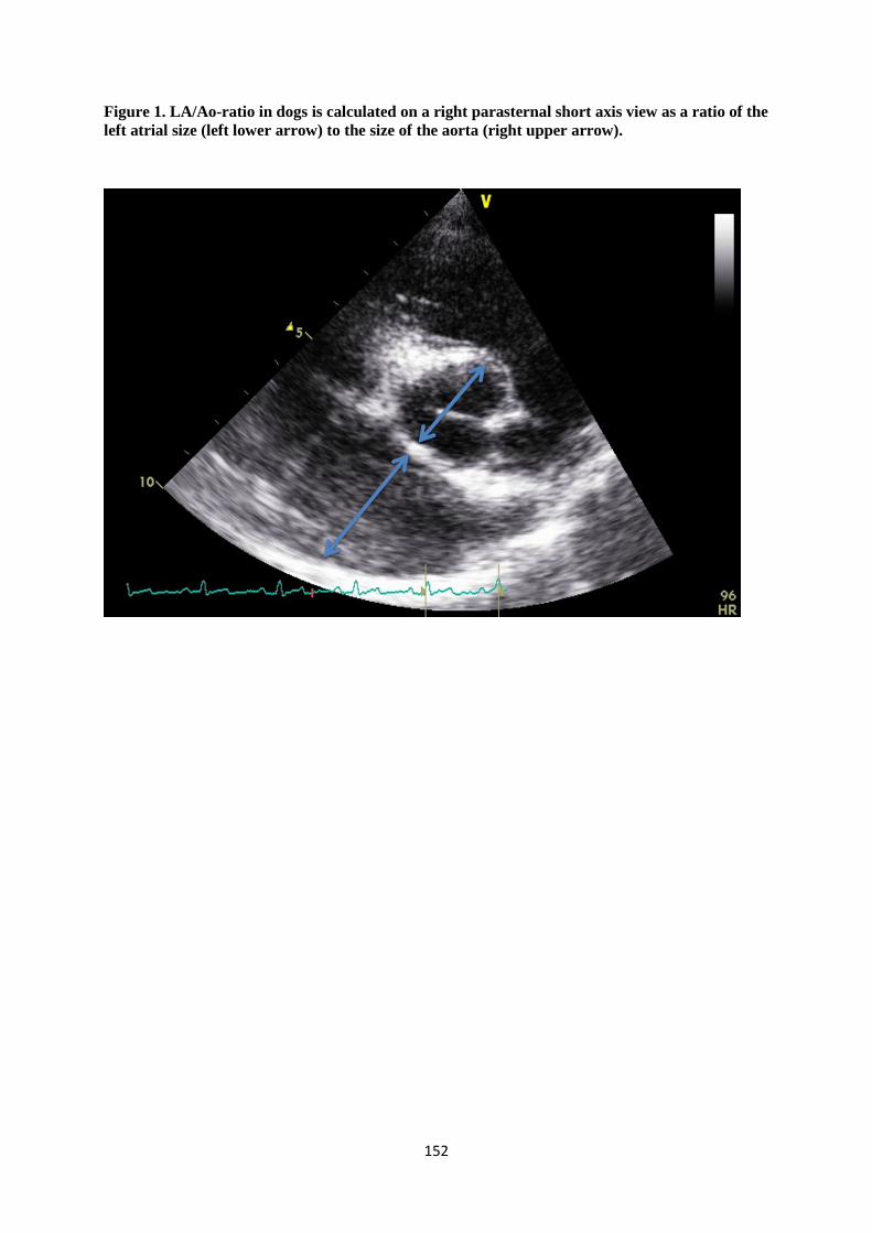
152
Figure 1. LA/Ao-ratio in dogs is calculated on a right parasternal short axis view as a ratio of the
left atrial size (left lower arrow) to the size of the aorta (right upper arrow).
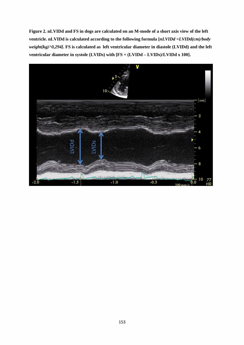
153
Figure 2. nLVIDd and FS in dogs are calculated on an M-mode of a short axis view of the left
ventricle. nLVIDd is calculated according to the following formula [nLVIDd =LVIDd(cm)/body
weight(kg)^0,294]. FS is calculated as left ventricular diameter in diastole (LVIDd) and the left
ventricular diameter in systole (LVIDs) with [FS = (LVIDd – LVIDs)/LVIDd x 100].

154
Figure 3. Boxplots of the heart rate at different time points in dogs with a clinical diagnosis of
SIRS.
The central line of the box plot indicates the median value, the upper and lower line of the box plot
illustrate the range of the 25% and 75% of the values, the outer lines at the end of the vertical lines
indicate the 95% and 5% range of the recorded values.

155
Figure 4. Boxplots of the systolic arterial pressure at different time points in dogs with a clinical
diagnosis of SIRS.
The central line of the box plot indicates the median value, the upper and lower line of the box plot
illustrate the range of the 25% and 75% of the values, the outer lines at the end of the vertical lines
indicate the 95% and 5% range of the recorded values.

156
Figure 5. Boxplots of the left atrium to aortic (LA/Ao) ratio at different time points in dogs with
a clinical diagnosis of SIRS.
The central line of the box plot indicates the median value, the upper and lower line of the box plot
illustrate the range of the 25% and 75% of the values, the outer lines at the end of the vertical lines
indicate the 95% and 5% range of the recorded values.

157
Figure 6. Boxplots of the normalized left ventricular internal dimension in diastole (nLVIDd) at
different time points in dogs with a clinical diagnosis of SIRS.
The central line of the box plot indicates the median value, the upper and lower line of the box plot
illustrate the range of the 25% and 75% of the values, the outer lines at the end of the vertical lines
indicate the 95% and 5% range of the recorded values.

158
Figure 7. Boxplots of the fractional shortening (FS) at different time points in dogs with a
clinical diagnosis of SIRS.
The central line of the box plot indicates the median value, the upper and lower line of the box plot
illustrate the range of the 25% and 75% of the values, the outer lines at the end of the vertical lines
indicate the 95% and 5% range of the recorded values.

159
Figure 8. Scatter plots of the heart rate at different time points in survivors and non survivors of
dogs with a clinical diagnosis of SIRS.
S = Survivor, NS = Non Survivors. The red line indicates the median value.

160
Figure 9. Scatter plots of the left atrium to aortic (LA/Ao) ratio at different time points in
survivors and non survivors of dogs with a clinical diagnosis of SIRS.
S = Survivors, NS = Non Survivors. The red line indicates the median value.
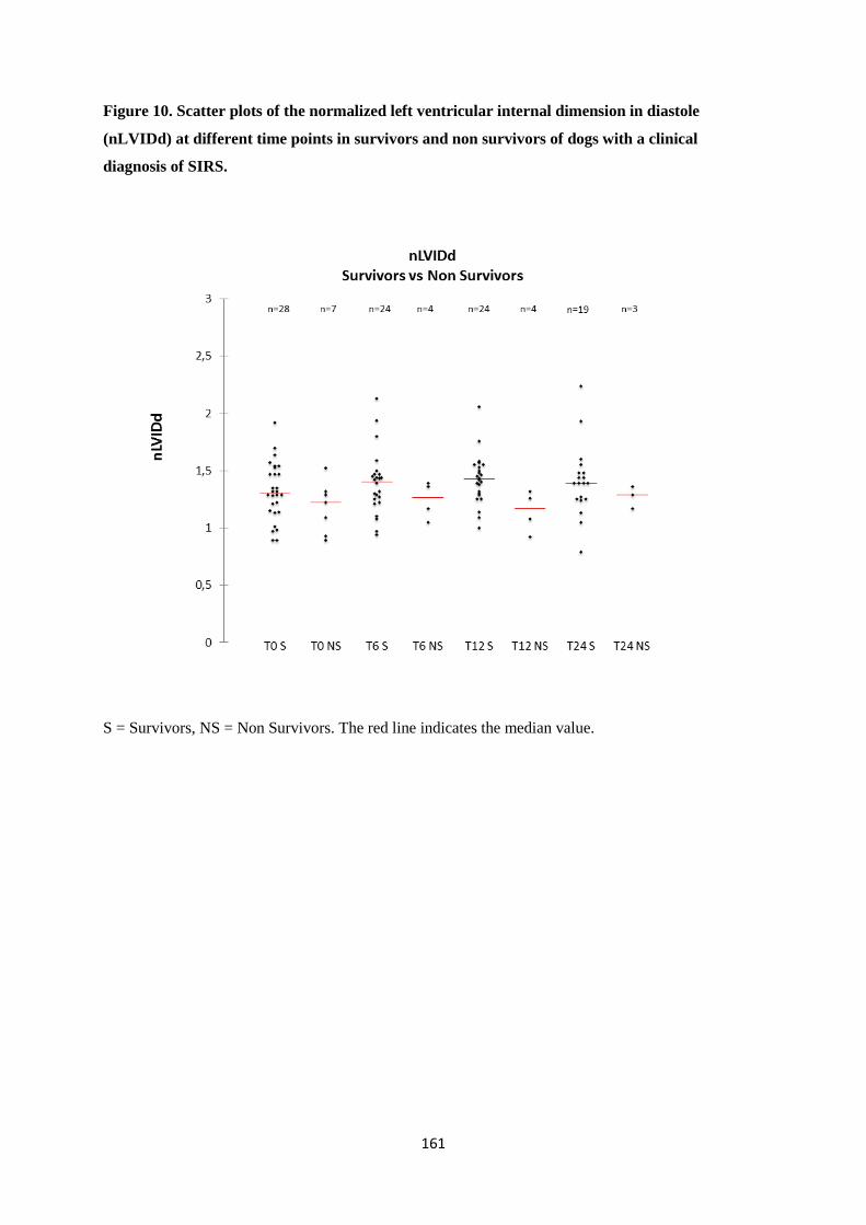
161
Figure 10. Scatter plots of the normalized left ventricular internal dimension in diastole
(nLVIDd) at different time points in survivors and non survivors of dogs with a clinical
diagnosis of SIRS.
S = Survivors, NS = Non Survivors. The red line indicates the median value.

162
Figure 11. Scatter plots of the fractional shortening (FS) at different time points in survivors and
non survivors of dogs with a clinical diagnosis of SIRS.
S = Survivors, NS = Non Survivors. The red line indicates the median value.

163
Figure 12. Scatter plots of the systolic arterial pressure (SAP) at different time points in
survivors and non survivors of dogs with a clinical diagnosis of SIRS.
S = Survivors, NS = Non Survivors. The red line indicates the median value.

164
165
4.4 CARDIAC BIOMARKERS IN CANINE EMERGENCIES WITH A CLINICAL
DIAGNOSIS OF SYSTEMIC INFLAMMATORY RESPONSE SYNDROME
The paper on cardiac biomarkers mostly confirms previous findings published by Hamacher and by
Langhorn on cardiac troponins, but gives some interesting information on NT-proBNP as well. First of
all, cTnT concentrations are supposedly undetectable in healthy dogs, and this was confirmed in our
study where none of the dogs demonstrated detectable concentrations at their control visit. However,
40.6% of dogs presented to the emergency department with a clinical diagnosis of SIRS displayed
detectable concentrations during hospitalization and cTnT changed significantly over time (p <0.0001).
cTnT concentrations were significantly different at T12, T24 and T72 compared to concentrations at
presentations and at the control visit (p <0.05). Moreover, the finding of detectable cTnT concentrations
was a negative prognostic marker in these dogs (p 0.011).
Our study also demonstrated that NT-proBNP changes significantly over time (p <0.001) in canine
emergencies with a clinical diagnosis of SIRS during hospitalization. Values at presentation, after 6 and
12 hours, and during the control visit were all significantly lower than values observed at T24, T72 and
T120. However, NT-proBNP concentrations were not significantly different between survivors and non
survivors (p 0.509). Neither cTnT nor NT-proBNP was correlated with the underlying disease category,
however groups were very small, and these findings should be confirmed in a larger population.
Despite the finding that NT-proBNP concentrations significantly changed over time, with higher
concentrations observed from 24 to 120 hours after hospitalization, the clinical value of this finding
remains unknown. NT-proBNP apparently rises late during hospitalization, and our study failed to
demonstrate an association of NT-proBNP concentrations with survival. Whether NT-proBNP and cTnT
serve as indirect markers of myocardial dysfunction should be determined in a larger population
including all dogs, regardless of their clinical condition, on which cardiac biomarkers and
echocardiography are assessed simultaneously.

166

167
CARDIAC BIOMARKERS IN CANINE EMERGENCIES WITH A CLINICAL DIAGNOSIS
OF SYSTEMIC INFLAMMATORY RESPONSE SYNDROME
K. Gommeren*, I. Desmas*, A. Garcia*, C. Clercx*, K. McEntee*/**, D. Peeters*
*Department of Clinical Sciences, School of Veterinary Medicine, University of Liège, Liège, Belgium,
** Laboratory of Physiology, Faculty of Medicine, Université Libre de Bruxelles, Brussels, Belgium,
Submitted (revised version) to the
Journal of Veterinary Emergency and Critical Care
168
Objective - The N-terminal fragment of pro-B-type natriuretic peptides (NT-proBNP) and cardiac
troponin T (cTnT) are associated with myocardial hibernation and provide prognostic information on
survival in human systemic inflammatory response syndrome (SIRS). In veterinary medicine little is
known about these cardiac biomarkers in dogs presented to an emergency department with a clinical
diagnosis of SIRS. We hypothesized that cTnT and NT-proBNP would (1) increase during
hospitalization, (2) vary in magnitude according to the underlying etiology, and (3) serve as prognostic
markers.
Design – Prospective, observational, clinical study.
Setting – Emergency department of a university teaching hospital.
Animals – Sixty-nine dogs presented to the emergency department with a clinical diagnosis of SIRS
were prospectively studied. Age in these patients ranged from 5 months to 15 years while weight varied
from 5.5 to 75 kg. Dogs were not sampled if blood collection was deemed unduly stressful.
Measurements and Main Results - Samples were obtained at presentation and during hospitalization
until discharge or death and at a control visit (T1m) over one month after discharge. cTnT was measured
with a validated immunoassay on an automated device, while NT-proBNP was assayed with a
commercially available canine ELISA-kit. A correlation procedure, mixed procedure on a linear model
and a logistic procedure were performed (p < 0.05). Forty-four patients survived, 19 of which had control
visits. cTnT and NT-proBNP both changed significantly over time. cTnT concentrations were
significantly higher from T12 to T72. In 28 dogs, cTnT was detected during hospitalization, but cTnT
was never detected at control visits. Higher cTnT were negatively associated with survival, irrespective
of disease category. NT-proBNP concentrations were significantly higher from T24 to T120, but were
not associated with survival.
Conclusions - NT-proBNP and cTnT increased significantly in canine SIRS, regardless of the
underlying disease process. Non survivors displayed significantly higher cTnT concentrations.

169
Introduction
The systemic inflammatory response syndrome (SIRS) characterizes the systemic repercussions of a
generalized state of inflammation and the possible secondarily created organ damage. The list of
underlying causes of SIRS is diverse, with sepsis, trauma and sterile inflammatory conditions such as
pancreatitis amongst the most well-known.1 The clinical diagnosis of SIRS is based on defined changes
in clinical (body temperature, heart rate and respiratory rate) and hematologic (leucocyte counts,
presence of a left shift) variables (Table 1) and the suspicion of an underlying disease process known to
trigger the systemic inflammatory response. Diagnosing a patient with SIRS recognizes the presence of
clinical signs compatible with systemic inflammation, but is overly sensitive and poorly specific.2
Cardiovascular impairment secondary to systemic inflammation has been reported in human medicine,
and is also known as myocardial hibernation.3,4 Myocardial hibernation has been reported in human
critically ill patients and studied in experimental sepsis models in dogs.4-7 It is characterized by increased
end-diastolic and end-systolic ventricular volumes8, and systolic3 and diastolic7 ventricular dysfunction.5
Whether myocardial hibernation serves as a protective mechanism of the body during systemic
inflammation, or whether it is in fact a negative prognostic factor remains a matter of debate.5,9
Myocardial hibernation has been associated with increased concentrations of cardiac troponins10,11 and
natriuretic peptides12-14 in human SIRS patients and some studies have found cardiac troponins (cTn)
and the N-terminal portion of probrain natriuretic peptide (NT-proBNP) to be correlated with the degree
of cardiac dysfunction and with concentrations of inflammatory cytokines.15,16 However, very little is
known about myocardial hibernation in canine clinical studies, although the scarce literature appears to
support its existence.17,18 Recent data from canine clinical studies suggest that cardiac biomarkers may
also have a role in SIRS patients.19-21 As these cardiac biomarkers may help in the diagnosis of cardiac
dysfunction and prognosis of human SIRS patients,9,22,23 they might be interesting and accessible tools
in canine SIRS.
Cardiac troponins (cTn) are sensitive and specific for the detection of myocardial ischemic necrosis or
minor myocardial injury and increased cTnT and cTnI concentrations are associated with negative
prognosis in critically ill human patients.24,25 Research on cTn in veterinary medicine has mainly focused
on primary cardiac disease, where cTns are early markers of cardiac lesions with a negative prognostic
value.26 Troponins are however also increased and associated with poor prognosis in many other canine
disease processes such as gastric dilation and volvulus (GDV)27,28, trauma27,29, infections30-34 and
SIRS.20,21,35
Brain natriuretic peptide (BNP) and the N-terminal fragment of the prohormone (NT-proBNP) are
quantitative markers of ventricular wall stress with high sensitivity and specificity for cardiac insult.12
Several studies demonstrated elevations of BNP and NT-proBNP in human sepsis and SIRS to be
associated with myocardial hibernation and poor prognosis.12,13,15,16 In veterinary medicine, papers found
170
increased NT-proBNP concentrations in canine babesiosis and non-cardiac disease such as traumatic,
neurological and gastrointestinal disease.34,36
Altogether, cardiac biomarkers might serve as non-invasive markers of myocardial hibernation and
might serve as prognostic tools in canine SIRS. We hypothesized that cardiac troponin T (cTnT) and the
N-terminal fragment of pro-BNP (NT-proBNP) would (1) increase during hospitalization, (2) vary in
magnitude according to the underlying etiology, and (3) serve as a prognostic marker in canine patients
with SIRS presented to an emergency department.
Materials and methods
All dogs presented to the emergency service of the XXX between January and August 2010 were
considered for inclusion. Dogs entered the study if a clinical diagnosis of SIRS was made based on the
suspicion of an underlying disease process known to trigger the systemic inflammatory response and
finding 2 or more abnormalities of the following clinical (temperature, heart rate and respiratory rate)
and basic laboratory parameters (abnormal leukocyte counts).2 The cut-off values for white blood cell
counts were modified from the original paper to adhere with the reference ranges of our own clinical
laboratory (Table 1) and the limits of normal body temperature were set at 38 to 39°C. An informed
consent was obtained from the owners of each dog and approval was obtained by the ethics committee
(letter 1709). All dogs presented to any other service, or dogs weighing less than 5kg were excluded as
were animals that were considered too unstable by the primary clinician to sustain any
additional/unnecessary stress. Patients were grouped into 7 different disease categories: patients with
neoplastic disease (N), infectious disease (I), GDV (GDV), other gastrointestinal disease (GI), traumatic
disease (T), renal disease (R), and miscellaneous or undetermined causes (M). Since NT-proBNP is
influenced by renal function, patients with renal insufficiency defined as azotemia or oliguria and anuria
that was unresponsive to fluid therapy were excluded from the NT-proBNP part of the study.37 Although
cTn concentrations also can be influenced by renal status, severity of renal failure does not correlate
with cTn concentrations,38,39 and cTn analysis remains useful in identifying myocardial injury in human
renal patients.40-42 Similarly, cTn concentrations can identify human patients with worse prognosis
despite concurrent renal failure and/or hemodialysis.40,43-48 Based on these findings, we did not reject
patients with renal impairment from the cTnT part of the study.
Baseline concentrations of cTnT, and NT-proBNP were assessed on blood sampled prior to starting any
treatment (T0). Other samples were taken after 6 (T6), 12 (T12) and 24 hours (T24) and every other day
thereafter (T72, T120, …) until discharge or death. Short term survival was defined as the patient leaving
the hospital, long term survival was defined as the patient being alive one month after discharge from
the hospitalization. All long term survivors were invited to a free control visit one month to one year
after discharge. Blood samples were divided into EDTA (4mL) and serum (2mL) tubes which were
centrifuged and separated within 15 minutes, and stored at -80°C until analysis.
171
A commercial electrochemiluminescence kit (Modular Analytics E®, Roche), with a lower limit of
detection at 0.010ng/mL, a limit of linearity at 25.00ng/mL, and a coefficient of variation under 5% for
values above 0.06ng/mL was used to measure cTnT.49 The kit detects 2 epitopes of the central part of
human cTnT (125-131 and 135-147), which are highly conserved in canine cTnT (one substitution in
the first epitope and 100% homology in the second), and has previously been used in veterinary
research.49,50 Reported values in healthy dogs are less than 0.010 ng/mL.29,51
A commercially available sandwich enzyme immune assay with an upper limit of detection of 3000
pmol/L (VetSign Canine Cardioscreen Test Kit®, Idexx Laboratories) was used to measure NT-proBNP.
In short, microtiter plates were provided with capture antibody anti-NT-proBNP bound to the wells in
which plasma (30 µL) was incubated (5 hours at 20°C) with an immunoaffinity purified sheep detection
antibody conjugated to horseradish peroxidase in a stabilizer solution (200 µL). Afterwards wells were
washed (5 x 350 µL), tetramethylbenzidine (200 µL) was added and left for 40 minutes, after which a
stop solution was added and bound NT-proBNP was quantified by an ELISA plate reader. All plates
were run with calibration and control solutions, yet for financial reasons only the first plate was run in
duplicate.
Statistical methods
Statistical analysis was performed using SASi. Unmeasurable samples were attributed the value of the
lower detection limit. A Shapiro-Wilk and Kolmogorov-Smirnov test (univariate procedure) and
normality QQplots were performed, on the raw data and after logarithmic transformation of the data.
For both cTnT and NT-proBNP the logarithmically transformed data were used after identification of a
nearly normal distribution of the residues on the QQplots. A mixed procedure on a generalized linear
model was used to assess the effect of clinical parameters on cardiac biomarkers. As the data were taken
repeatedly over time on the same animals, there is a possible correlation between successive data. This
correlation structure is reflected in the linear mixed model used (MIXED procedure, repeated by time
which was treated as a categorical variable). Correlation between different biomarkers was tested using
Spearman correlation (CORR procedure). A logistic analysis (LOGISTIC procedure) was performed in
order to evaluate the effect of cardiac biomarker concentrations on survival to discharge. Only dogs that
survived, died of natural causes or were euthanized for prognostic reasons were included for the
assessment of prognostic value of the evaluated parameters. Statistical significance was reached at a p
value < 0.05.
Results
Dogs
Fifty-eight pure breed and 11 mixed-breed dogs (69 dogs in total) were included in the study. The most
commonly represented breeds were Bernese mountain dog (n=8), German shepherd (n=6), Great Dane
172
(n=4), Jack Russell terrier (n=4) and Belgian shepherd (n=3). There were 38 male (29 intact and 9
castrated) and 31 female (17 intact and 14 neutered) dogs with a median age of 6.5 years (ranging
between 7 months and 15.2 years) and with a median weight of 30.3kg (ranging from 5.5 to 75kg).
Patients were included into each disease category (N=13; I=12; GDV=11; GI=5; T=6; R=3; and M=19).
Outcome and follow-up of our studied population has been represented in a flow diagram (Figure 1).
Forty-four patients were discharged, 8 died during hospitalization while 17 dogs were euthanized (8 for
prognostic, 7 for financial reasons and 2 for unspecified reasons). Thirty-four patients were still alive
more than one month after discharge and were available for a control visit, of which 19 presented for a
control visit (5 declined, 5 dogs were lost to follow-up and 5 died from related causes before the
scheduled control visit such as continued GI signs in 2 dogs, aspiration pneumonia secondary to a
megaesophagus, worsening hepatocutaneous syndrome and tumor recurrence with secondary
hemoabdomen in one dog each).
Biomarkers at different time points
cTnT and NT-proBNP both changed significantly over time (p <0.001), concentrations over time are
displayed in Figure 2 and 3. Twenty-eight out of 69 dogs had at least one time point during
hospitalization at which cTnT was detectable, while none of the dogs had measurable cTnT
concentrations at the control visit. cTnT was significantly higher at T12, T24 and T72 compared to
concentrations at presentation or at the control visit (Table 2). NT-proBNP concentrations were
measurable in all dogs at all time points. NT-proBNP concentrations were significantly higher at T24,
T72 and T120 compared to T0, T6, T12 and T1m. Median concentrations did not differ significantly
between T24 [661.391 (60.774-3000) pmol/L], T72 [888.806 (76.58-3000) pmol/L] and T120 [737.139
(0-3000) pmol/L] (Figure 3).
Association between biomarkers and underlying disease, prognosis and each other
Statistical analysis did not identify any influence of the underlying disease category on cTnT and NT-
proBNP concentrations (p 0.162 and 0.084 respectively). High cTnT concentrations (p =0.011) were
however associated with negative prognosis (Figure 4). In contrast NT-proBNP concentrations (p
=0.509) were not significantly correlated with survival to discharge (Figure 5). Finally, cTnT and NT-
proBNP were significantly and mildly correlated (p <0.001, with r 0.291).
Discussion
This study demonstrated changes in cardiac biomarkers during hospitalization in a population of canine
SIRS patients presented to an emergency department. Troponin concentrations rise within 8 hours after
an initial insult, and reportedly remain increased for over 50 hours in humans and dogs.10,26,52,53 cTnT
values in this cohort of dogs with a clinical diagnosis of SIRS presented to an emergency department
were detectable in 28 dogs during hospitalization, with concentrations significantly higher at T12, T24

173
and T72 compared to concentrations at presentation and at the control visit. Although the clinical nature
of this study on dogs suffering from different diseases does not allow us to identify the exact timing of
the insult in the majority of dogs, the timing of the changes in cTnT concentrations appear to agree with
the rather rapid rise and sustained increase described. In contrast, at their control visit, all dogs had
undetectable cTnT concentration (<0.01ng/mL). A study in GDV patients described rather similar
findings, with no significant changes in cTnT concentrations immediately after surgery, but increased
concentrations on day one and two after presentation.27,28 A study evaluating cTnI concentrations in
canine SIRS patients identified a higher prevalence of increased cTnI concentrations at presentation
(35/60 dogs) and during hospitalization, but similarly failed to find significant variations from day to
day.20 A study comparing cTnT and cTnI in SIRS patients admitted to the ICU found a higher
prevalence of increased cTnI concentrations. This difference in detection rate can be explained by the
lower sensitivity of cTnT tests, or by the timing of sampling compared to the start of the disease process
(as admission to the ICU likely later than admission to an emergency department).21
The lower sensitivity of cTnT results in cTnI usually being preferred over cTnT. The use of a cTnI assay
in the present study would probably have resulted in the detection of elevated concentrations in a larger
proportion of SIRS patients, but cTnT was chosen for financial reasons.28,30 In human medicine,
continuous test-improvement and increased sensitivity of cTnI assays resulted in lower detection limits.
These lower detection limits subsequently lead to an increased detection rate of cTnI elevations, which
are not necessarily attributable to acute processes.54 With different tests available for cTnI measurement,
it is therefore recommended to apply the 99th percentile as cut-off value, as each assay appears to be
unique and direct comparison between results is not possible.55-58 Lower cut-off percentiles allow for
the detection of more chronic cardiac disease, lowering the specificity of a single cTnI measurement to
screen for acute cardiac conditions.59
NT-proBNP changed significantly over time, with concentrations at T24, T72 and T120 significantly
higher than concentrations at T0, T6, T12 and the control visit. As BNP has a very short half-life and is
technically difficult to measure,60-62 we assessed NT-proBNP, which has got a longer half-life. Most of
the research performed in veterinary medicine on NT-proBNP has focused on cardiac disease.63-65 A
recent study evaluating BNP in dogs with non-cardiac disease (e.g. neurological and gastrointestinal
disease) demonstrated a moderate increase of natriuretic peptides in these patients.36 As our study
focused on dogs with SIRS presented to an emergency department, and SIRS has a high potential to
induce cardiac effects as is well described in human medicine, it is not surprising that NT-proBNP
concentrations in this study were more markedly elevated compared to this previous study.36 Finding
higher concentrations at T24, 72 and T120 is in agreement with studies on SIRS and sepsis in human
patients. The optimal timing of NT-proBNP measurement varied across studies in humans, from the day
of admission to day 2 and day 5 after admission,23,66 which is probably due to the difficulty in
determining the time of onset of the disease. Nevertheless, peak concentrations are likely to be found

174
more than two days after hospitalization in humans.15,16,22 Unfortunately, the clinical setting of this study
prevents determination of the exact time the insult triggering SIRS occurred for the majority of dogs.
The kinetics observed in this cohort do however seem to confirm that NT-proBNP should be expected
to rise during the first days of hospitalization in dogs presented with a clinical diagnosis of SIRS to an
emergency department. Elevated levels of NT-proBNP when screening for occult cardiac disease should
therefore be interpreted carefully in SIRS patients.
Whether increased cTn and NT-proBNP concentrations are indicative of myocardial hibernation in
canine emergencies with SIRS cannot be concluded from this paper, but merits further investigation.
Over the last decades, interest of echocardiography in the ICU has greatly increased in human medicine,
leading to increased availability of echocardiography.67-69 The performance characteristics of
echocardiography by non-specialists is largely determined by the hours of training, the quality of the
device, the patient characteristics and by the definition of a ‘successful examination’.70 Therefore
performing cardiac ultrasound in an emergency setting necessitates 24h availability of properly trained
intensivists, and such developments should be greatly encouraged in veterinary medicine.
An increase of cTnT during hospitalization was associated with poor short term prognosis. Cardiac
troponin T and I are well-accepted prognostic biomarkers in human intensive care units.10,11,24,71 In
veterinary medicine, increased concentrations of cardiac troponins have been observed in infectious
disease patients, trauma patients, GDV patients and patients suffering from systemic diseases27,28,30-
32,62,72-75 and are correlated with poor prognosis in some of these studies.27,28,30,76 Studies evaluating cTnI
and cTnT in canine SIRS patients already confirmed their prognostic value.20,21,35 These studies similarly
identified significant differences between survivors and non-survivors.20 However, additional sampling
to measure cTnI concentrations on day 2 or 3 (or evaluation of concentration changes) did not add
value.20 Although incidence of increased cTnI concentrations was higher than for cTnT, cTnI and cTnT
carried rather similar prognostic information.21,35 cTnT concentrations have been established as
interesting markers to evaluate prognosis of canine SIRS patients, but cut-off limits remain to be
determined in larger studies.35 As cTnT (and cTnI) can remain increased up to 7 or 10 days after the
insult, kinetics of cTnT are difficult to evaluate, and they are less useful for evaluation of disease
progression or treatment response.77,78
In the present study, NT-proBNP concentrations were not significantly correlated with prognosis.
Previous studies in dogs tended to evaluate natriuretic peptides at presentation, while higher
concentrations should be expected later during hospitalization. This delay in the rise of NT-proBNP
probably also limits its use as a prognostic marker in a clinical veterinary emergency care setting. At
later time points the group size in this study rapidly decreased, which may have impacted the likelihood
to identify significant differences between survivors and non survivors. A recently published meta-
analysis in human septic patients describing 12 studies on 1865 cases did conclude that (NT-pro)BNP

175
is significantly associated with risk of mortality.23 This meta-analysis also concluded that elevated (NT-
pro)BNP levels in the presence of SIRS or sepsis do not equal cardiac dysfunction due to low specificity,
but normal (NT-pro)BNP levels could be used to rule out the need for further cardiac investigation.23
Therefore the lack of a significant difference in NT-proBNP between survivors and non survivors in this
study definitely needs to be confirmed in a larger cohort of patients with a clinical diagnosis of SIRS.
The observed increased NT-proBNP concentrations in this study can not only be explained by
myocardial dysfunction, but also indirectly via increased wall stress after volume resuscitation,79 after
lung injury, acute respiratory distress syndrome or thromboembolism.6
There are several limitations to the present study. Firstly, dogs that were considered too unstable by the
attending clinician were removed from the study, and therefore more severely ill patients were less likely
to enter the study. Consequently findings would likely have been more significant if all dogs, regardless
of their clinical status would have been included. As previously mentioned, sampling times were
standardized with relation to the moment of presentation to the emergency department. Clinical signs
may have been present for variable times prior to presentation and this may have altered the kinetics of
these biomarkers. Unfortunately due to the clinical context, we could not retrieve accurate information
on the duration of disease for the majority of the patients, which impedes us to draw strong conclusions
regarding the kinetics of these parameters. Thirdly, a large proportion of our patients were euthanized
on the owners’ request, rather than based on specific study endpoints, which may have had an impact
on our findings regarding prognosis. In order to avoid any effect of financial considerations on the
prognostic evaluation, all dogs that were euthanized for financial or unspecified reasons were removed
for this analysis. Including dogs that were euthanized for prognostic reasons might still have influenced
our findings, however all these dogs had a deteriorating clinical condition that did not respond to
appropriate treatment or suffered life-threatening complications. In the present study, 44 patients
survived to discharge (64%), which is similar to80, or better than81 previous studies on clinical canine
SIRS patients.
Samples with NT-proBNP concentrations above the upper limit of the assay (3000pmol/L) were not
diluted to measure the exact concentration because of financial restrictions. Therefore, NT-proBNP
concentration was underestimated in some samples. This did however not prohibit the finding of
significant changes, and therefore probably even underestimated changes in NT-proBNP. Similarly, a
cTnT assay rather than a cTnI assay was used due to financial restrictions. The use of a cTnI assay would
probably have resulted in a higher detection rate of increased troponin concentrations, however our cTnT
assay already allowed us to obtain significant results.
Our study demonstrates that cardiac biomarkers are often elevated in dogs with SIRS presented to the
emergency department. Whether these increased concentrations are linked with myocardial hibernation
does however remain to be demonstrated. Additionally, this study confirms that cTn’s carry prognostic

176
value in dogs with SIRS. Our research is however merely observational, and does not allow to explain
our findings. Studies investigating the correlation of cardiac biomarkers with echocardiographic
findings and inflammatory cytokines in canine SIRS patients are therefore warranted.
Conclusion
In conclusion, the present study demonstrates increased concentrations of cTnT and NT-proBNP during
hospitalization of dogs presented to the emergency department with a clinical diagnosis of SIRS.
Moreover increased cTnT concentrations were associated with poor prognosis to survival in this cohort.
Further research is warranted to explain these findings, and to assess the potential use of cardiac
biomarkers to evaluate cardiac damage in SIRS.
Footnotes
i SAS; Statistical Analysis Software, Cary, United States.
177
References
1. de Laforcade AM. Systemic Inflammatory Response Syndrome. In: Silverstein DC, Hopper K,
editors. Small Animal Critical Care Medicine, 2nd ed. St. Louis, Missouri, United States: Saunders
Elsevier; 2015:31-34.
2. Hauptman JG, Walshaw R, Olivier NB. Evaluation of the sensitivity and specificity of diagnostic
criteria for sepsis in dogs. Vet Surg 1997;26:393-397.
3. Parker M, Shelhamer J, Bacharach S, et al. Profound but reversible myocardial depression in patients
with septic shock. Ann Intern Med 1984;100:483-490.
4. Werdan K, Schmidt H, Ebelt H, et al. Impaired regulation of cardiac function in sepsis, SIRS, and
MODS. Can J Physiol Pharmacol 2009;87:266-274.
5. Levy R, Piel D, Acton P, et al. Evidence of myocardial hibernation in the septic heart. Crit Care Med
2005;33:2752-2756.
6. Maeder M, Fehr T, Rickli H, et al. Sepsis-associated myocardial dysfunction: diagnostic and
prognostic impact of cardiac troponins and natriuretic peptides. Chest 2006;129:1349-1366.
7. Natanson C, Fink M, Ballantyne H, et al. Gram-negative bacteremia produces both severe systolic
and diastolic cardiac dysfunction in a canine model that simulates human septic shock. J Clin Invest
1986;78:259-270.
8. Marik P, Varon J. Sepsis: state of the art. Dis Mon 2001;47:465-532.
9. Charpentier J, Luyt CE, Fulla Y, et al. Brain natriuretic peptide: A marker of myocardial dysfunction
and prognosis during severe sepsis. Crit Care Med 2004;32:660-665.
10. Babuin L, Vasile V, Rio Perez J, et al. Elevated cardiac troponin is an independent risk factor for
short- and long-term mortality in medical intensive care unit patients. Crit Care Med 2008;36:759-765.
11. Ammann P, Maggiorini M, Bertel O, et al. Troponin as a risk factor for mortality in critically ill
patients without acute coronary syndromes. J Am Coll Cardiol 2003;41:2004-2009.
12. Chen Y, Li C. Prognostic significance of brain natriuretic peptide obtained in the ED in patients
with SIRS or sepsis. Am J Emerg Med 2009;27:701-706.
13. Meyer B, Huelsmann M, Wexberg P, et al. N-terminal pro-B-type natriuretic peptide is an
independent predictor of outcome in an unselected cohort of critically ill patients. Crit Care Med
2007;35:2268-2273.
14. Rudiger A, Fischler M, Harpes P, et al. In critically ill patients, B-type natriuretic peptide (BNP)
and N-terminal pro-BNP levels correlate with C-reactive protein values and leukocyte counts. Int J
Cardiol 2008;126:28-31.
15. Charpentier J, Luyt CE, Fulla Y, et al. Brain natriuretic peptide: A marker of myocardial dysfunction
and prognosis during severe sepsis. Crit Care Med 2004;32:660-665.
178
16. Witthaut R, Busch C, Fraunberger P, et al. Plasma atrial natriuretic peptide and brain natriuretic
peptide are increased in septic shock: impact of interleukin-6 and sepsis-associated left ventricular
dysfunction. Intensive Care Med 2003;29:1696-1702.
17. Dickinson A, Rozanski E, Rush J. Reversible myocardial depression associated with sepsis in a dog.
J Vet Intern Med 2007;21:1117-1120.
18. Nelson O, Thompson P. Cardiovascular dysfunction in dogs associated with critical illnesses. J Am
Anim Hosp Assoc 2006;42:344-349.
19. Langhorn R, Persson F, Åblad B, et al. Myocardial injury in dogs with snake envenomation and its
relation to systemic inflammation. J Vet Emerg Crit Care (San Antonio) 2014;24:174-181.
20. Hamacher L, Dorfelt R, Muller M, et al. Serum cardiac troponin I concentrations in dogs with
systemic inflammatory response syndrome. J Vet Intern Med 2015;29:164-170.
21. Langhorn R, Oyama MA, King LG, et al. Prognostic importance of myocardial injury in critically
ill dogs with systemic inflammation. J Vet Intern Med 2013;27:895-903.
22. Roch A, Allardet-Servent J, Michelet P, et al. NH2 terminal pro-brain natriuretic peptide plasma
level as an early marker of prognosis and cardiac dysfunction in septic shock patients. Crit Care Med
2005;33:1001-1007.
23. Wang F, Wu Y, Tang L, et al. Brain natriuretic peptide for prediction of mortality in patients with
sepsis: a systematic review and meta-analysis. Crit Care 2012;16:R74.
24. Spies C, Haude V, Fitzner R, et al. Serum cardiac troponin T as a prognostic marker in early sepsis.
Chest 1998;113:1055-1063.
25. Turner A, Tsamitros M, Bellomo R. Myocardial cell injury in septic shock. Crit Care Med
1999;27:1775-1780.
26. Fonfara S, Loureiro J, Swift S, et al. Cardiac troponin I as a marker for severity and prognosis of
cardiac disease in dogs. Vet J 184:334-339.
27. Burgener IA, Kovacevic A, Mauldin GN, et al. Cardiac troponins as indicators of acute myocardial
damage in dogs. J Vet Intern Med 2006;20:277-283.
28. Schober KE, Cornand C, Kirbach B, et al. Serum cardiac troponin I and cardiac troponin T
concentrations in dogs with gastric dilatation-volvulus. J Am Vet Med Assoc 2002;221:381-388.
29. Schober KE, Kirbach B, Oechtering G. Noninvasive assessment of myocardial cell injury in dogs
with suspected cardiac contusion. J Vet Cardiol 1999;1:17-25.
30. Lobetti R, Dvir E, Pearson J. Cardiac troponins in canine babesiosis. J Vet Intern Med 2002;16:63-
68.
31. Diniz PP, de Morais HS, Breitschwerdt EB, et al. Serum cardiac troponin I concentration in dogs
with ehrlichiosis. J Vet Intern Med 2008;22:1136-1143.
32. Mastrorilli C, Dondi F, Agnoli C, et al. Clinicopathologic features and outcome predictors of
Leptospira interrogans Australis serogroup infection in dogs: a retrospective study of 20 cases (2001-
2004). J Vet Intern Med 2007;21:3-10.

179
33. Silvestrini P, Piviani M, Alberola J, et al. Serum cardiac troponin I concentrations in dogs with
leishmaniasis: correlation with age and clinicopathologic abnormalities. Vet Clin Pathol 2012;41:568-
574.
34. Lobetti R, Kirberger R, Keller N, et al. NT-ProBNP and cardiac troponin I in virulent canine
babesiosis. Vet Parasitol 2012;190:333-339.
35. Langhorn R, Thawley V, Oyama MA, et al. Prediction of long-term outcome by measurement of
serum concentration of cardiac troponins in critically ill dogs with systemic inflammation. J Vet Intern
Med 2014;28:1492-1497.
36. Lee JA, Herndon WE, Rishniw M. The effect of noncardiac disease on plasma brain natriuretic
peptide concentration in dogs. J Vet Emerg Crit Care (San Antonio) 2011;21:5-12.
37. Boswood A, Dukes-McEwan J, Loureiro J, et al. The diagnostic accuracy of different natriuretic
peptides in the investigation of canine cardiac disease. J Small Anim Pract 2008;49:26-32.
38. De Zoysa JR. Cardiac troponins and renal disease. Nephrology (Carlton) 2004;9:83-88.
39. Lamb EJ, Webb MC, Abbas NA. The significance of serum troponin T in patients with kidney
disease: a review of the literature. Ann Clin Biochem 2004;41:1-9.
40. Martin GS, Becker BN, Schulman G. Cardiac troponin-I accurately predicts myocardial injury in
renal failure. Nephrol Dial Transplant 1998;13:1709-1712.
41. McCullough PA, Nowak RM, Foreback C, et al. Performance of multiple cardiac biomarkers
measured in the emergency department in patients with chronic kidney disease and chest pain. Acad
Emerg Med 2002;9:1389-1396.
42. McLaurin MD, Apple FS, Falahati A, et al. Cardiac troponin I and creatine kinase-MB mass to rule
out myocardial injury in hospitalized patients with renal insufficiency. Am J Cardiol 1998;82:973-975.
43. Rahman A, Broadley SA. Review article: elevated troponin: diagnostic gold or fool's gold? Emerg
Med Australas 2014;26:125-130.
44. Apple FS, Murakami MM, Pearce LA, et al. Predictive value of cardiac troponin I and T for
subsequent death in end-stage renal disease. Circulation 2002;106:2941-2945.
45. Ooi DS, Zimmerman D, Graham J, et al. Cardiac troponin T predicts long-term outcomes in
hemodialysis patients. Clin Chem 2001;47:412-417.
46. Apple FS, Sharkey SW, Hoeft P, et al. Prognostic value of serum cardiac troponin I and T in chronic
dialysis patients: a 1-year outcomes analysis. Am J Kidney Dis 1997;29:399-403.
47. Antman EM, Tanasijevic MJ, Thompson B, et al. Cardiac-specific troponin I levels to predict the
risk of mortality in patients with acute coronary syndromes. N Engl J Med 1996;335:1342-1349.
48. Stolear JC, Georges B, Shita A, et al. The predictive value of cardiac troponin T measurements in
subjects on regular haemodialysis. Nephrol Dial Transplant 1999;14:1961-1967.
49. Osathanon R, Moonarmart W, Suksantilap N, et al. Evaluation of Hematology Profiles and
Measurement of Serum Cardiac Troponin Level in Canine Monocytic Ehrlichiosis. Thai J Vet Med
2013;43:405-409.

180
50. Giannitsis E, Kurz K, Hallermayer K, et al. Analytical validation of a high-sensitivity cardiac
troponin T assay. Clin Chem 2010;56:254-261.
51. DeFrancesco TC, Atkins CE, Keene BW, et al. Prospective clinical evaluation of serum cardiac
troponin T in dogs admitted to a veterinary teaching hospital. J Vet Intern Med 2002;16:553-557.
52. Feng X, Taggart P, Hall L, et al. Limited additional release of cardiac troponin I and T in
isoproterenol-treated beagle dogs with cardiac injury. Clin Chem 2005;51:1305-1307.
53. Wu A. Increased troponin in patients with sepsis and septic shock: myocardial necrosis or reversible
myocardial depression? Intensive Care Med 2001;27:959-961.
54. Schulz O, Paul-Walter C, Lehmann M, et al. Usefulness of detectable levels of troponin, below the
99th percentile of the normal range, as a clue to the presence of underlying coronary artery disease. Am
J Cardiol 2007;100:764-769.
55. Larue C, Defacque-Lacquement H, Calzolari C, et al. New monoclonal antibodies as probes for
human cardiac troponin I: epitopic analysis with synthetic peptides. Mol Immunol 1992;29:271-278.
56. Panteghini M, Pagani F, Yeo KT, et al. Evaluation of imprecision for cardiac troponin assays at
low-range concentrations. Clin Chem 2004;50:327-332.
57. Apple FS. Clinical and analytical standardization issues confronting cardiac troponin I. Clin Chem
1999;45:18-20.
58. Ferrieres G, Calzolari C, Mani JC, et al. Human cardiac troponin I: precise identification of antigenic
epitopes and prediction of secondary structure. Clin Chem 1998;44:487-493.
59. Schober K, Kirbach B, Oechtering G. Noninvasive assessment of myocardial cell injury in dogs
with suspected cardiac contusion. J Vet Cardiol 1999;1:17-25.
60. MacDonald KA, Kittleson MD, Munro C, et al. Brain natriuretic peptide concentration in dogs with
heart disease and congestive heart failure. J Vet Intern Med 2003;17:172-177.
61. Asano K, Masuda K, Okumura M, et al. Plasma atrial and brain natriuretic peptide levels in dogs
with congestive heart failure. J Vet Med Sci 1999;61:523-529.
62. Prosek R, Sisson DD, Oyama MA, et al. Distinguishing cardiac and noncardiac dyspnea in 48 dogs
using plasma atrial natriuretic factor, B-type natriuretic factor, endothelin, and cardiac troponin-I. J Vet
Intern Med 2007;21:238-242.
63. Oyama MA, Sisson DD, Solter PF. Prospective screening for occult cardiomyopathy in dogs by
measurement of plasma atrial natriuretic peptide, B-type natriuretic peptide, and cardiac troponin-I
concentrations. Am J Vet Res 2007;68:42-47.
64. Hori Y, Tsubaki M, Katou A, et al. Evaluation of NT-pro BNP and CT-ANP as markers of
concentric hypertrophy in dogs with a model of compensated aortic stenosis. J Vet Intern Med
2008;22:1118-1123.
65. Noszczyk-Nowak A. NT-pro-BNP and troponin I as predictors of mortality in dogs with heart
failure. Pol J Vet Sci 2011;14:551-556.

181
66. Fromm RJ, Varon J. NH2 terminal pro-brain natriuretic peptide in cardiovascular dysfunction and
septic shock. Crit Care Med 2005;33:1156-1157.
67. Vieillard-Baron A, Slama M, Cholley B, et al. Echocardiography in the intensive care unit: from
evolution to revolution? Intensive Care Med 2008;34:243-249.
68. Beaulieu Y. Bedside echocardiography in the assessment of the critically ill. Crit Care Med
2007;35:S235-249.
69. Levitov A, Mayo PH, Slonim AD. Critical care ultrasonography. New York: McGraw Hill; 2009.
70. Griffee M, Merkel M, Wei K. The role of echocardiography in hemodynamic assessment of septic
shock. Crit Care Clin 2010;26:365-382.
71. Wu TT, Yuan A, Chen CY, et al. Cardiac troponin I levels are a risk factor for mortality and multiple
organ failure in noncardiac critically ill patients and have an additive effect to the APACHE II score in
outcome prediction. Shock 2004;22:95-101.
72. Hagman R, Lagerstedt AS, Fransson BA, et al. Cardiac troponin I levels in canine pyometra. Acta
Vet Scand 2007;49:6.
73. Porciello F, Rishniw M, Herndon WE, et al. Cardiac troponin I is elevated in dogs and cats with
azotaemia renal failure and in dogs with non-cardiac systemic disease. Aust Vet J 2008;86:390-394.
74. Pelander L, Hagman R, Häggström J. Concentrations of cardiac Troponin I before and after
ovariohysterectomy in 46 female dogs with pyometra. Acta Vet Scand 2008;50:35.
75. Barr S, Warner K, Kornreic B, et al. A cysteine protease inhibitor protects dogs from cardiac damage
during infection by Trypanosoma cruzi. Antimicrob Agents Chemother 2005;49:5160-5161.
76. Mastrorilli C, Dondi F, Agnoli C, et al. Clinicopathologic features and outcome predictors of
Leptospira interrogans Australis serogroup infection in dogs: a retrospective study of 20 cases (2001-
2004). J Vet Intern Med 2007;21:3-10.
77. O'Brien PJ, Dameron GW, Beck ML, et al. Cardiac troponin T is a sensitive, specific biomarker of
cardiac injury in laboratory animals. Lab Anim Sci 1997;47:486-495.
78. Katus HA, Remppis A, Scheffold T, et al. Intracellular compartmentation of cardiac troponin T and
its release kinetics in patients with reperfused and nonreperfused myocardial infarction. Am J Cardiol
1991;67:1360-1367.
79. Phua J, Lim TK, Lee KH. B-type natriuretic peptide: issues for the intensivist and pulmonologist.
Crit Care Med 2005;33:2094-2013.
80. Rau S, Kohn B, Richter C, et al. Plasma interleukin-6 response is predictive for severity and
mortality in canine systemic inflammatory response syndrome and sepsis. Vet Clin Pathol 2007;36:253-
260.
81. Yu DH, Nho DH, Song RH, et al. High-mobility group box 1 as a surrogate prognostic marker in
dogs with systemic inflammatory response syndrome. J Vet Emerg Crit Care (San Antonio)
2010;20:298-302.

182
Table 1: Clinical criteria for the diagnosis of SIRS
Parameter Limit Unit
Heart frequency > 120 bpm
Respiratory rate > 20 rpm
Temperature < 38 or > 39 °C
Leucocytosis/leucopenia > 16000 or < 5000 /µL
Left shift on blood smear > 3 %

183
Figure 1: Flow diagram off all patients throughout the study.
D=deceased; P=euthanized for prognostic reasons; F=euthanized for financial reasons; U=euthanized
for unclear reasons; R=died more than a month after discharge yet before a control visit was
performed; L=lost to follow-up.

184
Table 2: P-values for cTnT concentrations between different time points in all canine SIRS
patients. Significant differences are indicated in green.
T0 T6 T12 T24 T72 T120 T1m
T0 1 0.0501 0.0002 0.0004 0.0231 0.8514 0.0866
T6 0.0501 1 0.0677 0.0748 0.4441 0.368 0.0022
T12 0.0002 0.0677 1 0.9257 0.514 0.0572 <0.0001
T24 0.0004 0.0748 0.9257 1 0.4769 0.0536 <0.0001
T72 0.0231 0.4441 0.514 0.4769 1 0.174 0.001
T120 0.8514 0.368 0.0572 0.0536 0.174 1 0.1634
T1m 0.0866 0.0022 <0.0001 <0.0001 0.001 0.1634 1

185
Table 3: P-values for NT-proBNP concentrations between different time points in all canine
SIRS patients. Significant differences are indicated in green.
T0 T6 T12 T24 T72 T120 T1m
T0 1 0.2712 0.2125 0.008 0.0004 0.005 0.9357
T6 0.2712 1 0.8849 0.0003 <0.0001 0.0007 0.4015
T12 0.2125 0.8849 1 0.0002 <0.0001 0.0005 0.3475
T24 0.008 0.0003 0.0002 1 0.2061 0.2321 0.0626
T72 0.0004 <0.0001 <0.0001 0.2061 1 0.7928 0.0063
T120 0.005 0.0007 0.0005 0.2321 0.7928 1 0.0155
T1m 0.9357 0.4015 0.3475 0.0626 0.0063 0.0155 1

186
Figure 2: Scatter plots of serum concentrations of cTnT at different time points in all canine
SIRS patients.
The red line indicates the median value.

187
Figure 3: Plasma concentrations of NT-proBNP at different time points in all canine SIRS
patients.
The central line of the box plot indicates the median value, the upper and lower line of the box plot illustrate the
range of the 25% and 75% of the values, the outer lines at the end of the vertical lines indicate the 95% and 5%
range of the recorded values.
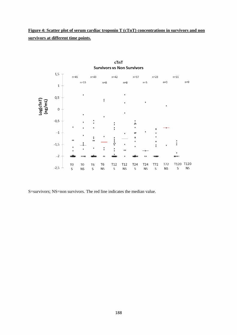
188
Figure 4: Scatter plot of serum cardiac troponin T (cTnT) concentrations in survivors and non
survivors at different time points.
S=survivors; NS=non survivors. The red line indicates the median value.
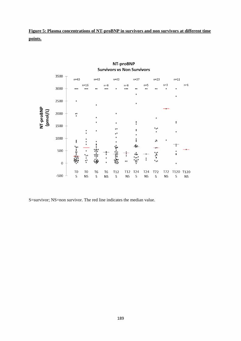
189
Figure 5: Plasma concentrations of NT-proBNP in survivors and non survivors at different time
points.
S=survivor; NS=non survivor. The red line indicates the median value.

190

191
5. DISCUSSION
The studies performed in this research hope to improve the initial diagnosis and stabilization of dogs
presented with SIRS. The results demonstrate several interesting points, which have an impact on
- the interpretation of a clinical diagnosis of SIRS in an emergency setting in dogs
- the development of echocardiography of SIRS/emergency dogs to evaluate fluid status and
cardiac function
- the interpretation of cardiac biomarkers in dogs with a clinical diagnosis of SIRS
After the brief discussion of the findings identified in the three papers, a closer look will be taken
between the correlations of the different parameters studied.
5.1 INFLAMMATORY CYTOKINES AND C-REACTIVE PROTEIN IN CANINE SIRS
The first study evaluating pro-inflammatory cytokines and CRP in emergency dogs with a clinical
diagnosis of SIRS demonstrates that the majority of these dogs have, or will soon develop, increased
CRP concentrations. As explained in the literature review, high CRP concentrations are indicative of an
acute phase inflammatory response. Therefore this study indicates that the clinical diagnosis of SIRS at
presentation to a canine emergency service might not be as unspecific as commonly assumed32.
Similarly, the majority of this cohort of dogs presented to the emergency room with a clinical diagnosis
of SIRS also displayed additional indicators of an active inflammatory process (i.e. increased
concentrations of pro-inflammatory cytokines). The main motivation of this study was to validate other
prospective studies on SIRS in an emergency department in which we would include dogs based on a
clinical diagnosis of SIRS, and the identification of systemic markers of inflammation in these dogs
validates this approach and the subsequent studies.
It should be noted that only 71.2% of dogs had increased CRP concentrations at presentation. Normal
CRP concentrations at presentation in several dogs is explained by the kinetics of APPs. In experimental
studies, CRP has been found to increase within 4 to 6 hours of stimulation and peak after 36 hours348. In
the present study some dogs were presented for hyperacute conditions such as GDV and trauma, and
thus entered the clinic within the 4 to 6 hour timeframe. In these patients CRP concentrations increased
during the initial hours of hospitalization, even if it was within normal limits at presentation. It is
interesting to note that a clinical diagnosis of SIRS may precede changes in APPs in dogs presenting to
the emergency room.
Reference ranges for IL-6 have not been established in dogs. IL-6 is one of the few cytokines that is
detectable in the plasma of healthy dogs, unlike TNF-α37. Similar to previous studies11, absolute values
of cytokines were not normally distributed and logarithmic values were used for statistical analysis. Our
study demonstrated significantly higher IL-6 concentrations at the beginning of hospitalization
compared to the follow-up visit. Opposed to IL-6, TNF-α was detected in only 20/69 dogs (29.0%)

192
throughout hospitalization. Several factors can explain this low prevalence of detectable TNF-α
concentrations. In the dog, TNF-α peaks within 2 hours but often becomes unmeasurable within 6 hours
and rarely remains present for longer than 24 hours35,37. The rapid decrease of TNF-α concentrations is
explained by inhibitory effects of IL-6 on TNF-α production via negative feedback and soluble TNF-α
receptors rendering the circulating TNF-α biologically inactive135. Most of the dogs in our cohort with
detectable TNF-α concentrations at presentation suffered from hyperacute disease such as GDV and
trauma. In agreement with literature, only 2/69 dogs in the present study had measurable TNF-α
concentrations for longer than 24 hours. It is likely that a rise in TNF-α occurred in other dogs prior to
presentation. Furthermore, TNF-α does not typically rise following elective surgery or accidental injury,
and increases in TNF-α may be relatively mild in localized inflammation in humans and dogs173,233.
Some of the dogs in our study likely failed to provoke an increase in TNF-α despite signs of SIRS and
increases in IL-6 and CRP. Other studies in dogs with SIRS and sepsis identified a higher proportion of
dogs with detectable TNF-α concentrations14,255. Such differences can be explained by assay
methodologies and variations in the enrolled cohort of dogs. ELISA techniques measure all the present
TNF-α in the sample, including the biologically inactivated TNF-α by TNF-α soluble receptors, while
bioassays only measure the biologically active TNF-α135. Besides the assays, differences in studied
population also may play an important role. Another study using a (different) bioassay found measurable
TNF-α concentrations in 39/42 dogs with SIRS or sepsis14. That study however looked at dogs at
admission to an intensive care unit, regardless of the presenting signs and previous history, and can
therefore not be easily compared with the present cohort of emergency patients. Additionally, the latter
study did not perform kinetic studies of TNF-α and we can therefore not evaluate the speed at which
TNF-α became undetectable again.
CRP, IL-6 and TNF-α were not associated with the underlying disease category, or with prognosis. APPs
are highly sensitive markers of inflammation but lack specificity regarding the underlying disease
process353. The magnitude of the increase in CRP depends on multiple factors such as initiating cause,
disease severity and extent of tissue damage17,23,24. Highest CRP values may occur at different time
points depending on the type of insult23,511. The clinical nature of the present study implies that dogs had
a great variety of initiating causes of SIRS, and were presented at different points in the process. Despite
these factors, CRP concentrations tended to be higher in dogs with SIRS due to an infectious cause at
presentation. This difference was not significant and should be evaluated in a larger cohort of dogs. The
use of CRP to discriminate septic from non-septic SIRS patients in human medicine has met with
variable results, and has generally been superseded by procalcitonin which also confers prognostic
value, yet unfortunately procalcitonin assays are not available in canine medicine404,405,424,427,442,448.
Our finding that CRP was not predictive of prognosis contradicts with several previous studies on APP-
kinetics and prognosis in canine SIRS30,503,524. When evaluating a single disease entity such as pyometra,
CRP may predict disease severity491. However, for conditions such as canine leptospirosis, with a more

193
variable clinical presentation, CRP was not found to be useful to predict prognosis114. A previous study
on CRP in canine SIRS found that while initial CRP concentrations were unhelpful, the 3-day change
in CRP predicted survival with survivors experiencing a bigger drop in CRP concentrations30. The utility
of CRP as a monitoring tool for treatment evaluation in the acute phase appears limited based on the
findings of this study. CRP concentrations were not significantly different between T6, T12, T24 and
T72, and therefore do not appear to be very informative to evaluate treatment efficacy.
The role of TNF-α as an early mediator of the acute phase inflammatory response with rapid
downregulation makes it a poor diagnostic and prognostic tool in critical care patients34,35,37,38,170. In the
present study, IL-6 was not related to outcome either. Mean IL-6 values for survivors were not
significantly higher at presentation compared to non-survivors, and were not significantly lower from
T6 onwards. These findings are in agreement with two other clinical studies on dogs that also failed to
detect significant differences in IL-6 and TNF-α related to outcome13,14. Research in human medicine
and a canine clinical study in SIRS and sepsis do however suggest prognostic value of IL-6
concentrations11,117,220. The clinical study on dogs included dogs that were hospitalized and dogs with
chronic conditions (mean sign of illness 6.7 days, range 1 to 65 days) and lacked trauma cases or dogs
with GDV. Population characteristics therefore differed significantly from our cohort11. It is our belief
that the short timespan during which TNF-α is detectable, and the cumbersome biological assays
required to measure biological active concentrations of TNF-α and IL-6, renders the utility of these
assays in a clinical setting extremely limited.

194
5.2 CARDIAC FINDINGS IN CANINE EMERGENCIES WITH A CLINICAL DIAGNOSIS
OF SYSTEMIC INFLAMMATORY RESPONSE SYNDROME WITHOUT
HYPOTENSION
The second paper of this PhD project evaluated echocardiographic findings in a cohort of dogs with
SIRS presented to a university emergency department. Dogs that participated in this arm of the PhD had
higher median heart rate, lower LA/Ao and nLVIDd at presentation. The increase of nLVIDd and LA/Ao
during hospitalization can either be explained by a decreasing heart rate (mediated by decreasing stress,
pain relief, anti-inflammatory treatment), or can indicate a mild degree of hypovolemia which improves
following appropriate treatment.
nLVIDd was significantly correlated with LA/Ao (p <0.001 and r 0.328) yet negatively correlated with
FS (p <0.001 and R -0.418). As both LA/Ao and nLVIDd estimate preload, this positive correlation is
not surprising. However, the negative correlation between nLVIDd and FS is in conflict with the Frank
Starling principle. As heart rate was positively correlated with FS, it is very likely that adrenergic and
sympathetic effects explain this correlation.
Only heart rate in this study was significantly associated with survival. However, median LA/Ao and
nLVIDd values were higher and median FS values were lower in survivors compared to non survivors
from T0 until T24. Myocardial hibernation in human beings is characterized by a decreased systolic
function, and an increased end diastolic left ventricular volume. Whether the trend towards lower FS
and higher nLVIDd in survivors observed in this study are consequences of changing heart rate and
sympathetic tone, explained by changes in volume status, or early signs of myocardial hibernation, can
unfortunately not be determined. In canine cardiology, LV systolic dysfunction is defined as a FS<26%,
although these percentiles depend on breed size, with a FS of 26% considered worse in small compared
to large breed dogs36. Only three dogs in the present study had a FS below 26%, and all these were
medium to large breed dogs (22.6, 31.8 and 56 kg). Therefore, even if these low values are considered
indicative of ventricular dysfunction, the incidence of ventricular systolic dysfunction in this study
should be considered low. Few papers have discussed systolic dysfunction in canine critical care
patients. A previous retrospective study described 16 dogs with cardiovascular dysfunction associated
with infectious (septic) and non-infectious (neoplastic and other disease) critical illness36. Unfortunately,
that study was not blinded, and underlying disease and the identification of myocardial dysfunction
might have influenced treatment decisions and prognosis36.
None of the dogs included in the present study was reported to experience any complication secondary
to echocardiography, and echocardiography only requires mild physical restraint during a couple of
minutes. Based on developments in human ICUs and on the findings of this paper, echocardiography
therefore should be considered as a relatively safe and promising procedure.
195
5.3 CARDIAC BIOMARKERS IN CANINE EMERGENCIES WITH A CLINICAL
DIAGNOSIS OF SYSTEMIC INFLAMMATORY RESPONSE SYNDROME
This study demonstrated changes in cardiac biomarkers during hospitalization in a population of canine
SIRS patients presented to an emergency department. cTnT values were detectable in 28 dogs during
hospitalization, with concentrations significantly higher at T12, T24 and T72 compared to
concentrations at presentation and at the control visit. The timing of the changes in cTnT concentrations
appear to agree with the rather rapid rise and sustained increase described in the literature review. In
contrast, at their control visit, all dogs had undetectable cTnT concentration (<0.01ng/mL). A study
evaluating cTnI concentrations in canine SIRS patients identified a higher prevalence of increased cTnI
concentrations at presentation (35/60 dogs) and during hospitalization, but similarly failed to find
significant variations from day to day.74 Another study comparing cTnT and cTnI in SIRS patients
admitted to the ICU found a higher prevalence of increased cTnI concentrations.75 This difference in
detection rate can be explained either by the lower sensitivity of cTnT tests, or by the timing of sampling
compared to the start of the disease process (as admission to the ICU likely later than admission to an
emergency department).75
NT-proBNP changed significantly over time, with concentrations at T24, T72 and T120 significantly
higher than concentrations at T0, T6, T12 and the control visit. Most of the research performed in
veterinary medicine on NT-proBNP has focused on cardiac disease.1311-1313 A recent study evaluating
BNP in dogs with non-cardiac disease (e.g. neurological and gastrointestinal disease) demonstrated a
moderate increase of natriuretic peptides in these patients.97 As our study focused on dogs with SIRS
presented to an emergency department, and SIRS has a high potential to induce cardiac effects as is well
described in human medicine, it is not surprising that NT-proBNP concentrations in the present study
were more markedly elevated compared to that previous study.97 Finding higher concentrations at T24,
72 and T120 is in agreement with studies on SIRS and sepsis in human patients. The optimal timing of
NT-proBNP measurement varied across studies in humans, from the day of admission to day 2 and day
5 after admission168,710,1092,1259,1274 The kinetics observed in this cohort seem to confirm these findings in
dogs presented with a clinical diagnosis of SIRS to an emergency department. Elevated levels of NT-
proBNP when screening for occult cardiac disease should therefore be interpreted carefully in SIRS
patients.
Regarding the association of cardiac biomarkers with prognosis, an increase of cTnT during
hospitalization was associated with poor short term prognosis. Cardiac troponin T and I are well-
accepted prognostic biomarkers in human intensive care units.62,64,72,1320 In veterinary medicine,
increased concentrations of cardiac troponins have been observed in patients suffering from infectious
disease, trauma, GDV or systemic disease93,818,824,825,861,953,1032,1042-1044 and are correlated with poor
prognosis in some of these studies.114,824,825,861 Studies evaluating cTnI and cTnT in canine SIRS patients
already confirmed their prognostic value.74-76 These studies similarly identified significant differences
196
between survivors and non-survivors.74 As cTnT (and cTnI) can remain increased up to 7 or 10 days
after the insult, kinetics of cTnT are difficult to evaluate, and they are less useful for evaluation of disease
progression or treatment response.58,865
In the present study, NT-proBNP concentrations were not significantly correlated with prognosis.
Previous studies in dogs tended to evaluate natriuretic peptides at presentation, while higher
concentrations should be expected later during hospitalization. This delay in the rise of NT-proBNP
probably also limits its use as a prognostic marker in a clinical veterinary emergency care setting. At
later time points the group size in this study rapidly decreased, which may have impacted the likelihood
to identify significant differences between survivors and non survivors. A recently published meta-
analysis in human septic patients describing 12 studies on 1865 cases did conclude that (NT-pro)BNP
is significantly associated with risk of mortality.1259 This meta-analysis also concluded that elevated
(NT-pro)BNP levels in the presence of SIRS or sepsis do not equal cardiac dysfunction due to low
specificity, but normal (NT-pro)BNP levels could be used to rule out the need for further cardiac
investigation.1259 Therefore, the lack of a significant difference in NT-proBNP between survivors and
non survivors observed in the present study definitely needs to be confirmed in a larger cohort of patients
with a clinical diagnosis of SIRS. The observed increased NT-proBNP concentrations can be explained
by myocardial dysfunction, but also via increased wall stress after volume resuscitation,1244 lung injury,
acute respiratory distress syndrome or thromboembolism.67

197
5.4 CORRELATION OF STUDIED MARKERS IN CANINE EMERGENCIES WITH A
CLINICAL DIAGNOSIS OF SYSTEMIC INFLAMMATORY RESPONSE
SYNDROME
As the reader undoubtedly has already understood, the different studies of this defense were all
performed on the same population, although only a subgroup entered the echocardiography study. The
hypothesis of our studies were that dogs with a clinical diagnosis of SIRS presented to an emergency
department would have measurable proof of systemic inflammation via inflammatory cytokines or
biomarkers. Moreover, we also wanted to investigate whether systemic inflammation does affect the
heart as it has been shown in human medicine and in experimental animal studies. It is therefore
particularly interesting to evaluate whether a correlation could be identified between the different
parameters assessed in the different studies.
The table 4 which is presented hereunder does allow the reader to visually assess the correlation of
different parameters, and the exact level of significance and correlation are given in tables 5 and 6.
Firstly, as already discussed in the separate papers, several parameters were significantly correlated
within each study. CRP and IL-6 were positively correlated, which was not surprising as IL-6 is the
major stimulatory cytokine for CRP production. TNF-α however was not significantly correlated with
both biomarkers, but this can be explained by the short presence of TNF-α in plasma. Regarding the
cardiac biomarkers, NT-proBNP concentrations were positively correlated with cTnT concentrations.
This again was expected as such a positive correlation has already been reported in human SIRS patients.
Regarding echocardiographic parameters, heart rate was negatively correlated with LA/Ao and nLVIDd,
yet positively correlated with FS, and not correlated with SAP. The lack of a correlation with SAP is
most likely explained by the bias in study population, with only less severely ill animals being included
in this study. As explained in the manuscript, the negative correlation with preload parameters is
explained by the effect of heart rate on cardiac filling. The correlation with FS might be due to
sympathetic and adrenergic effects. Furthermore, SAP was mildly positively correlated with LA/Ao,
which itself was positively correlated with nLVIDd. Both correlations may indicate the role of preload
in the generation of an adequate arterial pressure to maintain tissue perfusion. Finally, and most
interestingly, an increase in nLVIDd was negatively correlated with FS. This might be a very early sign
of myocardial dysfunction. However, this one finding in a small and biased study population should not
be emphasized, yet rather confirmed in a larger study including all SIRS patients.
Looking at the correlation between inflammatory and cardiac biomarkers, cTnT appears to be positively
correlated with TNF-α concentrations. This might be an indication of how systemic inflammation affects
cardiomyocyte integrity and function. However, at the same time, IL-6 concentrations appeared to be
negatively correlated with NT-proBNP concentrations. Therefore, rather than speculating about the
significance of these correlations, further research in larger cohorts appears warranted.
198
Heart rate was positively correlated with IL-6 and TNF-α, and SAP was negatively correlated with CRP
and IL-6. All of these findings support the effect of systemic inflammation on inducing hypotension and
tachycardia. Similarly, LA/Ao was negatively correlated with IL-6 and TNF-α, again supporting the
concept of dehydration and (relative) hypovolemia developing in SIRS patients via decreased water
intake or increased losses via vomiting, diarrhea, or shifting of water from the circulation. However,
nLVIDd and FS were not correlated with any of the inflammatory markers, although such a lack may
be explained by the small and biased study group in the echocardiography study.
Finally when evaluating the correlation of cardiac biomarkers with finding on echocardiography, first
of all a positive correlation between heart rate and cTnT was demonstrated. Rather than assuming a
direct link between heart rate and cTnT concentrations, this correlation may merely indirectly confirm
again that dogs with detectable cTnT concentrations were cardiovascularily more severely affected dogs,
and thus more likely to suffer from severe inflammatory disease. nLVIDd was mildly positively
correlated with NT-proBNP, which is not surprising as the main stimulus for NT-proBNP secretion is
increased ventricular wall stress. The mild correlation might have been more pronounced if more
severely affected patients were included. More surprisingly, nLVIDd was also positively correlated with
cTnT. This could be a sign of myocardial hibernation, as this process is characterized both by increased
ventricular size ad increased cTnT concentrations. However, decreased systolic function is also
considered a key element of myocardial hibernation, yet FS did not appear to be correlated with any of
the cardiac biomarkers, and neither was LA/Ao. Therefore, and as indicated before, drawing any
conclusions based on this small cohort of dogs that were not severely affected would be premature, and
these findings need to be confirmed in larger cohorts including all patients.

199
Table 4. Correlation table of studied parameters in canine emergencies with a clinical diagnosis
of systemic inflammatory response syndrome.
This correlation table represents an easily appreciable visual estimation of the correlation between two
parameters. Blue circles indicate a positive correlation, while red circles indicate a negative correlation.
The colour intensity is indicative of the value of the correlation coefficient with darker shades indicating
a value closer to 1. The size of the circle represents the level of significance of the correlation, with
bigger circles indicative of a lower p-value and thus higher significance. Whenever a black cross is
present, this indicates that the two parameters are not significantly correlated (p>0.05). As many of the
parameters that were evaluated were not normally distributed, we have used the Spearman correlation
for the evaluation of all parameters.
The exact values of the correlations and the p-values of each correlation are presented in table 5 and 6
respectively. Whenever the probability (p-value) is lower than 0.05 (5%), the test is significant and both
parameters are significantly correlated at a level of 5%.

200
Table 5. Spearman correlation of the different parameters studied throughout the different
papers. Values in green indicate a significant correlation (p <0.05) of the between the two
parameters.
Table 6. Spearman correlation values of the different parameters studied throughout the different
papers.

201
6. LIMITATIONS OF THE PERFORMED RESEARCH
6.1 GENERAL LIMITATIONS OF THE STUDIES
The first conclusion one should take is that we performed observational studies, and did not perform
any fundamental research. However, the definition of SIRS and its effects on the cardiovascular system
have been investigated in detail in experimental designs in laboratory animals including dogs, and have
been observed in human clinical studies. Observational studies were therefore justified. The hypothesis
of our research was that, in a clinical setting of dogs with SIRS presented to an emergency department,
cardiac effects could be detected, and our ultimate goal was not to explain such effects.
For observational studies, the clinical nature of the performed research in our opinion is one of the strong
points of it, yet simultaneously is one of the biggest limitations. We chose to include privately owned
dogs presented to the emergency department of a university small animal teaching hospital with a
clinical diagnosis of SIRS. This implies that we included dogs of different breeds, weights and ages,
presenting with a large variety of diseases, at different time points in the disease process. Rather than
developing experimental designs, we preferred to investigate whether cardiac consequences of SIRS can
be appreciated in a clinical emergency and critical care setting.
The clinical observational nature of the designs obliged us to perform the first research, evaluating
whether dogs included based on a clinical diagnosis of SIRS truly present biochemical evidence of
systemic inflammation. If biochemical findings such as increased inflammatory cytokine and CRP
concentrations were not substantiating the value of the clinical diagnosis of SIRS as a screening tool in
an emergency referral setting, this would have obliged us to adapt our designs.
A second implication of clinical studies is that owners have different levels of motivation, affecting
decision making and outcome of the dogs included. To limit the influence of this factor, we recorded
whether patients were euthanized for financial rather than for prognostic reasons, and those euthanized
for financial reasons were removed for statistical calculations regarding outcome. All dogs that were
euthanized for prognostic reasons had a deteriorating clinical condition that did not respond to
appropriate treatment or suffered life-threatening complications. In the presented studies, 64% (in the
papers on inflammatory cytokines and cardiac biomarkers) and 76% (in the paper on echocardiography)
of patients survived until discharge, which is comparable to or better than previous studies on clinical
canine SIRS patients. The higher survival rate in the echocardiography paper indicates that these patients
were less severely affected, and we will come back to the implications of these findings when discussing
the limitations of this paper in particular. Moreover, we only managed to convince less than half of
owners to come back for a control visit of their pet. As dogs were clinically healthy and owners did not
receive any compensation, many owners declined the control visit. This low percentile of control visits
also creates an important bias in our control population.

202
A third limitation of clinical studies on a clinical syndrome such as SIRS is that included dogs suffer
from various disease processes eliciting different pathophysiological responses. Empirically dividing
clinical cases over different categories is often complicated, resulting in many animals ending in a
‘miscellaneous’ group, and low numbers in specific disease categories. Therefore, observations in a
specific disease category should be tested in larger cohorts before drawing strong conclusions.
Another limitation in the design of our papers is that time points for evaluation were standardized with
relation to the moment of presentation to the emergency department. Clinical signs were present for
variable times prior to presentation, and as previously mentioned, different diseases induce different
responses, and this will have affected our findings. The fixed timing of sampling implied that the timing
varied with regard to surgical or medical interventions. For the analysis of our findings, the fixed timing
allowed us to describe the kinetics of cytokines, biomarkers and echocardiographic findings, but it did
not allow to determine peak concentrations or maximal variations in observations. As SIRS is a
syndrome, presented secondary to a wide variety of conditions, an exact moment for peak concentrations
and maximal variations would be unlikely to exist, and limiting our sampling points was considered
favourable for ethical and financial reasons. Studies to determine peak concentrations and maximal
variations should be reserved for well-described experimental designs on specific disease, not for
clinical studies.
6.2 INFLAMMATORY CYTOKINES AND C-REACTIVE PROTEIN IN CANINE SIRS
Samples were stored at -80°C prior to analysis of inflammatory cytokines and CRP. All these substances
remain stable at temperatures below -70°C, although no single publication investigated the maximum
storage time for these substances at this temperature. As samples were analyzed within a year, which is
comparable with many publications, we are convinced this did not affect our findings. Moreover, if
storage would have artificially decreased concentrations of these cytokines and biomarkers, this would
have resulted in less rather than more significant changes.
The bioassays applied for the determination of concentrations of TNF-α and IL-6 have been previously
validated and published. These bioassays allow for the detection of biologically active concentrations
of cytokines, in contrast to ELISA techniques, which also detect biologically inactive fractions. The
cumbersome methodology is however not applicable in a clinical setting, but gives a better reflection of
the clinical situation. Several previous publications in dogs described concentrations assessed using such
ELISA techniques. Findings from studies applying ELISA techniques should therefore not be compared
with our findings. From a clinical standpoint, concentrations of biologically active cytokines are more
relevant than total concentrations, and therefore we considered the technique used in this work
preferable over ELISA techniques.
Several samples were displaying signs of hemolysis, hyperbilirubinemia or lipemia, which theoretically
could interfere with the measurement of CRP. The assay we applied however appears to be fairly

203
insensitive to these effects, and we could not detect any influence of these interfering factors on our
findings.
6.3 CARDIAC FINDINGS IN CANINE EMERGENCIES WITH A CLINICAL DIAGNOSIS
OF SYSTEMIC INFLAMMATORY RESPONSE SYNDROME WITHOUT
HYPOTENSION
The ethical concerns raised to submit critical patients to an echocardiography, which was judged to be
an invasive procedure, was one of the major limitations to this paper. Ethical approval was obtained for
the study after owners signed an informed consent form, but case veterinarians could withdraw patients
if they considered them unstable for blood sampling or ultrasonography. None of the dogs was
withdrawn for blood sampling during the study, but many patients were excluded from the
echocardiographic part of the study for this reason. Whether a limited, basic echocardiography is more
demanding than blood sampling however requires to be determined, and this decision was based on the
perception of the clinician. As bedside echocardiography is a well-accepted procedure in human critical
care, and is considered harmless, this fear of echocardiography induced complications by attending
veterinarians seems to be debatable. None of the patients undergoing echocardiography in this study
demonstrated a complication secondary to the procedure. However, survival rates in this cohort of dogs
(75.7%) was higher than compared to previous studies on clinical canine SIRS patients11,13 and the two
other studies of the present thesis, so included patients did appear to be clinically in a better condition.
As heart rate during echocardiography at the control visit of clinically healthy patients was similar to
heart rate at presentation, it seems that echocardiography does at least provoke some stress in otherwise
healthy dogs. Therefore, although cage side echocardiography seems to be safe in a canine critical care
setting, this still needs to be confirmed in more critical patients.
The withdrawal of less stable patients may explain why we did not observe clear evidence of myocardial
hibernation in this clinical setting, as described in experimental canine studies and clinical human
papers. Myocardial dysfunction in human medicine is more severe and prevalent in human septic shock
patients and septic patients compared to SIRS patients. The degree of myocardial dysfunction has been
correlated with concentrations of cardiac troponins and brain natriuretic peptide67, which are also
correlated with the clinical condition1011, degree of hypotension1014, and clinical scores of these
patients65,86,1011. A design including all emergency patients with SIRS regardless of their cardiovascular
status (but excluding dogs with severe dyspnea to prevent complications due to the lateral recumbency)
is more likely to identify and evaluate myocardial hibernation better. However, if we want to develop
such studies, we need to have a properly trained staff to perform such short echocardiographies.
Echocardiographies were also not performed by cardiologists, but by interns previously trained by a
cardiologist, demonstrating the ability to perform repeatable and comparable echocardiographies in a
population of research beagles prior to the start of this study. Although ideally all echocardiographies
would be performed by a single cardiologist, this is not feasible in an emergency setting. As discussed

204
in the literature review, echocardiography in human critical care is also performed by criticalist receiving
a short training programme. Although the results of this study on echocardiography in SIRS patients did
not illustrate significant changes in systolic function throughout hospitalization, it did identify
significant changes in preload during hospitalization, and some interesting trends regarding the
association of echocardiographic findings and prognosis. This study therefore illustrates the huge need
for such short training programmes for criticalists in veterinary medicine, as well as continued research
in this field. As the two involved veterinarians were properly trained and demonstrated to be competent
in the performance of the short echocardiographic protocol, we believe this did not significantly
influence our findings.
Our echocardiography study focused on preload and LV dysfunction. Right ventricular dysfunction and
left and right ventricular diastolic dysfunction have also all been described in human myocardial
dysfunction and experimental canine studies55,56,654,737. However, such parameters are even harder to
assess, and we therefore consider that echocardiography by non-cardiologists should first focus on one-
dimensional parameters that could be assessed easily on standard windows, to improve performance of
the trainees88.
Due to the low amount of included patients based on cautiousness of the attending veterinarian, the study
was terminated with only low numbers of dogs in each disease category. The findings of this paper
should therefore not be over interpreted, yet need to be confirmed in larger studies including all dogs
with a clinical diagnosis of SIRS.
6.4 CARDIAC BIOMARKERS IN CANINE EMERGENCIES WITH A CLINICAL
DIAGNOSIS OF SYSTEMIC INFLAMMATORY RESPONSE SYNDROME
Samples were stored at -80°C prior to analysis of cardiac biomarkers, similarly to inflammatory
cytokines and CRP. Again, NT-proBNP and cTn are reportedly stable at temperatures below -70°C,
although the maximum storage time has not been described. These samples were also analyzed within
one year as reported previously, and if storage would have artificially decreased concentrations of these
cytokines and biomarkers, this would have resulted in less rather than more significant changes.
We evaluated cTnT rather than cTnI, as the cTnT assay was readily available. cTnI has received more
attention in veterinary medicine, as cTnI assays are more sensitive than cTnT to detect cardiac
involvement824,825. The use of a cTnI assay would probably have resulted in the detection of elevated
concentrations in a larger proportion of SIRS patients. A recent review however once more concluded
that cTnT and cTnI are probably equally valuable as prognostic markers.
The analysis of NT-proBNP concentrations was more costly and labour intensive. Subsequently, due to
financial restrictions, samples with concentrations above the upper limit of the assay (3000pmol/L) were
not diluted to measure the exact concentration. Therefore, NT-proBNP concentration was

205
underestimated in a small portion of samples. Similarly to the (unlikely) effects of freezing, such an
effect did not prohibit us from finding significant changes. If exact concentrations would have been
measured, our findings would probably have been even more significant.
7. CONCLUSIONS
This manuscript allows us to draw several meaningful conclusions regarding dogs presented to an
emergency department with a clinical diagnosis of SIRS.
- CRP is elevated in the majority of such cases at presentation, or will increase shortly after
presentation. This indicates that the clinical diagnosis of SIRS in this setting may be more
specific than previously considered. Whether CRP may be of additional value as a screening
and monitoring tool in these patients remains to be determined.
- Even in the absence of marked hypotension, such dogs have lower median LA/Ao and nLVIDd
at presentation. A trend was observed towards higher median LA/AO, nLVIDd and lower FS in
survivors during the initial hours of hospitalization. Whether these observations are valid, and
whether they represent an early sign of myocardial hibernation in these patients requires to be
confirmed in larger studies including all dogs with SIRS. Assessment of preload and myocardial
function via echocardiography merits further investigation in canine emergency and critical
care.
- cTn and NT-proBNP are often elevated in these patients and cTnT carries prognostic value in
dogs with SIRS presented to the emergency department. Whether such increases are linked with
myocardial hibernation remains to be demonstrated.
Based on these conclusions, we suggest multiple future perspectives that will be discussed in the next
chapter.

206
207
8. FUTURE PERSPECTIVES
The first ‘validating’ paper on biochemical markers of systemic inflammation mostly re-emphasizes the
role of APPs in SIRS. With the easy availability of benchtop devices to quantitatively measure CRP
concentrations in house in dogs, this parameter might be considered part of the minimal database in
future canine emergencies. Future studies could aim to evaluate CRP in a cohort of emergencies
regardless of the clinical diagnosis of SIRS. Such a design would allow us to comment on the agreement
between a clinical diagnosis of SIRS and CRP concentrations. A previous study in dogs with pyometra
demonstrated that CRP is associated with SIRS in this disease, but this has never been evaluated in a
cohort of emergency patients255. An increased CRP concentration at presentation gives objective proof
of systemic inflammation, pushing the veterinarian to investigate, and the owner to accept further work-
up of the emergency patient.
Moreover, although we did not find a significant association between CRP concentrations and prognosis
or diagnosis of the underlying disease category, this merits deeper evaluation. With current guidelines
for the diagnosis of sepsis and septic shock redefined, placing less emphasis on the component of
systemic inflammation, it would be interesting to investigate the added value of CRP to a simplified
clinical screening scale (such as the qSAP) regarding the likelihood of morbidity and mortality of these
patients.
CRP kinetics could help to evaluate the response to treatment of critical care patients. Evidence in human
literature demonstrates how CRP could be used to evaluate the efficacy of antibiotic therapy in
streptococcal meningitis. Similarly, CRP has been applied as a biomarker to evaluate the efficacy of
immunosuppressive therapy in canine steroid responsive meningitis and arteritis. As discussed in this
manuscript, CRP kinetics depend on the underlying pathology. Therefore, the use of CRP as a
monitoring tool should be evaluated in larger groups of dogs affected by a single disease category (e.g.
septic peritonitis, pneumonia or pancreatitis).
The most interesting developments might be expected via the development of echocardiography to
evaluate and monitor fluid status and cardiac function. Our paper on SIRS patients without hypotension
demonstrated rather low preload of these emergency patients, and although not statistically significant
suggest a trend towards a correlation between preload and survival. Fluid loading is an important aspect
of human emergency stabilization, and fluid responsiveness (defined as the potential to increase cardiac
output in response to a fluid challenge) is evaluated via several echographic parameters in human ECC
services552.
In veterinary medicine, the use of echocardiography in a canine emergency and critical care setting still
largely needs to be developed. Moreover, in order to perform many of the possible studies described
above, canine ECC departments require trained staff capable of performing such short
echocardiographic studies. According to the authors, the first step in this process therefore was to
208
develop a short training program, allowing non-cardiologists to record basic echocardiographic views
and respond to basic questions, as previously demonstrated in humans. The authors have developed and
tested such a program in association with the cardiology department, and an abstract on the performance
of repeatable cardiovascular focused assessment via sonography for triage (CV-FAST) after a 6-hour
training was presented at the ECVIM-CA congress in Goteborg (September 2016) (see appendix 1).
Findings of this study seem encouraging and the manuscript is in preparation to be submitted for
publication to the Journal of Emergency and Critical Care. The ability to train the entire veterinary staff
to perform such a CV-FAST exam should allow for clinical trials including all emergency patients.
Moreover, as patients in our paper on echocardiography in SIRS did not demonstrate any complications,
we consider that this should allow for ethical approval to perform such examinations even in patients in
hypotension.
Besides a large prospective study on CV-FAST echocardiography in canine emergency and critical care
cases in general, different common clinical scenarios could be evaluated. For instance, the effects of
known changes in blood volume (blood donation or blood transfusion) on volume status could be
evaluated. Volume status and cardiac function via CV-FAST techniques could also be evaluated in
septic peritonitis cases, or in cardiac patients in the critical care department. Similarly, experimental
designs could potentially be developed to evaluate CV-FAST findings in canine hypovolemic, septic or
cardiogenic shock models.
In human patients, the best echocardiographic parameter for the assessment of fluid responsiveness is
the change in the patient’s vena cava diameter with respiration, or the evaluation of stroke volume618.
The utility of inferior vena cava FAST assessment to estimate volume status and fluid responsiveness
in critically ill humans is well established1321,1322. Future research evaluating caudal vena cava size and
collapsibility in dogs to estimate preload and fluid responsiveness should therefore be developed. The
authors have received the EVECC - SCIL research grant to perform a study in collaboration with the
university of Calgary to standardize and evaluate the repeatability of the echographic evaluation of the
caudal vena cava in healthy dogs via a diaphragmatic, hepatic or the renal view (see appendix 2). Our
hypothesis is that caudal vena cava size will be related to body weight or metabolic weight, while caudal
vena cava collapsibility will be an index independent of body size. Results of that study are expected to
be presented at the next EVECC congress in Dublin, June 2017.
The paper on cardiac biomarkers also offers very interesting perspectives. At this time, we consider
cardiac biomarkers mostly indicated to identify patients with a high likelihood of cardiovascular disease
or complications in an emergency and critical care setting. Semi-quantitative point-of-care tests for NT-
proBNP are available and should be evaluated as screening tools to identify patients with primary or
secondary cardiovascular disease. It would be interesting to evaluate a larger cohort of emergency
patients via echocardiography and to test NT-proBNP concentrations in these patients regardless of a

209
clinical SIRS diagnosis. By comparing findings, it would be interesting to evaluate the sensitivity and
specificity of NT-proBNP to detect (primary versus secondary) cardiovascular disease in these patients.
As NT-proBNP rises fairly late in the course of the disease, it would be interesting to measure
concentrations at presentation and after 3 days and to evaluate the impact of the timing of sampling on
such findings. Due to the late rise, the use of NT-proBNP as an initial marker seems limited. Cardiac
troponins are probably more interesting as an initial screening tool to identify patients requiring thorough
cardiovascular evaluation or monitoring, as they rise earlier in the course of disease. Moreover our study
confirmed previous findings that cTns carry prognostic information in dogs with a clinical diagnosis of
SIRS. However, their interest as monitoring tools is probably even more limited as troponin
concentrations decrease very slowly.
Based on findings in human papers, the increases in NT-proBNP and troponin in SIRS patients may be
a reflection of and correlated with myocardial hibernation. Larger prospective studies should evaluate
the correlation between NT-proBNP and echocardiographic findings in emergency patients, recording
clinical SIRS diagnosis, but not limited to these patients only. It would also be interesting to record
simplified patient evaluating scores such as shock index, qSAP or APACHE scores, and evaluate their
correlation with cardiac biomarkers and echographic findings.
My hope remains that at the end of this journey, we will look back to this day, reassured that we have
learned to provide more appropriate cardiovascular care for our emergency and critical care patients. If
this PhD is nothing more than an introduction to this exciting story, then this PhD was worth the hours
of work behind my desk instead of in clinics…

210

211
9. BIBLIOGRAPHY
1. Bone RC, Balk RA, Cerra FB, et al. Definitions for sepsis and organ failure and guidelines for the use of innovative therapies in sepsis. The ACCP/SCCM Consensus Conference Committee. American College of Chest Physicians/Society of Critical Care Medicine. Chest 1992;101:1644-1655. 2. Herzum I, Renz H. Inflammatory markers in SIRS, sepsis and septic shock. Curr Med Chem 2008;15:581-587. 3. Beutler B, Milsark IW, Cerami AC. Passive immunization against cachectin/tumor necrosis factor protects mice from lethal effect of endotoxin. Science, 1985, 229(4716):869-871. J Immunol 2008;181:7-9. 4. Casey LC, Balk RA, Bone RC. Plasma cytokine and endotoxin levels correlate with survival in patients with the sepsis syndrome. Ann Int Med 1993;119:771-778. 5. Thalinger AR, Lefer AM. Cardiac actions of a myocardial depressant factor isolated from shock plasma. Proceedings of the Society for Experimental Biology and Medicine Society for Experimental Biology and Medicine (New York, NY) 1971;136:354-358. 6. Kubota T, Bounoutas GS, Miyagishima M, et al. Soluble tumor necrosis factor receptor abrogates myocardial inflammation but not hypertrophy in cytokine-induced cardiomyopathy. Circulation 2000;101:2518-2525. 7. Michie HR, Manogue KR, Spriggs DR, et al. Detection of circulating tumor necrosis factor after endotoxin administration. N Engl J Med 1988;318:1481-1486. 8. Fong YM, Marano MA, Moldawer LL, et al. The acute splanchnic and peripheral tissue metabolic response to endotoxin in humans. J Clin Invest 1990;85:1896-1904. 9. Tizard IR. Innate Immunity: Proinflammatory and Antimicrobial Mediators. In: Tizard IR, editor. Veterinary Immunology, 9th ed. St. Louis, Missouri: Elsevier Saunders; 2013:21-29. 10. Flower L, Ahuja RH, Humphries SE, et al. Effects of sample handling on the stability of interleukin 6, tumour necrosis factor-alpha and leptin. Cytokine 2000;12:1712-1716. 11. Rau S, Kohn B, Richter C, et al. Plasma interleukin-6 response is predictive for severity and mortality in canine systemic inflammatory response syndrome and sepsis. Vet Clin Pathol 2007;36:253-260. 12. Damas P, Ledoux D, Nys M, et al. Cytokine serum level during severe sepsis in human IL-6 as a marker of severity. Ann Surg 1992;215:356-362. 13. Yu DH, Nho DH, Song RH, et al. High-mobility group box 1 as a surrogate prognostic marker in dogs with systemic inflammatory response syndrome. J Vet Emerg Crit Care (San Antonio) 2010;20:298-302. 14. DeClue AE, Sharp CR, Harmon M. Plasma inflammatory mediator concentrations at ICU admission in dogs with naturally developing sepsis. J Vet Intern Med 2012;26:624-630. 15. Heinrich PC, Castell JV, Andus T. Interleukin-6 and the acute phase response. Biochem J 1990;265:621-636. 16. Pepys MB, Baltz ML. Acute phase proteins with special reference to C-reactive protein and related proteins (pentaxins) and serum amyloid A protein. Adv Immunol 1983;34:141-212. 17. Ceron JJ, Eckersall PD, Martynez-Subiela S. Acute phase proteins in dogs and cats: current knowledge and future perspectives. Vet Clin Pathol 2005;34:85-99. 18. Petersen HH, Nielsen JP, Heegaard PM. Application of acute phase protein measurements in veterinary clinical chemistry. Vet Res 2004;35:163-187. 19. Hirvonen J, Eklund K, Teppo AM, et al. Acute phase response in dairy cows with experimentally induced Escherichia coli mastitis. Acta Vet Scand 1999;40:35-46. 20. Hulten C, Demmers S. Serum amyloid A (SAA) as an aid in the management of infectious disease in the foal: comparison with total leucocyte count, neutrophil count and fibrinogen. Equine Vet J 2002;34:693-698. 21. Peltola HO. C-reactive protein for rapid monitoring of infections of the central nervous system. Lancet 1982;1:980-982. 22. Skinner JG, Brown RA, Roberts L. Bovine haptoglobin response in clinically defined field conditions. Vet Rec 1991;128:147-149.

212
23. Rikihisa Y, Yamamoto S, Kwak I, et al. C-reactive protein and alpha 1-acid glycoprotein levels in dogs infected with Ehrlichia canis. J Clin Microbiol 1994;32:912-917. 24. Yamamoto S, Shida T, Okimura T, et al. Determination of C-reactive protein in serum and plasma from healthy dogs and dogs with pneumonia by ELISA and slide reversed passive latex agglutination test. Vet Q 1994;16:74-77. 25. Yule TD, Roth MB, Dreier K, et al. Canine parvovirus vaccine elicits protection from the inflammatory and clinical consequences of the disease. Vaccine 1997;15:720-729. 26. Vigushin DM, Pepys MB, Hawkins PN. Metabolic and scintigraphic studies of radioiodinated human C-reactive protein in health and disease. J Clin Invest 1993;91:1351-1357. 27. Kjelgaard-Hansen M, Kristensen AT, Jensen AL. Evaluation of a commercially available enzyme-linked immunosorbent assay (ELISA) for the determination of C-reactive protein in canine serum. J Vet Med A Physiol Pathol Clin Med 2003;50:164-168. 28. Yamamoto S, Shida T, Miyaji S, et al. Changes in serum C-reactive protein levels in dogs with various disorders and surgical traumas. Vet Res Commun 1993;17:85-93. 29. Nakamura M, Takahashi M, Ohno K, et al. C-reactive protein concentration in dogs with various diseases. J Vet Med Sci 2008;70:127-131. 30. Gebhardt C, Hirschberger J, Rau S, et al. Use of C-reactive protein to predict outcome in dogs with systemic inflammatory response syndrome or sepsis. J Vet Emerg Crit Care (San Antonio) 2009;19:450-458. 31. Brady CA, Otto CM. Systemic inflammatory response syndrome, sepsis, and multiple organ dysfunction. Vet Clin North Am Small Anim Pract 2001;31:1147-1162, v-vi. 32. Hauptman JG, Walshaw R, Olivier NB. Evaluation of the sensitivity and specificity of diagnostic criteria for sepsis in dogs. Vet Surg 1997;26:393-397. 33. de Laforcade AM. Systemic Inflammatory Response Syndrome. In: Silverstein DC, Hopper K, editors. Small Animal Critical Care Medicine, 2nd ed. St. Louis, Missouri, United States: Saunders Elsevier; 2015:31-34. 34. Marks JD, Marks CB, Luce JM, et al. Plasma tumor necrosis factor in patients with septic shock. Mortality rate, incidence of adult respiratory distress syndrome, and effects of methylprednisolone administration. Am Rev Respir Dis 1990;141:94-97. 35. Miyamoto T, Fujinaga T, Yamashita K, et al. Changes of serum cytokine activities and other parameters in dogs with experimentally induced endotoxic shock. Jpn J Vet Res 1996;44:107-118. 36. Hesse DG, Tracey KJ, Fong Y, et al. Cytokine appearance in human endotoxemia and primate bacteremia. Surg Gynecol Obstet 1988;166:147-153. 37. LeMay DR, LeMay LG, Kluger MJ, et al. Plasma profiles of IL-6 and TNF with fever-inducing doses of lipopolysaccharide in dogs. Am J Physiol 1990;259:R126-132. 38. Moscovitz H, Shofer F, Mignott H, et al. Plasma cytokine determinations in emergency department patients as a predictor of bacteremia and infectious disease severity. Crit Care Med 1994;22:1102-1107. 39. Rudiger A, Singer M. Mechanisms of sepsis-induced cardiac dysfunction. Crit Care Med 2007;35:1599-1608. 40. Jardin F, Fourme T, Page B, et al. Persistent preload defect in severe sepsis despite fluid loading: A longitudinal echocardiographic study in patients with septic shock. Chest 1999;116:1354-1359. 41. Vignon P, Mentec H, Terre S, et al. Diagnostic accuracy and therapeutic impact of transthoracic and transesophageal echocardiography in mechanically ventilated patients in the ICU. Chest 1994;106:1829-1834. 42. Parker M, Shelhamer J, Bacharach S, et al. Profound but reversible myocardial depression in patients with septic shock. Ann Intern Med 1984;100:483-490. 43. Natanson C, Eichenholz PW, Danner RL, et al. Endotoxin and tumor necrosis factor challenges in dogs simulate the cardiovascular profile of human septic shock. J Exp Med 1989;169:823-832. 44. Bouhemad B, Nicolas-Robin A, Arbelot C, et al. Isolated and reversible impairment of ventricular relaxation in patients with septic shock. Crit Care Med 2008;36:766-774.

213
45. Reilly JM, Cunnion RE, Burch-Whitman C, et al. A circulating myocardial depressant substance is associated with cardiac dysfunction and peripheral hypoperfusion (lactic acidemia) in patients with septic shock. Chest 1989;95:1072-1080. 46. Merx MW, Weber C. Sepsis and the heart. Circulation 2007;116:793-802. 47. Harvey S, Harrison DA, Singer M, et al. Assessment of the clinical effectiveness of pulmonary artery catheters in management of patients in intensive care (PAC-Man): a randomised controlled trial. Lancet 2005;366:472-477. 48. Vieillard-Baron A, Slama M, Cholley B, et al. Echocardiography in the intensive care unit: from evolution to revolution? Intensive Care Med 2008;34:243-249. 49. Vieillard-Baron A, Prin S, Chergui K, et al. Hemodynamic instability in sepsis: bedside assessment by Doppler echocardiography. Am J Respir Crit Care Med 2003;168:1270-1276. 50. Appleton CP, Galloway JM, Gonzalez MS, et al. Estimation of left ventricular filling pressures using two-dimensional and Doppler echocardiography in adult patients with cardiac disease: Additional value of analyzing left atrial size, left atrial ejection fraction and the difference in duration of pulmonary venous and mitral flow velocity at atrial contraction. J Am Coll Cardiol 1993;22:1972-1982. 51. Rishniw M, Erb HN. Evaluation of four 2-dimensional echocardiographic methods of assessing left atrial size in dogs. J Vet Intern Med 2000;14:429-435. 52. Schiller NB, Shah PM, Crawford M, et al. Recommendations for quantitation of the left ventricle by two-dimensional echocardiography. American Society of Echocardiography Committee on Standards, Subcommittee on Quantitation of Two-Dimensional Echocardiograms. J Am Soc Echocardiograph 1989;2:358-367. 53. Thomas WP. TWO-DIMENSIONAL, REAL-TIME ECHOCARDIOGRAPHY IN THE DOG Technique and Anatomic Validation. Vet Radiol Ultrasound 1984;25:50-64. 54. Bonagura JD, O'Grady MR, Herring DS. Echocardiography. Principles of interpretation. Vet Clin North Am Small Anim Pract 1985;15:1177-1194. 55. Natanson C, Fink M, Ballantyne H, et al. Gram-negative bacteremia produces both severe systolic and diastolic cardiac dysfunction in a canine model that simulates human septic shock. J Clin Invest 1986;78:259-270. 56. Stahl TJ, Alden PB, Ring WS, et al. Sepsis-induced diastolic dysfunction in chronic canine peritonitis. Am J Physiol 1990;258:H625-633. 57. Nelson O, Thompson P. Cardiovascular dysfunction in dogs associated with critical illnesses. J Am Anim Hosp Assoc 2006;42:344-349. 58. O'Brien PJ, Dameron GW, Beck ML, et al. Cardiac troponin T is a sensitive, specific biomarker of cardiac injury in laboratory animals. Lab Anim Sci 1997;47:486-495. 59. Fredericks S, Merton GK, Lerena MJ, et al. Cardiac troponins and creatine kinase content of striated muscle in common laboratory animals. Clin Chim Acta 2001;304:65-74. 60. Ottani F, Galvani M, Ferrini D, et al. Direct comparison of early elevations of cardiac troponin T and I in patients with clinical unstable angina. Am Heart J 1999;137:284-291. 61. Missov E, Calzolari C, Pau B. Circulating cardiac troponin I in severe congestive heart failure. Circulation 1997;96:2953-2958. 62. Ammann P, Maggiorini M, Bertel O, et al. Troponin as a risk factor for mortality in critically ill patients without acute coronary syndromes. J Am Coll Cardiol 2003;41:2004-2009. 63. Lim W, Qushmaq I, Devereaux PJ, et al. Elevated cardiac troponin measurements in critically ill patients. Arch Intern Med 2006;166:2446-2454. 64. Spies C, Haude V, Fitzner R, et al. Serum cardiac troponin T as a prognostic marker in early sepsis. Chest 1998;113:1055-1063. 65. ver Elst KM, Spapen HD, Nguyen DN, et al. Cardiac troponins I and T are biological markers of left ventricular dysfunction in septic shock. Clin Chem 2000;46:650-657. 66. Prabhu SD. Cytokine-induced modulation of cardiac function. Circ Res 2004;95:1140-1153. 67. Maeder M, Fehr T, Rickli H, et al. Sepsis-associated myocardial dysfunction: diagnostic and prognostic impact of cardiac troponins and natriuretic peptides. Chest 2006;129:1349-1366.

214
68. Mehta NJ, Khan IA, Gupta V, et al. Cardiac troponin I predicts myocardial dysfunction and adverse outcome in septic shock. Int J Cardiol 2004;95:13-17. 69. Gunnewiek JM, Van Der Hoeven JG. Cardiac troponin elevations among critically ill patients. Curr Opin Crit Care 2004;10:342-346. 70. Feng X, Taggart P, Hall L, et al. Limited additional release of cardiac troponin I and T in isoproterenol-treated beagle dogs with cardiac injury. Clin Chem 2005;51:1305-1307. 71. Wu A. Increased troponin in patients with sepsis and septic shock: myocardial necrosis or reversible myocardial depression? Intensive Care Med 2001;27:959-961. 72. Babuin L, Vasile V, Rio Perez J, et al. Elevated cardiac troponin is an independent risk factor for short- and long-term mortality in medical intensive care unit patients. Crit Care Med 2008;36:759-765. 73. Fonfara S, Loureiro J, Swift S, et al. Cardiac troponin I as a marker for severity and prognosis of cardiac disease in dogs. Vet J 184:334-339. 74. Hamacher L, Dorfelt R, Muller M, et al. Serum cardiac troponin I concentrations in dogs with systemic inflammatory response syndrome. J Vet Intern Med 2015;29:164-170. 75. Langhorn R, Oyama MA, King LG, et al. Prognostic importance of myocardial injury in critically ill dogs with systemic inflammation. J Vet Intern Med 2013;27:895-903. 76. Langhorn R, Thawley V, Oyama MA, et al. Prediction of long-term outcome by measurement of serum concentration of cardiac troponins in critically ill dogs with systemic inflammation. J Vet Intern Med 2014;28:1492-1497. 77. Langhorn R, Persson F, Åblad B, et al. Myocardial injury in dogs with snake envenomation and its relation to systemic inflammation. J Vet Emerg Crit Care (San Antonio) 2014;24:174-181. 78. Levin ER, Gardner DG, Samson WK. Natriuretic peptides. N Engl J Med 1998;339:321-328. 79. Baughman KL. B-type natriuretic peptide -- a window to the heart. N Engl J Med 2002;347:158-159. 80. Richards AM, Doughty R, Nicholls MG, et al. Plasma N-terminal pro-brain natriuretic peptide and adrenomedullin: prognostic utility and prediction of benefit from carvedilol in chronic ischemic left ventricular dysfunction. Australia-New Zealand Heart Failure Group. J Am Coll Cardiol 2001;37:1781-1787. 81. Hunt PJ, Richards AM, Nicholls MG, et al. Immunoreactive amino-terminal pro-brain natriuretic peptide (NT-PROBNP): a new marker of cardiac impairment. Clin Endocrinol (Oxf) 1997;47:287-296. 82. Rudiger A, Gasser S, Fischler M, et al. Comparable increase of B-type natriuretic peptide and amino-terminal pro-B-type natriuretic peptide levels in patients with severe sepsis, septic shock, and acute heart failure. Crit Care Med 2006;34:2140-2144. 83. Balion CM, Santaguida P, McKelvie R, et al. Physiological, pathological, pharmacological, biochemical and hematological factors affecting BNP and NT-proBNP. Clin Biochem 2008;41:231-239. 84. Post F, Weilemann LS, Messow CM, et al. B-type natriuretic peptide as a marker for sepsis-induced myocardial depression in intensive care patients. Crit Care Med 2008;36:3030-3037. 85. Vila G, Resl M, Stelzeneder D, et al. Plasma NT-proBNP increases in response to LPS administration in healthy men. J Appl Physiol (1985) 2008;105:1741-1745. 86. McLean AS, Huang SJ, Nalos M, et al. The confounding effects of age, gender, serum creatinine, and electrolyte concentrations on plasma B-type natriuretic peptide concentrations in critically ill patients. Crit Care Med 2003;31:2611-2618. 87. Brueckmann M, Huhle G, Lang S, et al. Prognostic value of plasma N-terminal pro-brain natriuretic peptide in patients with severe sepsis. Circulation 2005;112:527-534. 88. Charpentier J, Luyt CE, Fulla Y, et al. Brain natriuretic peptide: A marker of myocardial dysfunction and prognosis during severe sepsis. Crit Care Med 2004;32:660-665. 89. Meyer B, Huelsmann M, Wexberg P, et al. N-terminal pro-B-type natriuretic peptide is an independent predictor of outcome in an unselected cohort of critically ill patients. Crit Care Med 2007;35:2268-2273. 90. Rhodes A, Tilley R, Barnes S, et al. A prospective study into the use of NT-proBNP measurements in critically ill patients. Clin Intensive Care 2004;15:31-36.

215
91. Chen Y, Li C. Prognostic significance of brain natriuretic peptide obtained in the ED in patients with SIRS or sepsis. Am J Emerg Med 2009;27:701-706. 92. Witthaut R. Science review: natriuretic peptides in critical illness. Crit Care 2004;8:342-349. 93. Prosek R, Sisson DD, Oyama MA, et al. Distinguishing cardiac and noncardiac dyspnea in 48 dogs using plasma atrial natriuretic factor, B-type natriuretic factor, endothelin, and cardiac troponin-I. J Vet Intern Med 2007;21:238-242. 94. Fine DM, DeClue AE, Reinero CR. Evaluation of circulating amino terminal-pro-B-type natriuretic peptide concentration in dogs with respiratory distress attributable to congestive heart failure or primary pulmonary disease. J Am Vet Med Assoc 2008;232:1674-1679. 95. Schmidt MK, Reynolds CA, Estrada AH, et al. Effect of azotemia on serum N-terminal proBNP concentration in dogs with normal cardiac function: a pilot study. J Vet Cardiol 2009;11 Suppl 1:S81-86. 96. Raffan E, Loureiro J, Dukes-McEwan J, et al. The cardiac biomarker NT-proBNP is increased in dogs with azotemia. J Vet Intern Med 2009;23:1184-1189. 97. Lee JA, Herndon WE, Rishniw M. The effect of noncardiac disease on plasma brain natriuretic peptide concentration in dogs. J Vet Emerg Crit Care (San Antonio) 2011;21:5-12. 98. Lobetti R, Kirberger R, Keller N, et al. NT-ProBNP and cardiac troponin I in virulent canine babesiosis. Vet Parasitol 2012;190:333-339. 99. Sakaue Y, Nezu Y, Yanagisawa S, et al. Effects of continuous low-dose infusion of lipopolysaccharide on expression of E-selectin and intercellular adhesion molecule-1 messenger RNA and neutrophil accumulation in specific organs in dogs. Am J Vet Res 2005;66:1259-1266. 100. De Laforcade AM, D.C. S. Shock. In: Silverstein DC, Hopper K, editors. Small Animal Critical Care Medicine, Second ed. St. Louis, Missouri: Elsevier Saunders; 2015:26-30. 101. Mittleman Boller E, Otto CM. Sepsis. In: Silverstein DC, Hopper K, editors. Small Animal Critical Care Medicine. St. Louis, Missouri, United States: Saunders Elsevier; 2009:454-458. 102. Singer M, Deutschman CS, Seymour CW, et al. The Third International Consensus Definitions for Sepsis and Septic Shock (Sepsis-3). JAMA 2016;315:801-810. 103. Silverstein DC, Hopper K. Small animal critical care medicine. St.Louis: Elsevier; 2009;954. 104. Andersen PH. Bacterial toxins in the veterinary clinic. Dansk veterinaertidskrift 1992;75:809-815. 105. Shoemaker WC, Appel PL, Kram HB, et al. Temporal hemodynamic and oxygen transport patterns in medical patients. Septic shock. Chest 1993;104:1529-1536. 106. Parrillo JE, Parker MM, Natanson C, et al. Septic shock in humans. Advances in the understanding of pathogenesis, cardiovascular dysfunction, and therapy. Ann Inter Med 1990;113:227-242. 107. Hotchkiss RS, Swanson PE, Freeman BD, et al. Apoptotic cell death in patients with sepsis, shock, and multiple organ dysfunction. Crit Care Med 1999;27:1230-1251. 108. Seymour CW, Liu VX, Iwashyna TJ, et al. Assessment of Clinical Criteria for Sepsis: For the Third International Consensus Definitions for Sepsis and Septic Shock (Sepsis-3). JAMA 2016;315:762-774. 109. Dellinger RP, Levy MM, Rhodes A, et al. Surviving sepsis campaign: international guidelines for management of severe sepsis and septic shock: 2012. Crit Care Med 2013;41:580-637. 110. Viriyakosol S, Matthias MA, Swancutt MA, et al. Toll-like receptor 4 protects against lethal Leptospira interrogans serovar icterohaemorrhagiae infection and contributes to in vivo control of leptospiral burden. Infect Immun 2006;74:887-895. 111. Diament D, Brunialti MK, Romero EC, et al. Peripheral blood mononuclear cell activation induced by Leptospira interrogans glycolipoprotein. Infection and immunity 2002;70:1677-1683. 112. Kaukonen KM, Bailey M, Pilcher D, et al. Systemic inflammatory response syndrome criteria in defining severe sepsis. N Engl J Med 2015;372:1629-1638. 113. Lavrentieva A, Kontakiotis T, Lazaridis L, et al. Inflammatory markers in patients with severe burn injury. What is the best indicator of sepsis? Burns 2007;33:189-194. 114. Mastrorilli C, Dondi F, Agnoli C, et al. Clinicopathologic features and outcome predictors of Leptospira interrogans Australis serogroup infection in dogs: a retrospective study of 20 cases (2001-2004). J Vet Intern Med 2007;21:3-10.

216
115. Scaffidi P, Misteli T, Bianchi ME. Release of chromatin protein HMGB1 by necrotic cells triggers inflammation. Nature 2002;418:191-195. 116. Tizard IR. Innate Immunity: The Recognition of Invaders. In: Tizard IR, editor. Veterinary Immunology, 9th ed. St. Louis, Missouri: Elsevier Saunders; 2013:11-20. 117. Oda S, Hirasawa H, Shiga H, et al. Sequential measurement of IL-6 blood levels in patients with systemic inflammatory response syndrome (SIRS)/sepsis. Cytokine 2005;29:169-175. 118. Baumann H, Gauldie J. The acute phase response. Immunol Today 1994;15:74-80. 119. Dinarello CA. Interleukin-1 and the pathogenesis of the acute-phase response. N Engl J Med 1984;311:1413-1418. 120. Steel DM, Whitehead AS. The major acute phase reactants: C-reactive protein, serum amyloid P component and serum amyloid A protein. Immunol Today 1994;15:81-88. 121. Le J, Vilcek J. Tumor necrosis factor and interleukin 1: cytokines with multiple overlapping biological activities. Lab Invest 1987;56:234-248. 122. Le JM, Vilcek J. Interleukin 6: a multifunctional cytokine regulating immune reactions and the acute phase protein response. Lab Invest 1989;61:588-602. 123. Day MJ, Schultz RD. Antigen presentation and cytokines. In: Day MJ, Schultz RD, editors. Veterinary Immunology Principles and Practice, 2nd ed. Boca Raton, Florida: CRC Press; 2014:81-94. 124. Suffredini AF, Fantuzzi G, Badolato R, et al. New insights into the biology of the acute phase response. J Clin Immunol 1999;19:203-214. 125. Tizard IR. Cell Signaling: Cytokines and Their Receptors. In: Tizard IR, editor. Veterinary Immunology, 9th ed. St. Louis, Missouri: Elsevier Saunders; 2013:74-83. 126. Kumar A, Thota V, Dee L, et al. Tumor necrosis factor alpha and interleukin 1beta are responsible for in vitro myocardial cell depression induced by human septic shock serum. J Exp Med 1996;183:949-958. 127. Beck G, Habicht GS. Isolation and characterization of a primitive interleukin-1-like protein from an invertebrate, Asterias forbesi. Proceedings of the National Academy of Sciences of the United States of America 1986;83:7429-7433. 128. Bird S, Wang T, Zou J, et al. The first cytokine sequence within cartilaginous fish: IL-1 beta in the small spotted catshark (Scyliorhinus canicula). J Immunol 2002;168:3329-3340. 129. Huang H, Potter AA, Campos M, et al. Pathogenesis of porcine Actinobacillus pleuropneumonia, part II: roles of proinflammatory cytokines. Can J Vet Res 1999;63:69-78. 130. Murtaugh MP. Porcine cytokines. Vet Immunol Immunopathol 1994;43:37-44. 131. Content J, De Wit L, Poupart P, et al. Induction of a 26-kDa-protein mRNA in human cells treated with an interleukin-1-related, leukocyte-derived factor. Eur J Biochem 1985;152:253-257. 132. Dinarello CA, Cannon JG, Wolff SM, et al. Tumor necrosis factor (cachectin) is an endogenous pyrogen and induces production of interleukin 1. J Exp Med 1986;163:1433-1450. 133. Nawroth PP, Bank I, Handley D, et al. Tumor necrosis factor/cachectin interacts with endothelial cell receptors to induce release of interleukin 1. J Exp Med 1986;163:1363-1375. 134. Mackiewicz A. Acute phase proteins and transformed cells. Int Rev Cytol 1997;170:225-300. 135. Aderka D, Le JM, Vilcek J. IL-6 inhibits lipopolysaccharide-induced tumor necrosis factor production in cultured human monocytes, U937 cells, and in mice. J Immunol 1989;143:3517-3523. 136. Jordan M, Otterness IG, Ng R, et al. Neutralization of endogenous IL-6 suppresses induction of IL-1 receptor antagonist. J Immunol 1995;154:4081-4090. 137. Mizuhara H, O'Neill E, Seki N, et al. T cell activation-associated hepatic injury: mediation by tumor necrosis factors and protection by interleukin 6. J Exp Med 1994;179:1529-1537. 138. Schindler R, Mancilla J, Endres S, et al. Correlations and interactions in the production of interleukin-6 (IL-6), IL-1, and tumor necrosis factor (TNF) in human blood mononuclear cells: IL-6 suppresses IL-1 and TNF. Blood 1990;75:40-47. 139. Natanson C, Danner RL, Elin RJ, et al. Role of endotoxemia in cardiovascular dysfunction and mortality. Escherichia coli and Staphylococcus aureus challenges in a canine model of human septic shock. J Clin Invest 1989;83:243-251. 140. Wardle EN. Endotoxin and acute renal failure. Nephron 1975;14:321-332.
217
141. Fox ES, Thomas P, Broitman SA. Hepatic mechanisms for clearance and detoxification of bacterial endotoxins. J Nutr Biochem 1990;1:620-628. 142. van Deventer SJ, Pauw W, ten Cate JW, et al. Clinical evaluation in febrile patients of an optimized endotoxin assay in blood. Prog Clin Biol Res 1987;231:489-499. 143. Marty C, Misset B, Tamion F, et al. Circulating interleukin-8 concentrations in patients with multiple organ failure of septic and nonseptic origin. Crit Care Med 1994;22:673-679. 144. Oberhoffer M, Karzai W, Meier-Hellmann A, et al. Sensitivity and specificity of various markers of inflammation for the prediction of tumor necrosis factor-alpha and interleukin-6 in patients with sepsis. Crit Care Med 1999;27:1814-1818. 145. Tomasdottir H, Hjartarson H, Ricksten A, et al. Tumor necrosis factor gene polymorphism is associated with enhanced systemic inflammatory response and increased cardiopulmonary morbidity after cardiac surgery. Anesth Analg 2003;97:944-949, table of contents. 146. Riese J, Woerner K, Zimmermann P, et al. Association of a TNFbeta gene polymorphism with complications after major abdominal operations. Shock 2003;19:1-4. 147. Ma P, Chen D, Pan J, et al. Genomic polymorphism within interleukin-1 family cytokines influences the outcome of septic patients. Crit Care Med 2002;30:1046-1050. 148. Schluter B, Raufhake C, Erren M, et al. Effect of the interleukin-6 promoter polymorphism (-174 G/C) on the incidence and outcome of sepsis. Crit Care Med 2002;30:32-37. 149. Stuber F, Petersen M, Bokelmann F, et al. A genomic polymorphism within the tumor necrosis factor locus influences plasma tumor necrosis factor-alpha concentrations and outcome of patients with severe sepsis. Crit Care Med 1996;24:381-384. 150. van Miert AS, Frens J. The reaction of different animal species to bacterial pyrogens. Zentralbl Veterinarmed A 1968;15:532-543. 151. Goodwin JK, Schaer M. Septic shock. Vet Clin North Am Small Anim Pract 1989;19:1239-1258. 152. Hagman R, Kindahl H, Lagerstedt AS. Pyometra in bitches induces elevated plasma endotoxin and prostaglandin F2alpha metabolite levels. Acta Vet Scand 2006;47:55-67. 153. Parrillo JE, Burch C, Shelhamer JH, et al. A circulating myocardial depressant substance in humans with septic shock. Septic shock patients with a reduced ejection fraction have a circulating factor that depresses in vitro myocardial cell performance. J Clin Invest 1985;76:1539-1553. 154. Schuette WH, Burch C, Roach PO, et al. Closed loop television tracking of beating heart cells in vitro. Cytometry 1987;8:101-103. 155. Jha P, Jacobs H, Bose D, et al. Effects of E. coli sepsis and myocardial depressant factor on interval-force relations in dog ventricle. The American journal of physiology 1993;264:H1402-1410. 156. Lefer AM, Martin J. Origin of myocardial depressant factor in shock. Am J Physiol 1970;218:1423-1427. 157. Gulick T, Chung MK, Pieper SJ, et al. Interleukin 1 and tumor necrosis factor inhibit cardiac myocyte beta-adrenergic responsiveness. Proceedings of the National Academy of Sciences of the United States of America 1989;86:6753-6757. 158. Balligand JL, Ungureanu D, Kelly RA, et al. Abnormal contractile function due to induction of nitric oxide synthesis in rat cardiac myocytes follows exposure to activated macrophage-conditioned medium. J Clin Invest 1993;91:2314-2319. 159. Finkel MS, Oddis CV, Jacob TD, et al. Negative inotropic effects of cytokines on the heart mediated by nitric oxide. Science 1992;257:387-389. 160. Kinugawa K, Takahashi T, Kohmoto O, et al. Nitric oxide-mediated effects of interleukin-6 on [Ca2+]i and cell contraction in cultured chick ventricular myocytes. Circ Res 1994;75:285-295. 161. Weisensee D, Bereiter-Hahn J, Schoeppe W, et al. Effects of cytokines on the contractility of cultured cardiac myocytes. Int J Immunopharmacol 1993;15:581-587. 162. Bozza M, Satoskar AR, Lin G, et al. Targeted disruption of migration inhibitory factor gene reveals its critical role in sepsis. J Exp Med 1999;189:341-346. 163. Evans DA, Jacobs DO, Revhaug A, et al. The effects of tumor necrosis factor and their selective inhibition by ibuprofen. Ann Surg 1989;209:312-321.

218
164. Eck MJ, Sprang SR. The structure of tumor necrosis factor-alpha at 2.6 A resolution. Implications for receptor binding. J Biol Chem 1989;264:17595-17605. 165. Mohler KM, Torrance DS, Smith CA, et al. Soluble tumor necrosis factor (TNF) receptors are effective therapeutic agents in lethal endotoxemia and function simultaneously as both TNF carriers and TNF antagonists. J Immunol 1993;151:1548-1561. 166. Roth J, Martin D, Storr B, et al. Neutralization of pyrogen-induced tumour necrosis factor by its type 1 soluble receptor in guinea-pigs: effects on fever and interleukin-6 release. J Physiol 1998;509 ( Pt 1):267-275. 167. Rogy MA, Coyle SM, Oldenburg HS, et al. Persistently elevated soluble tumor necrosis factor receptor and interleukin-1 receptor antagonist levels in critically ill patients. J Am Coll Surg 1994;178:132-138. 168. Witthaut R, Busch C, Fraunberger P, et al. Plasma atrial natriuretic peptide and brain natriuretic peptide are increased in septic shock: impact of interleukin-6 and sepsis-associated left ventricular dysfunction. Intensive Care Med 2003;29:1696-1702. 169. Stephens KE, Ishizaka A, Larrick JW, et al. Tumor necrosis factor causes increased pulmonary permeability and edema. Comparison to septic acute lung injury. Am Rev Respir Dis 1988;137:1364-1370. 170. Yousef AA, Suliman GA. The predictive prognostic values of serum TNF-alpha in comparison to SOFA score monitoring in critically ill patients. Biomed Res Int 2013;2013:258029. 171. Okusawa S, Yancey KB, van der Meer JW, et al. C5a stimulates secretion of tumor necrosis factor from human mononuclear cells in vitro. Comparison with secretion of interleukin 1 beta and interleukin 1 alpha. J Exp Med 1988;168:443-448. 172. Beutler BA, Milsark IW, Cerami A. Cachectin/tumor necrosis factor: production, distribution, and metabolic fate in vivo. J Immunol 1985;135:3972-3977. 173. Pullicino EA, Carli F, Poole S, et al. The relationship between the circulating concentrations of interleukin 6 (IL-6), tumor necrosis factor (TNF) and the acute phase response to elective surgery and accidental injury. Lymphokine Res 1990;9:231-238. 174. Morris DD, Crowe N, Moore JN. Correlation of clinical and laboratory data with serum tumor necrosis factor activity in horses with experimentally induced endotoxemia. Am J Vet Res 1990;51:1935-1940. 175. Yu DH, Kim B, Park J. Pathophysiologic and immunologic changes in a canine endotoxemia over a period of 24 hours. J Vet Med Sci 2012;74:537-544. 176. Tracey KJ, Lowry SF, Fahey TJ, 3rd, et al. Cachectin/tumor necrosis factor induces lethal shock and stress hormone responses in the dog. Surg Gynecol Obstet 1987;164:415-422. 177. Mathison JC, Wolfson E, Ulevitch RJ. Participation of tumor necrosis factor in the mediation of gram negative bacterial lipopolysaccharide-induced injury in rabbits. J Clin Invest 1988;81:1925-1937. 178. Smith RA, Baglioni C. The active form of tumor necrosis factor is a trimer. J Biol Chem 1987;262:6951-6954. 179. Fisch H, Gifford GE. In vitro production of rabbit macrophage tumor cell cytotoxin. Int J Cancer 1983;32:105-112. 180. Vincent JL, Bakker J, Marecaux G, et al. Administration of anti-TNF antibody improves left ventricular function in septic shock patients. Results of a pilot study. Chest 1992;101:810-815. 181. Brigham KL, Bowers RE, McKeen CR. Methylprednisolone prevention of increased lung vascular permeability following endotoxemia in sheep. J Clin Invest 1981;67:1103-1110. 182. Beutler B, Krochin N, Milsark IW, et al. Control of cachectin (tumor necrosis factor) synthesis: mechanisms of endotoxin resistance. Science 1986;232:977-980. 183. Tracey KJ, Beutler B, Lowry SF, et al. Shock and tissue injury induced by recombinant human cachectin. Science 1986;234:470-474. 184. Tracey KJ, Fong Y, Hesse DG, et al. Anti-cachectin/TNF monoclonal antibodies prevent septic shock during lethal bacteraemia. Nature 1987;330:662-664. 185. Hinshaw LB, Tekamp-Olson P, Chang AC, et al. Survival of primates in LD100 septic shock following therapy with antibody to tumor necrosis factor (TNF alpha). Circ Shock 1990;30:279-292.

219
186. Opal SM, Cross AS, Kelly NM, et al. Efficacy of a monoclonal antibody directed against tumor necrosis factor in protecting neutropenic rats from lethal infection with Pseudomonas aeruginosa. J Infect Dis 1990;161:1148-1152. 187. Mandrup-Poulsen T, Bendtzen K, Dinarello CA, et al. Human tumor necrosis factor potentiates human interleukin 1-mediated rat pancreatic beta-cell cytotoxicity. J Immunol 1987;139:4077-4082. 188. Okusawa S, Gelfand JA, Ikejima T, et al. Interleukin 1 induces a shock-like state in rabbits. Synergism with tumor necrosis factor and the effect of cyclooxygenase inhibition. J Clin Invest 1988;81:1162-1172. 189. Nishihara T, Ishihara Y, Noguchi T, et al. Membrane IL-1 induces bone resorption in organ culture. J Immunol 1989;143:1881-1886. 190. Blick M, Sherwin SA, Rosenblum M, et al. Phase I study of recombinant tumor necrosis factor in cancer patients. Cancer Res 1987;47:2986-2989. 191. Kreil EA, Greene E, Fitzgibbon C, et al. Effects of recombinant human tumor necrosis factor alpha, lymphotoxin, and Escherichia coli lipopolysaccharide on hemodynamics, lung microvascular permeability, and eicosanoid synthesis in anesthetized sheep. Circ Res 1989;65:502-514. 192. Remick DG, Kunkel RG, Larrick JW, et al. Acute in vivo effects of human recombinant tumor necrosis factor. Lab Invest 1987;56:583-590. 193. Schirmer WJ, Schirmer JM, Fry DE. Recombinant human tumor necrosis factor produces hemodynamic changes characteristic of sepsis and endotoxemia. Arch Surg 1989;124:445-448. 194. Fong Y, Tracey KJ, Moldawer LL, et al. Antibodies to cachectin/tumor necrosis factor reduce interleukin 1 beta and interleukin 6 appearance during lethal bacteremia. J Exp Med 1989;170:1627-1633. 195. Moser R, Schleiffenbaum B, Groscurth P, et al. Interleukin 1 and tumor necrosis factor stimulate human vascular endothelial cells to promote transendothelial neutrophil passage. J Clin Invest 1989;83:444-455. 196. Entman ML, Youker K, Shappell SB, et al. Neutrophil adherence to isolated adult canine myocytes. Evidence for a CD18-dependent mechanism. J Clin Invest 1990;85:1497-1506. 197. Tsujimoto M, Yokota S, Vilcek J, et al. Tumor necrosis factor provokes superoxide anion generation from neutrophils. Biochem Biophys Res Commun 1986;137:1094-1100. 198. Klebanoff SJ, Vadas MA, Harlan JM, et al. Stimulation of neutrophils by tumor necrosis factor. J Immunol 1986;136:4220-4225. 199. von Asmuth EJ, Leeuwenberg JF, van der Linden CJ, et al. Tumour necrosis factor-alpha induces neutrophil-mediated injury of cultured human endothelial cells. Scand J Immunol 1991;34:197-206. 200. Bevilacqua MP, Pober JS, Majeau GR, et al. Interleukin 1 (IL-1) induces biosynthesis and cell surface expression of procoagulant activity in human vascular endothelial cells. J Exp Med 1984;160:618-623. 201. Bevilacqua MP, Pober JS, Majeau GR, et al. Recombinant tumor necrosis factor induces procoagulant activity in cultured human vascular endothelium: characterization and comparison with the actions of interleukin 1. Proceedings of the National Academy of Sciences of the United States of America 1986;83:4533-4537. 202. Nawroth PP, Stern DM. Modulation of endothelial cell hemostatic properties by tumor necrosis factor. J Exp Med 1986;163:740-745. 203. Brooks GA. Lactate production under fully aerobic conditions: the lactate shuttle during rest and exercise. Fed Proc 1986;45:2924-2929. 204. Yokoyama T, Vaca L, Rossen RD, et al. Cellular basis for the negative inotropic effects of tumor necrosis factor-alpha in the adult mammalian heart. J Clin Invest 1993;92:2303-2312. 205. Suffredini AF, Fromm RE, Parker MM, et al. The cardiovascular response of normal humans to the administration of endotoxin. N Engl J Med 1989;321:280-287. 206. Hegewisch S, Weh HJ, Hossfeld DK. TNF-induced cardiomyopathy. Lancet 1990;335:294-295. 207. Millar AB, Foley NM, Singer M, et al. Tumour necrosis factor in bronchopulmonary secretions of patients with adult respiratory distress syndrome. Lancet 1989;2:712-714.
220
208. Weinberg JR, Boyle P, Meager A, et al. Lipopolysaccharide, tumor necrosis factor, and interleukin-1 interact to cause hypotension. J Lab Clin Med 1992;120:205-211. 209. Waage A, Espevik T. Interleukin 1 potentiates the lethal effect of tumor necrosis factor alpha/cachectin in mice. J Exp Med 1988;167:1987-1992. 210. Bozkurt B, Kribbs SB, Clubb FJ, Jr., et al. Pathophysiologically relevant concentrations of tumor necrosis factor-alpha promote progressive left ventricular dysfunction and remodeling in rats. Circulation 1998;97:1382-1391. 211. Kadokami T, Frye C, Lemster B, et al. Anti-tumor necrosis factor-alpha antibody limits heart failure in a transgenic model. Circulation 2001;104:1094-1097. 212. Kapadia S, Torre-Amione G, Yokoyama T, et al. Soluble TNF binding proteins modulate the negative inotropic properties of TNF-alpha in vitro. Am J Physiol 1995;268:H517-525. 213. Mann DL. Inflammatory mediators and the failing heart: past, present, and the foreseeable future. Circ Res 2002;91:988-998. 214. Wolfe F, Michaud K. Heart failure in rheumatoid arthritis: rates, predictors, and the effect of anti-tumor necrosis factor therapy. Am J Med 2004;116:305-311. 215. Rothstein JL, Schreiber H. Synergy between tumor necrosis factor and bacterial products causes hemorrhagic necrosis and lethal shock in normal mice. Proceedings of the National Academy of Sciences of the United States of America 1988;85:607-611. 216. Bachwich PR, Chensue SW, Larrick JW, et al. Tumor necrosis factor stimulates interleukin-1 and prostaglandin E2 production in resting macrophages. Biochem Biophys Res Commun 1986;136:94-101. 217. Cannon JG, Tompkins RG, Gelfand JA, et al. Circulating interleukin-1 and tumor necrosis factor in septic shock and experimental endotoxin fever. J Infect Dis 1990;161:79-84. 218. Waage A, Halstensen A, Espevik T. Association between tumour necrosis factor in serum and fatal outcome in patients with meningococcal disease. Lancet 1987;1:355-357. 219. Girardin E, Grau GE, Dayer JM, et al. Tumor necrosis factor and interleukin-1 in the serum of children with severe infectious purpura. N Engl J Med 1988;319:397-400. 220. Oberholzer A, Souza SM, Tschoeke SK, et al. Plasma cytokine measurements augment prognostic scores as indicators of outcome in patients with severe sepsis. Shock 2005;23:488-493. 221. Sullivan JS, Kilpatrick L, Costarino AT, Jr., et al. Correlation of plasma cytokine elevations with mortality rate in children with sepsis. J Pediatr 1992;120:510-515. 222. Calandra T, Baumgartner JD, Grau GE, et al. Prognostic values of tumor necrosis factor/cachectin, interleukin-1, interferon-alpha, and interferon-gamma in the serum of patients with septic shock. Swiss-Dutch J5 Immunoglobulin Study Group. J Infect Dis 1990;161:982-987. 223. McBride WT, Armstrong MA, Crockard AD, et al. Cytokine balance and immunosuppressive changes at cardiac surgery: contrasting response between patients and isolated CPB circuits. Br J Anaesth 1995;75:724-733. 224. Robertshaw HJ, Brennan FM. Release of tumour necrosis factor alpha (TNFalpha) by TNFalpha cleaving enzyme (TACE) in response to septic stimuli in vitro. Br J Anaesth 2005;94:222-228. 225. Guirao X, Lowry SF. Biologic control of injury and inflammation: much more than too little or too late. World J Surg 1996;20:437-446. 226. Waage A, Brandtzaeg P, Halstensen A, et al. The complex pattern of cytokines in serum from patients with meningococcal septic shock. Association between interleukin 6, interleukin 1, and fatal outcome. J Exp Med 1989;169:333-338. 227. Offner F, Philippe J, Vogelaers D, et al. Serum tumor necrosis factor levels in patients with infectious disease and septic shock. J Lab Clin Med 1990;116:100-105. 228. Damas P, Reuter A, Gysen P, et al. Tumor necrosis factor and interleukin-1 serum levels during severe sepsis in humans. Crit Care Med 1989;17:975-978. 229. Cain BS, Meldrum DR, Harken AH, et al. The physiologic basis for anticytokine clinical trials in the treatment of sepsis. J Am Coll Surg 1998;186:337-350. 230. Abraham E, Laterre PF, Garbino J, et al. Lenercept (p55 tumor necrosis factor receptor fusion protein) in severe sepsis and early septic shock: a randomized, double-blind, placebo-controlled, multicenter phase III trial with 1,342 patients. Crit Care Med 2001;29:503-510.

221
231. Neugebauer E, Rixen D, Raum M, et al. Thirty years of anti-mediator treatment in sepsis and septic shock--what have we learned? Langenbecks Arch Surg 1998;383:26-34. 232. Deitschel SJ, Kerl ME, Chang CH, et al. Age-associated changes to pathogen-associated molecular pattern-induced inflammatory mediator production in dogs. J Vet Emerg Crit Care (San Antonio) 2010;20:494-502. 233. Yamashita K, Fujinaga T, Miyamoto T, et al. Canine acute phase response: relationship between serum cytokine activity and acute phase protein in dogs. J Vet Med Sci 1994;56:487-492. 234. Nezu Y, Tagawa M, Sakaue Y, et al. Kinetics of endotoxin concentration and tumor necrosis factor-alpha, interleukin-1beta, and interleukin-6 activities in the systemic and portal circulation during small intestinal ischemia and reperfusion in dogs. Am J Vet Res 2002;63:1680-1686. 235. Pagani F, Baker L, Hsi C, et al. Left ventricular systolic and diastolic dysfunction after infusion of tumor necrosis factor-alpha in conscious dogs. J Clin Invest 1992;90:389-398. 236. MacLean LD, Mulligan WG, McLean AP, et al. Patterns of septic shock in man--a detailed study of 56 patients. Ann Surg 1967;166:543-562. 237. Natanson C, Danner RL, Fink MP, et al. Cardiovascular performance with E. coli challenges in a canine model of human sepsis. Am J Physiol 1988;254:H558-569. 238. Eichenholz PW, Eichacker PQ, Hoffman WD, et al. Tumor necrosis factor challenges in canines: patterns of cardiovascular dysfunction. Am J Physiol 1992;263:H668-675. 239. Freeman GL, Little WC, O'Rourke RA. Influence of heart rate on left ventricular performance in conscious dogs. Circ Res 1987;61:455-464. 240. Murray DR, Freeman GL. Tumor necrosis factor-alpha induces a biphasic effect on myocardial contractility in conscious dogs. Circ Res 1996;78:154-160. 241. Pohlman TH, Stanness KA, Beatty PG, et al. An endothelial cell surface factor(s) induced in vitro by lipopolysaccharide, interleukin 1, and tumor necrosis factor-alpha increases neutrophil adherence by a CDw18-dependent mechanism. J Immunol 1986;136:4548-4553. 242. Engler R, Covell JW. Granulocytes cause reperfusion ventricular dysfunction after 15-minute ischemia in the dog. Circ Res 1987;61:20-28. 243. Engler RL, Dahlgren MD, Morris DD, et al. Role of leukocytes in response to acute myocardial ischemia and reflow in dogs. Am J Physiol 1986;251:H314-323. 244. Engler RL, Schmid-Schonbein GW, Pavelec RS. Leukocyte capillary plugging in myocardial ischemia and reperfusion in the dog. Am J Pathol 1983;111:98-111. 245. Romson JL, Hook BG, Kunkel SL, et al. Reduction of the extent of ischemic myocardial injury by neutrophil depletion in the dog. Circulation 1983;67:1016-1023. 246. Simpson PJ, Todd RF, 3rd, Fantone JC, et al. Reduction of experimental canine myocardial reperfusion injury by a monoclonal antibody (anti-Mo1, anti-CD11b) that inhibits leukocyte adhesion. J Clin Invest 1988;81:624-629. 247. Huber M, Beutler B, Keppler D. Tumor necrosis factor alpha stimulates leukotriene production in vivo. Eur J Immunol 1988;18:2085-2088. 248. Meyer JD, Yurt RW, Duhaney R, et al. Tumor necrosis factor-enhanced leukotriene B4 generation and chemotaxis in human neutrophils. Arch Surg 1988;123:1454-1458. 249. Sun XM, Hsueh W. Bowel necrosis induced by tumor necrosis factor in rats is mediated by platelet-activating factor. J Clin Invest 1988;81:1328-1331. 250. Ezra D, Boyd LM, Feuerstein G, et al. Coronary constriction by leukotriene C4, D4, and E4 in the intact pig heart. Am J Cardiol 1983;51:1451-1454. 251. Mullane KM, Fornabaio D. Thromboxane synthetase inhibitors reduce infarct size by a platelet-dependent, aspirin-sensitive mechanism. Circ Res 1988;62:668-678. 252. Mullane KM, Read N, Salmon JA, et al. Role of leukocytes in acute myocardial infarction in anesthetized dogs: relationship to myocardial salvage by anti-inflammatory drugs. J Pharmacol Exp Ther 1984;228:510-522. 253. Przyklenk K, Kloner RA. Superoxide dismutase plus catalase improve contractile function in the canine model of the "stunned myocardium". Circ Res 1986;58:148-156.

222
254. Aziz N, Fahey JL, Detels R, et al. Analytical performance of a highly sensitive C-reactive protein-based immunoassay and the effects of laboratory variables on levels of protein in blood. Clin Diagn Lab Immunol 2003;10:652-657. 255. Fransson BA, Lagerstedt A-S, Bergstrom A, et al. C-reactive protein, tumor necrosis factor α, and interleukin-6 in dogs with pyometra and SIRS. J Vet Emerg Crit Care (San Antonio) 2007;17:373-381. 256. Otto CM, Drobatz KJ, Soter C. Endotoxemia and tumor necrosis factor activity in dogs with naturally occurring parvoviral enteritis. J Vet Intern Med 1997;11:65-70. 257. Coran AG, Drongowski RA, Paik JJ, et al. Ibuprofen intervention in canine septic shock: reduction of pathophysiology without decreased cytokines. J Surg Res 1992;53:272-279. 258. Auron PE, Webb AC, Rosenwasser LJ, et al. Nucleotide sequence of human monocyte interleukin 1 precursor cDNA. Proc. Natl. Acad. Sci. USA 1984. 81: 7907-7911. J Immunol 2007;178:5413-5417. 259. Dinarello CA. Interleukin-1 and its biologically related cytokines. Adv Immunol 1989;44:153-205. 260. Kluger MJ. Fever: role of pyrogens and cryogens. Physiol Rev 1991;71:93-127. 261. Luger A, Graf H, Schwarz HP, et al. Decreased serum interleukin 1 activity and monocyte interleukin 1 production in patients with fatal sepsis. Crit Care Med 1986;14:458-461. 262. Dinarello CA. Interleukin-1. Rev Infect Dis 1984;6:51-95. 263. Kampschmidt RF. Infection, inflammation, and interleukin 1 (IL-1). Lymphokine Res 1983;2:97-102. 264. Sayers TJ, Wiltrout TA, Bull CA, et al. Effect of cytokines on polymorphonuclear neutrophil infiltration in the mouse. Prostaglandin- and leukotriene-independent induction of infiltration by IL-1 and tumor necrosis factor. J Immunol 1988;141:1670-1677. 265. Bevilacqua MP, Pober JS, Wheeler ME, et al. Interleukin 1 acts on cultured human vascular endothelium to increase the adhesion of polymorphonuclear leukocytes, monocytes, and related leukocyte cell lines. J Clin Invest 1985;76:2003-2011. 266. Beasley D, Cohen RA, Levinsky NG. Interleukin 1 inhibits contraction of vascular smooth muscle. J Clin Invest 1989;83:331-335. 267. Saklatvala J, Curry VA, Sarsfield SJ. Purification to homogeneity of pig leucocyte catabolin, a protein that causes cartilage resorption in vitro. Biochem J 1983;215:385-392. 268. Ballou SP, Kushner I. C-reactive protein and the acute phase response. Adv Intern Med 1992;37:313-336. 269. Fauteux LJ, Osmond DG. IL-1 as systemic modifier of B lymphopoiesis. Recombinant IL-1 alpha binds to stromal cells and sinusoid endothelium in bone marrow and precursor B cell dynamics. J Immunol 1996;156:2376-2383. 270. Rossi V, Breviario F, Ghezzi P, et al. Prostacyclin synthesis induced in vascular cells by interleukin-1. Science 1985;229:174-176. 271. Dejana E, Breviario F, Erroi A, et al. Modulation of endothelial cell functions by different molecular species of interleukin 1. Blood 1987;69:695-699. 272. Albrightson CR, Baenziger NL, Needleman P. Exaggerated human vascular cell prostaglandin biosynthesis mediated by monocytes: role of monokines and interleukin 1. J Immunol 1985;135:1872-1877. 273. Conti P, Cifone MG, Alesse E, et al. In vitro enhanced thromboxane B2 release by polymorphonuclear leukocytes and macrophages after treatment with human recombinant interleukin 1. Prostaglandins 1986;32:111-115. 274. Tracey KJ, Lowry SF. The role of cytokine mediators in septic shock. Adv Surg 1990;23:21-56. 275. Evans HG, Lewis MJ, Shah AM. Interleukin-1 beta modulates myocardial contraction via dexamethasone sensitive production of nitric oxide. Cardiovasc Res 1993;27:1486-1490. 276. Wakabayashi G, Gelfand JA, Burke JF, et al. A specific receptor antagonist for interleukin 1 prevents Escherichia coli-induced shock in rabbits. FASEB J 1991;5:338-343. 277. Ohlsson K, Bjork P, Bergenfeldt M, et al. Interleukin-1 receptor antagonist reduces mortality from endotoxin shock. Nature 1990;348:550-552. 278. Thaik CM, Calderone A, Takahashi N, et al. Interleukin-1 beta modulates the growth and phenotype of neonatal rat cardiac myocytes. J Clin Invest 1995;96:1093-1099.

223
279. He Q, LaPointe MC. Interleukin-1beta regulation of the human brain natriuretic peptide promoter involves Ras-, Rac-, and p38 kinase-dependent pathways in cardiac myocytes. Hypertension 1999;33:283-289. 280. Wong GG, Clark SC. Multiple actions of interleukin 6 within a cytokine network. Immunol Today 1988;9:137-139. 281. Fey GH, Gauldie J. The acute phase response of the liver in inflammation. Prog Liver Dis 1990;9:89-116. 282. Diebel LN, Liberati DM, Ledgerwood AM, et al. Changes in lymph proteome induced by hemorrhagic shock: the appearance of damage-associated molecular patterns. J Trauma Acute Care Surg 2012;73:41-50; discussion 51. 283. Panacek E, Kaul M. IL-6 as a Marker of Excessive TNF-α Activity in Sepsis. Sepsis 1999;3:65-73. 284. Hack CE, De Groot ER, Felt-Bersma RJ, et al. Increased plasma levels of interleukin-6 in sepsis. Blood 1989;74:1704-1710. 285. Mohamed-Ali V, Pinkney JH, Coppack SW. Adipose tissue as an endocrine and paracrine organ. Int J Obes Relat Metab Disord 1998;22:1145-1158. 286. Shalaby MR, Waage A, Aarden L, et al. Endotoxin, tumor necrosis factor-alpha and interleukin 1 induce interleukin 6 production in vivo. Clin Immunol Immunopathol 1989;53:488-498. 287. Somers W, Stahl M, Seehra JS. 1.9 A crystal structure of interleukin 6: implications for a novel mode of receptor dimerization and signaling. EMBO J 1997;16:989-997. 288. Dinarello CA. The interleukin-1 family: 10 years of discovery. FASEB J 1994;8:1314-1325. 289. Luheshi G, Miller AJ, Brouwer S, et al. Interleukin-1 receptor antagonist inhibits endotoxin fever and systemic interleukin-6 induction in the rat. Am J Physiol 1996;270:E91-95. 290. Helle M, Brakenhoff JP, De Groot ER, et al. Interleukin 6 is involved in interleukin 1-induced activities. Eur J Immunol 1988;18:957-959. 291. Sironi M, Breviario F, Proserpio P, et al. IL-1 stimulates IL-6 production in endothelial cells. J Immunol 1989;142:549-553. 292. Innate Immunity. In: Murphy K, Travers P, Walport M, editors. Janeway's Immunobiology, 7th ed. New York: Garland Science, Taylor & Francis Group; 2008:39-108. 293. Maiolini A, Otten M, Hewicker-Trautwein M, et al. Interleukin-6, vascular endothelial growth factor and transforming growth factor beta 1 in canine steroid responsive meningitis-arteritis. BMC Vet Res 2013;9:23. 294. Tizard IR. Immunity at Body Surfaces. In: Tizard IR, editor. Veterinary Immunology: an introduction, 7th ed. Philadelphia, Pennsylvania: Saunders; 2004:234-246. 295. Geiger T, Andus T, Klapproth J, et al. Induction of rat acute-phase proteins by interleukin 6 in vivo. Eur J Immunol 1988;18:717-721. 296. Castell JV, Gomez-Lechon MJ, David M, et al. Recombinant human interleukin-6 (IL-6/BSF-2/HSF) regulates the synthesis of acute phase proteins in human hepatocytes. FEBS Lett 1988;232:347-350. 297. Nishimoto N, Yoshizaki K, Tagoh H, et al. Elevation of serum interleukin 6 prior to acute phase proteins on the inflammation by surgical operation. Clin Immunol Immunopathol 1989;50:399-401. 298. Schroder NW, Heine H, Alexander C, et al. Lipopolysaccharide binding protein binds to triacylated and diacylated lipopeptides and mediates innate immune responses. J Immunol 2004;173:2683-2691. 299. Carr C, Bild GS, Chang AC, et al. Recombinant E. coli-derived tissue factor pathway inhibitor reduces coagulopathic and lethal effects in the baboon gram-negative model of septic shock. Circ Shock 1994;44:126-137. 300. Levi M, Keller TT, van Gorp E, et al. Infection and inflammation and the coagulation system. Cardiovasc Res 2003;60:26-39. 301. Takai Y, Wong GG, Clark SC, et al. B cell stimulatory factor-2 is involved in the differentiation of cytotoxic T lymphocytes. J Immunol 1988;140:508-512. 302. Koike K, Nakahata T, Takagi M, et al. Synergism of BSF-2/interleukin 6 and interleukin 3 on development of multipotential hemopoietic progenitors in serum-free culture. J Exp Med 1988;168:879-890.

224
303. Sugishita K, Kinugawa K, Shimizu T, et al. Cellular basis for the acute inhibitory effects of IL-6 and TNF- alpha on excitation-contraction coupling. J Mol Cell Cardiol 1999;31:1457-1467. 304. Pathan N, Hemingway CA, Alizadeh AA, et al. Role of interleukin 6 in myocardial dysfunction of meningococcal septic shock. Lancet 2004;363:203-209. 305. Kuwahara K, Saito Y, Harada M, et al. Involvement of cardiotrophin-1 in cardiac myocyte-nonmyocyte interactions during hypertrophy of rat cardiac myocytes in vitro. Circulation 1999;100:1116-1124. 306. Tsutamoto T, Hisanaga T, Wada A, et al. Interleukin-6 spillover in the peripheral circulation increases with the severity of heart failure, and the high plasma level of interleukin-6 is an important prognostic predictor in patients with congestive heart failure. J Am Coll Cardiol 1998;31:391-398. 307. Pathan N, Sandiford C, Harding SE, et al. Characterization of a myocardial depressant factor in meningococcal septicemia. Crit Care Med 2002;30:2191-2198. 308. Dahaba AA, Metzler H. Procalcitonin's role in the sepsis cascade. Is procalcitonin a sepsis marker or mediator? Minerva Anestesiol 2009;75:447-452. 309. Levy MM, Fink MP, Marshall JC, et al. 2001 SCCM/ESICM/ACCP/ATS/SIS International Sepsis Definitions Conference. Crit Care Med 2003;31:1250-1256. 310. Dandona P, Nix D, Wilson MF, et al. Procalcitonin increase after endotoxin injection in normal subjects. J Clin Endocrinol Metab 1994;79:1605-1608. 311. Redl H, Schlag G, Togel E, et al. Procalcitonin release patterns in a baboon model of trauma and sepsis: relationship to cytokines and neopterin. Crit Care Med 2000;28:3659-3663. 312. Pettila V, Hynninen M, Takkunen O, et al. Predictive value of procalcitonin and interleukin 6 in critically ill patients with suspected sepsis. Intensive Care Med 2002;28:1220-1225. 313. Reinhart K, Karzai W, Meisner M. Procalcitonin as a marker of the systemic inflammatory response to infection. Intensive Care Med 2000;26:1193-1200. 314. Tabrizi AR, Zehnbauer BA, Freeman BD, et al. Genetic markers in sepsis. J Am Coll Surg 2001;192:106-117; quiz 145-106. 315. Selberg O, Hecker H, Martin M, et al. Discrimination of sepsis and systemic inflammatory response syndrome by determination of circulating plasma concentrations of procalcitonin, protein complement 3a, and interleukin-6. Crit Care Med 2000;28:2793-2798. 316. Geppert A, Steiner A, Zorn G, et al. Multiple organ failure in patients with cardiogenic shock is associated with high plasma levels of interleukin-6. Crit Care Med 2002;30:1987-1994. 317. Martin C, Boisson C, Haccoun M, et al. Patterns of cytokine evolution (tumor necrosis factor-alpha and interleukin-6) after septic shock, hemorrhagic shock, and severe trauma. Crit Care Med 1997;25:1813-1819. 318. Gebhard F, Pfetsch H, Steinbach G, et al. Is interleukin 6 an early marker of injury severity following major trauma in humans? Arch Surg 2000;135:291-295. 319. Berney T, Gasche Y, Robert J, et al. Serum profiles of interleukin-6, interleukin-8, and interleukin-10 in patients with severe and mild acute pancreatitis. Pancreas 1999;18:371-377. 320. Meduri GU, Headley S, Kohler G, et al. Persistent elevation of inflammatory cytokines predicts a poor outcome in ARDS. Plasma IL-1 beta and IL-6 levels are consistent and efficient predictors of outcome over time. Chest 1995;107:1062-1073. 321. Kuster H, Weiss M, Willeitner AE, et al. Interleukin-1 receptor antagonist and interleukin-6 for early diagnosis of neonatal sepsis 2 days before clinical manifestation. Lancet 1998;352:1271-1277. 322. Ebong S, Call D, Nemzek J, et al. Immunopathologic alterations in murine models of sepsis of increasing severity. Infect Immun 1999;67:6603-6610. 323. Calandra T, Gerain J, Heumann D, et al. High circulating levels of interleukin-6 in patients with septic shock: evolution during sepsis, prognostic value, and interplay with other cytokines. The Swiss-Dutch J5 Immunoglobulin Study Group. Am J Med 1991;91:23-29. 324. Kinasewitz GT, Yan SB, Basson B, et al. Universal changes in biomarkers of coagulation and inflammation occur in patients with severe sepsis, regardless of causative micro-organism [ISRCTN74215569]. Crit Care 2004;8:R82-90.

225
325. Riche FC, Cholley BP, Panis YH, et al. Inflammatory cytokine response in patients with septic shock secondary to generalized peritonitis. Crit Care Med 2000;28:433-437. 326. Watanabe E, Hirasawa H, Oda S, et al. Extremely high interleukin-6 blood levels and outcome in the critically ill are associated with tumor necrosis factor- and interleukin-1-related gene polymorphisms. Crit Care Med 2005;33:89-97; discussion 242-243. 327. Roumen RM, Hendriks T, van der Ven-Jongekrijg J, et al. Cytokine patterns in patients after major vascular surgery, hemorrhagic shock, and severe blunt trauma. Relation with subsequent adult respiratory distress syndrome and multiple organ failure. Ann Surg 1993;218:769-776. 328. Simmons EM, Himmelfarb J, Sezer MT, et al. Plasma cytokine levels predict mortality in patients with acute renal failure. Kidney Int 2004;65:1357-1365. 329. Taniguchi T, Koido Y, Aiboshi J, et al. Change in the ratio of interleukin-6 to interleukin-10 predicts a poor outcome in patients with systemic inflammatory response syndrome. Crit Care Med 1999;27:1262-1264. 330. Remick DG, Bolgos GR, Siddiqui J, et al. Six at six: interleukin-6 measured 6 h after the initiation of sepsis predicts mortality over 3 days. Shock 2002;17:463-467. 331. Goldie AS, Fearon KC, Ross JA, et al. Natural cytokine antagonists and endogenous antiendotoxin core antibodies in sepsis syndrome. The Sepsis Intervention Group. JAMA 1995;274:172-177. 332. Pinsky MR, Vincent JL, Deviere J, et al. Serum cytokine levels in human septic shock. Relation to multiple-system organ failure and mortality. Chest 1993;103:565-575. 333. Spittler A, Razenberger M, Kupper H, et al. Relationship between interleukin-6 plasma concentration in patients with sepsis, monocyte phenotype, monocyte phagocytic properties, and cytokine production. Clin Infect Dis 2000;31:1338-1342. 334. Yamashita K, Fujinaga T, Hagio M, et al. Bioassay for interleukin-1, interleukin-6, and tumor necrosis factor-like activities in canine sera. J Vet Med Sci 1994;56:103-107. 335. Preiser JC, Schmartz D, Van der Linden P, et al. Interleukin-6 administration has no acute hemodynamic or hematologic effect in the dog. Cytokine 1991;3:1-4. 336. Waage A, Brandtzaeg P, Espevik T, et al. Current understanding of the pathogenesis of gram-negative shock. Infect Dis Clin North Am 1991;5:781-791. 337. Hogenesch H, Snyder PW, Scott-Moncrieff JC, et al. Interleukin-6 activity in dogs with juvenile polyarteritis syndrome: effect of corticosteroids. Clin Immunol Immunopathol 1995;77:107-110. 338. Lowrie M. Steroid responsive meningitis-arteritis : a prospective study of potential disease markers, prednisolone treatment, and long-term outcome in 20 dogs (2006 – 2008). In: Faculty of Veterinary Medicine. Glasgow: University of Glasgow; 2011:105. 339. Filion LG, Graziani-Bowering G, Matusevicius D, et al. Monocyte-derived cytokines in multiple sclerosis. Clin Exp Immunol 2003;131:324-334. 340. Isomaki P, Punnonen J. Pro- and anti-inflammatory cytokines in rheumatoid arthritis. Ann Med 1997;29:499-507. 341. Pedersen LM, Sorensen PG. Mediators of inflammation correlate with microalbuminuria in patients with non-Hodgkin's lymphoma. Br J Haematol 2003;121:275-279. 342. Damas P, Canivet JL, de Groote D, et al. Sepsis and serum cytokine concentrations. Crit Care Med 1997;25:405-412. 343. Presterl E, Staudinger T, Pettermann M, et al. Cytokine profile and correlation to the APACHE III and MPM II scores in patients with sepsis. Am J Respir Crit Care Med 1997;156:825-832. 344. Slotman GJ. Prospectively validated prediction of physiologic variables and organ failure in septic patients: The Systemic Mediator Associated Response Test (SMART). Crit Care Med 2002;30:1035-1045. 345. van Dissel JT, van Langevelde P, Westendorp RG, et al. Anti-inflammatory cytokine profile and mortality in febrile patients. Lancet 1998;351:950-953. 346. Pellegrini JD, Puyana JC, Lapchak PH, et al. A membrane TNF-alpha/TNFR ratio correlates to MODS score and mortality. Shock 1996;6:389-396. 347. Schuetz P, Christ-Crain M, Muller B. Biomarkers to improve diagnostic and prognostic accuracy in systemic infections. Curr Opin Crit Care 2007;13:578-585.
226
348. Spapen HD, Hachimi-Idrissi S, Corne L, et al. Diagnostic markers of sepsis in the emergency department. Acta Clin Belg 2006;61:138-142. 349. Whicher JT. Acute Phase Proteins. In: Thompson RA, editor. Clinics in Immunology and Allergy. London: W.B. Saunders; 1985:425-446. 350. Matijatko V, Mrljak V, Kis I, et al. Evidence of an acute phase response in dogs naturally infected with Babesia canis. Vet Parasitol 2007;144:242-250. 351. Abernethy TJ, Avery OT. The occurrence during acute infections of a protein not normally present in the blood: I.Distribution of the reactive protein in patients’ sera and the effect of calcium on the flocculation reaction with C polysaccharide of pneumococcus. J Exp Med 1941;73:173-182. 352. Macleod CM, Avery OT. The occurrence during acute infections of a protein not normally present in the blood: II. Isolation and properties of the reactive protein. J Exp Med 1941;73:183-190. 353. Eckersall PD, Bell R. Acute phase proteins: Biomarkers of infection and inflammation in veterinary medicine. Vet J 2010;185:23-27. 354. Saini PK, Webert DW. Application of acute phase reactants during antemortem and postmortem meat inspection. J Am Vet Med Assoc 1991;198:1898-1901. 355. Gauldie J, Richards C, Northemann W, et al. IFN beta 2/BSF2/IL-6 is the monocyte-derived HSF that regulates receptor-specific acute phase gene regulation in hepatocytes. Ann N Y Acad Sci 1989;557:46-58; discussion 58-49. 356. Hochepied T, Berger FG, Baumann H, et al. Alpha(1)-acid glycoprotein: an acute phase protein with inflammatory and immunomodulating properties. Cytokine Growth Factor Rev 2003;14:25-34. 357. Tilg H, Dinarello CA, Mier JW. IL-6 and APPs: anti-inflammatory and immunosuppressive mediators. Immunol Today 1997;18:428-432. 358. Eckersall PD, Duthie S, Toussaint MJ, et al. Standardization of diagnostic assays for animal acute phase proteins. Adv Vet Med 1999;41:643-655. 359. Murata H, Shimada N, Yoshioka M. Current research on acute phase proteins in veterinary diagnosis: an overview. Vet J 2004;168:28-40. 360. Kajikawa T, Furuta A, Onishi T, et al. Changes in concentrations of serum amyloid A protein, alpha 1-acid glycoprotein, haptoglobin, and C-reactive protein in feline sera due to induced inflammation and surgery. Vet Immunol Immunopathol 1999;68:91-98. 361. Kent J. Acute phase proteins: their use in veterinary diagnosis. Br Vet J 1992;148:279-282. 362. Clark GH, Fraser CG. Biological variation of acute phase proteins. Ann Clin Biochem 1993;30 ( Pt 4):373-376. 363. Salonen EM, Vaheri A. C-reactive protein in acute viral infections. J Med Virol 1981;8:161-167. 364. Pepys MB, Baltz M, Gomer K, et al. Serum amyloid P-component is an acute-phase reactant in the mouse. Nature 1979;278:259-261. 365. Dedobbeleer C, Melot C, Renard M. C-reactive protein increase in acute myocardial infarction. Acta Cardiol 2004;59:291-296. 366. Tillett WS, Francis T. Serological reactions in pneumonia with a non-protein somatic fraction of pneumococcus. J Exp Med 1930;52:561-571. 367. Mitaka C. Clinical laboratory differentiation of infectious versus non-infectious systemic inflammatory response syndrome. Clin Chim Acta 2005;351:17-29. 368. Gotschlich EC, Edelman GM. C-reactive protein: a molecule composed of subunits. Proceedings of the National Academy of Sciences of the United States of America 1965;54:558-566. 369. Thompson D, Pepys MB, Wood SP. The physiological structure of human C-reactive protein and its complex with phosphocholine. Structure 1999;7:169-177. 370. Macy EM, Hayes TE, Tracy RP. Variability in the measurement of C-reactive protein in healthy subjects: implications for reference intervals and epidemiological applications. Clin Chem 1997;43:52-58. 371. McIntyre C, Harper I, Macdougall IC, et al. Serum C-reactive protein as a marker for infection and inflammation in regular dialysis patients. Clin Nephrol 1997;48:371-374. 372. Krabbe KS, Bruunsgaard H, Hansen CM, et al. Ageing is associated with a prolonged fever response in human endotoxemia. Clin Diagn Lab Immunol 2001;8:333-338.

227
373. Smith JW, Colombo JL, McDonald TL. Comparison of serum amyloid A and C-reactive protein as indicators of lung inflammation in corticosteroid treated and non-corticosteroid treated cystic fibrosis patients. J Clin Lab Anal 1992;6:219-224. 374. Harris KR, Digard NJ, Lee HA. Serum C-reactive protein. A useful and economical marker of immune activation in renal transplantation. Transplantation 1996;61:1593-1600. 375. Kushner I, Mackiewicz A. The acute phase response: an overview. In: Mackiewicz A, Kushner I, Baumann H, editors. Acute Phase Proteins: Molecular Biology, Biochemistry and Clinical Applications. London: CRC Press; 1993:3-19. 376. Gabay C, Kushner I. Acute-phase proteins and other systemic responses to inflammation. N Engl J Med 1999;340:448-454. 377. Castell JV, Gomez-Lechon MJ, David M, et al. Acute-phase response of human hepatocytes: regulation of acute-phase protein synthesis by interleukin-6. Hepatology 1990;12:1179-1186. 378. Sims JE, March CJ, Cosman D, et al. cDNA expression cloning of the IL-1 receptor, a member of the immunoglobulin superfamily. Science 1988;241:585-589. 379. Tizard IR. Systemic Response to Inflammation. In: Tizard IR, editor. Veterinary Immunology, 9th ed. St. Louis, Missouri: Elsevier Saunders; 2013:52-60. 380. Ebersole JL, Cappelli D. Acute-phase reactants in infections and inflammatory diseases. Periodontology 2000 2000;23:19-49. 381. Griselli M, Herbert J, Hutchinson WL, et al. C-reactive protein and complement are important mediators of tissue damage in acute myocardial infarction. J Exp Med 1999;190:1733-1740. 382. Volanakis JE. Human C-reactive protein: expression, structure, and function. Mol Immunol 2001;38:189-197. 383. Pepys MB, Hirschfield GM. C-reactive protein: a critical update. J Clin Invest 2003;111:1805-1812. 384. Gill R, Kemp JA, Sabin C, et al. Human C-reactive protein increases cerebral infarct size after middle cerebral artery occlusion in adult rats. J Cereb Blood Flow Metab 2004;24:1214-1218. 385. Gewurz H, Mold C, Siegel J, et al. C-reactive protein and the acute phase response. Adv Intern Med 1982;27:345-372. 386. Robey FA, Jones KD, Tanaka T, et al. Binding of C-reactive protein to chromatin and nucleosome core particles. A possible physiological role of C-reactive protein. J Biol Chem 1984;259:7311-7316. 387. Volanakis JE. Complement activation by C-reactive protein complexes. Ann N Y Acad Sci 1982;389:235-250. 388. Wolbink GJ, Bossink AW, Groeneveld AB, et al. Complement activation in patients with sepsis is in part mediated by C-reactive protein. J Infect Dis 1998;177:81-87. 389. Fiedel BA, Simpson RM, Gewurz H. Effects of C-reactive protein (CRP) on platelet function. Ann N Y Acad Sci 1982;389:263-273. 390. Mold C, Du Clos TW, Nakayama S, et al. C-reactive protein reactivity with complement and effects on phagocytosis. Ann N Y Acad Sci 1982;389:251-262. 391. de Beer FC, Baltz ML, Munn EA, et al. Isolation and characterization of C-reactive protein and serum amyloid P component in the rat. Immunology 1982;45:55-70. 392. Ballou SP, Lozanski G. Induction of inflammatory cytokine release from cultured human monocytes by C-reactive protein. Cytokine 1992;4:361-368. 393. Cermak J, Key NS, Bach RR, et al. C-reactive protein induces human peripheral blood monocytes to synthesize tissue factor. Blood 1993;82:513-520. 394. Pue CA, Mortensen RF, Marsh CB, et al. Acute phase levels of C-reactive protein enhance IL-1 beta and IL-1ra production by human blood monocytes but inhibit IL-1 beta and IL-1ra production by alveolar macrophages. J Immunol 1996;156:1594-1600. 395. Zouki C, Beauchamp M, Baron C, et al. Prevention of In vitro neutrophil adhesion to endothelial cells through shedding of L-selectin by C-reactive protein and peptides derived from C-reactive protein. J Clin Invest 1997;100:522-529. 396. Yeh ET, Willerson JT. Coming of age of C-reactive protein: using inflammation markers in cardiology. Circulation 2003;107:370-371.
228
397. Jialal I, Devaraj S. Inflammation and atherosclerosis: the value of the high-sensitivity C-reactive protein assay as a risk marker. Am J Clin Pathol 2001;116 Suppl:S108-115. 398. Venugopal SK, Devaraj S, Jialal I. C-reactive protein decreases prostacyclin release from human aortic endothelial cells. Circulation 2003;108:1676-1678. 399. Venugopal SK, Devaraj S, Yuhanna I, et al. Demonstration that C-reactive protein decreases eNOS expression and bioactivity in human aortic endothelial cells. Circulation 2002;106:1439-1441. 400. Smyth EM, FitzGerald GA. Human prostacyclin receptor. Vitam Horm 2002;65:149-165. 401. Moncada S, Vane JR. Pharmacology and endogenous roles of prostaglandin endoperoxides, thromboxane A2, and prostacyclin. Pharmacol Rev 1978;30:293-331. 402. Devaraj S, Xu DY, Jialal I. C-reactive protein increases plasminogen activator inhibitor-1 expression and activity in human aortic endothelial cells: implications for the metabolic syndrome and atherothrombosis. Circulation 2003;107:398-404. 403. Zou MH, Leist M, Ullrich V. Selective nitration of prostacyclin synthase and defective vasorelaxation in atherosclerotic bovine coronary arteries. Am J Pathol 1999;154:1359-1365. 404. Sierra R, Rello J, Bailen MA, et al. C-reactive protein used as an early indicator of infection in patients with systemic inflammatory response syndrome. Intensive Care Med 2004;30:2038-2045. 405. Falcoz PE, Laluc F, Toubin MM, et al. Usefulness of procalcitonin in the early detection of infection after thoracic surgery. Eur J Cardiothorac Surg 2005;27:1074-1078. 406. Ehl S, Gering B, Bartmann P, et al. C-reactive protein is a useful marker for guiding duration of antibiotic therapy in suspected neonatal bacterial infection. Pediatrics 1997;99:216-221. 407. Jaswal RS, Kaushal RK, Goel A, et al. Role of C-reactive protein in deciding duration of antibiotic therapy in neonatal septicemia. Indian Pediatr 2003;40:880-883. 408. Povoa P. C-reactive protein: a valuable marker of sepsis. Intensive Care Med 2002;28:235-243. 409. Strachan AF, Noakes TD, Kotzenberg G, et al. C reactive protein concentrations during long distance running. Br Med J (Clin Res Ed) 1984;289:1249-1251. 410. Chan YL, Liao HC, Tsay PK, et al. C-reactive protein as an indicator of bacterial infection of adult patients in the emergency department. Chang Gung Med J 2002;25:437-445. 411. Matson A, Soni N, Sheldon J. C-reactive protein as a diagnostic test of sepsis in the critically ill. Anaesth Intensive Care 1991;19:182-186. 412. Povoa P, Almeida E, Moreira P, et al. C-reactive protein as an indicator of sepsis. Intensive Care Med 1998;24:1052-1056. 413. Ugarte H, Silva E, Mercan D, et al. Procalcitonin used as a marker of infection in the intensive care unit. Crit Care Med 1999;27:498-504. 414. Suprin E, Camus C, Gacouin A, et al. Procalcitonin: a valuable indicator of infection in a medical ICU? Intensive Care Med 2000;26:1232-1238. 415. Miller PR, Munn DD, Meredith JW, et al. Systemic inflammatory response syndrome in the trauma intensive care unit: who is infected? J Trauma 1999;47:1004-1008. 416. Hambach L, Eder M, Dammann E, et al. Diagnostic value of procalcitonin serum levels in comparison with C-reactive protein in allogeneic stem cell transplantation. Haematologica 2002;87:643-651. 417. Reny JL, Vuagnat A, Ract C, et al. Diagnosis and follow-up of infections in intensive care patients: value of C-reactive protein compared with other clinical and biological variables. Crit Care Med 2002;30:529-535. 418. Fassbender K, Pargger H, Muller W, et al. Interleukin-6 and acute-phase protein concentrations in surgical intensive care unit patients: diagnostic signs in nosocomial infection. Crit Care Med 1993;21:1175-1180. 419. Luzzani A, Polati E, Dorizzi R, et al. Comparison of procalcitonin and C-reactive protein as markers of sepsis. Crit Care Med 2003;31:1737-1741. 420. Muller B, Becker KL, Schachinger H, et al. Calcitonin precursors are reliable markers of sepsis in a medical intensive care unit. Crit Care Med 2000;28:977-983. 421. Simon L, Gauvin F, Amre DK, et al. Serum procalcitonin and C-reactive protein levels as markers of bacterial infection: a systematic review and meta-analysis. Clin Infect Dis 2004;39:206-217.

229
422. Rau B, Steinbach G, Baumgart K, et al. The clinical value of procalcitonin in the prediction of infected necrosis in acute pancreatitis. Intensive Care Med 2000;26 Suppl 2:S159-164. 423. Harbarth S, Holeckova K, Froidevaux C, et al. Diagnostic value of procalcitonin, interleukin-6, and interleukin-8 in critically ill patients admitted with suspected sepsis. Am J Respir Crit Care Med 2001;164:396-402. 424. Uzzan B, Cohen R, Nicolas P, et al. Procalcitonin as a diagnostic test for sepsis in critically ill adults and after surgery or trauma: a systematic review and meta-analysis. Crit Care Med 2006;34:1996-2003. 425. Aikawa N, Fujishima S, Endo S, et al. Multicenter prospective study of procalcitonin as an indicator of sepsis. J Infect Chemother 2005;11:152-159. 426. Balc IC, Sungurtekin H, Gurses E, et al. Usefulness of procalcitonin for diagnosis of sepsis in the intensive care unit. Crit Care 2003;7:85-90. 427. Bell K, Wattie M, Byth K, et al. Procalcitonin: a marker of bacteraemia in SIRS. Anaesth Intensive Care 2003;31:629-636. 428. Brunkhorst FM, Wegscheider K, Forycki ZF, et al. Procalcitonin for early diagnosis and differentiation of SIRS, sepsis, severe sepsis, and septic shock. Intensive Care Med 2000;26 Suppl 2:S148-152. 429. Giamarellos-Bourboulis EJ, Mega A, Grecka P, et al. Procalcitonin: a marker to clearly differentiate systemic inflammatory response syndrome and sepsis in the critically ill patient? Intensive Care Med 2002;28:1351-1356. 430. Ruokonen E, Ilkka L, Niskanen M, et al. Procalcitonin and neopterin as indicators of infection in critically ill patients. Acta Anaesthesiol Scand 2002;46:398-404. 431. Uusitalo-Seppala R, Koskinen P, Leino A, et al. Early detection of severe sepsis in the emergency room: diagnostic value of plasma C-reactive protein, procalcitonin, and interleukin-6. Scand J Infect Dis 2011;43:883-890. 432. Muller B, Peri G, Doni A, et al. High circulating levels of the IL-1 type II decoy receptor in critically ill patients with sepsis: association of high decoy receptor levels with glucocorticoid administration. J Leukoc Biol 2002;72:643-649. 433. Guven H, Altintop L, Baydin A, et al. Diagnostic value of procalcitonin levels as an early indicator of sepsis. Am J Emerg Med 2002;20:202-206. 434. Tugrul S, Esen F, Celebi S, et al. Reliability of procalcitonin as a severity marker in critically ill patients with inflammatory response. Anaesth Intensive Care 2002;30:747-754. 435. Chirouze C, Schuhmacher H, Rabaud C, et al. Low serum procalcitonin level accurately predicts the absence of bacteremia in adult patients with acute fever. Clin Infect Dis 2002;35:156-161. 436. McCarthy PL, Frank AL, Ablow RC, et al. Value of the C-reactive protein test in the differentiation of bacterial and viral pneumonia. J Pediatr 1978;92:454-456. 437. Chalmers JD, Singanayagam A, Hill AT. C-reactive protein is an independent predictor of severity in community-acquired pneumonia. Am J Med 2008;121:219-225. 438. Castelli GP, Pognani C, Cita M, et al. Procalcitonin, C-reactive protein, white blood cells and SOFA score in ICU: diagnosis and monitoring of sepsis. Minerva Anestesiol 2006;72:69-80. 439. Lobo SM, Lobo FR, Bota DP, et al. C-reactive protein levels correlate with mortality and organ failure in critically ill patients. Chest 2003;123:2043-2049. 440. Muller B, Harbarth S, Stolz D, et al. Diagnostic and prognostic accuracy of clinical and laboratory parameters in community-acquired pneumonia. BMC Infect Dis 2007;7:10. 441. Kruger S, Ewig S, Marre R, et al. Procalcitonin predicts patients at low risk of death from community-acquired pneumonia across all CRB-65 classes. Eur Respir J 2008;31:349-355. 442. Silvestre J, Povoa P, Coelho L, et al. Is C-reactive protein a good prognostic marker in septic patients? Intensive Care Med 2009;35:909-913. 443. Meisner M, Tschaikowsky K, Palmaers T, et al. Comparison of procalcitonin (PCT) and C-reactive protein (CRP) plasma concentrations at different SOFA scores during the course of sepsis and MODS. Crit Care 1999;3:45-50.

230
444. Gaini S, Koldkjaer OG, Pedersen C, et al. Procalcitonin, lipopolysaccharide-binding protein, interleukin-6 and C-reactive protein in community-acquired infections and sepsis: a prospective study. Crit Care 2006;10:R53. 445. Sridharan P, Chamberlain RS. The efficacy of procalcitonin as a biomarker in the management of sepsis: slaying dragons or tilting at windmills? Surg Infect (Larchmt) 2013;14:489-511. 446. Marti L, Cervera C, Filella X, et al. Cytokine-release patterns in elderly patients with systemic inflammatory response syndrome. Gerontology 2007;53:239-244. 447. Reith HB, Mittelkotter U, Wagner R, et al. Procalcitonin (PCT) in patients with abdominal sepsis. Intensive Care Med 2000;26 Suppl 2:S165-169. 448. Silvestre J, Coelho L, Povoa P. Should C-reactive protein concentration at ICU discharge be used as a prognostic marker? BMC Anesthesiol 2010;10:17. 449. Castelli GP, Pognani C, Meisner M, et al. Procalcitonin and C-reactive protein during systemic inflammatory response syndrome, sepsis and organ dysfunction. Crit Care 2004;8:R234-242. 450. Legouffe E, Rodriguez C, Picot MC, et al. C-reactive protein serum level is a valuable and simple prognostic marker in non Hodgkin's lymphoma. Leuk Lymphoma 1998;31:351-357. 451. Engelken FJ, Bettschart V, Rahman MQ, et al. Prognostic factors in the palliation of pancreatic cancer. Eur J Surg Oncol 2003;29:368-373. 452. Chung YC, Chang YF. Serum C-reactive protein correlates with survival in colorectal cancer patients but is not an independent prognostic indicator. Eur J Gastroenterol Hepatol 2003;15:369-373. 453. Their M, Ronnholm K, Sairanen H, et al. Serum C-reactive protein in pediatric kidney and liver transplant patients. Pediatr Transplant 2002;6:153-160. 454. Yentis SM, Soni N, Sheldon J. C-reactive protein as an indicator of resolution of sepsis in the intensive care unit. Intensive Care Med 1995;21:602-605. 455. Schofield KP, Voulgari F, Gozzard DI, et al. C-reactive protein concentration as a guide to antibiotic therapy in acute leukaemia. J Clin Pathol 1982;35:866-869. 456. Squire EN, Jr., Reich HM, Merenstein GB, et al. Criteria for the discontinuation of antibiotic therapy during presumptive treatment of suspected neonatal infection. Pediatr Infect Dis 1982;1:85-90. 457. Eckersall PD. Acute phase proteins as markers of inflammatory lesions. Comp Haematol Int 1995;5:93-97. 458. Martinez-Subiela S, Tecles F, Eckersall PD, et al. Serum concentrations of acute phase proteins in dogs with leishmaniasis. Vet Rec 2002;150:241-244. 459. Kaneko JJ. Serum proteins and the dysproteinemias. In: Kaneko JJ, Harvey JW, Bruss ML, editors. Clinical Biochemistry of Domestic Animals, 4th ed. New York: Academic Press; 1997:117-138. 460. Caspi D, Baltz ML, Snel F, et al. Isolation and characterization of C-reactive protein from the dog. Immunology 1984;53:307-313. 461. Eckersall PD, Conner JG. Bovine and canine acute phase proteins. Vet Res Commun 1988;12:169-178. 462. Klenner S, Bauer N, Moritz A. Evaluation of three automated human immunoturbidimetric assays for the detection of C-reactive protein in dogs. J Vet Diagn Invest 2010;22:544-552. 463. Kjelgaard-Hansen M, Jensen AL, Kristensen AT. Evaluation of a commercially available human C-reactive protein (CRP) turbidometric immunoassay for determination of canine serum CRP concentration. Vet Clin Pathol 2003;32:81-87. 464. Kjelgaard-Hansen M, Stadler M, Jensen AL. Canine serum C-reactive protein detected by means of a near-patient test for human C-reactive protein. J Small Anim Pract 2008;49:282-286. 465. Kumagai K, Nakashima H, Saku K. The HMG-CoA reductase inhibitor atorvastatin prevents atrial fibrillation by inhibiting inflammation in a canine sterile pericarditis model. Cardiovasc Res 2004;62:105-111. 466. Onishi T, Inokuma H, Ohno K, et al. C-reactive Protein Concentrations in Normal and Diseased Dogs-Measured by Laser Nephelometric Immunoassay. J Jpn Vet Med Assoc 2000;53:595-601. 467. Eckersall PD, Conner JG, Harvie J. An immunoturbidimetric assay for canine C-reactive protein. Vet Res Commun 1991;15:17-24.

231
468. Fransson BA, Bergstrom A, Wardrop KJ, et al. Assessment of three automated assays for C-reactive protein determination in dogs. Am J Vet Res 2007;68:1281-1286. 469. Klenner S, Zielinsky S, Kneier N, et al. Validation of a new canine species-specific C-reactive protein assay on the Pentra 400. Abstract at the European Society of Veterinary Clinical Pathology (ESVCP)/ European College of Veterinary Clinical Pathology (ECVCP) 15th Annual Congress. Vet Clin Pathol 2013:29. 470. Hillstrom A, Hagman R, Tvedten H, et al. Validation of a commercially available automated canine-specific immunoturbidimetric method for measuring canine C-reactive protein. Vet Clin Pathol 2014;43:235-243. 471. Eckersall PD, Harvey MJ, Ferguson JM, et al. Acute phase proteins in canine pregnancy (Canis familiaris). J Reprod Fert Suppl 1993;47:159-164. 472. Kuribayashi T, Shimada T, Matsumoto M, et al. Determination of serum C-reactive protein (CRP) in healthy beagle dogs of various ages and pregnant beagle dogs. ExpAnim 2003;52:387-390. 473. Hayashi S, Jinbo T, Iguchi K, et al. A comparison of the concentrations of C-reactive protein and alpha1-acid glycoprotein in the serum of young and adult dogs with acute inflammation. Vet Res Commun 2001;25:117-126. 474. Otabe K, Sugimoto T, Jinbo T, et al. Physiological levels of C-reactive protein in normal canine sera. Vet Res Commun 1998;22:77-85. 475. Couto CG, Ceron JJ, Parra MD, et al. Acute phase protein concentrations in retired racing Greyhounds. Vet Clin Pathol 2009;38:219-223. 476. Wakshlag JJ, Stokol T, Geske SM, et al. Evaluation of exercise-induced changes in concentrations of C-reactive protein and serum biochemical values in sled dogs completing a long-distance endurance race. Am J Vet Res 2010;71:1207-1213. 477. Wakshlag JJ, Kraus MS, Gelzer AR, et al. The influence of high-intensity moderate duration exercise on cardiac troponin I and C-reactive protein in sled dogs. J Vet Intern Med 2010;24:1388-1392. 478. Borer LR, Peel JE, Seewald W, et al. Effect of carprofen, etodolac, meloxicam, or butorphanol in dogs with induced acute synovitis. Am J Vet Res 2003;64:1429-1437. 479. Bennett D, Eckersall PD, Waterston M, et al. The effect of robenacoxib on the concentration of C-reactive protein in synovial fluid from dogs with osteoarthritis. BMC Vet Res 2013;9:42-42. 480. Martinez-Subiela S, Tecles F, Ceron JJ. Critical differences of acute phase proteins in canine serum samples. Vet J 2003;166:233-237. 481. Martinez-Subiela S, Ginel PJ, Ceron JJ. Effects of different glucocorticoid treatments on serum acute phase proteins in dogs. Vet Rec 2004;154:814-817. 482. Parra MD, Tuomola M, Cabezas-Herrera J, et al. Use of a time-resolved immunofluorometric assay for determination of canine C-reactive protein concentrations in whole blood. Am J Vet Res 2005;66:62-66. 483. Parra MD, Tecles F, Martinez-Subiela S, et al. C-reactive protein measurement in canine saliva. J Vet Diagn Invest 2005;17:139-144. 484. Bathen-Noethen A, Carlson R, Menzel D, et al. Concentrations of acute-phase proteins in dogs with steroid responsive meningitis-arteritis. J Vet Intern Med 2008;22:1149-1156. 485. Parra MD, Papasouliotis K, Ceron JJ. Concentrations of C-reactive protein in effusions in dogs. Vet Rec 2006;158:753-757. 486. Riley RF, Zontine W. Further observations on the properties of dog C-reactive protein and the C-reactive protein response in the dog. J Lab Clin Med 1972;80:698-703. 487. Martinez-Subiela S, Ceron JJ. Effects of hemolysis, lipemia, hyperbilirrubinemia, and anticoagulants in canine C-reactive protein, serum amyloid A, and ceruloplasmin assays. Can Vet J 2005;46:625-629. 488. Yamamoto S, Tagata K, Nagahata H, et al. Isolation of canine C-reactive protein and characterization of its properties. Vet Immunol Immunopathol 1992;30:329-339. 489. Eckersall PD, Conner JG, Parton H. An enzyme-linked immunosorbent assay for canine C-reactive protein. Vet Rec 1989;124:490-491. 490. Caspi D, Snel FW, Batt RM, et al. C-reactive protein in dogs. Am J Vet Res 1987;48:919-921.

232
491. Fransson BA, Karlstam E, Bergstrom A, et al. C-reactive protein in the differentiation of pyometra from cystic endometrial hyperplasia/mucometra in dogs. J Am Anim Hosp Assoc 2004;40:391-399. 492. Nakajima Y, Momotani E, Murakami T, et al. Induction of acute phase protein by recombinant human interleukin-6 (IL-6) in calves. Vet Immunol Immunopathol 1993;35:385-391. 493. Lauritzen B, Lykkesfeldt J, Friis C. Evaluation of a single dose versus a divided dose regimen of danofloxacin in treatment of Actinobacillus pleuropneumoniae infection in pigs. Res Vet Sci 2003;74:271-277. 494. Lauritzen B, Lykkesfeldt J, Skaanild MT, et al. Putative biomarkers for evaluating antibiotic treatment: an experimental model of porcine Actinobacillus pleuropneumoniae infection. Res Vet Sci 2003;74:261-270. 495. Fagliari JJ, McClenahan D, Evanson OA, et al. Changes in plasma protein concentrations in ponies with experimentally induced alimentary laminitis. Am J Vet Res 1998;59:1234-1237. 496. Takiguchi M, Fujinaga T, Naiki M, et al. Isolation, characterization, and quantitative analysis of C-reactive protein from horses. Am J Vet Res 1990;51:1215-1220. 497. Eckersall PD, Saini PK, McComb C. The acute phase response of acid soluble glycoprotein, alpha(1)-acid glycoprotein, ceruloplasmin, haptoglobin and C-reactive protein, in the pig. Vet Immunol Immunopathol 1996;51:377-385. 498. Lampreave F, Gonzalez-Ramon N, Martinez-Ayensa S, et al. Characterization of the acute phase serum protein response in pigs. Electrophoresis 1994;15:672-676. 499. Burton SA, Honor DJ, Mackenzie AL, et al. C-reactive protein concentration in dogs with inflammatory leukograms. Am J Vet Res 1994;55:613-618. 500. Yamamoto S, Shida T, Honda M, et al. Serum C-reactive protein and immune responses in dogs inoculated with Bordetella bronchiseptica (phase I cells). Vet Res Commun 1994;18:347-357. 501. Ndung'u JM, Eckersall PD, Jennings FW. Elevation of the concentration of acute phase proteins in dogs infected with Trypanosoma brucei. Acta Trop 1991;49:77-86. 502. Seo K-w, Lee J-b, Ahn J-O, et al. C-reactive protein as an indicator of inflammatory responses to experimentally induced cystitis in dogs. J Vet Sci 2012;13:179-185. 503. Tecles F, Spiranelli E, Bonfanti U, et al. Preliminary studies of serum acute-phase protein concentrations in hematologic and neoplastic diseases of the dog. J Vet Intern Med 2005;19:865-870. 504. Jergens AE, Schreiner CA, Frank DE, et al. A scoring index for disease activity in canine inflammatory bowel disease. J Vet Intern Med 2003;17:291-297. 505. Kjelgaard-Hansen M, Jensen AL, Houser GA, et al. Use of serum C-reactive protein as an early marker of inflammatory activity in canine type II immune-mediated polyarthritis: case report. Acta Vet Scand 2006;48:9. 506. Griebsch C, Arndt G, Raila J, et al. C-reactive protein concentration in dogs with primary immune-mediated hemolytic anemia. Vet Clin Pathol 2009;38:421-425. 507. Nielsen L, Toft N, Eckersall PD, et al. Serum C-reactive protein concentration as an indicator of remission status in dogs with multicentric lymphoma. J Vet Intern Med 2007;21:1231-1236. 508. Mischke R, Waterston M, Eckersall PD. Changes in C-reactive protein and haptoglobin in dogs with lymphatic neoplasia. Vet J 2007;174:188-192. 509. Merlo A, Rezende BC, Franchini ML, et al. Serum C-reactive protein concentrations in dogs with multicentric lymphoma undergoing chemotherapy. J Am Vet Med Assoc 2007;230:522-526. 510. Otabe K, Ito T, Sugimoto T, et al. C-reactive protein (CRP) measurement in canine serum following experimentally-induced acute gastric mucosal injury. Lab Anim 2000;34:434-438. 511. Conner JG, Eckersall PD, Ferguson J, et al. Acute phase response in the dog following surgical trauma. Res Vet Sci 1988;45:107-110. 512. Bayramli G, Ulutas B. Acute phase protein response in dogs with experimentally induced gastric mucosal injury. Vet Clin Pathol 2008;37:312-316. 513. Holm JL, Rozanski EA, Freeman LM, et al. C-reactive protein concentrations in canine acute pancreatitis. J Vet Emerg Crit Care (San Antonio) 2004;14:183-186. 514. Chan DL, Rozanski EA, Freeman LM. Relationship among plasma amino acids, C-reactive protein, illness severity, and outcome in critically ill dogs. J Vet Intern Med 2009;23:559-563.

233
515. Galezowski AM, Snead EC, Kidney BA, et al. C-reactive protein as a prognostic indicator in dogs with acute abdomen syndrome. J Vet Diagn Invest 2010;22:395-401. 516. Torrente C, Manzanilla EG, Bosch L, et al. Plasma iron, C-reactive protein, albumin, and plasma fibrinogen concentrations in dogs with systemic inflammatory response syndrome. J Vet Emerg Crit Care (San Antonio) 2015;25:611-619. 517. Caldin M, Tasca S, Carli E, et al. Serum acute phase protein concentrations in dogs with hyperadrenocorticism with and without concurrent inflammatory conditions. Vet Clin Pathol 2009;38:63-68. 518. Goldstein RN, Ryu K, Khrestian C, et al. Prednisone prevents inducible atrial flutter in the canine sterile pericarditis model. J Cardiovasc Electrophysiol 2008;19:74-81. 519. Graham MF, Willey A, Zhu YN, et al. Corticosteroids repress the interleukin 1 beta-induced secretion of collagenase in human intestinal smooth muscle cells. Gastroenterology 1997;113:1924-1929. 520. Scheinman RI, Cogswell PC, Lofquist AK, et al. Role of transcriptional activation of I kappa B alpha in mediation of immunosuppression by glucocorticoids. Science 1995;270:283-286. 521. Solter PF, Hoffmann WE, Hungerford LL, et al. Haptoglobin and ceruloplasmin as determinants of inflammation in dogs. Am J Vet Res 1991;52:1738-1742. 522. Kjelgaard-Hansen M, Mikkelsen LF, Kristensen AT, et al. Study on biological variability of five acute-phase reactants in dogs. Comp Clin Path 2003;12:69-74. 523. Lowrie M, Penderis J, Eckersall PD, et al. The role of acute phase proteins in diagnosis and management of steroid-responsive meningitis arteritis in dogs. Vet J 2009;182:125-130. 524. Dabrowski R, Kostro K, Lisiecka U, et al. Usefulness of C-reactive protein, serum amyloid A component, and haptoglobin determinations in bitches with pyometra for monitoring early post-ovariohysterectomy complications. Theriogenology 2009;72:471-476. 525. Ulutas B, Bayramli G, Ulutas PA, et al. Serum concentration of some acute phase proteins in naturally occurring canine babesiosis: a preliminary study. Vet Clin Pathol 2005;34:144-147. 526. Tecles F, Caldin M, Zanella A, et al. Serum acute phase protein concentrations in female dogs with mammary tumors. J Vet Diagn Invest, Inc 2009;21:214-219. 527. Planellas M, Bassols A, Siracusa C, et al. Evaluation of serum haptoglobin and C-reactive protein in dogs with mammary tumors. Vet Clin Pathol 2009;38:348-352. 528. Ohno K, Yokoyama Y, Nakashima K, et al. C-reactive protein concentration in canine idiopathic polyarthritis. J Vet Med Sci 2006;68:1275-1279. 529. Mansfield CS, James FE, Robertson ID. Development of a clinical severity index for dogs with acute pancreatitis. J Am Vet Med Assoc 2008;233:936-944. 530. Spillman T, Korrell J, Wittker A, et al. Serum canine pancreatic elastase and canine C-reactive protein for the diagnosis and prognosis of acute pancreatitis in dogs. J Vet Intern Med 2002;16:365. 531. McCann TM, Ridyard AE, Else RW, et al. Evaluation of disease activity markers in dogs with idiopathic inflammatory bowel disease. J Small Anim Pract 2007;48:620-625. 532. Mitchell KD, Kruth SA, Wood RD, et al. Serum acute phase protein concentrations in dogs with autoimmune hemolytic anemia. J Vet Intern Med 2009;23:585-591. 533. Koster LS, Van Schoor M, Goddard A, et al. C-reactive protein in canine babesiosis caused by Babesia rossi and its association with outcome. J S Afr Vet Assoc 2009;80:87-91. 534. Kocaturk M, Martinez S, Eralp O, et al. Prognostic value of serum acute-phase proteins in dogs with parvoviral enteritis. J Small Anim Pract 2010;51:478-483. 535. Martinez-Subiela S, Bernal LJ, Ceron JJ. Serum concentrations of acute-phase proteins in dogs with leishmaniosis during short-term treatment. Am J Vet Res 2003;64:1021-1026. 536. Lowrie M, Penderis J, McLaughlin M, et al. Steroid responsive meningitis-arteritis: a prospective study of potential disease markers, prednisolone treatment, and long-term outcome in 20 dogs (2006-2008). J Vet Intern Med 2009;23:862-870. 537. Dabrowski W. Changes in intra-abdominal pressure and central venous and brain venous blood pressure in patients during extracorporeal circulation. Med Sci Monit 2007;13:Cr548-Cr554.

234
538. Niedziela P, Michalak J, Kostro K, et al. CRP protein as a marker for early diagnosis of post-implantation complications of aorto-difemoral graft. Med Weter 2001;57:50-53. 539. Mow T, Pedersen HD. Increased endothelin-receptor density in myxomatous canine mitral valve leaflets. J Cardiovasc Pharmacol 1999;34:254-260. 540. de Laforcade AM, Freeman LM, Rush JE. Serum nitrate and nitrite in dogs with spontaneous cardiac disease. J Vet Intern Med 2003;17:315-318. 541. Rush JE, Lee ND, Freeman LM, et al. C-reactive protein concentration in dogs with chronic valvular disease. J Vet Intern Med 2006;20:635-639. 542. Tarnow I, Falk T, Tidholm A, et al. Hemostatic biomarkers in dogs with chronic congestive heart failure. J Vet Intern Med 2007;21:451-457. 543. Ljungvall I, Hoglund K, Tidholm A, et al. Cardiac troponin I is associated with severity of myxomatous mitral valve disease, age, and C-reactive protein in dogs. J Vet Intern Med 2010;24:153-159. 544. Wilson AM, Ryan MC, Boyle AJ. The novel role of C-reactive protein in cardiovascular disease: risk marker or pathogen. Int J Cardiol 2006;106:291-297. 545. Cunningham SM, Rush JE, Freeman LM. Systemic inflammation and endothelial dysfunction in dogs with congestive heart failure. J Vet Intern Med 2012;26:547-557. 546. Tvarijonaviciute A, Martinez S, Gutierrez A, et al. Serum acute phase proteins concentrations in dogs during experimentally short-term induced overweight. A preliminary study. Res Vet Sci 2011;90:31-34. 547. Costachescu T, Denault A, Guimond JG, et al. The hemodynamically unstable patient in the intensive care unit: hemodynamic vs. transesophageal echocardiographic monitoring. Crit Care Med 2002;30:1214-1223. 548. Shephard JN, Brecker SJ, Evans TW. Bedside assessment of myocardial performance in the critically ill. Intensive Care Med 1994;20:513-521. 549. Price S, Nicol E, Gibson D, et al. Echocardiography in the critically ill: current and potential roles. Intensive Care Med 2006;32:48-59. 550. Iberti TJ, Fischer EP, Leibowitz AB, et al. A multicenter study of physicians' knowledge of the pulmonary artery catheter. Pulmonary Artery Catheter Study Group. JAMA 1990;264:2928-2932. 551. Swan HJ, Ganz W. Complications with flow-directed balloon-tipped catheters. Ann Int Med 1979;91:494. 552. Michard F, Teboul JL. Predicting fluid responsiveness in ICU patients: a critical analysis of the evidence. Chest 2002;121:2000-2008. 553. Crexells C, Chatterjee K, Forrester JS, et al. Optimal level of filling pressure in the left side of the heart in acute myocardial infarction. N Engl J Med 1973;289:1263-1266. 554. Russell RO, Jr., Rackley CE, Pombo J, et al. Effects of increasing left ventricular filling. Pressure in patients with acute myocardial infarction. J Clin Invest 1970;49:1539-1550. 555. Patterson SW, Starling EH. On the mechanical factors which determine the output of the ventricles. J Physiol 1914;48:357-379. 556. Sarnoff SJ, Berglund E. Ventricular function. I. Starling's law of the heart studied by means of simultaneous right and left ventricular function curves in the dog. Circulation 1954;9:706-718. 557. Yelderman ML, Ramsay MA, Quinn MD, et al. Continuous thermodilution cardiac output measurement in intensive care unit patients. J Cardiothorac Vasc Anesth 1992;6:270-274. 558. Parker MM, Ognibene FP, Parrillo JE. Peak systolic pressure/end-systolic volume ratio, a load-independent measure of ventricular function, is reversibly decreased in human septic shock. Crit Care Med 1994;22:1955-1959. 559. Griffee M, Merkel M, Wei K. The role of echocardiography in hemodynamic assessment of septic shock. Crit Care Clin 2010;26:365-382. 560. Fiddian-Green RG, Baker S. Predictive value of the stomach wall pH for complications after cardiac operations: comparison with other monitoring. Crit Care Med 1987;15:153-156.

235
561. Gys T, Van Esbroeck G, Hubens A. Assessment of the perfusion in peripheral tissue beds by subcutaneous oximetry and gastric intramucosal pH-metry in elective colorectal surgery. Intensive Care Med 1991;17:78-82. 562. Sandham JD, Hull RD, Brant RF, et al. A randomized, controlled trial of the use of pulmonary-artery catheters in high-risk surgical patients. N Engl J Med 2003;348:5-14. 563. Connors AF, Jr., Speroff T, Dawson NV, et al. The effectiveness of right heart catheterization in the initial care of critically ill patients. SUPPORT Investigators. JAMA 1996;276:889-897. 564. Gore JM, Goldberg RJ, Spodick DH, et al. A community-wide assessment of the use of pulmonary artery catheters in patients with acute myocardial infarction. Chest 1987;92:721-727. 565. Zion MM, Balkin J, Rosenmann D, et al. Use of pulmonary artery catheters in patients with acute myocardial infarction. Analysis of experience in 5,841 patients in the SPRINT Registry. SPRINT Study Group. Chest 1990;98:1331-1335. 566. Robin ED. Death by pulmonary artery flow-directed catheter. Time for a moratorium? Chest 1987;92:727-731. 567. Ramsey SD, Saint S, Sullivan SD, et al. Clinical and economic effects of pulmonary artery catheterization in nonemergent coronary artery bypass graft surgery. J Cardiothorac Vasc Anesth 2000;14:113-118. 568. Jardin F, Valtier B, Beauchet A, et al. Invasive monitoring combined with two-dimensional echocardiographic study in septic shock. Intensive Care Med 1994;20:550-554. 569. Vieillard Baron A, Schmitt JM, Beauchet A, et al. Early preload adaptation in septic shock? A transesophageal echocardiographic study. Anesthesiology 2001;94:400-406. 570. Hansen RM, Viquerat CE, Matthay MA, et al. Poor correlation between pulmonary arterial wedge pressure and left ventricular end-diastolic volume after coronary artery bypass graft surgery. Anesthesiology 1986;64:764-770. 571. Thys DM, Hillel Z, Goldman ME, et al. A comparison of hemodynamic indices derived by invasive monitoring and two-dimensional echocardiography. Anesthesiology 1987;67:630-634. 572. Michard F, Boussat S, Chemla D, et al. Relation between respiratory changes in arterial pulse pressure and fluid responsiveness in septic patients with acute circulatory failure. Am J Respir Crit Care Med 2000;162:134-138. 573. Tavernier B, Makhotine O, Lebuffe G, et al. Systolic pressure variation as a guide to fluid therapy in patients with sepsis-induced hypotension. Anesthesiology 1998;89:1313-1321. 574. Fontes ML, Bellows W, Ngo L, et al. Assessment of ventricular function in critically ill patients: limitations of pulmonary artery catheterization. Institutions of the McSPI Research Group. J Cardiothorac Vasc Anesth 1999;13:521-527. 575. Edler I, Hertz CH. The use of ultrasonic reflectoscope for the continuous recording of the movements of heart walls. 1954. Clin Physiol Funct Imaging 1954;24:118-136. 576. Robotham JL, Takata M, Berman M, et al. Ejection fraction revisited. Anesthesiology 1991;74:172-183. 577. Miyatake K, Yamagishi M, Tanaka N, et al. New method for evaluating left ventricular wall motion by color-coded tissue Doppler imaging: in vitro and in vivo studies. J Am Coll Cardiol 1995;25:717-724. 578. ten Cate FJ, Cornel JH, Serruys PW, et al. Quantitative assessment of myocardial blood flow by contrast two-dimensional echocardiography: initial clinical observations. Am J Physiol Imaging 1987;2:56-60. 579. Schwartz SL, Gillam LD, Weintraub AR, et al. Intracardiac echocardiography in humans using a small-sized (6F), low frequency (12.5 MHz) ultrasound catheter. Methods, imaging planes and clinical experience. J Am Coll Cardiol 1993;21:189-198. 580. Hisanaga K, Hisanaga A, Nagata K, et al. Transesophageal cross-sectional echocardiography. Am Heart J 1980;100:605-609. 581. Wang XF, Deng YB, Nanda NC, et al. Live three-dimensional echocardiography: imaging principles and clinical application. Echocardiography 2003;20:593-604. 582. Rhodes A, Cusack RJ, Newman PJ, et al. A randomised, controlled trial of the pulmonary artery catheter in critically ill patients. Intensive Care Med 2002;28:256-264.
236
583. Richard C, Warszawski J, Anguel N, et al. Early use of the pulmonary artery catheter and outcomes in patients with shock and acute respiratory distress syndrome: a randomized controlled trial. JAMA 2003;290:2713-2720. 584. Binanay C, Califf RM, Hasselblad V, et al. Evaluation study of congestive heart failure and pulmonary artery catheterization effectiveness: the ESCAPE trial. JAMA 2005;294:1625-1633. 585. Poelaert J, Schmidt C, Colardyn F. Transoesophageal echocardiography in the critically ill. Anaesthesia 1998;53:55-68. 586. Spencer KT, Anderson AS, Bhargava A, et al. Physician-performed point-of-care echocardiography using a laptop platform compared with physical examination in the cardiovascular patient. J Am Coll Cardiol 2001;37:2013-2018. 587. Slama MA, Novara A, Van de Putte P, et al. Diagnostic and therapeutic implications of transesophageal echocardiography in medical ICU patients with unexplained shock, hypoxemia, or suspected endocarditis. Intensive Care Med 1996;22:916-922. 588. Poelaert JI, Trouerbach J, De Buyzere M, et al. Evaluation of transesophageal echocardiography as a diagnostic and therapeutic aid in a critical care setting. Chest 1995;107:774-779. 589. Reichert CL, Visser CA, Koolen JJ, et al. Transesophageal echocardiography in hypotensive patients after cardiac operations. Comparison with hemodynamic parameters. J Thorac Cardiovasc Surg 1992;104:321-326. 590. Forrester JS, Diamond GA, Swan HJ. Correlative classification of clinical and hemodynamic function after acute myocardial infarction. Am J Cardiol 1977;39:137-145. 591. Eisenberg PR, Jaffe AS, Schuster DP. Clinical evaluation compared to pulmonary artery catheterization in the hemodynamic assessment of critically ill patients. Crit Care Med 1984;12:549-553. 592. Swenson JD, Bull D, Stringham J. Subjective assessment of left ventricular preload using transesophageal echocardiography: corresponding pulmonary artery occlusion pressures. J Cardiothorac Vasc Anesth 2001;15:580-583. 593. Vieillard-Baron A, Prin S, Chergui K, et al. Echo-Doppler demonstration of acute cor pulmonale at the bedside in the medical intensive care unit. Am J Respir Crit Care Med 2002;166:1310-1319. 594. Kaplan A, Mayo PH. Echocardiography performed by the pulmonary/critical care medicine physician. Chest 2009;135:529-535. 595. Beaulieu Y. Bedside echocardiography in the assessment of the critically ill. Crit Care Med 2007;35:S235-249. 596. Hwang JJ, Shyu KG, Chen JJ, et al. Usefulness of transesophageal echocardiography in the treatment of critically ill patients. Chest 1993;104:861-866. 597. Cook CH, Praba AC, Beery PR, et al. Transthoracic echocardiography is not cost-effective in critically ill surgical patients. J Trauma 2002;52:280-284. 598. Joseph MX, Disney PJ, Da Costa R, et al. Transthoracic echocardiography to identify or exclude cardiac cause of shock. Chest 2004;126:1592-1597. 599. Martin RP. Real time ultrasound quantification of ventricular function: has the eyeball been replaced or will the subjective become objective. J Am Coll Cardiol 1992;19:321-323. 600. Vandenberg BF, Rath LS, Stuhlmuller P, et al. Estimation of left ventricular cavity area with an on-line, semiautomated echocardiographic edge detection system. Circulation 1992;86:159-166. 601. Levitov A, Mayo PH, Slonim AD. Critical care ultrasonography. New York: McGraw Hill; 2009. 602. Quinones MA, Douglas PS, Foster E, et al. ACC/AHA clinical competence statement on echocardiography: a report of the American College of Cardiology/American Heart Association/American College of Physicians-American Society of Internal Medicine Task Force on clinical competence. J Am Soc Echocardiogr 2003;16:379-402. 603. Stewart WJ, Douglas PS, Sagar K, et al. Echocardiography in emergency medicine: a policy statement by the American Society of Echocardiography and the American College of Cardiology. The Task Force on Echocardiography in Emergency Medicine of the American Society of Echocardiography and the Echocardiography TPEC Committees of the American College of Cardiology. J Am Soc Echocardiogr 1999;12:82-84.

237
604. Vignon P, Dugard A, Abraham J, et al. Focused training for goal-oriented hand-held echocardiography performed by noncardiologist residents in the intensive care unit. Intensive Care Med 2007;33:1795-1799. 605. Manasia AR, Nagaraj HM, Kodali RB, et al. Feasibility and potential clinical utility of goal-directed transthoracic echocardiography performed by noncardiologist intensivists using a small hand-carried device (SonoHeart) in critically ill patients. J Cardiothorac Vasc Anesth 2005;19:155-159. 606. DeCara JM, Lang RM, Koch R, et al. The use of small personal ultrasound devices by internists without formal training in echocardiography. Eur J Echocardiogr 2003;4:141-147. 607. Kimura BJ, Amundson SA, Willis CL, et al. Usefulness of a hand-held ultrasound device for bedside examination of left ventricular function. Am J Cardiol 2002;90:1038-1039. 608. Alexander JH, Peterson ED, Chen AY, et al. Feasibility of point-of-care echocardiography by internal medicine house staff. Am Heart J 2004;147:476-481. 609. Grocott MPW, Mythen MG, Gan TJ. Perioperative fluid management and clinical outcomes in adults. Anesth Analg 2005;100:1093-1106. 610. Douglas PS, Edmunds LH, Sutton MS, et al. Unreliability of hemodynamic indexes of left ventricular size during cardiac surgery. Ann Thorac Surg 1987;44:31-34. 611. Troianos CA, Porembka DT. Assessment of left ventricular function and hemodynamics with transesophageal echocardiography. Crit Care Clin 1996;12:253-272. 612. Clements FM, Harpole DH, Quill T, et al. Estimation of left ventricular volume and ejection fraction by two-dimensional transoesophageal echocardiography: comparison of short axis imaging and simultaneous radionuclide angiography. Br J Anaesth 1990;64:331-336. 613. Tousignant CP, Walsh F, Mazer CD. The use of transesophageal echocardiography for preload assessment in critically ill patients. Anesth Analg 2000;90:351-355. 614. Vieillard-Baron A, Chergui K, Rabiller A, et al. Superior vena caval collapsibility as a gauge of volume status in ventilated septic patients. Intensive Care Med 2004;30:1734-1739. 615. Feissel M, Michard F, Faller JP, et al. The respiratory variation in inferior vena cava diameter as a guide to fluid therapy. Intensive Care Med 2004;30:1834-1837. 616. Barbier C, Loubieres Y, Schmit C, et al. Respiratory changes in inferior vena cava diameter are helpful in predicting fluid responsiveness in ventilated septic patients. Intensive Care Med 2004;30:1740-1746. 617. Slama M, Masson H, Teboul JL, et al. Respiratory variations of aortic VTI: a new index of hypovolemia and fluid responsiveness. Am J Physiol Heart Circ Physiol 2002;283:H1729-1733. 618. Charron C, Caille V, Jardin F, et al. Echocardiographic measurement of fluid responsiveness. Curr Opin Crit Care 2006;12:249-254. 619. Jue J, Chung W, Schiller NB. Does inferior vena cava size predict right atrial pressures in patients receiving mechanical ventilation? J Am Soc Echocardiogr 1992;5:613-619. 620. Jardin F, Vieillard-Baron A. Ultrasonographic examination of the venae cavae. Intensive Care Med 2006;32:203-206. 621. Feissel M, Michard F, Mangin I, et al. Respiratory changes in aortic blood velocity as an indicator of fluid responsiveness in ventilated patients with septic shock. Chest 2001;119:867-873. 622. Vieillard-Baron A, Augarde R, Prin S, et al. Influence of superior vena caval zone condition on cyclic changes in right ventricular outflow during respiratory support. Anesthesiology 2001;95:1083-1088. 623. Swenson JD, Harkin C, Pace NL, et al. Transesophageal echocardiography: an objective tool in defining maximum ventricular response to intravenous fluid therapy. Anesth Analg 1996;83:1149-1153. 624. Gunn SR, Pinsky MR. Implications of arterial pressure variation in patients in the intensive care unit. Curr Opin Crit Care 2001;7:212-217. 625. Van Dam I, Fast J, de Boo T, et al. Normal diastolic filling patterns of the left ventricle. Eur Heart J 1988;9:165-171. 626. Berk MR, Xie GY, Kwan OL, et al. Reduction of left ventricular preload by lower body negative pressure alters Doppler transmitral filling patterns. J Am Coll Cardiol 1990;16:1387-1392.

238
627. Masuyama T, St Goar FG, Alderman EL, et al. Effects of nitroprusside on transmitral flow velocity patterns in extreme heart failure: a combined hemodynamic and Doppler echocardiographic study of varying loading conditions. J Am Coll Cardiol 1990;16:1175-1185. 628. Leung JM, Levine EH. Left ventricular end-systolic cavity obliteration as an estimate of intraoperative hypovolemia. Anesthesiology 1994;81:1102-1109. 629. Kusumoto FM, Muhiudeen IA, Kuecherer HF, et al. Response of the interatrial septum to transatrial pressure gradients and its potential for predicting pulmonary capillary wedge pressure: an intraoperative study using transesophageal echocardiography in patients during mechanical ventilation. J Am Coll Cardiol 1993;21:721-728. 630. Sefidbakht S, Assadsangabi R, Abbasi HR, et al. Sonographic measurement of the inferior vena cava as a predictor of shock in trauma patients. Emerg Radiol 2007;14:181-185. 631. Yanagawa Y, Sakamoto T, Okada Y. Hypovolemic shock evaluated by sonographic measurement of the inferior vena cava during resuscitation in trauma patients. J Trauma 2007;63:1245-1248; discussion 1248. 632. Kircher BJ, Himelman RB, Schiller NB. Noninvasive estimation of right atrial pressure from the inspiratory collapse of the inferior vena cava. Am J Cardiol 1990;66:493-496. 633. Monnet X, Rienzo M, Osman D, et al. Passive leg raising predicts fluid responsiveness in the critically ill. Crit Care Med 2006;34:1402-1407. 634. Lamia B, Ochagavia A, Monnet X, et al. Echocardiographic prediction of volume responsiveness in critically ill patients with spontaneously breathing activity. Intensive Care Med 2007;33:1125-1132. 635. Bruch C, Comber M, Schmermund A, et al. Diagnostic usefulness and impact on management of transesophageal echocardiography in surgical intensive care units. Am J Cardiol 2003;91:510-513. 636. Etchecopar-Chevreuil C, Francois B, Clavel M, et al. Cardiac morphological and functional changes during early septic shock: a transesophageal echocardiographic study. Intensive Care Med 2008;34:250-256. 637. Grocott-Mason RM, Shah AM. Cardiac dysfunction in sepsis: new theories and clinical implications. Intensive Care Med 1998;24:286-295. 638. Mueller X, Stauffer JC, Jaussi A, et al. Subjective visual echocardiographic estimate of left ventricular ejection fraction as an alternative to conventional echocardiographic methods: comparison with contrast angiography. Clin Cardiol 1991;14:898-902. 639. Parrillo JE. Pathogenetic mechanisms of septic shock. N Engl J Med 1993;328:1471-1477. 640. Braunwald E. Assessment of cardiac function. In: Braunwald E, editor. Heart Disease: A Textbook of Cardiovascular Medicine. Philadelphia: W.B. Saunders; 1984:473-474. 641. Jardin F, Brun-Ney D, Auvert B, et al. Sepsis-related cardiogenic shock. Crit Care Med 1990;18:1055-1060. 642. Dellinger RP. Cardiovascular management of septic shock. Crit Care Med 2003;31:946-955. 643. Kumar A, Bunnell E, Lynn M, et al. Experimental human endotoxemia is associated with depression of load-independent contractility indices: prevention by the lipid a analogue E5531. Chest 2004;126:860-867. 644. Dickinson A, Rozanski E, Rush J. Reversible myocardial depression associated with sepsis in a dog. J Vet Intern Med 2007;21:1117-1120. 645. Feigenbaum H, Armstrong WF, Ryan T. Feigenbaum's echocardiography, 6th ed. Philadelphia: Lippincott, Williams & Wilkins; 2004. 646. Henein MY, Gibson DG. Long axis function in disease. Heart 1999;81:229-231. 647. Henein MY, Gibson DG. Normal long axis function. Heart 1999;81:111-113. 648. McDonald IG, Feigenbaum H, Chang S. Analysis of left ventricular wall motion by reflected ultrasound. Application to assessment of myocardial function. Circulation 1972;46:14-25. 649. Kyriakidis M, Antonopoulos A, Georgiakodis F, et al. Systolic time intervals after phenylephrine administration for early stratification of patients after acute myocardial infarction. Am J Cardiol 1994;73:6-10. 650. Atkins CE, Snyder PS. Systolic time intervals and their derivatives for evaluation of cardiac function. J Vet Intern Med 1992;6:55-63.

239
651. Orpello JM, Manasia AR, Goldman M. Goal-directed echocardiography in the ICU. In: Levitov A, Mayo PH, Slonim AD, editors. Critical care ultrasonography. New York: McGraw Medical; 2009:72-73. 652. Antonopoulos A, Kyriacou C. Apical ballooning syndrome or Takotsubo cardiomyopathy: a new challenge in acute cardiac care. Cardiol J 2008;15:572-577. 653. Stamos TD, Soble JS. The use of echocardiography in the critical care setting. Crit Care Clin 2001;17:253-270, v. 654. Poelaert J, Declerck C, Vogelaers D, et al. Left ventricular systolic and diastolic function in septic shock. Intensive Care Med 1997;23:553-560. 655. Jafri SM, Lavine S, Field BE, et al. Left ventricular diastolic function in sepsis. Crit Care Med 1990;18:709-714. 656. Stoddard MF, Pearson AC, Kern MJ, et al. Left ventricular diastolic function: comparison of pulsed Doppler echocardiographic and hemodynamic indexes in subjects with and without coronary artery disease. J Am Coll Cardiol 1989;13:327-336. 657. Oh JK, Appleton CP, Hatle LK, et al. The noninvasive assessment of left ventricular diastolic function with two-dimensional and Doppler echocardiography. J Am Soc Echocardiogr 1997;10:246-270. 658. Gibson DG, Francis DP. Clinical assessment of left ventricular diastolic function. Heart 2003;89:231-238. 659. Kitabatake A, Inoue M, Asao M, et al. Transmitral blood flow reflecting diastolic behavior of the left ventricle in health and disease--a study by pulsed Doppler technique. Jpn Circ J 1982;46:92-102. 660. Labovitz AJ, Pearson AC. Evaluation of left ventricular diastolic function: clinical relevance and recent Doppler echocardiographic insights. Am Heart J 1987;114:836-851. 661. Greenberg NL, Vandervoort PM, Firstenberg MS, et al. Estimation of diastolic intraventricular pressure gradients by Doppler M-mode echocardiography. Am J Physiol Heart Circ Physiol 2001;280:H2507-2515. 662. Nagueh SF, Sun H, Kopelen HA, et al. Hemodynamic determinants of the mitral annulus diastolic velocities by tissue Doppler. J Am Coll Cardiol 2001;37:278-285. 663. Garcia MJ, Ares MA, Asher C, et al. An index of early left ventricular filling that combined with pulsed Doppler peak E velocity may estimate capillary wedge pressure. J Am Coll Cardiol 1997;29:448-454. 664. Firstenberg MS, Levine BD, Garcia MJ, et al. Relationship of echocardiographic indices to pulmonary capillary wedge pressures in healthy volunteers. J Am Coll Cardiol 2000;36:1664-1669. 665. Lavine SJ, Arends D. Importance of the left ventricular filling pressure on diastolic filling in idiopathic dilated cardiomyopathy. Am J Cardiol 1989;64:61-65. 666. Bouchard MJ, Denault A, Couture P, et al. Poor correlation between hemodynamic and echocardiographic indexes of left ventricular performance in the operating room and intensive care unit. Crit Care Med 2004;32:644-648. 667. Garcia MJ, Smedira NG, Greenberg NL, et al. Color M-mode Doppler flow propagation velocity is a preload insensitive index of left ventricular relaxation: animal and human validation. J Am Coll Cardiol 2000;35:201-208. 668. Firstenberg MS, Greenberg NL, Main ML, et al. Determinants of diastolic myocardial tissue Doppler velocities: influences of relaxation and preload. J Appl Physiol (1985) 2001;90:299-307. 669. Jones CJ, Raposo L, Gibson DG. Functional importance of the long axis dynamics of the human left ventricle. Br Heart J 1990;63:215-220. 670. Jones CJ, Song GJ, Gibson DG. An echocardiographic assessment of atrial mechanical behaviour. Br Heart J 1991;65:31-36. 671. Keren G, Sonnenblick EH, LeJemtel TH. Mitral anulus motion. Relation to pulmonary venous and transmitral flows in normal subjects and in patients with dilated cardiomyopathy. Circulation 1988;78:621-629. 672. Bouhemad B, Nicolas-Robin A, Arbelot C, et al. Acute left ventricular dilatation and shock-induced myocardial dysfunction. Crit Care Med 2009;37:441-447.
240
673. Kennedy JW, Baxley WA, Figley MM, et al. Quantitative angiocardiography. I. The normal left ventricle in man. Circulation 1966;34:272-278. 674. Vuile C, Weyman AE. Left Ventricle I: General Considerations. In: Assessment of Chamber Size and Function. Philadelphia: Lea & Febiger; 1994. 675. Vieillard-Baron A, Schmitt JM, Augarde R, et al. Acute cor pulmonale in acute respiratory distress syndrome submitted to protective ventilation: incidence, clinical implications, and prognosis. Crit Care Med 2001;29:1551-1555. 676. Jardin F, Gueret P, Dubourg O, et al. Two-dimensional echocardiographic evaluation of right ventricular size and contractility in acute respiratory failure. Crit Care Med 1985;13:952-956. 677. Bunnell E, Parrillo JE. Cardiac dysfunction during septic shock. Clin Chest Med 1996;17:237-248. 678. Enger EL, O'Toole MF. Noncardiogenic mechanisms of right heart dysfunction. J Cardiovasc Nurs 1991;6:54-69. 679. Jardin F, Dubourg O, Bourdarias JP. Echocardiographic pattern of acute cor pulmonale. Chest 1997;111:209-217. 680. McConnell MV, Solomon SD, Rayan ME, et al. Regional right ventricular dysfunction detected by echocardiography in acute pulmonary embolism. Am J Cardiol 1996;78:469-473. 681. Heidenreich PA. Transesophageal echocardiography (TEE) in the critical care patient. Cardiol Clin 2000;18:789-805, ix. 682. Savino JS, Troianos CA, Aukburg S, et al. Measurement of pulmonary blood flow with transesophageal two-dimensional and Doppler echocardiography. Anesthesiology 1991;75:445-451. 683. Roewer N, Bednarz F, Schulte am Esch J. Continuous measurement of intracardiac and pulmonary blood flow velocities with transesophageal pulsed Doppler echocardiography: technique and initial clinical experience. J Cardiothorac Anesth 1987;1:418-428. 684. Estagnasie P, Djedaini K, Mier L, et al. Measurement of cardiac output by transesophageal echocardiography in mechanically ventilated patients. Comparison with thermodilution. Intensive Care Med 1997;23:753-759. 685. Ihlen H, Amlie JP, Dale J, et al. Determination of cardiac output by Doppler echocardiography. Br Heart J 1984;51:54-60. 686. Heidenreich PA, Stainback RF, Redberg RF, et al. Transesophageal echocardiography predicts mortality in critically ill patients with unexplained hypotension. J Am Coll Cardiol 1995;26:152-158. 687. Foster E, Schiller NB. Transesophageal echocardiography in the critical care patient. Cardiol Clin 1993;11:489-503. 688. Khoury AF, Afridi I, Quinones MA, et al. Transesophageal echocardiography in critically ill patients: feasibility, safety, and impact on management. Am Heart J 1994;127:1363-1371. 689. Domenech O, Oliveira P. Transoesophageal echocardiography in the dog. Vet J 2013;198:329-338. 690. Boysen SR, Lisciandro GR. The use of ultrasound for dogs and cats in the emergency room: AFAST and TFAST. Vet Clin North Am Small Anim Pract 2013;43:773-797. 691. Matsushima K, Frankel HL. Beyond focused assessment with sonography for trauma: ultrasound creep in the trauma resuscitation area and beyond. Curr Opin Crit Care 2011;17:606-612. 692. Lisciandro GR, Fosgate GT, Fulton RM. Frequency and number of ultrasound lung rockets (B-lines) using a regionally based lung ultrasound examination named vet BLUE (veterinary bedside lung ultrasound exam) in dogs with radiographically normal lung findings. Vet Radiol Ultrasound 2014;55:315-322. 693. Rademacher N, Pariaut R, Pate J, et al. Transthoracic lung ultrasound in normal dogs and dogs with cardiogenic pulmonary edema: a pilot study. Vet Radiol Ultrasound 2014;55:447-452. 694. Lichtenstein D. Fluid administration limited by lung sonography: the place of lung ultrasound in assessment of acute circulatory failure (the FALLS-protocol). Expert Rev Respir Med 2012;6:155-162. 695. Lichtenstein D, Karakitsos D. Integrating lung ultrasound in the hemodynamic evaluation of acute circulatory failure (the fluid administration limited by lung sonography protocol). J Crit Care 2012;27:533 e511-539.
241
696. Tse YC, Rush JE, Cunningham SM, et al. Evaluation of a training course in focused echocardiography for noncardiology house officers. J Vet Emerg Crit Care (San Antonio) 2013;23:268-273. 697. Haendchen RV, Povzhitkov M, Meerbaum S, et al. Evaluation of changes in left ventricular end-diastolic pressure by left atrial two-dimensional echocardiography. Am Heart J 1982;104:740-745. 698. Basnight MA, Gonzalez MS, Kershenovich SC, et al. Pulmonary venous flow velocity: relation to hemodynamics, mitral flow velocity and left atrial volume, and ejection fraction. J Am Soc Echocardiogr 1991;4:547-558. 699. Appleton CP, Hatle LK. The Natural History of Left Ventricular Filling Abnormalities: Assessment by Two-Dimensional and Doppler Echocardiography. Echocardiography 1992;9:437-457. 700. Nidorf SM, Picard MH, Triulzi MO, et al. New perspectives in the assessment of cardiac chamber dimensions during development and adulthood. J Am Coll Cardiol 1992;19:983-988. 701. Schreiber TL, Miller DH, Zola B. Management of myocardial infarction shock: current status. Am Heart J 1989;117:435-443. 702. Fournier PE, Etienne J, Harle JR, et al. Myocarditis, a rare but severe manifestation of Q fever: report of 8 cases and review of the literature. Clin Infect Dis 2001;32:1440-1447. 703. Acquatella H. Echocardiography in Chagas heart disease. Circulation 2007;115:1124-1131. 704. Bruneel F, D'Estanque J, Fournier PE, et al. Isolated right-sided Bartonella quintana endocarditis in an immunocompetent adult. Scand J Infect Dis 1998;30:424-425. 705. Doyle A, Bhalla KS, Jones JM, 3rd, et al. Myocardial involvement in rocky mountain spotted fever: a case report and review. Am J Med Sci 2006;332:208-210. 706. Hess ML, Hastillo A, Greenfield LJ. Spectrum of cardiovascular function during gram-negative sepsis. Prog Cardiovasc Dis 1981;23:279-298. 707. Werdan K, Schmidt H, Ebelt H, et al. Impaired regulation of cardiac function in sepsis, SIRS, and MODS. Can J Physiol Pharmacol 2009;87:266-274. 708. Artucio H, Digenio A, Pereyra M. Left ventricular function during sepsis. Crit Care Med 1989;17:323-327. 709. Quezado ZM, Natanson C. Systemic hemodynamic abnormalities and vasopressor therapy in sepsis and septic shock. Am J Kidney Dis 1992;20:214-222. 710. Charpentier J, Luyt CE, Fulla Y, et al. Brain natriuretic peptide: A marker of myocardial dysfunction and prognosis during severe sepsis. Crit Care Med 2004;32:660-665. 711. Hunter J, Doddi M. Sepsis and the heart. Br J Anaesth 2010;104:3-11. 712. Siegel JH, Greenspan M, Del Guercio LR. Abnormal vascular tone, defective oxygen transport and myocardial failure in human septic shock. Ann Surg 1967;165:504-517. 713. Winslow EJ, Loeb HS, Rahimtoola SH, et al. Hemodynamic studies and results of therapy in 50 patients with bacteremic shock. Am J Med 1973;54:421-432. 714. Wilson RF, Chiscano AD, Quadros E, et al. Some observations on 132 patients with septic shock. Anesth Analg 1967;46:751-763. 715. Rackow EC, Astiz ME. Mechanisms and management of septic shock. Crit Care Clin 1993;9:219-237. 716. Wilson RF, Thal AP, Kindling PH, et al. Hemodynamic measurements in septic shock. Arch Surg 1965;91:121-129. 717. Krausz MM, Perel A, Eimerl D, et al. Cardiopulmonary effects of volume loading in patients in septic shock. Ann Surg 1977;185:429-434. 718. Weisel RD, Vito L, Dennis RC, et al. Myocardial depression during sepsis. Am J Surg 1977;133:512-521. 719. Abraham E, Shoemaker WC, Bland RD, et al. Sequential cardiorespiratory patterns in septic shock. Crit Care Med 1983;11:799-803. 720. Wilson RF, Sarver EJ, LeBlanc PL. Factors affecting hemodynamics in clinical shock with sepsis. Ann Surg 1971;174:939-943. 721. Annane D, Bellissant E, Cavaillon JM. Septic shock. Lancet 2005;365:63-78.
242
722. Walley KR, Becker CJ, Hogan RA, et al. Progressive hypoxemia limits left ventricular oxygen consumption and contractility. Circ Res 1988;63:849-859. 723. Walley KR, Wood LDH. Ventricular dysfunction in critical illness. In: Hall JB, Schmidt GA, Wood LDH, editors. Principles of Critical Care, 2nd ed. New York: McGraw-Hill; 1998:303-322. 724. Nihoyannopoulos P, Gomez PM, Joshi J, et al. Cardiac abnormalities in systemic lupus erythematosus. Association with raised anticardiolipin antibodies. Circulation 1990;82:369-375. 725. Raper R, Sibbald WJ, Driedger AA, et al. Relative myocardial depression in normotensive sepsis. J Crit Care 1989;4:9-18. 726. Court O, Kumar A, Parrillo JE, et al. Clinical review: Myocardial depression in sepsis and septic shock. Crit Care 2002;6:500-508. 727. Krishnagopalan S, Kumar A, Parrillo JE. Myocardial dysfunction in the patient with sepsis. Curr Opin Crit Care 2002;8:376-388. 728. Levy R, Piel D, Acton P, et al. Evidence of myocardial hibernation in the septic heart. Crit Care Med 2005;33:2752-2756. 729. Singer M. Powering up failed organs. Am J Respir Crit Care Med 2007;176:733-734. 730. Flierl MA, Rittirsch D, Huber-Lang MS, et al. Molecular events in the cardiomyopathy of sepsis. Mol Med 2008;14:327-336. 731. Ozier Y, Gueret P, Jardin F, et al. Two-dimensional echocardiographic demonstration of acute myocardial depression in septic shock. Crit Care Med 1984;12:596-599. 732. Ellrodt AG, Riedinger MS, Kimchi A, et al. Left ventricular performance in septic shock: reversible segmental and global abnormalities. Am Heart J 1985;110:402-409. 733. Ognibene FP, Parker MM, Natanson C, et al. Depressed left ventricular performance. Response to volume infusion in patients with sepsis and septic shock. Chest 1988;93:903-910. 734. Vieillard-Baron A, Caille V, Charron C, et al. Actual incidence of global left ventricular hypokinesia in adult septic shock. Crit Care Med 2008;36:1701-1706. 735. Belcher E, Mitchell J, Evans T. Myocardial dysfunction in sepsis: no role for NO? Heart 2002;87:507-509. 736. Pasque MK, Van Trigt P, Pellom GL, et al. Assessment of the intrinsic contractile status of the heart during sepsis by myocardial pressure-dimension analysis. Ann Surg 1988;208:110-117. 737. Munt B, Jue J, Gin K, et al. Diastolic filling in human severe sepsis: an echocardiographic study. Crit Care Med 1998;26:1829-1833. 738. Pirracchio R, Cholley B, De Hert S, et al. Diastolic heart failure in anaesthesia and critical care. Br J Anaesth 2007;98:707-721. 739. Marik P, Varon J. Sepsis: state of the art. Dis Mon 2001;47:465-532. 740. Parker JO, Case RB. Normal left ventricular function. Circulation 1979;60:4-12. 741. Parker MM, McCarthy KE, Ognibene FP, et al. Right ventricular dysfunction and dilatation, similar to left ventricular changes, characterize the cardiac depression of septic shock in humans. Chest 1990;97:126-131. 742. Kimchi A, Ellrodt AG, Berman DS, et al. Right ventricular performance in septic shock: a combined radionuclide and hemodynamic study. J Am Coll Cardiol 1984;4:945-951. 743. Schneider AJ. Right ventricular performance in sepsis and septic shock. Neth J Med 1988;33:187-204. 744. Schneider AJ, Teule GJ, Groeneveld AB, et al. Biventricular performance during volume loading in patients with early septic shock, with emphasis on the right ventricle: a combined hemodynamic and radionuclide study. Am Heart J 1988;116:103-112. 745. Vieillard-Baron A, Loubieres Y, Schmitt JM, et al. Cyclic changes in right ventricular output impedance during mechanical ventilation. J Appl Physiol (1985) 1999;87:1644-1650. 746. Schmitt JM, Vieillard-Baron A, Augarde R, et al. Positive end-expiratory pressure titration in acute respiratory distress syndrome patients: impact on right ventricular outflow impedance evaluated by pulmonary artery Doppler flow velocity measurements. Crit Care Med 2001;29:1154-1158. 747. Hayes MA, Timmins AC, Yau EH, et al. Elevation of systemic oxygen delivery in the treatment of critically ill patients. N Engl J Med 1994;330:1717-1722.

243
748. Dhainaut J, Huyghebaert M, Monsallier J, et al. Coronary hemodynamics and myocardial metabolism of lactate, free fatty acids, glucose, and ketones in patients with septic shock. Circulation 1987;75:533-541. 749. Cunnion R, Schaer G, Parker M, et al. The coronary circulation in human septic shock. Circulation 1986;73:637-644. 750. Hinshaw LB. Sepsis/septic shock: participation of the microcirculation: an abbreviated review. Crit Care Med 1996;24:1072-1078. 751. Fink MP. Bench-to-bedside review: Cytopathic hypoxia. Crit Care 2002;6:491-499. 752. Solomon MA, Correa R, Alexander HR, et al. Myocardial energy metabolism and morphology in a canine model of sepsis. Am J Physiol 1994;266:H757-768. 753. Hotchkiss RS, Song SK, Neil JJ, et al. Sepsis does not impair tricarboxylic acid cycle in the heart. Am J Physiol 1991;260:C50-57. 754. Yang S, Chung CS, Ayala A, et al. Differential alterations in cardiovascular responses during the progression of polymicrobial sepsis in the mouse. Shock 2002;17:55-60. 755. Tao W, Deyo DJ, Traber DL, et al. Hemodynamic and cardiac contractile function during sepsis caused by cecal ligation and puncture in mice. Shock 2004;21:31-37. 756. Budinger GR, Duranteau J, Chandel NS, et al. Hibernation during hypoxia in cardiomyocytes. Role of mitochondria as the O2 sensor. J Biol Chem 1998;273:3320-3326. 757. Murakawa K, Kobayashi A. Effects of vasopressors on renal tissue gas tensions during hemorrhagic shock in dogs. Crit Care Med 1988;16:789-792. 758. Zanotti-Cavazzoni S, Hollenberg S. Cardiac dysfunction in severe sepsis and septic shock. Curr Opin Crit Care 2009;15:392-397. 759. Hotchkiss RS, Karl IE. The pathophysiology and treatment of sepsis. N Engl J Med 2003;348:138-150. 760. Rossi MA, Celes MR, Prado CM, et al. Myocardial structural changes in long-term human severe sepsis/septic shock may be responsible for cardiac dysfunction. Shock 2007;27:10-18. 761. Tavener SA, Kubes P. Is there a role for cardiomyocyte toll-like receptor 4 in endotoxemia? Trends Cardiovasc Med 2005;15:153-157. 762. Hosenpud JD. The effects of interleukin 1 on myocardial function and metabolism. Clin Immunol Immunopathol 1993;68:175-180. 763. Lefer AM. Role of a myocardial depressant factor in the pathogenesis of circulatory shock. Fed Proc 1970;29:1836-1847. 764. Lefer AM, Rovetto MJ. Influence of a myocardial depressant factor on physiologic properties of cardiac muscle. Proc Soc Exp Biol Med 1970;134:269-273. 765. Abel FL. Myocardial function in sepsis and endotoxin shock. Am J Physiol 1989;257:R1265-1281. 766. Goldhaber JI, Kim KH, Natterson PD, et al. Effects of TNF-alpha on [Ca2+]i and contractility in isolated adult rabbit ventricular myocytes. Am J Physiol 1996;271:H1449-1455. 767. Heard SO, Perkins MW, Fink MP. Tumor necrosis factor-alpha causes myocardial depression in guinea pigs. Crit Care Med 1992;20:523-527. 768. Mullane K, Hatala MA, Kraemer R, et al. Myocardial salvage induced by REV-5901: an inhibitor and antagonist of the leukotrienes. J Cardiovasc Pharmacol 1987;10:398-406. 769. Wildhirt SM, Dudek RR, Suzuki H, et al. Immunohistochemistry in the identification of nitric oxide synthase isoenzymes in myocardial infarction. Cardiovasc Res 1995;29:526-531. 770. Kelm M, Schafer S, Dahmann R, et al. Nitric oxide induced contractile dysfunction is related to a reduction in myocardial energy generation. Cardiovasc Res 1997;36:185-194. 771. Massion PB, Feron O, Dessy C, et al. Nitric oxide and cardiac function: ten years after, and continuing. Circulation Res 2003;93:388-398. 772. Massion PB, Moniotte S, Balligand JL. Nitric oxide: does it play a role in the heart of the critically ill? Curr Opin Crit Care 2001;7:323-336. 773. Vallance P, Moncada S. Role of endogenous nitric oxide in septic shock. New Horiz 1993;1:77-86. 774. Moncada S. Nitric oxide. J Hypertens Suppl 1994;12:S35-39.

244
775. Preiser JC, Zhang H, Vray B, et al. Time course of inducible nitric oxide synthase activity following endotoxin administration in dogs. Nitric oxide 2001;5:208-211. 776. Balligand JL, Ungureanu-Longrois D, Simmons WW, et al. Induction of NO synthase in rat cardiac microvascular endothelial cells by IL-1 beta and IFN-gamma. Am J Physiol 1995;268:H1293-1303. 777. Roberts AB, Vodovotz Y, Roche NS, et al. Role of nitric oxide in antagonistic effects of transforming growth factor-beta and interleukin-1 beta on the beating rate of cultured cardiac myocytes. Mol Endocrinol 1992;6:1921-1930. 778. Brady AJ, Warren JB, Poole-Wilson PA, et al. Nitric oxide attenuates cardiac myocyte contraction. Am J Physiol 1993;265:H176-182. 779. Brady AJ, Poole-Wilson PA. Circulatory failure in septic shock. Nitric oxide: too much of a good thing? Br Heart J 1993;70:103-105. 780. Rassaf T, Poll LW, Brouzos P, et al. Positive effects of nitric oxide on left ventricular function in humans. Eur Heart J 2006;27:1699-1705. 781. Kawaguchi H, Shin WS, Wang Y, et al. In vivo gene transfection of human endothelial cell nitric oxide synthase in cardiomyocytes causes apoptosis-like cell death. Identification using Sendai virus-coated liposomes. Circulation 1997;95:2441-2447. 782. Ullrich R, Scherrer-Crosbie M, Bloch KD, et al. Congenital deficiency of nitric oxide synthase 2 protects against endotoxin-induced myocardial dysfunction in mice. Circulation 2000;102:1440-1446. 783. Schulz R, Panas DL, Catena R, et al. The role of nitric oxide in cardiac depression induced by interleukin-1 beta and tumour necrosis factor-alpha. Br J Pharmacol 1995;114:27-34. 784. Schulz R, Nava E, Moncada S. Induction and potential biological relevance of a Ca(2+)-independent nitric oxide synthase in the myocardium. Br J Pharmacol 1992;105:575-580. 785. Pacher P, Beckman JS, Liaudet L. Nitric oxide and peroxynitrite in health and disease. Physiol Rev 2007;87:315-424. 786. Spitzer JJ. Lipid metabolism in endotoxic shock. Circ Shock Suppl 1979;1:69-79. 787. Nishino Y, Miura T, Miki T, et al. Ischemic preconditioning activates AMPK in a PKC-dependent manner and induces GLUT4 up-regulation in the late phase of cardioprotection. Cardiovasc Res 2004;61:610-619. 788. McFalls EO, Baldwin DR, Marx D, et al. Glucose uptake increases relative to oxygen consumption during short-term hibernation. Basic Res Cardiol 2000;95:39-46. 789. Tian R, Abel ED. Responses of GLUT4-deficient hearts to ischemia underscore the importance of glycolysis. Circulation 2001;103:2961-2966. 790. Sherman AJ, Klocke FJ, Decker RS, et al. Myofibrillar disruption in hypocontractile myocardium showing perfusion-contraction matches and mismatches. Am J Physiol Heart Circ Physiol 2000;278:H1320-1334. 791. Spitzer JJ, Bechtel AA, Archer LT, et al. Myocardial substrate utilization in dogs following endotoxin administration. Am J Physiol 1974;227:132-136. 792. Hazelwood RL, Ullrick WC. Glycogen mobilization and work in the rat heart. Am J Physiol 1961;200:999-1003. 793. Olson RE, Hoeschen RJ. Utilization of endogenous lipid by the isolated perfused rat heart. Biochem J 1967;103:796-801. 794. Kako K, Dubuc MJ. Changes in esterified fatty acids in the isolated, perfused rabbit heart. Can J Biochem 1968;46:1241-1246. 795. Levy RJ, Vijayasarathy C, Raj NR, et al. Competitive and noncompetitive inhibition of myocardial cytochrome C oxidase in sepsis. Shock 2004;21:110-114. 796. Adiseshiah M, Baird RJ. Correlation of the changes in diastolic myocardial tissue pressure and regional coronary blood flow in hemorrhagic and endotoxic shock. J Surg Res 1978;24:20-25. 797. Gertz EW, Wisneski JA, Neese R, et al. Myocardial lactate metabolism: evidence of lactate release during net chemical extraction in man. Circulation 1981;63:1273-1279. 798. Zhong J, Hwang TC, Adams HR, et al. Reduced L-type calcium current in ventricular myocytes from endotoxemic guinea pigs. Am J Physiol 1997;273:H2312-2324.

245
799. Raeburn CD, Calkins CM, Zimmerman MA, et al. Vascular cell adhesion molecule--1 expression is obligatory for endotoxin-induced myocardial neutrophil accumulation and contractile dysfunction. Surgery 2001;130:319-325. 800. Granton JT, Goddard CM, Allard MF, et al. Leukocytes and decreased left-ventricular contractility during endotoxemia in rabbits. Am J Respir Crit Care Med 1997;155:1977-1983. 801. Shepherd RE, Lang CH, McDonough KH. Myocardial adrenergic responsiveness after lethal and nonlethal doses of endotoxin. Am J Physiol 1987;252:H410-416. 802. Cariou A, Pinsky MR, Monchi M, et al. Is myocardial adrenergic responsiveness depressed in human septic shock? Intensive Care Med 2008;34:917-922. 803. Silverman HJ, Penaranda R, Orens JB, et al. Impaired beta-adrenergic receptor stimulation of cyclic adenosine monophosphate in human septic shock: association with myocardial hyporesponsiveness to catecholamines. Crit Care Med 1993;21:31-39. 804. Bersten AD, Hersch M, Cheung H, et al. The effect of various sympathomimetics on the regional circulations in hyperdynamic sepsis. Surgery 1992;112:549-561. 805. Bohm M, Kirchmayr R, Gierschik P, et al. Increase of myocardial inhibitory G-proteins in catecholamine-refractory septic shock or in septic multiorgan failure. Am J Med 1995;98:183-186. 806. Chung MK, Gulick TS, Rotondo RE, et al. Mechanism of cytokine inhibition of beta-adrenergic agonist stimulation of cyclic AMP in rat cardiac myocytes. Impairment of signal transduction. Circulation Res 1990;67:753-763. 807. Bouhemad B, Nicolas-Robin A, Arbelot C, et al. Isolated and reversible impairment of ventricular relaxation in patients with septic shock. Crit Care Med 2008;36:766-774. 808. Parker MM, Shelhamer JH, Natanson C, et al. Serial cardiovascular variables in survivors and nonsurvivors of human septic shock: heart rate as an early predictor of prognosis. Crit Care Med 1987;15:923-929. 809. Baumgartner JD, Vaney C, Perret C. An extreme form of the hyperdynamic syndrome in septic shock. Intensive Care Med 1984;10:245-249. 810. Groeneveld AB, Nauta JJ, Thijs LG. Peripheral vascular resistance in septic shock: its relation to outcome. Intensive Care Med 1988;14:141-147. 811. Pinsky M, Rico P. Cardiac contractility is not depressed in early canine endotoxic shock. Am J Respir Crit Care Med 2000;161:1087-1093. 812. Bruni FD, Komwatana P, Soulsby ME, et al. Endotoxin and myocardial failure: role of the myofibril and venous return. Am J Physiol 1978;235:H150-156. 813. Hess ML, Soulsby ME, Davis JA, et al. The influence of venous return on cardiac mechanical and sarcoplasmic reticulum function during endotoxemia. Circ Shock 1977;4:143-152. 814. Hinshaw LB, Archer LT, Spitzer JJ, et al. Effects of coronary hypotension and endotoxin on myocardial performance. Am J Physiol 1974;227:1051-1057. 815. Elkins RC, McCurdy JR, Brown PP, et al. Effects of coronary perfusion pressure on myocardial performance during endotoxin shock. Surg Gynecol Obstet 1973;137:991-996. 816. Peyton MD, Hinshaw LB, Greenfield LJ, et al. The effects of coronary vasodilatation on cardiac performance during endotoxin shock. Surg Gynecol Obstet 1976;143:533-538. 817. Brown DJ, Rush JE, MacGregor J, et al. M-mode echocardiographic ratio indices in normal dogs, cats, and horses: a novel quantitative method. J Vet Intern Med 2003;17:653-662. 818. Diniz PP, de Morais HS, Breitschwerdt EB, et al. Serum cardiac troponin I concentration in dogs with ehrlichiosis. J Vet Intern Med 2008;22:1136-1143. 819. Parker MM, Suffredini AF, Natanson C, et al. Responses of left ventricular function in survivors and nonsurvivors of septic shock. J Crit Care 4:19-25. 820. Cohen C. The protein switch of muscle contraction. Sci Am 1975;233:36-45. 821. Filatov VL, Katrukha AG, Bulargina TV, et al. Troponin: structure, properties, and mechanism of functioning. Biochemistry (Mosc) 1999;64:969-985. 822. Solaro RJ, Rosevear P, Kobayashi T. The unique functions of cardiac troponin I in the control of cardiac muscle contraction and relaxation. Biochem Biophys Res Commun 2008;369:82-87. 823. Wells SM, Sleeper M. Cardiac troponins. J Vet Emerg Crit Care (San Antonio) 2008;18:235-245.

246
824. Schober KE, Cornand C, Kirbach B, et al. Serum cardiac troponin I and cardiac troponin T concentrations in dogs with gastric dilatation-volvulus. J Am Vet Med Assoc 2002;221:381-388. 825. Lobetti R, Dvir E, Pearson J. Cardiac troponins in canine babesiosis. J Vet Intern Med 2002;16:63-68. 826. Schreier T, Kedes L, Gahlmann R. Cloning, structural analysis, and expression of the human slow twitch skeletal muscle/cardiac troponin C gene. J Biol Chem 1990;265:21247-21253. 827. Goldmann BU, Christenson RH, Hamm CW, et al. Implications of troponin testing in clinical medicine. Curr Control Trials Cardiovasc Med 2001;2:75-84. 828. Langhorn R, Willesen JL. Cardiac Troponins in Dogs and Cats. J Vet Intern Med 2016;30:36-50. 829. Saggin L, Gorza L, Ausoni S, et al. Cardiac troponin T in developing, regenerating and denervated rat skeletal muscle. Development 1990;110:547-554. 830. McLaurin MD, Apple FS, Voss EM, et al. Cardiac troponin I, cardiac troponin T, and creatine kinase MB in dialysis patients without ischemic heart disease: evidence of cardiac troponin T expression in skeletal muscle. Clin Chem 1997;43:976-982. 831. Anderson PA, Malouf NN, Oakeley AE, et al. Troponin T isoform expression in humans. A comparison among normal and failing adult heart, fetal heart, and adult and fetal skeletal muscle. Circ Res 1991;69:1226-1233. 832. Muller-Bardorff M, Hallermayer K, Schroder A, et al. Improved troponin T ELISA specific for cardiac troponin T isoform: assay development and analytical and clinical validation. Clin Chem 1997;43:458-466. 833. Anderson PA, Greig A, Mark TM, et al. Molecular basis of human cardiac troponin T isoforms expressed in the developing, adult, and failing heart. Circ Res 1995;76:681-686. 834. Bodor GS, Survant L, Voss EM, et al. Cardiac troponin T composition in normal and regenerating human skeletal muscle. Clin Chem 1997;43:476-484. 835. Toyota N, Shimada Y. Differentiation of troponin in cardiac and skeletal muscles in chicken embryos as studied by immunofluorescence microscopy. J Cell Biol 1981;91:497-504. 836. Bucher EA, Maisonpierre PC, Konieczny SF, et al. Expression of the troponin complex genes: transcriptional coactivation during myoblast differentiation and independent control in heart and skeletal muscles. Mol Cell Biol 1988;8:4134-4142. 837. Kobayashi S, Tanaka M, Tamura N, et al. Serum cardiac troponin T in polymyositis/dermatomyositis. Lancet 1992;340:726. 838. Ricchiuti V, Apple FS. RNA expression of cardiac troponin T isoforms in diseased human skeletal muscle. Clin Chem 1999;45:2129-2135. 839. Ricchiuti V, Voss EM, Ney A, et al. Cardiac troponin T isoforms expressed in renal diseased skeletal muscle will not cause false-positive results by the second generation cardiac troponin T assay by Boehringer Mannheim. Clin Chem 1998;44:1919-1924. 840. Wattanapermpool J, Guo X, Solaro RJ. The unique amino-terminal peptide of cardiac troponin I regulates myofibrillar activity only when it is phosphorylated. J Mol Cell Cardiol 1995;27:1383-1391. 841. Cummins P, Perry SV. Troponin I from human skeletal and cardiac muscles. Biochem J 1978;171:251-259. 842. Vallins WJ, Brand NJ, Dabhade N, et al. Molecular cloning of human cardiac troponin I using polymerase chain reaction. FEBS Lett 1990;270:57-61. 843. Perry SV. Troponin I: inhibitor or facilitator. Mol Cell Biochem 1999;190:9-32. 844. MacGeoch C, Barton PJ, Vallins WJ, et al. The human cardiac troponin I locus: assignment to chromosome 19p13.2-19q13.2. Hum Genet 1991;88:101-104. 845. Wade R, Eddy R, Shows TB, et al. cDNA sequence, tissue-specific expression, and chromosomal mapping of the human slow-twitch skeletal muscle isoform of troponin I. Genomics 1990;7:346-357. 846. Wilkinson JM, Grand RJ. Comparison of amino acid sequence of troponin I from different striated muscles. Nature 1978;271:31-35. 847. Babuin L, Jaffe AS. Troponin: the biomarker of choice for the detection of cardiac injury. CMAJ 2005;173:1191-1202.

247
848. Larue C, Defacque-Lacquement H, Calzolari C, et al. New monoclonal antibodies as probes for human cardiac troponin I: epitopic analysis with synthetic peptides. Mol Immunol 1992;29:271-278. 849. Bodor GS, Porterfield D, Voss EM, et al. Cardiac troponin-I is not expressed in fetal and healthy or diseased adult human skeletal muscle tissue. Clin Chem 1995;41:1710-1715. 850. Bhavsar PK, Dhoot GK, Cumming DV, et al. Developmental expression of troponin I isoforms in fetal human heart. FEBS Lett 1991;292:5-8. 851. Saggin L, Gorza L, Ausoni S, et al. Troponin I switching in the developing heart. J Biol Chem 1989;264:16299-16302. 852. Gallegos RP, Swingen C, Xu XJ, et al. Infarct extent by MRI correlates with peak serum troponin level in the canine model. J Surg Res 2004;120:266-271. 853. Collinson PO, Boa FG, Gaze DC. Measurement of cardiac troponins. Ann Clin Biochem 2001;38:423-449. 854. Adams JE, 3rd, Schechtman KB, Landt Y, et al. Comparable detection of acute myocardial infarction by creatine kinase MB isoenzyme and cardiac troponin I. Clin Chem 1994;40:1291-1295. 855. Adams JE, 3rd, Davila-Roman VG, Bessey PQ, et al. Improved detection of cardiac contusion with cardiac troponin I. Am Heart J 1996;131:308-312. 856. Sacks DB. Acute coronary ischemia: troponin I and T. Vasc Med 1999;4:253-256. 857. Jaffe AS, Landt Y, Parvin CA, et al. Comparative sensitivity of cardiac troponin I and lactate dehydrogenase isoenzymes for diagnosing acute myocardial infarction. Clin Chem 1996;42:1770-1776. 858. Bodor GS, Porter S, Landt Y, et al. Development of monoclonal antibodies for an assay of cardiac troponin-I and preliminary results in suspected cases of myocardial infarction. Clin Chem 1992;38:2203-2214. 859. Katus HA, Remppis A, Neumann FJ, et al. Diagnostic efficiency of troponin T measurements in acute myocardial infarction. Circulation 1991;83:902-912. 860. Kostin S, Pool L, Elsasser A, et al. Myocytes die by multiple mechanisms in failing human hearts. Circ Res 2003;92:715-724. 861. Burgener IA, Kovacevic A, Mauldin GN, et al. Cardiac troponins as indicators of acute myocardial damage in dogs. J Vet Intern Med 2006;20:277-283. 862. Hamm CW, Goldmann BU, Heeschen C, et al. Emergency room triage of patients with acute chest pain by means of rapid testing for cardiac troponin T or troponin I. N Engl J Med 1997;337:1648-1653. 863. Schober KE, Kirbach B, Oechtering G. Noninvasive assessment of myocardial cell injury in dogs with suspected cardiac contusion. J Vet Cardiol 1999;1:17-25. 864. Linklater AKJ, Lichtenberger MK, Thamm DH, et al. Serum concentrations of cardiac troponin I and cardiac troponin T in dogs with class IV congestive heart failure due to mitral valve disease. J Vet Emerg Crit Care (San Antonio) 2007;17:243-249. 865. Katus HA, Remppis A, Scheffold T, et al. Intracellular compartmentation of cardiac troponin T and its release kinetics in patients with reperfused and nonreperfused myocardial infarction. Am J Cardiol 1991;67:1360-1367. 866. Adin DB, Milner RJ, Berger KD, et al. Cardiac troponin I concentrations in normal dogs and cats using a bedside analyzer. J Vet Cardiol 2005;7:27-32. 867. Ricchiuti V, Sharkey SW, Murakami MM, et al. Cardiac troponin I and T alterations in dog hearts with myocardial infarction: correlation with infarct size. Am J Clin Pathol 1998;110:241-247. 868. Voss EM, Sharkey SW, Gernert AE, et al. Human and canine cardiac troponin T and creatine kinase-MB distribution in normal and diseased myocardium. Infarct sizing using serum profiles. Arch Pathol Lab Med 1995;119:799-806. 869. Katrukha AG, Bereznikova AV, Esakova TV, et al. Troponin I is released in bloodstream of patients with acute myocardial infarction not in free form but as complex. Clin Chem 1997;43:1379-1385. 870. Wu AH, Feng YJ, Moore R, et al. Characterization of cardiac troponin subunit release into serum after acute myocardial infarction and comparison of assays for troponin T and I. American Association for Clinical Chemistry Subcommittee on cTnI Standardization. Clin Chem 1998;44:1198-1208. 871. Katus HA, Remppis A, Looser S, et al. Enzyme linked immuno assay of cardiac troponin T for the detection of acute myocardial infarction in patients. J Mol Cell Cardiol 1989;21:1349-1353.
248
872. Feng YJ, Chen C, Fallon JT, et al. Comparison of cardiac troponin I, creatine kinase-MB, and myoglobin for detection of acute ischemic myocardial injury in a swine model. Am J Clin Pathol 1998;110:70-77. 873. Hamm CW, Ravkilde J, Gerhardt W, et al. The prognostic value of serum troponin T in unstable angina. N Engl J Med 1992;327:146-150. 874. Muller-Bardorff M, Weidtmann B, Giannitsis E, et al. Release kinetics of cardiac troponin T in survivors of confirmed severe pulmonary embolism. Clin Chem 2002;48:673-675. 875. La Vecchia L, Ottani F, Favero L, et al. Increased cardiac troponin I on admission predicts in-hospital mortality in acute pulmonary embolism. Heart 2004;90:633-637. 876. Neumayr G, Gaenzer H, Pfister R, et al. Plasma levels of cardiac troponin I after prolonged strenuous endurance exercise. Am J Cardiol 2001;87:369-371, A310. 877. Rifai N, Douglas PS, O'Toole M, et al. Cardiac troponin T and I, echocardiographic [correction of electrocardiographic] wall motion analyses, and ejection fractions in athletes participating in the Hawaii Ironman Triathlon. Am J Cardiol 1999;83:1085-1089. 878. Freda BJ, Tang WH, Van Lente F, et al. Cardiac troponins in renal insufficiency: review and clinical implications. J Am Coll Cardiol 2002;40:2065-2071. 879. Diris JH, Hackeng CM, Kooman JP, et al. Impaired renal clearance explains elevated troponin T fragments in hemodialysis patients. Circulation 2004;109:23-25. 880. Sleeper MM, Clifford CA, Laster LL. Cardiac troponin I in the normal dog and cat. J Vet Intern Med 2001;15:501-503. 881. Apple FS, Ler R, Chung AY, et al. Point-of-care i-STAT cardiac troponin I for assessment of patients with symptoms suggestive of acute coronary syndrome. Clin Chem 2006;52:322-325. 882. Beck ML, Dameron GW, O'Brien PJ. Effect of storage, platelet lysis, and hemolysis on blood determinations of CK-MB, LDH-1, and cardiac troponin T in rats. Clin Chem 1997;43:192. 883. Venge P, Johnston N, Lagerqvist B, et al. Clinical and analytical performance of the liaison cardiac troponin I assay in unstable coronary artery disease, and the impact of age on the definition of reference limits. A FRISC-II substudy. Clin Chem 2003;49:880-886. 884. O'Brien PJ, Smith DE, Knechtel TJ, et al. Cardiac troponin I is a sensitive, specific biomarker of cardiac injury in laboratory animals. Lab Anim 2006;40:153-171. 885. Eggers KM, Jaffe AS, Lind L, et al. Value of cardiac troponin I cutoff concentrations below the 99th percentile for clinical decision-making. Clin Chem 2009;55:85-92. 886. Jeremias A, Gibson CM. Narrative review: alternative causes for elevated cardiac troponin levels when acute coronary syndromes are excluded. Ann Intern Med 2005;142:786-791. 887. Zethelius B, Johnston N, Venge P. Troponin I as a predictor of coronary heart disease and mortality in 70-year-old men: a community-based cohort study. Circulation 2006;113:1071-1078. 888. Apple FS, Wu AH, Jaffe AS. European Society of Cardiology and American College of Cardiology guidelines for redefinition of myocardial infarction: how to use existing assays clinically and for clinical trials. Am Heart J 2002;144:981-986. 889. Roongsritong C, Warraich I, Bradley C. Common causes of troponin elevations in the absence of acute myocardial infarction: incidence and clinical significance. Chest 2004;125:1877-1884. 890. Stiegler H, Fischer Y, Vazquez-Jimenez JF, et al. Lower cardiac troponin T and I results in heparin-plasma than in serum. Clin Chem 2000;46:1338-1344. 891. Gerhardt W, Nordin G, Herbert AK, et al. Troponin T and I assays show decreased concentrations in heparin plasma compared with serum: lower recoveries in early than in late phases of myocardial injury. Clin Chem 2000;46:817-821. 892. Katrukha A, Bereznikova A, Filatov V, et al. Biochemical factors influencing measurement of cardiac troponin I in serum. Clin Chem Lab Med 1999;37:1091-1095. 893. Barison A, Pastormerlo LE, Giannoni A. Troponin in non-ischaemic dilated cardiomyopathy. Eur Cardiol 2011;7:220-224. 894. O'Brien PJ, Landt Y, Ladenson JH. Differential reactivity of cardiac and skeletal muscle from various species in a cardiac troponin I immunoassay. Clin Chem 1997;43:2333-2338.

249
895. Smith SC, Ladenson JH, Mason JW, et al. Elevations of cardiac troponin I associated with myocarditis. Experimental and clinical correlates. Circulation 1997;95:163-168. 896. Alpert JS, Thygesen K, Antman E, et al. Myocardial infarction redefined--a consensus document of The Joint European Society of Cardiology/American College of Cardiology Committee for the redefinition of myocardial infarction. J Am Coll Cardiol 2000;36:959-969. 897. Hasdemir C, Shah N, Rao AP, et al. Analysis of troponin I levels after spontaneous implantable cardioverter defibrillator shocks. J Cardiovasc Electrophysiol 2002;13:144-150. 898. Velmahos GC, Karaiskakis M, Salim A, et al. Normal electrocardiography and serum troponin I levels preclude the presence of clinically significant blunt cardiac injury. J Trauma 2003;54:45-50; discussion 50-41. 899. Falahati A, Sharkey SW, Christensen D, et al. Implementation of serum cardiac troponin I as marker for detection of acute myocardial infarction. Am Heart J 1999;137:332-337. 900. Collinson PO, Premachandram S, Hashemi K. Prospective audit of incidence of prognostically important myocardial damage in patients discharged from emergency department. BMJ 2000;320:1702-1705. 901. Gerhardt W, Katus H, Ravkilde J, et al. S-troponin T in suspected ischemic myocardial injury compared with mass and catalytic concentrations of S-creatine kinase isoenzyme MB. Clin Chem 1991;37:1405-1411. 902. Rice MS, MacDonald DC. Appropriate roles of cardiac troponins in evaluating patients with chest pain. J Am Board Fam Pract 1999;12:214-218. 903. Kontos MC, Shah R, Fritz LM, et al. Implication of different cardiac troponin I levels for clinical outcomes and prognosis of acute chest pain patients. J Am Coll Cardiol 2004;43:958-965. 904. Schulz O, Paul-Walter C, Lehmann M, et al. Usefulness of detectable levels of troponin, below the 99th percentile of the normal range, as a clue to the presence of underlying coronary artery disease. Am J Cardiol 2007;100:764-769. 905. Adams JE, 3rd, Bodor GS, Davila-Roman VG, et al. Cardiac troponin I. A marker with high specificity for cardiac injury. Circulation 1993;88:101-106. 906. Cummins B, Auckland ML, Cummins P. Cardiac-specific troponin-I radioimmunoassay in the diagnosis of acute myocardial infarction. Am Heart J 1987;113:1333-1344. 907. Olatidoye AG, Wu AH, Feng YJ, et al. Prognostic role of troponin T versus troponin I in unstable angina pectoris for cardiac events with meta-analysis comparing published studies. Am J Cardiol 1998;81:1405-1410. 908. Mair J, Wagner I, Morass B, et al. Cardiac troponin I release correlates with myocardial infarction size. Eur J Clin Chem Clin Biochem 1995;33:869-872. 909. Stanton EB, Hansen MS, Sole MJ, et al. Cardiac troponin I, a possible predictor of survival in patients with stable congestive heart failure. Can J Cardiol 2005;21:39-43. 910. Haastrup B, Gill S, Kristensen SR, et al. Biochemical markers of ischaemia for the early identification of acute myocardial infarction without St segment elevation. Cardiology 2000;94:254-261. 911. Huggon AM, Chambers J, Nayeem N, et al. Biochemical markers in the management of suspected acute myocardial infarction in the emergency department. Emerg Med J 2001;18:15-19. 912. Tucker JF, Collins RA, Anderson AJ, et al. Early diagnostic efficiency of cardiac troponin I and Troponin T for acute myocardial infarction. Acad Emerg Med 1997;4:13-21. 913. Zaninotto M, Altinier S, Lachin M, et al. Strategies for the early diagnosis of acute myocardial infarction using biochemical markers. Am J Clin Pathol 1999;111:399-405. 914. Latini R, Masson S, Anand IS, et al. Prognostic value of very low plasma concentrations of troponin T in patients with stable chronic heart failure. Circulation 2007;116:1242-1249. 915. Peacock WFt, De Marco T, Fonarow GC, et al. Cardiac troponin and outcome in acute heart failure. N Engl J Med 2008;358:2117-2126. 916. Perna ER, Macin SM, Canella JP, et al. Ongoing myocardial injury in stable severe heart failure: value of cardiac troponin T monitoring for high-risk patient identification. Circulation 2004;110:2376-2382.
250
917. La Vecchia L, Mezzena G, Zanolla L, et al. Cardiac troponin I as diagnostic and prognostic marker in severe heart failure. J Heart Lung Transplant 2000;19:644-652. 918. You JJ, Austin PC, Alter DA, et al. Relation between cardiac troponin I and mortality in acute decompensated heart failure. Am Heart J 2007;153:462-470. 919. Chen YN, Wei JR, Zeng LJ, et al. Monitoring of cardiac troponin I in patients with acute heart failure. Ann Clin Biochem 1999;36 ( Pt 4):433-437. 920. Briassoulis G, Papadopoulos G, Zavras N, et al. Cardiac troponin I in fulminant adenovirus myocarditis treated with a 24-hour infusion of high-dose intravenous immunoglobulin. Pediatr Cardiol 2000;21:391-394. 921. Allan JJ, Feld RD, Russell AA, et al. Cardiac troponin I levels are normal or minimally elevated after transthoracic cardioversion. J Am Coll Cardiol 1997;30:1052-1056. 922. Bonnefoy E, Chevalier P, Kirkorian G, et al. Cardiac troponin I does not increase after cardioversion. Chest 1997;111:15-18. 923. Grubb NR, Fox KA, Cawood P. Resuscitation from out-of-hospital cardiac arrest: implications for cardiac enzyme estimation. Resuscitation 1996;33:35-41. 924. van den Bos EJ, Constantinescu AA, van Domburg RT, et al. Minor elevations in troponin I are associated with mortality and adverse cardiac events in patients with atrial fibrillation. Eur Heart J 2011;32:611-617. 925. Bukkapatnam RN, Robinson M, Turnipseed S, et al. Relationship of myocardial ischemia and injury to coronary artery disease in patients with supraventricular tachycardia. Am J Cardiol 2010;106:374-377. 926. Brandt RR, Filzmaier K, Hanrath P. Circulating cardiac troponin I in acute pericarditis. Am J Cardiol 2001;87:1326-1328. 927. Mair P, Mair J, Koller J, et al. Cardiac troponin T in the diagnosis of heart contusion. Lancet 1991;338:693. 928. Salim A, Velmahos GC, Jindal A, et al. Clinically significant blunt cardiac trauma: role of serum troponin levels combined with electrocardiographic findings. J Trauma 2001;50:237-243. 929. Bertinchant JP, Polge A, Mohty D, et al. Evaluation of incidence, clinical significance, and prognostic value of circulating cardiac troponin I and T elevation in hemodynamically stable patients with suspected myocardial contusion after blunt chest trauma. J Trauma 2000;48:924-931. 930. Mahajan N, Mehta Y, Rose M, et al. Elevated troponin level is not synonymous with myocardial infarction. Int J Cardiol 2006;111:442-449. 931. Horwich TB, Patel J, MacLellan WR, et al. Cardiac troponin I is associated with impaired hemodynamics, progressive left ventricular dysfunction, and increased mortality rates in advanced heart failure. Circulation 2003;108:833-838. 932. Healey JS, Davies RF, Smith SJ, et al. Prognostic use of cardiac troponin T and troponin I in patients with heart failure. Can J Cardiol 2003;19:383-386. 933. Perna ER, Macin SM, Parras JI, et al. Cardiac troponin T levels are associated with poor short- and long-term prognosis in patients with acute cardiogenic pulmonary edema. Am Heart J 2002;143:814-820. 934. Del Carlo CH, Pereira-Barretto AC, Cassaro-Strunz C, et al. Serial measure of cardiac troponin T levels for prediction of clinical events in decompensated heart failure. J Card Fail 2004;10:43-48. 935. Sugiura T, Takase H, Toriyama T, et al. Circulating levels of myocardial proteins predict future deterioration of congestive heart failure. J Card Fail 2005;11:504-509. 936. Sato Y, Yamada T, Taniguchi R, et al. Persistently increased serum concentrations of cardiac troponin t in patients with idiopathic dilated cardiomyopathy are predictive of adverse outcomes. Circulation 2001;103:369-374. 937. Lauer B, Niederau C, Kuhl U, et al. Cardiac troponin T in patients with clinically suspected myocarditis. J Am Coll Cardiol 1997;30:1354-1359. 938. Lagi A, Meucci E, Cencetti S. Outcome of patients with elevated cardiac troponin I level after mild trauma. Am J Emerg Med 2008;26:248.e243-245.

251
939. Lankeit M, Friesen D, Aschoff J, et al. Highly sensitive troponin T assay in normotensive patients with acute pulmonary embolism. Eur Heart J 2010;31:1836-1844. 940. Giannitsis E, Muller-Bardorff M, Kurowski V, et al. Independent prognostic value of cardiac troponin T in patients with confirmed pulmonary embolism. Circulation 2000;102:211-217. 941. Vasile VC, Chai HS, Khambatta S, et al. Significance of elevated cardiac troponin T levels in critically ill patients with acute respiratory disease. Am J Med 2010;123:1049-1058. 942. Khan NA, Hemmelgarn BR, Tonelli M, et al. Prognostic value of troponin T and I among asymptomatic patients with end-stage renal disease: a meta-analysis. Circulation 2005;112:3088-3096. 943. Sommerer C, Beimler J, Schwenger V, et al. Cardiac biomarkers and survival in haemodialysis patients. Eur J Clin Invest 2007;37:350-356. 944. Ilva TJ, Eskola MJ, Nikus KC, et al. The etiology and prognostic significance of cardiac troponin I elevation in unselected emergency department patients. J Emerg Med 2010;38:1-5. 945. Ammann P, Fehr T, Minder EI, et al. Elevation of troponin I in sepsis and septic shock. Intensive Care Med 2001;27:965-969. 946. Atabek ME, Pirgon O, Oran B, et al. Increased cardiac troponin I concentration in diabetic ketoacidosis. J Pediatr Endocrinol Metab 2004;17:1077-1082. 947. Makwana N, Baines PB. Myocardial dysfunction in meningococcal septic shock. Curr Opin Crit Care 2005;11:418-423. 948. De Zoysa JR. Cardiac troponins and renal disease. Nephrology (Carlton) 2004;9:83-88. 949. Weinberg I, Cukierman T, Chajek-Shaul T. Troponin T elevation in lobar lung disease. Postgrad Med J 2002;78:244-245. 950. Khan IA, Tun A, Wattanasauwan N, et al. Elevation of serum cardiac troponin I in noncardiac and cardiac diseases other than acute coronary syndromes. Am J Emerg Med 1999;17:225-229. 951. Goldhaber SZ. Cardiac biomarkers in pulmonary embolism. Chest 2003;123:1782-1784. 952. Rahman A, Broadley SA. Review article: elevated troponin: diagnostic gold or fool's gold? Emerg Med Australas 2014;26:125-130. 953. Porciello F, Rishniw M, Herndon WE, et al. Cardiac troponin I is elevated in dogs and cats with azotaemia renal failure and in dogs with non-cardiac systemic disease. Aust Vet J 2008;86:390-394. 954. Martin GS, Becker BN, Schulman G. Cardiac troponin-I accurately predicts myocardial injury in renal failure. Nephrol Dial Transplant 1998;13:1709-1712. 955. McLaurin MD, Apple FS, Falahati A, et al. Cardiac troponin I and creatine kinase-MB mass to rule out myocardial injury in hospitalized patients with renal insufficiency. Am J Cardiol 1998;82:973-975. 956. Ellis K, Dreisbach AW, Lertora JL. Plasma elimination of cardiac troponin I in end-stage renal disease. South Med J 2001;94:993-996. 957. Lang K, Schindler S, Forberger C, et al. Cardiac troponins have no prognostic value for acute and chronic cardiac events in asymptomatic patients with end-stage renal failure. Clin Nephrol 2001;56:44-51. 958. Wayand D, Baum H, Schatzle G, et al. Cardiac troponin T and I in end-stage renal failure. Clin Chem 2000;46:1345-1350. 959. Ziebig R, Lun A, Hocher B, et al. Renal elimination of troponin T and troponin I. Clin Chem 2003;49:1191-1193. 960. Rishniw M, Porciello F, Herndon WE, et al. Cardiac Troponin I in dogs and cats with renal insufficiency. J Vet Intern Med 2004;18:775-796. 961. Wood GN, Keevil B, Gupta J, et al. Serum troponin T measurement in patients with chronic renal impairment predicts survival and vascular disease: a 2 year prospective study. Nephrol Dial Transplant 2003;18:1610-1615. 962. Apple FS, Murakami MM, Pearce LA, et al. Predictive value of cardiac troponin I and T for subsequent death in end-stage renal disease. Circulation 2002;106:2941-2945. 963. Willging S, Keller F, Steinbach G. Specificity of cardiac troponins I and T in renal disease. Clin Chem Lab Med 1998;36:87-92. 964. Hafner G, Thome-Kromer B, Schaube J, et al. Cardiac troponins in serum in chronic renal failure. Clin Chem 1994;40:1790-1791.

252
965. Labugger R, Organ L, Collier C, et al. Extensive troponin I and T modification detected in serum from patients with acute myocardial infarction. Circulation 2000;102:1221-1226. 966. Bodor GS, Oakeley AE, Allen PD, et al. Troponin I phosphorylation in the normal and failing adult human heart. Circulation 1997;96:1495-1500. 967. Lamb EJ, Webb MC, Abbas NA. The significance of serum troponin T in patients with kidney disease: a review of the literature. Ann Clin Biochem 2004;41:1-9. 968. McCullough PA, Nowak RM, Foreback C, et al. Performance of multiple cardiac biomarkers measured in the emergency department in patients with chronic kidney disease and chest pain. Acad Emerg Med 2002;9:1389-1396. 969. Ooi DS, Zimmerman D, Graham J, et al. Cardiac troponin T predicts long-term outcomes in hemodialysis patients. Clin Chem 2001;47:412-417. 970. Apple FS, Sharkey SW, Hoeft P, et al. Prognostic value of serum cardiac troponin I and T in chronic dialysis patients: a 1-year outcomes analysis. Am J Kidney Dis 1997;29:399-403. 971. Antman EM, Tanasijevic MJ, Thompson B, et al. Cardiac-specific troponin I levels to predict the risk of mortality in patients with acute coronary syndromes. N Engl J Med 1996;335:1342-1349. 972. Stolear JC, Georges B, Shita A, et al. The predictive value of cardiac troponin T measurements in subjects on regular haemodialysis. Nephrol Dial Transplant 1999;14:1961-1967. 973. Cardinale D, Sandri MT, Martinoni A, et al. Left ventricular dysfunction predicted by early troponin I release after high-dose chemotherapy. J Am Coll Cardiol 2000;36:517-522. 974. Lipshultz SE, Rifai N, Sallan SE, et al. Predictive value of cardiac troponin T in pediatric patients at risk for myocardial injury. Circulation 1997;96:2641-2648. 975. Hughes-Davies L, Sacks D, Rescigno J, et al. Serum cardiac troponin T levels during treatment of early-stage breast cancer. J Clin Oncol 1995;13:2582-2584. 976. Sandri MT, Cardinale D, Zorzino L, et al. Minor increases in plasma troponin I predict decreased left ventricular ejection fraction after high-dose chemotherapy. Clin Chem 2003;49:248-252. 977. Herman EH, Lipshultz SE, Rifai N, et al. Use of cardiac troponin T levels as an indicator of doxorubicin-induced cardiotoxicity. Cancer Res 1998;58:195-197. 978. Ellison GM, Waring CD, Vicinanza C, et al. Physiological cardiac remodelling in response to endurance exercise training: cellular and molecular mechanisms. Heart 2012;98:5-10. 979. Mingels A, Jacobs L, Michielsen E, et al. Reference population and marathon runner sera assessed by highly sensitive cardiac troponin T and commercial cardiac troponin T and I assays. Clin Chem 2009;55:101-108. 980. Wallace TW, Abdullah SM, Drazner MH, et al. Prevalence and determinants of troponin T elevation in the general population. Circulation 2006;113:1958-1965. 981. Eggers KM, Lagerqvist B, Venge P, et al. Persistent cardiac troponin I elevation in stabilized patients after an episode of acute coronary syndrome predicts long-term mortality. Circulation 2007;116:1907-1914. 982. Collinson PO, Gaynor GH, Gaze DC. Cardiac troponin I measurement using the ACS:180 to predict four-year cardiac event rate. Ann Clin Biochem 2008;45:184-188. 983. Fonarow GC, Peacock WF, Horwich TB, et al. Usefulness of B-type natriuretic peptide and cardiac troponin levels to predict in-hospital mortality from ADHERE. Am J Cardiol 2008;101:231-237. 984. Daniels LB, Laughlin GA, Clopton P, et al. Minimally elevated cardiac troponin T and elevated N-terminal pro-B-type natriuretic peptide predict mortality in older adults: results from the Rancho Bernardo Study. J Am Coll Cardiol 2008;52:450-459. 985. Higham H, Sear JW, Sear YM, et al. Peri-operative troponin I concentration as a marker of long-term postoperative adverse cardiac outcomes--a study in high-risk surgical patients. Anaesthesia 2004;59:318-323. 986. Adams JE, 3rd, Sicard GA, Allen BT, et al. Diagnosis of perioperative myocardial infarction with measurement of cardiac troponin I. N Engl J Med 1994;330:670-674. 987. McLaurin M, Apple FS, Henry TD, et al. Cardiac troponin I and T concentrations in patients with cocaine-associated chest pain. Ann Clin Biochem 1996;33 (Pt 3):183-186.
253
988. Trinquier S, Flecheux O, Bullenger M, et al. Highly specific immunoassay for cardiac troponin I assessed in noninfarct patients with chronic renal failure or severe polytrauma. Clin Chem 1995;41:1675-1676. 989. Lofberg M, Tahtela R, Harkonen M, et al. Cardiac troponins in severe rhabdomyolysis. Clin Chem 1996;42:1120-1121. 990. Cohen LF, Mohabeer AJ, Keffer JH, et al. Troponin I in hypothyroidism. Clin Chem 1996;42:1494-1495. 991. Wright RS, Williams BA, Cramner H, et al. Elevations of cardiac troponin I are associated with increased short-term mortality in noncardiac critically ill emergency department patients. Am J Cardiol 2002;90:634-636. 992. Lim W, Whitlock R, Khera V, et al. Etiology of troponin elevation in critically ill patients. J Crit Care 2010;25:322-328. 993. Guest TM, Ramanathan AV, Tuteur PG, et al. Myocardial injury in critically ill patients. A frequently unrecognized complication. JAMA 1995;273:1945-1949. 994. Kollef MH, Ladenson JH, Eisenberg PR. Clinically recognized cardiac dysfunction: an independent determinant of mortality among critically ill patients. Is there a role for serial measurement of cardiac troponin I? Chest 1997;111:1340-1347. 995. Hirsch R, Landt Y, Porter S, et al. Cardiac troponin I in pediatrics: normal values and potential use in the assessment of cardiac injury. J Pediatr 1997;130:872-877. 996. Altmann DR, Korte W, Maeder MT, et al. Elevated cardiac troponin I in sepsis and septic shock: no evidence for thrombus associated myocardial necrosis. PloS one 2010;5:e9017. 997. Mangano DT. Perioperative cardiac morbidity. Anesthesiology 1990;72:153-184. 998. Bottiger BW, Motsch J, Teschendorf P, et al. Postoperative 12-lead ECG predicts peri-operative myocardial ischaemia associated with myocardial cell damage. Anaesthesia 2004;59:1083-1090. 999. Mangano DT, Browner WS, Hollenberg M, et al. Association of perioperative myocardial ischemia with cardiac morbidity and mortality in men undergoing noncardiac surgery. The Study of Perioperative Ischemia Research Group. N Engl J Med 1990;323:1781-1788. 1000. Hussain N. Elevated cardiac troponins in setting of systemic inflammatory response syndrome, sepsis, and septic shock. ISRN Cardiology 2013;2013:723435. 1001. Chagnon F, Bentourkia M, Lecomte R, et al. Endotoxin-induced heart dysfunction in rats: assessment of myocardial perfusion and permeability and the role of fluid resuscitation. Crit Care Med 2006;34:127-133. 1002. Lush CW, Kvietys PR. Microvascular dysfunction in sepsis. Microcirculation 2000;7:83-101. 1003. Ogawa S, Gerlach H, Esposito C, et al. Hypoxia modulates the barrier and coagulant function of cultured bovine endothelium. Increased monolayer permeability and induction of procoagulant properties. J Clin Invest 1990;85:1090-1098. 1004. Piper HM, Schwartz P, Spahr R, et al. Early enzyme release from myocardial cells is not due to irreversible cell damage. J Mol Cell Cardiol 1984;16:385-388. 1005. Brett J, Gerlach H, Nawroth P, et al. Tumor necrosis factor/cachectin increases permeability of endothelial cell monolayers by a mechanism involving regulatory G proteins. J Exp Med 1989;169:1977-1991. 1006. Favory R, Neviere R. Bench-to-bedside review: Significance and interpretation of elevated troponin in septic patients. Crit Care 2006;10:224-224. 1007. Agewall S, Giannitsis E, Jernberg T, et al. Troponin elevation in coronary vs. non-coronary disease. Eur Heart J 2011;32:404-411. 1008. Turner A, Tsamitros M, Bellomo R. Myocardial cell injury in septic shock. Crit Care Med 1999;27:1775-1780. 1009. Wu AB. Increased troponin in patients with sepsis and septic shock: myocardial necrosis or reversible myocardial depression? Intensive Care Med 2001;27:959-961. 1010. Briassoulis G, Narlioglou M, Zavras N, et al. Myocardial injury in meningococcus-induced purpura fulminans in children. Intensive Care Med 2001;27:1073-1082.

254
1011. Thiru Y, Pathan N, Bignall S, et al. A myocardial cytotoxic process is involved in the cardiac dysfunction of meningococcal septic shock. Crit Care Med 2000;28:2979-2983. 1012. Fernandes CJ, Akamine N, Knobel E. Cardiac troponin: a new serum marker of myocardial injury in sepsis. Intensive Care Med 1999;25:1165-1168. 1013. Gurkan F, Alkaya A, Ece A, et al. Cardiac troponin-I as a marker of myocardial dysfunction in children with septic shock. Swiss Med Wkly 2004;134:593-596. 1014. Arlati S, Brenna S, Prencipe L, et al. Myocardial necrosis in ICU patients with acute non-cardiac disease: a prospective study. Intensive Care Med 2000;26:31-37. 1015. Relos RP, Hasinoff IK, Beilman GJ. Moderately elevated serum troponin concentrations are associated with increased morbidity and mortality rates in surgical intensive care unit patients. Crit Care Med 2003;31:2598-2603. 1016. Hamilton MA, Toner A, Cecconi M. Troponin in critically ill patients. Minerva Anestesiol 2012;78:1039-1045. 1017. O'Brien PJ, Dameron GW, Beck ML, et al. Differential reactivity of cardiac and skeletal muscle from various species in two generations of cardiac troponin-T immunoassays. Res Vet Sci 1998;65:135-137. 1018. Rishniw M, Barr SC, Simpson KW, et al. Cloning and sequencing of the canine and feline cardiac troponin I genes. Am J Vet Res 2004;65:53-58. 1019. O'Brien PJ. Deficiencies of myocardial troponin-T and creatine kinase MB isoenzyme in dogs with idiopathic dilated cardiomyopathy. Am J Vet Res 1997;58:11-16. 1020. Oyama MA, Solter PF. Validation of an immunoassay for measurement of canine cardiac troponin-I. J Vet Cardiol 2004;6:17-24. 1021. DeFrancesco TC, Atkins CE, Keene BW, et al. Prospective clinical evaluation of serum cardiac troponin T in dogs admitted to a veterinary teaching hospital. J Vet Intern Med 2002;16:553-557. 1022. Spratt DP, Mellanby RJ, Drury N, et al. Cardiac troponin I: evaluation I of a biomarker for the diagnosis of heart disease in the dog. J Small Anim Pract 2005;46:139-145. 1023. Tarducci A, Abate O, Borgarelli M, et al. Serum values of cardiac troponin-T in normal and cardiomyopathic dogs. Vet Res Commun 2004;28 Suppl 1:385-388. 1024. Oyama MA, Sisson DD. Cardiac troponin-I concentration in dogs with cardiac disease. J Vet Intern Med 2004;18:831-839. 1025. Schober K, Kirbach B, Oechtering G. Noninvasive assessment of myocardial cell injury in dogs with suspected cardiac contusion. J Vet Cardiol 1999;1:17-25. 1026. Donaldson A, Cove-Smith R. Cardiac troponin levels in patients with impaired renal function. Hosp Med 2001;62:86-89. 1027. LaVecchio D, Marin LM, Baumwart R, et al. Serum cardiac troponin I concentration in retired racing greyhounds. J Vet Intern Med 2009;23:87-90. 1028. Baumwart RD, Orvalho J, Meurs KM. Evaluation of serum cardiac troponin I concentration in Boxers with arrhythmogenic right ventricular cardiomyopathy. Am J Vet Res 2007;68:524-528. 1029. McKenzie EC, Jose-Cunilleras E, Hinchcliff KW, et al. Serum chemistry alterations in Alaskan sled dogs during five successive days of prolonged endurance exercise. J Am Vet Med Assoc 2007;230:1486-1492. 1030. Cummins B, Cummins P. Cardiac specific troponin-I release in canine experimental myocardial infarction: development of a sensitive enzyme-linked immunoassay. J Mol Cell Cardiol 1987;19:999-1010. 1031. Remppis A, Ehlermann P, Giannitsis E, et al. Cardiac troponin T levels at 96 hours reflect myocardial infarct size: a pathoanatomical study. Cardiology 2000;93:249-253. 1032. Pelander L, Hagman R, Häggström J. Concentrations of cardiac Troponin I before and after ovariohysterectomy in 46 female dogs with pyometra. Acta Vet Scand 2008;50:35. 1033. Schober KE. Biochemical markers of cardiovascular disease. In: Ettinger SJ, Feldman BF, editors. Veterinary Internal Medicine, 6th ed. Philadelphia: Elsevier; 2005:942. 1034. DeFrancesco TC, Atkins CE, Keene BW, et al. Evaluation of cardiac troponin T as a potential predictor of doxorubicin cardiotoxicity in dogs. J Vet Intern Med 2000;14:319-390.

255
1035. Church WM, Oyama MA, Bulmer BJ, et al. Troponin I Elevations in Dogs with Third Degree Atrioventricular Block (abstract). J Vet Intern Med 2006;20:697-802. 1036. Fonfara S, Loureiro JF, Swift S, et al. English springer spaniels with significant bradyarrhythmias--presentation, troponin I and follow-up after pacemaker implantation. J Small Anim Pract 2010;51:155-161. 1037. Baumwart RD, Meurs KM. Assessment of plasma brain natriuretic peptide concentration in Boxers with arrhythmogenic right ventricular cardiomyopathy. Am J Vet Res 2005;66:2086-2089. 1038. Shaw SP, Rozanski EA, Rush JE. Cardiac troponins I and T in dogs with pericardial effusion. J Vet Intern Med 2004;18:322-324. 1039. Linde A, Summerfield NJ, Sleeper MM, et al. Pilot study on cardiac troponin I levels in dogs with pericardial effusion. J Vet Cardiol 2006;8:19-23. 1040. Farace G, Beardow A, Carpenter C, et al. Troponin concentrations in patients with masses or tumors. J Vet Intern Med 2008;22:687-824. 1041. Polizopoulou ZS, Koutinas CK, Dasopoulou A, et al. Serial analysis of serum cardiac troponin I changes and correlation with clinical findings in 46 dogs with mitral valve disease. Vet Clin Pathol 2014;43:218-225. 1042. Mastrorilli C, Dondi F, Agnoli C, et al. Clinicopathologic features and outcome predictors of Leptospira interrogans Australis serogroup infection in dogs: a retrospective study of 20 cases (2001-2004). J Vet Intern Med 2007;21:3-10. 1043. Hagman R, Lagerstedt AS, Fransson BA, et al. Cardiac troponin I levels in canine pyometra. Acta Vet Scand 2007;49:6. 1044. Barr S, Warner K, Kornreic B, et al. A cysteine protease inhibitor protects dogs from cardiac damage during infection by Trypanosoma cruzi. Antimicrob Agents Chemother 2005;49:5160-5161. 1045. Sharkey LC, Berzina I, Ferasin L, et al. Evaluation of serum cardiac troponin I concentration in dogs with renal failure. J Am Vet Med Assoc 2009;234:767-770. 1046. Smith KF, Quinn RL, Rahilly LJ. Biomarkers for differentiation of causes of respiratory distress in dogs and cats: Part 1--Cardiac diseases and pulmonary hypertension. J Vet Emerg Crit Care (San Antonio) 2015;25:311-329. 1047. Jolobe OM. Troponin T elevation in lobar lung disease. Postgrad Med J 2002;78:443. 1048. Kennon S, Barakat K, Hitman GA, et al. Angiotensin-converting enzyme inhibition is associated with reduced troponin release in non-ST-elevation acute coronary syndromes. J Am Coll Cardiol 2001;38:724-728. 1049. Silvestrini P, Piviani M, Alberola J, et al. Serum cardiac troponin I concentrations in dogs with leishmaniasis: correlation with age and clinicopathologic abnormalities. Vet Clin Pathol 2012;41:568-574. 1050. Guglielmini C, Civitella C, Diana A, et al. Serum Cardiac Troponin I Concentration in Dogs with Precapillary and Postcapillary Pulmonary Hypertension. J Vet Intern Med 2010;24:145-152. 1051. Selting KA, Lana SE, Ogilvie GK, et al. Cardiac troponin I in canine patients with lymphoma and osteosarcoma receiving doxorubicin: comparison with clinical heart disease in a retrospective analysis. Vet Comp Oncol 2004;2:142-156. 1052. Pelander L, Ljungvall I, Haggstrom J. Myocardial cell damage in 24 dogs bitten by the common European viper (Vipera berus). Vet Rec 2010;166:687-690. 1053. Langhorn R, Persson F, Ablad B, et al. Myocardial injury in dogs with snake envenomation and its relation to systemic inflammation. J Vet Emerg Crit Care (San Antonio) 2013. 1054. Segev G, Ohad DG, Shipov A, et al. Cardiac arrhythmias and serum cardiac troponins in Vipera palaestinae envenomation in dogs. J Vet Intern Med 2008;22:106-113. 1055. Connolly DJ, Guitian J, Boswood A, et al. Serum troponin I levels in hyperthyroid cats before and after treatment with radioactive iodine. J Feline Med Surg 2005;7:289-300. 1056. Diniz P, de Morais H, Breitschwerdt E, et al. Serum cardiac troponin I concentration in dogs with ehrlichiosis. J Vet Intern Med 2008;22:1136-1143. 1057. Barr SC, Warner KL, Kornreic BG, et al. A cysteine protease inhibitor protects dogs from cardiac damage during infection by Trypanosoma cruzi. Antimicrob Agents Chemother 2005;49:5160-5161.
256
1058. Kocaturk M, Martinez S, Eralp O, et al. Tei index (myocardial performance index) and cardiac biomarkers in dogs with parvoviral enteritis. Res Vet Sci 2012;92:24-29. 1059. Packer M. The neurohormonal hypothesis: a theory to explain the mechanism of disease progression in heart failure. J Am Coll Cardiol 1992;20:248-254. 1060. Borgeson DD, Stevens TL, Heublein DM, et al. Activation of myocardial and renal natriuretic peptides during acute intravascular volume overload in dogs: functional cardiorenal responses to receptor antagonism. Clin Sci (Lond) 1998;95:195-202. 1061. Kangawa K, Matsuo H. Purification and complete amino acid sequence of α-human atrial natriuretic polypeptide (α-hANP). Biochem Biophys Res Commun 1984;118:131-139. 1062. Yasue H, Yoshimura M, Sumida H, et al. Localization and mechanism of secretion of B-type natriuretic peptide in comparison with those of A-type natriuretic peptide in normal subjects and patients with heart failure. Circulation 1994;90:195-203. 1063. Saito Y, Nakao K, Itoh H, et al. Brain natriuretic peptide is a novel cardiac hormone. Biochem Biophys Res Commun 1989;158:360-368. 1064. Ogawa Y, Nakao K, Mukoyama M, et al. Rat brain natriuretic peptide – Tissue distribution and molecular form. Endocrinology 1990;126:2225-2227. 1065. Mukoyama M, Nakao K, Hosoda K, et al. Brain natriuretic peptide as a novel cardiac hormone in humans. Evidence for an exquisite dual natriuretic peptide system, atrial natriuretic peptide and brain natriuretic peptide. J Clin Invest 1991;87:1402. 1066. Schweitz H, Vigne P, Moinier D, et al. A new member of the natriuretic peptide family is present in the venom of the green mamba (Dendroaspis angusticeps). J Biol Chem 1992;267:13928-13932. 1067. Takei Y, Takahashi A, Watanabe TX, et al. A novel natriuretic peptide isolated from eel cardiac ventricles. FEBS Lett 1991;282:317-320. 1068. Komatsu Y, Nakao K, Suga S, et al. C-type natriuretic peptide (CNP) in rats and humans. Endocrinology 1991;129:1104-1106. 1069. Sudoh T, Kangawa K, Minamino N, et al. A new natriuretic peptide in porcine brain. Nature 1988;332:78-81. 1070. Koller KJ, Goeddel DV. Molecular biology of the natriuretic peptides and their receptors. Circulation 1992;86:1081-1088. 1071. De Bold AJ. Atrial natriuretic factor: a hormone produced by the heart. Science 1985;230:767-770. 1072. Sugawara A, Nakao K, Morii N, et al. α-Human atrial natriuretic polypeptide is released from the heart and circulates in the body. Biochem Biophys Res Commun 1985;129:439-446. 1073. Kambayashi Y, Nakao K, Mukoyama M, et al. Isolation and sequence determination of human brain natriuretic peptide in human atrium. FEBS Lett 1990;259:341-345. 1074. Morita H, Nishida Y, Motochigawa H, et al. Effects of brain natriuretic peptide on renal nerve activity in conscious rabbits. Am J Physiol 1989;256:R792-R796. 1075. Nishida Y, Morita H, Minamino N, et al. Effects of brain natriuretic peptide on hemodynamics and renal function in dogs. Jpn J Physiol 1990;40:531-540. 1076. Tidholm A, Haggstrom J, Hansson K. Effects of dilated cardiomyopathy on the renin-angiotensin-aldosterone system, atrial natriuretic peptide activity, and thyroid hormone concentrations in dogs. Am J Vet Res 2001;62:961-967. 1077. Sudoh T, Minamino N, Kangawa K, et al. C-type natriuretic peptide (CNP): a new member of natriuretic peptide family identified in porcine brain. Biochem Biophys Res Commun 1990;168:863-870. 1078. Kambayashi Y, Nakao K, Itoh H, et al. Isolation and sequence determination of rat cardiac natriuretic peptide. Biochem Biophys Res Commun 1989;163:233-240. 1079. Kambayashi Y, Nakao K, Kimura H, et al. Biological characterization of human brain natriuretic peptide (BNP) and rat BNP: species-specific actions of BNP. Biochem Biophys Res Commun 1990;173:599-605.
257
1080. Sawada Y, Suda M, Yokoyama H, et al. Stretch-induced hypertrophic growth of cardiocytes and processing of brain-type natriuretic peptide are controlled by proprotein-processing endoprotease furin. J Biol Chem 1997;272:20545-20554. 1081. Nakagawa O, Ogawa Y, Itoh H, et al. Rapid transcriptional activation and early mRNA turnover of brain natriuretic peptide in cardiocyte hypertrophy. Evidence for brain natriuretic peptide as an "emergency" cardiac hormone against ventricular overload. J Clin Invest 1995;96:1280-1287. 1082. Yoshimura M, Yasue H, Okumura K, et al. Different secretion patterns of atrial natriuretic peptide and brain natriuretic peptide in patients with congestive heart failure. Circulation 1993;87:464-469. 1083. Thibault G, Charbonneau C, Bilodeau J, et al. Rat brain natriuretic peptide is localized in atrial granules and released into the circulation. Am J Physiol 1992;263:R301-309. 1084. Sudoh T, Maekawa K, Kojima M, et al. Cloning and sequence analysis of cDNA encoding a precursor for human brain natriuretic peptide. Biochem Biophys Res Commun 1989;159:1427-1434. 1085. Hama N, Itoh H, Shirakami G, et al. Rapid ventricular induction of brain natriuretic peptide gene expression in experimental acute myocardial infarction. Circulation 1995;92:1558-1564. 1086. Mukoyama M, Nakao K, Obata K, et al. Augmented secretion of brain natriuretic peptide in acute myocardial infarction. Biochem Biophys Res Commun 1991;180:431-436. 1087. Morita E, Yasue H, Yoshimura M, et al. Increased plasma levels of brain natriuretic peptide in patients with acute myocardial infarction. Circulation 1993;88:82-91. 1088. Lang CC, Choy AM, Turner K, et al. The effect of intravenous saline loading on plasma levels of brain natriuretic peptide in man. J Hypertens 1993;11:737-741. 1089. Magga J, Marttila M, Mantymaa P, et al. Brain natriuretic peptide in plasma, atria, and ventricles of vasopressin- and phenylephrine-infused conscious rats. Endocrinology 1994;134:2505-2515. 1090. Valli N, Gobinet A, Bordenave L. Review of 10 years of the clinical use of brain natriuretic peptide in cardiology. J Lab Clin Med 1999;134:437-444. 1091. Struthers AD. Plasma concentrations of brain natriuretic peptide: will this new test reduce the need for cardiac investigations? Br Heart J 1993;70:397-398. 1092. Roch A, Allardet-Servent J, Michelet P, et al. NH2 terminal pro-brain natriuretic peptide plasma level as an early marker of prognosis and cardiac dysfunction in septic shock patients. Crit Care Med 2005;33:1001-1007. 1093. Vanderheyden M, Bartunek J, Goethals M. Brain and other natriuretic peptides: molecular aspects. Eur J Heart Fail 2004;6:261-268. 1094. Charles CJ, Espiner EA, Richards AM, et al. Comparative bioactivity of atrial, brain, and C-type natriuretic peptides in conscious sheep. Am J Physiol 1996;270:R1324-1331. 1095. Charles CJ, Espiner EA, Richards AM, et al. Biological actions and pharmacokinetics of C-type natriuretic peptide in conscious sheep. Am J Physiol 1995;268:R201-207. 1096. Nugent AM, Onuoha GN, McEneaney DJ, et al. Variable patterns of atrial natriuretic peptide secretion in man. Eur J Clin Invest 1994;24:267-274. 1097. Thibault G, Murthy KK, Gutkowska J, et al. NH2-terminal fragment of rat pro-atrial natriuretic factor in the circulation: identification, radioimmunoassay and half-life. Peptides 1988;9:47-53. 1098. Hammerer-Lercher A, Puschendorf B, Mair J. Cardiac natriuretic peptides: new laboratory parameters in heart failure patients. Clin Lab 2001;47:265-277. 1099. Itoh H, Nakao K, Sugawara A, et al. Gamma-atrial natriuretic polypeptide (gamma ANP)-derived peptides in human plasma: cosecretion of N-terminal gamma ANP fragment and alpha ANP. J Clin Endocrinol Metab 1988;67:429-437. 1100. Dickstein K, Aarsland T, Hall C. Plasma N-terminal atrial natriuretic factor: a predictor of survival in patients with congestive heart failure. J Card Fail 1997;3:83-89. 1101. Berendes E, Walter M, Cullen P, et al. Secretion of brain natriuretic peptide in patients with aneurysmal subarachnoid haemorrhage. Lancet 1997;349:245-249. 1102. Sviri GE, Feinsod M, Soustiel JF. Brain natriuretic peptide and cerebral vasospasm in subarachnoid hemorrhage. Clinical and TCD correlations. Stroke 2000;31:118-122. 1103. Gerbes AL, Dagnino L, Nguyen T, et al. Transcription of brain natriuretic peptide and atrial natriuretic peptide genes in human tissues. J Clin Endocrinol Metab 1994;78:1307-1311.

258
1104. Espiner EA, Leikis R, Ferch RD, et al. The neuro-cardio-endocrine response to acute subarachnoid haemorrhage. Clin Endocrinol (Oxf) 2002;56:629-635. 1105. Magga J, Vuolteenaho O, Tokola H, et al. B-type natriuretic peptide: a myocyte-specific marker for characterizing load-induced alterations in cardiac gene expression. Ann Med 1998;30 Suppl 1:39-45. 1106. Omland T, Aakvaag A, Bonarjee VV, et al. Plasma brain natriuretic peptide as an indicator of left ventricular systolic function and long-term survival after acute myocardial infarction. Comparison with plasma atrial natriuretic peptide and N-terminal proatrial natriuretic peptide. Circulation 1996;93:1963-1969. 1107. Luchner A, Stevens TL, Borgeson DD, et al. Differential atrial and ventricular expression of myocardial BNP during evolution of heart failure. Am J Physiol 1998;274:H1684-1689. 1108. Ogawa T, Linz W, Stevenson M, et al. Evidence for load-dependent and load-independent determinants of cardiac natriuretic peptide production. Circulation 1996;93:2059-2067. 1109. Bianciotti LG, de Bold AJ. Modulation of cardiac natriuretic peptide gene expression following endothelin type A receptor blockade in renovascular hypertension. Cardiovasc Res 2001;49:808-816. 1110. Lang CC, Choy AM, Struthers AD. Atrial and brain natriuretic peptides: a dual natriuretic peptide system potentially involved in circulatory homeostasis. Clin Sci (Lond) 1992;83:519-527. 1111. Nakao K, Ogawa Y, Suga S, et al. Molecular biology and biochemistry of the natriuretic peptide system. I: Natriuretic peptides. J Hypertens 1992;10:907-912. 1112. Liang F, Wu J, Garami M, et al. Mechanical strain increases expression of the brain natriuretic peptide gene in rat cardiac myocytes. J Biol Chem 1997;272:28050-28056. 1113. Magga J, Vuolteenaho O, Tokola H, et al. Involvement of transcriptional and posttranscriptional mechanisms in cardiac overload-induced increase of B-type natriuretic peptide gene expression. Circ Res 1997;81:694-702. 1114. Tulevski, II, Mulder BJ, van Veldhuisen DJ. Utility of a BNP as a marker for RV dysfunction in acute pulmonary embolism. J Am Coll Cardiol 2002;39:2080. 1115. Wiese S, Breyer T, Dragu A, et al. Gene expression of brain natriuretic peptide in isolated atrial and ventricular human myocardium: influence of angiotensin II and diastolic fiber length. Circulation 2000;102:3074-3079. 1116. Moertl D, Berger R, Huelsmann M, et al. Short-term effects of levosimendan and prostaglandin E1 on hemodynamic parameters and B-type natriuretic peptide levels in patients with decompensated chronic heart failure. Eur J Heart Fail 2005;7:1156-1163. 1117. Bianciotti LG, De Bold AJ. Effect of selective ET(A) receptor blockade on natriuretic peptide gene expression in DOCA-salt hypertension. Am J Physiol Heart Circ Physiol 2000;279:H93-H101. 1118. Bruneau BG, Piazza LA, de Bold AJ. BNP gene expression is specifically modulated by stretch and ET-1 in a new model of isolated rat atria. Am J Physiol 1997;273:H2678-2686. 1119. Maeda K, Tsutamoto T, Wada A, et al. Plasma brain natriuretic peptide as a biochemical marker of high left ventricular end-diastolic pressure in patients with symptomatic left ventricular dysfunction. Am Heart J 1998;135:825-832. 1120. Tjeerdsma G, de Boer RA, Boomsma F, et al. Rapid bedside measurement of brain natriuretic peptide in patients with chronic heart failure. Int J Cardiol 2002;86:143-149; discussion 149-152. 1121. Zhang Q, Moalem J, Tse J, et al. Effects of natriuretic peptides on ventricular myocyte contraction and role of cyclic GMP signaling. Eur J Pharmacol 2005;510:209-215. 1122. Yoshimura M, Yasue H, Morita E, et al. Hemodynamic, renal, and hormonal responses to brain natriuretic peptide infusion in patients with congestive heart failure. Circulation 1991;84:1581-1588. 1123. Imura H, Nakao K, Itoh H. The natriuretic peptide system in the brain: implications in the central control of cardiovascular and neuroendocrine functions. Front Neuroendocrinol 1992;13:217-249. 1124. Takeda T, Kohno M. Brain natriuretic peptide in hypertension. Hypertens Res 1995;18:259-266. 1125. Protter AA, Wallace AM, Ferraris VA, et al. Relaxant effect of human brain natriuretic peptide on human artery and vein tissue. Am J Hypertens 1996;9:432-436.

259
1126. Calderone A, Thaik CM, Takahashi N, et al. Nitric oxide, atrial natriuretic peptide, and cyclic GMP inhibit the growth-promoting effects of norepinephrine in cardiac myocytes and fibroblasts. J Clin Invest 1998;101:812-818. 1127. Appel RG. Growth-regulatory properties of atrial natriuretic factor. Am J Physiol 1992;262:F911-918. 1128. Arjona AA, Hsu CA, Wrenn DS, et al. Effects of natriuretic peptides on vascular smooth-muscle cells derived from different vascular beds. Gen Pharmacol 1997;28:387-392. 1129. Ogawa Y, Tamura N, Chusho H, et al. Brain natriuretic peptide appears to act locally as an antifibrotic factor in the heart. Can J Physiol Pharmacol 2001;79:723-729. 1130. Hermel M. Influence of atrial natriuretic peptide, brain natriuretic peptide and urodilatin on the histamine-induced bronchoconstriction in the conscious guinea pig. Inflammopharmacology 1998;6:159-178. 1131. Kalra PR, Anker SD, Coats AJ. Water and sodium regulation in chronic heart failure: the role of natriuretic peptides and vasopressin. Cardiovasc Res 2001;51:495-509. 1132. Holmes SJ, Espiner EA, Richards AM, et al. Renal, endocrine, and hemodynamic effects of human brain natriuretic peptide in normal man. J Clin Endocrinol Metab 1993;76:91-96. 1133. Abraham WT, Lowes BD, Ferguson DA, et al. Systemic hemodynamic, neurohormonal, and renal effects of a steady-state infusion of human brain natriuretic peptide in patients with hemodynamically decompensated heart failure. J Card Fail 1998;4:37-44.
1134. Hall C. Essential biochemistry and physiology of (NT‐pro) BNP. Eur J Heart Fail 2004;6:257-260. 1135. D’souza S, Baxter G. B Type natriuretic peptide: a good omen in myocardial ischaemia? Heart 2003;89:707-709. 1136. Baxter GF. Natriuretic peptides and myocardial ischaemia. Basic Res Cardiol 2004;99:90-93. 1137. Brunner-La Rocca HP, Kaye DM, Woods RL, et al. Effects of intravenous brain natriuretic peptide on regional sympathetic activity in patients with chronic heart failure as compared with healthy control subjects. J Am Coll Cardiol 2001;37:1221-1227. 1138. Filippatos GS, Gangopadhyay N, Lalude O, et al. Regulation of apoptosis by vasoactive peptides. Am J Physiol Lung Cell Mol Physiol 2001;281:L749-L761. 1139. Tamura N, Ogawa Y, Chusho H, et al. Cardiac fibrosis in mice lacking brain natriuretic peptide. Proc Natl Acad Sci USA 2000;97:4239-4244. 1140. Kuhn M, Holtwick R, Baba H, et al. Progressive cardiac hypertrophy and dysfunction in atrial natriuretic peptide receptor (GC-A) deficient mice. Heart 2002;87:368-374. 1141. D'souza SP, Yellon DM, Martin C, et al. B-type natriuretic peptide limits infarct size in rat isolated hearts via KATP channel opening. Am J Physiol Heart Circ Physiol 2003;284:H1592-H1600. 1142. Nakamura M, Arakawa N, Yoshida H, et al. Vasodilatory effects of B-type natriuretic peptide are impaired in patients with chronic heart failure. Am Heart J 1998;135:414-420. 1143. Tsutamoto T, Wada A, Maeda K, et al. Attenuation of compensation of endogenous cardiac natriuretic peptide system in chronic heart failure: prognostic role of plasma brain natriuretic peptide concentration in patients with chronic symptomatic left ventricular dysfunction. Circulation 1997;96:509-516. 1144. Selvais PL, Donckier JE, Robert A, et al. Cardiac natriuretic peptides for diagnosis and risk stratification in heart failure: influences of left ventricular dysfunction and coronary artery disease on cardiac hormonal activation. Eur J Clin Invest 1998;28:636-642. 1145. Nagaya N, Nishikimi T, Okano Y, et al. Plasma brain natriuretic peptide levels increase in proportion to the extent of right ventricular dysfunction in pulmonary hypertension. J Am Coll Cardiol 1998;31:202-208. 1146. Buckley MG, Marcus NJ, Yacoub MH, et al. Prolonged stability of brain natriuretic peptide: importance for non-invasive assessment of cardiac function in clinical practice. Clin Sci (Lond) 1998;95:235-239. 1147. Murdoch DR, Byrne J, Morton JJ, et al. Brain natriuretic peptide is stable in whole blood and can be measured using a simple rapid assay: implications for clinical practice. Heart 1997;78:594-597.

260
1148. Omland T, Hagve TA. Natriuretic peptides: physiologic and analytic considerations. Heart Fail Clin 2009;5:471-487. 1149. Shimizu H, Aono K, Masuta K, et al. Stability of brain natriuretic peptide (BNP) in human blood samples. Clin Chim Acta 1999;285:169-172. 1150. Yeo KT, Wu AH, Apple FS, et al. Multicenter evaluation of the Roche NT-proBNP assay and comparison to the Biosite Triage BNP assay. Clin Chim Acta 2003;338:107-115. 1151. Yandle TG, Richards AM, Gilbert A, et al. Assay of brain natriuretic peptide (BNP) in human plasma: evidence for high molecular weight BNP as a major plasma component in heart failure. J Clin Endocrinol Metab 1993;76:832-838. 1152. Hobbs FD, Davis RC, Roalfe AK, et al. Reliability of N-terminal pro-brain natriuretic peptide assay in diagnosis of heart failure: cohort study in representative and high risk community populations. BMJ 2002;324:1498. 1153. Nageh T, Chin D, Cooke JC, et al. Interpretation of plasma brain natriuretic peptide concentrations may require adjustment for patient's age. Ann Clin Biochem 2002;39:151-153. 1154. Sayama H, Nakamura Y, Saito N, et al. Relationship between left ventricular geometry and brain natriuretic peptide levels in elderly subjects. Gerontology 2000;46:71-77. 1155. Wang TJ, Larson MG, Levy D, et al. Impact of age and sex on plasma natriuretic peptide levels in healthy adults. Am J Cardiol 2002;90:254-258. 1156. Raymond I, Groenning B, Hildebrandt Py, et al. The influence of age, sex and other variables on the plasma level of N-terminal pro brain natriuretic peptide in a large sample of the general population. Heart 2003;89:745-751. 1157. Jensen KT, Carstens J, Ivarsen P, et al. A new, fast and reliable radioimmunoassay of brain natriuretic peptide in human plasma. Reference values in healthy subjects and in patients with different diseases. Scand J Clin Lab Invest 1997;57:529-540. 1158. Cuthbertson BH, Patel RR, Croal BL, et al. B-type natriuretic peptide and the prediction of outcome in patients admitted to intensive care. Anaesthesia 2005;60:16-21. 1159. Richards AM, Crozier IG, Espiner EA, et al. Plasma brain natriuretic peptide and endopeptidase 24.11 inhibition in hypertension. Hypertension 1993;22:231-236. 1160. Wilkins MA, Su XL, Palayew MD, et al. The effects of posture change and continuous positive airway pressure on cardiac natriuretic peptides in congestive heart failure. Chest 1995;107:909-915. 1161. Willis MS, Lee ES, Grenache DG. Effect of anemia on plasma concentrations of NT-proBNP. Clin Chim Acta 2005;358:175-181. 1162. McCord J, Mundy BJ, Hudson MP, et al. Relationship between obesity and B-type natriuretic peptide levels. Arch Intern Med 2004;164:2247-2252. 1163. Hermann-Arnhof K-M, Hanusch-Enserer U, Kaestenbauer T, et al. N-terminal pro-B-type natriuretic peptide as an indicator of possible cardiovascular disease in severely obese individuals: comparison with patients in different stages of heart failure. Clin Chem 2005;51:138-143. 1164. Mehra MR, Uber PA, Park MH, et al. Obesity and suppressed B-type natriuretic peptide levels in heart failure. J Am Coll Cardiol 2004;43:1590-1595. 1165. Arakawa N, Nakamura M, Aoki H, et al. Relationship between plasma level of brain natriuretic peptide and myocardial infarct size. Cardiology 1994;85:334-340. 1166. Motwani JG, McAlpine H, Kennedy N, et al. Plasma brain natriuretic peptide as an indicator for angiotensin-converting-enzyme inhibition after myocardial infarction. Lancet 1993;341:1109-1113. 1167. Talwar S, Squire IB, Downie PF, et al. Profile of plasma N-terminal proBNP following acute myocardial infarction; correlation with left ventricular systolic dysfunction. Eur Heart J 2000;21:1514-1521. 1168. Ogawa A, Seino Y, Yamashita T, et al. Difference in elevation of N-terminal pro-BNP and conventional cardiac markers between patients with ST elevation vs non-ST elevation acute coronary syndrome. Circ J 2006;70:1372-1378. 1169. Sabatine MS, Morrow DA, de Lemos JA, et al. Acute changes in circulating natriuretic peptide levels in relation to myocardial ischemia. J Am Coll Cardiol 2004;44:1988-1995.

261
1170. Ohba H, Takada H, Musha H, et al. Effects of prolonged strenuous exercise on plasma levels of atrial natriuretic peptide and brain natriuretic peptide in healthy men. Am Heart J 2001;141:751-758. 1171. Bogen DK, Rabinowitz SA, Needleman A, et al. An analysis of the mechanical disadvantage of myocardial infarction in the canine left ventricle. Circ Res 1980;47:728-741. 1172. Omland T, Aakvaag A, Bonarjee VV, et al. Plasma brain natriuretic peptide as an indicator of left ventricular systolic function and long-term survival after acute myocardial infarction comparison with plasma atrial natriuretic peptide and n-terminal proatrial natriuretic peptide. Circulation 1996;93:1963-1969. 1173. Kikuta K, Yasue H, Yoshimura M, et al. Increased plasma levels of B-type natriuretic peptide in patients with unstable angina. Am Heart J 1996;132:101-107. 1174. Talwar S, Squire IB, Downie PF, et al. Plasma N terminal pro-brain natriuretic peptide and cardiotrophin 1 are raised in unstable angina. Heart 2000;84:421-424. 1175. Braunwald E. Unstable angina. A classification. Circulation 1989;80:410-414. 1176. Horio T, Shimada K-e, Kohno M, et al. Serial changes in atrial and brain natriuretic peptides in patients with acute myocardial infarction treated with early coronary angioplasty. Am Heart J 1993;126:293-299. 1177. Nagaya N, Nishikimi T, Goto Y, et al. Plasma brain natriuretic peptide is a biochemical marker for the prediction of progressive ventricular remodeling after acute myocardial infarction. Am Heart J 1998;135:21-28. 1178. de Lemos JA, Morrow DA, Bentley JH, et al. The prognostic value of B-type natriuretic peptide in patients with acute coronary syndromes. N Engl J Med 2001;345:1014-1021. 1179. Richards AM, Nicholls MG, Yandle TG, et al. Neuroendocrine prediction of left ventricular function and heart failure after acute myocardial infarction. The Christchurch Cardioendocrine Research Group. Heart 1999;81:114-120. 1180. Arakawa N, Nakamura M, Aoki H, et al. Plasma brain natriuretic peptide concentrations predict survival after acute myocardial infarction. J Am Coll Cardiol 1996;27:1656-1661. 1181. Darbar D, Davidson NC, Gillespie N, et al. Diagnostic value of B-type natriuretic peptide concentrations in patients with acute myocardial infarction. Am J Cardiol 1996;78:284-287. 1182. Richards AM, Nicholls MG, Yandle TG, et al. Plasma N-terminal pro-brain natriuretic peptide and adrenomedullin: new neurohormonal predictors of left ventricular function and prognosis after myocardial infarction. Circulation 1998;97:1921-1929. 1183. Jernberg T, Stridsberg M, Venge P, et al. N-terminal pro brain natriuretic peptide on admission for early risk stratification of patients with chest pain and no ST-segment elevation. J Am Coll Cardiol 2002;40:437-445. 1184. Hall C, Rouleau JL, Moyè L, et al. N-terminal proatrial natriuretic factor. An independent predictor of long-term prognosis after myocardial infarction. Circulation 1994;89:1934-1942. 1185. Choy AM, Darbar D, Lang CC, et al. Detection of left ventricular dysfunction after acute myocardial infarction: comparison of clinical, echocardiographic, and neurohormonal methods. Br Heart J 1994;72:16-22. 1186. Gottlieb SS, Kukin ML, Ahern D, et al. Prognostic importance of atrial natriuretic peptide in patients with chronic heart failure. J Am Coll Cardiol 1989;13:1534-1539. 1187. Cheng V, Kazanagra R, Garcia A, et al. A rapid bedside test for B-type peptide predicts treatment outcomes in patients admitted for decompensated heart failure: a pilot study. J Am Coll Cardiol 2001;37:386-391. 1188. Hulsmann M, Berger R, Sturm B, et al. Prediction of outcome by neurohumoral activation, the six-minute walk test and the Minnesota Living with Heart Failure Questionnaire in an outpatient cohort with congestive heart failure. Eur Heart J 2002;23:886-891. 1189. Maisel AS, Krishnaswamy P, Nowak RM, et al. Rapid measurement of B-type natriuretic peptide in the emergency diagnosis of heart failure. N Engl J Med 2002;347:161-167. 1190. Pfister R, Scholz M, Wielckens K, et al. Use of NT-proBNP in routine testing and comparison to BNP. Eur J Heart Fail 2004;6:289-293.

262
1191. Wei C-M, Heublein DM, Perrella MA, et al. Natriuretic peptide system in human heart failure. Circulation 1993;88:1004-1009. 1192. Tikkanen I, Metsärinne K, Fyhrquist F, et al. Plasma atrial natriuretic peptide in cardiac disease and during infusion in healthy volunteers. Lancet 1985;326:66-69. 1193. Shenker Y, Sider RS, Ostafin EA, et al. Plasma levels of immunoreactive atrial natriuretic factor in healthy subjects and in patients with edema. J Clin Invest 1985;76:1684. 1194. Burnett J, Kao P, Hu D, et al. Atrial natriuretic peptide elevation in congestive heart failure in the human. Science 1986;231:1145-1147. 1195. Saito Y, Nakao K, Nishimura K, et al. Clinical application of atrial natriuretic polypeptide in patients with congestive heart failure: beneficial effects on left ventricular function. Circulation 1987;76:115-124. 1196. Sugawara A, Nakao K, Morii N, et al. Synthesis of atrial natriuretic polypeptide in human failing hearts. Evidence for altered processing of atrial natriuretic polypeptide precursor and augmented synthesis of beta-human ANP. J Clin Invest 1988;81:1962. 1197. Mukoyama M, Nakao K, Saito Y, et al. Human brain natriuretic peptide, a novel cardiac hormone. Lancet 1990;335:801-802. 1198. Mukoyama M, Nakao K, Saito Y, et al. Increased human brain natriuretic peptide in congestive heart failure. N Engl J Med 1990;323:757-758. 1199. Clerico A, Iervasi G, Del Chicca MG, et al. Circulating levels of cardiac natriuretic peptides (ANP and BNP) measured by highly sensitive and specific immunoradiometric assays in normal subjects and in patients with different degrees of heart failure. J Endocrinol Invest 1998;21:170-179. 1200. Dao Q, Krishnaswamy P, Kazanegra R, et al. Utility of B-type natriuretic peptide in the diagnosis of congestive heart failure in an urgent-care setting. J Am Coll Cardiol 2001;37:379-385. 1201. McDonagh TA, Robb SD, Murdoch DR, et al. Biochemical detection of left-ventricular systolic dysfunction. Lancet 1998;351:9-13. 1202. Cowie MR, Struthers AD, Wood DA, et al. Value of natriuretic peptides in assessment of patients with possible new heart failure in primary care. Lancet 1997;350:1349-1353. 1203. Raine AE, Erne P, Burgisser E, et al. Atrial natriuretic peptide and atrial pressure in patients with congestive heart failure. N Engl J Med 1986;315:533-537. 1204. White HD, Norris RM, Brown MA, et al. Left ventricular end-systolic volume as the major determinant of survival after recovery from myocardial infarction. Circulation 1987;76:44-51. 1205. MacDonald KA, Kittleson MD, Munro C, et al. Brain natriuretic peptide concentration in dogs with heart disease and congestive heart failure. J Vet Intern Med 2003;17:172-177. 1206. Detaint D, Messika-Zeitoun D, Avierinos JF, et al. B-type natriuretic peptide in organic mitral regurgitation: determinants and impact on outcome. Circulation 2005;111:2391-2397. 1207. Nessmith MG, Fukuta H, Brucks S, et al. Usefulness of an elevated B-type natriuretic peptide in predicting survival in patients with aortic stenosis treated without surgery. Am J Cardiol 2005;96:1445-1448. 1208. Berger R, Stanek B, Frey B, et al. B-type natriuretic peptides (BNP and PRO-BNP) predict longterm survival in patients with advanced heart failure treated with atenolol. J Heart Lung Transplant 2001;20:251. 1209. Maisel A, Hollander JE, Guss D, et al. Primary results of the Rapid Emergency Department Heart Failure Outpatient Trial (REDHOT). A multicenter study of B-type natriuretic peptide levels, emergency department decision making, and outcomes in patients presenting with shortness of breath. J Am Coll Cardiol 2004;44:1328-1333. 1210. Januzzi JL, van Kimmenade R, Lainchbury J, et al. NT-proBNP testing for diagnosis and short-term prognosis in acute destabilized heart failure: an international pooled analysis of 1256 patients: the International Collaborative of NT-proBNP Study. Eur Heart J 2006;27:330-337. 1211. Januzzi JL, Jr., Sakhuja R, O'Donoghue M, et al. Utility of amino-terminal pro-brain natriuretic peptide testing for prediction of 1-year mortality in patients with dyspnea treated in the emergency department. Arch Intern Med 2006;166:315-320.

263
1212. Murdoch DR, McDonagh TA, Byrne J, et al. Titration of vasodilator therapy in chronic heart failure according to plasma brain natriuretic peptide concentration: randomized comparison of the hemodynamic and neuroendocrine effects of tailored versus empirical therapy. Am Heart J 1999;138:1126-1132. 1213. Troughton RW, Frampton CM, Yandle TG, et al. Treatment of heart failure guided by plasma aminoterminal brain natriuretic peptide (N-BNP) concentrations. Lancet 2000;355:1126-1130. 1214. Lubien E, DeMaria A, Krishnaswamy P, et al. Utility of B-natriuretic peptide in detecting diastolic dysfunction: comparison with Doppler velocity recordings. Circulation 2002;105:595-601. 1215. Yamamoto K, Burnett Jr J, Jougasaki M. Superiority of brain natriuretic peptide is related to diastolic dysfunction in hypertension. Clin Exp Pharmacol Physiol 1997;24:966-968. 1216. Yu CM, Sanderson JE, Shum IO, et al. Diastolic dysfunction and natriuretic peptides in systolic heart failure. Higher ANP and BNP levels are associated with the restrictive filling pattern. Eur Heart J 1996;17:1694-1702. 1217. Landray MJ, Lehman R, Arnold I. Measuring brain natriuretic peptide in suspected left ventricular systolic dysfunction in general practice: cross-sectional study. BMJ 2000;320:985-986. 1218. McClure SJ, Caruana L, Davie AP, et al. Cohort study of plasma natriuretic peptides for identifying left ventricular systolic dysfunction in primary care. BMJ 1998;317:516-519. 1219. Wong WF, Gold S, Fukuyama O, et al. Diastolic dysfunction in elderly patients with congestive heart failure. Am J Cardiol 1989;63:1526-1528. 1220. Iwanaga Y, Nishi I, Furuichi S, et al. B-type natriuretic peptide strongly reflects diastolic wall stress in patients with chronic heart failure: comparison between systolic and diastolic heart failure. J Am Coll Cardiol 2006;47:742-748. 1221. Dahlstrom U. Can natriuretic peptides be used for the diagnosis of diastolic heart failure? Eur J Heart Fail 2004;6:281-287. 1222. Krishnaswamy P, Lubien E, Clopton P, et al. Utility of B-natriuretic peptide levels in identifying patients with left ventricular systolic or diastolic dysfunction. Am J Med 2001;111:274-279. 1223. Struthers AD. Introducing a new role for BNP: as a general indicator of cardiac structural disease rather than a specific indicator of systolic dysfunction only. Heart 2002;87:97-98. 1224. Almeida SS, Azevedo A, Castro A, et al. B-type natriuretic peptide is related to left ventricular mass in hypertensive patients but not in athletes. Cardiology 2002;98:113-115. 1225. Mizuno Y, Yoshimura M, Harada E, et al. Plasma levels of A-and B-type natriuretic peptides in patients with hypertrophic cardiomyopathy or idiopathic dilated cardiomyopathy. Am J Cardiol 2000;86:1036-1040. 1226. Yamamoto K, Burnett JC, Jr., Jougasaki M, et al. Superiority of brain natriuretic peptide as a hormonal marker of ventricular systolic and diastolic dysfunction and ventricular hypertrophy. Hypertension 1996;28:988-994. 1227. Pedersen F, Raymond I, Kistorp C, et al. N-terminal pro-brain natriuretic peptide in arterial hypertension: a valuable prognostic marker of cardiovascular events. J Card Fail 2005;11:S70-75. 1228. Hildebrandt P, Boesen M, Olsen M, et al. N-terminal pro brain natriuretic peptide in arterial hypertension--a marker for left ventricular dimensions and prognosis. Eur J Heart Fail 2004;6:313-317. 1229. Fruhwald FM, Fahrleitner A, Watzinger N, et al. Natriuretic peptides in patients with diastolic dysfunction due to idiopathic dilated cardiomyopathy. Eur Heart J 1999;20:1415-1423. 1230. Lang CC, Coutie WJ, Struthers AD, et al. Elevated levels of brain natriuretic peptide in acute hypoxaemic chronic obstructive pulmonary disease. Clin Sci (Lond) 1992;83:529-533. 1231. Ando T, Ogawa K, Yamaki K, et al. Plasma concentrations of atrial, brain, and C-type natriuretic peptides and endothelin-1 in patients with chronic respiratory diseases. Chest 1996;110:462-468. 1232. Hosoda K, Nakao K, Mukoyama M, et al. Expression of brain natriuretic peptide gene in human heart. Production in the ventricle. Hypertension 1991;17:1152-1155. 1233. Hill NS, Klinger JR, Warburton RR, et al. Brain natriuretic peptide: possible role in the modulation of hypoxic pulmonary hypertension. Am J Physiol 1994;266:L308-315.

264
1234. Ishii J, Nomura M, Ito M, et al. Plasma concentration of brain natriuretic peptide as a biochemical marker for the evaluation of right ventricular overload and mortality in chronic respiratory disease. Clin Chim Acta 2000;301:19-30. 1235. Ogawa Y, Nakao K, Mukoyama M, et al. Natriuretic peptides as cardiac hormones in normotensive and spontaneously hypertensive rats. The ventricle is a major site of synthesis and secretion of brain natriuretic peptide. Circ Res 1991;69:491-500. 1236. Tulevski II, Hirsch A, Sanson B-J, et al. Increased brain natriuretic peptide as a marker for right ventricular dysfunction in acute pulmonary embolism. Thromb Haemost 2001;86:1193-1196. 1237. Pruszczyk P, Kostrubiec M, Bochowicz A, et al. N-terminal pro-brain natriuretic peptide in patients with acute pulmonary embolism. Eur Respir J 2003;22:649-653. 1238. Kucher N, Printzen G, Doernhoefer T, et al. Low pro-brain natriuretic peptide levels predict benign clinical outcome in acute pulmonary embolism. Circulation 2003;107:1576-1578. 1239. Kucher N, Printzen G, Goldhaber SZ. Prognostic role of brain natriuretic peptide in acute pulmonary embolism. Circulation 2003;107:2545-2547. 1240. Rogers RK, Stoddard GJ, Greene T, et al. Usefulness of adjusting for clinical covariates to improve the ability of B-type natriuretic peptide to distinguish cardiac from noncardiac dyspnea. Am J Cardiol 2009;104:689-694. 1241. Franz M, Woloszczuk W, Horl WH. Plasma concentration and urinary excretion of N-terminal proatrial natriuretic peptides in patients with kidney diseases. Kidney Int 2001;59:1928-1934. 1242. Mair J, Friedl W, Thomas S, et al. Natriuretic peptides in assessment of left-ventricular dysfunction. Scand J Clin Lab Invest Suppl 1999;230:132-142. 1243. DeFilippi C, van Kimmenade RR, Pinto YM. Amino-terminal pro-B-type natriuretic peptide testing in renal disease. Am J Cardiol 2008;101:82-88. 1244. Phua J, Lim TK, Lee KH. B-type natriuretic peptide: issues for the intensivist and pulmonologist. Crit Care Med 2005;33:2094-2013. 1245. Taylor JA, Christenson RH, Rao K, et al. B-type natriuretic peptide and N-terminal pro B-type natriuretic peptide are depressed in obesity despite higher left ventricular end diastolic pressures. Am Heart J 2006;152:1071-1076. 1246. Oana S, Terai M, Tanabe M, et al. Plasma brain natriuretic peptides and renal hypertension. Pediatr Nephrol 2000;14:813-815. 1247. Clerico A, Caprioli R, Del Ry S, et al. Clinical relevance of cardiac natriuretic peptides measured by means of competitive and non-competitive immunoassay methods in patients with renal failure on chronic hemodialysis. J Endocrinol Invest 2001;24:24-30. 1248. Ortega O, Gallar P, Muñoz M, et al. Association between C-reactive protein levels and N-terminal pro-B-type natriuretic peptide in pre-dialysis patients. Nephron Clin Pract 2004;97:c125-c130. 1249. Goei D, Schouten O, Boersma E, et al. Influence of renal function on the usefulness of N-terminal pro-B-type natriuretic peptide as a prognostic cardiac risk marker in patients undergoing noncardiac vascular surgery. Am J Cardiol 2008;101:122-126. 1250. Iversen PO, Woldbaek PR, Tønnessen T, et al. Decreased hematopoiesis in bone marrow of mice with congestive heart failure. Am J Physiol Regul Integr Comp Physiol 2002;282:R166-R172. 1251. Blum A, Miller H. Pathophysiological role of cytokines in congestive heart failure. Annu Rev Med 2001;52:15-27. 1252. Brucks S, Little WC, Chao T, et al. Relation of anemia to diastolic heart failure and the effect on outcome. Am J Cardiol 2004;93:1055-1057. 1253. Scharhag J, Herrmann M, Urhausen A, et al. Independent elevations of N-terminal pro-brain natriuretic peptide and cardiac troponins in endurance athletes after prolonged strenuous exercise. Am Heart J 2005;150:1128-1134. 1254. Scharhag J, Urhausen A, Herrmann M, et al. No difference in N-terminal pro-brain natriuretic peptide (NT-proBNP) concentrations between endurance athletes with athlete’s heart and healthy untrained controls. Heart 2004;90:1055-1056.

265
1255. Steele I, McDowell G, Moore A, et al. Responses of atrial natriuretic peptide and brain natriuretic peptide to exercise in patients with chronic heart failure and normal control subjects. Eur J Clin Invest 1997;27:270-276. 1256. Kindermann W, Keul J, Reindell H. [Principles of the evaluation of achievement-physiological adaptation]. Dtsch Med Wochenschr 1974;99:1372-1379. 1257. Das SR, Drazner MH, Dries DL, et al. Impact of body mass and body composition on circulating levels of natriuretic peptides: results from the Dallas Heart Study. Circulation 2005;112:2163-2168. 1258. Vickery S, Price CP, John RI, et al. B-type natriuretic peptide (BNP) and amino-terminal proBNP in patients with CKD: relationship to renal function and left ventricular hypertrophy. Am J Kidney Dis 2005;46:610-620. 1259. Wang F, Wu Y, Tang L, et al. Brain natriuretic peptide for prediction of mortality in patients with sepsis: a systematic review and meta-analysis. Crit Care 2012;16:R74. 1260. Kushner I. The phenomenon of the acute phase response. Ann N Y Acad Sci 1982;389:39-48. 1261. Kojima M, Minamino N, Kangawa K, et al. Cloning and sequence analysis of cDNA encoding a precursor for rat brain natriuretic peptide. Biochem Biophys Res Commun 1989;159:1420-1426. 1262. Shaw G, Kamen R. A conserved AU sequence from the 3' untranslated region of GM-CSF mRNA mediates selective mRNA degradation. Cell 1986;46:659-667. 1263. He Q, LaPointe MC. Interleukin-1beta regulates the human brain natriuretic peptide promoter via Ca(2+)-dependent protein kinase pathways. Hypertension 2000;35:292-296. 1264. Abo A, Pick E, Hall A, et al. Activation of the NADPH oxidase involves the small GTP-binding protein p21rac1. Nature 1991;353:668-670. 1265. Tomaru Ki K, Arai M, Yokoyama T, et al. Transcriptional activation of the BNP gene by lipopolysaccharide is mediated through GATA elements in neonatal rat cardiac myocytes. J Mol Cell Cardiol 2002;34:649-659. 1266. Ma KK, Ogawa T, de Bold AJ. Selective upregulation of cardiac brain natriuretic peptide at the transcriptional and translational levels by pro-inflammatory cytokines and by conditioned medium derived from mixed lymphocyte reactions via p38 MAP kinase. J Mol Cell Cardiol 2004;36:505-513. 1267. Tanaka T, Kanda T, Takahashi T, et al. Interleukin-6-induced reciprocal expression of SERCA and natriuretic peptides mRNA in cultured rat ventricular myocytes. J Int Med Res 2004;32:57-61. 1268. Rudiger A, Fischler M, Harpes P, et al. In critically ill patients, B-type natriuretic peptide (BNP) and N-terminal pro-BNP levels correlate with C-reactive protein values and leukocyte counts. Int J Cardiol 2008;126:28-31. 1269. McLean AS, Huang SJ, Hyams S, et al. Prognostic values of B-type natriuretic peptide in severe sepsis and septic shock. Crit Care Med 2007;35:1019-1026. 1270. Tung RH, Garcia C, Morss AM, et al. Utility of B-type natriuretic peptide for the evaluation of intensive care unit shock. Crit Care Med 2004;32:1643-1647. 1271. Ueda S, Nishio K, Akai Y, et al. Prognostic value of increased plasma levels of brain natriuretic peptide in patients with septic shock. Shock 2006;26:134-139. 1272. Maeder M, Ammann P, Kiowski W, et al. B-type natriuretic peptide in patients with sepsis and preserved left ventricular ejection fraction. Eur J Heart Fail 2005;7:1164-1167. 1273. Rivers EP, McCord J, Otero R, et al. Clinical utility of B-type natriuretic peptide in early severe sepsis and septic shock. J Intensive Care Med 2007;22:363-373. 1274. Fromm RJ, Varon J. NH2 terminal pro-brain natriuretic peptide in cardiovascular dysfunction and septic shock. Crit Care Med 2005;33:1156-1157. 1275. Forfia PR, Watkins SP, Rame JE, et al. Relationship between B-type natriuretic peptides and pulmonary capillary wedge pressure in the intensive care unit. J Am Coll Cardiol 2005;45:1667-1671. 1276. Kandil E, Burack J, Sawas A, et al. B-type natriuretic peptide: a biomarker for the diagnosis and risk stratification of patients with septic shock. Arch Surg 2008;143:242-246; discussion 246. 1277. Chung CP, Solus JF, Oeser A, et al. N-Terminal Pro-Brain Natriuretic Peptide in Systemic Lupus Erythematosus: Relationship with Inflammation, Augmentation Index, and Coronary Calcification. J Rheumatol 2008;35:1314-1319.

266
1278. Hammerer-Lercher A, Neubauer E, Muller S, et al. Head-to-head comparison of N-terminal pro-brain natriuretic peptide, brain natriuretic peptide and N-terminal pro-atrial natriuretic peptide in diagnosing left ventricular dysfunction. Clin Chim Acta 2001;310:193-197. 1279. Hoffmann U, Brueckmann M, Bertsch T, et al. Increased plasma levels of NT-proANP and NT-proBNP as markers of cardiac dysfunction in septic patients. Clin Lab 2005;51:373-379. 1280. Januzzi JL, Jr. Natriuretic peptide testing: a window into the diagnosis and prognosis of heart failure. Cleve Clin J Med 2006;73:149-152, 155-147. 1281. Brueckmann M, Huhle G, Lang S, et al. Prognostic value of plasma N-terminal pro-brain natriuretic peptide in patients with severe sepsis. Circulation 2005;112:527-534. 1282. Berendes E, Van Aken H, Raufhake C, et al. Differential secretion of atrial and brain natriuretic peptide in critically ill patients. Anesth Analg 2001;93:676-682. 1283. Provenchere S, Berroeta C, Reynaud C, et al. Plasma brain natriuretic peptide and cardiac troponin I concentrations after adult cardiac surgery: association with postoperative cardiac dysfunction and 1-year mortality. Crit Care Med 2006;34:995-1000. 1284. Kerbaul F, Giorgi R, Oddoze C, et al. High concentrations of N-BNP are related to non-infectious severe SIRS associated with cardiovascular dysfunction occurring after off-pump coronary artery surgery. Br J Anaesth 2004;93:639-644. 1285. Mclean AS, Poh G, Huang SJ. The effects of acute fluid loading on plasma B-type natriuretic peptide levels in a septic shock patient. Anaesth Intensive Care 2005;33:528-530. 1286. Vela-Zárate P, Varon J. BNP this, BNP that... Now in sepsis? Am J Emerg Med 2009;27:707-708. 1287. Latour-Perez J, Coves-Orts FJ, Abad-Terrado C, et al. Accuracy of B-type natriuretic peptide levels in the diagnosis of left ventricular dysfunction and heart failure: a systematic review. Eur J Heart Fail 2006;8:390-399. 1288. Chen HH, Burnett JC, Jr. The natriuretic peptides in heart failure: diagnostic and therapeutic potentials. Proc Assoc Am Physicians 1999;111:406-416. 1289. Oikawa S, Imai M, Inuzuka C, et al. Structure of dog and rabbit precursors of atrial natriuretic polypeptides deduced from nucleotide sequence of cloned cDNA. Biochemical and biophysical Res Commun 1985;132:892-899. 1290. Solter PF, Oyama MA, Sisson DD. Canine heterophilic antibodies as a source of false-positive B-type natriuretic peptide sandwich ELISA results. Vet Clin Pathol 2008;37:86-95. 1291. Boswood A, Dukes-McEwan J, Loureiro J, et al. The diagnostic accuracy of different natriuretic peptides in the investigation of canine cardiac disease. J Small Anim Pract 2008;49:26-32. 1292. Liu ZL, Wiedmeyer CE, Sisson DD, et al. Cloning and characterization of feline brain natriuretic peptide. Gene 2002;292:183-190. 1293. Thomas CJ, Woods RL. Haemodynamic action of B-type natriuretic peptide substantially outlasts its plasma half-life in conscious dogs. Clin Exp Pharmacol Physiol 2003;30:369-375. 1294. Woods RL, Jones MJ. Atrial, B-type, and C-type natriuretic peptides cause mesenteric vasoconstriction in conscious dogs. Am J Physiol 1999;276:R1443-1452. 1295. Woods RL. Contribution of the kidney to metabolic clearance of atrial natriuretic peptide. Am J Physiol 1988;255:E934-941. 1296. Asano K, Masuda K, Okumura M, et al. Plasma atrial and brain natriuretic peptide levels in dogs with congestive heart failure. J Vet Med Sci 1999;61:523-529. 1297. Asano K, Murakami M, Endo D, et al. Complementary DNA cloning, tissue distribution, and synthesis of canine brain natriuretic peptide. Am J Vet Res 1999;60:860-864. 1298. Schellenberg S, Grenacher B, Kaufmann K, et al. Analytical validation of commercial immunoassays for the measurement of cardiovascular peptides in the dog. Vet J 2008;178:85-90. 1299. Pemberton CJ, Johnson ML, Yandle TG, et al. Deconvolution analysis of cardiac natriuretic peptides during acute volume overload. Hypertension 2000;36:355-359. 1300. Moe GW, Grima EA, Wong NL, et al. Plasma and cardiac tissue atrial and brain natriuretic peptides in experimental heart failure. J Am Coll Cardiol 1996;27:720-727.

267
1301. Morita H, Hagiike M, Horiba T, et al. Effects of brain natriuretic peptide and C-type natriuretic peptide infusion on urine flow and jejunal absorption in anesthetized dogs. Jpn J Physiol 1992;42:349-353. 1302. Sjovall H, Butcher P, Biber B, et al. Carotid sinus baroreceptor modulation of fluid transport and blood flow in the feline jejunum. Am J Physiol 1986;250:G736-741. 1303. Sjovall H, Redfors S, Biber B, et al. Evidence for cardiac volume-receptor regulation of feline jejunal blood flow and fluid transport. Am J Physiol 1984;246:G401-410. 1304. Connolly DJ, Hezzell MJ, Fuentes VL, et al. The effect of protease inhibition on the temporal stability of NT-proBNP in feline plasma at room temperature. J Vet Cardiol 2011;13:13-19. 1305. Waku S, Iida N, Ishihara T. Significance of brain natriuretic peptide measurement as a diagnostic indicator of cardiac function. Methods Inf Med 2000;39:249-253. 1306. Eriksson AS, Jarvinen AK, Eklund KK, et al. Effect of age and body weight on neurohumoral variables in healthy Cavalier King Charles spaniels. Am J Vet Res 2001;62:1818-1824. 1307. Haggstrom J, Hansson K, Karlberg BE, et al. Plasma concentration of atrial natriuretic peptide in relation to severity of mitral regurgitation in Cavalier King Charles Spaniels. Am J Vet Res 1994;55:698-703. 1308. O'Sullivan ML, O'Grady MR, Minors SL. Plasma big endothelin-1, atrial natriuretic peptide, aldosterone, and norepinephrine concentrations in normal Doberman Pinschers and Doberman Pinschers with dilated cardiomyopathy. J Vet Intern Med 2007;21:92-99. 1309. Vollmar AM, Montag C, Preusser U, et al. Atrial natriuretic peptide and plasma volume of dogs suffering from heart failure or dehydration. J Vet Med A 1994;41:548-557. 1310. Chetboul V, Tessier-Vetzel D, Escriou C, et al. Diagnostic potential of natriuretic peptides in the occult phase of golden retriever muscular dystrophy cardiomyopathy. J Vet Intern Med 2004;18:845-850. 1311. Oyama MA, Sisson DD, Solter PF. Prospective screening for occult cardiomyopathy in dogs by measurement of plasma atrial natriuretic peptide, B-type natriuretic peptide, and cardiac troponin-I concentrations. Am J Vet Res 2007;68:42-47. 1312. Hori Y, Tsubaki M, Katou A, et al. Evaluation of NT-pro BNP and CT-ANP as markers of concentric hypertrophy in dogs with a model of compensated aortic stenosis. J Vet Intern Med 2008;22:1118-1123. 1313. Noszczyk-Nowak A. NT-pro-BNP and troponin I as predictors of mortality in dogs with heart failure. Pol J Vet Sci 2011;14:551-556. 1314. Haggstrom J, Hansson K, Kvart C, et al. Relationship between different natriuretic peptides and severity of naturally acquired mitral regurgitation in dogs with chronic myxomatous valve disease. J Vet Cardiol 2000;2:7-16. 1315. Haggstrom J, Hansson K, Kvart C, et al. Effects of naturally acquired decompensated mitral valve regurgitation on the renin-angiotensin-aldosterone system and atrial natriuretic peptide concentration in dogs. Am J Vet Res 1997;58:77-82. 1316. Moe GW, Grima EA, Wong NL, et al. Dual natriuretic peptide system in experimental heart failure. J Am Coll Cardiol 1993;22:891-898. 1317. Grantham JA, Borgeson DD, Burnett JC, Jr. BNP: pathophysiological and potential therapeutic roles in acute congestive heart failure. Am J Physiol 1997;272:R1077-1083. 1318. Alves de Souza RC, Camacho AA. Neurohormonal, hemodynamic, and electrocardiographic evaluations of healthy dogs receiving long-term administration of doxorubicin. Am J Vet Res 2006;67:1319-1325. 1319. DeFrancesco TC, Rush JE, Rozanski EA, et al. Prospective clinical evaluation of an ELISA B-type natriuretic peptide assay in the diagnosis of congestive heart failure in dogs presenting with cough or dyspnea. J Vet Intern Med 2007;21:243-250. 1320. Wu TT, Yuan A, Chen CY, et al. Cardiac troponin I levels are a risk factor for mortality and multiple organ failure in noncardiac critically ill patients and have an additive effect to the APACHE II score in outcome prediction. Shock 2004;22:95-101.

268
1321. Kalantari K, Chang JN, Ronco C, et al. Assessment of intravascular volume status and volume responsiveness in critically ill patients. Kidney Int 2013;83:1017-1028. 1322. Rivers E, Nguyen B, Havstad S, et al. Early goal-directed therapy in the treatment of severe sepsis and septic shock. New Engl J Med 2001;345:1368-1377.
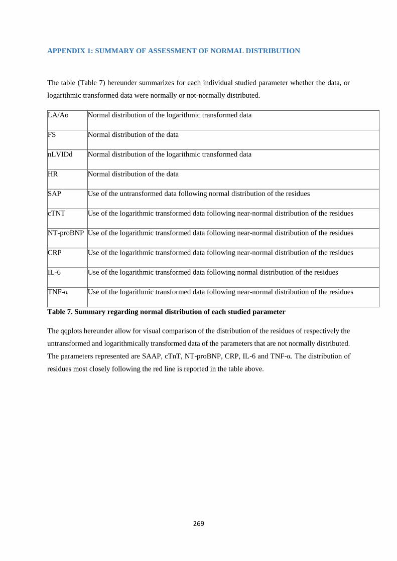
269
APPENDIX 1: SUMMARY OF ASSESSMENT OF NORMAL DISTRIBUTION
The table (Table 7) hereunder summarizes for each individual studied parameter whether the data, or
logarithmic transformed data were normally or not-normally distributed.
LA/Ao Normal distribution of the logarithmic transformed data
FS Normal distribution of the data
nLVIDd Normal distribution of the logarithmic transformed data
HR Normal distribution of the data
SAP Use of the untransformed data following normal distribution of the residues
cTNT Use of the logarithmic transformed data following near-normal distribution of the residues
NT-proBNP Use of the logarithmic transformed data following near-normal distribution of the residues
CRP Use of the logarithmic transformed data following near-normal distribution of the residues
IL-6 Use of the logarithmic transformed data following normal distribution of the residues
TNF-α Use of the logarithmic transformed data following near-normal distribution of the residues
Table 7. Summary regarding normal distribution of each studied parameter
The qqplots hereunder allow for visual comparison of the distribution of the residues of respectively the
untransformed and logarithmically transformed data of the parameters that are not normally distributed.
The parameters represented are SAAP, cTnT, NT-proBNP, CRP, IL-6 and TNF-α. The distribution of
residues most closely following the red line is reported in the table above.

270

271
Although several parameters failed to demonstrate normal distribution, the statistical model that was
used is considered strong enough to compensate for a moderate lack of perfectly normally distributed
data. As the QQ-plots demonstrate that residues of these values only diverge from the line at the lower
and higher extremes, the statistical model applied is therefore considered valid.

272

273
APPENDIX 2: CVC DIAMETER AND BASIC ECHOCARDIOGRAPHY BY NON-
CARDIOLOGIST VETERINARIANS FOLLOWING A 6-HOUR TRAINING COURSE
Elodie Darnis, DMV, University of Liège, Belgium
Anne Christine Merveille, DipECVIM-CA (cardiology), PhD,University of Liège, Belgium
Loïc Desquilbet, PhD, biostatistics and clinical epidemiology, Ecole nationale veterinaire d’Alfort,
Maisons-Alfort, France
Soren Boysen, DVM, DACVECC, University of Calgary, Canada
Kris Gommeren, DMV, DipECVIM-CA (internal medicine), University of Liège, Belgium
INTRO: Clinical parameters, including blood pressure, do not reliably predict intravascular volume
status. In human medicine, assessment of the inferior vena cava diameter (IVCD) and focused
echocardiographic parameters (La/Ao, LAminor, LVIDd, LVIDs, FS) have been used to rapidly evaluate
volume status and systolic function in critically ill patients. Recently, focused training courses in
echocardiography for human criticalists and internists have been described.
OBJECTIVE: This prospective, observational study aimed to quantify inter-observer (IEO agreements
between a cardiologist and 2 non-cardiologists who underwent a training course in echocardiography
for the ultrasonographic IVCD and focused echocardiographic parameters in healthy beagle dogs.
M&M: Two veterinary internists (one resident and one specialist), novice in echocardiography,
underwent a 6-hour echocardiography training course. One month later, 15 healthy beagle dogs were
examined 3 times by the two internists and one cardiologist. IVCD was assessed via a subxiphoid
window (IVC-SX) and a dorsolateral window (IVC-DL), caudal only to the last rib. Bland-Altman
analysis was used to assess IEO agreement between two series of clinical measurements; coefficients of
variation (CV) were calculated to quantify IEO variability.
RESULTS: The widest 95% limits of agreement (LOA) for LAminor, LVIDd, LVIDs, LA/Ao, and FS
were 5mm, 9mm, 5mm, 0.68, and 19%, and CV were 6%, 13%, 12%, 8%, and 17%, respectively.
For IVCD-SX, the 95% LOA for IVCDmin and IVCDmax were 0.68 cm and 0.05 cm with CV of 37%
respectively. For IVCD-LD, the 95% LOA were 0.34 cm with a CV of 11%.
DISCUSSION: Based on inter-observer reproducibility, minimal training in EC seems sufficient for
measurement of standard cardiac parameters. Evaluation of IVC-LD was considered good, based on

274
narrow 95% IOA. However, IVCD-SX was considered unacceptable. This may be due to variation in
measurements of the IVCD at the IVC-SX, and the effect of the respiratory cycle on the minimal and
maximal measurements. Standardization of the IVC-SX technique and investigation of the impact of the
respiratory phase on IVCD in dogs are needed.
CONCLUSION: A 6-hour training course in echocardiography seems sufficient to train non
cardiologist veterinarians to measure IVCD-LD and basic echocardiographic parameters in healthy
beagle dogs. Further studies are needed to determine whether IEO is acceptable with other breeds of
different body conformation. Values of these measurements to estimate the volume status in clinical
setting remain to be determined. IVCD-SX measurements require further standardization to allow for
quantitative analysis.
275
APPENDIX 3: EVECC – SCIL RESEARCH GRANT 2016
Applicant: Ms Elodie Darnis
Qualifications: DVM
Position: Resident in internal medicine
Institution: Liège University
Work Address:
Liège University,
Clinique des Petits Animaux
Quartier VALLEE 2
Avenue de Cureghem, 3
4000 Liège
Belgium
E-mail address: [email protected]
Tel: 0033645759874
Title of Project: Cardiac-FAST
Duration:
(max 12 months) 12 months
Lay Summary
Emergency and critical care patients often suffer from low blood pressure. Rapid and correct
assessment of these patients is required to initiate appropriate treatment and improve outcomes.
Hypotension has many underlying causes, and although many patients will require large volumes of
intravenous fluids to improve blood pressure, fluid requirements are not universal across all patients.
Furthermore end points of resuscitation and the fluid requirements needed to correct hypovolemia are
difficult to predict. In veterinary medicine, there is currently no readily available non-invasive technique
to monitor intravascular volume status and the subsequent need for fluid resuscitation. In human medicine,
measurement of caudal vena cava (CVC) diameter and the change in diameter between inspiration and
expiration, as well as the size of the left atrium accurately predicts the need for additional fluid therapy.
Evaluation of left atrial size is rarely performed in veterinary emergency and critical care (ECC), and
standardization of CVC diameter measurements and its collapsibility in dogs is lacking.
The goals of the current research project are to develop a standardized and objective method to
assess CVC diameter and collapsibility, and to determine reference ranges for different dog breeds and
sizes. Once a standardized technique and reference intervals have been established, this technique can then
be used in a clinical setting.
Co-applicant
name(s) & e-
mail address(es):
- Kris Gommeren, [email protected]
- Soren Boysen, [email protected]

276
Specific Aims
A pilot study was undertaken by two of the primary investigators (internal medicine resident and
head of the emergency and critical care department). Both investigators were trained by a cardiologist
in the imaging of the CVC via a subxiphoid transdiaphragmatic and a renal view obtained by placing the
probe directly caudal to the right costal arch with dogs positioned in right lateral recumbency. This pilot
study demonstrated good reproducibility and repeatability of imaging of the CVC in a colony of research
beagles.
The current study aims to describe reference ranges for CVC-related parameters such as maximal
and minimal diameter and collapsibility during inspiration and expiration in in a large variety of
spontaneously breathing healthy dogs from different breeds, weights, ages and sizes. The investigators
aim to describe a technique to assess these parameters, will assess intra and interobserver variability and
perform subgroup analysis to determine if breed or size impacts CVC diameter, or percentage change in
diameter measurement (the latter will form an index or ratio if it is consistent across different breeds).
In order to obtain sufficient data sets, a minimum of 120 healthy dogs will be required according
to established literature describing methodologies to establish reference intervals; these dogs will be
recruited at dog clubs and training centers and at dog shows. The investigators will go to central meeting
points at fixed dates to scan a maximum number of dogs and compare findings. Contact has already been
established with several dog clubs, training centers and show organizers, and 15 days of data collection
are anticipated to ensure an adequate number of healthy dogs are enrolled.
For the current phase of the study the hypothesis are; 1) that the CVC diameter can be measured
in dogs with minimal inter or intraobserver variability, 2) that changes in expiratory and inspiratory CVC
diameter in healthy dogs will be less than 60%, and 3) that although the CVC maximal diameter will
vary between breeds and size of dogs, the percentage change in CVC diameter at end inspiration and
expiration will be consistent across healthy dogs of different breeds and sizes.
Background
Echocardiography offers the benefits of direct visualization, allowing for real-time assessment
of cardiovascular structure and function1. Several human ICUs already have more than 15 years of
experience guiding the initial management of acute circulatory failure solely based on the use of
echocardiography, essentially replacing pulmonary artery catheters (PACs)2,3.
Over the last decade, interest in applying echocardiography within the ICU has greatly increased
in human medicine, leading to an increased availability of echography and echocardiography1,4,5, and the
incorporation of training programs for intensivists into some human ICU fellowships1.
Focused goal-oriented ultrasound training for human non-cardiologists involving as little as 3
hours of theoretical training and 5 hours of hands-on training have been described with positive results6.
These assessments allow the clinician to categorize the cause of shock and help direct therapy7. In
summary, echocardiography has found a place in human critical care, with the most common reason for
requesting an echocardiography being the assessment of volume status and left and/or right ventricular
function8. New echographic parameters to assess volume status have been developed in human patients.
Under controlled ventilation respiratory changes in the inferior vena cava (IVC) diameter are considered
the most useful echocardiographic parameters to assess fluid responsiveness9. Changes in intrathoracic
pressure throughout the respiratory cycle will impact the amount of blood in the vena cava. Changes in
the IVC diameter during respiration have been positively correlated with volume responsiveness in septic
shock10,11. Although spontaneously breathing patients are harder to evaluate, several parameters have
been suggested to evaluate volume responsiveness in these patients as well. An IVC diameter <1-2cm is
indicative of a low preload and volume responsiveness in hypotensive human patients12,13. In humans,
277
collapse of more than 50-60 % of the IVC between expiration and inspiration has a strong correlation
with decreased intravascular volume14,15. Assessment and monitoring of CVC size is however rarely
performed and lacks standardization in veterinary medicine.
How are the results likely to benefit pets?
If measurement of the CVC in dogs could be standardized, and similar changes in association
with respiration could be identified objectively, as has been described in humans, this methodology
would offer a huge amount of information for ECC-staff to more objectively assess the intravascular
volume in shock patients and could be tremendously helpful to monitor fluid responsiveness in the
critical care setting.
Experimental Design
The aim of this study is to define reference values for the evaluation of minimal and maximal
CVC size and CVC collapsibility during expiration and inspiration in spontaneously breathing healthy
dogs. The investigators aim to establish a reliable methodology to assess these parameters and compare
findings regardless of breed or size. If the percentage change in CVC change between expiration and
expiration is consistent across breeds it will serve to establish a repeatable index or ratio.
In order to achieve these goals, veterinarians will assess the CVC in a large variety of healthy dogs
from different breeds, weights, ages and sizes. Veterinarians will assess intra- and interobserver
repeatability by scanning each dog twice by two different investigators (4 scans in total per dog). In order
to obtain sufficient data sets, a minimum of 120 healthy dogs will be recruited at dog clubs and training
centers and at dog shows. Contacts have already been established with different dog training facilities and
dates are currently being fixed. The investigators will go to several central meeting points at fixed dates
to scan dogs and compare findings. Ethical approval for this part of the research project has already been
obtained from the universities ethical committee (14-1749)
In order to be included, dogs should be in between 1 and 8 years old, and present no abnormalities
on physical examination. Dogs will be excluded if they suffer from any known disease or were treated for
any disease (e.g. vomiting and diarrhea) within the prior month; whenever a murmur, pulse deficit,
arrhythmia other than sinus arrhythmia or any other abnormality is detected on cardiac examination;
whenever respiratory abnormalities are detected; or whenever the dog is considered to be severely
dehydrated on clinical examination. If the work performed in this part of the study has positive results, the
next phase would be to correlate these findings with those obtained in critically ill dogs.

278
Has funding been applied for elsewhere?
No, the SCIL research grant is the only funding that currently has been applied for.
279
Appendix: Literature
1. Vieillard-Baron A, Slama M, Cholley B, et al. Echocardiography in the intensive care unit:
from evolution to revolution? Intensive Care Med 2008;34:243-249.
2. Vieillard-Baron A, Prin S, Chergui K, et al. Hemodynamic instability in sepsis: bedside
assessment by Doppler echocardiography. Am J Respir Crit Care Med 2003;168:1270-1276.
3. Vieillard-Baron A, Prin S, Chergui K, et al. Echo-Doppler demonstration of acute cor
pulmonale at the bedside in the medical intensive care unit. Am J Respir Crit Care Med 2002;166:1310-
1319.
4. Beaulieu Y. Bedside echocardiography in the assessment of the critically ill. Crit Care Med
2007;35:S235-249.
5. Levitov A, Mayo PH, Slonim AD. Critical care ultrasonography. New York: McGraw Hill;
2009.
6. Vignon P, Dugard A, Abraham J, et al. Focused training for goal-oriented hand-held
echocardiography performed by noncardiologist residents in the intensive care unit. Intensive Care Med
2007;33:1795-1799.
7. Kaplan A, Mayo PH. Echocardiography performed by the pulmonary/critical care medicine
physician. Chest 2009;135:529-535.
8. Price S, Nicol E, Gibson D, et al. Echocardiography in the critically ill: current and potential
roles. Intensive Care Med 2006;32:48-59.
9. Charron C, Caille V, Jardin F, et al. Echocardiographic measurement of fluid responsiveness.
Curr Opin Crit Care 2006;12:249-254.
10. Feissel M, Michard F, Faller JP, et al. The respiratory variation in inferior vena cava
diameter as a guide to fluid therapy. Intensive Care Med 2004;30:1834-1837.
11. Barbier C, Loubieres Y, Schmit C, et al. Respiratory changes in inferior vena cava diameter
are helpful in predicting fluid responsiveness in ventilated septic patients. Intensive Care Med
2004;30:1740-1746.
12. Sefidbakht S, Assadsangabi R, Abbasi HR, et al. Sonographic measurement of the inferior
vena cava as a predictor of shock in trauma patients. Emerg Radiol 2007;14:181-185.
13. Yanagawa Y, Sakamoto T, Okada Y. Hypovolemic shock evaluated by sonographic
measurement of the inferior vena cava during resuscitation in trauma patients. J Trauma 2007;63:1245-
1248; discussion 1248.
14. Nagdev AD, Merchant RC, Tirado-Gonzalez A, et al. Emergency department bedside
ultrasonographic measurement of the caval index for noninvasive determination of low central venous
pressure. Ann Emerg Med 2010;55:290-295.
15. Stawicki SP, Braslow BM, Panebianco NL, et al. Intensivist use of hand-carried
ultrasonography to measure IVC collapsibility in estimating intravascular volume status: correlations
with CVP. J Am Coll Surg 2009;209:55-61.
Related Documents