Abusive Head Trauma in Children WWW.RN.ORG® Reviewed December, 2015, Expires December, 2017 Provider Information and Specifics available on our Website Unauthorized Distribution Prohibited ©2015 RN.ORG®, S.A., RN.ORG®, LLC By Wanda Lockwood, RN, BA, MA The purpose of this course is to explain the pathophysiology of abusive head trauma and outline common injuries, assessment and treatment options, and injuries typical of accidental injuries. Upon completion of this course, the healthcare provider should be able to: Explain the difference among the terms shaken baby syndrome, shaken impact syndrome, and abusive head trauma. Discuss incidence of abusive head injury. Discuss at least 5 factors that make infants and young children more susceptible to head injury. Discuss 4 secondary injuries that can occur with abusive head injury. Explain implications of retinal hemorrhage and 5 causes of retinal hemorrhage unrelated to abusive head injury. Discuss scalp injury/laceration, contusion, concussion, and skull fractures. Discuss subarachnoid hemorrhage, IVH, and epidural and subdural hematoma. Discuss diffuse axonal injury and head-injury associated seizures. Explain the ABCs of assessment for abusive head injury. Explain the differences in symptoms of increased intracranial pressure (ICP) in infants and children and list late signs. Explain normal values for ICP for children from infancy to 18 years. Discuss at least 5 types of ICP monitoring. Discuss cerebral perfusion pressure (CPP) monitoring. Goals Purpose

Welcome message from author
This document is posted to help you gain knowledge. Please leave a comment to let me know what you think about it! Share it to your friends and learn new things together.
Transcript

Abusive Head Trauma in Children WWW.RN.ORG®
Reviewed December, 2015, Expires December, 2017 Provider Information and Specifics available on our Website
Unauthorized Distribution Prohibited
©2015 RN.ORG®, S.A., RN.ORG®, LLC By Wanda Lockwood, RN, BA, MA
The purpose of this course is to explain the
pathophysiology of abusive head trauma and outline common injuries, assessment and treatment options, and
injuries typical of accidental injuries.
Upon completion of this course, the healthcare provider
should be able to:
Explain the difference among the terms shaken baby syndrome, shaken impact syndrome, and abusive head trauma.
Discuss incidence of abusive head injury. Discuss at least 5 factors that make infants and young children
more susceptible to head injury. Discuss 4 secondary injuries that can occur with abusive head
injury. Explain implications of retinal hemorrhage and 5 causes of
retinal hemorrhage unrelated to abusive head injury. Discuss scalp injury/laceration, contusion, concussion, and skull
fractures. Discuss subarachnoid hemorrhage, IVH, and epidural and
subdural hematoma.
Discuss diffuse axonal injury and head-injury associated seizures.
Explain the ABCs of assessment for abusive head injury. Explain the differences in symptoms of increased intracranial
pressure (ICP) in infants and children and list late signs. Explain normal values for ICP for children from infancy to 18
years. Discuss at least 5 types of ICP monitoring.
Discuss cerebral perfusion pressure (CPP) monitoring.
Goals
Purpose

Explain the differences between mannitol and hypertonic saline
solutions. Discuss the use of neuromuscular blocking agents,
hyperventilation, hypothermia, barbiturate-induced coma, corticosteroids, anticonvulsants, and positioning.
Discuss 2 methods of CSF decompression. Discuss indications for decompressive craniectomy.
Discuss the Glasgow coma scale, including scoring. Discuss injuries consistent with accidents related to various
types of falls.
Introduction People rarely admit to abusing infants and children. Instead, they invent “accidents,” such as the child’s falling out of a crib or highchair,
falling down the stairs, or being hit by other children. Unless head injuries are probable, such as with an unconscious child, healthcare
providers may overlook signs and symptoms of abusive head injury.
However, there are patterns of injuries that are suggestive of child abuse, and the healthcare provider should be aware of the difference
between typical injuries associated with accidents and those associated with abuse.
The term “shaken baby syndrome” has been commonly used to
describe brain injury symptoms consistent with a person vigorously
shaking an infant or small child. The injuries most consistent with shaken baby syndrome include unilateral or bilateral subdural
hemorrhage, bilateral retinal hemorrhages, and diffuse brain injury.
However, while children can be injured by shaking alone, authorities often find injuries that exceed those just from shaking and include
evidence of blunt trauma, so a more inclusive term, “shaken impact syndrome” is frequently used. An even broader term, “abusive head
trauma,” includes not only injuries associated with shaking and direct blunt trauma but also with dropping or throwing a child.
“Abusive head trauma” is now the term recommended by the
American Academy of Pediatrics. Because this term describes the type of injury rather than the mechanism of injury (such as shaking), the
use of “abusive head injury” may have legal significance as the specific
means of injury can rarely be ascertained, so indicating a child has “shaken baby syndrome” may preclude evidence of other types of
injuries and allow for more challenge in court.

Most victims of abusive head trauma are less than a year old, typically between 3 and 8 months, but these characteristic injuries can occur in
children up to 5 years of age or older. In most cases, the perpetrator is a parent or caregiver, with 65% to 90% being male, usually the
father or mother’s boyfriend. The National Center for Shaken Baby Syndrome (also now using the term abusive head injury) estimates
that approximately 1200 to 1400 children each year are injured or killed by abusive head injuries in the United States.
Abusive head trauma is the leading cause of death and disability in
infants and young children, resulting from child abuse. Child abuse has been identified as the cause of brain injuries in 24% of children <2
and suspected as the cause in an additional 32%.
Traumatic injury
The infant and young child are more susceptible to head injury than
older children or adults because of a combination of factors:
The head is large in proportion to the rest of the body, so
children tend to land headfirst when they fall. The child’s brain has a higher water content (88%) than adults
(77%), so the brain is softer and more likely to experience injury from acceleration-deceleration injuries.
The head and neck are unstable with support more from ligamentous structures than bony structures.
The unmyelinated brain is more likely to have shear injuries. The skull is easily deformed and presses in on brain tissue when
impacted with most injuries coup rather than contrecoup (as is
common in adults). Axons are disrupted because of shearing of long white matter
tracts with acceleration-deceleration injuries, leading to cell death.

When a child is shaken, struck in the head, or thrown against a wall,
the child suffers not only the primary injuries (hemorrhage, skull fracture, contusion), but also a number of secondary injuries:
Inflammatory biomolecular changes lead to interruptions in the
microcirculation of the brain and disintegration of neurons. Pathophysiological changes caused by the primary injury result
in increased ICP, hyperemia, and ischemia. Cerebral blood flow is impacted because of increased ICP.
Autoregulation of cerebral blood flow is impaired.
Retinal hemorrhage is one of the cardinal signs of abusive head trauma. Numerous
studies have been conducted to examine the relationship between various types of head trauma and retinal
hemorrhage. These studies clearly show that retinal hemorrhage is most common in infants and children who have suffered abusive head
trauma and should trigger further examination of the child for other types of injuries.
One retrospective study of 811 pediatric patients with traumatic injury
found that 27 had retinal hemorrhages with 22 believed associated
with abuse, 1 with a motor vehicle accident, and 4 with falls. Another study of 100 children <24 months with head injuries found 10 cases of
retinal hemorrhage with 9 associated with probable abusive head trauma and 1 associated with motor vehicle accident. Study after
study supports basically the same type of findings. Additionally, bilateral hemorrhages are associated with higher morbidity.
While retinal hemorrhage can occur without abusive head trauma,
retinal hemorrhage is uncommon. However, the healthcare provider must always consider other possibilities. Other causes of retinal
hemorrhage include:
Birth trauma: Retinal hemorrhage occurs in about a third of neonates but usually resolves within 2 weeks. The lowest
incidence is with Caesarean. Motor vehicle accident: The degree of head trauma may be
similar to abusive head injury, resulting in retinal hemorrhage, but some studies were completed before universal use of
restraints for children and placing of children in the back seat, so current incidence is less clear.
Retinal hemorrhage
Fall: Most falls do not cause retinal hemorrhages, but this
depends on the age, height of fall, site of impact, and other factors.
Drowning: Retinal hemorrhages are sometimes found in drowning victims.
Disease: Retinal hemorrhage has been found with various conditions, such as disseminated intravascular coagulopathy
(DIC) and AVM.
Lacerations commonly occur in abusive head trauma,
and—especially in small infants—the bleeding associated with the injury to the scalp may pose a
risk of hypovolemia. The area under a scalp injury must be examined carefully as underlying injuries, such as skull
fracture or cerebral extrusion, may be present.
With direct impact to the head, bruising and
tearing of brain tissue can occur, especially in the temporal and frontal areas. Bruising and
tearing of structures and vessels can occur throughout the brain, disrupting function. Contusions are generally localized and
macroscopic. Symptoms can include deterioration of neurological status secondary to cerebral edema, infarcts, or developing
hematomas.
Acceleration-deceleration injuries occur when a blow to the head
causes the skull to change shape and push against the brain, causing to move sharply backward, striking the opposite side of the skull.
Bruising may result from the point of impact (coup), the most common in children, or the opposite side (contrecoup). In some
cases, blood may pool in areas and result in pressure and distortion on the brain.
Concussion is the most frequent head injury and may
result in temporary impairment of function and cerebral blood flow but does not result in structural
abnormalities and outcome is generally good. Concussion is more widespread than contusion. Pooling of blood is not characteristic, and
bleeding is more likely to be microscopic. However, a child may have
BOTH a concussion as well as contusions.
Concussion
Scalp injury/
laceration
Cerebral contusion

Symptoms of concussion include changes in level of consciousness,
loss of muscle tone, reflexes, pupillary abnormalities, cortical blindness, heart rate abnormalities, nausea, vomiting, lethargy, slurred speech,
and confusion.
Secondary seizures are more common in infants and young children, often associated with delayed somnolence and vomiting, while older
children are more prone to posttraumatic amnesia. Older children also commonly have variations in mental status with alterations coming
and going.
Skull fractures Skull fractures may or not be present with abusive head trauma. Studies show that skull fractures occur in about 5-15%
of children with mild head injuries but in 30-70% of head injuries associated with child abuse. Most skull fractures in children are linear
and diastatic (separated) so they are clearly seen on radiograph. Skull
fractures often occur with lacerations. Any evidence of CSF warrants further exploration for damage to the dura. Depressed fractures may
cause dural or cortical lacerations.
Basal skull fractures result from a blow to the back of the head (occurring in up to 14% of children with head trauma). Signs include:
Battle
sign
Bruising over mastoid process:
Raccoon eyes
Periorbital ecchymosis:

Ocular nerve entrapment and CSH drainage from the nose or ear may also occur. Symptoms include extended nausea, vomiting, and
general malaise.
Another type of skull fracture that may occur with severe injuries to
infants, usually <12 months, is the cranial burst skull fracture. This fracture is a closed diastatic skull fracture with cerebral extrusion
under the scalp. It is characterized by severe swelling of the scalp in the area of extrusion.
SAH is the most common type of hemorrhage associated with head
trauma, occurring when small vessels in the cerebral cortex are disrupted, with the hemorrhage most often
occurring along the falx cerebri or tentorium and the outer cortical surface. Symptoms typically include restlessness, nausea, vomiting,
headache, fever, and nuchal rigidity.
Subarachnoid hemorrhage

Epidural hematoma occurs if an artery or
vein is lacerated. The size of the hematoma associated with arterial laceration usually peaks at 6-8 hours and
those associated with venous laceration at 24 hours.
In some cases, a skull fracture may overlay the hematoma, most commonly occurring in the
temporal, frontal, or occipital lobes. The classic periods of lucidity between initial loss of
consciousness and neurological deterioration, common in adults, is less likely to occur in children.
Symptoms can include loss of consciousness, hemiparesis, pupillary changes, and posturing.
Subdural hematoma may occur as a result
of birth trauma but is also common with
shaken baby syndrome when the veins that bridge the dura are torn or acceleration-deceleration forces tear
cortical arteries. If related to birth trauma, symptoms (seizures, tense fontanel, anisocoria, and respiratory distress)
usually occur within 12 hours.
Subdural hematoma
Epidural hematoma
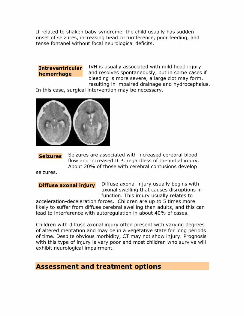
If related to shaken baby syndrome, the child usually has sudden
onset of seizures, increasing head circumference, poor feeding, and tense fontanel without focal neurological deficits.
IVH is usually associated with mild head injury
and resolves spontaneously, but in some cases if bleeding is more severe, a large clot may form,
resulting in impaired drainage and hydrocephalus. In this case, surgical intervention may be necessary.
Seizures are associated with increased cerebral blood flow and increased ICP, regardless of the initial injury.
About 20% of those with cerebral contusions develop seizures.
Diffuse axonal injury usually begins with
axonal swelling that causes disruptions in function. This injury usually relates to
acceleration-deceleration forces. Children are up to 5 times more likely to suffer from diffuse cerebral swelling than adults, and this can
lead to interference with autoregulation in about 40% of cases.
Children with diffuse axonal injury often present with varying degrees
of altered mentation and may be in a vegetative state for long periods of time. Despite obvious morbidity, CT may not show injury. Prognosis
with this type of injury is very poor and most children who survive will exhibit neurological impairment.
Assessment and treatment options
Diffuse axonal injury
Seizures
Intraventricular
hemorrhage

One of the problems with providing treatment to children who suffer
from abusive head injury is that often only those with severe injuries that result in death or coma are brought to the attention of healthcare
providers because abusers want to hide children’s injuries. Thus, many undiagnosed abused children suffer long-term effects, such as
learning disabilities or hearing or vision impairment.
A wide range of treatment options are available for those children brought to healthcare providers for care, depending on the severity of
injury, levels of ICP, and response to initial interventions.
As with most emergency interventions, the first
step is to ensure ABCDEs (airway, breathing, circulation, dysfunction/disability, and external exam.
ABCDEs
Airway Examine the airway for obstructions, such as loose
teeth, foreign bodies. Lacerations and bone instability may be obstructive. Examine the trachea for deviation
and observe for signs of circumoral cyanosis (sign of
hypoxia). Auscultate the airway and listen for turbulence.
Breathing Hypoventilation and apnea are common with severe head injuries and require immediate
intubation. Note indications of Cushing’s triad (bradycardia,
hypertension, abnormal respirations), which indicate herniation. Evaluate possible causes for
hypotension. Hypotension found with bradycardia often indicates spinal cord injury.
Cheyne-Stokes respirations are associated with
damage to cerebral hemispheres or diencephalon. Hyperventilation is associated with damage to
rostral brain stem or tegmentum.
Circulation Note blood pressure, pulse, temperature, color, and
indications of cyanosis (circumoral, peripheral).
Dysfunction/ disability
Assess responsiveness with alert, verbal, pain, unresponsive (AVPU) system and Glasgow Coma
Scale (using the pediatric version for those <5). Assess pupillary size and responsiveness:
o Ipsilateral dilatation with no response to light
Initial emergency treatment
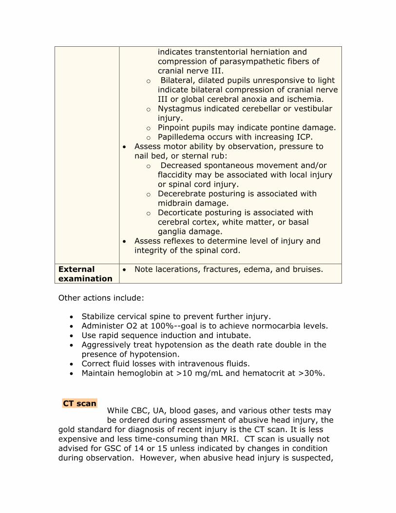
indicates transtentorial herniation and
compression of parasympathetic fibers of cranial nerve III.
o Bilateral, dilated pupils unresponsive to light indicate bilateral compression of cranial nerve
III or global cerebral anoxia and ischemia. o Nystagmus indicated cerebellar or vestibular
injury. o Pinpoint pupils may indicate pontine damage.
o Papilledema occurs with increasing ICP. Assess motor ability by observation, pressure to
nail bed, or sternal rub:
o Decreased spontaneous movement and/or flaccidity may be associated with local injury
or spinal cord injury. o Decerebrate posturing is associated with
midbrain damage. o Decorticate posturing is associated with
cerebral cortex, white matter, or basal ganglia damage.
Assess reflexes to determine level of injury and integrity of the spinal cord.
External examination
Note lacerations, fractures, edema, and bruises.
Other actions include:
Stabilize cervical spine to prevent further injury. Administer O2 at 100%--goal is to achieve normocarbia levels.
Use rapid sequence induction and intubate. Aggressively treat hypotension as the death rate double in the
presence of hypotension. Correct fluid losses with intravenous fluids.
Maintain hemoglobin at >10 mg/mL and hematocrit at >30%.
While CBC, UA, blood gases, and various other tests may
be ordered during assessment of abusive head injury, the gold standard for diagnosis of recent injury is the CT scan. It is less
expensive and less time-consuming than MRI. CT scan is usually not advised for GSC of 14 or 15 unless indicated by changes in condition
during observation. However, when abusive head injury is suspected,
CT scan
CTs are usually taken because the caregiver’s history of injury may not
be reliable.
While CT scans are relatively safe, they do pose a risk of future malignancy because children are more susceptible to ionizing radiation
than adults, especially with repeated CTs, so unnecessary CTs should be avoided. A recent study indicates that a period of observation for
those with mild to moderate head trauma cuts use of CT by approximately 50%.
Intracranial pressure is that within the skull, in the tissue of the brain and
cerebrospinal fluid. An increase in intracranial pressure occurs secondary to
other injuries. Symptoms of increased intracranial pressure in infants
and children include:
Infants: Fontanels bulge and normal pulsations absent, scalp veins distend, and head circumference increases. Poor feeding is
common and infants may appear irritable and cry in a high-pitched tone.
Children: Symptoms are more typical with headache, vomiting
(but without nausea), diplopia, blurred vision, and seizures. Children may exhibit behavioral and personality changes with
increased lethargy, inability to follow directions, and evidence of amnesia and/or memory loss.
Late signs: Further decrease in level of consciousness, motor
response, and response to painful stimuli. Pupillary changes
occur, and posturing (decerebrate or decorticate), respiratory depression with Cheyne-Stokes and papilledema. Cushing’s triad
may be evident: o Increased systolic pressure associated with widened pulse
pressure. o Bradycardia.
o Decreased respirations.
Adequate ICP is essential in order to maintain cerebral perfusion pressure and prevent ischemia and avoid cerebral herniation.
Additionally, ICP monitoring allows for early identification of
intracerebral hemorrhage. ICP monitoring is usually indicated for those with GSC 5 or GSC <8 with abnormal CT or with other symptoms,
Intracranial Pressure monitoring

such as posturing or systemic hypotension, that are of concern. ICP
monitoring is usually also recommended for children who are unconscious or in shock or exhibit deterioration in neurological
functioning.
Studies have been somewhat inconsistent, and some are small, but they generally indicate that ICP <20 mm Hg is rarely associated with
death while ICP of 20-40 is associated with up to 28% death rate and 40-60 with almost 100% death rate. Although critical values
associated with various physiological variables have not been determined, the goal is usually to maintain the ICP <20.
Normal ICP values
Infant 1.5-6 mm Hg.
Toddler, young child 3-7 mm Hg.
Older child 2-7 mm Hg.
18 years 15 mm Hg.
It’s important to remember that in a fluid-filled space, the pressure is
usually the same wherever it’s checked; however, with brain injury, various interfering factors, such as decreased CSF because of brain
swelling, may interfere, so that the pressure obtained by the catheter or sensor may reflect only the pressure at that site rather than
pressure within the ventricular system (the site of the most accurate CSF pressure).
ICP can be monitored in
a number of different ways, with each method
associated with advantages and
disadvantages.
According to the Monroe-Kellie hypothesis, in order to maintain ICP
within normal range, a change in the volume of one compartment
requires compensatory change in the volume of another compartment.
The three components are 1) brain tissue, 2) cerebrospinal fluid (CSF), and 3) blood. Since tissue cannot easily accommodate change,
medical interventions must focus on blood flow and drainage of CSF.
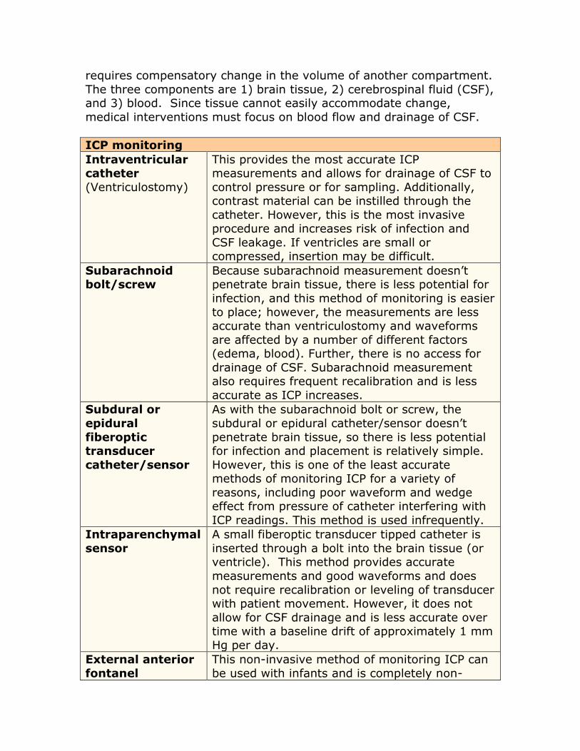
ICP monitoring
Intraventricular catheter
(Ventriculostomy)
This provides the most accurate ICP measurements and allows for drainage of CSF to
control pressure or for sampling. Additionally, contrast material can be instilled through the
catheter. However, this is the most invasive procedure and increases risk of infection and
CSF leakage. If ventricles are small or compressed, insertion may be difficult.
Subarachnoid bolt/screw
Because subarachnoid measurement doesn’t penetrate brain tissue, there is less potential for
infection, and this method of monitoring is easier
to place; however, the measurements are less accurate than ventriculostomy and waveforms
are affected by a number of different factors (edema, blood). Further, there is no access for
drainage of CSF. Subarachnoid measurement also requires frequent recalibration and is less
accurate as ICP increases.
Subdural or
epidural
fiberoptic transducer
catheter/sensor
As with the subarachnoid bolt or screw, the
subdural or epidural catheter/sensor doesn’t
penetrate brain tissue, so there is less potential for infection and placement is relatively simple.
However, this is one of the least accurate methods of monitoring ICP for a variety of
reasons, including poor waveform and wedge effect from pressure of catheter interfering with
ICP readings. This method is used infrequently.
Intraparenchymal
sensor
A small fiberoptic transducer tipped catheter is
inserted through a bolt into the brain tissue (or ventricle). This method provides accurate
measurements and good waveforms and does
not require recalibration or leveling of transducer with patient movement. However, it does not
allow for CSF drainage and is less accurate over time with a baseline drift of approximately 1 mm
Hg per day.
External anterior
fontanel
This non-invasive method of monitoring ICP can
be used with infants and is completely non-

tonometer invasive, but it can be less accurate and does
not allow for CSF drainage. The external anterior fontanel tonometer should only be used if there
is no edema of the scalp and skin turgor is normal.
Cerebral perfusion pressure is based on
mean arterial pressure (MAP) and intracranial pressure (ICP):
CPP = MAP – ICP.
Cerebral profusion pressure is the pressure controlling blood flow to the brain and must be maintained within narrow limits as too high
results in increased intracranial pressure and too low in ischemia. Cerebral blood flow is usually maintained at a constant and adequate
level with a mean arterial pressure between 60 and 150 mm Hg and ICP of about 10.
An increase in mean arterial pressure (MAP) increases the CPP while
an increase in ICP lowers the CPP, so monitoring ICP is especially critical. CPP may be decreased by vasospasms associated with the
brain injury as well as local pressure from intracranial hemorrhage or concussion.
While studies vary and definitive guidelines have not been established
for different pediatric age groups, generally CPP levels should be
maintained to at least 60 mm Hg in children (compared to 70-90 in adults). Mortality rates are significant with CPP <40 and survival is
high with CPP >40.
NMBAs, such as propofol, have been used in some studies to determine if they can reduce ICP
and improve outcomes. However, while NMBAs reduce oxygen consumption by about 10%, the FDA does not
recommend the use of NMBAs in pediatric patients with severe brain injury because of the potential for severe adverse effects and
inadequate evidence of benefit.
One method of decreasing increased
Cerebral perfusion pressure monitoring
Neuromuscular
blocking agents
CSF decompression

intracranial pressure is to drain cerebrospinal fluid. This can be done
through external ventricular drainage (EVD) per the ventriculostomy or through lumbar drainage or a combination. Lumbar drainage can lead
to tonsillar herniation.
CSF drainage is recommended if increased ICP is refractory to other treatments either through EVD by itself or a combination of EVD and
lumbar drainage.
Studies indicate that hypertonic saline solution
(HSS) or mannitol are both effective in reducing ICP. Hyperosmolar solutions function to reduce
cerebral edema through osmosis, improving circulation. HSS further expands plasma in order to increase CPP and reduces the
hyponatremia that results from brain injury and is one of the causes of
increased ICP.
One study showed that mannitol reduced ICP by >10% after 116 boluses, and other studies show benefit of 3% HSS. At present, either
mannitol or HSS is recommended as first-line hyperosmolar agent.
Hyperosmolar agents
HSS Hypertonic saline solutions has a sodium concentration that exceeds that of NS (0.9%). While concentrations
range from 2-23.4%, 3% solutions have been used in most pediatric studies.
Peripheral lines can only be used for solutions <3%, so
solutions with concentrations of 3% require central lines.
HSS is usually given continuously at varying rates (age-
dependent, at about 5 mL/kg per dose. In the event of severely increased pressure or transtentorial herniation,
boluses of higher concentrations may be given over 15 minute periods.
Monitoring for hypovolemia is critical as this can increase risk of renal failure. Serum sodium (normal value 145-155
mmol/L) most be monitored every 6 hours and serum osmolality (normal value 320 mOsmol/L) every 12 hours.
Mannitol Mannitol, an osmotic diuretic, decreases ICP and reduces cerebral edema by increasing sodium and fluid excretion.
Mannitol 15% to 25% solution is given per intravenous infusion at 0.25 to 2 g/kg, depending on age, over 30-60
Hyperosmolar solutions
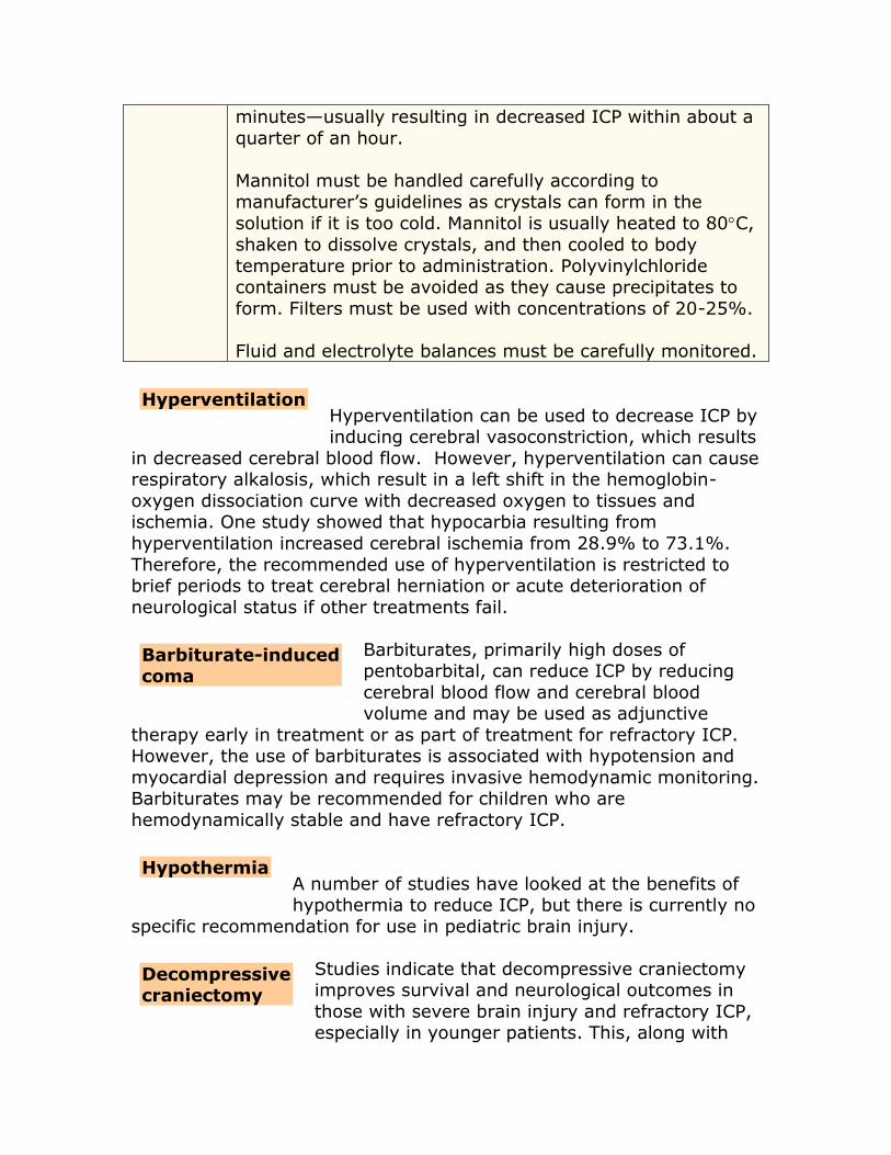
minutes—usually resulting in decreased ICP within about a
quarter of an hour.
Mannitol must be handled carefully according to manufacturer’s guidelines as crystals can form in the
solution if it is too cold. Mannitol is usually heated to 80C,
shaken to dissolve crystals, and then cooled to body
temperature prior to administration. Polyvinylchloride containers must be avoided as they cause precipitates to
form. Filters must be used with concentrations of 20-25%.
Fluid and electrolyte balances must be carefully monitored.
Hyperventilation can be used to decrease ICP by inducing cerebral vasoconstriction, which results
in decreased cerebral blood flow. However, hyperventilation can cause respiratory alkalosis, which result in a left shift in the hemoglobin-
oxygen dissociation curve with decreased oxygen to tissues and ischemia. One study showed that hypocarbia resulting from
hyperventilation increased cerebral ischemia from 28.9% to 73.1%.
Therefore, the recommended use of hyperventilation is restricted to brief periods to treat cerebral herniation or acute deterioration of
neurological status if other treatments fail.
Barbiturates, primarily high doses of pentobarbital, can reduce ICP by reducing
cerebral blood flow and cerebral blood volume and may be used as adjunctive
therapy early in treatment or as part of treatment for refractory ICP. However, the use of barbiturates is associated with hypotension and
myocardial depression and requires invasive hemodynamic monitoring. Barbiturates may be recommended for children who are
hemodynamically stable and have refractory ICP.
A number of studies have looked at the benefits of hypothermia to reduce ICP, but there is currently no
specific recommendation for use in pediatric brain injury.
Studies indicate that decompressive craniectomy improves survival and neurological outcomes in
those with severe brain injury and refractory ICP, especially in younger patients. This, along with
Hyperventilation
Barbiturate-induced coma
Hypothermia
Decompressive craniectomy

barbiturate-induced coma, is a second tier therapy. A bifrontal
craniectomy is most common, with studies showing this procedure resolved refractory increased ICP in about 44%.
Decompression is indicated for rapidly expanding epidural or subdural
hematoma. Subdural hematoma associated with midline shift or altered mental status requires emergency drainage although smaller
hematomas with no midline shift or indications of increasing pressure may be managed conservatively.
Depressed skull fractures must be elevated surgically if they depress
>1 cm, if the depression is thicker than the calvaria (skull cap), or if fragments are resulting in compression of brain tissue.
Complications can include brain shift from over drainage of CSF and
bone resorption up to 50%. Additionally, bone flap storage for
children can pose challenges.
Corticosteroids have been used for a wide range
of neurological disorders and reduce cerebral edema; however, they increase risk of infection
and gastrointestinal bleeding, and studies show corticosteroids do not make a difference in outcomes. Therefore, they are not recommended
for use with pediatric brain injuries.
Anticonvulsants have been used both to treat post-traumatic seizures (PTS) and as prophylaxis
to prevent them. Studies have varied in outcomes with some showing reduction in early PTS and improved
survival rates and others showing no reduction in late PTS. Current
recommendations are that anticonvulsants are not recommended as prophylaxis to prevent late PTS in pediatric patients. However, if
seizures are present (as may be the case with severe injury), initial treatment is usually with a short-acting benzodiazepine, such as
lorazepam or diazepam, followed by maintenance with phenytoin or phenobarbitol.
There is no consensus regarding positioning, but
elevating the head to 30-45 may show a small
improvement in ICP; however, elevation must be
avoided with hypotension, and some ICP monitoring may be affected.
Corticosteroids
Anticonvulsants
Positioning

Many authorities recommend elevating the head to 30 and
maintaining the head in midline position to increase venous drainage and decrease the ICP without impairing cerebral blood flow.
While protocols may vary somewhat, that
outlined by Critical Care Medicine is fairly standard:
CT is indicated with GS 8 or with lesion, diffuse or focal
swelling.
Insertion of EVD or ICP monitor. First tier interventions:
o CSF diversion; EVD at 0-3 cm above tragus.
o Sedation and/or paralysis. o Elevate HOB to 30,
o Maintain paCO2 at 35-38 m hg. o Maintain O2 saturation 92%
If inadequate response from first tier intervention, repeat CT. Second tier interventions:
o Mild hyperventilation (paCO2 30-35 mm Hg). o Mannitol (0.25-1.0 g/kg IV q 4-6 hours) with serum
osmolarity <320. o HSS 3% boluses or slow infusion with serum osmolarity
<370. o Low dose barbiturates (5 mg/kg/IV q 6 hrs.) with
escalation to burst suppressed, barbiturate coma). If no response to second tier interventions, repeat CT.
Third tier interventions:
o Decompressive craniectomy or temporal lobectomy. o Moderate hypothermia (32-33C).
o Transient hyperventilation to PaCO1 < 30 mm Hg)
Glasgow coma scale and AVPU
The Glasgow coma scale (GCS) is a measure of the depth and
duration of coma or impaired level of consciousness and is used for postoperative and neurological injury assessment. The GCS measures
best eye response, best verbal response, and best motor response. The same scale used for older children and adults is used with slight
modifications (indicated in parentheses) for infants and young children
(5).
Protocol example

Glasgow coma scale
Eye opening
4: Spontaneous. 3: To verbal stimuli (shout for infants <1 year)
2: To pain [not of face]. 1: No response.
Verbal 5: Oriented. (Infant/child: Smiles, exhibits appropriate
interactions). 4: Conversation confused, but can answer questions.
(Infant/child: crying but consolable) 3: Uses inappropriate words. (Infant/child: Moaning,
crying or screaming to pain, sometimes inconsolable.) 2: Speech incomprehensible. (Infant/child:
Inconsolable, agitated, grunts or moans to pain). 1: No response.
Motor 6: Moves on command. (Infant/child: Moves
spontaneously or with purpose). 5: Moves purposefully to respond to pain.
4: Withdraws in response to pain. 3: Decorticate posturing [flexion] in response to pain.
2: Decerebrate posturing [extension] in response to pain.
1: No response.
The total possible scores range from 3 to 15, with lower scores
indicating increasing morbidity. Injuries and/or conditions are classified according to the total score:
3-8 Coma
8 Severe head injury
9-12 Moderate head injury
13-15 Mild head injury.
The AVPU assessment is a quick assessment done to determine the patient’s level of consciousness:
AVPU
A Alert and awake, aware of
person, place, time, and condition.
Yes No
V Responds to verbal stimuli. Yes No
P Responds to painful stimuli but not verbal.
Yes No
U Unconscious, does to respond Yes No
AVPU
to painful or verbal stimuli.
Those who are not responding to painful stimuli (P) usually have an
equivalent score of about 8 on the GSC, consistent with severe injury.
Injuries consistent with accidental falls
While children can develop serious head injuries from falls, the vast majority of accidental falls do not cause serious head trauma, so
almost any child presenting with severe injuries or multiple injuries related to a fall should be examined further for evidence of abuse:
Falls from bed (2-4 feet): Injuries are usually minor although a
small number may have a skull or other fracture, such as a clavicle. Severe head trauma is rare and should be suspect. The
degree of injury may depend on the type of flooring, as carpeting is associated with lower levels of injury.
Falls from walkers: Most injuries result from falling down stairs in the walker rather than out of the walker onto the floor
although falling onto a concrete floor increases risk. Studies show about 1-8% may develop intracranial bleeds.
Falls down stairs: Injuries are less severe in free falls than if someone was holding and dropped the child down the stairs.
Single injuries to the head or extremities predominate when children fall down stairs. Multiple injuries are not common, and
stairway injuries are generally less severe than other free falls of the same total distance.
Falls from various heights: Short vertical falls (4 feet) rarely
cause severe head trauma or multiple injuries unless associated
with abuse. One study of deaths in children related to minor falls found that those who died after a fall of 1-4 feet had evidence of
other injuries consistent with abuse. Another study looked at

children who died after falls of 5-6 feet and found that 74% had
evidence of abuse.
Even falls from 10 feet or higher rarely result in death unless the
height is extreme, such as from a 2nd or 3rd floor. The greater the height is, the greater the incidence of skull or other fractures.
A study of 75,000 cases from the US Consumer Product Safety Commission found that only 18 suffered fall-related injury deaths
over 11 years, with 7 falling from swing and 11 from a horizontal surface, such as a ladder.
Conclusion Healthcare providers often have ethical concerns about the legal and social implications of a diagnosis of abusive head trauma, but
healthcare providers are legally and morally required to report suspected abuse to child protective services. Because of this, taking
time to do a complete assessment to rule out differential diagnoses is critical—and necessary should abuse result in legal action against the
caregiver.
Early identification of abusive head trauma is especially important. One retrospective study of 5 cases of death associated with abusive head
trauma but with delayed diagnosis showed that 4 out of the 5 may have survived with earlier intervention. While infants <2 are most
often victims of abusive head trauma and most often exhibit the
classic signs associated with shaken baby syndrome, one study of older children (2-7) showed that they exhibited very similar symptoms
with acute subdural hematoma, bilateral retinal hemorrhages, and diffuse axonal injury.
A chilling recent study (2011, April) shows cases of abusive head
trauma during the recent recession increased to twice the usual number, suggesting that the stress of dealing with the recession has
triggered increased violence toward children.
References
Arbour, A. (2004). Intracranial hypertension. Critical Care Nurse.
Retrieved July 21, 2011, from http://ccn.aacnjournals.org/content/24/5/19.full
Barclay, L. (2009, April 28). Guidelines issued on management of abusive head trauma in infants and children. Medscape

Nurses. Retrieved July 21, 2011, from
http://www.medscape.com/viewarticle/701998 Behen, M. (2011, April 13). Abusive head trauma in infants
doubled during recession: Study. Retrieved July 21, 2011, from http://health.usnews.com/health-news/family-health/brain-and-
behavior/articles/2011/04/13/abusive-head-trauma-in-infants-doubled-during-recession-study
Children's Hospital Boston (2011, May 9). CT scans are frequently unnecessary after head injury in children, large study finds. ScienceDaily. Retrieved July 25, 2011, from http://www.sciencedaily.com /releases/2011/05/110509065531.htm
Head/brain injury. (2011). Children’s Hospital Boston. Retrieved
July 21, 2011, from http://www.childrenshospital.org/az/Site985/mainpageS985P0.h
tml Kuppermann, N. et al. (2009, September 15). Identification of
children at very low risk of clinically-important brain injuries after trauma: a prospective cohort study. The Lancet 374: 1160-
70. Retrieved July 21, 2011, from http://www.pecarn.org/documents/Kuppermann_2009_The-
Lancet.pdf
Levin, A.V. (2010, October 4). Retinal hemorrhage in abusive head trauma. Pediatrics. Retrieved July 21, 2011, from
http://pediatrics.aappublications.org/content/early/2010/10/04/peds.2010-1220.abstract
Makoroff, K. (2011). Review of retinal hemorrhages. Cincinnati’s Children’s. Retrieved July 21, 2011, from
http://www.cincinnatichildrens.org/svc/alpha/c/child-abuse/tools/retinal-hemorrhage.htm
Pediatric head injuries from falls. (2011). Cincinnati Children’s.
Retrieved July 21, 2011, from http://www.cincinnatichildrens.org/svc/alpha/c/child-
abuse/tools/falls.htm Pattisapu, J.V. (2007). Pediatric head injury [PowerPoint].The
Third International Neurosurgery Conference. Retrieved July 21, 2011, from http://ccn.aacnjournals.org/content/24/5/19.full
Pediatricians to say ‘abusive head trauma,’ not ‘shaken baby syndrome.’ (2009, April 27). USA Today. Retrieved July 21,
2011, from http://www.usatoday.com/news/health/2009-04-27-shaken-babies_N.htm
Singh, H. (n.d.). Management of severe pediatric traumatic brain
injury. Thomas Jefferson University Hospital. Retrieved July 21, 2011, from
http://www.jefferson.edu/neurosurgery/documents/management_of_pediatric_head_trauma.pdf
Stock, A. (2011, June 7). Pediatric head trauma. Medscape Reference. Retrieved July 21, 2011, from
http://emedicine.medscape.com/article/907273-overview
Related Documents