Welcome message from author
This document is posted to help you gain knowledge. Please leave a comment to let me know what you think about it! Share it to your friends and learn new things together.
Transcript

AACN Essentials of Critical CareNursing—Pocket Handbook

Notice
Medicine is an ever-changing science. As new research and clinical experience broaden our knowledge, changes in treatment and drug therapy arerequired. The editor and publisher of this work have checked with sources believed to be reliable in their efforts to provide information that iscomplete and generally in accord with the standards accepted at the time of publication. However, in view of the possibility of human error orchanges in medical sciences, neither the editors nor the publisher nor any other party who has been involved in the preparation or publication ofthis work warrants that the information contained herein is in every respect accurate or complete, and they disclaim all responsibility for any errorsor omissions or for the results obtained from use of the information contained in this work. Readers are encouraged to confirm the informationcontained herein with other sources. For example and in particular, readers are advised to check the product information sheet included in thepackage of each drug they plan to administer to be certain that the information contained in this work is accurate and that changes have not beenmade in the recommended dose or in the contraindications for administration. This recommendation is of particular importance in connection withnew or infrequently used drugs.

AACN Essentials of Critical Care NursingPocket Handbook
Marianne Chulay, RN, PhD, FAANConsultant, Critical Care Nursing and Clinical Research
Gainesville, Florida
Suzanne M. Burns RN, MSN, RRT, ACNP, CCRN, FAAN, FCCM, FAANPProfessor of Nursing, Acute and Specialty Care
School of NursingAdvanced Practice Nurse Level 2, Director Professional Nursing Staff Organization Research Program
University of Virginia Health SystemCharlottesville, Virginia
New York Chicago San Francisco Lisbon London Madrid Mexico City MilanNew Delhi San Juan Seoul Singapore Sydney Toronto
Second Edition

Copyright © 2010, 2006 by The McGraw-Hill Companies, Inc. All rights reserved. Except as permitted under the United States Copyright Act of 1976, no part of this publication may be reproduced or distributed in any form or by any means, or stored in a database or retrieval system, without the prior written permission of the publisher.
ISBN: 978-0-07-170273-7
MHID: 0-07-170273-3
The material in this eBook also appears in the print version of this title: ISBN: 978-0-07-166408-0, MHID: 0-07-166408-4.
All trademarks are trademarks of their respective owners. Rather than put a trademark symbol after every occurrence of a trademarked name, we use names in an editorial fashion only, and to the benefi t of the trademark owner, with no intention of infringement of the trademark. Where such designa-tions appear in this book, they have been printed with initial caps.
McGraw-Hill eBooks are available at special quantity discounts to use as premiums and sales promotions, or for use in corporate training programs. To contact a representative please e-mail us at [email protected].
TERMS OF USE
This is a copyrighted work and The McGraw-Hill Companies, Inc. (“McGrawHill”) and its licensors reserve all rights in and to the work. Use of this work is subject to these terms. Except as permitted under the Copyright Act of 1976 and the right to store and retrieve one copy of the work, you may not decompile, disassemble, reverse engineer, reproduce, modify, create derivative works based upon, transmit, distribute, disseminate, sell, publish or sublicense the work or any part of it without McGraw-Hill’s prior consent. You may use the work for your own noncommercial and personal use; any other use of the work is strictly prohibited. Your right to use the work may be terminated if you fail to comply with these terms.
THE WORK IS PROVIDED “AS IS.” McGRAW-HILL AND ITS LICENSORS MAKE NO GUARANTEES OR WARRANTIES AS TO THE ACCURACY, ADEQUACY OR COMPLETENESS OF OR RESULTS TO BE OBTAINED FROM USING THE WORK, INCLUDING ANY INFORMATION THAT CAN BE ACCESSED THROUGH THE WORK VIA HYPERLINK OR OTHERWISE, AND EXPRESSLY DISCLAIM ANY WARRANTY, EXPRESS OR IMPLIED, INCLUDING BUT NOT LIMITED TO IMPLIED WARRANTIES OF MERCHANTABILITY OR FITNESS FOR A PARTICULAR PURPOSE. McGraw-Hill and its licensors do not warrant or guarantee that the functions contained in the work will meet your requirements or that its operation will be uninterrupted or error free. Neither McGraw-Hill nor its licensors shall be liable to you or anyone else for any inaccuracy, error or omission, regardless of cause, in the work or for any damages resulting therefrom. McGraw-Hill has no responsibil-ity for the content of any information accessed through the work. Under no circumstances shall McGraw-Hill and/or its licensors be liable for any indirect, incidental, special, punitive, consequential or similar damages that result from the use of or inability to use the work, even if any of them has been advised of the possibility of such damages. This limitation of liability shall apply to any claim or cause whatsoever whether such claim or cause arises in contract, tort or otherwise.
ContributorsEarnest Alexander, PharmD, FCCMManager, Clinical Pharmacy ServicesTampa General HospitalClinical Assistant ProfessorUniversity of Florida and Florida
A&M UniversityTampa, Florida
Suzanne M. Burns, RN, MSN, RRT, ACNP,CCRN, FAAN, FCCM, FAANPProfessor of Nursing, Acute and Specialty
CareAdvanced Practice Nuse Level 2, Director
Professional Nursing Staff OrganizationResearch Program
School of NursingUniversity of Virginia Health SystemCharlottesville, Virginia
Marianne Chulay, RN, PhD, FAANConsultant, Critical Care Nursing and
Clinical ResearchGainesville, Florida
v

vi
Carol Jacobson, RN, MNDirector, Quality Education ServicesSeattle, Washington
Barbara Leeper, MN, RN, CCRNClinical Nurse Specialist, Cardiovascular
ServicesBaylor University Medical CenterDallas, Texas
Dea Mahanes, RN, MSN, CCRN, CNRN,CCNSAPN1, Nerancy Neuro-ICUUniversity of Virginia Health SystemCharlottesville, Virginia
Leanna R. Miller, RN, MN, CCRN, CEN, NPEducator for Trauma, Neuro, FlightVanderbilt University Medical CenterNashville, Tennessee
Maureen Seckel, RN, APN, ACNS, BCClinical Nurse Specialist, Medical
Pulmonary Critical CareChristiana Care Health SystemNewark, Delaware
Robert E. St. John, MSN, RN, RRTMarketing Manager Covidien Imaging &
Pharmaceutical Solutions Hazelwood, Missouri
Mary Fran Tracy, PhD, RN, CCRN, CCNSCritical Care CNSFairview—University Medical CenterMinneapolis, Minnesota

vii
Preface / xiDedication / xii
Section 1. Normal Values .........................................1
1.1 Normal Values Table / 2
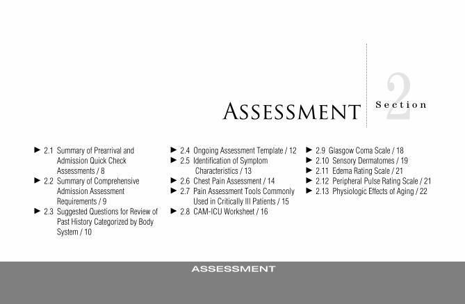
Section 2. Assessment .............................................7
2.1 Summary of Prearrival and Admission QuickCheck Assessments / 8
2.2 Summary of Comprehensive Admission Assessment Requirements / 9
2.3 Suggested Questions for Review of Past HistoryCategorized by Body System / 10
2.4 Ongoing Assessment Template / 122.5 Identification of Symptom Characteristics / 132.6 Chest Pain Assessment / 142.7 Pain Assessment Tools Commonly Used
in Critically Ill Patients / 152.8 CAM-ICU Worksheet / 162.9 Glasgow Coma Scale / 182.10 Sensory Dermatomes / 192.11 Edema Rating Scale / 21
2.12 Peripheral Pulse Rating Scale / 212.13 Physiologic Effects of Aging / 22
Section 3. ECG Concepts .......................................23
3.1 ECG Lead Placement for a Three-Wire System / 25
3.2 ECG Lead Placement for a Five-Wire System / 273.3 Twelve-Lead ECG Placement / 283.4 Right Side ECG Chest Lead
Placement / 29
Contents
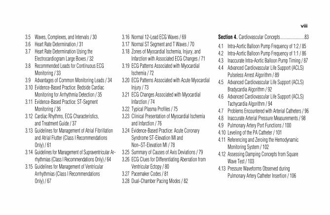
viii
3.5 Waves, Complexes, and Intervals / 303.6 Heart Rate Determination / 313.7 Heart Rate Determination Using the
Electrocardiogram Large Boxes / 323.8 Recommended Leads for Continuous ECG
Monitoring / 333.9 Advantages of Common Monitoring Leads / 343.10 Evidence-Based Practice: Bedside Cardiac
Monitoring for Arrhythmia Detection / 353.11 Evidence-Based Practice: ST-Segment
Monitoring / 363.12 Cardiac Rhythms, ECG Characteristics,
and Treatment Guide / 373.13 Guidelines for Management of Atrial Fibrillation
and Atrial Flutter (Class I RecommendationsOnly) / 61
3.14 Guidelines for Management of Supraventricular Ar-rhythmias (Class I Recommendations Only) / 64
3.15 Guidelines for Management of Ventricular Arrhythmias (Class I Recommendations Only) / 67
3.16 Normal 12-Lead ECG Waves / 693.17 Normal ST Segment and T Waves / 703.18 Zones of Myocardial Ischemia, Injury, and
Infarction with Associated ECG Changes / 713.19 ECG Patterns Associated with Myocardial
Ischemia / 723.20 ECG Patterns Associated with Acute Myocardial
Injury / 733.21 ECG Changes Associated with Myocardial
Infarction / 743.22 Typical Plasma Profiles / 753.23 Clinical Presentation of Myocardial Ischemia
and Infarction / 763.24 Evidence-Based Practice: Acute Coronary
Syndrome ST-Elevation MI and Non–ST-Elevation MI / 78
3.25 Summary of Causes of Axis Deviations / 793.26 ECG Clues for Differentiating Aberration from
Ventricular Ectopy / 803.27 Pacemaker Codes / 813.28 Dual-Chamber Pacing Modes / 82
Section 4. Cardiovascular Concepts .......................83
4.1 Intra-Aortic Balloon Pump Frequency of 1:2 / 854.2 Intra-Aortic Balloon Pump Frequency of 1:1 / 864.3 Inaccurate Intra-Aortic Balloon Pump Timing / 874.4 Advanced Cardiovascular Life Support (ACLS)
Pulseless Arrest Algorithm / 894.5 Advanced Cardiovascular Life Support (ACLS)
Bradycardia Algorithm / 924.6 Advanced Cardiovascular Life Support (ACLS)
Tachycardia Algorithm / 944.7 Problems Encountered with Arterial Catheters / 964.8 Inaccurate Arterial Pressure Measurements / 984.9 Pulmonary Artery Port Functions / 1004.10 Leveling of the PA Catheter / 1014.11 Referencing and Zeroing the Hemodynamic
Monitoring System / 1024.12 Assessing Damping Concepts from Square
Wave Test / 1034.13 Pressure Waveforms Observed during
Pulmonary Artery Catheter Insertion / 106

ix
4.14 Pulmonary Artery Waveform and Components / 108
4.15 Effect of a Mechanical Ventilator Breath on PAWaveform / 109
4.16 Reading End Expiration Before a SpontaneousBreath / 110
4.17 Evidence-Based Practice: Pulmonary Artery Pres-sure Measurement / 111
4.18 Problems Encountered with Pulmonary ArteryCatheters / 112
4.19 Inaccurate Pulmonary Artery Pressure Measurements / 118
4.20 Troubleshooting Problems with ThermodilutionCardiac Output Measurements / 121
4.21 Common Inotropic Therapies in Treating Abnormal Hemodynamics / 125
4.22 Common Preload Reducers for Abnormal Hemodynamics / 125
4.23 Common Afterload Reducing Agents / 126
Section 5. Respiratory Concepts...........................127
5.1 Normal Chest X-Ray / 1285.2 Mediastinal Structures Visible on a Chest
X-Ray / 1295.3 Chest X-Ray of COPD / 1305.4 Chest X-Ray of Pneumothorax / 1315.5 Chest X-Ray of Right Lower Lobe Pneumonia / 1325.6 Chest X-Ray Showing Carina and Right
Bronchus / 1335.7 Chest X-Ray with PA Catheter, ET Tube, and
Chest Tube / 1345.8 Acid-Base Abnormalities / 1355.9 Indications for Mechanical Ventilation / 1365.10 Pulmonary Specific Wean Criteria
Thresholds / 1375.11 Burns’ Wean Assessment Program (BWAP) / 1385.12 Algorithm for Management of Ventilator Alarms
and/ or Development of Acute Respiratory Distress / 140
5.13 Algorithm to Correct Hypoxaemia in an AcuteCOPD Patient / 141
Section 6. Neurologic Concepts ...........................143
6.1 Glasgow Coma Scale / 1446.2 Cranial Nerve Function / 1456.3 Circle of Willis / 1466.4 Incomplete Spinal Cord Injury Syndromes / 1476.5 Spinal Cord Injury–Functional Goals for Specific
Levels of Complete Injury / 1486.6 Intracranial Pressure Monitoring Systems / 152
Section 7. Pharmacology Tables ..........................153
7.1 Intravenous Medication Administration Guidelines / 154
7.2 Neuromuscular Blocking Agents / 1797.3 Vasoactive Agents / 1827.4 Antiarrhythmic Agents / 1857.5 Therapeutic Drug Monitoring / 1917.6 Tips for Calculating IV Medication Infusion
Rates / 194

This page intentionally left blank

xi
Given the complexity of critical care practice today, it’simpossible for even experienced clinicians to rememberall the information required to give safe and effective careto critically ill patients. Clinicians frequently need to use avariety of clinical resources to verify drug information,normal laboratory and physiologic values, ECG and he-modynamic monitoring information, emergency algo-rithms, and other essential facts of patient management.
To save time and avoid frustration, clinicians oftencreate their own “pocket guides” by cutting and past-ing together information from a variety of sources sothey always have a quick reference source available.The AACN Essentials of Critical Care Nursing PocketHandbook is designed to provide busy clinicians with
an easy to use resource that can, literally, be kept intheir pockets. The pocket handbook contains selectedtables and figures from the textbook, AACN Essentialsof Critical Care Nursing, and includes items that clini-cians are most likely to need at their fingertips:
• Critical care drug tables (common vasoactive drugs,neuromuscular blocking agents, antiarrhythmics, IVmedication guidelines)
• Normal values table for laboratory tests and physio-logic parameters
• Lists of assessment components• Cardiac rhythms: ECG characteristics and treatment
guides including sample rhythm strips
• 12-lead ECG changes in acute myocardial ischemiaand infarct
• Troubleshooting guides for hemodynamic monitoringequipment
• Indications for mechanical ventilation• Weaning assessment tool• Chest x-ray interpretationWe hope this pocket book will, indeed, be placed in yourpocket and assist you in making a difference in the livesof the patients and families you encounter.
Marianne ChulaySuzi Burns
Preface
xii
To our critical care nursing colleagues around the world whose wonderful work and efforts ensure the safe passage of patients through the critical care environment.

1Normal ValuesS e c t i o n
� 1.1 Normal Values Table / 2
NORMAL VALUES

1.1 � Normal Values TableAbbreviation Definition Normal Value Formula
BSA Body surface area Meters squared (m2) Value obtained from a nomogram based on height and weightC(a � v)O2 Arteriovenous oxygen content 4-6 mL/100 mL C(a � v)O2 (mL/100 mL or vol %) � CaO2 � CvO2
differenceCaO2 Arterial oxygen content Will vary with hemoglobin CaO2 (mL O2/100 mL blood or vol %) �
concentration and PaO2 on (Hb � 1.39) SaO2 � (PaO2 � 0.0031)air from 19-20 mL/100 mL
CI Cardiac index 2.5-3.0 L/min/m2 CI (L/min/m2) �
CK Creatinine kinase �150 mcg/LCK-MB Creatinine kinase MB band �10 ng/mL or �3% of totalCO Cardiac output 4-6 L/min CO � Stroke volume � heart rateCvO2 Mixed venous oxygen content Will vary with CaO2, cardiac
output, and O2 consumptionfrom 14-15 mL/100 mL
CVP Central venous pressure 2-8 mm Hgdp/dt First time derivative of left 13-14 seconds
ventricular pressure
EDC Effective dynamic compliance 35-45 mL/cm H2O women EDC (mL/cm H2O) �40-50 mL/cm H2O men
EDV End-diastolic volume 50-90 mL
EF Ejection fraction 70% Ejection fraction �SV
EDV
tidal volume (mL)
peak airway pressure (cm H2O)
cardiac output (L/min)
body surface area (m2)
2

1.1 � Normal Values Table (continued )
Abbreviation Definition Normal Value Formula
FRC Functional residual capacity 2400 mLHR Heart rate 60-90 beats/minIF Inspiratory force 75-100 cm H2OLVSW Left ventricular stroke work 8-10 g/m/m2 LVSW � SI � MAP � 0.0144
MAP Mean systemic arterial pressure � 70 mm Hg Map estimate �
O2 availability Oxygen availability 550-650 mL/min/m2 O2 availability (mL/min/m2) � CI � CaO2 � 10
O2 extraction ratio Oxygen extraction ratio 0.25 O2 extraction ratio �
P(A � a)O2 Alveolar-arterial oxygen gradient 25-65 mm Hg at FiO2 � 1.0 P(A � a)O2 (mm Hg) � PAO2 � PaO2
P(A � a)o2 Mean partial pressure of oxygen 104 mm Hgin alveolus
P(A � a)co2 Partial pressure of carbon dioxide 40 mm Hgin alveolus
PacO2 Partial pressure of carbon 35-45 mm Hgdioxide in arterial blood
PAD Pulmonary artery diastolic 5-12 mm Hg
C(a � v)O2
CaO2
(Systolic � 2 Diastolic)
3
3

4
1.1 � Normal Values Table (continued )
Abbreviation Definition Normal Value Formula
PaO2 Partial pressure of oxygen in Will vary with patient’s agearterial blood and the FiO2. On room air:
80-95 mm Hg. On 100% O2: 640 mm Hg
PAS Pulmonary artery 54 systolic 16-24 mm Hgpressure
PCWP Mean pulmonary capillary wedge 5-12 mm Hgpressure
PvCO2 Partial pressure of carbon 41-51 mm Hgdioxide in mixed venous blood
PvO2 Partial pressure of oxygen in Will vary with the FiO2,mixed venous blood cardiac output, and oxygen
consumption from 35-40 mm Hg
PVR Pulmonary vascular resistance 120-200 dynes/s/cm5 PVR 5 � (dynes/s/cm5) �1.5-2.5 mm Hg
QS/QT Right-to-left shunt (percentage 5%-8% QS/QT(%) � � 100of cardiac output flowing pastnonventilated alveoli or the Valid only when arterial blood is 100% saturatedequivalent
0.0031 � P(A � a)O2
C(a � v)O2 � (0.0031 � P[A � a]O2)
(PA [mm Hg] � PCWP [mm Hg]) � 79.9
cardiac output (L/min)

5
1.1 � Normal Values Table (continued )
Abbreviation Definition Normal Value Formula
R or RQ Respiratory quotient 0.8 RQ �
RVSW Right ventricular stroke work 51-61 g/m/m2 RVSW � SI � MPAP � 0.0144
SaO2 Percentage of oxyhemoglobin 96%-100% (air)saturation of arterial blood
SI Stroke index 35-50 mL/m2 SI (mL/min/m2) �
SV Stroke volume 50-100 mL/beat SV (mL/beat) �
SvO2 Percentage of oxyhemoglobin 70-80% (air)saturation of mixed venous blood
SVR Systemic vascular resistance 900-1200 dynes/s/cm5 SVR (TPR) (dynes/s/cm5) �10-15 mm Hg(mm Hg � 80 � dynes/s/cm5)
Troponin I Troponin I �0.4 ng/mL
Troponin T Troponin T �0.1 ng/mL
(MAP [mm Hg] � CVP [mm Hg]) � 79.9
cardiac output (L/min)
cardiac output (mL)
heart rate
stroke volume
body surface area
VCO2
VO2

6
1.1 � Normal Values Table (continued )
Abbreviation Definition Normal Value Formula
VC Vital capacity 65-75 mL/kg
VCO2 Carbon dioxide production 192 mL/min
VD Dead space 150 mL VD/VT �
VD/VT Dead space to tidal volume ratio 0.25-0.40
VO2 Oxygen consumption 115-165 mL/min/m2 O2 extraction ratio �
VT Tidal volume 6-8 mL/kg
Adapted from: Hall J, Schmidt G, Wood L. Principles of critical care. 3rd ed. New York: McGraw Hill, 2005; cover tables I-IV.
C(a � v)O2
CaO2
PaCO2 � PEco2
Paco2
2AssessmentS e c t i o n
� 2.1 Summary of Prearrival andAdmission Quick CheckAssessments / 8
� 2.2 Summary of ComprehensiveAdmission AssessmentRequirements / 9
� 2.3 Suggested Questions for Review ofPast History Categorized by BodySystem / 10
� 2.4 Ongoing Assessment Template / 12� 2.5 Identification of Symptom
Characteristics / 13� 2.6 Chest Pain Assessment / 14� 2.7 Pain Assessment Tools Commonly
Used in Critically Ill Patients / 15� 2.8 CAM-ICU Worksheet / 16
� 2.9 Glasgow Coma Scale / 18� 2.10 Sensory Dermatomes / 19� 2.11 Edema Rating Scale / 21� 2.12 Peripheral Pulse Rating Scale / 21� 2.13 Physiologic Effects of Aging / 22
ASSESSMENT

8
2.1 � Summary of Prearrival and Admission Quick Check AssessmentsPrearrival Assessment• Abbreviated report on patient (age, gender, chief complaint, diagnosis,
pertinent history, physiologic status, invasive devices, equipment, and status of laboratory/diagnostic tests)
• Complete room setup, including verification of proper equipment functioning
Admission Quick Check Assessment• General appearance (consciousness)• Airway:
PatencyPosition of artificial airway (if present)
• Breathing:Quantity and quality of respirations (rate, depth, pattern,
symmetry, effort, use of accessory muscles)Breath soundsPresence of spontaneous breathing
• Circulation and Cerebral Perfusion:ECG (rate, rhythm, and presence of ectopy)Blood pressurePeripheral pulses and capillary refillSkin, color, temperature, moisturePresence of bleedingLevel of consciousness, responsiveness
• Chief Complaint:Primary body systemAssociated symptoms
• Drugs and Diagnostic Tests:Drugs prior to admission (prescribed, over-the-counter, illicit)Current medicationsReview diagnostic test results
• Equipment:Patency of vascular and drainage systemsAppropriate functioning and labeling of all equipment connected to patient
• Allergies

9
2.2 � Summary of Comprehensive Admission Assessment RequirementsPast Medical History• Medical conditions, surgical procedures• Psychiatric/emotional problems• Hospitalizations• Medications (prescription, over-the-counter, illicit drugs) and
time of last medication dose• Allergies• Review of body systems (see Table 1-7)Social History• Age, gender• Ethnic origin• Height, weight• Highest educational level completed• Occupation• Marital status• Primary family members/significant others• Religious affiliation• Advance Directive and Durable Power of Attorney for Health Care• Substance use (alcohol, drugs, caffeine, tobacco)• Domestic Abuse or Vulnerable Adult Screen
Psychosocial Assessment• General communication• Coping styles• Anxiety and stress• Expectations of critical care unit• Current stresses• Family needsSpirituality• Faith/spiritual preference• Healing practicesPhysical Assessment• Nervous system• Cardiovascular system• Respiratory system• Renal system• Gastrointestinal system• Endocrine, hematologic, and immune systems• Integumentary system

10
2.3 � Suggested Questions for Review of Past History Categorized by Body SystemBody System History Questions
Nervous • Have you ever had a seizure?• Have you ever fainted, blacked out, or had
delirium tremens (DTs)?• Do you ever have numbness, tingling, or
weakness in any part of your body?• Do you have any difficulty with your hearing,
vision,or speech?• Has your daily activity level changed due to your
present condition?• Do you require any assistive devices such
as canes?Cardiovascular • Have you experienced any heart problems
or disease such as heart attacks or strokes?• Do you have any problems with extreme fatigue?• Do you have an irregular heart rhythm?• Do you have high blood pressure?• Do you have a pacemaker or an implanted
defibrillator?Respiratory • Do you ever experience shortness of breath?
• Do you have any pain associated with breathing?• Do you have a persistent cough? Is it productive?• Have you had any exposure to environmental
agents that might affect the lungs?• Do you have sleep apnea?
Body System History QuestionsRenal • Have you had any change in frequency of
urination?• Do you have any burning, pain, discharge,
or difficulty when you urinate?• Have you had blood in your urine?
Gastrointestinal • Has there been any recent weight loss or gain?• Have you had any change in appetite?• Do you have any problems with nausea
or vomiting?• How often do you have a bowel movement and
has there been a change in the normal pattern? Do you have blood in your stools?
• Do you have dentures?• Do you have any food allergies?
Integumentary • Do you have any problems with your skin?Endocrine • Do you have any problems with bleeding?Hematologic • Do you have problems with chronic infections?Immunologic • Have you recently been exposed to a contagious
illness?

11
Body System History QuestionsPsychosocial • Do you have any physical conditions which make
communication difficult (hearing loss, visual disturbances, language barriers, etc)?
• How do you best learn? Do you need information repeated several times and/or require information in advance of teaching sessions?
• What are the ways you cope with stress, crises, or pain?• Who are the important people in your “family” or
network? Who do you want to make decisions with you, or for you?
• Have you had any previous experiences with critical illness?• Have you ever been abused?• Have you ever experienced trouble with anxiety, irritability,
being confused, mood swings, or suicide attempts?• What are the cultural practices, religious influences,
and values that are important to the family?• What are family members’ perceptions and expectations
of the critical care staff and the setting?
2.3 � Suggested Questions for Review of Past History Categorized by Body System (continued )
Body System History QuestionsSpiritual • What is your faith or spiritual preference?
• What practices help you heal or deal withstress?
• Would you like to see a chaplain, priest, or other type of healer?
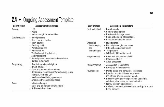
12
2.4 � Ongoing Assessment TemplateBody System Assessment Parameters
Nervous • LOC• Pupils• Motor strength of extremities
Cardiovascular • Blood pressure• Heart rate and rhythm• Heart sounds• Capillary refill• Peripheral pulses• Patency of IVs• Verification of IV solutions
and medications• Hemodynamic pressures and waveforms• Cardiac output data
Respiratory • Respiratory rate and rhythm• Breath sounds• Color and amount of secretions• Noninvasive technology information (eg, pulse
oximetry, end-tidal CO2)• Mechanical ventilatory parameters• Arterial and venous blood gases
Renal • Intake and output• Color and amount of urinary output• BUN/creatinine values
Body System Assessment ParametersGastrointestinal • Bowel sounds
• Contour of abdomen• Position of drainage tubes• Color and amount of secretions• Bilirubin and albumin values
Endocrine, • Fluid balancehematologic, • Electrolyte and glucose valuesand • CBC and coagulation valuesimmunologic • Temperature
• WBC with differential countIntegumentary • Color and temperature of skin
• Intactness of skin• Areas of redness
Pain/discomfort • Assessed in each system• Response to interventions
Psychosocial • Mental status and behavioral responses• Reaction to critical illness experience
(eg, stress, anxiety, coping, mood)• Presence of cognitive impairments (dementia,
delirium), depression, or demoralization• Family functioning and needs• Ability to communicate needs and participate in care• Sleep patterns
13
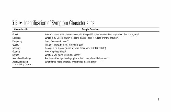
2.5 � Identification of Symptom CharacteristicsCharacteristic Sample Questions
Onset How and under what circumstances did it begin? Was the onset sudden or gradual? Did it progress?Location Where is it? Does it stay in the same place or does it radiate or move around?Frequency How often does it occur?Quality Is it dull, sharp, burning, throbbing, etc?Intensity Rank pain on a scale (numeric, word description, FACES, FLACC)Quantity How long does it last?Setting What are you doing when it happens?Associated findings Are there other signs and symptoms that occur when this happens?Aggravating and What things make it worse? What things make it better
alleviating factors
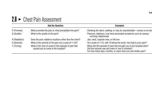
2.6 � Chest Pain AssessmentAsk the Question Examples
P (Provoke) What provokes the pain or what precipitates the pain? Climbing the stairs, walking; or may be unpredictable—comes on at restQ (Quality) What is the quality of the pain? Pressure, tightness; may have associated symptoms such as nausea,
vomiting, diaphoresisR (Radiation) Does the pain radiate to locations other than the chest? Jaw, neck, scapular area, or left armS (Severity) What is the severity of the pain (on a scale of 1-10)? On a scale of 1-10, with 10 being the worst, how bad is your pain?T (Timing) What is the time of onset of this episode of pain that When did this episode of pain that brought you to the hospital start?
caused you to come to the hospital? Did this episode wax and wane or was it constant?For how many days, months, or years have you had similar pain?
14
15
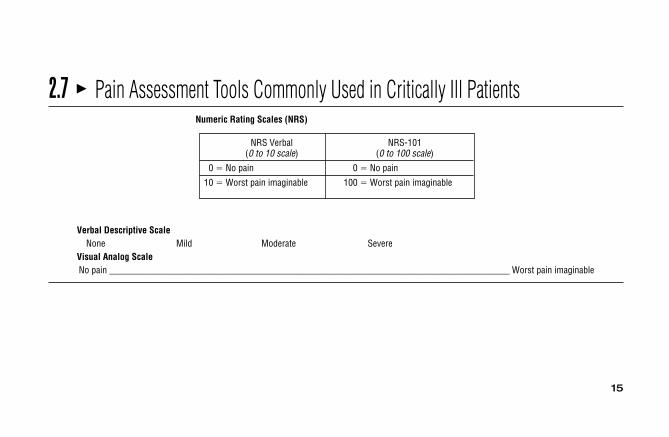
2.7 � Pain Assessment Tools Commonly Used in Critically Ill PatientsNumeric Rating Scales (NRS)
NRS Verbal NRS-101(0 to 10 scale) (0 to 100 scale)
0 � No pain 0 � No pain
10 � Worst pain imaginable 100 � Worst pain imaginable
Verbal Descriptive ScaleNone Mild Moderate Severe
Visual Analog ScaleNo pain ______________________________________________________________________________________ Worst pain imaginable
16
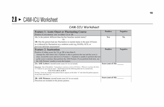
2.8 � CAM-ICU Worksheet
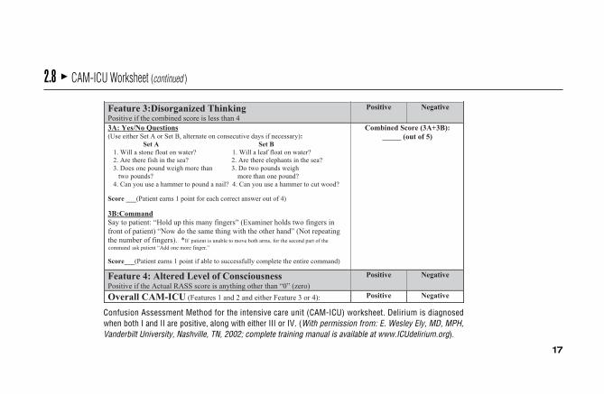
17
2.8 � CAM-ICU Worksheet (continued )
Confusion Assessment Method for the intensive care unit (CAM-ICU) worksheet. Delirium is diagnosedwhen both I and II are positive, along with either III or IV. (With permission from: E. Wesley Ely, MD, MPH,Vanderbilt University, Nashville, TN, 2002; complete training manual is available at www.ICUdelirium.org).
18
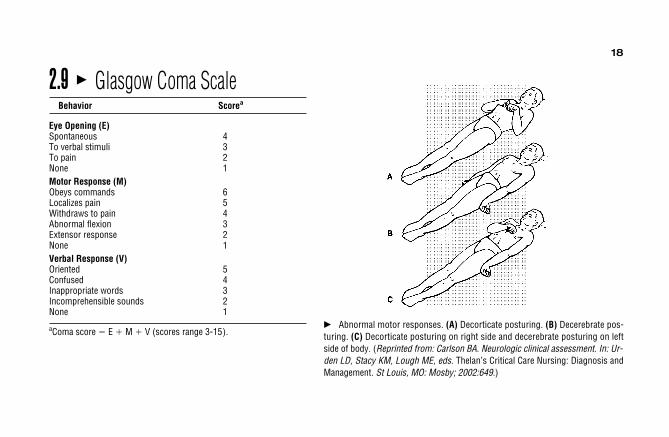
2.9 � Glasgow Coma ScaleBehavior Scorea
Eye Opening (E)Spontaneous 4To verbal stimuli 3To pain 2None 1Motor Response (M)Obeys commands 6Localizes pain 5Withdraws to pain 4Abnormal flexion 3Extensor response 2None 1Verbal Response (V)Oriented 5Confused 4Inappropriate words 3Incomprehensible sounds 2None 1
aComa score � E � M � V (scores range 3-15).� Abnormal motor responses. (A) Decorticate posturing. (B) Decerebrate pos-turing. (C) Decorticate posturing on right side and decerebrate posturing on leftside of body. (Reprinted from: Carlson BA. Neurologic clinical assessment. In: Ur-den LD, Stacy KM, Lough ME, eds. Thelan’s Critical Care Nursing: Diagnosis andManagement. St Louis, MO: Mosby; 2002:649.)

19
2.10 � Sensory Dermatomes.
A
(A) Anterior view.

20
2.10 � Sensory Dermatomes (continued )
B
(B) Posterior view. (Reprinted from: Carlson BA. Neurologic anatomy and physiology. In: Urden LD, Stacy KM, Lough ME, eds.Thelan’s Critical Care Nursing: Diagnosis and Management. St Louis, MO: Mosby; 2002: 641.)

21
2.11 � Edema Rating ScaleFollowing the application and removal of firm digital pressure against thetissue, the edema is evaluated for one of the following responses:
• 0 No depression in tissue• �1 Small depression in tissue, disappearing in �1 second• �2 Depression in tissue disappears in �1-2 second• �3 Depression in tissue disappears in �2-3 second• �4 Depression in tissue disappears in �4 second
2.12 � Peripheral Pulse Rating Scale• 0 Absent pulse• �1 Palpable but thready; easily obliterated with light pressure• �2 Normal; cannot obliterate with light pressure• �3 Full• �4 Full and bounding

22
2.13 � Physiologic Effects of AgingBody System Effects
Nervous Diminished hearing and vision, short-term memory loss, altered motor coordination, decreased muscle tone and strength, slower response to verbal and motor stimuli, decreased ability to synthesize new information, increased sensitivity to
altered temperature states, increased sensitivity to sedation (confusion or agitation), decreased alertness levelsCardiovascular Increased effects of atherosclerosis of vessels and heart valves, decreased stroke volume with resulting decreased cardiac
output, decreased myocardial compliance, increased workload of heart, diminished peripheral pulsesRespiratory Decreased compliance and elasticity, decreased vital capacity, increased residual volume, less effective cough, decreased
response to hypercapniaRenal Decreased glomerular filtration rate, increased risk of fluid and electrolyte imbalancesGastrointestinal Increased presence of dentition problems, decreased intestinal mobility, decreased hepatic metabolism, increased risk of
altered nutritional statesEndocrine, hematologic, Increased incidence of diabetes, thyroid disorders, and anemia; decreased antibody response and cellular immunity
and immunologicIntegumentary Decreased skin turgor, increased capillary fragility and bruising, decreased elasticityMiscellaneous Altered pharmacokinetics and pharmacodynamics, decreased range of motion of joints and extremitiesPsychosocial Difficulty falling asleep and fragmented sleep patterns, increased incidence of depression and anxiety, cognitive impairment
disorders, difficulty with change

3ECG ConceptsS e c t i o n
� 3.1 ECG Lead Placement for a Three-Wire System / 25
� 3.2 ECG Lead Placement for a Five-WireSystem / 27
� 3.3 Twelve-Lead ECG Placement / 28� 3.4 Right Side ECG Chest Lead
Placement / 29� 3.5 Waves, Complexes, and Intervals / 30� 3.6 Heart Rate Determination / 31
� 3.7 Heart Rate Determination Using theElectrocardiogram Large Boxes / 32
� 3.8 Recommended Leads forContinuous ECG Monitoring / 33
� 3.9 Advantages of Common MonitoringLeads / 34
� 3.10 Evidence-Based Practice: BedsideCardiac Monitoring for ArrhythmiaDetection / 35
� 3.11 Evidence-Based Practice: ST-Segment Monitoring / 36
� 3.12 Cardiac Rhythms, ECGCharacteristics, and TreatmentGuide / 37
� 3.13 Guidelines for Management ofAtrial Fibrillation and Atrial Flutter(Class I Recommendations Only) / 61
NORMAL VALUES

24
� 3.14 Guidelines for Management ofSupraventricular Arrhythmias (Class I Recommendations Only) / 64
� 3.15 Guidelines for Management ofVentricular Arrhythmias (Class IRecommendations Only) / 67
� 3.16 Normal 12-Lead ECG Waves / 69� 3.17 Normal ST Segment and
T Waves / 70� 3.18 Zones of Myocardial Ischemia,
Injury, and Infarction withAssociated ECG Changes / 71
� 3.19 ECG Patterns Associated withMyocardial Ischemia / 72
� 3.20 ECG Patterns Associated withAcute Myocardial Injury / 73
� 3.21 ECG Changes Associated withMyocardial Infarction / 74
� 3.22 Typical Plasma Profiles / 75� 3.23 Clinical Presentation of Myocardial
Ischemia and Infarction / 76� 3.24 Evidence-Based Practice: Acute
Coronary Syndrome ST-ElevationMI and Non–ST-Elevation MI / 78
� 3.25 Summary of Causes of AxisDeviations / 79
� 3.26 ECG Clues for DifferentiatingAberration from Ventricular Ectopy / 80
� 3.27 Pacemaker Codes / 81� 3.28 Dual-Chamber Pacing Modes / 82
25
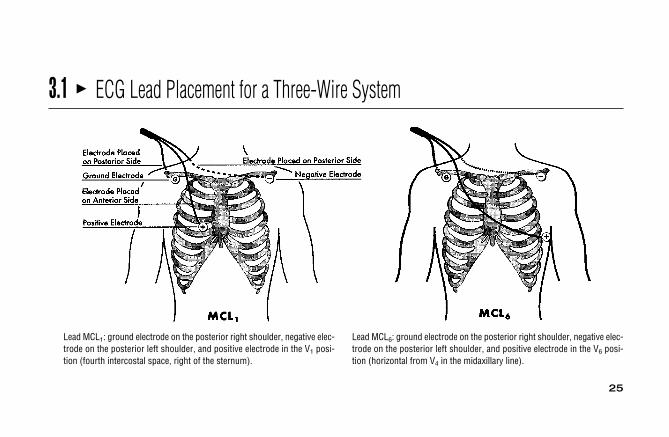
3.1 � ECG Lead Placement for a Three-Wire System
Lead MCL1: ground electrode on the posterior right shoulder, negative elec-trode on the posterior left shoulder, and positive electrode in the V1 posi-tion (fourth intercostal space, right of the sternum).
Lead MCL6: ground electrode on the posterior right shoulder, negative elec-trode on the posterior left shoulder, and positive electrode in the V6 posi-tion (horizontal from V4 in the midaxillary line).

26
3.1 � ECG Lead Placement for a Three-Wire System (continued )
Lead III: the positive electrode is placed on the upper left abdomen. Lead II: ground electrode on the left shoulder, negative electrode on rightshoulder, and positive electrode on the left lower rib cage.

27
3.2 � ECG Lead Placement for a Five-Wire System
(A) Correct electrode placement for using a 5-wire monitoring cable. Right and left arm electrodes are placed on the shoulders and right and left leg electrodes are placed low on thethorax or on the hips. With the arm and leg electrodes placed as illustrated, leads I, II, III, aVR, aVL, and aVF can be obtained by selecting the desired lead on the bedside monitor. Toobtain lead V1 place the chest lead in the fourth intercostal space at the right sternal border and select “V” on the bedside monitor. To obtain lead V6, place the chest lead in the fifth in-tercostal space at the left midaxillary line and select “V” on the bedside monitor. (B) Correct lead placement for obtaining MCL1 and MCL6 using a 3-wire lead system. Place the rightarm electrode on the left shoulder; the left arm electrode in the fourth intercostal space at the right sternal border; and the left leg electrode in the fifth intercostal space at the left mi-daxillary line. To monitor in MCL1, select lead I on the bedside monitor. To monitor in MCL6, select lead II on the bedside monitor. (Adapted from Drew BJ. Bedside electrocardiogrammonitoring. AACN Clin Issues Crit Care Nurs. 1993;4:26, 28.)
A
Angle ofLouis
B
Angle ofLouis

28
3.3 � Twelve-Lead ECG Placement
(A) Limb electrodes can be placed anywhere on arms and legs. Standard placement is shown here on wrists and ankles.(B) Chest electrode placement. V1 � fourth intercostal space to right of sternum; V2 � fourth intercostal space to left of ster-num; V3 � halfway between V2 and V4 in a straight line; V4 � fifth intercostal space at midclavicular line; V5 � same levelas V4 at anterior axillary line; V6 � same level as V4 at midaxillary line.
Angle of Louis
A
B
29
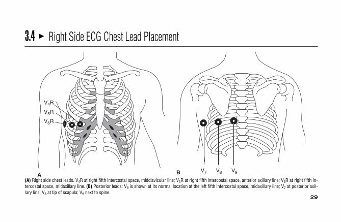
3.4 � Right Side ECG Chest Lead Placement
V4R
V5R
V6R
AV7 V8 V9B
(A) Right side chest leads. V4R at right fifth intercostal space, midclavicular line; V5R at right fifth intercostal space, anterior axillary line; V6R at right fifth in-tercostal space, midaxillary line. (B) Posterior leads: V6 is shown at its normal location at the left fifth intercostal space, midaxillary line; V7 at posterior axil-lary line; V8 at tip of scapula; V9 next to spine.
30
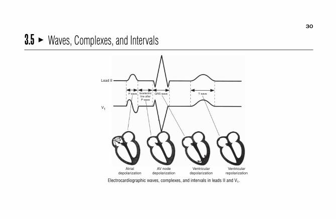
3.5 � Waves, Complexes, and Intervals
Atrialdepolarization
AV nodedepolarization
Ventriculardepolarization
Ventricularrepolarization
T waveP wave
Lead II
V1
QRS waveIsoelectricline afterP wave
Electrocardiographic waves, complexes, and intervals in leads II and V1.
31
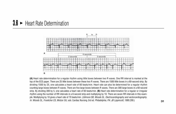
3.6 � Heart Rate Determination
(A) Heart rate determination for a regular rhythm using little boxes between two R waves. One RR interval is marked at thetop of the ECG paper. There are 25 little boxes between these two R waves. There are 1500 little boxes in a 60-second strip. Bydividing 1500 by 25, one calculates a heart rate of 60 beats/min. Heart rate can also be determined for a regular rhythmcounting large boxes between R waves. There are five large boxes between R waves. There are 300 large boxes in a 60-secondstrip. By dividing 300 by 5, one calculates a heart rate of 60 beats/min. (B) Heart rate determination for a regular or irregularrhythm using the number of RR intervals in a 6-second strip and multiplying by 10. There are seven RR intervals in this exam-ple. Multiplying by 10 gives a heart rate of 70 beats/min. (Gilmore SB, Woods SL. Electrocardiography and vectorcardiography.In: Woods SL, Froelicher ES, Motzer SU, eds. Cardiac Nursing 3rd ed. Philadelphia, PA: JB Lippincott; 1995:295.)
R to R
6 seconds3 seconds3 seconds
A
B

32
3.7 � Heart Rate Determination Using the Electrocardiogram Large BoxesNumber of Large Boxes Between R Waves Heart Rate (Beats/Min)
1 3002 1503 1004 755 606 507 408 389 33
10 30

3.8 � Recommended Leads for Continuous ECG MonitoringPurpose Best Leads
Arrhythmia detection V1 (V6 next best)RCA ischemia, inferior MI III, aVFLAD ischemia, anterior MI V3
Circumflex ischemia, lateral MI I, aVL (III, aVF best limb leads)RV infarction V4RAxis shifts I and aVF together
33

34
3.9 � Advantages of Common Monitoring LeadsLead Advantages
Preferred Monitoring LeadsV1 and V6 (or MCL1 and MCL6 Differentiate between right and left bundle branch block
if using a 3-wire system) Morphology clues to differentiate between ventricular beats and supraventricular beats withaberrant conduction
Differentiate between right and left ventricular ectopyDifferentiate between right and left ventricular pacingUsually shows well-formed P wavesPlacement of electrodes keeps apex clear for auscultation or defibrillation
Other Monitoring LeadsLead II Usually shows well-formed P waves
Often best lead for identification of atrial flutter wavesUsually has tall, upright QRS complex on which to synchronize machine for cardioversionAllows identification of retrograde P waves
Lead III or aVF Assists in diagnosis of hemiblockAllows identification of retrograde P wavesAllows Identification of atrial flutter wavesBest limb leads for ST-segment monitoring
Lewis Lead (negative electrode at Often best lead to identify P wavessecond right intercostal space,positive electrode at fourthright intercostal space)

3.10 � Evidence-Based Practice: Bedside Cardiac Monitoring for Arrhythmia DetectionElectrode Application• Make sure skin is clean and dry before applying
monitoring electrodes.• Place arm electrodes on shoulder (front, top, or
back) as close as possible to where arm joinstorso.
• Place leg electrodes below the rib cage or on hips.• Place V1 electrode at the fourth intercostal space
at right sternal border.• Place V6 electrode at the fifth intercostal space at
left midaxillary line.• Replace electrodes every 48 hours or more often if
skin irritation occurs.• Mark electrode position with indelible ink to
ensure consistent lead placement.
Lead Selection• Use lead V1 as the primary arrhythmia monitoring
lead whenever possible.• Use lead V6 if lead V1 is not available.• If using a 3-wire system, use MCL1 as the primary
lead and MCL6 as the second choice lead.
Compiled from Jacobson (2010); Drew, Califf, Funk, et al (2004); and the American Association of Critical-Care Nurse (2004).
35
Alarm Limits• Set heart rate alarms as appropriate for patient’s current heart rate and clinical condition.• Never turn heart rate alarms off while patient’s rhythm is being monitored.• Set alarm limits on other parameters if using a computerized arrhythmia monitoring system.
Documentation• Document the monitoring lead on every rhythm strip.• Document heart rate, PR interval, QRS width, QT interval with every shift and with any
significant rhythm change.• Document rhythm strip with every significant rhythm change:
– Onset and termination of tachycardias.– Symptomatic bradycardias or tachycardias.– Conversion into or out of atrial flutter or atrial fibrillation.– All rhythms requiring immediate treatment.
• Place rhythm strips flat on page (avoid folding or winding strips into chart).
Transporting Monitored Patients• Continue cardiac monitoring using a portable, battery-operated monitor-defibrillator if patient
is required to leave a monitored unit for diagnostic or therapeutic procedures.• Monitored patients must be accompanied by a health-care provider skilled in ECG
interpretation and defibrillation during transport.

363.11 � Evidence-Based Practice: ST-Segment MonitoringPatient SelectionClass I: ST-segment monitoring recommended for the followingtypes of patients:• Patients in the early phase of acute coronary syndromes
(unstable angina, “rule-out MI, ST elevation MI, non–ST-elevation MI).a,c
• Patients presenting to emergency department with chest painor anginal equivalent symptoms.a,c
• Patients and who have undergone nonurgent percutaneouscoronary intervention who have suboptimal angiographicresults.a,c
• Patients with possible variant angina due to coronaryvasospasm.a,c
Class II: ST-segment monitoring may be of benefit in somepatients but is not considered essential for all:• Patients with post-acute MI (after 24–48 h).a• Patients who have undergone nonurgent, uncomplicated
percutaneous coronary intervention 1.• Patients at high risk for ischemia after cardiac or noncardiac
surgery.a• Pediatric patients at risk of ischemia or infarction due to
congenital or acquired conditions.a
Electrode Application• Make sure skin is clean and dry before applying monitoring
electrodes.a,b,c
• Place electrodes according to manufacturer recommendationswhen using a derived 12-lead ECG system.a
Data compiled from aDrew (2004); bJacobson (2007); and cAACN (2004).
• When using a 3- or 5-wire-monitoring system, place electrodes as follows:– Place arm electrodes in infraclavicular fossa close to shouldera or on top or back
of shoulder as close to where arm joins torso as possible.– Place leg electrodes at lowest point on rib cage or on hips.a,b
– Place V1 electrode at the fourth intercostal space at right sternal border.b– Place V6 electrode at the fifth intercostal space at left midaxillary line.b
• Mark electrode placement with indelible ink.a,c
• Replace electrodes every 48 hours or more often if skin irritation occurs.b
Lead Selection• Monitor all 12 leads continuously if using a 12-lead monitoring system.b• Use V1 (or V6 if V1 is not possible due to dressings, etc.) for arrhythmia monitoring
in all multilead combinations.b• Choose the ST-segment monitoring lead according to the patient’s “ischemic
fingerprint” obtained during an ischemic event whenever possible.b,c Use the leadwith the largest ST-segment deviation (elevation or depression).b
• If no ischemic fingerprint is available, use either lead IIIb,c or aVF (whichever hastallest QRS complex)b for ST-segment monitoring.
• Lead V3 is the best lead for detecting anterior wall ST-segment deviation,c but canonly be used if the chest lead is not being used for arrhythmia monitoring in lead V1.
Alarm Limits• Establish baseline ST level with patient in the supine position.a,c
• Set ST alarm parameters at 1 mm above and below the patient’s baseline ST levelin patients at high risk for ischemia.a
• Set ST alarm parameters at 2 mm above and below the patient’s baseline ST levelin more stable patients.a

37
3.12 � Cardiac Rhythms, ECG Characteristics, and Treatment GuideRhythm ECG Characteristics Treatment
Normal sinus • Rate: 60-100 beats/min. • None.rhythm (NSR) • Rhythm: Regular.
• P waves: Precede every QRS; consistent shape.• PR interval: 0.12-0.20 second.• QRS complex: 0.04-0.10 second.
38
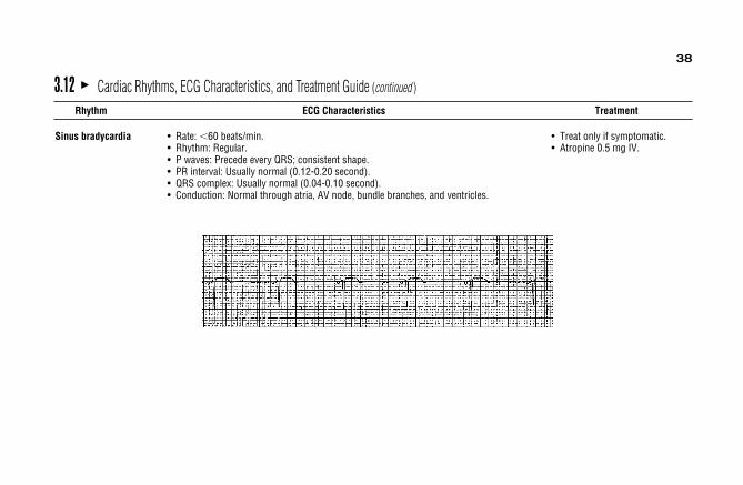
Sinus bradycardia • Rate: �60 beats/min. • Treat only if symptomatic.• Rhythm: Regular. • Atropine 0.5 mg IV.• P waves: Precede every QRS; consistent shape.• PR interval: Usually normal (0.12-0.20 second).• QRS complex: Usually normal (0.04-0.10 second).• Conduction: Normal through atria, AV node, bundle branches, and ventricles.
3.12 � Cardiac Rhythms, ECG Characteristics, and Treatment Guide (continued )
Rhythm ECG Characteristics Treatment
39
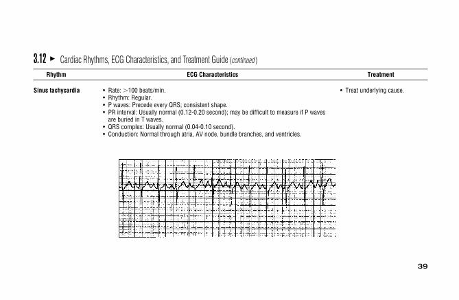
Sinus tachycardia • Rate: �100 beats/min. • Treat underlying cause.• Rhythm: Regular.• P waves: Precede every QRS; consistent shape.• PR interval: Usually normal (0.12-0.20 second); may be difficult to measure if P waves
are buried in T waves.• QRS complex: Usually normal (0.04-0.10 second).• Conduction: Normal through atria, AV node, bundle branches, and ventricles.
3.12 � Cardiac Rhythms, ECG Characteristics, and Treatment Guide (continued )
Rhythm ECG Characteristics Treatment

40
Sinus arrhythmia • Rate: 60-100 beats/min. • Treatment is usually not required.• Rhythm: Irregular; phasic increase and decrease in rate, which may or may not be • Hold digoxin if due to digitalis
related to respiration. toxicity.• P waves: Precede every QRS; consistent shape.• PR interval: Usually normal.• QRS complex: Usually normal.• Conduction: Normal through atria, AV node, bundle branches, and ventricles.
3.12 � Cardiac Rhythms, ECG Characteristics, and Treatment Guide (continued )
Rhythm ECG Characteristics Treatment
41
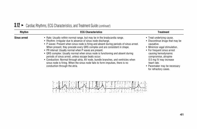
Sinus arrest • Rate: Usually within normal range, but may be in the bradycardia range. • Treat underlying cause.• Rhythm: Irregular due to absence of sinus node discharge. • Discontinue drugs that may be• P waves: Present when sinus node is firing and absent during periods of sinus arrest. causative.
When present, they precede every QRS complex and are consistent in shape. • Minimize vagal stimulation.• PR interval: Usually normal when P waves are present. • For frequent sinus arrest• QRS complex: Usually normal when sinus node is functioning and absent during causing hemodynamic
periods of sinus arrest, unless escape beats occur. compromise, atropine• Conduction: Normal through atria, AV node, bundle branches, and ventricles when 0.5 mg IV may increase
sinus node is firing. When the sinus node fails to form impulses, there is no heart rate.conduction through the atria. • Pacemaker may be necessary
for refractory cases.
3.12 � Cardiac Rhythms, ECG Characteristics, and Treatment Guide (continued )
Rhythm ECG Characteristics Treatment

42
Premature atrial • Rate: Usually within normal range. • Treatment is usually not contraction • Rhythm: Usually regular except when PACs occur, resulting in early beats. PACs necessary.
usually have a noncompensatory pause. • Treat underlying cause.• P waves: Precede every QRS. The configuration of the premature P wave differs from • Drugs (eg, beta-blockers,
that of the sinus P waves. calcium channel blockers,• PR interval: May be normal or long depending on the prematurity of the beat. Very early procainamide) can be used
PACs may find the AV junction still partially refractory and unable to conduct at a normal if necessary.rate, resulting in a prolonged PR interval.
• QRS complex: May be normal, aberrant (wide), or absent, depending on the prematurityof the beat.
• Conduction: PACs travel through the atria differently from sinus impulses because theyoriginate from a different spot. Conduction through the AV node, bundle branches, andventricles is usually normal unless the PAC is very early.
3.12 � Cardiac Rhythms, ECG Characteristics, and Treatment Guide (continued )
Rhythm ECG Characteristics Treatment
PACs conducted abnormally in the ventricle.PACs conducted normally in the ventricle.
43
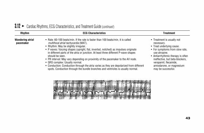
Wandering atrial • Rate: 60-100 beats/min. If the rate is faster than 100 beats/min, it is called • Treatment is usually notpacemaker multifocal atrial tachycardia (MAT). necessary.
• Rhythm: May be slightly irregular. • Treat underlying cause.• P waves: Varying shapes (upright, flat, inverted, notched) as impulses originate • For symptoms from slow rate,
in different parts of the atria or junction. At least three different P-wave shapes use atropine.should be seen. • Antiarrhythmic therapy is often
• PR interval: May vary depending on proximity of the pacemaker to the AV node. ineffective, but beta-blockers,• QRS complex: Usually normal. verapamil, flecainide, • Conduction: Conduction through the atria varies as they are depolarized from different amiodarone, or magnesium
spots. Conduction through the bundle branches and ventricles is usually normal. may be successful.
3.12 � Cardiac Rhythms, ECG Characteristics, and Treatment Guide (continued )
Rhythm ECG Characteristics Treatment
43

44
Atrial tachycardia • Rate: Atrial rate is 120-250 beats/min. • Eliminate underlying cause and• Rhythm: Regular unless there is variable block at the AV node. decrease ventricular rate.• P waves: Differ in shape from sinus P waves because they are ectopic. Precede each • Sedation.
QRS complex but may be hidden in preceding T wave. When block is present, more • Vagal stimulation.than one P wave appears before each QRS complex. • Digitalis (unless it is the cause
• PR interval: May be shorter than normal but often difficult to measure because of atrial tachycardia with block).of hidden P waves. • Propranolol, verapamil, or
• QRS complex: Usually normal but may be wide if aberrant conduction is present. diltiazem can slow ventricular • Conduction: Usually normal through the AV node and into the ventricles. In atrial rate.
tachycardia with block some atrial impulses do not conduct into the ventricles. • Procainamide, flecainide,Aberrant ventricular conduction may occur if atrial impulses are conducted into the amiodarone may be effective toventricles while the ventricles are still partially refractory. prevent recurrences.
• Radiofrequency ablation is oftensuccessful.
3.12 � Cardiac Rhythms, ECG Characteristics, and Treatment Guide (continued )
Rhythm ECG Characteristics Treatment

45
Atrial flutter • Rate: Atrial rate varies between 250 and 350 beats/min, most commonly 300. • Treatment depends onVentricular rate varies depending on the amount of block at the AV node. hemodynamic consequences
• Rhythm: Atrial rhythm is regular. Ventricular rhythm may be regular or irregular of arrhythmia.due to varying AV block. • Cardioversion is preferred
• P waves: F waves (flutter waves) are seen, characterized by a very regular, for markedly reduced cardiac“sawtooth” pattern. One F wave is usually hidden in the QRS complex, and when output.2:1 conduction occurs, F waves may not be readily apparent. • Beta-blockers, calcium channel
• FR interval (flutter wave to the beginning of the QRS complex): May be consistent blockers are used to slow or may vary. ventricular rate.
• QRS complex: Usually normal; aberration can occur. • Procainamide, flecainide,• Conduction: Usually normal through the AV node and ventricles. amiodarone, ibutilide, dofetilide,
sotalol may convert to sinus.• Use drugs that slow atrial rate
(procainamide, flecainide,propafenone) only after priortreatment to ensure AV block(eg, beta-blockers, calciumchannel blockers).
• Radiofrequency ablation isusually successful.
3.12 � Cardiac Rhythms, ECG Characteristics, and Treatment Guide (continued )
Rhythm ECG Characteristics Treatment

46
Atrial fibrillation • Rate: Atrial rate is 400-600 beats/min or faster. Ventricular rate varies depending • Eliminate underlying cause.on the amount of block at the AV node. In new atrial fibrillation, the ventricular • Cardiovert if hemodynamically response is usually quite rapid, 160-200 beats/min; in treated atrial fibrillation, unstable.the ventricular rate is controlled in the normal range of 60-100 beats/min. • Calcium channel blockers and
• Rhythm: Irregular. One of the distinguishing features of atrial fibrillation is the marked beta-blockers are used to slowirregularity of the ventricular response. ventricular rate. Procainamide,
• P waves: Not present. Atrial activity is chaotic with no formed atrial impulses visible. disopyramid, flecainide,Irregular F waves are often seen and vary in size from coarse to very fine. propafenone, amiodarone,
• PR interval: Not measurable; there are no P waves. sotalol, ibutilide, dofetilide• QRS complex: Usually normal; aberration is common. are used to convert to sinus.• Conduction: Conduction within the atria is disorganized and follows a very irregular • Radiofrequency ablation may be
pattern. Most of the atrial impulses are blocked within the AV junction. Those impulses successful.that are conducted through the AV junction are usually conducted normally through theventricles. If an atrial impulse reaches the bundle branch system during its refractoryperiod, aberrant intraventricular conduction can occur.
3.12 � Cardiac Rhythms, ECG Characteristics, and Treatment Guide (continued )
Rhythm ECG Characteristics Treatment

47
Premature junctional • Rate: 60-100 beats/min or whatever the rate of the basic rhythm. • Treatment is usually not complexes • Rhythm: Regular except for occurrence of premature beats. necessary.
• P waves: May occur before, during, or after the QRS complex of the premature beatand are usually inverted.
• PR interval: Short, usually 0.10 second or less, when P waves precede the QRS.• QRS complex: Usually normal but may be aberrant if the PJC occurs very early and
conducts into the ventricles during the refractory period of a bundle branch.• Conduction: Retrograde through the atria; usually normal through the ventricles.
3.12 � Cardiac Rhythms, ECG Characteristics, and Treatment Guide (continued )
Rhythm ECG Characteristics Treatment
↑ PJC

48
Junctional rhythm • Rate: Junctional rhythm, 40-60 beats/min; accelerated junctional rhythm, • Treatment is rarely needed 60-100 beats/min; junctional tachycardia, 100-250 beats/min. unless rate is too slow or too
• Rhythm: Regular. fast to maintain adequate CO.• P waves: May precede or follow QRS. • Atropine is used to increase • PR interval: Short, 0.11 second or less if P waves precede QRS. rate.• QRS complex: Usually normal. • Verapamil, propranolol, or• Conduction: Retrograde through the atria; normal through the ventricles. beta-blockers is used to decrease . rate.
• Withhold digitalis if digitalistoxicity is suspected.
3.12 � Cardiac Rhythms, ECG Characteristics, and Treatment Guide (continued )
Rhythm ECG Characteristics Treatment

49
Premature ventricular • Rate: 60-100 beats/min or the rate of the basic rhythm. • Eliminate underlying cause.complexes • Rhythm: Irregular because of the early beats. • Drug therapy is not usually
• P waves: Not related to the PVCs. Sinus rhythm is usually not interrupted by the used, but, if desired, lidocaine,premature beats, so sinus P waves can often be seen occurring regularly amiodarone, procainamide,throughout the rhythm. beta-blockers may be effective.
• PR interval: Not present before most PVCs. If a P wave happens, by coincidence, to precede a PVC, the PR interval is short.
• QRS complex: Wide and bizarre; � 0.10 second in duration. May vary inmorphology (size, shape) if they originate from more than one focus in the ventricles.
• Conduction: Wide QRS complexes. Some PVCs may conduct retrograde into the atria,resulting in inverted P waves following the PVC.
3.12 � Cardiac Rhythms, ECG Characteristics, and Treatment Guide (continued )
Rhythm ECG Characteristics Treatment

50
Ventricular rhythm • Rate: �50 beats/min for ventricular rhythm and 50-100 beats/min for accelerated • For ventricular escape rhythms,ventricular rhythm. use atropine to increase sinus
• Rhythm: Usually regular. rate and overdrive ventricular• P waves: May be seen but at a slower rate than the ventricular focus, with dissociation rhythm.
from the QRS. • Use ventricular pacing• PR interval: Not measured. to increase ventricular rate• QRS complex: Wide and bizarre. if escape rhythm is too slow.• Conduction: If sinus rhythm is the basic rhythm, atrial conduction is normal.
Impulses originating in the ventricles conduct via muscle cell-to-cell conduction,resulting in the wide QRS complex.
3.12 � Cardiac Rhythms, ECG Characteristics, and Treatment Guide (continued )
Rhythm ECG Characteristics Treatment
51
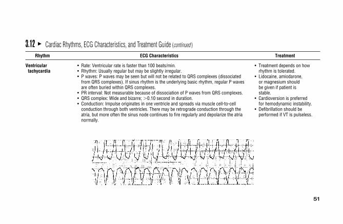
Ventricular • Rate: Ventricular rate is faster than 100 beats/min. • Treatment depends on howtachycardia • Rhythm: Usually regular but may be slightly irregular. rhythm is tolerated.
• P waves: P waves may be seen but will not be related to QRS complexes (dissociated • Lidocaine, amiodarone,from QRS complexes). If sinus rhythm is the underlying basic rhythm, regular P waves or magnesium should are often buried within QRS complexes. be given if patient is
• PR interval: Not measurable because of dissociation of P waves from QRS complexes. stable.• QRS complex: Wide and bizarre; �0.10 second in duration. • Cardioversion is preferred• Conduction: Impulse originates in one ventricle and spreads via muscle cell-to-cell for hemodynamic instability.
conduction through both ventricles. There may be retrograde conduction through the • Defibrillation should be atria, but more often the sinus node continues to fire regularly and depolarize the atria performed if VT is pulseless.normally.
3.12 � Cardiac Rhythms, ECG Characteristics, and Treatment Guide (continued )
Rhythm ECG Characteristics Treatment

52
Ventricular • Rate: Rapid, uncoordinated, ineffective. • Immediate defibrillation.fibrillation • Rhythm: Chaotic, irregular. • CPR required until defibrillator
• P waves: None seen. is available.• PR interval: None. • Amiodarone, lidocaine,• QRS complex: No formed QRS complexes seen; rapid, irregular undulations without magnesium are commonly
any specific pattern. used.• Conduction: Multiple ectopic foci firing simultaneously in ventricles and depolarizing • After conversion, use
them irregularly and without any organized pattern. Ventricles are not contracting. IV antiarrhythmic that facilitatesconversion to preventrecurrence.
3.12 � Cardiac Rhythms, ECG Characteristics, and Treatment Guide (continued )
Rhythm ECG Characteristics Treatment
53
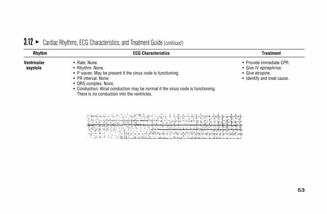
Ventricular • Rate: None. • Provide immediate CPR.asystole • Rhythm: None. • Give IV epinephrine.
• P waves: May be present if the sinus node is functioning. • Give atropine.• PR interval: None. • Identify and treat cause.• QRS complex: None.• Conduction: Atrial conduction may be normal if the sinus node is functioning.
There is no conduction into the ventricles.
3.12 � Cardiac Rhythms, ECG Characteristics, and Treatment Guide (continued )
Rhythm ECG Characteristics Treatment

54
First-degree AV • Rate: Can occur at any sinus rate, usually 60-100 beats/min. • Treatment is usually notblock • Rhythm: Regular. necessary.
• P waves: Normal; precede every QRS.• PR interval: Prolonged above 0.20 second.• QRS complex: Usually normal.• Conduction: Normal through the atria, usually delayed through the AV node.
Ventricular conduction is normal.
3.12 � Cardiac Rhythms, ECG Characteristics, and Treatment Guide (continued )
Rhythm ECG Characteristics Treatment
55
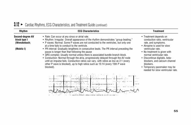
Second-degree AV • Rate: Can occur at any sinus or atrial rate. • Treatment depends onblock type I • Rhythm: Irregular. Overall appearance of the rhythm demonstrates “group beating.” conduction ratio, ventricular(Wenckebach; • P waves: Normal. Some P waves are not conducted to the ventricles, but only one rate, and symptoms.
at a time fails to conduct to the ventricle. • Atropine is used for slow (Mobitz I) • PR interval: Gradually lengthens in consecutive beats. The PR interval preceding the ventricular rate.
pause is longer than that following the pause • No treatment is given with • QRS complex: Usually normal unless there is associated bundle branch block. normal ventricular rate.• Conduction: Normal through the atria, progressively delayed through the AV node • Discontinue digitalis, beta-
until an impulse fails. Conduction ratios can vary, with ratios as low as 2:1 (every blockers, and calcium channelother P wave is blocked), up to high ratios such as 15:14 (every 15th P wave blockers.blocked). • Temporary pacemaker may be
needed for slow ventricular rate.
3.12 � Cardiac Rhythms, ECG Characteristics, and Treatment Guide (continued )
Rhythm ECG Characteristics Treatment

56
Second-degree AV • Rate: Can occur at any basic rate. • Pacemaker is often needed.block type II • Rhythm: Irregular due to blocked beats. • Atropine is not recommended.(Mobitz II) • P waves: Usually regular and precede each QRS. Periodically a P wave is not
followed by a QRS complex.• PR interval: Constant before conducted beats. The PR interval preceding the pause
is the same as that following the pause.• QRS complex: Usually wide due to associated bundle branch block.• Conduction: Normal through the atria and through the AV node but intermittently
blocked in the bundle branch system and fails to reach the ventricles. Conduction through the ventricles is abnormally slow due to associated bundle branch block. Conduction ratios can vary from 2:1 to only occasional blocked beats.
3.12 � Cardiac Rhythms, ECG Characteristics, and Treatment Guide (continued )
Rhythm ECG Characteristics Treatment

57
High-Grade (Advanced) • Rate: Atrial rate <135 beats/min. • Treatment is necessary if patientHigh AV block • Rhythm: Regular or irregular, depending on conduction pattern. is symptomatic.
• P waves: Normal; present before every conducted QRS, but two or more • Atropine may increaseconsecutive P waves may not be followed by QRS complexes. ventricular rate.
• PR interval: Constant before conducted beats; may be normal or prolonged. • Pacemaker is often required.• QRS complex: Usually normal in type I and wide in type II advanced blocks.• Conduction: Normal through the atria. Two or more consecutive atrial impulses fail
to conduct to the ventricles. Ventricular conduction is normal in type I and abnormally slow in type II advanced blocks.
3.12 � Cardiac Rhythms, ECG Characteristics, and Treatment Guide (continued )
Rhythm ECG Characteristics Treatment

58
Third-degree AV • Rate: Atrial rate is usually normal; ventricular rate is �45 beats/min. • Pacemaker.block (complete) • Rhythm: Regular. • Atropine is usually not effective.
• P waves: Normal but dissociated from QRS complexes. • With severely decreased cardiac• PR interval: No consistent PR intervals because there is no relationship between output, perform CPR until
P waves and QRS complexes. pacemaker available.• QRS complex: Normal if ventricles controlled by a junctional rhythm; wide if
controlled by a ventricular rhythm.• Conduction: Normal through the atria. All impulses are blocked at the AV node or in
the bundle branches, so there is no conduction to the ventricles. Conduction through the ventricles is normal if a junctional escape rhythm occurs, and abnormally slow if a ventricular escape rhythm occurs.
3.12 � Cardiac Rhythms, ECG Characteristics, and Treatment Guide (continued )
Rhythm ECG Characteristics Treatment

59
Ventricular paced • Rate: Depends on type of pacemaker. • None.rhythm with • Rhythm: Regular.capture • P waves: Absent or present but dissociated from QRS complexes.
• PR interval: None.• QRS complex: Pacemaker spike followed immediately by wide, bizarre QRS complex.
3.12 � Cardiac Rhythms, ECG Characteristics, and Treatment Guide (continued )
Rhythm ECG Characteristics Treatment

60
Ventricular paced • Conduction: Abnormal. • If hemodynamically stable,rhythm without • ECG characteristics depend on nature of intrinsic rhythm. elective correction/replacementcapture • Pacemaker spike has no fixed relationship to QRS complexes. of pacemaker.
• If hemodynamically unstable, treatment as for third-degree AV block.
3.12 � Cardiac Rhythms, ECG Characteristics, and Treatment Guide (continued )
Rhythm ECG Characteristics Treatment
61
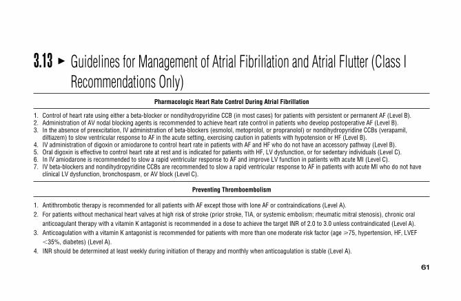
3.13 � Guidelines for Management of Atrial Fibrillation and Atrial Flutter (Class IRecommendations Only)
Pharmacologic Heart Rate Control During Atrial Fibrillation
1. Control of heart rate using either a beta-blocker or nondihydropyridine CCB (in most cases) for patients with persistent or permanent AF (Level B).2. Administration of AV nodal blocking agents is recommended to achieve heart rate control in patients who develop postoperative AF (Level B).3. In the absence of preexcitation, IV administration of beta-blockers (esmolol, metoprolol, or propranolol) or nondihydropyridine CCBs (verapamil,
diltiazem) to slow ventricular response to AF in the acute setting, exercising caution in patients with hypotension or HF (Level B).4. IV administration of digoxin or amiodarone to control heart rate in patients with AF and HF who do not have an accessory pathway (Level B).5. Oral digoxin is effective to control heart rate at rest and is indicated for patients with HF, LV dysfunction, or for sedentary individuals (Level C).6. In IV amiodarone is recommended to slow a rapid ventricular response to AF and improve LV function in patients with acute MI (Level C).7. IV beta-blockers and nondihydropyridine CCBs are recommended to slow a rapid ventricular response to AF in patients with acute MI who do not have
clinical LV dysfunction, bronchospasm, or AV block (Level C).
Preventing Thromboembolism
1. Antithrombotic therapy is recommended for all patients with AF except those with lone AF or contraindications (Level A).2. For patients without mechanical heart valves at high risk of stroke (prior stroke, TIA, or systemic embolism; rheumatic mitral stenosis), chronic oral
anticoagulant therapy with a vitamin K antagonist is recommended in a dose to achieve the target INR of 2.0 to 3.0 unless contraindicated (Level A).3. Anticoagulation with a vitamin K antagonist is recommended for patients with more than one moderate risk factor (age 75, hypertension, HF, LVEF
�35%, diabetes) (Level A).4. INR should be determined at least weekly during initiation of therapy and monthly when anticoagulation is stable (Level A).

62
3.13 � Guidelines for Management of Atrial Fibrillation and Atrial Flutter (Class I Recommendations Only) (continued )
Preventing Thromboembolism
5. Aspirin 325 mg daily is an alternative to vitamin K antagonists in low-risk patients or those with contraindications to anticoagulation (Level A).6. For patients with mechanical heart valves, the target intensity of anticoagulation should be based on the type of prosthesis, maintaining an INR of at least
2.5 (Level B).7. For patients with AF of 48 hours duration, or when the duration is unknown, anticoagulation (INR: 2.0-3.0) is recommended for at least 3 weeks prior
to and 4 weeks after cardioversion (electrical or pharmacologic) (Level B).8. For patients with AF of �48 hours duration requiring immediate cardioversion, heparin should be administered concurrently (unless contraindicated) by
an initial IV bolus followed by a continuous infusion in a dose adjusted to prolong the aPTT to 1.5 to 2 times the reference control value. Oralanticoagulation (INR: 2.0-3.0) should be given for at least 4 weeks after cardioversion. Limited data support SQ administration of LMWH in this categoryof patient condition (Level C).
9. For patients with AF of �48 hours duration and hemodynamic instability (angina, MI, shock, or pulmonary edema), cardioversion should be performedimmediately without delay for prior anticoagulation (Level C).
Cardioversion of Atrial Fibrillation
1. Administration of flecainide, dofetilide, propafenone, or ibutilide is recommended for pharmacologic cardioversion (Level A).2. Immediate electrical (direct-current) cardioversion is recommended for patients with AF involving preexcitation when very rapid tachycardia
or hemodynamic instability occurs (Level B).3. When a rapid ventricular response does not respond promptly to pharmacologic measures in patients with myocardial ischemia, symptomatic
hypotension, angina, or HF, immediate R-wave-synchronized cardioversion is recommended (Level C).
63
3.13 � Guidelines for Management of Atrial Fibrillation and Atrial Flutter (Class I Recommendations Only) (continued )
Cardioversion of Atrial Fibrillation
4. Electrical cardioversion is recommended in patients without hemodynamic instability when symptoms of AF are unacceptable to the patient. In case ofearly relapse of AF after cardioversion, repeated electrical cardioversion attempts may be made following administration of antiarrhythmic medication(Level C).
5. Electrical cardioversion is recommended for patients with acute MI and severe hemodynamic compromise, intractable ischemia, or inadequate ratecontrol with drugs (Level C).
There are no Class I recommendations for pharmacologic conversion of atrial fibrillation.
Maintenance of Sinus Rhythm
1. An oral beta-blocker to prevent postoperative AF is recommended for patients undergoing cardiac surgery (unless contraindicated) (Level A).2. Before initiating antiarrhythmic drug therapy, treatment of precipitating or reversible causes of AF is recommended (Level C).
Level of Evidence Definitions:Level A: Data derived from multiple randomized clinical trials or meta-analyses.Level B: Data derived from a single randomized trial or nonrandomized studies.Level C: Only consensus opinion of experts, case studies, or standard of care.
Fuster V, Ryden LE, Asinger RW, Cannom DS, Crijns HJ, Frye RL, et al. ACC/AHA/ESC Guidelines for the Management of Patients With Atrial Fibrillation: ex-ecutive summary: a report of the American College of Cardiology/American Heart Association Task Force on Practice Guidelines and the European Society ofCardiology Committee for Practice Guidelines (Writing Committee to Revise the 2001 Guidelines for the Management of Patients with Atrial Fibrillation). Cir-culation. 2006;114:700-752.Abbreviations: AF, atrial fibrillation; aPTT, activated partial thromboplastin time; CCB, calcium channel blocker; HF, heart failure; INR, international normalized ra-tio; LMWH, low molecular weight heparin; LV, left ventricular; MI, myocardial infarction; TIA, transient ischemic attack.

64
3.14 � Guidelines for Management of Supraventricular Arrhythmias (Class IRecommendations Only)
Acute Management of Hemodynamically Stable and Regular Tachycardia
Narrow QRS (SVT) and SVT with BBB:1. Vagal maneuvers (Valsalva, CSM) (Level B)2. Adenosine (Level A)3. Verapamil, diltiazem (Level A)
Preexcited SVT/AF:1. Flecainide (Level B)2. Ibutilide (Level B)3. Procainamide (Level B)4. Electrical cardioversion (Level C)
Wide QRS Tachycardia of Unknown Origin:1. Procainamide (Level B)2. Sotalol (Level B)3. Amiodarone (Level B)4. Electrical cardioversion (Level B)
Wide QRS Tachycardia of Unknown Origin in Patients with Poor LV Function:1. Amiodarone (Level B)2. Lidocaine (Level B)3. Electrical cardioversion (Level B)

65
3.14 � Guidelines for Management of Supraventricular Arrhythmias (Class I Recommendations Only) (continued )
Long-Term Treatment of Recurrent AVNRT
1. Catheter ablation (Level B)2. Verapamil for recurrent symptomatic AVNRT (Level B)3. Diltiazem or beta-blockers for recurrent symptomatic AVNRT (Level C)
Infrequent, Well Tolerated Episodes of AVNRT:1. Vagal maneuvers (Level B)2. Pill-in-the-pocket (single-dose oral diltiazem plus propranolol) (Level B)3. Verapamil, diltiazem, beta-blockers, catheter ablation (Level B)
Focal and Nonparoxysmal Junctional Tachycardia Syndromes
Nonparoxysmal Junctional Tachycardia:1. Reverse digitalis toxicity (Level C)2. Correct hypokalemia (Level C)3. Treat myocardial ischemia (Level C)
Long-Term Therapy of Accessory Pathway–Mediated Arrhythmias
1. Catheter ablation for WPW syndrome (preexcitation and symptomatic arrhythmias) that are well tolerated; or with AF and rapid conduction or poorlytolerated CMT (Level B)
2. Vagal maneuvers for single or infrequent episodes (Level B)3. Pill-in-the-pocket (verapamil, diltiazem, beta-blockers) for single or infrequent episodes (Level B)
Contraindicated: Verapamil, diltiazem, digoxin

66
3.14 � Guidelines for Management of Supraventricular Arrhythmias (Class I Recommendations Only) (continued )
Treatment of Focal Atrial Tachycardia
Acute Treatment:1. Electrical cardioversion if hemodynamically unstable (Level B)2. Beta-blockers, verapamil, diltiazem for rate control (in absence of digitalis therapy) (Level C)Prophylactic Therapy:1. Catheter ablation for recurrent symptomatic or incessant AT (Level B)2. Beta blockers, verapamil, diltiazem (Level C)Level of Evidence Definitions:Level A: Data derived from multiple randomized clinical trials or meta-analysesLevel B: Data derived from a single randomized trial or nonrandomized studiesLevel C: Only consensus opinion of experts, case studies, or standard-of-care
Blomstrom-Lundqvist C, Scheinman MM, Aliot EM, et al. ACC/AHA/ESC Guidelines for the Management of Patients with Supraventricular Arrhythmias–exec-utive summary: a report of the American College of Cardiology/American Heart Association Task Force on Practice Guidelines and the European Society ofCardiology Committee for Practice Guidelines (Writing Committee to Develop Guidelines for the Management of Patients with Supraventricular Arrhythmias).Circulation. 2003: 108: 1871-1909.Abbreviations: AF, atrial fibrillation; AVNRT, atrioventricular nodal reentry tachycardia; BBB, bundle branch block; CMT, circus movement tachycardia; LV,left ventricular; SVT, supraventricular tachycardia; WPW, Wolff-Parkinson-White.

67
3.15 � Guidelines for Management of Ventricular Arrhythmias (Class I Recommendations Only)
Sustained Monomorphic Ventricular Tachycardia
1. Wide QRS tachycardia should be presumed to be VT if the diagnosis is unclear. (Level C)2. Electrical cardioversion with sedation is recommended with hemodynamically unstable sustained monomorphic VT (Level C).Contraindicated: Calcium channel blockers (verapamil, diltiazem) should not be used to terminate wide QRS tachycardia of unknown origin, especially withhistory of myocardial dysfunction.
Polymorphic Ventricular Tachycardia
1. Electrical cardioversion with sedation is recommended for sustained PVT with hemodynamic compromise (Level B).2. IV beta-blockers are useful if ischemia is suspected or cannot be excluded (Level B).3. IV amiodarone is useful for recurrent PVT in the absence of QT prolongation (congenital or acquired) (Level C).4. Urgent angiography and revascularization should be considered with PVT when myocardial ischemia cannot be excluded (Level C).
Torsades de Pointes
1. Withdrawal of any offending drugs and correction of electrolyte abnormalities are recommended for TdP (Level A).2. Acute and long-term pacing is recommended for TdP due to heart block and symptomatic bradycardia (Level A).
Incessant Ventricular Tachycardia
1. Revascularization and beta blockade followed by IV antiarrhythmic drugs such as procainamide or amiodarone are recommended for recurrent orincessant PVT (Level B).
68
3.15 � Guidelines for Management of Ventricular Arrhythmias (Class I Recommendations Only) (continued )
Level of Evidence Definitions:
Level A: Data derived from multiple randomized clinical trials or meta-analyses.Level B: Data derived from a single randomized trial or nonrandomized studies.Level C: Only consensus opinion of experts, case studies, or standard of care.
Zipes DP, Camm JA, Borggrefe M., Buxton AE, Chaitman B, Fromer M, et al. ACC/AHA/ESC 2006 Guidelines for Management of Patients with Ventricular Ar-rhythmias and the Prevention of Sudden Cardiac Death: executive summary: a report of the American College of Cardiology/American Heart Association TaskForce and the European Society of Cardiology Committee for Practice Guidelines (Writing Committee to Revise the 2001 Guidelines for the Management ofPatients with Atrial Fibrillation). Circulation. 2006;114:1088-1132.Abbreviations PVT, polymorphic ventricular tachycardia; TdP, torsades de pointes; VT, ventricular tachycardia.

69
3.16 � Normal 12-Lead ECG Waves
(A) Normal sequence of depolarization through the heart as recorded byeach of the frontal plane leads.
(B) Cross-section of the thorax illustrating how the six precordial leadsrecord normal electrical activity in the ventricles. The small arrow (1) showsthe initial direction of depolarization through the septum, followed by the di-rection of ventricular depolarization, indicated by the larger arrow (2).

70
3.17 � Normal ST Segment and T Waves
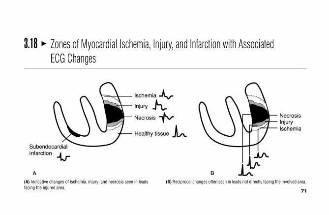
71
(A) Indicative changes of ischemia, injury, and necrosis seen in leadsfacing the injured area.
(B) Reciprocal changes often seen in leads not directly facing the involved area.
3.18 � Zones of Myocardial Ischemia, Injury, and Infarction with Associated ECG Changes
A B

72
3.19 � ECG Patterns Associated with Myocardial Ischemia
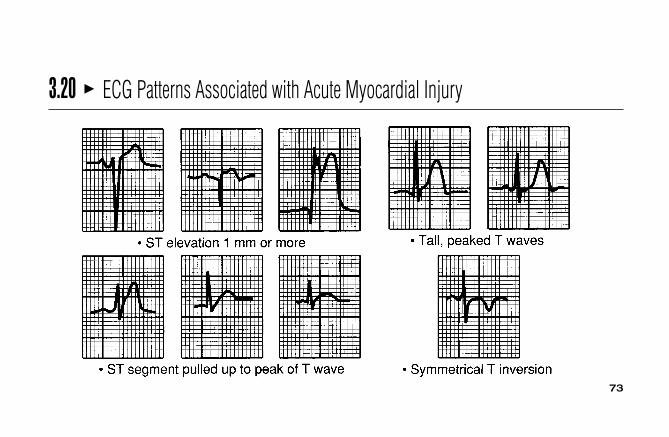
73
3.20 � ECG Patterns Associated with Acute Myocardial Injury
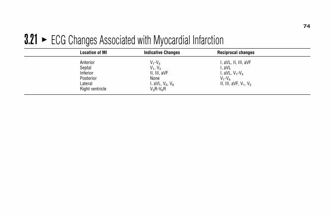
74
3.21 � ECG Changes Associated with Myocardial InfarctionLocation of MI Indicative Changes Reciprocal changes
Anterior V1-V4 I, aVL, II, III, aVFSeptal V1, V2 I, aVLInferior II, III, aVF I, aVL, V1-V4Posterior None V1-V4Lateral I, aVL, V5, V6 II, III, aVF, V1, V2Right ventricle V3R-V6R

75
3.22 � Typical Plasma Profiles
8
6
4
2
0Pla
sma
enzy
me
leve
ls x
ba
selin
e
0 1 2 3 4 5 6 7
Days after onset of chest pain
MB-CK (Q-wave)
MB-CK (reperfusion)
MB-CK(non-Q)
AST LDH
Typical plasma profiles for the MB isoenzymes of creatine kinase (MB-CK), aspartate amino transferase (AST), and lactate dehydrogenase (LDH) activitiesfollowing onset of acute myocardial infarction. (Used with permission, Alexander R, Pratt C. Diagnosis and management of acute myocardial infarction. In: Fuster V, Alexander R, O’Rouke R, eds. Hurst’s The Heart. 10th ed. New York, NY: McGraw-Hill; 2001.)
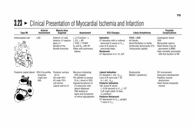
76
Posterior septal lateral RCA Circumflex Posterior surface Murmurs indicating Lateral Indicative: Bradycardia RV involvementbranches of LV, VSD (septal) ST elevation I, aVL, V5, 6 Mobitz I (posterior) Aneurysm development(right and SA node 45% PA catheter to assess Loss of R wave and ↑ ST Papillary muscleleft) AV node 10% R to L shunt in VSD in I, aVL, V5-6 dysfunction
Left atrium Signals/symptoms of Posterior Indicative: Heart blocks frequentlyLateral wall of LV LV aneurysm with Tall, broad R waves resolve
lateral displaced (�0.04 second) in V1-3 ↑ STPMI leading to V4R (right sided 12 lead, signs and symptoms V4 position)of mitral regurgitation Posterior Reciprocal:
ST depression in V1,2, uprightT wave in V1,2
3.23 � Clinical Presentation of Myocardial Ischemia and InfarctionArterial Muscle Area Possible
Type MI Involvement Supplied Assessment ECG Changes Likely Arrhythmias Complications
Anteroseptal wall LAD Anterior LV wall, ↓ LV Function → Indicative: RBBB, LBBB Cardiogenic shockAnterior LV septum ↓ CO, ↓ BP ST elevation with or without AV blocks VSDApex LV ↑ PAD, ↑ PCWP abnormal Q waves in V1-4 Atrial fibrillation or flutter Myocardial ruptureBundle of His S3 and S4, with HF Loss of R waves in Ventricular tachycardia (VT) Heart blocks may be Bundle branches Rales with pulmonary precordial leads Tachycardia (septal) permanent (LBBB)
edema Reciprocal: High mortality associatedST depression in II, III, aVF. with this location of MI

77
Right ventricular RCA RA, RV, Inferior LV Kussmaul’s sign Indicative: First-degree AV block Hypotension requiringinfarction SA Node JVD 1- to 2-mm ST-segment Second-degree AV large volumes initially to
AV Node Hypotension elevation in V4R block, type I maintain systemicPosterior ↑ SVR, ↓ PCWP ↑ CVP ST- and T-wave elevation Incomplete RBBB pressure. Once RV
IV septum S3 with noncompliant RV in II, III, aVF Transient CHB contractility improves Clear breath sounds Q waves in II, III, aVF Atrial fibrillation fluids will mobilize, initially ST-elevation decreases in VT/VF possibly requiring
Hepatomegaly; peripheral amplitude over V1–6 diuresisedema: cool, clammy,pale skin
Arterial Muscle Area PossibleType MI Involvement Supplied Assessment ECG Changes Likely Arrhythmias Complications
Inferior or RCA RV, RA Symptomatic bradycardia: Indicative: AV blocks; often progress Hiccups“diaphragmatic” SA Node 50% ↓ BP LOC changes ↑ ST segments in II, III, aVF to CHB which may be Nausea/vomiting
AV Node 90% diaphoresis Q waves in II, III, aVF transient or permanent; Papillary muscle RA, RV ↓ CO ↑ PAD ↑ PCWP Reciprocal: Wenckebach; dysfunctionInferior LV Murmurs: associated ST depression in I, aVL, bradyarrhythmias MRPosterior with papillary muscle V1-4 Septal rupture (0.5%-1%)
IV Septum dysfunction mid/ RV involvement Posterior holosystolic rales, associated with atrialLBBB pulmonary edema, infarcts especially with
Posterior nausea atrial arrhythmiasLV
3.23 � Clinical Presentation of Myocardial Ischemia and Infarction (continued )

78
3.24 � Evidence-Based Practice: Acute Coronary Syndrome ST-Elevation MI and Non–ST-Elevation MI
Diagnosis• Diagnosis of AMI is based on two of three findings:a,b
1. History of ischemic-like symptoms2. Changes on serial ECGs3. Elevation and fall in level of serum cardiac biomarkers
• Of AMI patients, 50% do not present with ST-segment elevation. Other indicators:a,b
1. ST-segment depression may indicate non–ST-elevation MI (NSTEMI).2. New LBBB.3. ST-segment depression that resolves with relief of chest pain.4. T-wave inversion in all chest leads may indicate NSTEMI with a critical stenosis in the proximal LAD.
Acute Management• Optimal time for initiation of therapy is within 1 hour of symptom onset. Rarely feasible due to delay in treatment-seeking behavior.a,b
• Initial ECG should be obtained within 10 minutes of emergency department arrival.a,b
• Oxygen, nitroglycerine, and aspirin should be administered if not contraindicated.a,b
• Reperfusion strategy: STEMI only.a,b
1. Fibrinolytic agent should be initiated within 30 minutes of arrival if no contraindication2. If primary PCI to be done, culprit vessel should be opened within 90 minutes of arrival
• Reperfusion strategy for NSTEMI.a,b
1. Fibrinolytics not recommended2. PCI to be done within 24 hours of arrival
• Weight-based heparin or low-molecular-weight heparin.a,b
• IV beta-blocker should be given within 12 hours of arrival.a,b
• Lipid-lowering agent should be initiated.a,b
Data compiled from: aAntman, et al (2004); bCasey (2002).

79
3.25 � Summary of Causes of Axis DeviationsAxis: �30° to �90°• NormalLeft Axis Deviation: �31° to �90°• Left ventricular hypertrophy• Left anterior fascicular block• Inferior myocardial infarction• Left bundle branch block• Congenital defects• Ventricular tachycardia• Wolff-Parkinson-White syndromeRight Axis Deviation: �91° to �180°• Right ventricular hypertrophy• Left posterior fascicular block• Right bundle branch block• Dextrocardia• Ventricular tachycardia• Wolff-Parkinson-White syndromeIndeterminant Axis: �90° to �180°• Ventricular tachycardia• Bifascicular block
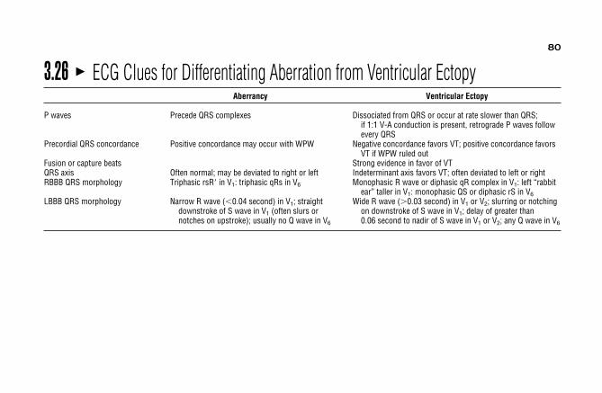
80
3.26 � ECG Clues for Differentiating Aberration from Ventricular EctopyAberrancy Ventricular Ectopy
P waves Precede QRS complexes Dissociated from QRS or occur at rate slower than QRS;if 1:1 V-A conduction is present, retrograde P waves followevery QRS
Precordial QRS concordance Positive concordance may occur with WPW Negative concordance favors VT; positive concordance favorsVT if WPW ruled out
Fusion or capture beats Strong evidence in favor of VTQRS axis Often normal; may be deviated to right or left Indeterminant axis favors VT; often deviated to left or rightRBBB QRS morphology Triphasic rsR in V1: triphasic qRs in V6 Monophasic R wave or diphasic qR complex in V1: left “rabbit
ear” taller in V1: monophasic QS or diphasic rS in V6LBBB QRS morphology Narrow R wave (�0.04 second) in V1; straight Wide R wave (�0.03 second) in V1 or V2; slurring or notching
downstroke of S wave in V1 (often slurs or on downstroke of S wave in V1; delay of greater thannotches on upstroke); usually no Q wave in V6 0.06 second to nadir of S wave in V1 or V2; any Q wave in V6

81
3.27 � Pacemaker CodesFirst Letter: Second Letter: Third Letter: Fourth Letter: Fifth Letter:
Chamber Paced Chamber Sensed Response to Sensing Rate Modulation Multisite Pacinga
0 � None 0 � None 0 � None 0 � None 0 � NoneA � Atrium A � Atrium I � Inhibited R � Rate modulation A � AtrialV � Ventricle V � Ventricle T � Triggered V � VentricularD � Dual (A&V) D � Dual (A&V) D � Dual (I&T) D � Dual
a Multiside indicates either pacing in both atria or both ventricles or pacing multiple sites with a chamber.

82
3.28 � Dual-Chamber Pacing ModesMode Chamber(s) Paced Chamber(s) Sensed Response to Sensing
DVI Atrium and ventricle Ventricle InhibitedVDD Ventricle Atrium and ventricle Atrial sensing triggers ventricular pacing
Ventricular sensing inhibits ventricular pacingDDI Atrium and ventricle Atrium and ventricle InhibitedDDD Atrium and ventricle Atrium and ventricle Atrial sensing inhibits atrial pacing, triggers ventricular pacing
Ventricular sensing inhibits atrial and ventricular pacing

4Cardiovascular conceptsS e c t i o n
� 4.1 Intra-Aortic Balloon Pump Frequencyof 1:2 / 85
� 4.2 Intra-Aortic Balloon Pump Frequencyof 1:1 / 86
� 4.3 Inaccurate Intra-Aortic Balloon PumpTiming / 87
� 4.4 Advanced Cardiovascular LifeSupport (ACLS) Pulseless ArrestAlgorithm / 89
� 4.5 Advanced Cardiovascular LifeSupport (ACLS) BradycardiaAlgorithm / 92
� 4.6 Advanced Cardiovascular LifeSupport (ACLS) TachycardiaAlgorithm / 94
� 4.7 Problems Encountered with ArterialCatheters / 96
� 4.8 Inaccurate Arterial PressureMeasurements / 98
� 4.9 Pulmonary Artery Port Functions / 100
� 4.10 Leveling of the PA Catheter / 101� 4.11 Referencing and Zeroing the
Hemodynamic Monitoring System / 102
� 4.12 Assessing Damping Concepts from Square Wave Test / 103
CARDIOVASCULAR CONCEPTS
84
� 4.13 Pressure Waveforms Observedduring Pulmonary Artery CatheterInsertion / 106
� 4.14 Pulmonary Artery Waveform andComponents / 108
� 4.15 Effect of a Mechanical VentilatorBreath on PA Waveform / 109
� 4.16 Reading End Expiration Before aSpontaneous Breath / 110
� 4.17 Evidence-Based Practice:Pulmonary Artery PressureMeasurement / 111
� 4.18 Problems Encountered withPulmonary Artery Catheters / 112
� 4.19 Inaccurate Pulmonary ArteryPressure Measurements / 118
� 4.20 Troubleshooting Problems withThermodilution Cardiac OutputMeasurements / 121
� 4.21 Common Inotropic Therapies inTreating Abnormal Hemodynamics/ 125
� 4.22 Common Preload Reducers forAbnormal Hemodynamics / 125
� 4.23 Common Afterload ReducingAgents / 126

85
4.1 � Intra-Aortic Balloon Pump Frequency of 1:2
Unassisted systole
Unassisted aorticend-diastolic
pressureAssisted aorticend-diastolic
pressure
Assistedsystole
Diastolicaugmentation
140
120
100
80
mmHg
(Datascope Corporation: Mechanics of intra-aortic balloon counterpulsation. Montvale, NJ: Datascope, 1989.)

86
4.2 � Intra-Aortic Balloon Pump Frequency of 1:1
Assisted aorticend-diastolic
pressure
Assistedsystole
Assistedsystole
Diastolicaugmentation
140
120
100
80
mmHg
(Datascope Corporation: Mechanics of intra-aortic balloon counterpulsation. Montvale, NJ: Datascope, 1989.)

87
4.3 � Inaccurate Intra-Aortic Balloon Pump Timing
Assisted aorticend-diastolic
pressure
Assisted aorticend-diastolic
pressure
Assistedsystole
Assistedsystole
Unassistedsystole
Unassistedsystole
Diastolicaugmentation Diastolic
augmentation
A B
Dicroticnotch
(A) Early inflation. (B) Late inflation.

88
(C) Early deflation. (D) Late deflation. (Datascope Corporation: Mechanics of intra-aortic balloon counterpulsation. Montvale, NJ: Datascope, 1989.)
Unassistedaortic
end-diastolicpressure
Assisted aorticend-diastolic
pressure
Assisted aorticend-diastolic
pressure
Assistedsystole Unassisted
systole
Diastolicaugmentation Diastolic
augmentation
Widenedappearance
Prolonged rateof rise of
assisted systole
DC
4.3 � Inaccurate Intra-Aortic Balloon Pump Timing (continued )

4.4 � Advanced Cardiovascular Life Support (ACLS) Pulseless Arrest Algorithm
89
1
2
3
Pulseless arrest• BLS algorithm: Call for help, give CPR• Give oxygen when available• Attach monitor/defibrillator when available
Check rhythmShockable rhythm?
Asystole/PEAVF/VT
Shockable Not shockable
9
(continued )

90
4
5
6
7
12
13
8
Check rhythmShockable rhythm?
Check rhythmShockable rhythm?
Check rhythmShockable rhythm?
Give 1 shock• Manual biphasic: Device specific (typically 120-200 J) Note: If unknown, use 200 J• AED: Device specific• Monophasic: 360 JResume CPR immediately
Continue CPR while defibrillator is chargingGive 1 shock• Manual biphasic: Device specific (same as first shock or higher dose) Note: If unknown, use 200 J• AED: Device specific• Monophasic: 360 JResume CPR immediately after the shockwhen IV/IO available, give vasopressor during CPR(before or after the shock)• Epinephrine 1 mg IV/IO
Repeat every 3-5 minutes or• May give 1 dose of vasopressin 40 U IV/IO to replace first or second dose of epinephrine
Resume CPR immediately for 5 cycleswhen IV/IO available, give vasopressor• Epinephrine 1 mg IV/IO
Repeat every 3-5 minutes or• May give 1 dose of vasopressin 40 U IV/IO to replace first or second dose of epinephrine
Consider atropine 1 mg IV/IO for asystole or slow PEA rate repeat every 3-5 minutes (up to 3 doses)
• If asystole, go to box 10• If electrical activity, check pulse; if no pulse, go to box 10• If pulse present, begin postresuscitation care
Go tobox 4
Notshockable Shockable
10
11
Give 5 cycles of CPR
Give 5 cycles of CPR
Give 5 cycles of CPR
Shockable
Shockable
No
No
4.4 � Advanced Cardiovascular Life Support (ACLS) Pulseless Arrest Algorithm (continued )

91
8
Continue CPR while defibrillator is chargingGive 1 shock• Manual biphasic: Device specific
(same as first shock or higher dose) Note: If unknown, use 200 J• AED: Device specific• Monophasic: 360 JResume CPR immediately after the shockConsider antiarrhythmics; give during CPR (before or after the shock)
amiodarone (300 mg IV/IO once, then consider additional 150 mg IV/IO once) or
lidocaine (1-1.5 mg/kg first dose, then 0.5- 0.75 mg/kg IV/IO, maximum 3 doses or 3 mg/kg)Consider magnesium, loading dose 1 to 2 g IV/IO for torsades de pointesAfter 5 cycles of CPR, go to box 5 above
During CPR
• Push hard and fast (100/min)• Ensure full chest recoil• Minimize interruptions in chest compressions• One cycle of CPR: 30 compressions then 2 breaths; 5 cycles ≈2 minutes• Avoid hyperventilation• Secure airway and confirm placement
* After an advanced airway is placed, rescuers no longer deliver “cycles” of CPR; give continuous chest com- pressions without pauses for breaths; give 8-10 breaths/min; check rhythm every 2 minutes
• Rotate compressors every 2 minutes with rhythm checks• Search for and treat possible contributing factors:- Hypovolemia- Hypoxia- Hydrogen ion (acidosis)- Hypo-/hyperkalemia- Hypoglycemia- Hypothermia- Toxins- Tamponade, cardiac- Tension pneumothorax- Thrombosis (coronary or pulmonary)- Trauma
Shockable
(Used with permission from 2005 American Heart Association Guidelines for Cardiopulmonary Resuscitation andEmergency Cardiovascular Care. Circultion. Dec 13, 2005;112[24 suppl]:IV59. http://circ.ahajournals.org/con-tent/vol112/24_suppl/. Accessed July 15, 2009.) Abbreviations: AED, automated external defibrillator; BLS, basic lifesupport; CPR, cardiopulmonary resuscitation; IO, intraosseous; IV, intravenous; PEA, pulseless electrical activity;U, units; VF, ventricular fibrillation; VT, ventricular tachycardia.

92
4.5 � Advanced Cardiovascular Life Support (ACLS) Bradycardia Algorithm
• Maintain patent airway; assist breathing as needed• Give oxygen• Monitor ECG (identify rhythm), blood pressure, oximetry • Establish IV access
3
2
1
BradycardiaHeart rate <60 bpm and
inadequate for clinical condition
Signs or symptoms of poor perfusion caused by the bradycardia?
(eg, acute altered mental status, ongoing chest pain, hypotension, or other signs of shock)
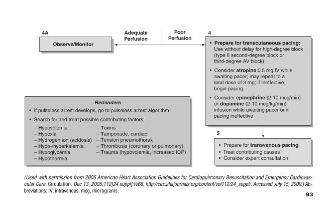
93
Reminders
• If pulseless arrest develops, go to pulseless arrest algorithm
• Search for and treat possible contributing factors:
– Hypovolemia– Hypoxia– Hydrogen ion (acidosis)– Hypo-/hyperkalemia– Hypoglycemia– Hypothermia
– Toxins– Tamponade, cardiac– Tension pneumothorax– Thrombosis (coronary or pulmonary)– Trauma (hypovolemia, increased ICP)
• Prepare for transvenous pacing • Treat contributing causes• Consider expert consultation
5
• Prepare for transcutaneous pacing; Use without delay for high-degree block (type II second-degree block or third-degree AV block)
• Consider atropine 0.5 mg IV while awaiting pacer; may repeat to a total dose of 3 mg; if ineffective, begin pacing
• Consider epinephrine (2-10 mcg/min) or dopamine (2-10 mcg/kg/min) infusion while awaiting pacer or if pacing ineffective
PoorPerfusion
AdequatePerfusion
Observe/Monitor
4A 4
(Used with permission from 2005 American Heart Association Guidelines for Cardiopulmonary Resuscitation and Emergency Cardiovas-cular Care. Circulation. Dec 13, 2005;112[24 suppl]:IV68. http://circ.ahajournals.org/content/vol112/24_suppl/. Accessed July 15, 2009.) Ab-breviations: IV, intravenous; mcg, micrograms.
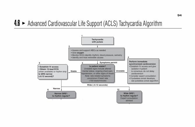
94
4.6 � Advanced Cardiovascular Life Support (ACLS) Tachycardia Algorithm
Tachycardiawith pulses
• Assess and support ABCs as needed• Give oxygen• Monitor ECG (identify rhythm), blood pressure, oximetry• Identify and treat reversible causes
Symptoms persist
Stable Unstable
• Establish IV access• Obtain 12-lead ECG(when available) or rhythm stripIs QRS narrow(<0.12 second)?
Is patient stable?Unstable signs include altered
mental status, ongoing chest pain,hypotension, or other signs of shock
Note: rate-related symptomsuncommon if heart rate
<150 beats/minutes
Perform immediatesynchronized cardioversion• Establish IV access and give sedation if patient is conscious; do not delay cardioversion• Consider expert consultation• If pulseless arrest develops, see pulseless arrest algorithm
Narrow
Narrow QRS*:Is rhythm regular?
Regular Irregular
Wide (≥0.12 seconds)
Wide QRS*:Is rhythm regular?Expert consultation
advised
1
4
2
5
6
7 11
3
12

95
Regular
Regular
Irregular
Irregular• Attempt vagal maneuvers• Give adenosine 6-mg rapidIV push. If no conversion,give 12-mg rapid IV push;may repeat 12 mg dose once
Does rhythmconvert?
Note: Considerexpert consultation
Converts Does not convert
If rhythm converts.probable reentry SVT(reentry supraventriculartachycardia):• Observe for recurrence• Treat recurrence with adenosine or longer- acting AV nodal-blocking agents (eg, diltiazem. β-blockers)
If rhythm does not convert,possible atrial flutter,extopic atrial tachycardia,or junctional tachycardia:• Control rate (eg, diltiazem,
β-blockers; use β-blockers with caution in pulmonary disease or HF)• Treat underlying cause• Consider expert consultation
Irregular narrow-complextachycardiaProbable atrial fibrillation orpossible atrial flutter or MAT(multifocal atrial tachycardia)• Consider expert consultation• Control rate (eg, diltiazem,β-blockers; use β-blockers withcaution in pulmonary diseaseor HF)
Note: If patient becomesunstable, go to Box 4.
During evaluation Treat contributing factors:• Secure, verify airway and vascular access when possible• Consider expert consultation• Prepare for cardioversion
- Hypovolemia- Hypoxia- Hydrogen ion (acidosis)- Hypo-/hyperkalemia- Hypoglycemia- Hypothermia
- Toxins- Tamponade, cardiac- Tension pneumothorax- Thrombosis (coronary or pulmonary- Trauma (hypovolemia)
advised
If ventriculartachycardia oruncertain rhythm• Amiodarone 150 mg IV over 10 minutes repeat as needed to maximum dose of 2.2 g/24 hours• Prepare for elective synchronized cardioversion
If SVT with aberrancy• Give adenosine (go to Box 7)
If atrial fibrillation withaberrancy• See irregular narrow- complex tachycardia (Box 11)
If pre-excited atrialfibrillation (AF + WPW)• Expert consultation advised• Avoid AV nodal blocking agents (eg,
adenosine, digoxin, diltiazem, verapamil)• Consider antiarrhyth- mics (eg, amiodarone 150 mg IV over 10 minutes)If recurrent polymor-phic VT, seek expertconsultationIf torsade de pointes,give magnesium(load with 1-2 g over5-60 minutes, then infusion)
7 11
8
9 10
13 14
(Used with permission from 2005 American Heart Association Guidelines for Cardiopulmonary Resuscitation and Emergency Cardiovascular Care. Circula-tion. Dec 13, 2005;112[24 suppl]:IV70. http://circ.ahajournals.org/content/vol112/24_suppl/. Accessed July 15 2009.) Abbreviations: AF, atrial fibrillation; HF, heart failure; SVT, supraventricular tachycardia; WPW, Wolff-Parkinson-White.

96
4.7 � Problems Encountered with Arterial CathetersProblem Cause Prevention Treatment
Hematoma after withdrawal of needle
Decreased or absent pulse distal to puncture site
Bleedback into tubing or transducer
Bleeding or oozing at puncture site
Spasm of artery
Thrombosis of artery
Insufficient pressure on IV bagLoose connections
Maintain firm pressure on site during withdrawal of catheter and for 5-15 minutes (as necessary) afterwithdrawal.
Apply elastic tape (Elastoplast) firmly over puncture site.
For femoral arterial puncture sites, leave a sandbag on site for 1-2 hoursto prevent oozing.
If patient is receiving unfractionated, discontinue 2 hours before catheterremoval.
Introduce arterial needle cleanly, nontraumatically.
Use 1 U of unfractionated/1 mL IV fluid.
Maintain 300 mm Hg pressure on IV bag.
Use Luer-Lock stopcocks; tighten periodically.
Continue to hold pressure to puncture site until oozing stops.
Apply sandbag to femoral puncture site for 1-2 hours after removal ofcatheter.
Call physician to inject lidocaine locally at insertion site and 10 mg into arterialcatheter.
Arteriotomy and Fogarty catheterization may be needed both distally andproximally from the puncture site resultin return of pulse in �90% of cases ifbrachial or femoral artery is used.
Replace transducer. “Fast flush” through system.
Tighten all connections.
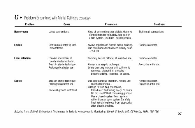
97
Hemorrhage
Emboli
Local infection
Sepsis
Adapted from: Daily E, Schroeder J. Techniques in Bedside Hemodynamic Monitoring. 5th ed. St Louis, MO: CV Mosby; 1994: 165-166.
4.7 � Problems Encountered with Arterial Catheters (continued )
Problem Cause Prevention Treatment
Loose connections
Clot from catheter tip into bloodstream
Forward movement of contaminated catheter
Break in sterile techniqueProlonged catheter use
Break in sterile techniqueProlonged catheter use
Bacterial growth in IV fluid
Keep all connecting sites visible. Observeconnecting sites frequently. Use built-inalarm system. Use Luer-Lock stopcocks.
Always aspirate and discard before flushing.Use continuous flush device. Gently flush�2-4 mL.
Carefully secure catheter at insertion site.
Always use aseptic technique.Leave dressing in place until catheter is
removed, changed, or dressingbecomes damp, loosened, or soiled.
Use percutaneous insertion. Always useaseptic technique.
Change IV fluid bag, stopcocks,transducer, and tubing every 72 hours.Do not use IV fluid containing glucose.Use a closed-system flush systemrather than an open system Carefullyflush remaining blood from stopcocksafter blood sampling.
Tighten all connections.
Remove catheter.
Remove catheter.
Prescribe antibiotic.
Remove catheter.Prescribe antibiotic.

98
4.8 � Inaccurate Arterial Pressure MeasurementsProblem Cause Prevention Treatment
Damped pressure tracing Catheter tip against vessel wall Usually cannot be avoided. Call physician to pull back, rotate, or reposition catheter whileobserving pressure waveform.
Partial occlusion of catheter tip by clot Use continuous infusion under Aspirate clot with syringe and flush pressure. Briefly “fast flush” with saline (�2-4 mL). Consider after blood withdrawal (2-4 mL). line removal.
Clot in stopcock or transducer Carefully flush catheter after blood Flush stopcock and transducer; if no withdrawal and reestablish IV improvement, change stopcock and drip. Use continuous flush device. transducer.
Air bubbles in transducer or connector Carefully flush transducer and tubing Check system; flush rapidly; disconnecttubing when setting up system and attaching transducer and flush out air bubbles.
to catheter.Compliant tubing Use stiff, short tubing. Shorten tubing or replace softer
tubing with stiffer tubing.

99
Abnormally high or low Change in transducer air-reference Maintain air-reference port of Recheck patient and transducer readings level transducer at midchest and/or positions.
catheter tip level for serial pressure measurements.
No pressure available Transducer not open to catheter Follow routine, systematic steps for Check system—stopcocks, monitor, Settings on monitor amplifiers setting up system and turning and amplifier setup.incorrect—still on zero, cal, or off stopcocks.
Incorrect scale selection Select scale appropriate to expected Select appropriate scale.range of physiologic signal.
Adapted from: Daily E, Schroeder J. Techniques in Bedside Hemodynamic Monitoring. 5th ed. St Louis, MO: CV Mosby; 1994:161
4.8 � Inaccurate Arterial Pressure Measurements (continued )
Problem Cause Prevention Treatment
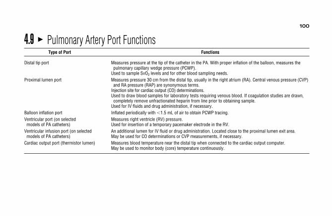
100
4.9 � Pulmonary Artery Port FunctionsType of Port Functions
Distal tip port Measures pressure at the tip of the catheter in the PA. With proper inflation of the balloon, measures the pulmonary capillary wedge pressure (PCWP).
Used to sample SvO2 levels and for other blood sampling needs.Proximal lumen port Measures pressure 30 cm from the distal tip, usually in the right atrium (RA). Central venous pressure (CVP)
and RA pressure (RAP) are synonymous terms.Injection site for cardiac output (CO) determinations.Used to draw blood samples for laboratory tests requiring venous blood. If coagulation studies are drawn,
completely remove unfractionated heparin from line prior to obtaining sample.Used for IV fluids and drug administration, if necessary.
Balloon inflation port Inflated periodically with �1.5 mL of air to obtain PCWP tracing.Ventricular port (on selected Measures right ventricle (RV) pressure.models of PA catheters) Used for insertion of a temporary pacemaker electrode in the RV.
Ventricular infusion port (on selected An additional lumen for IV fluid or drug administration. Located close to the proximal lumen exit area.models of PA catheters) May be used for CO determinations or CVP measurements, if necessary.
Cardiac output port (thermistor lumen) Measures blood temperature near the distal tip when connected to the cardiac output computer.May be used to monitor body (core) temperature continuously.

101
4.10 � Leveling of the PA Catheter
Stopcock opento atmosphere
Midaxillary Line(where distance “A” = distance “B”)
Typical leveling of PA catheter with stopcock attached to the transducer for mounting on a pole. The stopcock close to thetransducer is opened to atmospheric pressure (air) horizontal to the fourth ICS at the midaxillary line.
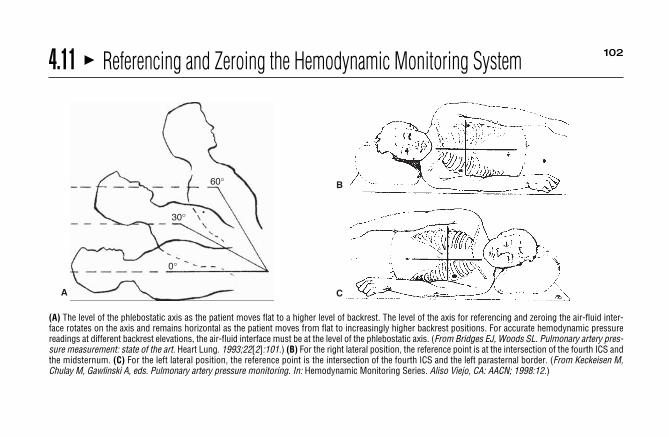
102
60°
30°
0°
A
(A) The level of the phlebostatic axis as the patient moves flat to a higher level of backrest. The level of the axis for referencing and zeroing the air-fluid inter-face rotates on the axis and remains horizontal as the patient moves from flat to increasingly higher backrest positions. For accurate hemodynamic pressurereadings at different backrest elevations, the air-fluid interface must be at the level of the phlebostatic axis. (From Bridges EJ, Woods SL. Pulmonary artery pres-sure measurement: state of the art. Heart Lung. 1993;22[2]:101.) (B) For the right lateral position, the reference point is at the intersection of the fourth ICS andthe midsternum. (C) For the left lateral position, the reference point is the intersection of the fourth ICS and the left parasternal border. (From Keckeisen M,Chulay M, Gawlinski A, eds. Pulmonary artery pressure monitoring. In: Hemodynamic Monitoring Series. Aliso Viejo, CA: AACN; 1998:12.)
C
B
4.11 � Referencing and Zeroing the Hemodynamic Monitoring System

103
4.12 � Assessing Damping Concepts from Square Wave TestSquare Wave Test Clinical Effect Corrective Action
Optimally damped Produces accurate waveform None required.and pressure.
Optimally damped system. When the fast flushof the continuous flush system is activatedand quickly released, a sharp upstroke termi-nates in a flat line at the maximal indicator onthe monitor and hard copy. This is followedby an immediate and rapid downstroke ex-tending below the baseline with just 1 or 2 os-cillations within 0.12 second (minimal ringing)
Observedwaveform
and a quick return to baseline. The patient’s pressure waveform is also clearlydefined with all components of the waveform, such as the dicrotic notch onan arterial waveform, clearly visible. Intervention: There is no adjustment in themonitoring system required. (Reprinted from Darovic GO, Vanriper S, VanriperJ. Fluid-filled monitoring systems. In: Darovic GO, ed. Hemodynamic Monitoring:Invasive and Noninvasive Clinical Application. 3rd ed. Philadelphia, PA: WBSaunders Co; 1995:161-162. Used with permission from Elsevier.)
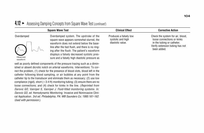
104
Observedwaveform
Overdamped Produces a falsely low Check the system for air, blood,systolic and high loose connections or kinksdiastolic value. in the tubing or catheter.
Verify extension tubing has notbeen added.
4.12 � Assessing Damping Concepts from Square Wave Test (continued )
Square Wave Test Clinical Effect Corrective Action
well as poorly defined components of the pressure tracing such as a dimin-ished or absent dicrotic notch on arterial waveforms. Interventions: To cor-rect the problem, (1) check for the presence of blood clots, blood left in thecatheter following blood sampling, or air bubbles at any point from thecatheter tip to the transducer and eliminate them as necessary; (2) use lowcompliance (rigid), short (�3-4 ft) monitoring tubing; (3) ensure there are noloose connections; and (4) check for kinks in the line. (Reprinted fromDarovic GO, Vanriper S, Vanriper J. Fluid-filled monitoring systems. In:Darovic GO, ed. Hemodynamic Monitoring: Invasive and Noninvasive Clini-cal Application. 3rd ed. Philadelphia, PA: WB Saunders Co; 1995:161-162.Used with permission.)
Overdamped system. The upstroke of thesquare wave appears somewhat slurred, thewaveform does not extend below the base-line after the fast flush, and there is no ring-ing after the flush. The patient’s waveformdisplays a falsely decreased systolic pres-sure and a falsely high diastolic pressure as

105
Observedwaveform
Underdamped Produces a falsely high Remove unnecessary tubing systolic and low and stopcocks.diastolic value. Add a damping device.
4.12 � Assessing Damping Concepts From Square Wave Test (continued )
Square Wave Test Clinical Effect Corrective Action
system. Use large bore, shorter tubing, or a damping device. (Reprinted fromDarovic GO, Vanriper S, Vanriper J. Fluid-filled monitoring systems. In:Darovic GO, ed. Hemodynamic Monitoring: Invasive and Noninvasive Clini-cal Application. 3rd ed. Philadelphia, PA: WB Saunders Co; 1995:161-162.Used with permission from Elsevier.)
Underdamped system. The waveform ischaracterized by numerous amplified oscilla-tions above and below the baseline followingthe fast flush. The monitored pressure wavedisplays falsely high systolic pressure(overshoot), possibly falsely low diastolicpressures, and “ringing” artifacts on thewaveform. Intervention: To correct theproblem, remove all air bubbles in the fluid

106
4.13 � Pressure Waveforms Observed During Pulmonary Artery Catheter InsertionLocation Pressure Waveform Normal Pressures
Right atrium 2-8 mm Hg
Right ventricle Systolic, 20-30 mm HgDiastolic, 0-5 mm Hg
60
40
20
0
60
40
20
0

107
4.13 � Pressure Waveforms Observed During Pulmonary Artery Catheter Insertion (continued )
Location Pressure Waveform Normal Pressures
Pulmonary artery Systolic, 20-30 mm HgDiastolic, 10-15 mm Hg
Pulmonary artery wedge 8-12 mm Hg
With permission from: Boggs R, Wooldridge-King M. AACN Procedure Manual. 3rd ed. Philadelphia, PA: WB Saunders; 1993:308, 324, 326, 334.
60
40
20
0
60
40
20
0

108
1, PA systole; 2, dichrotic notch; 3, PA end diastole; 4, anacrotic notch of PA valve opening. (From: Boggs R, Wooldridge-King M. AACN Procedure Manual for Critical Care, 3rd ed. Philadelphia, PA: WB Saunders; 1993:316.)
4.14 � Pulmonary Artery Waveform and Components

109
(From: Ahrens TS. Hemodynamic Waveform Recognition. Philadelphia, PA: WB Saunders; 1993:92.)
4.15 � Effect of a Mechanical Ventilator Breath on PA Waveform
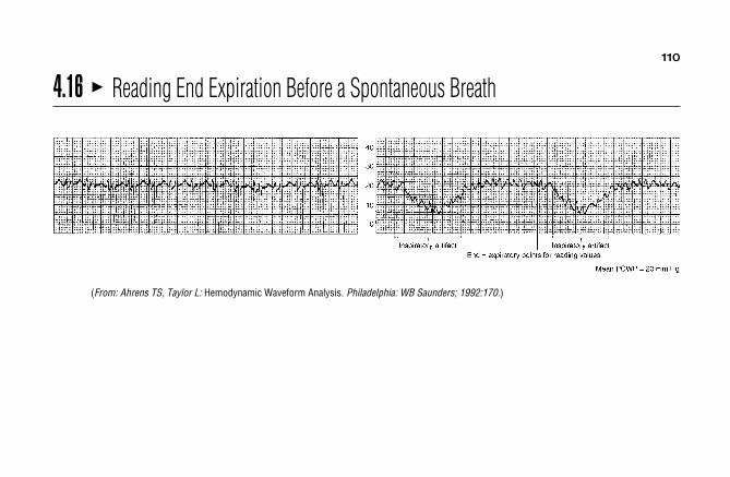
110
(From: Ahrens TS, Taylor L: Hemodynamic Waveform Analysis. Philadelphia: WB Saunders; 1992:170.)
4.16 � Reading End Expiration Before a Spontaneous Breath

111
4.17 � Evidence-Based Practice: Pulmonary Artery Pressure Measurement• Verify the accuracy of the transducer-patient interface by performing a square waveform test at the beginning of each shift.a,b
• Position the patient supine prior to PAP/RAP (CVP)/PCWP measurements. Head of bed elevation can be at any angle from 0° (flat) to 60°.a,b
• Level the transducer air-fluid interface to the phlebostatic axis (4th ICS/1⁄2 AP diameter of the chest) with the patient in a supine position prior toPAP/RAP/PCWP measurements.a,b
• Obtain PAP/RAP/PCWP measurements from a graphic (analog) tracing at end-expiration.a,b
• Use a simultaneous ECG tracing to assist with proper PAP/RAP/PCWP waveform identification.a,b
• PA catheters can be safely withdrawn and removed by competent registered nurses.a,b
Data from: aAmerican Association of Critical-Care Nurses (2004); bKeckeisen (1998).

112
Phlebitis or local infection at insertion site
Ventricular irritability
Mechanical irritation or contamination
Looping of excess catheter in RA
Migration of catheter from PA to RV
Irritation of the endocardium during catheter passage
Prepare skin properly before insertion. Use sterile technique during insertionand dressing change. Insert smoothlyand rapidly. Use Teflon-coatedintroducer. Change IV fluid bag,transducer, stopcocks, and connectingtubing every 72 hours. Removecatheter or change insertion site.
Carefully secure catheter at insertion site; check chest film.
Position catheter tip in main right orleft PA.
Keep balloon inflated during advancement; advance gently.
Remove catheter.Apply warm compresses.Give pain medication as necessary.
Call physician to reposition catheter; remove loop.
Inflate balloon to encourage catheter flotation out to PA.
4.18 � Problems Encountered with Pulmonary Artery CathetersProblem Cause Prevention Treatmen

113
4.18 � Problems Encountered with Pulmonary Artery Catheters (continued )
Problem Cause Prevention Treatment
Apparent wedging of catheter with balloon deflated
Aspirate blood from catheter; if catheter is wedged, sample will bearterialized and obtained withdifficulty.
If wedged, call physician to slowly pull back catheter until PA waveformappears. If not wedged, gentlyaspirate and flush catheter withsaline; catheter tip can partially clot,causing damping that resemblesdamped PAW waveform.
Forward migration of catheter tipcaused by blood flow, excessive loopin RV, or inadequate suturing ofcatheter at insertion site
Check catheter tip by radiograph or fluoroscopy; position in main rightor left PA. Carefully secure catheterat insertion site.
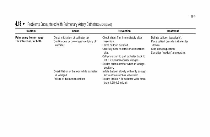
114
4.18 � Problems Encountered with Pulmonary Artery Catheters (continued )
Problem Cause Prevention Treatment
Pulmonary hemorrhage or infarction, or both
Distal migration of catheter tipContinuous or prolonged wedging of catheter
Overinflation of balloon while catheter is wedged
Failure of balloon to deflate
Check chest film immediately after insertion.
Leave balloon deflated.Carefully secure catheter at insertion site.
Call physician to pull catheter back to PA if it spontaneously wedges.
Do not flush catheter when in wedgeposition.
Inflate balloon slowly with only enoughair to obtain a PAW waveform.
Do not inflate 7-Fr catheter with more than 1.25-1.5 mL air.
Deflate balloon (passively).Place patient on side (catheter tip down).
Stop anticoagulation.Consider “wedge” angiogram.
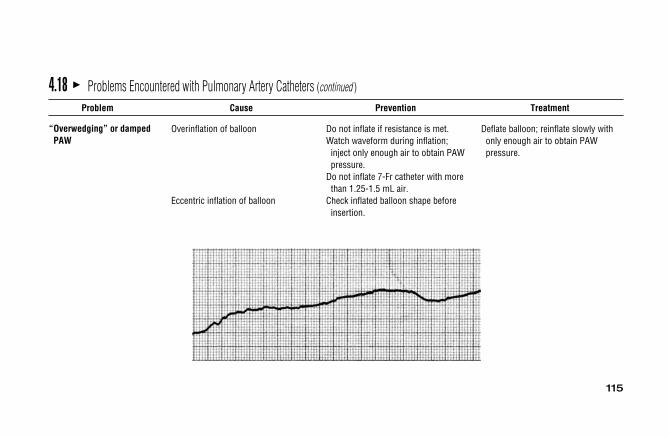
115
4.18 � Problems Encountered with Pulmonary Artery Catheters (continued )
Problem Cause Prevention Treatment
“Overwedging” or damped PAW
Overinflation of balloon
Eccentric inflation of balloon
Do not inflate if resistance is met.Watch waveform during inflation; inject only enough air to obtain PAWpressure.
Do not inflate 7-Fr catheter with more than 1.25-1.5 mL air.
Check inflated balloon shape before insertion.
Deflate balloon; reinflate slowly with only enough air to obtain PAWpressure.

116
4.18 � Problems Encountered with Pulmonary Artery Catheters (continued )
Problem Cause Prevention Treatment
PA balloon rupture
Infection
Overinflation of balloonFrequent inflations of balloonSyringe deflation damaging wall ofballoon
Nonsterile insertion techniques
Contamination via skin
Contamination through stopcockports or catheter hub
Inflate slowly with only enough air to obtain a PAW pressure.
Monitor PAD pressure as reflection of PAW and LVEDP.
Allow passive deflation of balloon.Remove syringe after inflation.
Use sterile techniques.Use sterile catheter sleeve.Prepare skin with effective antiseptic (chlorhexidine).
Reassess need for catheter after 3 days.
Avoid internal jugular approach.Use a closed-system flush system rather than an open system.
Use sterile dead-end caps on all stopcock ports.
Change tubing, continuous flush device transducer, and flush solutionevery 72 hours.
Do not use IV flush solution that contains glucose.
Remove syringe to prevent further air injection.
Monitor PAD pressure.
Remove catheter.Use antibiotics.
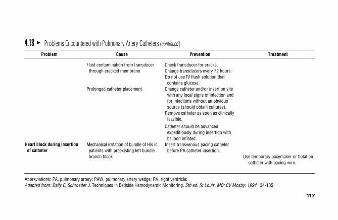
117
4.18 � Problems Encountered with Pulmonary Artery Catheters (continued )
Problem Cause Prevention Treatment
Heart block during insertion of catheter
Abbreviations: PA, pulmonary artery; PAW, pulmonary artery wedge; RV, right ventricle; Adapted from: Daily E, Schroeder J. Techniques in Bedside Hemodynamic Monitoring. 5th ed. St Louis, MO: CV Mosby; 1994:134-135
Fluid contamination from transducerthrough cracked membrane
Prolonged catheter placement
Mechanical irritation of bundle of His inpatients with preexisting left bundlebranch block
Check transducer for cracks.Change transducers every 72 hours.Do not use IV flush solution that contains glucose.
Change catheter and/or insertion site with any local signs of infection andfor infections without an obvioussource (should obtain cultures).
Remove catheter as soon as clinically feasible.
Catheter should be advanced expeditiously during insertion with balloon inflated.
Insert transvenous pacing catheter before PA catheter insertion.
Use temporary pacemaker or flotationcatheter with pacing wire.
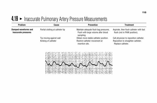
118
4.19 � Inaccurate Pulmonary Artery Pressure MeasurementsProblem Cause Prevention Treatment
Damped waveforms and inaccurate pressures
Partial clotting at catheter tip
Tip moving against wallKinking of catheter
Maintain adequate flush bag pressures.Flush with large volume after bloodsampling.
Obtain more stable catheter position.Restrict catheter movement at insertion site.
Aspirate, then flush catheter with fast flush (not in PAW position).
Call physician to reposition catheter.Reposition to straighten catheter. Replace catheter.

119
4.19 � Inaccurate Pulmonary Artery Pressure Measurements (continued )
Problem Cause Prevention Treatment
Abnormally low or negative pressures
Abnormally high pressure reading
Inappropriate pressure waveform
Incorrect air-reference level (above midchest level)
Incorrect zeroing and calibration of monitor
Loose connection
Pressure trapped by improper sequence of stopcock operation
Incorrect air-reference level (below midchest level)
Migration of catheter tip (eg, in RV or PAW instead of in PA)
Maintain transducer air-reference port at midchest level; rezero after patientposition changes.
Zero and calibrate monitor properly.Use Luer-Lock stopcocks.Use Luer-Lock stopcocks.
Turn stopcocks in proper sequence when two pressures are measuredon one transducer.
Maintain transducer air-reference portat midchest level; recheck and rezeroafter patient position changes.
Establish optimal position carefully when introducing catheter initially.
Secure catheter at insertion site andtape catheter to patient’s skin.
Remeasure level of transducer air-reference and reposition atmidchest level; rezero.
Recheck zero and calibration of monitor.
Check all connections.
Thoroughly flush transducers with IV solution; rezero and turn stopcocksin proper sequence.
Check air-reference level; reset at midchest and rezero.
Review waveform; if RV, call physician to reposition catheter.Check position under x-ray and/orfluoroscopy after reposition.
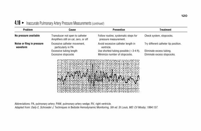
120
4.19 � Inaccurate Pulmonary Artery Pressure Measurements (continued )
Problem Cause Prevention Treatment
No pressure available
Noise or fling in pressure waveform
Abbreviations: PA, pulmonary artery; PAW, pulmonary artery wedge; RV, right ventricle.Adapted from: Daily E, Schroeder J. Techniques in Bedside Hemodynamic Monitoring. 5th ed. St Louis, MO: CV Mosby; 1994:137.
Transducer not open to catheterAmplifiers still on cal, zero, or off
Excessive catheter movement,particularly in PA
Excessive tubing lengthExcessive stopcocks
Follow routine, systematic steps for pressure measurement.
Avoid excessive catheter length inventricle.
Use shortest tubing possible (�3-4 ft).Minimize number of stopcocks.
Check system, stopcocks.
Try different catheter tip position.
Eliminate excess tubing.Eliminate excess stopcocks.
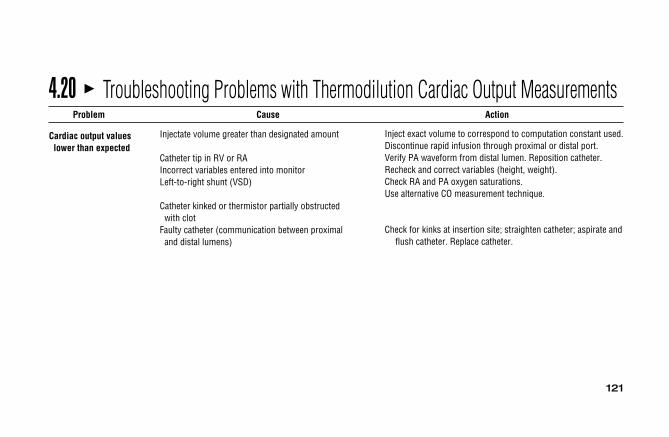
121
Injectate volume greater than designated amount
Catheter tip in RV or RAIncorrect variables entered into monitorLeft-to-right shunt (VSD)
Catheter kinked or thermistor partially obstructed with clot
Faulty catheter (communication between proximal and distal lumens)
Inject exact volume to correspond to computation constant used.Discontinue rapid infusion through proximal or distal port.Verify PA waveform from distal lumen. Reposition catheter.Recheck and correct variables (height, weight).Check RA and PA oxygen saturations.Use alternative CO measurement technique.
Check for kinks at insertion site; straighten catheter; aspirate andflush catheter. Replace catheter.
4.20 � Troubleshooting Problems with Thermodilution Cardiac Output MeasurementsProblem Cause Action
Cardiac output valueslower than expected
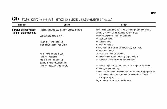
122
4.20 � Troubleshooting Problems with Thermodilution Cardiac Output Measurements (continued )
Problem Cause Action
Cardiac output values higher than expected
Injectate volume less than designated amount
Catheter too distal (PAW)
RA port lies within sheathThermistor against wall of PA
Fibrin covering thermistor Incorrect variablesRight-to-left shunt (VSD)Severe tricuspid regurgitationIncorrect injectate temperature
Inject exact volume to correspond to computation constant.Carefully remove all air bubbles from syringe.Verify PA waveform from distal lumen.Pull catheter back.Advance catheter.Reposition patient.Rotate catheter to turn thermistor away from wall.Reposition catheter.Check a-vDo2; change catheter.Recheck and correct variables (height, weight).Use alternative CO measurement technique.
Use closed injectate system with in-line temperature probe.Handle syringe minimally.Do not turn stopcock to reestablish IV infusion through proximal
port between injections; reduce or discontinue IV flowthrough VIP port.
Try to determine cause of interference.

123
4.20 � Troubleshooting Problems with Thermodilution Cardiac Output Measurements (continued )
Problem Cause Action
Irregular upslope of CO curve
Magnetic interference producing numerous spikes inCO curve
Long lag time between injection and upstroke of curve
Uneven injection techniqueRA port partially occluded with clot
Catheter partially kinked
Press start button after injection completed to delay computer sampling time.
Inject smoothly and quickly (10 mL in �4 seconds).Always check catheter patency by withdrawing, then flushing proximal port before CO determinations.
Check for kinks, particularly at insertion site; straighten catheter; reposition patient.

1244.20 � Troubleshooting Problems with Thermodilution Cardiac Output Measurements (continued )
Problem Cause Action
Irregular downslope of CO curve
Abbreviations: AV, atrioventricular; CO, cardiac output; CPR, cardiopulmonary resuscitation; ECG, electrocardiogram; IV, intravenous; NSR, normal sinusrhythm; MAT, multifocal atrial tachycardia; PAC, premature atrial contraction; PJC, premature junctional complex; PVC, premature ventricular complexes; VT,ventricular tachycardia.Adaption from: Daily E, Schroeder J. Techniques in Bedside Hemodynamic Monitoring. 5th ed. St Louis, MO; CV Mosby; eds, 1994: 183–184 Cardiac outputwaveforms from: Gardner P. Cardiac output: theory, technique and troubleshooting. In: Underhill SL, Woods S, Froelicher E, et al. Cardiac Nursing. 2nd ed. Philadelphia, PA: JB Lippincott; 1989:465.
Cardiac arrhythmias (PVC, AF, etc)
Marked movement of catheter tip
Marked variation in PA baseline temperature
Curve prematurely terminated
Right-to-left shunt
Note ECG during CO determinations.Try to inject during a stable period.Increase the number of CO determinations.Obtain x-ray film to determine position of tip.Advance catheter tip away from pulmonic value.Use iced temperature injectate to increase signal/noise ratio.Increase the number of CO determinations.Inject at various times during respiratory cycle.Press start button after injection completed to delay computer sampling time.
Use alternative CO measurement technique.

125
4.21 � Common Inotropic Therapies inTreating AbnormalHemodynamics
Onset ofDrug Dosage Action Route
Dobutamine 1-20 mcg/kg/min 1-2 minutes IV(Dobutrex)
Dopamine (Intropin) 2-10 mcg/kg/min 1-2 minutes IV
Milrinone (Primacor) Loading 0.75 mg/kg, �5 minutes IVthen 5-10 mcg/kg/min
Digoxin (Lanoxin) 0.5 mg at first; then(normally not used 0.25 every 6 hoursin acute LV failure) until desired effect, 1-2 hours IV
then 0.125-0.25 mg/day
4.22 � Common Preload Reducers forAbnormal Hemodynamics
Onset ofDrug Dosage Action Route
Diuretic AgentsFurosemide (Lasix) 20 mg or higher �5 minutes IV/POBumetanide (Bumex) 0.5-10 mg/day �5 minutes IV/POEthacrynic Acid (Edecrin) 50-100 mg/day �5 minutes IV/POChlorothiazide (Diuril) 500-2000 mg/day 1-2 hours IV/POMetolazone (Zaroxolyn) 2.5-20 mg/day 1 hour POMannitol (Osmitrol) 12.5-200 g/day �5 minutes IVVasodilating AgentsDopamine (Intropin) 1-2 mcg/kg/min �5 minutes IVNitroglycerine 5-400 mcg 1-2 minutes IV(Tridil, Nitrostat IV)

126
4.23 � Common Afterload Reducing AgentsDrug Dose Onset of Action Route
Smooth Muscle Relaxants and Alpha InhibitorsNitroprusside (Nipride) 0.5-10 mcg/kg/min 1-2 minutes IVNitroglycerine (Tridil, Nitrostat IV) 5-400 mcg 1-2 minutes IVDiazoxide (Hyperstat IV) 50-150 mg 1-2 minutes IVHydralazine (Apresoline) 10-40 mg 10-20 minutes IV/IMMethyldopa (Aldomet) 250 mg-1 g 2 hours IVTrimethaphan (Arfonad) 3-6 mg/min 1-2 minutes IVPhentolamine (Regitine) 0.1-2 mg/min �1 minute IV
Angiotension-Converting Enzyme InhibitorsCaptopril (Capoten) 25-400 mg/day in 2-3 doses 15-30 minutes POEnalapril/Enalaprilat (Vasotec/Vasotec IV) 2.5-4.0 mg/day 15 minutes PO/IV
Lisinopril (Zestril) 10-40 mg/day 1 hour PO

5Respiratory ConceptsS e c t i o n
� 5.1 Normal Chest X-Ray / 128� 5.2 Mediastinal Structures Visible on a
Chest X-Ray / 129� 5.3 Chest X-Ray of COPD / 130� 5.4 Chest X-Ray of Pneumothorax / 131� 5.5 Chest X-Ray of Right Lower Lobe
Pneumonia / 132� 5.6 Chest X-Ray Showing Carina
and Right Bronchus / 133
� 5.7 Chest X-Ray with PA Catheter, ETTube, and Chest Tube / 134
� 5.8 Acid-Base Abnormalities / 135� 5.9 Indications for Mechanical
Ventilation / 136� 5.10 Pulmonary Specific Wean Criteria
Thresholds / 137� 5.11 Burns’ Wean Assessment Program
(BWAP) / 138
� 5.12 Algorithm for Management ofVentilator Alarms and/orDevelopment of Acute RespiratoryDistress / 140
� 5.13 Algorithm to Correct Hypoxemia inan Acute COPD Patient / 141
RESPIRATORY SYSTEM

5.1 � Normal Chest X-Ray128
Normal chest x-ray film taken of a 28-year-old woman from a PA view. Some anatomic structures can be seen on the x-ray:(A) diaphragm; (B) costophrenic angle; (C) left ventricle; (D) rightatrium; (E) aortic arch (referred to as aortic knob); (F) superiorvena cava; (G) trachea; (H) right bronchus (right hilum); (I) leftbronchus (left hilum); and (J) breast shadows.

5.2 � Mediastinal Structures Visible on a Chest X-Ray
129
(1) Trachea, (2) first rib, (3) superior vena cava, (4) aortic knob, (5) pul-monary artery, (6) left main bronchus, (7) right main bronchus, (8) leftatrium, (9) right atrium, and (10) left ventricle. (From: Sanchez F. Funda-mentals of chest x-ray interpretation. Crit Care Nurse. 1986;6[5]:53.)
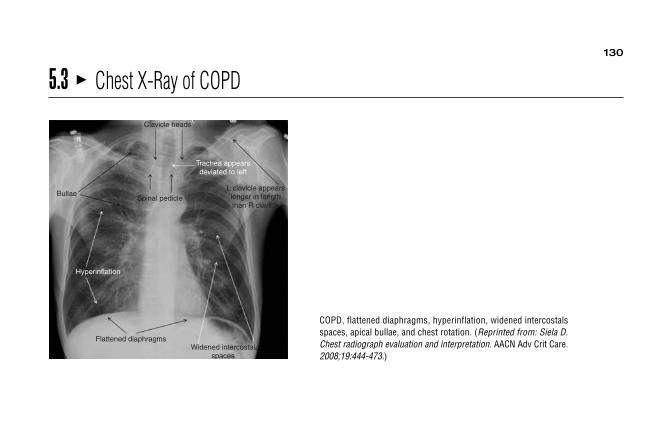
130
5.3 � Chest X-Ray of COPD
COPD, flattened diaphragms, hyperinflation, widened intercostalsspaces, apical bullae, and chest rotation. (Reprinted from: Siela D.Chest radiograph evaluation and interpretation. AACN Adv Crit Care.2008;19:444-473.)
Bullae
Clavicle heads
Spinal pedicle
Trachea appearsdeviated to left
L clavicle appearslonger in lengththan R clavicle
Hyperinflation
Flattened diaphragmsWidened intercostal
spaces

131
5.4 � Chest X-Ray of Pneumothorax
Left pneumothorax, hyperlucency, and widened intercostals spaces.(Reprinted from: Siela D. Chest radiograph evaluation and interpre-tation. AACN Adv Crit Care. 2008;19:444-473.)
Slight mediastinal shift
Widened intercostalspaces in left thorax
Hyperlucency
Lung

132
5.5 � Chest X-Ray of Right Lower Lobe Pneumonia
Right lower lobe pneumonia with minor fissure visualized.(Reprinted from: Siela D. Chest radiograph evaluation and interpre-tation. AACN Adv Crit Care. 2008;19:444-473.)
Minor fissure
Right heart bordervisualized
Right lower lobe

133
5.6 � Chest X-Ray Showing Carina and Right Bronchus
Carina and right bronchus. (Reprinted from: Siela D. Chest radio-graph evaluation and interpretation. AACN Adv Crit Care.2008;19:444-473.)
R bronchus L bronchus
Carina

134
5.7 � Chest X-Ray with PA Catheter, ET Tube, and Chest Tube
Pulmonary artery catheter, endotracheal tube, and left chest tube.(Reprinted from: Siela D. Chest radiograph evaluation and interpre-tation. AACN Adv Crit Care. 2008;19:444-473.)
PAC
ET tube
ET tube tip
Chesttube
PAC
PAC

135
5.8 � Acid-Base AbnormalitiesPrimary ABG Abnormalities ABG Changes with Compensation (if present)
Acid-Base Abnormality pH PaCO2 HCO3� Respiratory (PaCO2) Metabolic (HCO3
�)
AlkalemiaMetabolic ↑ ↑ ↑Respiratory ↑ ↓ ↓
AcidemiaMetabolic ↓ ↓ ↓Respiratory ↓ ↑ ↑
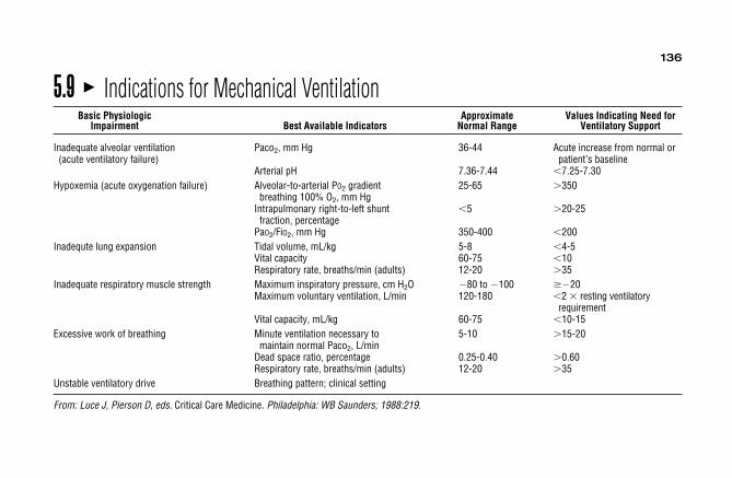
5.9 � Indications for Mechanical VentilationBasic Physiologic Approximate Values Indicating Need for
Impairment Best Available Indicators Normal Range Ventilatory Support
Inadequate alveolar ventilation Paco2, mm Hg 36-44 Acute increase from normal or(acute ventilatory failure) patient’s baseline
Arterial pH 7.36-7.44 �7.25-7.30Hypoxemia (acute oxygenation failure) Alveolar-to-arterial PO2 gradient 25-65 �350
breathing 100% O2, mm HgIntrapulmonary right-to-left shunt �5 �20-25
fraction, percentagePaO2/FiO2, mm Hg 350-400 �200
Inadequte lung expansion Tidal volume, mL/kg 5-8 �4-5Vital capacity 60-75 �10Respiratory rate, breaths/min (adults) 12-20 �35
Inadequate respiratory muscle strength Maximum inspiratory pressure, cm H2O �80 to �100 ��20Maximum voluntary ventilation, L/min 120-180 �2 � resting ventilatory
requirementVital capacity, mL/kg 60-75 �10-15
Excessive work of breathing Minute ventilation necessary to 5-10 �15-20maintain normal Paco2, L/min
Dead space ratio, percentage 0.25-0.40 �0.60Respiratory rate, breaths/min (adults) 12-20 �35
Unstable ventilatory drive Breathing pattern; clinical setting
From: Luce J, Pierson D, eds. Critical Care Medicine. Philadelphia: WB Saunders; 1988:219.
136

137
5.10 � Pulmonary Specific Wean Criteria ThresholdsTraditional Weaning Criteria• Negative inspiratory pressure (NIP) � −20 cm H2O• Positive expiratory pressure (PEP) � +30 cm H2O• Spontaneous tidal volume (SVT) �5 mL/kg• Vital capacity (VC) �15 mL/kg• Fraction of inspired oxygen (FiO2) �50%• Minute ventilation (MV) �10 L/minIntegrated Weaning Criteria• Index of rapid shallow breathing or frequency tidal volume ratio (fx/Vt) �105
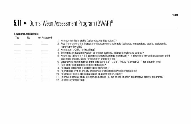
5.11 � Burns’ Wean Assessment Program (BWAP)a
I. General AssessmentYes No Not Assessed
_____ _____ _____ 1. Hemodynamically stable (pulse rate, cardiac output)?_____ _____ _____ 2. Free from factors that increase or decrease metabolic rate (seizures, temperature, sepsis, bacteremia,
hypo/hyperthyroid)?_____ _____ _____ 3. Hematocrit �25% (or baseline)?_____ _____ _____ 4. Systemically hydrated (weight at or near baseline, balanced intake and output)?_____ _____ _____ 5. Nourished (albumin �2.5, parenteral/enteral feedings maximized)? *If albumin is low and anasarca or third
spacing is present, score for hydration should be “no.”_____ _____ _____ 6. Electrolytes within normal limits (including Ca��, Mg�, PO4)? *Correct Ca�� for albumin level._____ _____ _____ 7. Pain controlled (subjective determination)?_____ _____ _____ 8. Adequate sleep/rest (subjective determination)?_____ _____ _____ 9. Appropriate level of anxiety and nervousness (subjective determination)?_____ _____ _____ 10. Absence of bowel problems (diarrhea, constipation, ileus)?_____ _____ _____ 11. Improved general body strength/endurance (ie, out of bed in chair, progressive activity program)?_____ _____ _____ 12. Chest x-ray improving?
138

139
5.11 � Burns’ Wean Assessment Program (BWAP)a (continued )
II. Respiratory AssessmentYes No Not Assessed
Gas Flow and Work of Breathing_____ _____ _____ 13. Eupneic respiratory rate and pattern (spontaneous RR �25, without dyspnea, absence of accessory muscle
use)? *This is assessed off the ventilator while measuring #20-23._____ _____ _____ 14. Absence of adventitious breath sounds (rhonchi, rales, wheezing)?_____ _____ _____ 15. Secretions thin and minimal?_____ _____ _____ 16. Absence of neuromuscular disease/deformity?_____ _____ _____ 17. Absence of abdominal distention/obesity/ascites?_____ _____ _____ 18. Oral ETT � #7.5 or trach � #6.5?
Airway Clearance_____ _____ _____ 19. Cough and swallow reflexes adequate?
Strength_____ _____ _____ 20. NIP �20 (negative inspiratory pressure)?_____ _____ _____ 21. PEP �30 (positive expiratory pressure)?
Endurance_____ _____ _____ 22. STV �5 mL/kg (spontaneous tidal volume)?_____ _____ _____ 23. VC �10-15 mL/kg (vital capacity)?
ABGs_____ _____ _____ 24. pH 7.30-7.45?_____ _____ _____ 25. PaCO2, 40 mm Hg (or baseline) with mV �10 L/min? *This is evaluated while on ventilator._____ _____ _____ 26. PaO2 �60 on FiO2 �40%?
aThe BWAP score is obtained by dividing the total number of BWAP factors scored as “yes” by 26. ©Burns 1990.

5.12 � Algorithm for Management of Ventilator Alarms and/or Development of AcuteRespiratory Distress
140
PR
OB
LE
MS
:• S
igns/symptom
s of respiratory distress• S
udden change in clinical condition• V
entilator alarm• A
bnormal ventilator function
Is the patient inrespiratory distress?
orhem
odynamic
instability?
Check ventilator to
determine if problem
exists• R
emove patient from
ventilator and ventilate
m
anually with 100%
O2
• Perform
rapid exam,
w
ith CP
emphasis
Does the
condition resolve?
Check ventilator to
determine if problem
exists• V
erify O2
delivery
appropriate• S
uction to remove
secretions and verify
airw
ay patency• A
scultate chest• C
heck vasoactive
drips
NO
NO
YE
SYE
S
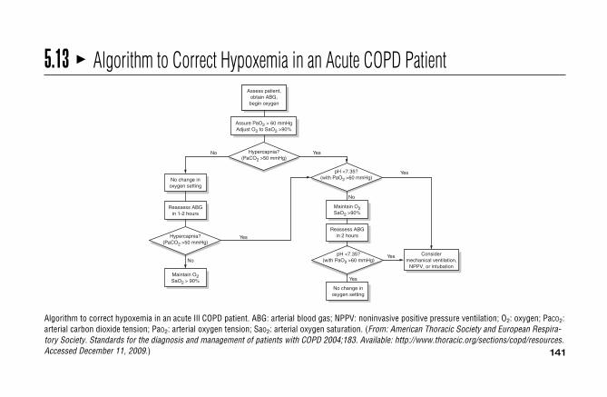
141
5.13 � Algorithm to Correct Hypoxemia in an Acute COPD Patient
Algorithm to correct hypoxemia in an acute Ill COPD patient. ABG: arterial blood gas; NPPV: noninvasive positive pressure ventilation; O2: oxygen; PaCO2:arterial carbon dioxide tension; PaO2: arterial oxygen tension; SaO2: arterial oxygen saturation. (From: American Thoracic Society and European Respira-tory Society. Standards for the diagnosis and management of patients with COPD 2004;183. Available: http://www.thoracic.org/sections/copd/resources.Accessed December 11, 2009.)
Assess patient,obtain ABG,begin oxygen
Assure PaO2 > 60 mmHgAdjust O2 to SaO2 >90%
Hypercapnia?(PaCO2 >50 mmHg)
No change inoxygen setting
Reassess ABGin 1-2 hours
Hypercapnia?(PaCO2 >50 mmHg)
Maintain O2SaO2 > 90%
pH <7.35?(with PaO2 >60 mmHg)
Maintain O2SaO2 >90%
Reassess ABGin 2 hours
pH <7.35?(with PaO2 >60 mmHg)
No change inoxygen setting
Considermechanical ventilation,
NPPV, or intubation
No
No
Yes
Yes
Yes
Yes
Yes
No

This page intentionally left blank

6Neurologic ConceptsS e c t i o n
� 6.1 Glasgow Coma Scale / 144� 6.2 Cranial Nerve Function / 145� 6.3 Circle of Willis / 146� 6.4 Incomplete Spinal Cord Injury
Syndromes / 147
� 6.5 Spinal Cord Injury–FunctionalGoals for Specific Levels ofComplete Injury / 148
NEUROLOGIC CONCEPTS
� 6.6 Intracranial Pressure MonitoringSystems / 152

144
6.1 � Glasgow Coma ScaleBehavior Scorea
Eye Opening (E)Spontaneous 4To verbal stimuli 3To pain 2None 1
Motor Response (M)Obeys commands 6Localizes pain 5Withdraws to pain 4Abnormal flexion 3Extensor response 2None 1
Verbal Response (V)Oriented 5Confused 4Inappropriate words 3Incomprehensible sounds 2None 1
aComa score � E � M � V (scores range from 3-15).
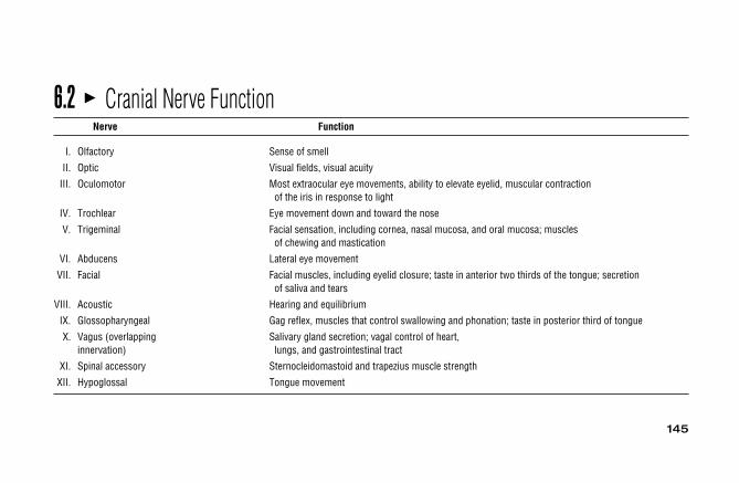
145
6.2 � Cranial Nerve FunctionNerve Function
I. Olfactory Sense of smell
II. Optic Visual fields, visual acuity
III. Oculomotor Most extraocular eye movements, ability to elevate eyelid, muscular contraction of the iris in response to light
IV. Trochlear Eye movement down and toward the nose
V. Trigeminal Facial sensation, including cornea, nasal mucosa, and oral mucosa; muscles of chewing and mastication
VI. Abducens Lateral eye movement
VII. Facial Facial muscles, including eyelid closure; taste in anterior two thirds of the tongue; secretion of saliva and tears
VIII. Acoustic Hearing and equilibrium
IX. Glossopharyngeal Gag reflex, muscles that control swallowing and phonation; taste in posterior third of tongue
X. Vagus (overlapping Salivary gland secretion; vagal control of heart,innervation) lungs, and gastrointestinal tract
XI. Spinal accessory Sternocleidomastoid and trapezius muscle strength
XII. Hypoglossal Tongue movement
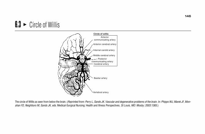
146
6.3 � Circle of WillisCircle of willis
Anteriorcommunicating artery
Anterior cerebral artery
Internal carotid artery
Middle cerebral artery
Posteriorcommunicating artery
Cerebral artery
Basilar artery
Vertebral artery
The circle of Willis as seen from below the brain. (Reprinted from: Perry L, Sands JK. Vascular and degenerative problems of the brain. In: Phipps WJ, Marek JF, Mon-ahan FD, Neighbors M, Sands JK, eds. Medical-Surgical Nursing: Health and Illness Perspectives. St Louis, MO: Mosby; 2003:1365.)

147
6.4 � Incomplete Spinal Cord Injury SyndromesSensory Function Below Level
Syndrome Pathophysiolgy Motor Function Below Level of Injury of Injury
Central cord syndrome Injury to central gray matter with Weakness/paralysis of upper Sensory loss greater in upper preservation of outer white matter extremities greater than extremities than lower extremities
lower extremities
Anterior cord syndrome Injury to anterior portion of spinal Paralysis Loss of pain and temperature with cord, disruption of blood flow preservation vibration and position through anterior spinal artery sense
Posterior cord syndrome Injury to posterior column None Loss of vibration and position sense with preservation of pain and temperature sensation
Brown-Séquard syndrome Lateral injury to one side of the cord Ipsilateral motor paralysis Ipsilateral loss of vibration and position sense
Contralateral loss of pain and temperature sensation
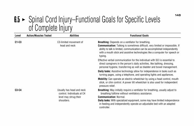
148
6.5 � Spinal Cord Injury–Functional Goals for Specific Levels of Complete Injury
Level Action/Muscles Tested Abilities Functional Goals
C1-C3 C3-limited movement of Breathing: Depends on a ventilator for breathing.head and neck Communication: Talking is sometimes difficult, very limited or impossible. If
ability to talk is limited, communication can be accomplished independentlywith a mouth stick and assistive technologies like a computer for speech ortyping.
Effective verbal communication for the individual with SCI is essential to direct caregivers in the person’s daily activities, like bathing, dressing,personal hygiene, transferring as well as bladder and bowel management.
Daily tasks: Assistive technology allow for independence in tasks such asturning pages, using a telephone, and operating lights and appliances.
Mobility: Can operate an electric wheelchair by using a head control, mouth stick, or chin control. A power tilt wheelchair is also used for independentpressure relief.
C3-C4 Usually has head and neck Breathing: May initially require a ventilator for breathing, usually adjust to control. Individuals at C4 breathing fulltime without ventilatory assistance.level may shrug their Communication: Normal.shoulders. Daily tasks: With specialized equipment, some may have limited independence
in feeding and independently operate an adjustable bed with an adaptedcontroller.
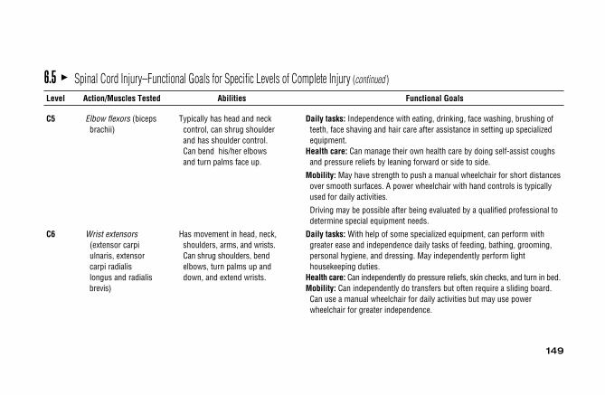
149
6.5 � Spinal Cord Injury–Functional Goals for Specific Levels of Complete Injury (continued )
Level Action/Muscles Tested Abilities Functional Goals
C5 Elbow flexors (biceps Typically has head and neck Daily tasks: Independence with eating, drinking, face washing, brushing of brachii) control, can shrug shoulder teeth, face shaving and hair care after assistance in setting up specialized
and has shoulder control. equipment.Can bend his/her elbows Health care: Can manage their own health care by doing self-assist coughs and turn palms face up. and pressure reliefs by leaning forward or side to side.
Mobility: May have strength to push a manual wheelchair for short distancesover smooth surfaces. A power wheelchair with hand controls is typicallyused for daily activities.
Driving may be possible after being evaluated by a qualified professional todetermine special equipment needs.
C6 Wrist extensors Has movement in head, neck, Daily tasks: With help of some specialized equipment, can perform with (extensor carpi shoulders, arms, and wrists. greater ease and independence daily tasks of feeding, bathing, grooming, ulnaris, extensor Can shrug shoulders, bend personal hygiene, and dressing. May independently perform light carpi radialis elbows, turn palms up and housekeeping duties.longus and radialis down, and extend wrists. Health care: Can independently do pressure reliefs, skin checks, and turn in bed.brevis) Mobility: Can independently do transfers but often require a sliding board.
Can use a manual wheelchair for daily activities but may use power wheelchair for greater independence.

150
6.5 � Spinal Cord Injury–Functional Goals for Specific Levels of Complete Injury (continued )
Level Action/Muscles Tested Abilities Functional Goals
C7 Elbow extensors Has similar movement as an Daily tasks: Able to perform household duties. Need fewer adaptive aids in (triceps brachii) individual with C6, with independent living.
added ability to straighten Health care: Able to do wheelchair pushups for pressure reliefs.his/her elbows. Mobility: Daily use of manual wheelchair. Can transfer with greater ease.
C8 Finger flexors (flexor Has added strength and Daily tasks: Can live independently without assistive devices in feeding, digitorum profundus- precision of fingers that bathing, grooming, oral and facial hygiene, dressing, bladder management, distal phalanx of the result in limited or natural and bowel management.middle finger) hand function.
T1 Finger abductors(abductor digitiminimi)
T2-T6 Has normal motor function Mobility: Has increased ability to do some unsupported seated activities. in head, neck, shoulders, A few individuals are capable of limited walking with orthodic aids. This arms, hands, and requires extremely high energy and puts stress on the upper body, offering fingers. Has increased no functional advantage. Can lead to damage of upper joints.use of rib and chest muscles, or trunk control.

151
6.5 � Spinal Cord Injury–Functional Goals for Specific Levels of Complete Injury (continued )
Level Action/Muscles Tested Abilities Functional Goals
T7-L1 Has added motor function from Daily tasks: Able to perform unsupported seated activities.increased abdominal control. Health care: Has improved cough effectiveness.
L2 Hip flexors (iliopsoas) Has additional return of motor Mobility: Walking can be a viable function, with the help of specialized leg movement in the hips and ankle braces. Lower levels walk with greater ease with the help ofand knees. assistive devices.
L3 Knee extensors (quadriceps femoris)
L4 Ankle dorsiflexors(tibialis anterior)
L5 Long toe extensors(hallucis longus)
S1-S5 Ankle plantar flexors Depending on level of injury, Mobility: Increased ability to walk with fewer or no supportive devices.(gastrocnemius) there are various degrees
of return of voluntary bladder, bowel and sexual functions.

152
6.6 � Intracranial Pressure Monitoring Systems
On the right, a ventriculostomy connects to a CSF collection system. A three-way stopcock allows either pressure monitoring or drainage of CSF. On theleft, a transducer is inserted into the brain parenchyma and anchored to the skull by a bolt mechanism. The transducer is attached to an external monitor-ing device. (Reprinted from: Bergsneider M, Becker DP. Intracranial pressure monitoring. In: Shoemaker WC, Ayres SM, Grenvik A, Holbrook PR, eds. Text-book of Critical Care. 3rd ed. Philadelphia, PA: WB Saunders;1995, p. 313.)

7Pharmacology TablesS e c t i o n
� 7.1 Intravenous MedicationAdministration Guidelines / 154
� 7.2 Neuromuscular Blocking Agents / 179
� 7.3 Vasoactive Agents / 182� 7.4 Antiarrhythmic Agents / 185� 7.5 Therapeutic Drug
Monitoring / 191
� 7.6 Tips for Calculating IV MedicationInfusion Rates / 194
PHARMACOLOGY TABLES
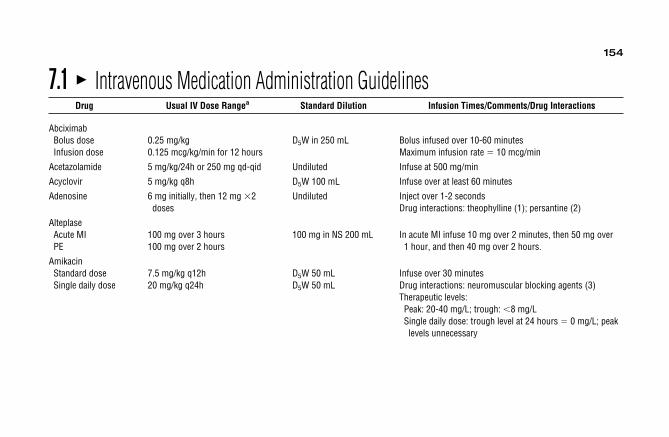
7.1 � Intravenous Medication Administration GuidelinesDrug Usual IV Dose Rangea Standard Dilution Infusion Times/Comments/Drug Interactions
AbciximabBolus dose 0.25 mg/kg D5W in 250 mL Bolus infused over 10-60 minutesInfusion dose 0.125 mcg/kg/min for 12 hours Maximum infusion rate � 10 mcg/min
Acetazolamide 5 mg/kg/24h or 250 mg qd-qid Undiluted Infuse at 500 mg/min
Acyclovir 5 mg/kg q8h D5W 100 mL Infuse over at least 60 minutes
Adenosine 6 mg initially, then 12 mg �2 Undiluted Inject over 1-2 secondsdoses Drug interactions: theophylline (1); persantine (2)
AlteplaseAcute MI 100 mg over 3 hours 100 mg in NS 200 mL In acute MI infuse 10 mg over 2 minutes, then 50 mg overPE 100 mg over 2 hours 1 hour, and then 40 mg over 2 hours.
AmikacinStandard dose 7.5 mg/kg q12h D5W 50 mL Infuse over 30 minutesSingle daily dose 20 mg/kg q24h D5W 50 mL Drug interactions: neuromuscular blocking agents (3)
Therapeutic levels:Peak: 20-40 mg/L; trough: �8 mg/LSingle daily dose: trough level at 24 hours � 0 mg/L; peak levels unnecessary
154

AminophyllineLoading dose 6 mg/kg D5W 50 mL Infuse loading dose over 30 minutes
Maximum loading infusion rate 25 mg/minAminophylline � 80% theophylline
Infusion dose 500 mg in D5W 500 mL Drug interactions: cimetidine, ciprofloxacin, erythromycin,CHF 0.3 mg/kg/h clarithromycin (4)Normal 0.6 mg/kg/h Therapeutic levels: 10-20 mg/LSmoker 0.9 mg/kg/h
Ammonium chloride mEq Cl � Cl deficit (in mEq/L) 100 mEq in NS 500 mL Maximum infusion rate is 5 mL/min of a 0.2-mEq/mL � 0.2 � wt (kg) solution; correct 1/3 to 1/2 of Cl deficit while monitoring pH
and Cl; administer remainder as neededAmphotericin B 0.5-1.5 mg/kg q24h D5W 250 mL Infuse over 2-6 hours
Do not mix in electrolyte solutions (eg, saline, lactated Ringer solution)
155
7.1 � Intravenous Medication Administration Guidelines (continued )
Drug Usual IV Dose Rangea Standard Dilution Infusion Times/Comments/Drug Interactions
a Usual dose ranges are listed; refer to appropriate disease state for specific dose.Abbreviations: bid, twice a day; HF, heart failure; conc, concentration; D5W, destrose-5%-water; DVT, deep venous thrombosis; HPLC, high-performance liq-uid chromatography; IM, intramuscular; IV, intravenous; IVP, IV push; IVPB, IV piggyback; MI, myocardial infarction; NS, normal saline; NSAID, nonsteroidalanti-inflammatory drug; PCP, Pneumocystis carinii pneumonia; PE, pulmonary embolism; PO, orally; prn, as needed; qd, daily; SW, sterile water.Drug interactions: (1) antagonizes adenosine effect; (2) potentiates adenosine effect; (3) potentiates effect of neuromuscular blocking agents; (4) inhibits theo-phylline metabolism; (5) antagonizes effect of neuromuscular blocking agents: (6) metabolism inhibited by cimetidine; (7) metabolism inhibited by ciprofloxacin;(8) increased digoxin concentrations; (9) metabolism inhibited by erythromycin; (10) increased nephrotoxicity; (11) increased heparin requirements.

156
Ampicillin 0.5-3 g q4-6h NS 100 mL Infuse over 15-30 minutesAmpicillin/sulbactam 1.5-3 g q6h NS 100 mL Infuse over 15-30 minutesAnistreplase (APSAC) 30 U IV SW 5 mL Infuse over 5 minutes, give with aspirin 325 mg PO
immediatelyPreparation should be discarded if not used within 6 hours
ArgatrobanBolus dose 350 mcg/kg 250 mg in NS 250 mL Titrate to aPTT or ACTInfusion dose 25 mcg/kg/min
Atenolol 5 mg IV over 5 minutes, 5 mg IV Undiluted Inject 1 mg/min10 minutes later
AtracuriumIntubating dose 0.4-0.5 mg/kg Undiluted Inject over 60 sec to prevent histamine releaseMaintenance dose 0.08-0.1 mg/kg Undiluted Inject over 60 sec to prevent histamine releaseInfusion dose 5-9 mcg/kg/min 1000 mg in D5W 150 mL Continuous infusion. Final volume � 250 mL, conc �
4 mg/mLDrug interactions: aminoglycosides (3); anticonvulsants (5)
Aztreonam 0.5-2 g q6-12h D5W 100 mL Infuse over 15-30 minutes
7.1 � Intravenous Medication Administration Guidelines (continued )
Drug Usual IV Dose Rangea Standard Dilution Infusion Times/Comments/Drug Interactions

157
BivalirudinBolus dose 1 mg/kg 250 mg in D5W 500 mL Infuse bolus over 2 minInfusion dose 2.5 mg/kg/h �4 hours; if necessary Titrate to aPTT or ACT
0.2 mg/kg/h for up to 20 hoursBumetanideBolus dose 0.5-1 mg Undiluted Maximum injection rate: 1 mg/minInfusion dose 0.08-0.3 mg/h 2.4 mg in NS 100 mL Continuous infusion
Calcium (elemental) 100-200 mg of elemental 1000 mg in NS 1000 mL Ca chloride 1 g � 272 mg (13.6 mEq) of elemental calciumcalcium IV over 15 minutes Ca gluconate 1 g � 90 mg (4.65 mEq) of elemental calciumfollowed by 100 mg/h
Cefazolin 0.5-1 g q6-8h D5W 50 mL Infuse over 15-30 minutesCefepime 1-2 g q8-12h 1-2 g in D5W 100 mL Infuse over 15 minutesCefonicid 1-2 g q24h D5W 50 mL Infuse over 15-30 minutesCefoperazone 1-2 g q12h D5W 50 mL Infuse over 15-30 minutesCefotaxime 1-2 g q4-6h D5W 50 mL Infuse over 15-30 minutesCefotetan 1-2 g q12h D5W 50 mL Infuse over 15-30 minutesCefoxitin 1-2 g q4-6h D5W 50 mL Infuse over 15-30 minutesCeftazidime 0.5-2 g q8-12h D5W 50 mL Infuse over 15-30 minutesCeftizoxime 1-2 g q8-12h D5W 50 mL Infuse over 15-30 minutesCeftriaxone 0.5-2 g q12-24h D5W 50 mL Infuse over 15-30 minutesCefuroxime 0.75-1.5 g q8h D5W 50 mL Infuse over 15-30 minutes
7.1 � Intravenous Medication Administration Guidelines (continued )
Drug Usual IV Dose Rangea Standard Dilution Infusion Times/Comments/Drug Interactions

158
Chlorothiazide 0.5-1 g qd-bid SW 18 mL Inject over 3-5 minutesChlorpromazine 10-50 mg q4-6h Dilute with NS to a final Inject at 1 mg/minutes
concentration of1 mg/mL
CimetidineIVPB 300 mg q6-8h D5W 50 mL Infuse over 15-30 minutes
IVP dose may be injected over at least 5 minutesInfusion dose 37.5 mg/h D5W 250 mL Continuous infusion
Drug interactions: theophylline, warfarin, phenytoin, lidocaine, benzodiazepines (6)
Ciprofloxacin 200-400 mg q8-12h Premix solution 2 mg/mL Infuse over 60 minutesDrug interactions: theophylline, warfarin (7)
CisatricuriumInfusion dose 1-3 mcg/kg/min
Clevidipine 1-16 mg/h Undiluted Continuous infusionClindamycin 150-900 mg q8h D5W 250 mL Infuse over 30-60 minutesConivaptanBolus dose 20 mg D5W 100 mL Infuse over 30 minutesInfusion dose 20 mg D5W 250 mL Infuse over 24 hours
7.1 � Intravenous Medication Administration Guidelines (continued )
Drug Usual IV Dose Rangea Standard Dilution Infusion Times/Comments/Drug Interactions

159
Conjugated 0.6 mg/kg/d �5 days NS 50 mL Infuse over 15-30 minutesestrogens
Cosyntropin 0.25 mg IV Undiluted Inject over 60 secondsCyclosporine 5-6 mg/kg q24h D5W 100 mL Infuse over 2-6 hours
Drug interactions: digoxin (8); erythromycin (9); amphotericin,NSAID (10)
IV dose � 1/3 PO doseTherapeutic levels: trough: 50-150 ng/mL (whole blood—HPLC)
DantroleneBolus dose 1-2 mg/kg SW 60 mL Administer as rapidly as possibleMaximum dose 10 mg/kg Do not dilute in dextrose or electrolyte-containing solutionsMaintenance dose 2.5 mg/kg q4h �24h SW 60 mL Infuse over 60 minutes
Daptomycin 4-6 mg/kg q24h 250 or 500 mg in Infuse over 30 minutesNS 50 mL
Desmopressin 0.3 mg/kg NS 50 mL Infuse over 15-30 minutesDexamethasone 0.5-20 mg NS 50 mL May give doses �10 mg undiluted IVP over 60 secondsDexmedetomidineBolus dose 1 mcg/kg 200 mcg in NS 50 mL Infuse bolus over 10 minutesInfusion dose 0.2-1 mcg/kg/h
7.1 � Intravenous Medication Administration Guidelines (continued )
Drug Usual IV Dose Rangea Standard Dilution Infusion Times/Comments/Drug Interactions

160
Diazepam 2.5-5 mg q2-4h Undiluted Inject 2-5 mg/minutesActive metabolites contribute to activity
Diazoxide 50-150 mg q5-15 min Undiluted Inject over 30 secondsMaximum 150 mg/dose
DigoxinDigitalizing dose 0.25 mg q4-6h up to 1 mg Undiluted Inject over 3-5 minMaintenance dose 0.125-0.25 mg q24h Drug interactions: amiodarone, cyclosporine, quinidine,
verapamil (8)Therapeutic levels: 0.5-2.0 ng/mL
DiltiazemBolus dose 0.25-0.35 mg/kg Undiluted Inject over 2 minutesInfusion dose 5-15 mg/h 125 mg in D5W 100 mL Continuous infusion (final conc � 1 mg/mL)
Diphenhydramine 25-100 mg IV q 2-4h Undiluted Inject over 3-5 minutesCompetitive histamine antagonist, doses �1000 mg/24 h may be required in some instances
Dobutamine 2.5-20 mcg/kg/min 500 mg in D5W 250 mL Continuous infusionDolasetron 1.8 mg/kg or 100 mg Undiluted or 100 mg in Infuse undiluted drug over at least 30 seconds
D6W 50 mL Infuse piggyback over 15 minutesAdminister 30 L, minutes prior to chemo or 1 hour prior to anesthesia
7.1 � Intravenous Medication Administration Guidelines (continued )
Drug Usual IV Dose Rangea Standard Dilution Infusion Times/Comments/Drug Interactions

161
DopamineRenal dose �5 mcg/kg/min 400 mg in D5W 250 mL Continuous infusionInotrope 5-10 mcg/kg/min 400 mg in D5W 250 mL Continuous infusionPressor �10 mcg/kg/min 400 mg in D5W 250 mL Continuous infusion
Doripenem 500 mg q-8h D5W or NS 100 mL Infuse over 60 minutes-4 hoursDoxacuriumIntubating dose 0.025-0.08 mg/kg Undiluted Inject over 5-10 secondsMaintenance dose 0.005-0.01 mg/kg Undiluted Inject over 5-10 secondsInfusion dose 0.25 mcg/kg/min 25 mg in D5W 50 mL Continuous infusion
Dose based on lean body weightDrug interactions: aminoglycosides (3); anticonvulsants (5)
Doxycycline 100-200 mg q12-24h D5W 250 mL Infuse over 60 minutesDrotrecogin alfa 24 mcg/kg/h 100 or 200 mcg/mL Infuse through dedicated line or lumen
dilution in NS (multilumen catheter).Total infusion time is 96 hours
7.1 � Intravenous Medication Administration Guidelines (continued )
Drug Usual IV Dose Rangea Standard Dilution Infusion Times/Comments/Drug Interactions
a Usual dose ranges are listed; refer to appropriate disease state for specific dose.Abbreviations: bid, twice a day; HF, heart failure; conc, concentration; D5W, destrose-5%-water; DVT, deep venous thrombosis; HPLC, high-performance liq-uid chromatography; IM, intramuscular; IV, intravenous; IVP, IV push; IVPB, IV piggyback; MI, myocardial infarction; NS, normal saline; NSAID, nonsteroidalanti-inflammatory drug; PCP, Pneumocystis carinii pneumonia; PE, pulmonary embolism; PO, orally; prn, as needed; qd, daily; SW, sterile water.Drug interactions: (1) antagonizes adenosine effect; (2) potentiates adenosine effect; (3) potentiates effect of neuromuscular blocking agents; (4) inhibits theo-phylline metabolism; (5) antagonizes effect of neuromuscular blocking agents: (6) metabolism inhibited by cimetidine; (7) metabolism inhibited by ciprofloxacin;(8) increased digoxin concentrations; (9) metabolism inhibited by erythromycin; (10) increased nephrotoxicity; (11) increased heparin requirements.

162
Droperidol 0.625-10 mg q1-4h Undiluted Inject over 3-5 minutesEnalaprilat 0.625-1.25 mg q6h Undiluted Inject over 5 minutes
Initial dose for patients on diuretics is 0.625 mgEpinephrine 1-4 mcg/min 1 mg in D5W 250 mL Continuous infusionEptifibatideBolus dose 180 mcg/kg Undiluted Maximum infusion duration of 72 hoursInfusion dose 2 mcg/kg/min until discharge or
CABGErtapenem 1 g q24h 1 g in NS 50 mL Infuse over 30 minutesErythromycin 0.5-1 g q6h NS 250 mL Infuse over 60 minutes
Drug interactions: theophylline (4); cyclosporine (9)Erythropoietin 12.5-600 U/kg 1-3 �per week Undiluted Inject over 3-5 minutesEsmololBolus dose 500 mcg/kg Undiluted Inject over 60 secondsInfusion dose 50-400 mcg/kg/min 5 g in D5W 500 mL Continuous infusion
Ethacrynic acid 50 mg D5W 50 mL Inject over 3-5 minutesMay repeat �1 Maximum single dose 100 mg
Etidronate 7.5 mg/kg qd �3 days NS or D5W 500 mL Infuse over at least 2 hoursFamotidine 20 mg q12h D5W 100 mL Infuse over 15-30 minutesFenoldopamInfusion dose 0.1-1.6 mcg/kg/min 20 mg in D5W 250 mL Titrate to BP
7.1 � Intravenous Medication Administration Guidelines (continued )
Drug Usual IV Dose Rangea Standard Dilution Infusion Times/Comments/Drug Interactions

163
FentanylBolus dose 25-75 mcg q1-2h Undiluted Inject over 5-10 secondsInfusion dose 50-100 mcg/h Undiluted Continuous infusion
Filgastrim 1-20 mcg/kg �2-4 weeks D5W Preferred route of administration is subcutaneousFluconazole 100-800 mg q24h Premix solution 2 mg/mL Maximum infusion rate 200 mg/h (IV rate is 15-30 minutes)FlumazenilReversal of conscious 0.2 mg initially, then Undiluted Inject over 15 secondssedation 0.2 mg q60 sec to a Maximum dose of 3 mg in any 1-hour period
total of 1 mgBenzodiazepine 0.2 mg initially, then 0.3 Undiluted Inject over 30 secondsoverdose mg �1 dose, then 0.5 Maximum dose of 3 mg in any 1-hour period
mg q30s up to atotal of 3 mg
Continuous infusion 0.1-0.5 mg/h 5 mg in D5W 1000 mL Continuous infusionFoscarnetInduction dose 60 mg/kg q8h Undiluted Infuse over 1 hourMaintenance dose 90-120 mg/kg q24h Undiluted Infuse over 2 hours
7.1 � Intravenous Medication Administration Guidelines (continued )
Drug Usual IV Dose Rangea Standard Dilution Infusion Times/Comments/Drug Interactions

164
Fosphenytoin NS 250 mL Infuse no faster than 150 mg/minStatus epilepticusLoading dose 15-20 mg/kg
NonemergencyLoading dose 10-20 mg/kgMaintenance dose 4-6 mg/kg/day
FurosemideBolus dose 10-100 mg q1-6h Undiluted Maximum injection rate 40 mg/minInfusion dose 1-15 mg/h 100 mg in NS 100 mL Continuous infusion
Gallium nitrate 100-200 mg/m2 qd �5 days D5W 1000 mL Infuse over 24 hoursGanciclovir 2.5 mg/kg q12h D5W 100 mL Infuse over 1 hourGentamicinLoading dose 2-3 mg/kg D5W 50 mL Infuse over 30 minutesMaintenance dose 1.5-2.5 mg/kg q8-24h D5W 50 mL Infuse over 30 minutesSingle daily dose 5-7 mg/kg q24h D5W 50 mL Infuse over 30 minutes
Critically ill patients have an increased volume of distribution requiring increased doses
Drug interactions: neuromuscular blocking agentsTherapeutic levels:Peak: 4-10 mg/LTrough: �2 mg/LSingle daily dose: trough level at 24 hours � 0 mg/L;
peak levels unnecessary
7.1 � Intravenous Medication Administration Guidelines (continued )
Drug Usual IV Dose Rangea Standard Dilution Infusion Times/Comments/Drug Interactions

165
Glycopyrrolate 5-15 mcg/kg Undiluted Inject over 60 secondsGranisetron 10 mcg/kg D5W 50 mL Infuse over 15 minutesHaloperidol (lactate)Bolus dose 1-10 mg q2-4h Undiluted Inject over 3-5 minutesInfusion dose 10 mg/h 100 mg in D5W 100 mL Continuous infusion
In urgent situations the dose may be doubled every 20-30 minutes until an effect is obtained
Decanoate salt is only for IM administrationHeparin 10-25 U/kg/h 25,000 U in D5W 500 mL Drug interactions: nitroglycerin (11)Hydralazine 10-25 mg q2-4h UndilutedHydrochloric acid mEq � (0.5 � BW � 100 mEq in SW 1000 mL Maximum infusion rate � 0.2 mEq/kg/h
(103 � serum Cl))Hydrocortisone 12.5-100 mg q6-12h Undiluted Inject over 60 secondsHydromorphone 0.5-2 mg q4-6h Undiluted Inject over 60 seconds
Dilaudid-HP available as 10 mg/mLIbutilide Infuse over 10 minutesPatient �60 kg 1 mg NS 50 mL Repeat dose possible 10 minutes after completion of initial bolusPatient �60 kg 0.01 mg/kg
7.1 � Intravenous Medication Administration Guidelines (continued )
Drug Usual IV Dose Rangea Standard Dilution Infusion Times/Comments/Drug Interactions
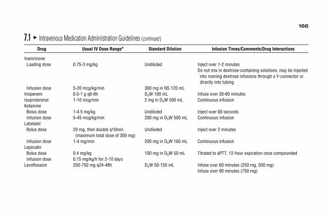
166
InamrinoneLoading dose 0.75-3 mg/kg Undiluted Inject over 1-2 minutes
Do not mix in dextrose-containing solutions; may be injected into running dextrose infusions through a Y-connector or directly into tubing
Infusion dose 5-20 mcg/kg/min 300 mg in NS 120 mLImipenem 0.5-1 g q6-8h D5W 100 mL Infuse over 30-60 minutesIsoproterenol 1-10 mcg/min 2 mg in D5W 500 mL Continuous infusionKetamineBolus dose 1-4.5 mg/kg Undiluted Inject over 60 secondsInfusion dose 5-45 mcg/kg/min 200 mg in D5W 500 mL Continuous infusion
LabetalolBolus dose 20 mg, then double q10min Undiluted Inject over 2 minutes
(maximum total dose of 300 mg)Infusion dose 1-4 mg/min 200 mg in D5W 160 mL Continuous infusion
LepirudinBolus dose 0.4 mg/kg 100 mg in D5W 50 mL Titrated to aPTT, 12-hour expiration once compoundedInfusion dose 0.15 mg/kg/h for 2-10 days
Levofloxacin 250-750 mg q24-48h D5W 50-150 mL Infuse over 60 minutes (250 mg, 500 mg)Infuse over 90 minutes (750 mg)
7.1 � Intravenous Medication Administration Guidelines (continued )
Drug Usual IV Dose Rangea Standard Dilution Infusion Times/Comments/Drug Interactions
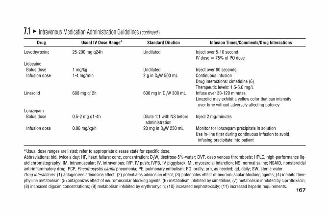
167
Levothyroxine 25-200 mg q24h Undiluted Inject over 5-10 secondIV dose � 75% of PO dose
LidocaineBolus dose 1 mg/kg Undiluted Inject over 60 secondsInfusion dose 1-4 mg/min 2 g in D5W 500 mL Continuous infusion
Drug interactions: cimetidine (6)Therapeutic levels: 1.5-5.0 mg/L
Linezolid 600 mg q12h 600 mg in D5W 300 mL Infuse over 30-120 minutesLinezolid may exhibit a yellow color that can intensify over time without adversely affecting potency
LorazepamBolus dose 0.5-2 mg q1-4h Dilute 1:1 with NS before Inject 2 mg/minutes
administrationInfusion dose 0.06 mg/kg/h 20 mg in D5W 250 mL Monitor for lorazepam precipitate in solution
Use in-line filter during continuous infusion to avoid infusing precipitate into patient
7.1 � Intravenous Medication Administration Guidelines (continued )
Drug Usual IV Dose Rangea Standard Dilution Infusion Times/Comments/Drug Interactions
a Usual dose ranges are listed; refer to appropriate disease state for specific dose.Abbreviations: bid, twice a day; HF, heart failure; conc, concentration; D5W, destrose-5%-water; DVT, deep venous thrombosis; HPLC, high-performance liq-uid chromatography; IM, intramuscular; IV, intravenous; IVP, IV push; IVPB, IV piggyback; MI, myocardial infarction; NS, normal saline; NSAID, nonsteroidalanti-inflammatory drug; PCP, Pneumocystis carinii pneumonia; PE, pulmonary embolism; PO, orally; prn, as needed; qd, daily; SW, sterile water.Drug interactions: (1) antagonizes adenosine effect; (2) potentiates adenosine effect; (3) potentiates effect of neuromuscular blocking agents; (4) inhibits theo-phylline metabolism; (5) antagonizes effect of neuromuscular blocking agents: (6) metabolism inhibited by cimetidine; (7) metabolism inhibited by ciprofloxacin;(8) increased digoxin concentrations; (9) metabolism inhibited by erythromycin; (10) increased nephrotoxicity; (11) increased heparin requirements.

168
Magnesium (elemental) Magnesium1 g � 8 mEqMagnesium 25 mEq over 24 hours followed 25 mEq in D5W 1000 mL Continuous infusiondeficiency by 6 mEq over the next
12 hoursAcute myocardial 15-45 mEq over 24-48 25 mEq in D5W 1000 mL Continuous infusion
infarction hours followed by 12.5 mEq/dayfor 3 days
Ventricular 16 mEq over 1 hour followed by 40 mEq in D5W 1000 mL 16 mEq (2 g) may be diluted in 100 mLarrythmias 40 mEq over 6 hours D5W and infused over 1 hour
MannitolDiuretic Undiluted Inject over 30-60 minutesBolus dose 0.25-0.5 g/kgMaintenance dose 0.25-0.5 g/kg q4hCerebral edema 1.5-2 g/kg over 30-60 minutes
Meperidine 25-100 mg q2-4h Undiluted Inject over 60 secondsAvoid in renal failure
Meropenem 0.5-2 g q8-24h NS 50 mL or undiluted Infuse over 15-30 min or bolus dose over 3-5 minutesMethadone 5-20 mg qd Undiluted Inject over 3-5 minutes
Accumulation with repetitive dosingMethyldopate 0.25-1 g q6h D5W 100 mL Infuse over 30-60 minutesMethylprednisolone 10-500 mg q6h Undiluted Inject over 60 seconds
7.1 � Intravenous Medication Administration Guidelines (continued )
Drug Usual IV Dose Rangea Standard Dilution Infusion Times/Comments/Drug Interactions
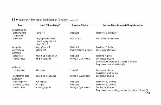
169
MetoclopramideSmall intestine 10 mg � 1 Undiluted Inject over 3-5 minutes
intubationAntiemetic 2 mg/kg before chemo, D5W 50 mL Infuse over 15-30 minutes
then 2 mg/kg q2h �2,then q3h �3
Metoprolol 5 mg q2min �3 Undiluted Inject over 3-5 minMetronidazole 500 mg q6h Premix solution 5 mg/mL Infuse over 30 minutesMidazolamBolus dose 0.025-0.35 mg/kg q1-2h Undiluted Inject 0.5 mg/minInfusion dose 0.5-5 mcg/kg/min 50 mg in D5W 100 mL Continuous infusion
Unpredictable clearance in critically ill patientsDrug interactions: cimetidine (6)
MilrinoneLoading dose 50 mcg/kg 1 mg/mL Infuse over 10 min
Available in 5-mL syringeMaintenance dose 0.375-0.75 mcg/kg/min 50 mg in D5W 250 mL Continuous infusion
MivacuriumIntubating dose 0.25 mg/kg Undiluted Inject over 60 secondsMaintenance dose 0.1 mg/kg Undiluted Inject over 60 secondsInfusion dose 9-10 mcg/kg/min 50 mg in D5W 100 mL Continuous infusion
Drug interactions: aminoglycosides (3); anticonvulsants (5)
7.1 � Intravenous Medication Administration Guidelines (continued )
Drug Usual IV Dose Rangea Standard Dilution Infusion Times/Comments/Drug Interactions

170
MorphineBolus dose 2-10 mg Undiluted Inject over 60 secondsInfusion dose 2-5 mg/h 100 mg in D5W 100 mL Continuous infusion
Moxifloxacin 400 mg q24h 400 mg in NS 250 mL Infuse over 60 minutesNafcillin 0.5-2 g q4-6h D5W 100 mL Infuse over 30-60 minutesNaloxonePostoperative opiatedepression
Loading dose 0.1-0.2 mg q2-3min Undiluted Infuse over 60 minutesInfusion dose 3-5 mcg/kg/h 2 mg in D5W 250 mL Continuous infusion
Opiate overdoseLoading dose 0.4-2 mg q2-3min Undiluted Infuse over 60 secondsInfusion dose 2.5-5 mcg/kg/h 2 mg in D5W 250 mL Continuous infusion
Neostigmine 25-75 mcg/kg Undiluted Inject over 60 secondsNesiritideBolus dose 2 mcg/kg 1.5 mg in Monitor for hypotension
preservative-freeInfusion dose 0.01 mcg/kg/min D5W 250 mL
Nitroglycerin 10-300 mcg/min 50 mg in D5W 250 mL Continuous infusionDrug interactions: heparin (11)
7.1 � Intravenous Medication Administration Guidelines (continued )
Drug Usual IV Dose Rangea Standard Dilution Infusion Times/Comments/Drug Interactions
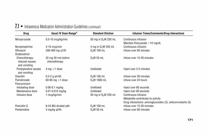
171
Nitroprusside 0.5-10 mcg/kg/min 50 mg in D5W 250 mL Continuous infusionMaintain thiocyanate �10 mg/dL
Norepinephrine 4-10 mcg/min 4 mg in D5W 250 mL Continuous infusionOfloxacin 200-400 mg q12h D5W 100 mL Infuse over 60 minutesOndansetronChemotherapy- 32 mg 30 min before D5W 50 mL Infuse over 15-30 minutesinduced nausea chemotherapyand vomiting
Postoperative nausea 4 mg �1 dose Undiluted Inject over 2-5 minutesand vomiting
Oxacillin 0.5-2 g q4-6h D5W 100 mL Infuse over 30 minutesPamidronate 60-90 mg �1 dose D5W 1000 mL Infuse over 24 hoursPancuroniumIntubating dose 0.06-0.1 mg/kg Undiluted Inject over 60 secondsMaintenance dose 0.01-0.015 mg/kg Undiluted Inject over 60 secondsInfusion dose 1 mcg/kg/min 50 mg in D5W 250 mL Continuous infusion
Metabolite contributes to activityDrug interactions: aminoglycosides (3); anticonvulsants (5)
Penicillin G 8-24 MU divided q4h D5W 100 mL Infuse over 15-30 minutesPentamidine 4 mg/kg q24h D5W 50 mL Infuse over 60 minutes
7.1 � Intravenous Medication Administration Guidelines (continued )
Drug Usual IV Dose Rangea Standard Dilution Infusion Times/Comments/Drug Interactions

172
PentobarbitalBolus dose 5-10 mg/kg NS 100 mL Infuse over 2 hoursInfusion dose 0.5-1 mg/kg/h initially, then NS 250 mL Continuous infusion
0.5-4 mg/kg/h 2 g in NS 250 mL Therapeutic levels: 20-50 mg/LPhenobarbital NS 100 mL Infuse over 2 hoursBolus dose 5-10 mg/kg NS 250 mL Continuous infusionInfusion dose 0.5-1 mg/kg/h initially, then 2 g in NS 250 mL Therapeutic levels: 20-50 mg/L
0.5-4 mg/kg/hPhentolamineBolus dose 2.5-10 mg prn q5-15min Undiluted Inject over 3-5 minutesContinuous infusion 1-10 mg/min 50 mg in D5W 100 mL Continuous infusion
Phenylephrine 20-30 mcg/min 15 mg in D5W 250 mL Continuous infusion; 0.5 mg over 20-30 secondsPhenytoin Maximum infusion rate is 50 mg/minStatus epilepticus Undiluted Drug interactions: cimetidine; neuromuscular blocking agentsBolus dose 15-20 mg/kg Therapeutic levels: 10-20 mg/LInfusion dose 5 mg/kg/day (divided into 2 or
3 doses)Phosphate (potassium) 0.08-0.64 mmol/kg Function of K� Infuse over 6-8 hours
concentration 1 mmol of PO4 � P 31 mgSolution should be made no more concentrated than 0.4 mEq/mL K�
7.1 � Intravenous Medication Administration Guidelines (continued )
Drug Usual IV Dose Rangea Standard Dilution Infusion Times/Comments/Drug Interactions

173
Piperacillin 2-4 g q4-6h D5W 100 mL Infuse over 15-30 minutesPiperacillin/tazobactam 3.375 g IV q6h D5W 100 mL Infuse over 30 minutes
Each 2.25-g vial contains 2 g piperacillin and 0.25 g tazobactam
Potassium chloride 5-40 mEq/h 40 mEq in 1000 mL Cardiac monitoring should be used with infusion rates (NS, D5W, etc) �20 mEq/h
Prednisolone 4-60 mg q24h Undiluted Inject over 60 secondsProcainamideLoading dose 15 mg/kg D5W 50 mL Maximum infusion rate 25-50 mg/minInfusion dose 1-4 mg/min 2 g in D5W 500 mL Continuous infusion
Therapeutic levels:Procainamide: 4-10 mg/LNAPA: 10-20 mg/L
7.1 � Intravenous Medication Administration Guidelines (continued )
Drug Usual IV Dose Rangea Standard Dilution Infusion Times/Comments/Drug Interactions
a Usual dose ranges are listed; refer to appropriate disease state for specific dose.Abbreviations: bid, twice a day; HF, heart failure; conc, concentration; D5W, destrose-5%-water; DVT, deep venous thrombosis; HPLC, high-performance liq-uid chromatography; IM, intramuscular; IV, intravenous; IVP, IV push; IVPB, IV piggyback; MI, myocardial infarction; NS, normal saline; NSAID, nonsteroidal anti-inflammatory drug; PCP, Pneumocystis carinii pneumonia; PE, pulmonary embolism; PO, orally; prn, as needed; qd, daily; SW, sterile water.Drug interactions: (1) antagonizes adenosine effect; (2) potentiates adenosine effect; (3) potentiates effect of neuromuscular blocking agents; (4) inhibits theo-phylline metabolism; (5) antagonizes effect of neuromuscular blocking agents: (6) metabolism inhibited by cimetidine; (7) metabolism inhibited by ciprofloxacin;(8) increased digoxin concentrations; (9) metabolism inhibited by erythromycin; (10) increased nephrotoxicity; (11) increased heparin requirements.
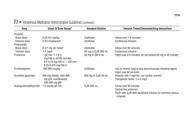
174
PropofolBolus dose 0.25-0.5 mg/kg Undiluted Infuse over 1-2 minutesInfusion dose 5-50 mcg/kg/min Undiluted Continuous infusion
PropranololBolus dose 0.5-1 mg q5-15min Undiluted Infuse over 60 secondsInfusion dose 1-4 mg/h 50 mg in D5W 500 mL Continuous infusion
Protamine �30 min: 1-1.5 U 50 mg in SW 5 mL Inject over 3-5 minutes; do not exceed 50 mg in 10 minutesmg/100 U; 30-60 minutes:0.5-0.75 mg/100 U; �120 min:0.25-0.375 mg/100 U
Pyridostigmine 100-300 mcg/kg Undiluted Use to reverse long-acting neuromuscular blocking agentsInject over 60 seconds
Quinidine gluconate 600 mg initially, then 400 800 mg in D5W 50 mL Infusion rate 1 mg/min; use cardiac monitormg q2h, maintenance Therapeutic levels: 1.5-5 mg/L200-300 mg q6h
Quinupristin/dalfopristin 7.5 mg/kg q8-12h D5W 250 mL Infuse over 60 minutesCentral line preferredFlush with D5W after peripheral infusion to minimize venous
irritation
7.1 � Intravenous Medication Administration Guidelines (continued )
Drug Usual IV Dose Rangea Standard Dilution Infusion Times/Comments/Drug Interactions

175
RanitidineIVPB 50 mg q6-8h D5W 50 mL Infuse over 15-30 minutes
IVP dose should be injected over at least 5 minutesInfusion dose 6.25 mg/h 150 mg in D5W 150 mL Continuous infusion
Reteplase 10-U bolus � 2 SW 10 mL Inject over 2 minutes, use dedicated IV line, flush heparin-coated catheters with NS D5W after use
RocuroniumIntubating dose 0.45-1.2 mg/kg Undiluted Inject over 60 secondsMaintenance dose 0.075-0.15 mg/kg Undiluted Inject over 60 secondsInfusion dose 10-14 mcg/kg/min 50 mg in D5W 100 mL Continuous infusion
StreptokinaseAcute MI 1.5 MU D5W 45 mL Infuse over 30 minutesDVT, PE 250,000 U over 30 minutes, D5W 90 mL Continuous infusion
then 100,000 U/hover 24-72 hours
Succinylcholine 0.6-2 mg/kg Undiluted Inject over 60 secondsTacrolimus 50-100 mcg/kg/day 5 mg in D5W 250 mLTenecteplase 30-50 mg SW 10 mL Inject over 5 secondst-PA 100 mg 100 mg in D5W 100 mL Infuse 60 mg/h during first hour, then 20 mg/h for 2 hoursTheophylline Smokers: 0.9 mg/kg/hBolus dose 6 mg/kg 800 mg in 500 mL Nonsmokers: 0.6 mg/kg/hInfusion dose 0.3-0.9 mg/kg/h premixed Liver and heart failure: 0.3 mg/kg/h
7.1 � Intravenous Medication Administration Guidelines (continued )
Drug Usual IV Dose Rangea Standard Dilution Infusion Times/Comments/Drug Interactions

176
Thiamine 100 mg qd � 3 D5W 50 mL Infuse over 15-30 minutesThiopental 3-4 mg/kg Undiluted Inject over 3-5 minutesTicarcillin 3 g q3-6h D5W 100 mL Infuse over 15-30 minutesTicarcillin/clavulanate 3.1 g q4-6h D5W 100 mL Infuse over 15-30 minutesTirofibanBolus dose 0.4 mcg/kg/h 25 mg in D5W 500 mL Bolus infused over 30 minutesInfusion dose 0.1 mcg/kg/min for 12-24 hours
after angioplasty or arthrectomyTobramycinLoading dose 2-3 mg/kg D5W 50 mL Infuse over 30 minutesMaintenance dose 1.5-2.5 mg/kg q8-24h D5W 50 mL Infuse over 30 minutes
Critically ill patients have an increased volume of distribution requiring increased doses
Drug interactions: neuromuscular blocking agents (3)Therapeutic levelsPeak: 4-10 mg/LTrough: �2 mg/L
Torsemide 5-20 mg qd Undiluted Inject over 60 secondsTrimethaphan 0.5-5 mg/min 500 mg in D5W 500 mL Continuous infusion
7.1 � Intravenous Medication Administration Guidelines (continued )
Drug Usual IV Dose Rangea Standard Dilution Infusion Times/Comments/Drug Interactions

177
Trimethaprim-sulfamethoxazoleCommon infections 4-5 mg/kg q12h TMP 16 mg-SMX 80 mg Infuse over 60 minutes
per D5W 25 mLPCP 5 mg/kg q6h TMP 16 mg-SMX 80 mg Infuse over 60 minutes
per D5W 25 mL Therapeutic levels: 100-150 mg/LUrokinase 4400 U/kg over 10 minutes, D5W 195 mL Continuous infusionPulmonary then 4400 U/hembolism over 12 hours
Vancomycin 1 g q12h D5W 250 mL Infuse over at least 1 hour to avoid “red-man” syndromeTherapeutic levelsPeak: 20-40 mg/LTrough: �10 mg/L
VasopressinGI hemorrhage 0.2-0.3 U/min 100 U in D5W 250 mL Maximum infusion rate 0.9 U/minSeptic shock 0.01-0.04 U/min
7.1 � Intravenous Medication Administration Guidelines (continued )
Drug Usual IV Dose Rangea Standard Dilution Infusion Times/Comments/Drug Interactions

178
VecuroniumIntubating dose 0.1-0.28 mg/kg Undiluted Inject over 60 secondsMaintenance dose 0.01-0.015 mg/kg Undiluted Inject over 60 secondsInfusion dose 1 mcg/kg/min 20 mg in D5W 100 mL Continuous infusion
Metabolite contributes to activityDrug interactions: aminoglycosides (3); anticonvulsants (5)
VerapamilBolus dose 0.075-0.15 mg/kg Undiluted Inject over 1-2 minutes
Continuous infusionDrug interactions:
digoxin (8)
a Usual dose ranges are listed; refer to appropriate disease state for specific dose.Abbreviations: bid, twice a day; HF, heart failure; conc, concentration; D5W, destrose-5%-water; DVT, deep venous thrombosis; HPLC, high-performance liq-uid chromatography; IM, intramuscular; IV, intravenous; IVP, IV push; IVPB, IV piggyback; MI, myocardial infarction; NS, normal saline; NSAID, nonsteroidalanti-inflammatory drug; PCP, Pneumocystis carinii pneumonia; PE, pulmonary embolism; PO, orally; prn, as needed; qd, daily; SW, sterile water.Drug interactions: (1) antagonizes adenosine effect; (2) potentiates adenosine effect; (3) potentiates effect of neuromuscular blocking agents; (4) inhibits theo-phylline metabolism; (5) antagonizes effect of neuromuscular blocking agents: (6) metabolism inhibited by cimetidine; (7) metabolism inhibited by ciprofloxacin;(8) increased digoxin concentrations; (9) metabolism inhibited by erythromycin; (10) increased nephrotoxicity; (11) increased heparin requirements.
7.1 � Intravenous Medication Administration Guidelines (continued )
Drug Usual IV Dose Rangea Standard Dilution Infusion Times/Comments/Drug Interactions
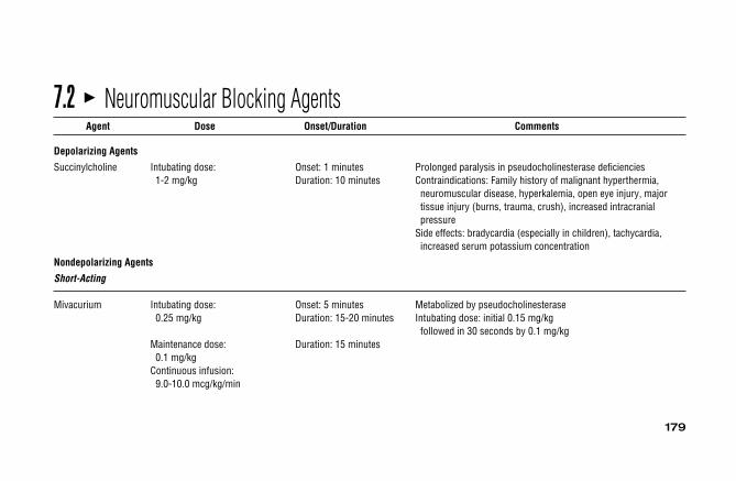
179
7.2 � Neuromuscular Blocking AgentsAgent Dose Onset/Duration Comments
Depolarizing Agents
Succinylcholine Intubating dose: Onset: 1 minutes Prolonged paralysis in pseudocholinesterase deficiencies1-2 mg/kg Duration: 10 minutes Contraindications: Family history of malignant hyperthermia,
neuromuscular disease, hyperkalemia, open eye injury, majortissue injury (burns, trauma, crush), increased intracranialpressure
Side effects: bradycardia (especially in children), tachycardia,increased serum potassium concentration
Nondepolarizing Agents
Short-Acting
Mivacurium Intubating dose: Onset: 5 minutes Metabolized by pseudocholinesterase0.25 mg/kg Duration: 15-20 minutes Intubating dose: initial 0.15 mg/kg
followed in 30 seconds by 0.1 mg/kgMaintenance dose: Duration: 15 minutes
0.1 mg/kgContinuous infusion:9.0-10.0 mcg/kg/min

180
Intermediate-Acting
Atracurium Intubating dose: Onset: 2 minutes Histamine release with bolus doses �0.6 mg/kg and may0.5 mg/kg Duration: 30-40 minutes precipitate asthma or hypotension
Maintenance dose: Duration: 15-25 minutes Elimination independent of renal hepatic function0.08-0.10 mg/kg Metabolized in the plasma by Hofmann elimination and ester
Continuous infusion: hydrolysis5-9 mcg/kg/min Duration not prolonged by renal or liver failure
Used when succinylcholine is contraindicated or not preferred
Cisatricurium Intubating dose: Onset: 2 minutes Decreased histamine release compared to atracurium0.15-0.2 mg/kg Duration: 30-90 minutes Elimination independent of renal or hepatic function
Maintenance dose: Duration: 15-30 minutes Metabolized in the plasma by Hofmann elimination and0.03 mg/kg ester hydrolysis
Continuous infusion: Duration not prolonged by renal or liver failure1-3 mcg/kg/min
Rocuronium Intubating dose: Onset: 0.7-1.3 minutes Not associated with histamine release0.45-1.2 mg/kg Duration: 22-67 minutes Used when succinylcholine is contraindicated or not preferred
Maintenance dose: Duration: 12-17 minutes Metabolized by liver; duration not significantly prolonged by 0.075-0.15 mg/kg renal failure, but prolonged in patients with liver disease
Continuous infusion: No adverse cardiovascular effects10-14 mcg/kg/min
7.2 � Neuromuscular Blocking Agents (continued )
Agent Dose Onset/Duration Comments
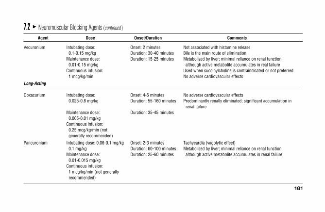
Vecuronium Intubating dose: Onset: 2 minutes Not associated with histamine release0.1-0.15 mg/kg Duration: 30-40 minutes Bile is the main route of elimination
Maintenance dose: Duration: 15-25 minutes Metabolized by liver; minimal reliance on renal function, 0.01-0.15 mg/kg although active metabolite accumulates in real failure
Continuous infusion: Used when succinylcholine is contraindicated or not preferred1 mcg/kg/min No adverse cardiovascular effects
Long-Acting
Doxacurium Intubating dose: Onset: 4-5 minutes No adverse cardiovascular effects0.025-0.8 mg/kg Duration: 55-160 minutes Predominantly renally eliminated; significant accumulation in
renal failureMaintenance dose: Duration: 35-45 minutes
0.005-0.01 mg/kgContinuous infusion:0.25 mcg/kg/min (notgenerally recommended)
Pancuronium Intubating dose: 0.06-0.1 mg/kg Onset: 2-3 minutes Tachycardia (vagolytic effect)0.1 mg/kg Duration: 60-100 minutes Metabolized by liver; minimal reliance on renal function,
Maintenance dose: Duration: 25-60 minutes although active metabolite accumulates in renal failure0.01-0.015 mg/kg
Continuous infusion:1 mcg/kg/min (not generallyrecommended)
7.2 � Neuromuscular Blocking Agents (continued )
Agent Dose Onset/Duration Comments
181
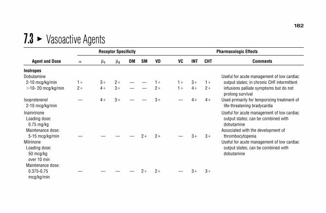
182
7.3 � Vasoactive AgentsReceptor Specificity Pharmacologic Effects
Agent and Dose � �1 �2 DM SM VD VC INT CHT Comments
InotropesDobutamine Useful for acute management of low cardiac 2-10 mcg/kg/min 1� 3� 2� — — 1� 1� 3� 1� output states; in chronic CHF intermittent�10- 20 mcg/kg/min 2� 4� 3� — — 2� 1� 4� 2� infusions palliate symptoms but do not
prolong survivalIsoproterenol — 4� 3� — — 3� — 4� 4� Used primarily for temporizing treatment of2-10 mcg/kg/min life-threatening bradycardia
Inamrinone Useful for acute management of low cardiac Loading dose: output states; can be combined with 0.75 mg/kg dobutamine
Maintenance dose: Associated with the development of 5-15 mcg/kg/min — — — — 2� 2� — 3� 3� thrombocytopenia
Milrinone Useful for acute management of low cardiac Loading dose: output states; can be combined with 50 mcg/kg dobutamineover 10 min
Maintenance dose:0.375-0.75 — — — — 2� 2� — 3� 3�
mcg/kg/min

183
MixedDopamine Doses �20-30 mcg/kg/min usually produce no 2-5 mcg/kg/min — 3� — 4� — — — 2� 1� added response; 2 mcg/kg/min may protect 5-10 mcg/kg/min — 4� 2� 4� — — — 4� 2� kidneys when giving other vasopressors10-20 mcg/kg/min 3� 4� 1� — — — 3� 3� 3�
Epinephrine Mixed vasoconstrictor/inotrope; stronger 0.01-0.05 mcg/kg/min 1� 4� 2� — — 1� 1� 4� 2� inotrope than norepinephrine; does not �0.05 mcg/kg/min 4� 3� 1� — — — 3� 3� 3� constrict coronary or cerebral vessels; give
as needed to maintain BPVasopressorsNorepinephrine Mixed vasoconstrictor/inotrope; useful when 2-20 mcg/min 4� 2� — — — — 4� 1� 2� dopamine inadequate; give as needed to titrate to effect maintain BP (usually �20 mcg/min)
Phenylephrine Pure vasoconstrictor without direct cardiac Start at 30 mcg/min 4� — — — — — 4� — — effect; may cause reflex bradycardia; usefulIV and titrate when other pressors cause tachyarrhythmias;
give as much as needed to maintain BP
Vasopressin Pure vasoconstrictor without direct cardiac 0.01-0.04 U/min — — — — — — 4� — — effect; may cause gut ischemia if dose is
increased �0.04 U/min
7.3 � Vasoactive Agents (continued )
Receptor Specificity Pharmacologic Effects
Agent and Dose � �1 �2 DM SM VD VC INT CHT Comments

184
VasodilatorsNitroglycerin Tachyphylaxis, headache
20-100 mcg/min — — — — 4� 4� — — 1�
A�VNitroprusside — — — — 4� 4� — — 1� Monitor thiocyanate levels if infusion duration 0.5-10 mcg/kg/min A�V �48 hours; maintain thiocyanate level
�10 mg/dL
Abbreviations: α1: α1-adrenergic; β1; β1-adrenergic; β2: β2-adrenergic; DM: dopaminergic; SM: smooth muscle; VD: vasodilator; VC: vasoconstrictor; INT:inotropic; CHT: chronotropic. Vasoconstrictors usually are given by central vein and should be used only in conjunction with adequate volume repletion. Allcan precipitate myocardial ischemia. All except phenylephrine can cause tachyarrhythmias.Modified from: Gonzalez ER, Meyers DG. Assessment and management of cardiogenic shock. In Oronato JC, ed. Clinics in Emergency Medicine:Cardiovascular Emergencies. New York, NY: Churchill Livingstone; 1986:125, with permission.
7.3 � Vasoactive Agents (continued )
Receptor Specificity Pharmacologic Effects
Agent and Dose � �1 �2 DM SM VD VC INT CHT Comments

185
7.4 � Antiarrhythmic AgentsAgents Indications Dosage Comments
Class IAProcainamide
Quinidine
Disopyramide
Ventricular ectopy; conversion of atrial fibrillation and atrial flutter; WPW
Ventricular ectopy; conversion of atrial fibrillation and atrial flutter; WPW
Ventricular ectopy; conversion of atrial fibrillation and atrial flutter; WPW
Loading dose: (IV) 15 mg/kg at 25-50 mg/min
Maintenance dose: (IV) 2-5 mg/min
Quinidine sulfate: 200-300 mg PO q6hQuinidine sulfate: 324-648 mg PO q8h
100-300 mg PO q6h; SR: 100-300 mg PO q12h
N-acetyl procainamide is active metabolite; lupus-like syndrome; rash;agranulocytosis; QT prolongation
Therapeutic range: PA 4-10 mg/L, NAPA 10-20 mg/L
Diarrhea, nausea, headache dizziness; hypersensitivity reactions includingthrombocytopenia; hemolysis; feverhepatitis; rash QT prolongation; increaseddigoxin level
Dosage adjustment should be made when switching from one salt to another:Quinidine sulfate (83% quinidine),gluconate (62% quinidine),polygalacturonate (60% quinidine)
Therapeutic range: 2.5-5 mg/L
Anticholinergic effects; negative inotropy; QT prolongation
Therapeutic range: 2-4 mg/L

186
Class IBLidocaine
Mexiletine
Tocainide
7.4 � Antiarrhythmic Agents (continued )
Agents Indications Dosage Comments
Malignant ventricular ectopy; WPW
Malignant ventricular ectopy
Malignant ventricular ectopy
1.5 mg/kg IV over 2 minutes, then1-4 mg/min
150-300 mg PO q6-8h with food
200-600 mg PO q8h with food
No benefit in atrial arrhythmiasSeizures; paresthesias; delirium; levelsincreased by cimetidine; minimalhemodynamic effects
Therapeutic range: 1.5-5 mg/L
No benefit in atrial arrhythmiasLess effective than IA and IC agentsNausea; tremor; dizziness; delirium; levels increased by cimetidine
Therapeutic range: 0.5-2 mg/L
No benefit in atrial arrhythmiasLess effective than IA and IC agentsNausea; tremor; dizziness; delirium; agranulocytosis; pneumonitis; minimalhemodynamic effects
Therapeutic range: 4-10 mg/L
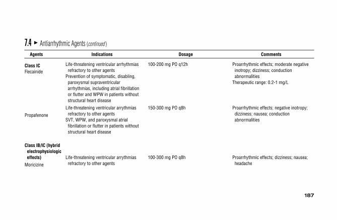
187
Class ICFlecainide
Propafenone
Class IB/IC (hybrid electrophysiologiceffects)
Moricizine
7.4 � Antiarrhythmic Agents (continued )
Agents Indications Dosage Comments
Life-threatening ventricular arrhythmiasrefractory to other agents
Prevention of symptomatic, disabling, paroxysmal supraventriculararrhythmias, including atrial fibrillationor flutter and WPW in patients withoutstructural heart disease
Life-threatening ventricular arrythmias refractory to other agents
SVT, WPW, and paroxysmal atrial fibrillation or flutter in patients withoutstructural heart disease
Life-threatening ventricular arrythmias refractory to other agents
100-200 mg PO q12h
150-300 mg PO q8h
100-300 mg PO q8h
Proarrhythmic effects; moderate negative inotropy; dizziness; conductionabnormalities
Therapeutic range: 0.2-1 mg/L
Proarrhythmic effects; negative inotropy; dizziness; nausea; conductionabnormalities
Proarrhythmic effects; dizziness; nausea; headache

188
Class II (beta-blocking agents)
Propranolol
Esmolol
Metoprolol
Class IIIAmiodarone
7.4 � Antiarrhythmic Agents (continued )
Agents Indications Dosage Comments
Slowing ventricular rate in atrial fibrillation, atrial flutter, and SVT;suppression of PVCs
Slowing ventricular rate in atrial fibrillation,atrial flutter, SVT, and MAT
Slowing ventricular rate in atrial fibrillation, atrial flutter, SVT, and MAT
Life-threatening ventricular arrhythmias, supraventricular arrhythmias, includingWPW refractory to other agents
Up to 0.5-1 mg IV, then 1-4 mg/h(or 10-100 mg PO q6h)
Loading dose: 500 mcg/over 1 minuteMaintenance dose: 50 mcg/kg/min; rebolus and increase q5min by 50mcg/kg/min to maximum of 400
Initial IV dose: 5 mg q5min up to 15 mg, then 25-100 mg PO q8-12h
800-1600 mg PO qd for 1-3 weeks, then 600-800 mg PO qd for 4 weeks, then100-400 mg PO qd
Not cardioselective; hypotension; bronchospasm; negative inotropy
Cardioselective at low doses; hypotension; negative inotropy; very short half-life
Cardioselective at low doses; hypotension; negative inotropy
Half-life �50 days; pulmonary fibrosis; corneal microdeposits; hypo/hyperthyroidism; bluish skin; hepatitis;photosensitivity; conduction abnormalities;mild negative inotropy; increased effect ofcoumadin; increased digoxin level
Therapeutic range: 1-2.5 mg/L

189
Bretylium
Sotalol
Dofetilide
Class IV (calcium channelantagonists)
Verapamil
Diltiazem
7.4 � Antiarrhythmic Agents (continued )
Agents Indications Dosage Comments
Refractory ventricular tachycardiaand ventricular fibrillation
Life-threatening ventricular arrythmias
Conversion of atrial fibrillation
Conversion of SVT; slowing ventricular rate in atrial fibrillation, atrial flutter, and MAT
Conversion of SVT; slowing ventricular rate in atrial fibrillation, atrial flutter, and MAT
5-10 mg/kg IV boluses q10 min up to 30 mg/kg, then 0.5-2 mg/min
80-160 mg PO q12h; may increase up to 160 mg PO q8h
250-500 mcg orally twice a day
IV bolus: 5-10 mg over 2-3 minutes(repeat in 30 min prn), continuousinfusion: 2.5-5 mcg/kg/min
PO: 40-160 mg PO q8h
IV bolus: 0.25 mg/kg over 2 minutes (repeatin 15 minutes prn with 0.35 mg/kgIV); Maintenance infusion: 5-15 mg/h
PO: 30-90 mg PO q6h
Initial hypertension, then postural hypotension; nausea and vomiting;parotitis; catecholamine sensitivity
Beta-blocker with class III properties; proarrhythmic effects; QT prolongation
Dose adjusted based on QTc interval and creatinine clearance
Hypotension; negative inotropy; conduction disturbances; increased digoxin level;generally contraindicated in WPW
Hypotension; less negative inotropy than verapamil; conduction disturbances; rarehepatic injury; generally contraindicated inWPW
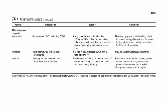
190
Miscellaneousagents
Adenosine
Atropine
Digitalis
Abbreviations: AV, atrioventricular; MAT, multifocal atrial tachycardia; SR: sustained release; SVT, supraventricular tachycardia; WPW, Wolff-Parkinson-White.
7.4 � Antiarrhythmic Agents (continued )
Agents Indications Dosage Comments
Conversion of SVT, including WPW
Initial therapy for symptomatic bradycardia
Slowing AV conduction in atrial fibrillation and atrial flutter
6-mg rapid IV bolus; if ineffective, 12-mg rapid IV bolus 2 minutes later;follow bolus with fast flush; use smallerdoses if giving through central venousline
0.5-mg IV bolus; repeat q5min prn to total of 2 mg IV
Loading dose: 0.5 mg IV, then 0.25 mg IVq4-6h up to 1 mg; Maintenance dose:0.125-0.375 mg PO/IV qd
Flushing; dyspnea; nodal blocking effect increased by dipyridamole and decreasedby theophylline and caffeine; very shorthalf-life (�10 seconds)
May induce tachycardia and ischemia
Heart block; arrhythmias; nausea; yellow vision; numerous drug interactions,generally contraindicated in WPW
Therapeutic range: 0.5-2.0 mg/mL

191
7.5 � Therapeutic Drug MonitoringDrug Usual Therapeutic Range Usual Sampling Time
AntibioticsAmikacin Peak: 20-40 mg/L Peak: 30-60 minutes after a 30-minute infusion
Trough: �10 mg/L Trough: Just before next doseChloramphenicol Peak: 10-25 mg/L Peak: 30-90 minutes after a 30-minute infusion
Trough: 5-10 mg/L Trough: Just before the next doseFlucytosine Peak: 50-100 mg/L Peak: 1-2 hours after an oral dose
Trough: �25 mg/L Trough: Just before the next doseGentamicin Peak: 4-10 mg/L Peak: 30-60 minutes after a 30-minute infusion
Trough: �2 mg/L Trough: Just before the next doseTobramycin Peak: 4-10 mg/L Peak: 30-60 minutes after a 30-minutes infusion
Trough: �2 mg/L Trough: Just before the next doseNetilmicin Peak: 4-10 mg/L Peak: 30-60 minutes after a 30-minutes infusion
Trough: �2 mg/L Trough: Just before the next doseVancomycin Peak: 20-40 mg/L Peak: 1 hour after end of a 1-hour infusion
Trough: �20 mg/L Trough: Just before the next doseSulfonamides Peak: 100-150 mg/L Peak: 2 hours after 1-hour infusion(sulfamethoxazole, Trough: Not applicablesulfadiazine, cotrimoxazole)
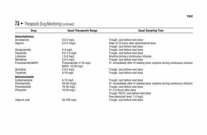
192
AntiarrhythmicsAmiodarone 0.5-2 mg/L Trough: Just before next doseDigoxin 0.5-2 mcg/L Peak: 8-12 hours after administered dose
Trough: Just before next doseDisopyramide 2-4 mg/L Trough: Just before next doseFlecainide 0.2-1.0 mg/L Trough: Just before next doseLidocaine 1.5-5 mg/L Anytime during a continuous infusionMexiletine 0.5-2 mg/L Trough: Just before next doseProcainamide/NAPA Procainamide: 4-10 mg/L IV: Immediately after IV loading dose: anytime during continuous infusion
NAPA: 10-20 mg/LQuinidine 2.5-5 mg/L Trough: Just before next doseTocainide 4-10 mg/L Trough: Just before next doseAnticonvulsantsCarbamazepine 4-12 mg/L Trough: Just before next dosePentobarbital 20-50 mcg/L IV: Immediately after IV loading dose: anytime during continuous infusionPhenobarbital 15-40 mg/L Trough: Just before next dosePhenytoin 10-20 mg/L IV: 2-4 hours after dose
Trough: PO/IV: Just before next doseFree phenytoin level: 1-2 mg/L
Valproic acid 50-100 mg/L Trough: Just before next dose
7.5 � Therapeutic Drug Monitoring (continued )
Drug Usual Therapeutic Range Usual Sampling Time

193
BronchodilatorsTheophylline 10-20 mg/L IV: Prior to IV bolus dose, 30 minutes after end of bolus dose, anytime during
continuous infusionPO: peak: 2 hours after rapid-release product, 4 hours after sustained-release product
Trough: Just before next doseMiscellaneousCyclosporine 50-150 ng/mL (whole blood, HPLC) Trough: IV, PO: Just before next dose
7.5 � Therapeutic Drug Monitoring (continued )
Drug Usual Therapeutic Range Usual Sampling Time

194
7.6 � Tips for Calculating IV Medication Infusion RatesInformation Required to Calculate IV Infusion Rates to Deliver Specific Medication Doses
• Dose to be infused (eg, mg/kg/min, mg/min, mg/h)• Concentration of IV solution (eg, dopamine 400 mg in D5W 250 mL � 1.6 mg/mL; nitroglycerin 50 mg in D5W 250 mL � 200 mcg/mL)• Patient’s weight
1. Calculate the IV infusion rate in milliliters per hour for a 70-kg patient requiring dobutamine 5 mcg/kg/min using a dobutamine admixture of 500 mg inD5W 250 mL.• Dose to be infused: 5 mcg/kg/min• Dobutamine concentration: 500 mg/250 mL � 2 mg/mL or 2000 mcg/mL• Patient weight: 70 kg
Calculation:5 mcg/kg/min � 70 kg � 350 mcg/min 350 mcg/min � 60 min/h � 21,000 mcg/h 21,000 mcg/h � 2000 mcg/mL � 10.5 mL/h
Answer: Setting the infusion pump at 10.5 mL/h will deliver dobutamine at a dose of 5 mcg/kg/min.2. Calculate the IV infusion rate in milliliters per hour for a 70-kg patient requiring nitroglycerin 50 mcg/min using a nitroglycerin admixture of 50 mg in
D5W 250 mL.• Dose to be infused: 50 mcg/min• Nitroglycerin concentration: 50 mg/250 mL � 0.2 mg/mL or 200 mcg/mL• Patient weight: 70 kg
Calculation:50 mcg/min � 60 min/h � 3000 mcg/h3000 mcg/h � 200 mcg/mL � 15 mL/h
Answer: Setting the infusion pump at 15 mL/h will deliver nitroglycerin at a dose of 50 mcg/min.
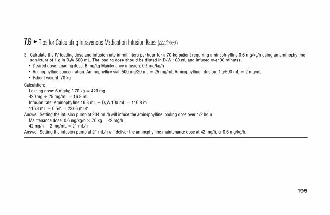
195
3. Calculate the IV loading dose and infusion rate in milliliters per hour for a 70-kg patient requiring aminoph-ylline 0.6 mg/kg/h using an aminophyllineadmixture of 1 g in D5W 500 mL. The loading dose should be diluted in D5W 100 mL and infused over 30 minutes.
• Desired dose: Loading dose: 6 mg/kg Maintenance infusion: 0.6 mg/kg/h• Aminophylline concentration: Aminophylline vial: 500 mg/20 mL � 25 mg/mL Aminophylline infusion: 1 g/500 mL � 2 mg/mL• Patient weight: 70 kg
Calculation:Loading dose: 6 mg/kg 3 70 kg � 420 mg420 mg � 25 mg/mL � 16.8 mLInfusion rate: Aminophylline 16.8 mL � D5W 100 mL � 116.8 mL116.8 mL � 0.5/h � 233.6 mL/h
Answer: Setting the infusion pump at 234 mL/h will infuse the aminophylline loading dose over 1/2 hourMaintenance dose: 0.6 mg/kg/h � 70 kg � 42 mg/h42 mg/h � 2 mg/mL � 21 mL/h
Answer: Setting the infusion pump at 21 mL/h will deliver the aminophylline maintenance dose at 42 mg/h, or 0.6 mg/kg/h.
7.6 � Tips for Calculating Intravenous Medication Infusion Rates (continued )
Related Documents