Applicant Name (Last, first, middle): Hensley, Kenneth RESEARCH PLAN Page ____ Proposal Narrative 1 A. Specific Aims, Rationale and Significance This goal of this project is to develop an innovative treatment for amyotrophic lateral sclerosis (ALS) based on pharmacological manipulation of collapsin response mediator protein-2 (CRMP2) pathways. We hypothesize that agents which boost CRMP2 function would help protect motor axons from degeneration, thus slowing motor functional decline in ALS. CRMP2 was originally identified as a crucial mediator of axon growth in the developing central nervous system (CNS). There, CRMP2 responds to signals from the axon repellence protein semaphorin-3 (SEMA3A) to collapse growth cones. Recent reports of anomalous SEMA3A expression in terminal Schwann cells of SOD1 G93A mice (a model of familial ALS) suggest this system could be involved in motor neuron disease as well. Interestingly, the SEMA3A receptor neuropilin-1 (NRP1) also binds vascular endothelial growth factor (VEGF), whose deficiency has been independently linked to ALS. Thus, aberrant NRP1 function might help explain VEGF role in ALS. Experimentally, CRMP2 suppression causes neurite retraction, partly mimicking ALS axonopathy, whereas increasing CRMP2 causes axon growth. The P.I. recently reported that a CNS metabolite called lanthionine ketimine (LK) binds CRMP2, altering its protein:protein interactions and promoting neurite growth (J. Neuroscience 2010). A cell-permeable LK-ethyl ester (LKE, invented by the P.I. and granted U.S. patent 7,683,055 in 2010) promotes neurite elongation in cell culture at low nanomolar concentrations. Furthermore, LKE protects neurons against glutamate or H 2 O 2 ; suppresses microglial activation; and protects motor neurons from microglial toxicity, all of which activities should benefit CNS tissue afflicted by ALS. Notably, LKE slows disease progression in the SOD1 G93A mouse model of ALS when administered late in the disease (Molecules 2010). Due to its novel apparent mechanism of CRMP2 action, LKE potentially could become a “first-in-class” drug for treating neurodegeneration in ALS. In strikingly complementary work, Mileusnic and Rose describe a CNS-penetrating, patented neurotrophic peptide called Ac-rER designed to mimic the secreted amyloid precursor protein- (sAPP) of Alzheimer’s disease (AD) (J. Neurochemistry 2011). Proteomics studies identified CRMP2 as the high-affinity Ac-rER binding target. The common CRMP2-binding action and neurotrophic behavior shared by LK and Ac- rER compel our wish to determine whether Ac-rER also could treat ALS. These recent findings motivate us to aggressively explore whether inappropriate signaling through CRMP2 contributes to ALS axonopathy in a manner that is amenable to pharmacological manipulation. We therefore propose three focused AIMS to test specific strategies for supporting CRMP2 function to slow ALS: AIM 1 will test the working hypothesis that LKE treatment beginning prior to distal axonopathy (40 d) will better delay onset and slow disease progression and diminish histopathology in the SOD1 G93A mouse model of ALS, compared to effects we have observed when LKE treatment is begun late in the disease. Such early treatment would be practical for dominant hereditary forms of ALS (10-15% of cases), wherein patients often know their risk status so treatment could begin in prodromal (preclinical) phases. AIM 2 (A) will test the working hypothesis that Ac-rER will slow disease progression and mitigate neuropathology in the SOD1 G93A mouse when administered either prior to onset of distal axonopathy, or later during the timeframe of clinically-evident disease. AIM 2 (B) will test the working hypothesis that Ac-rER affects CRMP2-dependent aspects of glial inflammatory activation in a fashion that mimics the observed glial effects of LKE. AIM 3 will test the working hypothesis that SEMA3A signaling to CRMP2 through NRP1 contributes to axon degeneration in ALS in a fashion that can be modulated with NRP1-binding, monoclonal antibodies developed by Genentech and currently in phase I human clinical trials for cancer. SOD1 G93A mice will be treated with either anti-NRP1 A (which blocks SEMA3A binding to NRP1) or anti-NRP1 B (which blocks VEGF binding to NRP1) beginning prior to onset of axonopathy (40 d) or at clinically-evident disease (90 d). Functional and histological progression of ALS will be evaluated. Success in this project would launch a new drug development program centered on CRMP2 function-boosting therapeutics, the long-term goals of which would be creation of investigational new drug (IND) application(s) and ultimately, clinical trials against ALS. A specific therapy development plan concludes this proposal.

Welcome message from author
This document is posted to help you gain knowledge. Please leave a comment to let me know what you think about it! Share it to your friends and learn new things together.
Transcript
Applicant Name (Last, first, middle): Hensley, Kenneth
RESEARCH PLAN
Page ____ Proposal Narrative 1
A. Specific Aims, Rationale and Significance This goal of this project is to develop an innovative treatment for amyotrophic lateral sclerosis (ALS)
based on pharmacological manipulation of collapsin response mediator protein-2 (CRMP2) pathways. We hypothesize that agents which boost CRMP2 function would help protect motor axons from degeneration, thus slowing motor functional decline in ALS.
CRMP2 was originally identified as a crucial mediator of axon growth in the developing central nervous system (CNS). There, CRMP2 responds to signals from the axon repellence protein semaphorin-3 (SEMA3A) to collapse growth cones. Recent reports of anomalous SEMA3A expression in terminal Schwann cells of SOD1G93A mice (a model of familial ALS) suggest this system could be involved in motor neuron disease as well. Interestingly, the SEMA3A receptor neuropilin-1 (NRP1) also binds vascular endothelial growth factor (VEGF), whose deficiency has been independently linked to ALS. Thus, aberrant NRP1 function might help explain VEGF role in ALS. Experimentally, CRMP2 suppression causes neurite retraction, partly mimicking ALS axonopathy, whereas increasing CRMP2 causes axon growth.
The P.I. recently reported that a CNS metabolite called lanthionine ketimine (LK) binds CRMP2, altering its protein:protein interactions and promoting neurite growth (J. Neuroscience 2010). A cell-permeable LK-ethyl ester (LKE, invented by the P.I. and granted U.S. patent 7,683,055 in 2010) promotes neurite elongation in cell culture at low nanomolar concentrations. Furthermore, LKE protects neurons against glutamate or H2O2; suppresses microglial activation; and protects motor neurons from microglial toxicity, all of which activities should benefit CNS tissue afflicted by ALS. Notably, LKE slows disease progression in the SOD1G93A mouse model of ALS when administered late in the disease (Molecules 2010). Due to its novel apparent mechanism of CRMP2 action, LKE potentially could become a “first-in-class” drug for treating neurodegeneration in ALS.
In strikingly complementary work, Mileusnic and Rose describe a CNS-penetrating, patented neurotrophic peptide called Ac-rER designed to mimic the secreted amyloid precursor protein- (sAPP) of Alzheimer’s disease (AD) (J. Neurochemistry 2011). Proteomics studies identified CRMP2 as the high-affinity Ac-rER binding target. The common CRMP2-binding action and neurotrophic behavior shared by LK and Ac-rER compel our wish to determine whether Ac-rER also could treat ALS.
These recent findings motivate us to aggressively explore whether inappropriate signaling through CRMP2 contributes to ALS axonopathy in a manner that is amenable to pharmacological manipulation. We therefore propose three focused AIMS to test specific strategies for supporting CRMP2 function to slow ALS:
AIM 1 will test the working hypothesis that LKE treatment beginning prior to distal axonopathy (40 d) will better delay onset and slow disease progression and diminish histopathology in the SOD1G93A mouse model of ALS, compared to effects we have observed when LKE treatment is begun late in the disease. Such early treatment would be practical for dominant hereditary forms of ALS (10-15% of cases), wherein patients often know their risk status so treatment could begin in prodromal (preclinical) phases.
AIM 2 (A) will test the working hypothesis that Ac-rER will slow disease progression and mitigate neuropathology in the SOD1G93A mouse when administered either prior to onset of distal axonopathy, or later during the timeframe of clinically-evident disease. AIM 2 (B) will test the working hypothesis that Ac-rER affects CRMP2-dependent aspects of glial inflammatory activation in a fashion that mimics the observed glial effects of LKE.
AIM 3 will test the working hypothesis that SEMA3A signaling to CRMP2 through NRP1 contributes to axon degeneration in ALS in a fashion that can be modulated with NRP1-binding, monoclonal antibodies developed by Genentech and currently in phase I human clinical trials for cancer. SOD1G93A mice will be treated with either anti-NRP1A (which blocks SEMA3A binding to NRP1) or anti-NRP1B (which blocks VEGF binding to NRP1) beginning prior to onset of axonopathy (40 d) or at clinically-evident disease (90 d). Functional and histological progression of ALS will be evaluated.
Success in this project would launch a new drug development program centered on CRMP2 function-boosting therapeutics, the long-term goals of which would be creation of investigational new drug (IND) application(s) and ultimately, clinical trials against ALS. A specific therapy development plan concludes this proposal.
Applicant Name (Last, first, middle): Hensley, Kenneth
RESEARCH PLAN
Page ____ Proposal Narrative 2
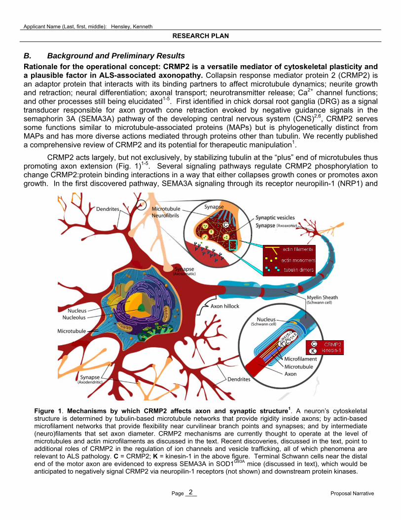
B. Background and Preliminary Results Rationale for the operational concept: CRMP2 is a versatile mediator of cytoskeletal plasticity and a plausible factor in ALS-associated axonopathy. Collapsin response mediator protein 2 (CRMP2) is an adaptor protein that interacts with its binding partners to affect microtubule dynamics; neurite growth and retraction; neural differentiation; axonal transport; neurotransmitter release; Ca2+ channel functions; and other processes still being elucidated1-5. First identified in chick dorsal root ganglia (DRG) as a signal transducer responsible for axon growth cone retraction evoked by negative guidance signals in the semaphorin 3A (SEMA3A) pathway of the developing central nervous system (CNS)2,6, CRMP2 serves some functions similar to microtubule-associated proteins (MAPs) but is phylogenetically distinct from MAPs and has more diverse actions mediated through proteins other than tubulin. We recently published a comprehensive review of CRMP2 and its potential for therapeutic manipulation1.
CRMP2 acts largely, but not exclusively, by stabilizing tubulin at the “plus” end of microtubules thus promoting axon extension (Fig. 1)1-5. Several signaling pathways regulate CRMP2 phosphorylation to change CRMP2:protein binding interactions in a way that either collapses growth cones or promotes axon growth. In the first discovered pathway, SEMA3A signaling through its receptor neuropilin-1 (NRP1) and
Figure 1. Mechanisms by which CRMP2 affects axon and synaptic structure1. A neuron’s cytoskeletal structure is determined by tubulin-based microtubule networks that provide rigidity inside axons; by actin-based microfilament networks that provide flexibility near curvilinear branch points and synapses; and by intermediate (neuro)filaments that set axon diameter. CRMP2 mechanisms are currently thought to operate at the level of microtubules and actin microfilaments as discussed in the text. Recent discoveries, discussed in the text, point to additional roles of CRMP2 in the regulation of ion channels and vesicle trafficking, all of which phenomena are relevant to ALS pathology. C = CRMP2; K = kinesin-1 in the above figure. Terminal Schwann cells near the distal end of the motor axon are evidenced to express SEMA3A in SOD1G93A mice (discussed in text), which would be anticipated to negatively signal CRMP2 via neuropilin-1 receptors (not shown) and downstream protein kinases.
Applicant Name (Last, first, middle): Hensley, Kenneth
RESEARCH PLAN
Page ____ Proposal Narrative 3
co-receptor tyrosine kinases triggers Rac1 activation, ultimately activating cyclin-dependent kinase-5 (Cdk5) and glycogen synthase kinase 3 (GSK3) which phosphorylate CRMP2 on Ser-523 and Thr-509/ Thr-514, respectively2,6-8. Phosphorylated CRMP2 releases tubulin heterodimers, thereby reducing microtubule growth at the distal end of axons, encouraging axon retraction2-8. Conversely, neurotrophin-3 and brain-derived neurotrophic factor (BDNF) inhibit GSK3 via the phosphatidylinositol-3-kinase (PI3K)/AKT pathway, reducing CRMP2 phosphorylation and promoting axon growth9.
A second pathway through which CRMP2 affects neurites is an anterograde transport mechanism (Fig. 1). CRMP2 adapts the microtubule motor kinesin-1 to transport vesicles carrying the neurotrophin receptor tyrosine kinase TrkB4-5,10. After insertion into the cell membrane, TrkB promotes axon growth and synaptogenesis by signaling for accumulation and polymerization of F-actin in distal axon shafts and growth cones4-5,11. GSK3-phosphorylated CRMP2 releases kinesin-1, impeding TrkB function and reducing structural integrity of the actin-based cytoskeleton in distal axons, growth cones, and synapses10.
A third means of CRMP2-dependent neuritic remodeling operates through anterograde transport of the Sra1/WAVE1 (specifically Rac1-associated protein-1/ WASP family verprolin-homologous protein-1) complex12. Analogous to the case with TrkB, CRMP2 links kinesin-1 to Sra1/WAVE1 for transport to distal axons and synapses (Fig. 1). There, WAVE1 activates the ARP2/3 complex which in turn nucleates actin monomers. Otherwise, the actin monomers would be kinetically impeded from polymerization into microfilaments13. RNA interference of CRMP2 delocalizes WAVE1 from growth cones, triggering cone collapse12. Similarly, siRNA knockdown of Sra1 and WAVE1 cancels CRMP2-induced axon outgrowth12 indicating that proper connection of CRMP2 to Sra1/WAVE1 is essential to preserve integrity of distal actin networks. In normal rodent neurons, CRMP2 seems to be limiting for neurite growth because increasing CRMP2 expression is sufficient to stimulate neurite extension2-4,14.
Distal axonopathy is a significant feature of ALS pathology and a logical target for therapeutic intervention. In human ALS and rodent models, actual motor neuron death is markedly preceded in time by motor end plate denervation and progressive die-back of distal axons15-19. This is evident histologically and electrophysiologically15-19, and by the accumulation of axon degeneration markers in cerebrospinal fluid of ALS patients20. In the SOD1G93A mouse model of ALS, denervation of the neuromuscular junction begins as early as 47 d of age15. By 80 d of age, these mice suffer up to 60% loss of ventral root axons without loss of cell bodies15-16. Moreover, cultured motor neurons from SOD1G93A mice display clear evidence of deficient anterograde axonal transport18. Thus, the practical scientific question is whether the axon degeneration or, rather, the somatic events are likely to be better drug targets. Thus far, strategies aimed at preventing cell death have met with relatively poor success in ALS mice21. Studies aimed at supporting axon structure are fewer, but there has been some encouraging progress on this front: In 2007 Fanara et al. reported that the microtubule stabilizing agent noscapine extended lifespan of SOD1G93A mice by >10%, restored axonal transport deficits, and reduced motor neuron death22.
Aberrant expression of the CRMP4 isoform in ALS further argues a reason to boost CRMP2 function. To date, CRMP2 expression changes have not been reported in ALS and we ourselves find no change in CRMP2 protein expression or phosphorylation in SOD1G93A mouse spinal cord (data not shown, and studies are still underway to address CRMP2 nearer the neuromuscular junction). However, Dr. Brigitte Pettmann’s group reports anomalous expression of the CRMP4 isoform in motor neurons of ALS mice prior to onset of frank motor neuron loss23. CRMP4 is usually only expressed in embryos so its appearance in adult motor neurons may represent an inappropriate ontological recapitulation. Dr. Pettman reports that CRMP4 is induced in cultured motor neurons by exposure to nitric oxide (●NO), suggesting that ALS neuroinflammation may predispose motor neuron damage via CRMP4 expression23. Forced adeno-associated virus (AAV)-mediated expression of CRMP4 in wild-type motoneurons triggers axon degeneration and cell death, whereas silencing of CRMP4 in mSOD1 motoneurons protects them from ●NO-induced death23. Thus, ectopic CRMP4 seems to oppose CRMP2 and promote neurodegeneration.
Applicant Name (Last, first, middle): Hensley, Kenneth
RESEARCH PLAN
Page ____ Proposal Narrative 4
Boosting CRMP2 function would be expected to compensate for CRMP4 in order to promote healthy neuritic structure and function.
CRMP2 is a valid target for CNS therapy development. In a current paper (Nature Med 2011)24 Dr. Rajesh Khanna of Indiana University demonstrates that deliberate disruption of CRMP2 binding to one of its interaction partners, the voltage-gated Ca2+ channel CaV2.2, using a Tat protein-CRMP2 chimeric decoy peptide, treats neuropathic pain in animal models. This exciting study formally proves that it is possible to subtly alter CRMP2:binding partner interactions in order to mitigate certain neuropathologies.
Taken together these data strongly suggest that agents which increase CRMP2 function or block repulsive signaling through CRMP2 pathways could help stabilize axonal degeneration and slow disease progression in ALS. We propose to test three such agents in complementary AIMS as follows.
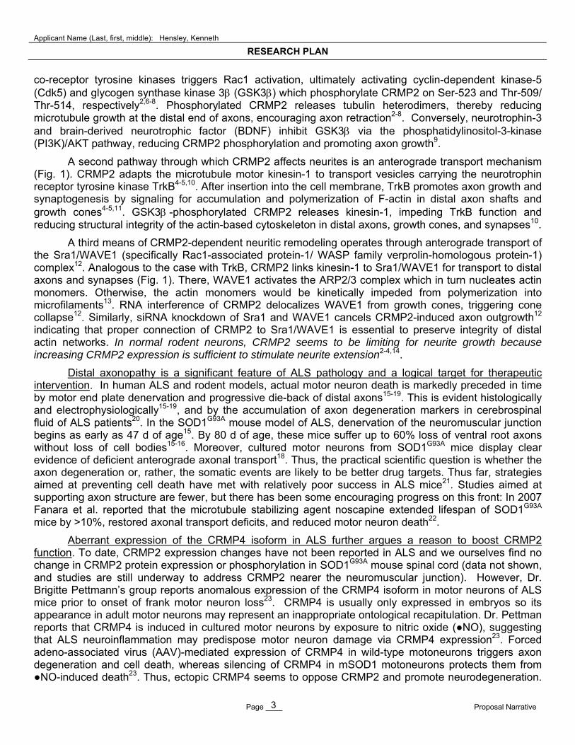
Rationale for AIM 1 and evidence for feasibility: LKE is a potent multifunctional neurotrophic agent with a novel mode of action that is highly relevant to ALS. Lanthionine ketimine (LK, Fig. 2) is a natural brain metabolite formed through non-canonical reactions catalyzed by the transsulfuration enzyme cystathionine -synthase (CS)25-30. In 2007-2010 the P.I. began to document unexpected bioactivities of LK and he synthesized novel cell-permeating derivatives25-28. One such derivative, LKE, promotes growth factor-dependent elongation of neurites in NSC-34 motor neuron cultures25. Subsequent work showed this effect also occurs in primary chick dorsal root ganglia neurons, and is dose-dependent down to LKE concentrations of 1-10 nM1 (approximating the physiological CNS concentration of LK)30 (Fig. 3). In addition to its potency as a neurite-supporting agent,
S
NHO2C CO2H
H
H
S
NH
HO2C CO2H
HS
NHO2C CO2H
H
H
S
NHO2C CO2H
H
H
S
NH
HO2C CO2H
H S
NH
HO2C CO2H
S
NH
HO2C CO2H
H
Figure 2. Lanthionine ketimine. The compound naturally exists in tautomeric equilibrium as shown.
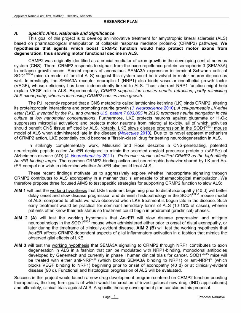
Figure 3. LKE promotes neurite growth in chick DRG cultures. A: DIV3 neurons were treated 48 h with vehicle or with LKE then treated with PromegaLive/Dead® fluorescent reagent, photomicrographed, and quantitatively assessed for morphometry by an analyst blinded to treatment group1. Data indicate mean ± SEM for 10-30 microscopic fields at each LKE concentration. Control neurite length (no drug) was 239 ± 23 m (dashed line). *p<0.05 by t-test. B, C: In separate experiments DIV 6 DRG neurons were treated ±LKE for 72 h, fixed and labeled with fluoro-taxol to visualize microtubules. Note longer, thicker, more numerous microtubules in LKE-treated cells.
LKE (M)
10-11 10-10 10-9 10-8 10-7 10-6 10-5 10-4
mea
n ne
urite
leng
th ( m
per
cel
l)
0
100
200
300
400
500
600
* * * ** *
LKE (M)
10-11 10-10 10-9 10-8 10-7 10-6 10-5 10-4
mea
n ne
urite
leng
th ( m
per
cel
l)
0
100
200
300
400
500
600
* * * ** *
B
DRG, DIV9, (-) LKE
C
DRG, DIV9, (+) 10 nM LKE from DIV6
A
Applicant Name (Last, first, middle): Hensley, Kenneth
RESEARCH PLAN
Page ____ Proposal Narrative 5
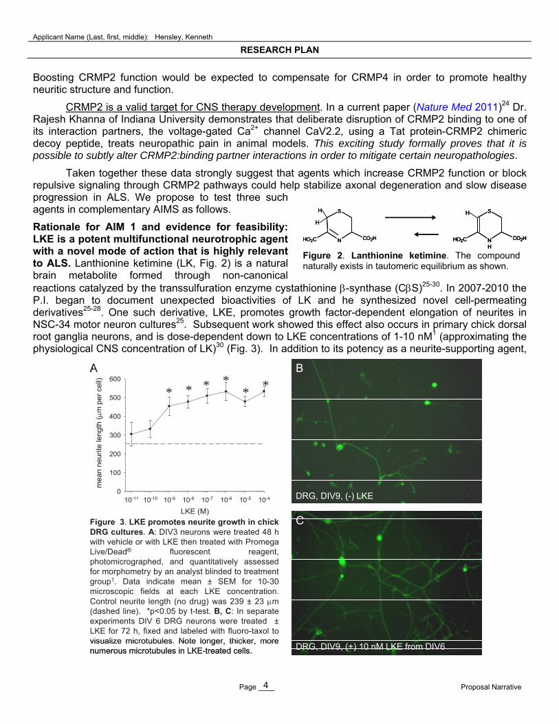
LKE suppresses tumor necrosis factor- (TNF)-stimulated microglial ●NO production at low M concentrations (Fig. 4)25; reduces the neurotoxicity of microglia-conditioned medium at sub-M concentrations (Fig. 5)26; blocks H2O2 toxicity25; and protects neurons from glutamate toxicity26. All these actions theoretically should aid ALS-afflicted neurons.
Ultimately we were able to demonstrate binding of immobilized LK to CRMP2 in whole brain lysates25, thus providing a rational LK target that could explain the extraordinary neurotrophic potency of LKE. The ability of LK to functionally interact with CRMP2 was corroborated in experiments where CRMP2 was immunoprecipitated from brain lysates in the presence or absence of LK, which increased CRMP2 interaction with some binding partners but decreased interaction with others25.
LKE promotes neuron growth in C. elegans to partially rescue the neuroanatomical phenotype of CRMP2 (UNC-33) hypomorphs. Further corroboration that LKE acts through CRMP2 comes from the P.I.’s collaborator Dr. Andrea Holgado (Southwestern Oklahoma State University) who finds that LKE increases dorso-ventral neural commissure development in normal C. elegans, and partially rescues the phenotype of incomplete commissure development in unc-33 (CRMP2 homolog) hypomorphs (manuscript in preparation). Whereas developing wild-type C. elegans typically extend commissural neuron processes from the ventral nerve cord to form dorsal or ventral neuromuscular junctions, unc-33 (e204) mutant worms extend commissures that prematurely terminate without reaching the target muscle cells31. Thus Tsuboi et al.31 suggested that nematode UNC-33 isoforms act in vivo by promoting axon growth and commissural extension; loss-of-function in UNC-33 results in abnormal neural networks and uncoordinated locomotion. To investigate LKE effect in vivo, Dr. Holgado first cultured the RM3128 strain of C. elegans (provided courtesy of Dr. James Rand, Oklahoma Medical Research Foundation). RM3128 nematodes express yellow fluorescent-protein (YFP) in cholinergic neuron cell bodies and neurites (driven by punc-17)32, allowing visualization of these cholinergic circuits. Next, she crossed unc-33 (e204) animals to the RM3128 strain and determined that treatment with 0.5 mM LKE for 96 h partially rescued the neuroanatomical phenotype of early commissure termination (Fig. 6). Furthermore, LKE increased the defecation rate of these worms from 5.2 ± 2.5 (SD) events/10 minutes to 7.2 ± 1.2 events/10 min (p<0.05), an indication of normalized neuromuscular function. This data strongly suggests that LKE has the capability of rearranging the synaptic network to overcome lack of normal UNC-33 (CRMP2) function.
Peripherally-administered LKE reaches mouse brain. We have developed an LC-MS assay for LKE and in preliminary experiments we find that a 200 mg/kg bolus i.p. dose of LKE results in mouse blood plasma [LKE]15 min = 98 M and brain LKE15 min = 151 ng/g wet weight after perfusion with saline (to wash out any blood vessel luminal LKE), or 377 ng/g in unperfused brain (N=4 mice total). While more detailed studies are underway (ongoing collaboration with P2D, Inc., Cincinnati, OH; see Therapy Development
LKE (M)
0 20 40 60 80 100
% v
iabi
lity
NS
C-3
4 ce
lls
0
20
40
60
80
100
EC50% = 764 nM
LKE (M)
0 20 40 60 80 100
% v
iabi
lity
NS
C-3
4 ce
lls
0
20
40
60
80
100
EC50% = 764 nM
Figure 5. LKE protects NSC-34 motor neuron-like cells against toxicity of microglia-conditioned medium26. EOC-20 murine microglia were stimulated with TNF and interferon- for 24 h then medium added to NSC-34 cultures ± LKE. Viability was assessed 24 h later by a tetrazolium reduction assay.
0 20 40 60 80 1000
5
10
15
20
25
30
net c
ytok
ine-
stim
ula
ted
NO
2-
(M
)
LKE (M)
0 20 40 60 80 1000
5
10
15
20
25
30
net c
ytok
ine-
stim
ula
ted
NO
2-
(M
)
LKE (M)
0 20 40 60 80 1000
5
10
15
20
25
30
net c
ytok
ine-
stim
ula
ted
NO
2-
(M
)
LKE (M) Figure 4. LKE inhibits TNF-stimulated ●NO release (indexed as NO2
) in EOC-20 microglial cultures. Cells were stimulated for 24 h and NO2
measured in culture medium with the Griess assay. Basal NO2
in control wells was <2 M. **p<0.01 by t-test.

Applicant Name (Last, first, middle): Hensley, Kenneth
RESEARCH PLAN
Page ____ Proposal Narrative 6
Plan section at end of this proposal), this preliminary data encourages us that LKE reaches the brain after peripheral administration and thus is quite feasible for further development as a CNS-acting drug.
LKE slows ALS progression in SOD1G93A mice when administered after onset of motor neuron death. The combination of potent activities inherent to LKE makes it a compelling molecule to test against ALS where oxidative stress, neuroinflammation, and cytoskeletal perturbation are all relevant features. In our experience, LKE is well tolerated by mice and in fact slows progression of paralysis when chronically injected into the SOD1G93A mice beginning at 90 d of age (Fig. 7)26-27. At this age however, axon deterioration is well under way and one even sees significant loss of -motoneuron somas in spinal cord ventral horns21. We therefore wish to complete in vivo studies of LKE by (1) treating mice at an earlier age prior to axonopathy, which is medically relevant to dominant hereditary forms of ALS where most individuals at risk for disease know their status and could be treated during pre-clinical stages; and (2) performing detailed histology to ascertain the full range of LKE effects.
Rationale for AIM 2 and evidence for feasibility: Ac-rER is a novel brain-penetrating, CRMP2 binder that displays neurotrophic potency and in vivo benefits. Prior to January 2011, LKE was the only small molecule drug candidate known to the P.I. that could conceivably benefit neurons through direct CRMP2 interaction. This changed in 2011 due to a publication from Mileusnic and Rose of the Open University, Great Britain33, who were studying the neurotrophic secreted extracellular fragment of Alzheimer’s disease-associated amyloid precursor protein (sAPP). These researchers built on prior findings that the sAPP neurotrophic action maps to a specific CRMP2-binding epitope, RERMS34. Neither sAPP nor RERMS is particularly attractive as a CNS drug because of their large size and limited penetrance through the blood-brain barrier (BBB). To overcome these limitations, Mileusnic and Rose designed and patented35 a novel tripeptide Ac-rER [N-terminal acetylated, (D)Arg-(L)-Glu-(L)-Arg] that retains the neurotrophic action of sAPP and RERMS. Because of its arginine-rich composition Ac-rER readily and demonstrably penetrates an intact BBB when administered peripherally35-36 and has less protease sensitivity and longer plasma lifetime than other RERMS mimics (Dr. R. Mileusnic, personal communications). The Ac-rER restores passive avoidance memory ability in a
age (days)
rota
rod
perf
orm
ance
(% o
f 90
d ba
selin
e)
control (N=29)LKE (N=15)
90 100 110 120 130 140 1500
25
50
75
100
125
150
*
*
age (days)
rota
rod
perf
orm
ance
(% o
f 90
d ba
selin
e)
control (N=29)LKE (N=15)control (N=29)LKE (N=15)
90 100 110 120 130 140 1500
25
50
75
100
125
150
*
*
vehicle (N=29) LKE (N=15)
age of frank paralysis (d) 113.7 ± 1.4 121.6 ± 2.5 *
median age at death (d) 132 139 †
minimum age at death (d) 117 124
maximum age at death (d) 142 152
Figure 7. Effect of LKE on motor function, paralysis and lifespan in SOD1G93A mice26. Mice were treated with LKE 100 mg/kg i.p. from 90 d until death and assessed regularly by a rotarod motor function task. *p<0.05 by t-test; †p<0.05 by log-rank test.
Figure 6. LKE reduces the frequency of premature commissure terminations in RM3128 x unc-33 (e204) C. elegans. A: Epifluorescence image showing normal commissure (arrow). B: Commissure that terminates prematurely (arrow). C: Bars represent mean ± SD for N=5 independent experiments (total of 69 control and 103 LKE-treated animals).
Applicant Name (Last, first, middle): Hensley, Kenneth
RESEARCH PLAN
Page ____ Proposal Narrative 7
chick model of amnestic impairment caused by antisense knockdown of APP35-36. In affinity proteomics studies, Ac-rER bound selectively to CRMP234. We anticipate that similar binding interactions of Ac-rER and LKE predict similar neurochemical effects though perhaps with important differences in potency and dynamics. We have fully executed agreements with the Open University to test Ac-rER (available upon request).
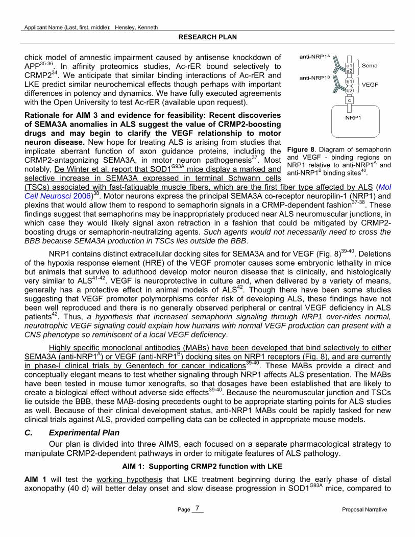
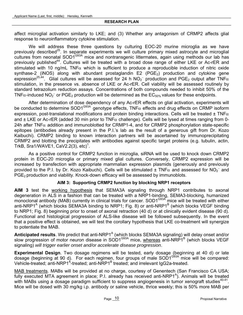
Rationale for AIM 3 and evidence for feasibility: Recent discoveries of SEMA3A anomalies in ALS suggest the value of CRMP2-boosting drugs and may begin to clarify the VEGF relationship to motor neuron disease. New hope for treating ALS is arising from studies that implicate aberrant function of axon guidance proteins, including the CRMP2-antagonizing SEMA3A, in motor neuron pathogenesis37. Most notably, De Winter et al. report that SOD1G93A mice display a marked and selective increase in SEMA3A expressed in terminal Schwann cells (TSCs) associated with fast-fatiguable muscle fibers, which are the first fiber type affected by ALS (Mol Cell Neurosci 2006)38. Motor neurons express the principal SEMA3A co-receptor neuropilin-1 (NRP1) and plexins that would allow them to respond to semaphorin signals in a CRMP-dependent fashion37-38. These findings suggest that semaphorins may be inappropriately produced near ALS neuromuscular junctions, in which case they would likely signal axon retraction in a fashion that could be mitigated by CRMP2-boosting drugs or semaphorin-neutralizing agents. Such agents would not necessarily need to cross the BBB because SEMA3A production in TSCs lies outside the BBB.
NRP1 contains distinct extracellular docking sites for SEMA3A and for VEGF (Fig. 8)39-40. Deletions of the hypoxia response element (HRE) of the VEGF promoter causes some embryonic lethality in mice but animals that survive to adulthood develop motor neuron disease that is clinically, and histologically very similar to ALS41-42. VEGF is neuroprotective in culture and, when delivered by a variety of means, generally has a protective effect in animal models of ALS42. Though there have been some studies suggesting that VEGF promoter polymorphisms confer risk of developing ALS, these findings have not been well reproduced and there is no generally observed peripheral or central VEGF deficiency in ALS patients42. Thus, a hypothesis that increased semaphorin signaling through NRP1 over-rides normal, neurotrophic VEGF signaling could explain how humans with normal VEGF production can present with a CNS phenotype so reminiscent of a local VEGF deficiency.
Highly specific monoclonal antibodies (MABs) have been developed that bind selectively to either SEMA3A (anti-NRP1A) or VEGF (anti-NRP1B) docking sites on NRP1 receptors (Fig. 8), and are currently in phase-I clinical trials by Genentech for cancer indications39-40. These MABs provide a direct and conceptually elegant means to test whether signaling through NRP1 affects ALS presentation. The MABs have been tested in mouse tumor xenografts, so that dosages have been established that are likely to create a biological effect without adverse side effects39-40. Because the neuromuscular junction and TSCs lie outside the BBB, these MAB-dosing precedents ought to be appropriate starting points for ALS studies as well. Because of their clinical development status, anti-NRP1 MABs could be rapidly tasked for new clinical trials against ALS, provided compelling data can be collected in appropriate mouse models.
C. Experimental Plan
Our plan is divided into three AIMS, each focused on a separate pharmacological strategy to manipulate CRMP2-dependent pathways in order to mitigate features of ALS pathology.
AIM 1: Supporting CRMP2 function with LKE
AIM 1 will test the working hypothesis that LKE treatment beginning during the early phase of distal axonopathy (40 d) will better delay onset and slow disease progression in SOD1G93A mice, compared to
c
b2
b1
a2a1
anti-NRP1A
anti-NRP1B
Sema
VEGF
NRP1
Figure 8. Diagram of semaphorin and VEGF - binding regions on NRP1 relative to anti-NRP1A and anti-NRP1B binding sites40.
Applicant Name (Last, first, middle): Hensley, Kenneth
RESEARCH PLAN
Page ____ Proposal Narrative 8
effects observed when treatment is begun later. This data would inform treatment strategies for dominant familial ALS where risk status is known long before onset of symptoms. In this AIM we will also perform new histology studies to fully ascertain the effects of LKE on neuropathology of the SOD1G93A mouse.
Anticipated results. We anticipate that LKE treatment beginning at 40 d (prior to axonopathy)15 will delay onset of paralysis, slow motor functional loss, increase lifespan and diminish histopathology of ALS mice; and the magnitude of effects will be greater than effects seen when treatment is begun later in disease.
Experimental design. Male and female mice will be treated with i.p. injections of LKE 100 mg/kg/d or saline vehicle beginning at 40 d. Twenty male and female mice will be used in each group (based on power analyses using historical data for motor function and survival variance in our SOD1G93A colony)21.
Animals. Mice expressing high copy numbers of human SOD1G93A will be obtained from Jackson Laboratories [Bar Harbor ME; strain B6SJL-TgN-(SOD1-G93A)-1-Gur]21. Mice will be maintained in the hemizygous state by mating SOD1G93A males with B6SJL-TGN females.
Motor function and lifespan. Rotarod motor function tests will be performed by a technician blinded to the treatment groups using a standard rotarod device (Columbus Instruments) set to accelerate at 1 rpm/10 s. Trials will begin 10 d prior to first dosage and will be repeated at 10 d intervals. Mice will be assigned to groups with similar initial mean rotarod performance times and equal numbers of male and female animals (10 mice/sex/group). No more than one mouse from a single litter will be included in any one group, and care will be taken to equalize litter representation amongst groups, to minimize bias arising from small-group genetic or gestational influences. Rotarod performance times will be recorded in quadruplicate tests and the best three times averaged at each trial for each mouse. Mouse weights will be recorded at each trial period. Mice will be killed when they can no longer perform the rotarod task or right themselves within 10 s of being placed on their sides. In all cases motor function tests will be conducted and euthanasia decisions made by trained personnel who are blinded to mouse treatment groups.
Spinal histology: 120 d old mice near the normal midpoint of their clinical disease progression21 will be deeply anesthetized and transcardially perfused with buffered formalin. Spinal cords will be removed, blocked, and post-fixed. Blocks will be cryoprotected in 30% sucrose and cryostat-sectioned in 40 μm sections. Adjacent lumber (L1-L3) or cervical (C5-C7) sections will be processed. For neuron visualization, alternating sections will be Nissl-stained or stained with antibodies against choline acetyltransferase (ChaT). For assessing gliosis parallel sections will be stained with anti-GFAP. Microglial proliferation will be determined by counting Iba1+ cells. Neurons will be counted in ventral horns bilaterally under a 40X objective by an experimenter blind to the treatment groups.
Axon counts: Animals will be perfused with 2% paraformaldehyde/ 2% glutaraldehyde in sodium cacodylate buffer and ventral spinal cord, nerve, and muscles postfixed in 2% osmium tetroxide, dehydrated, and embedded in Araldite. Myelinated axon counts will be made in 1 m sections of the 4th lumbar ventral root (L4VR) and medial gastroneimial (MG) nerves after toluidine blue staining41. Mitochondria will be assessed by electron microscopy43.
Statistics. Statistical differences in motor performance between groups will be assessed using repeated measures analysis of variance (ANOVA) with post-hoc analysis using Student’s t-tests at individual time points. Survival curves will be analyzed by log rank statistical methods. Data will be stratified by gender post-hoc if statistically justified. Histological parameters will be assessed by appropriate parametric tests.
Potential pitfalls and alternatives. Prior experience with LKE indicates that the drug is safe and well tolerated by mice receiving daily i.p. injections. We ourselves have treated mice for over one month (e.g. Fig. 7 above) and our collaborator Dr. Marni Harris-White of the University of California, Los Angeles, has been injecting LKE into a mouse model for Alzheimer’s disease for 5 months with no adverse effects (Dr. Harris-White, personal communication). Nonetheless, it is conceivable that SOD1G93A mice may exhibit unanticipated adverse reactions to either LKE or Ac-rER (AIM 2 below). In either case we will consider
Applicant Name (Last, first, middle): Hensley, Kenneth
RESEARCH PLAN
Page ____ Proposal Narrative 9
alternative routes of administration such as mini-osmotic pumps. Ample time to manage such contingencies has been allocated to the proposed research timetable (Fig. 9 below).
AIM 2: Supporting CRMP2 function with Ac-rER
AIM 2 (A) will test the working hypothesis that Ac-rER will slow disease progression and mitigate neuropathology in the SOD1G93A mouse when administered either prior to onset of distal axonopathy, or later during clinically-evident disease. AIM 2 (B) will test the working hypothesis that Ac-rER affects CRMP2-dependent aspects of glial inflammatory activation in a fashion that mimics the observed glial effects of LKE.
Anticipated results. We anticipate that Ac-rER treatment will slow motor function decline, slow weight loss, delay onset of paralysis, delay death, and improve histological correlates of neurodegeneration (i.e. maintain motor neuron, ventral root, and nerve axon numbers and reduce microglia) in SOD1G93A mice. We further anticipate that Ac-rER will suppress glial inflammatory (M1) activation in cell culture and that generally, microglial responses will be sensitive to experimental manipulation of CRMP2 expression.
Experimental design for AIM 2(A): Animal therapy studies. A dose-ranging design will be employed wherein male SOD1G93A mice will be treated with vehicle; 50, 200, or 500 mg/kg/d Ac-rER beginning at 40 d (before neuromuscular denervation) or 90 d (clinically evident disease) and continuing through the remaining animal lifespan.
Treatments. Ac-rER will be synthesized as the acetate salt through contract with AnaSpec (Fremont CA USA; quote attached), dissolved in physiological saline, and injected i.p. 5 d/week. Vehicle will be saline with matched sodium acetate content.
Motor function and histology. Motor function will be assessed as described under AIM 1. Histology will be performed on separate, specifically designated groups of mice treated with vehicle or the most effective Ac-rER regimen (in terms of improvements in lifespan and motor function). Ten (10) male nontransgenic mice will be compared with groups of 10 Ac-rER-treated and 10 vehicle-treated SOD1G93A mice.
Experimental Design for AIM 2(B): Glial cell culture studies. As noted above (Fig. 4), LKE suppresses microglial activation by inflammatory cytokines in EOC-20 microglial cell culture. Aberrant neuroinflammatory activation of microglia and astrocytes has been repeatedly linked to pathogenesis in the SOD1G93A mouse, and evidence suggests that neuroinflammatory mechanisms are highly relevant to human sporadic ALS as well21. Interestingly, we have shown that primary astroglia from SOD1G93A mice are much more sensitive to inflammatory activation in culture than are normal nontransgenic glia44, while Dr. Stanley Appel has replicated our findings in SOD1G93A microglia and showed that these cells are more inherently toxic to motor neurons when activated as compared to wild-type glia45. The precise molecular reason for this inflammatory hypersensitivity and SOD1G93A microglial hyper-toxicity remains a mystery but Dr. Appel reports that the degree of microglial toxicity is directly proportional to microglial nitrite production, suggesting a neurotoxic role for ●NO45. Activated SOD1G93A glia were particularly capable of damaging motor neuritic structure in cell culture, which is broadly consistent with Dr. Pettmann’s observation23 of ●NO-dependent CRMP4 expression sensitivity in motor neurons (discussed above).
The LKE effects we observe in glia are noteworthy, in part, because CRMP2 has never before been seriously studied in a glial cell context. If CRMP2 proves important in microglial dynamics, then the study of CRMP2’s microglial role(s) could shed new light on neuroinflammatory mechanisms in ALS, and could lead to identification of novel new microglia-acting drugs. Ac-rER has already been studied by Mileusnic et al. with respect to its CRMP2 binding properties and neurotrophic actions, both of which are very reminiscent of LKE properties, but has never been tested in glial cell systems. Ac-rER is thus very compelling to test against possible CRMP2-dependent microglial activation events.
We therefore wish to determine whether (1) Glia express CRMP isoforms and/or post-translationally process CRMP2 (or other isoforms) in a fashion that is sensitive to pro-inflammatory stimulation; (2) Whether a structurally distinct compound such as Ac-rER that also binds CRMP2 can
Applicant Name (Last, first, middle): Hensley, Kenneth
RESEARCH PLAN
Page ____ Proposal Narrative 10
affect microglial activation similarly to LKE; and (3) Whether any antagonism of CRMP2 affects glial response to neuroinflammatory cytokine stimulation.
We will address these three questions by culturing EOC-20 murine microglia as we have previously described25. In separate experiments we will culture primary mixed astrocyte and microglial cultures from neonatal SOD1G93A mice and nontransgenic littermates, again using methods our lab has previously published44. Cultures will be treated with a broad dose range of either LKE or Ac-rER and stimulated with 10 ng/mL TNF which is sufficient to produce a reproducible induction of nitric oxide synthase-2 (iNOS) along with abundant prostaglandin E2 (PGE2) production and cytokine gene expression25,44. Glial cultures will be assessed for 24 h NO2
production and PGE2 output after TNF stimulation, in the presence vs. absence of LKE or Ac-rER. Cell viability will be assessed routinely by standard tetrazolium reduction assays. Concentrations of both compounds needed to inhibit 50% of the TNF-induced NO2
or PGE2 production will be determined as the EC50% values for these endpoints.
After determination of dose dependency of any Ac-rER effects on glial activation, experiments will be conducted to determine SOD1G93A genotype effects, TNF effects and drug effects on CRMP isoform expression, post-translational modifications and protein binding interactions. Cells will be treated ± TNF and ± LKE or Ac-rER (added 30 min prior to TNF challenge). Cells will be lysed at times ranging from 0-24h after TNF addition and immunoblotted for CRMP1-4, and for CRMP2 phosphorylation state-specific epitopes (antibodies already present in the P.I.’s lab as the result of a generous gift from Dr. Kozo Kaibuchi). CRMP2 binding to known interaction partners will be ascertained by immunoprecipitating CRMP2 and blotting the precipitates with antibodies against specific target proteins (e.g. tubulin, actin, TrkB, Sra1/WAVE1, CaV2.2(3), etc)1.
As a positive control for CRMP2 function in microglia, siRNA will be used to knock down CRMP2 protein in EOC-20 microglia or primary mixed glial cultures. Conversely, CRMP2 expression will be increased by transfection with appropriate mammalian expression plasmids (generously and previously provided to the P.I. by Dr. Kozo Kaibuchi). Cells will be stimulated ± TNF and assessed for NO2
and PGE2 production and viability. Knock-down efficacy will be assessed by immunoblots.
AIM 3: Supporting CRMP2 function by blocking NRP1 receptors
AIM 3 test the working hypothesis that SEMA3A signaling through NRP1 contributes to axonal degeneration in ALS in a fashion that can be treated with a NRP1-binding, SEMA3-blocking, humanized monoclonal antibody (MAB) currently in clinical trials for cancer. SOD1G93A mice will be treated with either anti-NRP1A (which blocks SEMA3A binding to NRP1; Fig. 8) or anti-NRP1B (which blocks VEGF binding to NRP1; Fig. 8) beginning prior to onset of axonal retraction (40 d) or at clinically evident disease (90 d). Functional and histological progression of ALS-like disease will be followed subsequently. In the event that a positive effect is obtained, we will test the corollary hypothesis that LKE co-treatment will synergize to potentiate the MAB.
Anticipated results. We predict that anti-NRP1A (which blocks SEMA3A signaling) will delay onset and/or slow progression of motor neuron disease in SOD1G93A mice, whereas anti-NRP1B (which blocks VEGF signaling) will trigger earlier onset and/or accelerate disease progression.
Experimental Design. Two dosage regimens will be tested, early dosage (beginning at 40 d) or late dosage (beginning at 90 d). For each regimen, four groups of male SOD1G93A mice will be compared: Vehicle-treated; anti-NRP1A-treated; anti-NRP1B treated; and irrelevant IgG2a-treated.
MAB treatments. MABs will be provided at no charge, courtesy of Genentech (San Francisco CA USA; fully executed MTA agreement in place; P.I. already has received anti-NRP1A). Animals will be treated with MABs using a dosage paradigm sufficient to suppress angiogenesis in tumor xenograft studies40-41. Mice will be dosed with 30 mg/kg i.p. antibody or saline vehicle, thrice weekly; this is 50% more MAB per
Applicant Name (Last, first, middle): Hensley, Kenneth
RESEARCH PLAN
Page ____ Proposal Narrative 11
dose than needed to slow angiogenesis in mouse studies, and one more dose per week. Dosage will begin either at 40 d (prior to neuromuscular denervation)15 or at 90 d (clinically-evident disease when mice have lost ca. 10% of ventral horn -motoneurons).
Motor function and histology will be assessed as described in AIMS 1-2.
Potential pitfalls and alternatives. It is conceivable that mice chronically treated with humanized anti-NRP1 could display an immune reaction to the antibodies resulting in too-rapid clearance of the MAB or an allergic response. This is relatively unlikely since the classic strategy used to deliberately generate mouse anti-(other species) IgG in vivo requires adjuvants and much more aggressive, deliberate, systemic immune activation than is likely to occur within our protocol. Empirically, passive immunization with anti-amyloid MABs has shown benefits in mouse models of AD even when mice were treated nine months with weekly i.p. injections46. Also, twice-weekly delivery of XOMA 052, a fully humanized IgG2 MAB directed against IL1, was safe and efficacious in an immune-competent mouse model of diet-induced obesity even after 13 weeks of MAB treatment47. Other examples exist in the literature. Nonetheless we can test for the presence of serum antibodies against anti-NRP1A and monitor for signs of allergic reaction. The use of an irrelevant IgG2a control arm also should mitigate the risk of committing an error of interpretation in our results. If necessary we may adjust the MAB dosing frequency downward or explore the use of non-humanized murine MABs or F(ab’)2 fragments. Ample time to manage such contingencies has been allocated to the proposed timetable (Fig. 9).
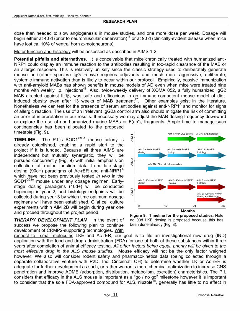
TIMELINE. The P.I.’s SOD1G93A mouse colony is already established, enabling a rapid start to the project if it is funded. Because all three AIMS are independent but mutually synergistic, they will be pursued concurrently (Fig. 9) with initial emphasis on collection of motor function data from late-stage dosing (90d+) paradigms of Ac-rER and anti-NRP1A which have not been previously tested in vivo in the SOD1G93A mouse under any dosage regimen. Early-stage dosing paradigms (40d+) will be conducted beginning in year 2; and histology endpoints will be collected during year 3 by which time optimum dosage regimens will have been established. Glial cell culture experiments within AIM 2B will begin during year one and proceed throughout the project period.
THERAPY DEVELOPMENT PLAN. In the event of success we propose the following plan to continue development of CRMP2-supporting technologies. With respect to small molecules LKE and Ac-rER, our goal is to file an investigational new drug (IND) application with the food and drug administration (FDA) for one of both of these substances within three years after completion of animal efficacy testing. All other factors being equal, priority will be given to the most effective drug in the ALS mouse studies. Mouse efficacy will not be the only factor weighed however: We also will consider rodent safety and pharmacokinetics data (being collected through a separate collaborative venture with P2D, Inc. Cincinnati OH) to determine whether LK or Ac-rER is adequate for further development as such, or rather warrants more chemical optimization to increase CNS penetration and improve ADME (adsorption, distribution, metabolism, excretion) characteristics. The P.I. considers that efficacy in the ALS mouse is important as a “go / no go” milestone however it is important to consider that the sole FDA-approved compound for ALS, riluzole48, generally has little to no effect in
0 12 24 36
AIM 1: 40d+ LKE dosing AIM 1: LKE histology
AIM 2A: 90d+ Ac-rER AIM 2A: 40d+ Ac-rER AIM 2A: Ac-rERdosing dosing histology
AIM 3: 90d+ anti-NRP1A AIM 3: 40d+ anti-NRP1A AIM 3: anti-NRP1A
dosing dosing histology
AIM 3: 40d+ anti-NRP1B
dosing and histology
AIM 2B: Glial cell culture studies
AIM
3
A
IM 2
AIM
1
Months Figure 9. Timeline for the proposed studies. Note no 90d LKE dosing is proposed because this has been done already (Fig. 8).
Applicant Name (Last, first, middle): Hensley, Kenneth
RESEARCH PLAN
Page ____ Proposal Narrative 12
ALS mouse trials49 despite its having consistently shown a small human clinical benefit (but only offers several month’s life extension over a typical three-five year ALS progression period)48. Conversely, a number of compounds that have modest to impressive effects in ALS mice have failed in the clinic due to patient intolerance or inadequate pharmacological behavior48. Thus, a very safe compound that has desirable pharmacokinetic parameters and formulation potential and displays even a modest but reproducible effect in ALS mouse models is justifiable to pursue as a lead compound for human clinical development.
Both LKE and Ac-rER already have composition-of-matter patent status26,36 which would greatly enable further development. Our preferred plan is to partner with a development-stage company in order to conduct GMP-compliant toxicology and pharmacology in support of IND(s). We are in discussion with P2D regarding the details of this strategy. P2D’s business model is to license technologies like LKE or Ac-rER and develop to the point of Phase II clinical trials, before partnering with a major pharmaceutical company to complete trials. An alternative strategy is to create a new business enterprise based on the development of CRMP2-acting agents. In either event we will seek private capital plus SBIR and/or U01 funds to help support the endeavor.
Development of LKE would necessitate a licensing agreement with the patent assignee, the Oklahoma Medical Research Foundation (OMRF) where the P.I. invented the compound. Such a license is currently being negotiated between OMRF and the P.I.’s development partner, P2D Inc. Development of Ac-rER would necessitate separate agreements with the assignee of its patents, Open University, and these discussions have been initiated (confidentiality and research agreements already are in place between University of Toledo and the Open University).
With respect to biologics (NRP1 monoclonal antibodies), this technology is held by Genentech. The P.I. has discussed the project with Dr. Ryan Watts, head of Neurodegeneration Laboratories at Genentech. In the event of a successful proof-of-concept test, The P.I. will work with Genentech to explore anti-NRP1 MAB potential in ALS and other MNDs. Necessary confidentiality and material transfer agreements are in place and are available upon request.
.
PRINCIPAL INVESTIGATOR: Kenneth Hensley, PhD
REFERENCES FOR LITERATURE CITED IN THIS APPLICATION
(Key citations in blue are hyperlinked to URLs for reviewer convenience)
1. Hensley K, Christov A, Venkova K, Gunning W, Park J (2011) Collapsin response mediator protein-2: An emerging pathologic feature and therapeutic target for neurodisease indications. Mol Neurobiol 43: 180-191. PMID: 21271304
2. Goshima Y, Nakamura F, Strittmatter P, Strittmatter SM (1995) Collapsin-induced growth cone collapse mediated by an intracellular protein related to UNC-33. Nature 376: 509-514.PMID:7637782
3. Inagaki N, Chihara K, Arimura N, Menager C, Kawano Y, Matsuo N, Nishimura T, Amano M, Kaibuchi K (2001) CRMP-2 induces axons in cultured hippocampal neurons. Nature Neurosci 4: 781-782. PMID: 11477421
4. Quach TT, Duchemin A-M, Rogemond V, Aguera M, Honnorat J, Belin M-F, Kolattukudy PE (2004) Involvement of collapsing response mediator proteins in the neurite extension induced by neurotrophins in dorsal root ganglion neurons. Mol Cell Neurosci 25: 433-443. PMID:15033171
5. Charrier E, Riebel S, Rogemond V, Aguera M, Thomasset N, Honnorat J (2003) Collapsin response mediator proteins (CRMPs): Involvement in nervous system development and neurodegenerative disorders. Mol Neurobio 203: 51-63. PMID: 14514985
6. Deo RC,Schmidt EF, Elhabazi A, Togashi H, Burley SK, Strittmatter SM (2004) Structural bases for CRMP function in plexin-dependent semaphoring 3A signaling. EMBO J 23: 9-22. PMCID: PMC1271659
7. Yoshimura T,Kawano Y, Arimura N, Kawabata S, Kikuchi A, Kaibuchi K (2005) GSK-3 regulates phosphorylation of CRMP-2 and neuronal polarity. Cell 120: 137-149. PMID: 15652488
8. Uchida Y, Ohshima T, Sasaki Y, Suzuki H, Yanai S, Yamashita N, Nakamura F, Takei K, Ihara Y, Mikoshiba K, Kolattukudy P, Honnorat J, Goshima Y (2005) Semaphorin3A signaling is mediated via sequential Cdk5 and GSK3beta phosphorylation of CRMP2: implication of common phosphorylating mechanism underlying axon guidance and Alzheimer's disease. Genes Cells 10: 165-179. PMID: 15676027
9. Li T, Hawkes C, Qureshi HY, Kar S, Paudel HK (2006) Cyclin-dependent protein kinase 5 primes microtubule-associated protein tau site-specifically for glycogen synthase kinase 3beta. Biochemistry 14: 3134-3145. PMID: 16519508
10. Lykissas MG, Batistatou AK, Konstantinos A, Charalabopoulos KA, Beris AE (2007) The role of neurotrophins in axon guidance, growth and regeneration. Curr Neurovas Res 4: 143-151.
PMID: 17504212 11. Arimura N, Kimura T, Nakamuta S, Taya S, Funahashi Y, Hattori A, ShimALSa A, Menager C,
Kawabata S, Jujii K, Iwamatsu A, Segal RA, Fukuda M, Kaibuchi K (2009) Anterogrde transport of TrkB in axons is mediated by direct interaction with Slp1 and Rab27. Dev Cell 15: 675-686. NIHMS PMCID: PMC2781534
12. Kawano Y, Yoshimura T, Tsuboi D, Kawabata S, Kaneko-Kawano T, Shirataki H, Takenawa T, Kaibuchi K (2005) CRMP-2 is involved in kinesin-1-dependent transport of the Swa-1/WAVE1 complex and axon formation. Moll Cell Biol 25: 9920-9935. PMCID: PMC1280248
13. Pollitt A, Insall RH (2009) WASP and SCAR/WAVE1 proteins: The drivers of actin assembly. J Cell Sci 122: 2575-2578. PMID: 19625501
14. Chu CC, Wang JJ, Chen KT, Shieh JP, Wang LK, Shui HA, Ho ST (2010) Neurotrophic effects of tianeptine on hippocampal neurons: A proteomic approach. J Proteome Res 9: 936-944.
PMID: 20000655 15. Fischer LR, Culver DG, Tennant P, Davis AA, Wang M, Castellano-Sanchez A, Khan J, Polak
MA, Glass JD (2004) Amyotrophic lateral sclerosis is a distal axonopathy: Evidence in mice and man. Exp Neurol 185: 232-240. PMID: 14736504
16. Fischer LR, Glass JD (2007) Axonal degeneration in motor neuron disease. Neurodegener Dis 4: 431-442. PMID: 17934327
17. Hegedus J, Putman CT, Gordon T (2007) Time course of preferential motor unit loss in the SOD1
G93A mouse model of amyotrophic lateral sclerosis. Neurobiol Dis 28: 154-164. PMID: 17766128 18. Vickers JC, King AE, Woodhouse A, Kirkcaldie T, Staal JA, McCormack GH, Blizzard CA,
Musgrove REJ, Mitew S, Liu Y, Chuckowree IA, Bibari O, Dickson TC (2009) Axonopathy and cytoskeletal disruption in degenerative diseases of the central nervous system. Br Res Bull 80: 217-223. PMID: 19683034
19. Dupuis L, Loeffler JP (2009) Neuromuscular junction destruction during amyotrophic lateral sclerosis: Insights from transgenic models. Curr Opin Pharmacol 9: 341-346. PMID: 19386549
20. Brettschneider J, Petzold A, Sussmuth SD, Ludolph AC, Tumani H (2006) Axonal damage markers in cerebrospinal fluid are increased in ALS. Neurology 66: 852-866. PMID: 16567701
21. Hensley K, Mhatre MC, Mou S, Pye QN, Stewart CA, West MS, Williamson KS (2006) Lessons learned from the SOD1-G93A mouse model of amyotrophic lateral sclerosis. Antioxid Redox Signal 8: 2075-2087. PMID: 17034351
22. Fanara P, Banerjee J, Hueck RV, Harper MR, Awada M, Turner H, Husted KH, Brandt R, Hellerstein MK (2007) Stabilization of hyperdynamic microtubules is neuroprotective in amyotrophic lateral sclerosis. J Biol Chem 282: 23465-23472. PMID: 17567579
23. Duplan J, Bernard N, Casseron W, Dudley K, Thouvenot E, Honnorat J, Rogemond V, De Bovis B, Aebischer P, Marin P, Raoul C, Henderson CE, Pettmann B (2010) Collapsin response mediator protein 4a (CRMP4a) is upregulated in motoneurons of mutant SOD1 mice and can trigger motoneuron axonal degeneration and cell death. J Neurosci 30: 785-796. PMID: 20071543
24. Brittain JM, Duarte DB, Wilson SM, Zhu W, Ballard C, Johnson PL, L iu N, Xiong W, Ripsch MS, Wang Y, Fehrenbacher JC, Fitz SD, Khanna M, Park CK, Schmutzler BS, Cheon BM, Due MR, Brustovetsky T, Ashpole NM, Hudmon A, Meroueh SO, Hingtgen CM, Brustovetsky N, Ji RR, Hurley JH, Jin X, Shekhar A, Xu XM, Oxford GS, Vasko MR, White FA, Khanna R (2011) Suppression of inflammatory and neuropathic pain by uncoupling CRMP-2 from the presynaptic Ca2+ channel complex. Nature Med: Epub ahead of print. PMID: 2164297
25. Hensley K, Christov A, Kamat S, Zhang XC, Jackson KW, Snow S, Post J (2010) Proteomic Identification of Binding Partners for the Brain Metabolite Lanthionine Ketimine (LK) and Documentation of LK Effects on Microglia and Motoneuron Cell Cultures. J Neurosci 30: 2979-2988. PMID: 20181595, PMCID:PMC 2836831
26. Hensley, K. Lanthionine-related compounds for the treatment of inflammatory diseases. U.S. Patent No. 7,683,055. Issued 3/23/2010.
27. Hensley, K. (2010) Emerging Biological Importance of Central Nervous System Lanthionines. Molecules 15: 5581-5594. PMID: 20714314
28. Chung, C., Kurien, B.T., Mehta, P., Mhatre, M.C., Mou, S., Pye, Q.N., Stewart, C.A., West, M.S., Williamson, K.S., Post, J., Liu, L., Wang, R., Hensley, K. (2007) Identification of lanthionine synthase C-like protein 1 (LanCL1) as a prominent glutathione binding protein expressed in the mammalian central nervous system. Biochemistry 46: 3262-3269. PMID: 17305318
29. Cavallini D, Ricci G, Dupre S, Pecci L, Costa M, Matarese RM, Pensa B, Antonuci A, Solinas SP, Fontana M (1991) Sulfur-containing cyclic ketimines and imino acids A novel family of endogenous products in search for a role Eur J Biochem 202: 217-223. PMID: 1761027
30. Ricci G, Vesci L, nardini M, Arduini A, Storto S, Rosato N, Cavallini D (1989) Detection of 2H-1,4-thiazine-5,6-dihydro-3,5-dicarboxylic acid (lanthionine ketimine) in the bovine brain by a fluorometric assay. Biochim Biophys Acta 990: 211-215. PMID: 2917179
31. Mileusnic R, Rose SP (2011) The memory enhancing effect of the APP-derived tripeptide Ac-rER is mediated through CRMP2. J Neurochem 2011 (Epub ahead of print, doi: 10.1111/j.1471-4159.2011.07193.x) PMID: 21255016
32. Pawlik M, Otero DA, Park M, Fischer WH, Levy E, Saitoh T (2007) Proteins that bind to the RERMS region of beta amyloid precursor protein. Biochem Biophys Res Commun 355: 907-912.
PMID: 17335780, PMCID:PMC 1896148 33. Mileusnic R, Lancashire C, Clark J, Rose SP (2007) Protection against Abeta-induced memory
loss by tripeptide D-Arg-L-Glu-L-Arg. Behav Pharmacol 18: 231-238. PMID: 17426487 34. Mileusnic R, Rose SPR. Polypeptides related to amyloid precursor protein, pharmaceutical
compositions thereof, and methods of treatment using the same. U.S. Patent 7,491,702. Issued Feb. 17, 2009.
35. Schmidt ERE, Pasterkamp RJ, van den Berg LH (2009) Axon guidance proteins: Novel therapeutic targets for ALS? Progress in Neurobiol 88: 286-301. PMID: 19523502
36. De Winter F, Vo T, Stam FJ, Wisman LA, Bar PR, Niclou SP, van Muiswinkel FL, Verhaagen J (2006) The expression of the chemorepellent semaphoring 3A is selectively induced in terminal Schwann cells of a subset of neuromuscular synapses that display limited anatomical plasticity and enhanced vulnerability in motor neuron disease. Mol Cell Neurosci 32: 102-117. PMID: 16677822
37. Liang W-C, Dennis MS, Stawicki Y, Chanthery Y, Pan Q, Chen Y, Eigenbrot C, Yin J, Koch AW, Wu X, Ferrara N, Bagri A, Tessier-Lavigne M, Watts RJ, Wu Y (2007) Function blocking antibodies to neuropilin-1 generated from a designed human synthetic antibody phage library. J Mol Biol 366: 815-829. PMID: 17196977
38. Pan Q, Chanthery Y, Liang W-C, Stawicki S, Mak J, Rathore N, Tong RK, Kowalski J, Yee SF, Pacheco G, Ross S, Cheng Z, Couter J, Plowman G, Peale F, Koch AW, Wu Y, Bagri A, Tessler-Lavigne M, Wattts RJ (2007) Blocking neuropilin-1 function has an additive effect with anti-VEGF to inhibit tumor growth. Cancer Cell 11: 53-67. PMID: 17222790
39. Oosthuyse B, Moons L, Storkebaum E, Beck H, Nuyens D, Brusselmans K, Van Dorpe J, Hellings P, Gorselink M, Heymans S, Theilmeier G, Dewerchin M, Laudenbach V, Vermylen P, Raat H, Acker T, Vleminckx V, Ven Den Bosch L, Cashman N, Fujisawa H, Drost MR, Sciot R, Bruyninckx F, Hicklin DJ, Ince C, Gressens P, Lupu F, Plate KH, Robberecht W, Herbert J-M, Collen D, Carmeliet P (2001) Deletion of the hypoxia-response element in the vascular endothelial growth factor promoter causes motor neuron degeneration. Nat Genet 28: 131-138. PMID: 11381259
40. Sathasivam S (2008) VEGF and ALS. Neurosci Res 62: 71-77. PMID: 18656504 41. Gould TW, Buss RR, Vinsant S, Prevette D, Sun W, Knudson CM, Milligan CE, Oppenheim
RW (2006) Complete dissociation of motor neuron death from motor dysfunction by Bax deletion in a mouse model of ALS. J Neurosci 26: 8774-8786. PMID: 16928866
42. Aggarwal S, Cudkowicz M (2008) ALS drug development: Reflections from the past and a way forward. Neurotherapeutics: Journal of the American Society for Experimental Neurotherapeutics 5: 516-527. PMID: 19019302
43. Scott S, Kranz JE, Cole J, Lincecum JM, Thompson K, Kelly N, Bostrom A, Theodoss A, Al-Nakhala BM, Viera FG, Ramasubbu J, Heywood JA (2008) Design, power, and interpretation of studies in the standard murine model of ALS. Amyotroph Lateral Scler 9: 4-15. PMID 18273714
Related Documents