LETTER Communicated by Klaus Stiefel A Simulation Study Investigating the Impact of Dendritic Morphology and Synaptic Topology on Neuronal Firing Patterns Jen-Yung Chen [email protected] Department of Psychology, Binghamton University, Binghamton, NY 13902-6000, U.S.A. In the brain, complex information interactions among neurons span sev- eral spatial and temporal scales, making it extremely difficult to identify the principles governing neural information processing. In this study, we used computational models to investigate the impact of dendritic mor- phology and synaptic topology on patterns of neuronal firing. We first constructed Hodgkin-Huxley-type neuron models that possessed den- drites with different morphological features. We then simulated the re- sponses of these neurons to a number of spatiotemporal input patterns. The similarity between neuronal responses to different patterned inputs was effectively evaluated by a novel combination of metric space analysis and multidimensional scaling analyses. The results showed that neurons with different morphological or anatomical features exhibit differences in stimulus-specific temporal encoding and firing reliability. These find- ings support the idea that in addition to biophysical membrane prop- erties, the dendritic morphology and the synaptic topology of a neuron can play a significant role in neuronal information processing and may directly contribute to various brain functions. 1 Introduction Neurons, the elemental operating units of the brain, communicate with each other via synapses using a series of discrete electrical signals known as action potentials. A single neuron can receive hundreds to thousands of synaptic inputs, both excitatory and inhibitory, distributed over various locations of its dendrites. These inputs are integrated spatiotemporally at dendrites before they converge onto the cell body. On sufficient depolar- ization, a new spike train can be generated at the cell body (axon hillock) and transmitted to the following neurons. For decades, neuroscientists have been interested in studying the underlying biological mechanisms that func- tionally manipulate the input-output relationships of individual neurons. It has been thought that there are reliable computational principles that can quantitatively and logically interpret how information is integrated Neural Computation 22, 1086–1111 (2010) C 2009 Massachusetts Institute of Technology

Welcome message from author
This document is posted to help you gain knowledge. Please leave a comment to let me know what you think about it! Share it to your friends and learn new things together.
Transcript

LETTER Communicated by Klaus Stiefel
A Simulation Study Investigating the Impact of DendriticMorphology and Synaptic Topology on NeuronalFiring Patterns
Jen-Yung [email protected] of Psychology, Binghamton University, Binghamton,NY 13902-6000, U.S.A.
In the brain, complex information interactions among neurons span sev-eral spatial and temporal scales, making it extremely difficult to identifythe principles governing neural information processing. In this study, weused computational models to investigate the impact of dendritic mor-phology and synaptic topology on patterns of neuronal firing. We firstconstructed Hodgkin-Huxley-type neuron models that possessed den-drites with different morphological features. We then simulated the re-sponses of these neurons to a number of spatiotemporal input patterns.The similarity between neuronal responses to different patterned inputswas effectively evaluated by a novel combination of metric space analysisand multidimensional scaling analyses. The results showed that neuronswith different morphological or anatomical features exhibit differencesin stimulus-specific temporal encoding and firing reliability. These find-ings support the idea that in addition to biophysical membrane prop-erties, the dendritic morphology and the synaptic topology of a neuroncan play a significant role in neuronal information processing and maydirectly contribute to various brain functions.
1 Introduction
Neurons, the elemental operating units of the brain, communicate witheach other via synapses using a series of discrete electrical signals knownas action potentials. A single neuron can receive hundreds to thousandsof synaptic inputs, both excitatory and inhibitory, distributed over variouslocations of its dendrites. These inputs are integrated spatiotemporally atdendrites before they converge onto the cell body. On sufficient depolar-ization, a new spike train can be generated at the cell body (axon hillock)and transmitted to the following neurons. For decades, neuroscientists havebeen interested in studying the underlying biological mechanisms that func-tionally manipulate the input-output relationships of individual neurons.It has been thought that there are reliable computational principles thatcan quantitatively and logically interpret how information is integrated
Neural Computation 22, 1086–1111 (2010) C© 2009 Massachusetts Institute of Technology
Impact of Morphology and Anatomy on Firing Patterns 1087
and encoded in the brain. Early efforts have primarily focused on howmembrane properties of neurons mediate unitary excitatory and inhibitorypostsynaptic potentials (EPSP/IPSP) at dendrites (see Gulledge, Kampa, &Stuart, 2005; and Reyes, 2001, for recent reviews).
In addition to biophysical membrane properties, recent evidence hasshown that dendritic morphology and synaptic topology can also play asignificant role in neuronal information processing. For instance, in the au-ditory brain stem, neurons with stereotyped bipolar dendrites have beenfound such that each dendrite receives input from only one ear (Smith &Rubel, 1979). The dendritic morphology of these binaural neurons facilitatesthe performance of a significant function: that of detecting interaural timedifferences between auditory stimuli incident on each ear to help localizethe source of the sound (Agmon-Snir, Carr, & Rinzel, 1998; Joris, Smith,& Yin, 1998). Another well-characterized example of dendritic computa-tion is the directional selectivity of starbust amacrine cells in the retina.Extracellular recording and calcium imaging have revealed that the localsynaptic inputs at a dendrite sum with global signals only when visualstimuli move in the preferred direction across the dendritic tree and nototherwise (Euler, Detwiler, & Denk, 2002; Tukker, Taylor, & Smith, 2004;Vanay & Taylor, 2002). These reports highlighted the potential contributionof dendritic morphology and synaptic topology to neuronal informationprocessing. The goal of the study reported here is to address this issuesystematically by computational modeling.
First, we constructed Hodgkin-Huxley-type model neurons with dif-ferent morphological attributes that mimic the morphological features ofindividual neurons in the brain. During simulations, the input signals withspecific spatial and temporal patterns were sent to these model neurons,and the resulting firing patterns were faithfully recorded. These neuronalfiring patterns were further evaluated by a novel combination of quantita-tive methods, including metric space analysis and multidimensional scal-ing analyses. The results show that the length of dendrites, the dendriticbranching pattern, and the spatial distribution of synaptic inputs can besignificantly involved in information integration and further determine thepatterns of firing outputs. These results support the idea that in addition tobiophysical membrane properties, the morphological and anatomical fea-tures of individual neurons may directly contribute to neural coding andvarious brain functions.
2 Methods
2.1 Model. In this study, three Hodgkin-Huxley types of model neuronsfrom the nucleus of the solitary tract (NTS) were constructed in NEURON(Hines & Carnevale, 1997). In order to systematically assess the impactof dendritic morphology on neuronal firing activities, the morphologicalvariables were set the same across these three model neurons with the

1088 J. Chen
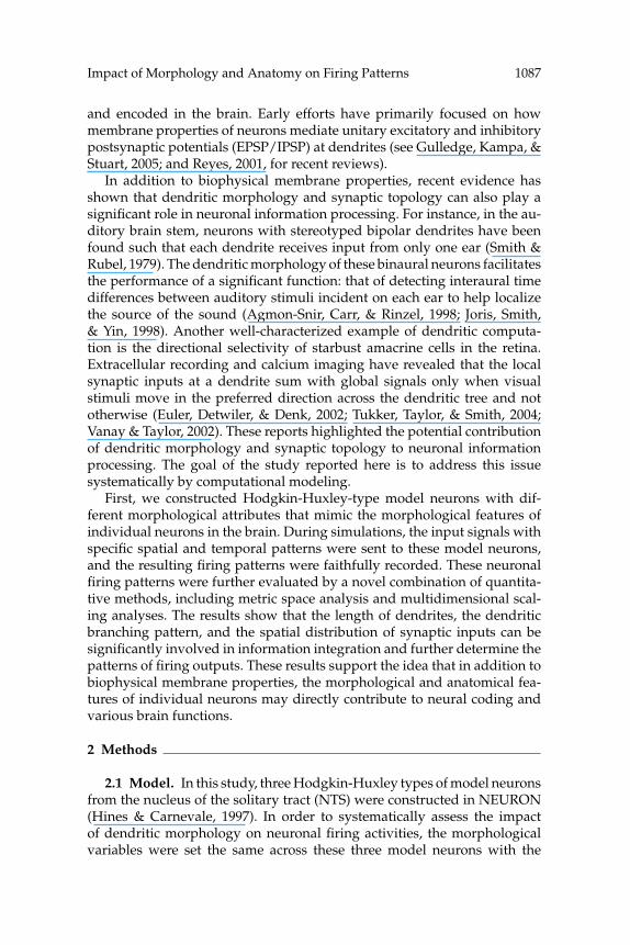
exception of the length of their primary dendrite (750 μm for a large neuronand a branching neuron and 350 μm for a small neuron) and the number ofbranching points at their primary dendrite (0 for a large neuron and a smallneuron and 2 for a branching neuron). Details of the morphological settingsare listed in Table 1. The goal was to vary one feature systematically andstudy its effects while keeping all other features constant.
The membrane properties of these model neurons were obtained fromthe results of in vitro experiments (Schild et al., 1993). Each compartmentin the model neuron contained a fast Na+ current (INa), a delayed rectifiercurrent (IK), a long-lasting Ca2+ current (ICaL), a transient outward potas-sium current (IA), a Ca2+ activated potassium current (IKCa), a delay current(ID), an inward rectifier current (IR), a linear leakage current (IB), an Na+-Ca2+ exchange current (INaCa), an Na+-K+ pump current (INaK), and a Ca2+
pump current (ICaP). Details of the channel kinetics are listed in appendixesA and B. The extracellular concentration of ions ([Ca2+]o, [Na+]o, [K+]o)was assumed to be constant. In all model neurons, calcium buffering wasalso included (see appendix A).
Since the distribution of ion channels can undergo significant changeduring nervous system development (Maletic-Savatic, Lenn, & Trimmer,1995; Miyashita & Kubo, 1997), precise channel distributions for most neu-rons in the brain are still unknown. In this study, the membrane properties,which were distributed evenly across the dendrites and the soma, weretested here. At the axon, only a fast Na+ current (INa) and a delayed rectifiercurrent (IK) were included.
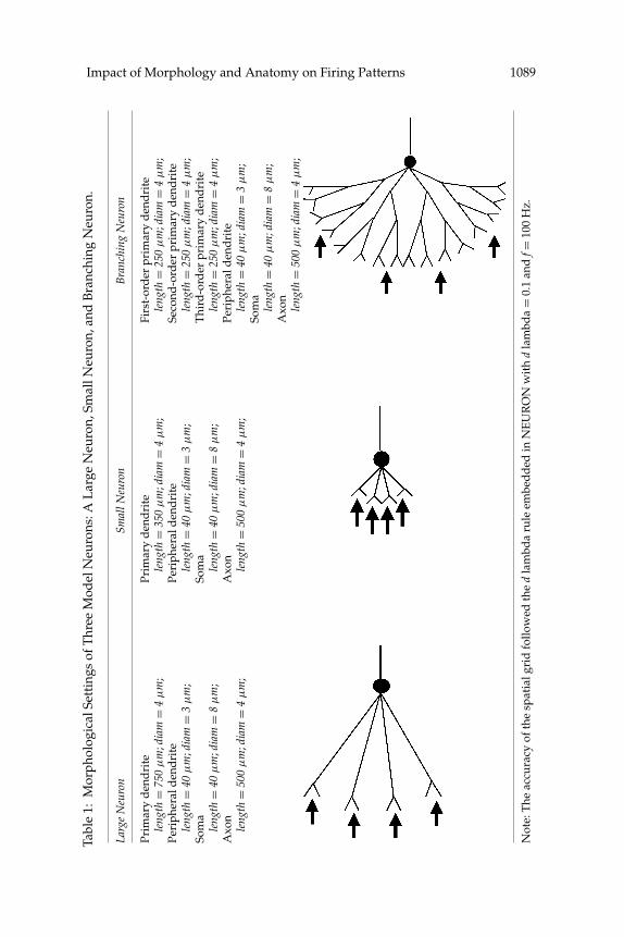
Next, a computational model of an excitatory synapse characterized byseveral variables, the rising time constant, the decaying time constant, andthe reversal potential based on several reports (Chen, Horowitz, & Bonham,1999; Schild et al., 1993; Wang & Bradley, 1995) was constructed. Their valuesare listed in appendix B. In addition, two types of connection paradigms,each distinguished by its distribution of synapses across the dendritic tree(dispersive or concentrated), were also examined. The placement of synap-tic inputs for these two different paradigms is illustrated in Table 2.
2.2 Metric Space Analysis. In order to examine the similarity of tempo-ral patterns among spike trains, metric space analysis (Victor, 2005; Victor& Purpura, 1996, 1997) was applied here. Using this analysis, a family ofmetrics could be derived to quantitatively measure the distance (degreeof dissimilarity between temporal patterns) between spike trains. The dis-tance between two spike trains was simply represented by the minimumtotal cost of transforming one spike train into the other through several se-quences of elementary steps: moving, inserting, or deleting specific spikes.First, the cost of moving a spike by an amount of time t was counted as qt,where q (sec−1) was the cost of moving a spike per unit time (1 second).Further, each spike that was deleted or added incurred a cost of 1. Basedon this algorithm, the costs of all possible sequences to transform one spike
Impact of Morphology and Anatomy on Firing Patterns 1089Ta
ble
1:M
orph
olog
ical
Sett
ings
ofT
hree
Mod
elN
euro
ns:A
Lar
geN
euro
n,Sm
allN
euro
n,an
dB
ranc
hing
Neu
ron.
Larg
eN
euro
nSm
allN
euro
nB
ranc
hing
Neu
ron
Prim
ary
den
dri
tePr
imar
yd
end
rite
Firs
t-or
der
prim
ary
den
dri
tele
ngth
=75
0μ
m;d
iam
=4
μm
;le
ngth
=35
0μ
m;d
iam
=4
μm
;le
ngth
=25
0μ
m;d
iam
=4
μm
;Pe
riph
eral
den
dri
tePe
riph
eral
den
dri
teSe
cond
-ord
erpr
imar
yd
end
rite
leng
th=
40μ
m;d
iam
=3
μm
;le
ngth
=40
μm
;dia
m=
3μ
m;
leng
th=
250
μm
;dia
m=
4μ
m;
Som
aSo
ma
Thi
rd-o
rder
prim
ary
den
dri
tele
ngth
=40
μm
;dia
m=
8μ
m;
leng
th=
40μ
m;d
iam
=8
μm
;le
ngth
=25
0μ
m;d
iam
=4
μm
;A
xon
Axo
nPe
riph
eral
den
dri
tele
ngth
=50
0μ
m;d
iam
=4
μm
;le
ngth
=50
0μ
m;d
iam
=4
μm
;le
ngth
=40
μm
;dia
m=
3μ
m;
Som
ale
ngth
=40
μm
;dia
m=
8μ
m;
Axo
nle
ngth
=50
0μ
m;d
iam
=4
μm
;
Not
e:T
heac
cura
cyof
the
spat
ialg
rid
follo
wed
the
dla
mbd
aru
leem
bed
ded
inN
EU
RO
Nw
ith
dla
mbd
a=
0.1
and
f=10
0H
z.
1090 J. Chen
Table 2: Two Types of Synaptic Topology in the Model Neurons.
Dispersive Inputs Concentrated Inputs
train into the other were calculated, and the sequence that yielded a min-imum cost was chosen. In this study, the parameter q was set to 20 (at atemporal precision of 50 ms). Based on this measure, if spiking patternswere to depend on the dendritic morphology, we expect that the distancebetween patterns generated by different neuron types would be greaterthan patterns generated by similar neurons. (See Di Lorenzo & Victor, 2003;Di Lorenzo, Chen, & Victor, 2009; and Roussin, Victor, Chen, & Di Lorenzo,2008, for other examples where metric space analysis was used.)
2.3 Multidimensional Scaling Analyses (MDS). A matrix of pairwisedistances between spike patterns was generated using the metric spaceanalysis introduced above. The values of this matrix were then chosen asinputs to multidimensional scaling analyses (MDS) to visualize the under-lying relationships among the spike trains in terms of the similarity of theirtemporal patterns.
MDS provides a geometrical representation of data similarity or dis-similarity and has often been used to visualize the underlying structureof relationships among individual entities or groups of entities under

Impact of Morphology and Anatomy on Firing Patterns 1091
Table 3: Six Categories of Inputs with Different Frequency (5/40 Hz) or Levelof Correlation (Random/Medium Correlated/Highly Correlated).
Length of First Fixed Time Mean ofCategories of inputs Number Each Time Point at the First Added Poisson(various degree of correlation) of Sets Window Time Window Interval
5 Hz random inputs—low 20 setscorrelation
5 Hz medium-correlated inputs 20 sets 200 ms 2050 50 ms5 Hz highly correlated inputs 20 sets 200 ms 2090 10 ms40 Hz random inputs—low 20 sets
correlation40 Hz medium-correlated 20 sets 25 ms 2006.25 6.25 ms
inputs40 Hz highly correlated inputs 20 sets 25 ms 2009.5 3 ms
investigation (Kruskal & Wish, 1978). In the beginning, the objects (enti-ties) were moved around in a space of required dimensions. Then the bestfit-configuration of objects in this multidimensional space could be decidedafter a series of evaluations of how well a particular configuration repro-duced the observed distance matrix by simply summing the squared devi-ations of observed distances (obtained from metric space analysis) from thereproduced distances (obtained from a particular configuration of MDS). Inthis study, objects, the input or output spike trains, were embedded in a hy-pothetical space such that the distances from one to another correspondedto the relative similarity of their temporal patterns measured by the metricspace analysis. When there were morphology- or anatomy-dependent fir-ing patterns, the spike trains (objects) from the same model neuron or thesame anatomical setting would tend to get together and form individualclouds in the MDS space. According to the geometrical configuration ofthese clouds, new relationships among received inputs and firing outputscan be revealed. The actual orientation of axes in the MDS space is arbitrary.The meaning of an axe can be abstract (interpreted by user). In this study,they are kept as dimension I, dimension II, and dimension III.
2.4 Simulations. Six categories (120 sets) of artificial spike trains wereconstructed and served as inputs to the model neurons. They are 5/40 HzPoisson random inputs, 5/40 Hz medium-correlated inputs, and 5/40 Hzhighly correlated inputs (see Table 3). Those random inputs were generatedusing a Poisson process. The correlated inputs were constructed by addinga random interval to a fixed time point in each time window (see Figure 1).Since the level of correlation among input spike trains can be recognized asthe level of jitter (synchronization) among them, higher-correlated inputsare made by adding smaller random intervals (lower mean value) to a fixedtime point. Detailed information of correlated inputs is listed in Table 3.

1092 J. Chen
B.A. 5 Hz 40 Hz
0
4.2
8.4
2000 2200 2400 2600 2800 3000
Time (ms)
0
4.2
2050 ms + Poisson interval (mean=50ms)
Ran
do
mIn
pu
ts
Co
rrela
ted
Inp
uts
0
4.2
8.4
2000 2100 2200 2300 2400
Time (ms)
2006.25 ms + Poisson interval (mean=6.25ms)
Length of each window = 200 ms Length of each window = 25 ms
Ran
do
mIn
pu
ts
Co
rrela
ted
Inp
uts
0
4.2
2000 2006.25 2012.5 20252000 2050 2100 2200)sm(emiTTime (ms)
Figure 1: Inputs sent to model neurons could be 5 or 40 Hz random or corre-lated spike trains. Random spike trains were generated by a Poisson process.Correlated spike trains were generated by adding a Poisson interval at the initialtime point to each time window. (A) In a 5 Hz correlated spike train, a series oftime windows with length equal to 200 ms (1/5 sec) was first decided. An event(spike) was inserted in each time window by adding a Poisson interval to theinitial time point. The initial time point is set at the end of the first quarter ofeach time window, and the mean of the Poisson interval is set to be equal to aquarter of a time window (50 ms in this case). The raster plots of 5 Hz Poissonrandom spike trains and 5 Hz medium-correlated spike trains are shown here.(B) A similar idea is applied to generate 40 Hz correlated inputs. Instead of200 ms, a time window of 40 Hz correlated inputs is set to be 25 ms (1/40 sec).After the initial time points of each time window are decided (at the end of thefirst quarter of each window), a Poisson interval with mean equal to 6.25 ms (aquarter of 25 ms) is added. The raster plots of 40 Hz random spike trains and40 Hz medium-correlated spike trains are shown here.
During each run of the simulation, one set of the inputs (that included40 different spike trains) was assigned to 40 different synapses distributedon the neuron. Each run consisted of an initial 2 seconds of silence followedby a spike train that lasted 5 seconds and another 1 second where noinput was presented to the neuron. The timing of individual spikes wasrecorded from the middle of an axon when the action potential reached athreshold of 10 mV. A total of 800 trials of simulation were conducted. Those

Impact of Morphology and Anatomy on Firing Patterns 1093
Tabl
e4:
Com
plet
eL
isto
fSim
ulat
ions
Exe
cute
din
Thi
sSt
udy.
Syna
ptic
Ind
ivid
ualS
ets
ofIn
puts
Topo
logy
Lar
geC
ell
Smal
lCel
lB
ranc
hing
Cel
l
40Po
isso
nra
ndom
inpu
ts(5
Hz)
Dis
pers
ive
Ran
20tr
ials
(set
s)R
an20
tria
ls(s
ets)
Ran
20tr
ials
(set
s)(s
ynap
tic
wei
ghts
=0.
01)
Con
cent
rate
dR
an20
tria
ls(s
ets)
Ran
20tr
ials
(set
s)R
an20
tria
ls(s
ets)
40Po
isso
nra
ndom
inpu
ts(4
0H
z)D
ispe
rsiv
eR
an20
tria
ls(s
ets)
Ran
20tr
ials
(set
s)R
an20
tria
ls(s
ets)
(syn
apti
cw
eigh
ts=
0.01
)C
once
ntra
ted
Ran
20tr
ials
(set
s)R
an20
tria
ls(s
ets)
Ran
20tr
ials
(set
s)40
med
ium
corr
elat
edin
puts
(5H
z)D
ispe
rsiv
eR
an20
tria
ls(s
ets)
Ran
20tr
ials
(set
s)R
an20
tria
ls(s
ets)
(syn
apti
cw
eigh
ts=
0.01
)C
once
ntra
ted
Ran
20tr
ials
(set
s)R
an20
tria
ls(s
ets)
Ran
20tr
ials
(set
s)40
med
ium
corr
elat
edin
puts
(40
Hz)
Dis
pers
ive
Ran
20tr
ials
(set
s)R
an20
tria
ls(s
ets)
Ran
20tr
ials
(set
s)(s
ynap
tic
wei
ghts
=0.
01)
Con
cent
rate
dR
an20
tria
ls(s
ets)
Ran
20tr
ials
(set
s)R
an20
tria
ls(s
ets)
40hi
ghly
corr
elat
edin
puts
(5H
z)D
ispe
rsiv
eR
an20
tria
ls(s
ets)
(syn
apti
cw
eigh
ts=
0.01
)C
once
ntra
ted
Ran
20tr
ials
(set
s)40
high
lyco
rrel
ated
inpu
ts(4
0H
z)D
ispe
rsiv
eR
an20
tria
ls(s
ets)
(syn
apti
cw
eigh
ts=
0.01
)C
once
ntra
ted
Ran
20tr
ials
(set
s)4
Pois
son
rand
omin
puts
(5H
z)D
ispe
rsiv
eR
an20
tria
ls(s
ets)
Ran
20tr
ials
(set
s)R
an20
tria
ls(s
ets)
(syn
apti
cw
eigh
ts=
1)C
once
ntra
ted
Ran
20tr
ials
(set
s)R
an20
tria
ls(s
ets)
Ran
20tr
ials
(set
s)4
Pois
son
rand
omin
puts
(40
Hz)
Dis
pers
ive
Ran
20tr
ials
(set
s)R
an20
tria
ls(s
ets)
Ran
20tr
ials
(set
s)(s
ynap
tic
wei
ghts
=1)
Con
cent
rate
dR
an20
tria
ls(s
ets)
Ran
20tr
ials
(set
s)R
an20
tria
ls(s
ets)

1094 J. Chen
trials are summarized in Table 4. In addition, to give a clearer picture of theanalysis combining metric space analysis and MDS, a flowchart using 5 Hzrandom inputs and 5 Hz correlated inputs as an example is presented inFigure 2.
0
40
2 Time (sec)
7
Represent the distance (cost) among spike trains in a hypothetical space via multidimensional scaling analysis (MDS)
Set 1: 5 Hz random inputs Set 2: 5 Hz medium correlated inputs
Imp
uls
es /
se
c
0
20
2Time (sec)
7
Tria
ls
7
0 2
Time (sec)
40
Imp
uls
es /
se
c
0
20
2 Time (sec)
Tria
ls
Evaluate similarity of temporal patterns between spike trains via metric space analysis
-0.1
-0.05
0
0.05-0.05
0
0.05
0.1
-0.08
-0.04
0
0.04
0.08
Dimension I
Dimension II
Dimension III
Set 1
Set 2
Set 2
Set 1
between spike trainsDistance (cost)
7
Set 1: 5 Hz random inputs
Set 2: 5 Hz medium correlated inputs ˚

Impact of Morphology and Anatomy on Firing Patterns 1095
3 Results
3.1 Model Neurons Demonstrate an Ability to Integrate ComplexSynaptic Inputs. Figures 3 and 4 show the poststimulus time histograms(PSTH) used to illustrate neuronal responses in relation to the input spiketrains they receive. No apparent pattern was found as model neurons re-ceived low-frequency (5 Hz) random inputs (see Figure 3A). However,when the neurons received medium-correlated inputs at the same frequency(5 Hz), a clear resonance (see Figure 3B) was observed in all model neu-rons regardless of differences in their dendritic morphology and synaptictopology. In Figure 4A, a resonance was seen in responses to 40 Hz ran-dom inputs only if these inputs were distributed locally. Further, in Figure4B, 40 Hz medium-correlated inputs resulted in resonance across all neu-rons. The resonance became stronger as the inputs were distributed morewidely.
Next, using metric space analysis and MDS, the neuronal response pat-terns were compared with the patterns of random inputs they received. InFigure 5, each point in the MDS plot represents a 5 second spike train withrandom inputs being shown by the open circles and the firing outputs bythe filled circles. In Figure 5A, for instance, 20 open circles represent the5 Hz random inputs received by a large model neuron, and 20 filled circlesindicate its firing outputs. Here, the firing outputs form a cluster (cloud) thatcan be separated from the cloud consisting of the random inputs it received.Such clustering was evidenced in all model neurons that received disper-sive low- (5 Hz) or high- (40 Hz) frequency random inputs (see Figures 5Athrough 5F). This geometrical structure suggests that neurons can reliablyregulate their firing patterns even when inputs arrive randomly and aredistributed widely across the dendritic tree. Furthermore, the outputs inFigures 5B, 5D, and 5F clustered closely compared to those in Figures 5A,
Figure 2: Flowchart introducing the procedure combining metric space analysisand multidimensional scaling analyses (MDS). First, the similarity of temporalpatterns among spike trains is evaluated by metric space analysis. Then theoutcomes of metric space analysis are used as inputs to MDS. After MDS, amultidimensional MDS plot can be acquired with each object in this space rep-resenting an individual spike train. The relative distance among them simplyrepresents the degree of similarity of their temporal patterns. Spike trains dis-playing more similar temporal patterns are closer to each other in this MDSplot. When 5 Hz medium-correlated spike trains and 5 Hz random spike trainsare used as an example, the temporal patterns of 5 Hz correlated spike trains areproved to be more similar to each other than 5 Hz random spike trains in thisfigure. This is evidenced by a more condensed cloud (filled circles) consistingof 5 Hz correlated spike trains in this MDS plot.

1096 J. Chen
Firing outputs (When received 5 Hz dispersive random inputs)
Firing outputs (When received 5 Hz concentrated random inputs)
A.
Time (sec)
2 7
0
100 5 Hz random inputs
Large Cell
Large Cell
Small Cell
Small Cell
Branching Cell
Branching Cell
Impulses per
second
100
100
001001
001001
00
0 00
0
22 777 2
B.
Large Cell
Firing outputs (When received 5 Hz dispersive correlated inputs)
Firing outputs (When received 5 Hz concentrated correlated inputs)
Time (sec)
72
0
100 5 Hz correlated inputs
Large Cell
Small Cell
Small Cell
Branching Cell
Branching Cell
Impulses per
second
100 001001
001 001 100
0 00
00 0
2 227 7 7
Figure 3: (A) PSTHs of 5 Hz random inputs and the neuronal responses theyevoked. No special response pattern is found in these cases. (B) PSTHs of 5 Hzmedium-correlated inputs and the neuronal responses they evoked. Clear res-onance can be seen in all neurons.
5C, and 5E, indicating that output patterns tend to be more reliable (repro-ducible) as the input frequency gets higher.
3.2 Effects of Dendritic Morphology on Temporal Firing Patterns. Inorder to explore if morphological features of dendrites can affect neuronalfiring activity, the firing patterns from three model neurons were com-pared to each other as they received the same sets of inputs. In Figures 6A

Impact of Morphology and Anatomy on Firing Patterns 1097
Large cell (dispersive)
Large cell (concentrated)
Small cell (dispersive)
Small cell (concentrated)
40 Hz correlated inputs
7 2 Time (sec)0
200
Branching cell (dispersive)
B.
Branching cell (concentrated)
7
40 Hz random inputs
Large cell (dispersive)
Large cell (concentrated)
Small cell (dispersive)
Small cell (concentrated)
Branching cell (dispersive)
A. 200
0
2 Time (sec)
Branching cell (concentrated)
Impuls
es / s
ec
Impuls
es / s
ec
200
200
200
200
200
200
200
200
200
200
200
200
0
0
0
0
0
0
0
0
0
0
0
0
Figure 4: (A) PSTHs of 40 Hz random inputs and neuronal responses theyevoked. Neurons are found to be able to regulate a high frequency of randominputs and generate resonant outputs, especially when the synaptic inputs dis-tribute closer to each other. (B) PSTHs of 40 Hz medium-correlated inputs andthe neuronal responses they evoked. Resonant firing outputs can be seen in allcases. Moreover, it is found that the degree of resonance can change dramati-cally in large neurons and small neurons as the synaptic topology changes froma dispersive to a concentrated distribution.
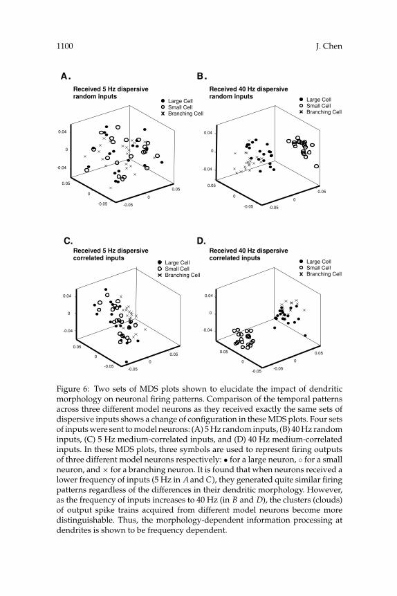
and 6B, the similarity of neuronal firing patterns was investigated as threemodel neurons received the same sets of 5 Hz or 40 Hz random inputs.First, all three neurons generated very similar firing patterns in responseto the low frequency (5 Hz) of random inputs as evidenced by the ex-tensive overlap among the corresponding clusters (circle, dot, and cross)in Figure 6A. In Figure 6B, an increase in frequency of inputs (40 Hz)led to dramatically different outcomes. The firing patterns generated bythe three model neurons clearly separated from each other. This strik-ing change suggests a morphology-dependent impact on neuronal firingpatterns. That is, different dendritic morphology can perform as differentarithmetic, which decides the way of neuronal information integration atdendrites and further determines the temporal patterns of firing outputs.This morphology-dependent consequence is more apparent as the neuronsreceive higher frequency of inputs. Similar results were observed as neuronsreceived 5 Hz or 40 Hz medium-correlated inputs (see Figures 6C and 6D).

1098 J. Chen
-0.05
0
0.05
0.1
-0.1
-0.05
0
0.05
-0.05
0
0.05
-0.05
0
0.05
0.1
-0.1
-0.05
0
0.05
-0.05
0
0.05
-0.1
-0.05
0
0.05 -0.1
-0.05
0
0.05
-0.05
0
0.05
-0.05
0
0.05 0.1
-0.05
0
0.05
0.1
-0.05
0
0.05
-0.05
0 0.05
0.1
-0.05
0
0.05
0.1
-0.05
0
0.05
5 Hz random inputs Firing outputs
40 Hz random inputs Firing outputs
5 Hz random inputs Firing outputs
40 Hz random inputs Firing outputs
5 Hz random inputs Firing outputs
00.05
0.1
-0.05
0
0.05
0.1
-0.05
0
0.05
40 Hz random inputs Firing outputs
A. B.
C. D.
F. E.
3.3 Effects of Synaptic Topology on Temporal Firing Patterns. Next,the impact of anatomical features on neuronal firing patterns was assessed.As shown in Table 2, two types of connections were tested here. During thesimulations, each model neuron received the same set of inputs through adispersive or a concentrated distribution of synapses. The response firingpatterns were then systematically evaluated by metric space analysis and

Impact of Morphology and Anatomy on Firing Patterns 1099
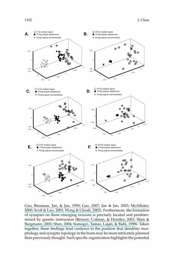
MDS. The results are shown in Figure 7. In general, when the distributionof synaptic inputs changed, a new cluster of firing outputs in the MDSplot was formed. One exceptional case is observed in Figure 7F. In thisfigure, when a high frequency of random inputs was sent to a branchingneuron, all firing patterns were very similar regardless of the distributionof synapses. In addition, a highly reliable firing pattern (indicated by anextremely dense cloud in a MDS plot) was commonly observed as neuronsreceived concentrated high-frequency inputs (see Figures 7B, 7D, and 7F).According to these results, the synaptic topology can also involve neuronalinformation processing, and this processing is frequency dependent.
3.4 Impact of Level of Correlation of Inputs. Next, we explored thedependence of output patterns on the correlation of input patterns. Sixcategories of input patterns with different frequency and different level ofcorrelation were sent to large model neurons via dispersive or concentrateddistribution of synapses. These six categories of inputs are listed in Table 3.Their raster plots are shown in Figures 8A and 9A.
The impacts of correlation of input patterns on neuronal firing patternsare shown in Figures 8B and 9B. In Figure 8B, when inputs were at lowfrequency (5 Hz in this case), inputs with different levels of correlationgenerally evoked distinguishable patterns of outputs (separable clouds inthe MDS plot). The output patterns evoked by higher-correlated inputs areusually more reliable (indicated by an extremely condense cloud in the MDSplot). Furthermore, it was noticed that as a neuron received low-frequencyinputs, variation of the synaptic topology (dispersive or concentrated) didnot change the configuration of firing outputs in the MDS plots. Interest-ingly, when the input frequency was increased to 40 Hz, the configurations
Figure 5: Six MDS plots that introduce the similarity of temporal patterns ofparticular inputs (5 Hz or 40 Hz random inputs) and the firing outputs theyevoked in each model neuron. The input spike trains were delivered to eachneuron via dispersive distribution of synapses. (A) An MDS plot containing 5Hz random inputs (◦) and the firing outputs they evoked (•) in a large neuron.(B) An MDS plot showing 40 Hz random inputs (◦) and the firing outputs theyevoked (•) in a large neuron. (C) An MDS plot exhibiting 5 Hz random inputs(◦) and the firing outputs they evoked (•) in a small neuron. (D) An MDS plotcontaining 40 Hz random inputs (◦) and the firing outputs they evoked (•) ina small neuron. (E) An MDS plot containing 5 Hz random inputs (◦) and thefiring outputs they evoked (•) in a branching neuron. (F) An MDS plot showing40 Hz random inputs (◦) and the firing outputs they evoked (•) in a branchingneuron. In these MDS plots, the clusters consisting of input and output spiketrains are clearly separable. This particular arrangement of spike trains in theseMDS plots indicates that neurons are able to systematically regulate the inputsignals they receive and generate reliable firing patterns. This performance isapparent as a neuron receives a higher frequency of inputs.
1100 J. Chen
-0.05
0
0.05
-0.05
0
0.05
-0.04
0
0.04
-0.05
0
0.05
-0.05
0
0.05
-0.04
0
0.04
Large Cell Small Cell Branching Cell
Large Cell Small Cell Branching Cell
Received 5 Hz dispersive random inputs
Received 40 Hz dispersive random inputs
.B .A
Received 5 Hz dispersive correlated inputs
Large Cell Small Cell Branching Cell
Received 40 Hz dispersive correlated inputs
Large Cell Small Cell Branching Cell
D.C.
-0.05
0
0.05
-0.05
0
0.05
-0.04
0
0.04
-0.05
0
0.05
-0.05
0
0.05
-0.04
0
0.04
Figure 6: Two sets of MDS plots shown to elucidate the impact of dendriticmorphology on neuronal firing patterns. Comparison of the temporal patternsacross three different model neurons as they received exactly the same sets ofdispersive inputs shows a change of configuration in these MDS plots. Four setsof inputs were sent to model neurons: (A) 5 Hz random inputs, (B) 40 Hz randominputs, (C) 5 Hz medium-correlated inputs, and (D) 40 Hz medium-correlatedinputs. In these MDS plots, three symbols are used to represent firing outputsof three different model neurons respectively: • for a large neuron, ◦ for a smallneuron, and × for a branching neuron. It is found that when neurons received alower frequency of inputs (5 Hz in A and C), they generated quite similar firingpatterns regardless of the differences in their dendritic morphology. However,as the frequency of inputs increases to 40 Hz (in B and D), the clusters (clouds)of output spike trains acquired from different model neurons become moredistinguishable. Thus, the morphology-dependent information processing atdendrites is shown to be frequency dependent.

Impact of Morphology and Anatomy on Firing Patterns 1101
of firing outputs in the MDS plots dramatically changed as the synaptictopology changed. In Figure 9B (right), firing patterns became quite similar(overlap among clouds) as the 40 Hz inputs were delivered to the neuronby a concentrated distribution of synapses.
3.5 Impact of Number of Inputs. Membrane potential traces recordedfrom the soma of a large model neuron when the number of inputs was re-duced from 40 to 4 and the strength of synaptic conductance was increasedfrom 0.01 to 1 are shown in Figures 10A and 10B. When the neuron receivedfewer though stronger inputs, the membrane potential trace was less noisy(see Figure 10A) than the one recorded when a large number of compara-tively weak inputs were presented (see Figure 10B). In the former case, theimpact of dendritic morphology and synaptic topology on neuronal firingpatterns (see Figures 10C and 10D) was found to be similar to our resultspresented in previous sections (see Figures 6 and 7). Thus, as the number ofinputs and the strength of conductance changed, the impacts of dendriticmorphology and synaptic topology on neuronal firing patterns are similar(showing similar configurations in MDS plots).
4 Discussion
Identifying the principles of neural computation in the brain is a challeng-ing task due to the number of biological variables that can interact witheach other dynamically at various spatial and temporal scales. Fortunately,the development of a computational modeling technique offers an effectiveway to resolve this difficulty. When current is injected into the soma, var-ious firing patterns can be reproduced from the reconstructed neocorticalmodel neurons (Mainen & Sejnowski, 1996). In the current study, whichused a different simulation strategy, multiple model neurons with simpli-fied morphological structures were constructed first. Then the changes inneuronal responses caused by specific morphological or anatomical featureswere systematically evaluated using a novel combination of metric spaceanalysis and MDS. These quantitative methods together provide a morerigorous way to investigate neuronal firing activities than conventional rep-resentations (e.g., raster plot or PSTH) and measures (e.g., discharging rateor distribution of interspike interval). Our results reveal the contributionof dendritic morphology and synaptic topology on neuronal informationprocessing (neural coding). These ideas are consistent with several recentexperiments discussed below:
• Dendritic morphology and synaptic topology can be very specific. Recentprogress in our understanding of dendrite growth and axon guidance showsthat both intracellular and extracellular molecular mechanisms, in conjunc-tion with ongoing synaptic activity, are involved in the process of dendriticbranching and targeting during nervous system development (Cline, 2001;
1102 J. Chen
-0.1
-0.05
0
0.05
0.1
-0.05
0
0.05
-0.05
0
0.05
5 Hz random inputs Firing outputs (dispersive)
Firing outputs (concentrated)
-0.1
-0.05
0
0.05-0.05
0
0.05
-0.05
0
0.05
-0.1
-0.05
0
0.05
0.1
-0.05
0
0.05
-0.05
0
0.05
-0.15
-0.1
-0.05
0
0.05
0.1-0.05
0
0.05
-0.05
0
0.05
B. A.
D. C.
-0.1
-0.05
0
0.05
0.1
-0.05
0
0.05
-0.05
0
0.05
E.
-0.15
-0.1
-0.05
0
0.05-0.05
0
0.05
-0.05
0
0.05
F.
40 Hz random inputs Firing outputs (dispersive)
Firing outputs (concentrated)
5 Hz random inputs Firing outputs (dispersive)
Firing outputs (concentrated)
40 Hz random inputs Firing outputs (dispersive)
Firing outputs (concentrated)
5 Hz random inputs Firing outputs (dispersive)
Firing outputs (concentrated)
40 Hz random inputs Firing outputs (dispersive)
Firing outputs (concentrated)
Gao, Brenman, Jan, & Jan, 1999; Gao, 2007; Jan & Jan, 2003; McAllister,2000; Scott & Luo, 2001; Wong & Ghosh, 2002). Furthermore, the formationof synapses on these emerging neurons is precisely located and predeter-mined by genetic instruction (Benson, Colman, & Huntley, 2001; Shen &Bargmann, 2003; Shen, 2004; Somogyi, Tamas, Lujan, & Buhl, 1998). Takentogether, these findings lend credence to the position that dendritic mor-phology and synaptic topology in the brain may be more intricately plannedthan previously thought. Such specific organization highlights the potential

Impact of Morphology and Anatomy on Firing Patterns 1103
contribution of dendritic morphology and synaptic topology as key featuresin neuronal information processing.
• Dendritic arithmetic. For decades, neuroscientists have devoted consid-erable effort to deriving principles of dendritic computation. Two modesof integrating information, each related to the dendritic morphology andsynaptic topology, have been observed (Carter, Soler-Llavina, & Sabatini,2007; Poirazi, Brannon, & Mel, 2003; Polsky, Mel, & Schiller, 2004). First,when high-frequency synchronous inputs activate a restricted portion of thelocal dendrite, a nonlinear summation of inputs was observed. The thresh-old of local dendritic spikes increase in a manner that prevents subsequentspikes (Ariav, Polsky, & Schiller, 2003; Gasparini, Migliore, & Magee, 2004;Gasparini & Magee, 2006; Poirazi et al., 2003; Polsky et al., 2004). In contrast,spatially distributed inputs were found to sum linearly, making the neuronmore sensitive to the changes of input frequency and timing (Ariav et al.,2003; Gasparini & Magee, 2006; Poirazi et al., 2003; Polsky et al., 2004). Theseresults illustrate several fundamental computational principles regardingthe integration of information at dendrites. In order to connect these com-putational principles to higher-level brain functions, the performance ofneuronal computation in a longer time period was investigated in thisstudy (temporal patterns of 5 seconds spike trains). Our simulation resultshighlight the potential contribution of dendritic morphology and synaptictopology on neuronal computations. Structural characteristics may directlyor indirectly contribute to various brain functions.
• From “form” to “function.” CA1 pyramidal neurons can produce differ-ent firing patterns associated with different information integration modesat their dendrites (Gasparini & Magee, 2006). Different modes of informa-tion integration at the dendrites can be independently engaged by differentbehavioral states in animals. For instance, a linear integration mode at den-drites can be associated with theta states, during which a neuron increasesits firing rate and shifts its spike timing forward. In addition, a nonlinear
Figure 7: In order to realize the impact of synaptic topology on neuronal firingpatterns, firing outputs from individual neurons as they receive the same setsof inputs via different synaptic topology (dispersive or concentrated) are inves-tigated. (A) A large neuron receives 5 Hz dispersive or concentrated randominputs. (B) A large neuron receives 40 Hz dispersive or concentrated random in-puts. (C) A small neuron receives 5 Hz dispersive or concentrated random in-puts. (D) A small neuron receives 40 Hz dispersive or concentrated randominputs. (E) A branching neuron receives 5 Hz dispersive or concentrated ran-dom inputs. (F) A branching neuron receives 40 Hz dispersive or concentratedrandom inputs. In general, different synaptic topology evokes different firingpatterns. One exception is observed in F as a branching neuron receives a highfrequency of inputs. In addition, it is found that firing patterns are commonlymore reliable when neurons receive a higher frequency of concentrated inputs.This is indicated by extremely condense clouds of × in B, D, and F .
1104 J. Chen
B.
-0.05
0
0.05
-0.05
0
0.05
-0.04
0
0.04
Dispersive inputs – Concentrated inputs – 5 Hz zH5
-0.04
-0.05
0
0.05-0.05
0
0.05
0
0.04
Firing outputs (5Hz random inputs)
Firing outputs (5 Hz medium-corr inputs)
Firing outputs (5 Hz high-corr inputs)
Random Inputs (5 Hz) Medium Correlated Inputs (5 Hz)
(mean of random interval = 50 ms)Highly Correlated Inputs (5 Hz)
(mean of random interval = 10 ms)A.
2000 20002000 30003000 3000Time (ms) Time (ms) Time (ms)
Tri
als
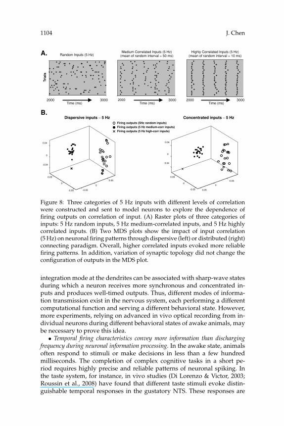
Figure 8: Three categories of 5 Hz inputs with different levels of correlationwere constructed and sent to model neurons to explore the dependence offiring outputs on correlation of input. (A) Raster plots of three categories ofinputs: 5 Hz random inputs, 5 Hz medium-correlated inputs, and 5 Hz highlycorrelated inputs. (B) Two MDS plots show the impact of input correlation(5 Hz) on neuronal firing patterns through dispersive (left) or distributed (right)connecting paradigm. Overall, higher correlated inputs evoked more reliablefiring patterns. In addition, variation of synaptic topology did not change theconfiguration of outputs in the MDS plot.
integration mode at the dendrites can be associated with sharp-wave statesduring which a neuron receives more synchronous and concentrated in-puts and produces well-timed outputs. Thus, different modes of informa-tion transmission exist in the nervous system, each performing a differentcomputational function and serving a different behavioral state. However,more experiments, relying on advanced in vivo optical recording from in-dividual neurons during different behavioral states of awake animals, maybe necessary to prove this idea.
• Temporal firing characteristics convey more information than dischargingfrequency during neuronal information processing. In the awake state, animalsoften respond to stimuli or make decisions in less than a few hundredmilliseconds. The completion of complex cognitive tasks in a short pe-riod requires highly precise and reliable patterns of neuronal spiking. Inthe taste system, for instance, in vivo studies (Di Lorenzo & Victor, 2003;Roussin et al., 2008) have found that different taste stimuli evoke distin-guishable temporal responses in the gustatory NTS. These responses are

Impact of Morphology and Anatomy on Firing Patterns 1105
A.Random Inputs (40 Hz)
Medium Correlated Inputs (40 Hz)(mean of random interval = 6.25 ms)
Tri
als
2000 2400Time (ms)
2000 2400Time (ms)
Highly Correlated Inputs (40 Hz) (mean of random interval = 3 ms)
00420002Time (ms)
B.
-0.08
-0.04
0
0.04
0.08
-0.04
-0.02
0
0.02
-0.02
0
0.02
-0.1
-0.05
0
0.05
0.1
-0.05
0
0.05
0.1
0.15
-0.04
0
0.04
Dispersive inputs – Concentrarted inputs – 40 Hz zH 04
Firing outputs (40Hz random inputs)Firing outputs (40 Hz medium-corr inputs)Firing outputs (40 Hz high-corr inputs)
Figure 9: Another three categories of 40 Hz inputs with different levels of cor-relation were constructed and sent to model neurons to explore the dependenceof firing outputs on correlation of input. (A) Raster plots of three categoriesof inputs: 40 Hz random inputs, 40 Hz medium-correlated inputs, and 40 Hzhighly correlated inputs. (B) Two MDS plots show the impact of input correla-tion (40 Hz) on neuronal firing patterns through a dispersive (left) or distributed(right) connecting paradigm. It was found that the configuration of outputs inthese MDS plots dramatically changed as the connecting paradigm changed.When inputs were sent to a model neuron by a concentrated distribution ofsynapses, the neuron generated quite similar output patterns regardless of thelevel of correlation of inputs.
highly reproducible in repeated trials. It was established using metric spaceanalysis that information contained in the temporal firing characteristicswas significantly higher than information contained in the average dis-charge rate. Similar experiments emphasizing the significance of temporalfiring precision during neuronal information processing have also beenreported in other sensory systems (Agmon-Snir et al., 1998; Joris et al., 1998;Lestienne, 2001; Singer, 1999; Stopfer & Laurent, 1999).
In sum, by using computational modeling technique along with novelcombination of metric space analysis and MDS, the results of this studyshow that there are morphology- and anatomy-dependent computationalmechanisms that influence the spiking activity of individual neurons. In thenear future, it will be critical to see how these morphology- and anatomy-dependent mechanisms contribute to the network level of computationsand eventually to various brain functions.

1106 J. Chen
A. B.
C.
D.
100 ms
20 mV
100 ms
20 mV
4 synaptic inputs, synaptic weights = 1
40 synaptic inputs, synaptic weights = 0.01
-0.05
0
0.05
-0.05
0
0.05
-0.05
0
0.05
-0.08
-0.04
0
0.04
-0.04
0
0.04
-0.05
0
0.05
Large Cell Small Cell Branching Cell
Received 5 Hz random inputs
Received 40 Hz random inputs
-0.05
0
0.05
0.1
-0.05
0
0.05
-0.05
0
0.05
5 Hz random inputs
Firing outputs (dispersive inputs)
Firing outputs (concentrated inputs)
40 Hz random inputs
Firing outputs (dispersive inputs)
Firing outputs (concentrated inputs)
-0.05
0
0.05
0.1
-0.05
0
0.05
-0.05
0
0.05
Large Cell Small Cell Branching Cell
Figure 10: The somatic membrane potential traces of a large model neuron areintroduced in A and B. (A) A large model neuron receives 4 synaptic inputswith synaptic weights equal to 1. (B) A large model neuron receives 40 synapticinputs with synaptic weights equal to 0.01. (C) The impact of dendritic mor-phology on neuronal firing patterns was evaluated as all three model neuronsreceived 4 synaptic inputs with synaptic weights equal to 1. The results areconsistent with Figure 6, showing morphology- and frequency-dependent fir-ing activities. (D) The impact of synaptic topology on neuronal firing patternswas systematically evaluated. In general, the outcomes are consistent with theresults illustrated in Figure 7 except that extensive overlap between two cloudsof firing outputs evoked by 5 Hz random inputs is observed here in D (left).

Impact of Morphology and Anatomy on Firing Patterns 1107A
ppen
dix
A:
Lis
tofC
hann
elK
inet
ics.
Cur
rent
sK
inet
icM
odel
sSt
ate
Var
iabl
es
Fast
sod
ium
curr
ent(
I Na)
I Na
=g N
am
3h
(V–
EN
a)α
m=
0.36
(V+3
5)1−
exp[
−(V
+35)
/3]
βm
=−0
.4(V
+53)
1−ex
p[(V
+53)
/20
]
τ m=
0.05
+(2/
(3.4
88∗ (
αm
+βm
)))
m∞
=α
m/
(αm
+β
m)
τ h=
1.15
exp[
−(0.
055)
2 (V+
29.6
2)2 ]+
0.15
h∞=
11+
exp[
(V+2
9.62
)/5.
035]
Lon
g-la
stin
gca
lciu
mcu
rren
t(I C
aL)
I Ca
L=
g CaL
d2f(
V−
EC
a)
τ d=
10.
3426
exp(
0.09
25V
)+1.
8818
exp(
−0.0
0732
V)+
0.1
d∞
=1
1+ex
p[(−
(V+2
.8)/
9.85
]
τ f=
10.
2419
exp(
0.14
5V)+
0.04
34ex
p(−0
.020
13V
)
f∞=
11+
exp[
(V+1
4.6)
/4.
5]
Del
ayre
ctifi
ercu
rren
t(I K
)I K
=g K
n2 (V−
EK
)τ n
=60
exp[
−(0.
027)
2 (V+
32)2 ]+
4
n∞=
11+
exp[
(V−7
.187
01)/
(−11
.774
68)]
Ear
lytr
ansi
ento
utw
ard
pota
ssiu
mcu
rren
t(I A
)I A
=g A
p3(0
.5q 1
–0.
5q2)
(V–
EK
)τ
p=
5.5
exp[
−(0.
022)
2 (V+
65)2 ]+
1
P∞=
11+
exp[
(V+5
1.83
144)
/(−
13.8
0238
)]
q 1∞
=1
1+ex
p[(V
+73)
/4.
6666
7];τ
q1=
70
q 2∞
=1
1+ex
p[(V
+73)
/4.
6666
7];τ
q2=
550
Slow
lyin
acti
vati
ngd
elay
curr
ent(
I D)
I D=
g Dx3
y(V
−E
K)
τ x=
5.5
exp[
−(0.
022)
2 (V+
65)2 ]+
1
x∞=
11+
exp[
(V+3
1.83
144)
/(−
13.1
3572
)]
τ y=
6500
,
y∞=
11+
exp[
(V+6
3)/4.
6666
7]

1108 J. Chen
App
end
ixA
:(C
onti
nued
).
Cur
rent
sK
inet
icM
odel
sSt
ate
Var
iabl
es
Cal
cium
-act
ivat
edpo
tass
ium
curr
ent(
I Kca
)I K
ca=
g KC
ac(
V−
EK
)α
C=
7.5
[Ca2+
]exp
[(V
+20
)/24
]
βC
=0.
2ex
p[(V
+20
)/(−
24)]
Inw
ard
rect
ifier
curr
ent(
I R)
I R=
g R(V
−Ek−
36)
1+ex
p[(V
−Ek+
140)
ZF
/R
T]
Bac
kgro
und
curr
ent(
I NaB
,IK
B)
I NaB
=g N
aB
(V−
EN
a)
I KB
=g K
B(V
−E
K)
Sod
ium
-cal
cium
exch
ange
curr
ent(
I NaC
a)I N
aCa
=K
∗ NaC
a(D
F in
−D
F out
)/S
S=
1+D
NaC
a([
Ca2+
] i[N
a+] o
r+
[Ca2+
] o[N
a+] ir )
DF i
n=
[Ca2+
] o[N
a+] ir
exp(
(r−2
)γV
FR
T)
DF o
ut=
[Ca2+
] i[N
a+] o
rex
p((r
−2)(γ−1
)VF
RT
)
Cal
cium
pum
pcu
rren
t(I C
aP)
I CaP
=I C
aP∗
([C
a2+] i
[Ca2
+] i
+KM
.Ca
P)
Sod
ium
-pot
assi
umpu
mp
curr
ent(
I NaK
)I N
aK=
I NaK
∗(
[Na+
] i[N
a+] i
+KM
.Na
)∗
([K
+] o
[K+
] o+K
M.K
)∗(V
+150
V+2
00)
Cal
cium
dyn
amic
sO
C=
k U[C
a2+] i
∗(1
−OC
)−K
RO
C
[Ca2+
] i=
(IC
a/
2VC
aF
)–(n
[B] i
OC
’)
Sour
ce:S
child
etal
.(19
93).

Impact of Morphology and Anatomy on Firing Patterns 1109
Appendix B: Values of Biophysical Variables.
• Resting membrane potential (– 42.11630 mV) • F = 96,500 C mol−1
• ENa = 75 mV, EK = −80 mV, ECaL = 50 mV R = 8314 J kg−1 mol−1 K−1
• gNa = 0.5 uS, gCaL = 0.075 uS T = 310 K, Z = 1gK = 1.1 uS, g A = 0.205 uS • VCa = 2003∗ 10−12 cm3
gD = 0.3 uS, gR = 0.1 uS • DNaCa = 0.05 mM−4,gK ca = 0.185 uS, gNa B = 0.0009315 uS • KNaCa = 0.00016 nA mM−4
gK B = 0.0009935 uS KM.Ca P = 0.0005 mM• Membrane capacitance Cm = 0.052 nF Km.Na = 5.46 mM• I Na K = 0.15 nA, I Ca P = 0.01 nA KM.K = 0.621 mM• [Na]o = 154 mM, [Na]i = 8.71 mM • [B]i = 0.04 mM
[K]o = 5.9 mM, [K]i = 155 mM • KU = 100 mM−1 ms−1
[Ca]o = 2.4 mM • K R = 0.238 ms−1
• r = 4, n = 4, γ = 0.5• Initial value of state variables:
m = 0.041904, h = 0.923234n = 0.015467, d = 0.018110f = 0.998023, p = 0.666418x = 0.311018, y = 0.056171q1 = 0.004915, C = 0.002932q2 = 0.008894, Oc = 0.020856
• [Ca2+]i = 0.000049• Excitatory Synapses:
Rising time constant = 2.5 msDecaying time constant = 5.6 msReversal potential = −7 mV
Sources: Chen et al. (1999), Schild et al. (1993), Wang & Bradley (1995).
Acknowledgments
I thank N. T. Carnevale for help with the simulations using NEURON.I would also acknowledge a number of valuable comments from P. DiLorenzo, J. Victor, and C. Assisi. This study was supported by NIDCDgrant RO1-DC006914 to P. Di Lorenzo.
References
Agmon-Snir, H., Carr, C. E., & Rinzel, J. (1998). The role of dendrites in auditorycoincidence detection. Nature, 393, 268–272.
Ariav, G., Polsky, A., & Schiller, J. (2003). Submillisecond precision of the input-output transformation function mediated by fast sodium dendritic spikes inbasal dendrites of CA1 pyramidal neurons. J. Neurosci., 23(21), 7750–7758.
Benson, D. L., Colman, D. R., & Huntley, G. W. (2001). Molecules, maps and synapsespecificity Nat. Rev. Neurosci., 2, 899–909.
Carter, A. G., Soler-Llavina, G. J., & Sabatini, B. L. (2007). Timing and location ofsynaptic inputs determine modes of subthreshold integration in striatal mediumspiny neurons. J. Neurosci., 27(33), 8967–8977.
1110 J. Chen
Chen, C. Y., Horowitz, J. H., & Bonham, A. C. (1999). A presynaptic mechanismcontributes to depression of autonomic signal transmission in NTS. Am. J. Physiol.,277, H1350–H1360.
Cline, H. T. (2001). Dendritic arbor development and synaptogenesis. Curr. Opin.Neurobiol., 11, 118–126.
Di Lorenzo, P. M., & Victor, J. D. (2003). Taste response variability and temporalcoding in the nucleus of the solitary tract of the rat. J. Neurophysiol., 90, 1418–1431.
Di Lorenzo, P. M., Chen, J.-Y., & Victor, J. D. (2009). Quality time: Representation ofa multidimensional sensory domain through temporal coding. J. Neurosci., 29(9),9227–9238.
Euler, T., Detwiler, P. B., & Denk W. (2002). Directionally selective calcium signals indendrites of starburst amacrine cells. Nature, 418, 845–852.
Gao, F.-B. (2007). Molecular and cellular mechanisms of dendritic morphogenesis.Curr. Opin. Neurobiol., 17, 1–8.
Gao, F.-B., Brenman, J. E., Jan, L. Y., & Jan, Y.-N. (1999). Genes regulating dendriticoutgrowth, branching, and routing in Drosophila. Gene Dev., 13, 2549–2561.
Gasparini, S., & Magee, J. C. (2006). State-dependent dendritic computation inhippocampal CA1 pyramidal neurons. J. Neurosci., 26(7), 2088–2100.
Gasparini, S., Migliore, M., & Magee, J. C. (2004). On the initiation and propagationof dendritic spikes in CA1 pyramidal neurons. J. Neurosci., 24(49), 11046–11056.
Gulledge, A. T., Kampa, B. M., & Stuart, G. J. (2005). Synaptic integration in dendritictrees. J. Neurobiol., 64, 75–90.
Hines M. L., & Carnevale, N. T. (1997). The NEURON simulation environment.Neural Comp., 9, 1179–1209.
Jan, Y.-N., & Jan, L. Y. (2003). The control of dendritic development. Neuron, 40,229–242.
Joris, P. X., Smith, P. H., & Yin, C. T. (1998). Coincidence detection in the auditorysystem: 50 years after Jeffres. Neuron, 21, 1235–1238.
Kruskal, J. B., & Wish, M. (1978). Multidimensional scaling. Beverly Hills, CA: Sage.Lestienne, R. (2001). Spike timing, synchronization and information processing on
the sensory side of the central nervous system. Prog. Neurobiol., 65, 545–591.Mainen, Z. F., & Sejnowski, T. R. (1996). Influence of dendritic structure on firing
pattern in model neocortical neurons. Nature, 382, 363–366.Maletic-Savatic, M., Lenn, N. J., & Trimmer, J. S. (1995). Differential spatiotemporal
expression of K+ channel polypeptides in rat hippocampal neurons developingin situ and in vitro. J Neurosci., 15, 3840–3851.
McAllister, A. K. (2000). Cellular and molecular mechanisms of dendrite growth.Cereb. Cortex, 10, 963–973.
Miyashita, T., & Kubo, Y. (1997). Localization and developmental changes of theexpression of two inward rectifying K+ channel proteins in the rat brain. BrainRes., 750, 251–263.
Poirazi, P., Brannon, T., & Mel, B. W. (2003). Arithmetic of subthreshold synapticsummation in a model CA1 pyramidal cell. Neuron, 37, 977–987.
Polsky, A., Mel, B. W., & Schiller, J. (2004). Computational subunits in thin dendritesof pyramidal cells. Nat. Neurosci., 7(6), 621–627.
Reyes, A. (2001). Influence of dendritic conductances on the input-output propertiesof neurons. Annu. Rev. Neurosci., 24, 653–675.

Impact of Morphology and Anatomy on Firing Patterns 1111
Roussin, A. T., Victor, J. D., Chen, J.-Y., & Di Lorenzo, P. M. (2008). Variability inresponses and temporal coding of tastants of similar quality in the nucleus ofthe solitary tract of the rat. J. Neurophysiol., 99(2), 644–655.
Schild, J. H., Khushalani, S., Clark, J. W., Andresen, M. C., Kunze, D. L., & Yang,M. (1993). An ionic current model for neurons in the rat medial nucleus tractussolitary receiving sensory afferent input. J. Physiol., 469, 341–363.
Scott, E. K., & Luo, L. (2001). How do dendrites take their shape? Nat. Neurosci.,4(4), 359–365.
Shen, K. (2004). Molecular mechanisms of target specificity during synapseformation. Curr. Opin. Neurobiol., 14, 83–88.
Shen, K., & Bargmann, C. I. (2003). The immunoglobulin superfamily protein SYG-1determines the location of specific synapses in C. elegans. Cell, 112, 619–630.
Singer, W. (1999). Neuronal synchrony: A versatile code for the definition ofrelations? Neuron, 24, 49–65.
Smith, D. J., & Rubel, E. W. (1979). Organization and development of brain stemauditory nuclei of the chicken: Dendritic gradients in nucleus laminaris. J. Comp.Neurol., 186, 213–239.
Somogyi, P., Tamas, G., Lujan, R., & Buhl, E. H. (1998). Salient features of synapticorganization in the cerebral cortex. Brain Res. Rev., 26, 113–135.
Stopfer, M., & Laurent, G. (1999). Short-term memory in olfactory networkdynamics. Nature, 402, 664–668.
Tukker, J. J., Taylor, W. R., & Smith, R. G. (2004). Direction selectivity in a model ofthe starburst amacrine cell. Visual Neurosci., 21, 611–625.
Vanay, D. I., & Taylor, W. R. (2002). Direction selectivity in the retina. Curr. Opin.Neurobio., 12, 405–410.
Victor, J. D. (2005). Spike train metrics. Curr. Opin. Neurobiol., 15(5), 585–592.Victor, J. D., & Purpura K. P. (1996). Nature and precision of temporal coding in
visual cortex: A metric-space analysis. J. Neurophysiol., 76, 1310–1326.Victor, J. D., & Purpura K. P. (1997). Metric-space analysis of spike trains: Theory,
algorithms, and application. Network, 8, 127–164.Wang, L., & Bradley, R. M. (1995). In vitro study of afferent synaptic transmission in
the rostral gustatory zone of the rat nucleus of the solitary tract. Brain Res., 702,188–198.
Wong, R. O. L., & Ghosh, A. (2002). Activity-dependent regulation of dendriticgrowth and patterning. Nat. Rev. Neurosci., 3, 803–812.
Received November 18, 2008; accepted July 17, 2009.
Related Documents