Review A review of diffusion tensor imaging studies in schizophrenia Marek Kubicki a,b , Robert McCarley a, * , Carl-Fredrik Westin b , Hae-Jeong Park a,b,d , Stephan Maier c , Ron Kikinis b , Ferenc A. Jolesz c , Martha E. Shenton a,b, * a Clinical Neuroscience Division, Laboratory of Neuroscience, Boston VA Health Care System-Brockton Division, Department of Psychiatry, Harvard Medical School, 940 Belmont Street, Brockton, Boston, MA 02301, United States b Surgical Planning Laboratory, MRI Division, Department of Radiology, Brigham and WomenÕs Hospital, Harvard Medical School, Boston, MA, United States c Department of Radiology, Brigham and WomenÕs Hospital, Harvard Medical School, Boston, MA, United States d Department of Diagnostic radiology, Yonsei University, College of Medicine, Seoul, Republic of Korea Received 18 February 2005; received in revised form 1 May 2005; accepted 6 May 2005 Abstract Both post-mortem and neuroimaging studies have contributed significantly to what we know about the brain and schizophrenia. MRI studies of volumetric reduction in several brain regions in schizophrenia have confirmed early speculations that the brain is disordered in schizophrenia. There is also a growing body of evidence suggesting that a disturbance in connectivity between different brain regions, rather than abnormalities within the separate regions themselves, are responsible for the clinical symptoms and cog- nitive dysfunctions observed in this disorder. Thus an interest in white matter fiber tracts, subserving anatomical connections between distant, as well as proximal, brain regions, is emerging. This interest coincides with the recent advent of diffusion tensor imaging (DTI), which makes it possible to evaluate the organization and coherence of white matter fiber tracts. This is an important advance as conventional MRI techniques are insensitive to fiber tract direction and organization, and have not consistently dem- onstrated white matter abnormalities. DTI may, therefore, provide important new information about neural circuitry, and it is increasingly being used in neuroimaging studies of psychopathological disorders. Of note, in the past five years 18 DTI studies in schizophrenia have been published, most describing white matter abnormalities. Questions still remain, however, regarding what we are measuring that is abnormal in this disease, and how measures obtained using one method correspond to those obtained using other methods? Below we review the basic principles involved in MR-DTI, followed by a review of the different methods used to evaluate diffusion. Finally, we review MR-DTI findings in schizophrenia. Ó 2005 Elsevier Ltd. All rights reserved. Keywords: Diffusion tensor imaging; Schizophrenia; White matter; Fiber tracts Contents 1. Introduction ............................................................................ 16 2. The theory behind diffusion tensor imaging ...................................................... 16 3. Acquisition of diffusion tensor images in the brain ................................................. 18 4. Quantitative representation of diffusion tensor .................................................... 19 0022-3956/$ - see front matter Ó 2005 Elsevier Ltd. All rights reserved. doi:10.1016/j.jpsychires.2005.05.005 * Corresponding authors. Tel.: +1 508 583 4500x1371/2473; fax: +1 508 580 0059. E-mail addresses: [email protected] (R. McCarley), [email protected] (M.E. Shenton). www.elsevier.com/locate/jpsychires Journal of Psychiatric Research 41 (2007) 15–30 J OURNAL OF P SYCHIATRIC RESEARCH

Welcome message from author
This document is posted to help you gain knowledge. Please leave a comment to let me know what you think about it! Share it to your friends and learn new things together.
Transcript
JOURNAL OF
www.elsevier.com/locate/jpsychires
Journal of Psychiatric Research 41 (2007) 15–30
PSYCHIATRIC
RESEARCH
Review
A review of diffusion tensor imaging studies in schizophrenia
Marek Kubicki a,b, Robert McCarley a,*, Carl-Fredrik Westin b, Hae-Jeong Park a,b,d,Stephan Maier c, Ron Kikinis b, Ferenc A. Jolesz c, Martha E. Shenton a,b,*
a Clinical Neuroscience Division, Laboratory of Neuroscience, Boston VA Health Care System-Brockton Division, Department of Psychiatry,
Harvard Medical School, 940 Belmont Street, Brockton, Boston, MA 02301, United Statesb Surgical Planning Laboratory, MRI Division, Department of Radiology, Brigham and Women�s Hospital, Harvard Medical School,
Boston, MA, United Statesc Department of Radiology, Brigham and Women�s Hospital, Harvard Medical School, Boston, MA, United States
d Department of Diagnostic radiology, Yonsei University, College of Medicine, Seoul, Republic of Korea
Received 18 February 2005; received in revised form 1 May 2005; accepted 6 May 2005
Abstract
Both post-mortem and neuroimaging studies have contributed significantly to what we know about the brain and schizophrenia.MRI studies of volumetric reduction in several brain regions in schizophrenia have confirmed early speculations that the brain isdisordered in schizophrenia. There is also a growing body of evidence suggesting that a disturbance in connectivity between differentbrain regions, rather than abnormalities within the separate regions themselves, are responsible for the clinical symptoms and cog-nitive dysfunctions observed in this disorder. Thus an interest in white matter fiber tracts, subserving anatomical connectionsbetween distant, as well as proximal, brain regions, is emerging. This interest coincides with the recent advent of diffusion tensorimaging (DTI), which makes it possible to evaluate the organization and coherence of white matter fiber tracts. This is an importantadvance as conventional MRI techniques are insensitive to fiber tract direction and organization, and have not consistently dem-onstrated white matter abnormalities. DTI may, therefore, provide important new information about neural circuitry, and it isincreasingly being used in neuroimaging studies of psychopathological disorders. Of note, in the past five years 18 DTI studiesin schizophrenia have been published, most describing white matter abnormalities. Questions still remain, however, regarding whatwe are measuring that is abnormal in this disease, and how measures obtained using one method correspond to those obtained usingother methods? Below we review the basic principles involved in MR-DTI, followed by a review of the different methods used toevaluate diffusion. Finally, we review MR-DTI findings in schizophrenia.� 2005 Elsevier Ltd. All rights reserved.
Keywords: Diffusion tensor imaging; Schizophrenia; White matter; Fiber tracts
Contents
1. Introduction . . . . . . . . . . . . . . . . . . . . . . . . . . . . . . . . . . . . . . . . . . . . . . . . . . . . . . . . . . . . . . . . . . . . . . . . . . . . 162. The theory behind diffusion tensor imaging . . . . . . . . . . . . . . . . . . . . . . . . . . . . . . . . . . . . . . . . . . . . . . . . . . . . . . 163. Acquisition of diffusion tensor images in the brain . . . . . . . . . . . . . . . . . . . . . . . . . . . . . . . . . . . . . . . . . . . . . . . . . 184. Quantitative representation of diffusion tensor . . . . . . . . . . . . . . . . . . . . . . . . . . . . . . . . . . . . . . . . . . . . . . . . . . . . 19
0022-3956/$ - see front matter � 2005 Elsevier Ltd. All rights reserved.
doi:10.1016/j.jpsychires.2005.05.005
* Corresponding authors. Tel.: +1 508 583 4500x1371/2473; fax: +1 508 580 0059.E-mail addresses: [email protected] (R. McCarley), [email protected] (M.E. Shenton).
16 M. Kubicki et al. / Journal of Psychiatric Research 41 (2007) 15–30
5. Applications of DTI to schizophrenia research . . . . . . . . . . . . . . . . . . . . . . . . . . . . . . . . . . . . . . . . . . . . . . . . . . . . 226. DTI schizophrenia findings . . . . . . . . . . . . . . . . . . . . . . . . . . . . . . . . . . . . . . . . . . . . . . . . . . . . . . . . . . . . . . . . . . 227. Implications, conclusions and future directions. . . . . . . . . . . . . . . . . . . . . . . . . . . . . . . . . . . . . . . . . . . . . . . . . . . . 26
Acknowledgments . . . . . . . . . . . . . . . . . . . . . . . . . . . . . . . . . . . . . . . . . . . . . . . . . . . . . . . . . . . . . . . . . . . . . . . . 27References. . . . . . . . . . . . . . . . . . . . . . . . . . . . . . . . . . . . . . . . . . . . . . . . . . . . . . . . . . . . . . . . . . . . . . . . . . . . . . 28
1. Introduction
While a great deal of progress has been made in delin-eating gray matter abnormalities in schizophrenia usingmagnetic resonance imaging (MRI) (for a review seeShenton et al., 2001), far less progress has been madein evaluating white matter abnormalities, or in evaluat-ing white matter fiber tracts interconnecting brain re-gions, particularly those that connect the frontal andtemporal lobes, tracts that have long been thought tobe abnormal in schizophrenia (e.g., Wernicke, 1906;Kraepelin, 1919/1971).
Of particular note, unlike studies of gray matter, MRstructural studies of white matter have not been as infor-mative. More specifically, only a small number of MRIstudies have evaluated white matter volume differencesbetween patients with schizophrenia and controls, andthese findings have been largely negative (e.g., Suddathet al., 1990; Wible et al., 1995). Moreover, in one of thefew studies reporting white matter volume reduction inschizophrenia, Breier and coworkers (Breier et al., 1992)noted a correlation between prefrontal white matterreduction and amygdala–hippocampal complex volumereduction, thus highlighting the potential importance offrontal–temporal interactions in schizophrenia. More re-cently, two studies using voxel-based analyses haveshown white matter abnormalities in both temporal andfrontal lobe regions (Sigmudsson et al., 2001), and bilat-erally in the frontal lobe (Paillere-Martinot et al., 2001).
There are also only a few post-mortem studies ofwhite matter in schizophrenia, and these findings, simi-lar to the MRI findings, are inconclusive. Specifically,two recent studies report decreased fiber number anddensity in the anterior commissure and the corpus callo-sum in women but not in men with schizophrenia (High-ley et al., 1999a,b), and no differences in the number anddensity of fibers in the uncinate fasciculus (Highleyet al., 2002). There is also growing evidence to suggestthat glial cells, particularly oligodendrocytes, whichform myelin sheaths around axons, are abnormal inschizophrenia (Hakak et al., 2001; Uranova et al.,2001, 2004). For example, Hakak et al. study reportedabnormal expression of myelin related genes in schizo-phrenia, which suggests a disruption in oligodendrocytefunction. Furthermore, Uranova et al. (2001) study,using electron microscopy, showed both qualitativeand quantitative abnormalities in post-mortem brains
of schizophrenics in the oligodentroglia in the prefrontalcortex and caudate nucleus, including a marked increasein the density of concentric lamellar bodies (indicatingdamage to myelinated fibers) in the caudate nucleus inpost-mortem brains of patients diagnosed with schizo-phrenia, as well as decreased density of the oligodendro-cytes in layer IV of the prefrontal cortex inschizophrenia (Uranova et al., 2004). In another study,Hof et al. (2003) found decreased oligodendrocyte num-ber and density in layer III of Brodmann area 9 and ingyral prefrontal white matter in schizophrenia.
In addition, Benes� (1993) work suggests delayedmyelination in the prefrontal cortex in patients withschizophrenia. Importantly, if glial dysfunction is con-firmed in schizophrenia, this alone could explain abnor-mal neuronal cytoarchitecture and functional deficitsincluding functional fronto-temporal disconnections ob-served in schizophrenia (see also the recent review ofwhite matter in schizophrenia by Davis et al. (2003). Fi-nally, Akbarian et al. (1996) reported a maldistributionof the interstitial neurons in both prefrontal and tempo-ral white matter in schizophrenia brains. The observedaltered neuronal distributions in both prefrontal andtemporal lobe regions suggested to these investigatorsthat neurodevelopmental abnormalities were present inat least a subgroup of patients, and that such abnormal-ities could alter the connectivity of these brain regions.
Taken together, these studies serve to underscore theimportance of evaluating white matter fiber tract abnor-malities in schizophrenia.
Below, we provide a brief review of the principles ofDTI, including some of the different measures em-ployed, followed by a review of the findings to date inschizophrenia. For the selection of published DTI stud-ies in schizophrenia, we used PubMed and Medline,where we used the key words diffusion tensor imagingand schizophrenia. We end with a review of future appli-cations of DTI techniques to schizophrenia, which willlikely further our understanding of the neuropathologyof schizophrenia.
2. The theory behind diffusion tensor imaging
One of the main reasons for inconsistent findingsregarding white matter abnormalities in schizophreniais the difficulty in evaluating this brain compartment

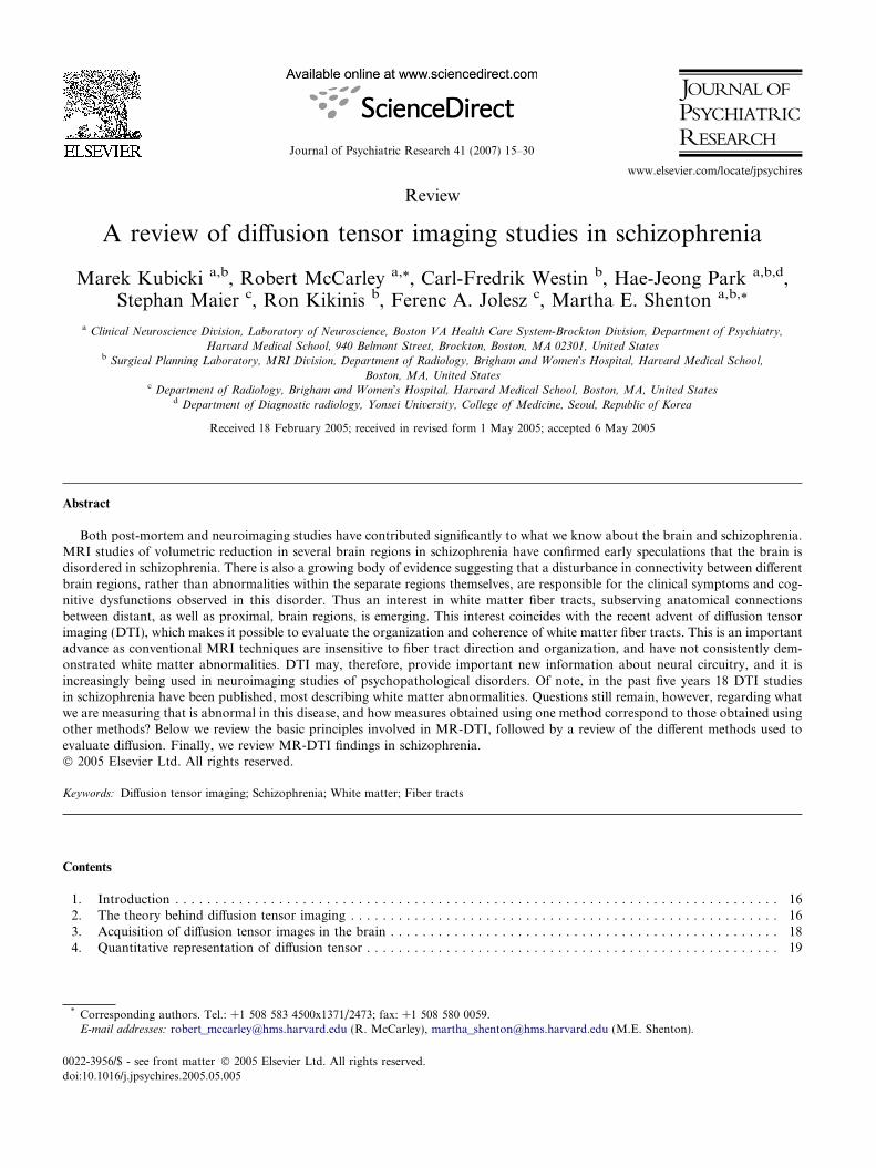
Fig. 1. Schematic of diffusion tensor imaging.
M. Kubicki et al. / Journal of Psychiatric Research 41 (2007) 15–30 17
using conventional MRI. This is primarily because whitematter appears homogeneous in conventional MRI, asthis methodology is not sensitive to discerning fiber tractdirection and/or organization. In contrast, recently, dif-fusion tensor MRI techniques have been developedwhich have proven to be useful in the evaluation of dif-ferent white matter components. DTI is, in fact, anexciting neuroimaging technique that affords a uniqueopportunity to quantify the diffusion of water in braintissue. It is based upon the phenomenon of water diffu-sion known as Brownian motion, named after the Eng-lish botanist Robert Brown, who in 1827 observed theconstant movement of minute particles that were sus-pended within grains of pollen. As this molecular mo-tion is affected by the properties of the medium inwhich it occurs, diffusion within biological tissues re-flects both tissue structure and architecture at the micro-scopic level. Further, due to the fact that diffusion is notuniform throughout the brain (differing, for example,between gray matter, white matter, and cerebrospinalfluid), researchers can use this characteristic of DTI toevaluate different brain tissue.
Specifically, in brain tissue, the motion of watermolecules can be restricted at many levels, as for exam-ple by interacting with different tissue components,such as cell membranes, macromolecules, fibers, myelinsheaths and fiber tracts. Thus within white matter, themobility of water is restricted in directions that are per-pendicular to the fiber tracts. (Parenthetically, this re-stricted mobility of water is described as anisotropic,whereas unrestricted mobility of water, such as wouldbe observed for cerebrospinal fluid, is described as iso-tropic.) And, while myelination is not essential for dif-fusion anisotropy of nerves, see for example studies ofnon-myelinated garfish olfactory nerves (Beaulieu andAllen, 1994) and studies of neonate brains prior tothe appearance of myelin (Huppi et al., 1998), tightlypacked multiple myelin membranes accompanying ax-ons are generally assumed to be the major barrier todiffusion in myelinated fiber tracts. Moreover, the tis-sue properties of white matter fiber tracts, includingthe density of the fibers, the average fiber diameter,the thickness of the myelin sheaths, and the direction-ality (or coherence) of the fibers in each voxel, all affectthe diffusion of water molecules, and thus, in turn, pro-vide useful information about white matter fiber tractorganization. Hence because of the physical propertiesof white matter, diffusion tensor imaging can be usedto investigate white matter in the brain in a mannernot possible with conventional MRI imagingtechniques.
Since its introduction to clinical use in 1986 by Le Bi-han et al. (1986), brain diffusion MRI has become one ofthe most important tools in clinical neuroimaging.Using this technique, diffusion is encoded in the MRIsignal by magnetic field gradient pulses. Accordingly,
the displacement of water molecules (diffusion) causesrandomization of the NMR spin phase, which, in turn,results in signal reduction. The amount of reductionprovides a quantitative measure of the diffusion in thegradient direction, and thus only diffusion in the direc-tion of this particular gradient can be detected. Sincediffusion is truly a three-dimensional process, at leastthree orthogonal measurements are needed to calculatethe mean diffusion for each voxel. Basser et al. (1994)introduced a multi-dimensional approach to the assess-ment of diffusion data in vivo, called diffusion tensorimaging (DTI), followed by the first clinical applicationin 1996 (Pierpaoli et al., 1996). This method requires atleast six different, non-collinear and non-planar diffu-sion measurements, and provides the rotationally invari-ant (independent to the gradient and head position)estimations of the diffusion and diffusion anisotropy.The diffusion for each voxel is then described as a3 · 3 matrix and called a diffusion tensor. In an isotropicmedia, such as cerebrospinal fluid, where diffusion in allthree main axes is equal, the tensor is symmetrical in alldirections, and it can be visualized as a sphere. In ananisotropic media, where the diffusion is different alongeach axis, the tensor is visualized as an ellipsoid, with itslongest axis pointing toward the longest of the so-calledprincipal directions (see Fig. 1).
The shape of the tensor ellipsoid thus depends uponthe strength of the diffusion along three principal direc-tions (i.e., its eigenvectors). The geometric nature of thediffusion tensors can then be used to characterize quan-titatively the local structure in tissues such as white mat-ter in the brain, because the direction of the majordiffusion axis will always be aligned in the direction ofthe white matter fiber tracts. The DTI images are ac-quired on high field, high performance MR systems,and analyzed in terms of diffusion anisotropy using ded-icated software.
18 M. Kubicki et al. / Journal of Psychiatric Research 41 (2007) 15–30
3. Acquisition of diffusion tensor images in the brain
In order to detect water diffusion along differentdirections, Stejskal and Tanner (1965) method is used.This method uses two strong gradient pulses, symmetri-cally positioned around a refocusing pulse, allowing fordiffusion weighting. To eliminate the dependence of spindensity, T1 and T2, at least two measurements of diffu-sion-weighting must be obtained, that are differentlysensitized to diffusion but remain identical in all otherrespects.
As noted previously, in order to correctly assess thediffusion tensor, data from at least six independent diffu-sion encoding directions is acquired for each voxel. Themost popular acquisition technique to obtain DTIimages is a single-shot diffusion-weighted, echo-planar(EPI) protocol, introduced in 1990 by Turner et al.(1990). It is fast, thus being robust to motion artifacts,although it is limited in spatial resolution, and very sen-sitive to image artifacts and distortions.
While DTI EPI remains the most popular techniqueto acquire diffusion tensor images, other protocols haveemerged as alternatives to the low resolution EPI tech-nique. Line-scan-diffusion-imaging (LSDI), for example,was introduced by Gudbartsson et al. (1996). Unlike thesingle-shot EPI, where a complete image is acquiredafter each radio-frequency excitation, LSDI is basedon the sequential acquisition of parallel columns lyingin the image plane. The sequential collection of this linedata in independent single-shot acquisitions makes thesequence largely insensitive to bulk motion artifactssince no phase encoding is used. Because of the acquisi-tion method, however, the LSDI sequence is four to sixtimes slower than single-shot diffusion-weighted EPI.On the other hand, it allows for better spatial resolution,which is especially important when small fiber bundlesare being evaluated. It is also relatively free from theartifacts inherent in single-shot EPI methods. Anotheradvantage of this technique is that it can be used onhigher field strengths, with signal to noise (S/N) gain,and without the dramatic increase of the aforemen-tioned artifacts.
More recently several new techniques have been pro-posed that further minimize susceptibility and chemicalshift artifacts, as well as allowing for higher than EPIresolution and/or signal to noise ratio, and shorter thanLSDI scan times. These newer techniques include: (1)diffusion weighted radial acquisition of data (DIFRAD)(Trouard et al., 1999); (2) slab scan diffusion imaging(SSDI) (Maier, 2001); (3) segmented EPI with navigatorechoes correction (Ordidge et al., 1994); and (4) single-shot EPI with parallel imaging (Bammer et al., 2002).
DTI acquisitions are generally performed on highfield, high performance gradient systems where 3–4 Te-sla magnets are becoming more and more popular inclinical research. However, the 1.5 Tesla magnet is still
the preferred MR field strength for clinical imaging.Lower field strengths systems require longer acquisitiontimes (due to the increased number of repetitions tocompensate for the decreased signal to noise ratio).Higher field strengths, on the other hand, allow forpotentially faster acquisitions and higher S/N ratios,but they are not free of limitations. Such limitations,in fact, include (depending on the acquisition tech-nique), eddy current distortions, susceptibility andchemical shift artifacts as well as geometrical distortionsof the images, which are, at least in the case of the fastertechniques, much higher that on lower field strength ma-chines. Nonetheless, the development of acquisitiontools such as high order shimming, which make possiblebetter quality images, as well as post-processing tools,which minimize image distortions and artifacts, shouldall help to ensure that clinical research can be movedto higher field magnets.
To return, however, to the image acquisition, and asmentioned previously, in order to calculate the tensor, atleast six non-colinear measurements of diffusion are re-quired. Some recent schizophrenia studies, however,use even more diffusion directions (Wang et al., 2003;Sun et al., 2003). Of note, multiple direction acquisitionshave numerous advantages, including the fact that mul-tiple direction acquisitions allow for the creation ofmore symmetrical tensors, they minimize noise, as wellas make the guiding of fiber tracking more precise.Moreover, higher angular data can be used to solvethe problem of fiber crossings, and to guide fiber track-ing in areas where the anisotropy is low, and whereuncertainty exists with respect to the major directionof diffusion (Tuch et al., 2002; Frank, 2002). This ap-proach, however, requires complicated mathematicalmodels such as in the recent work by Hagmann et al.(2004). In this work, the authors show tractography re-sults of fiber crossing in the centrum semiovale. Wenote, however, that while the direction of this work ispromising, it needs further development and is not vali-dated at this time.
Two final issues that should be taken into accountwhen planning a diffusion experiment, both of whichinfluence the S/N ratio, are the number of excitations(NEX), or repetitions, and the image resolution. TheS/N ratio for single-shot EPI is relatively low (due tomovement artifacts and short excitation times), and inorder to increase it, signal either needs to be averagedover many repeated scans, or scan resolution needs tobe decreased. The first results in longer scanner times,while the second results in less detailed data. Becauseof motion-related phase variations in the MR signal,complex averaging cannot be applied, but magnitudesignal must be used instead. This entails that not onlythe signal, but also the noise is being averaged. It isadvisable, for a good estimation of FA, to maintain anSNR for the diffusion-weighted image data of 10:1.
M. Kubicki et al. / Journal of Psychiatric Research 41 (2007) 15–30 19
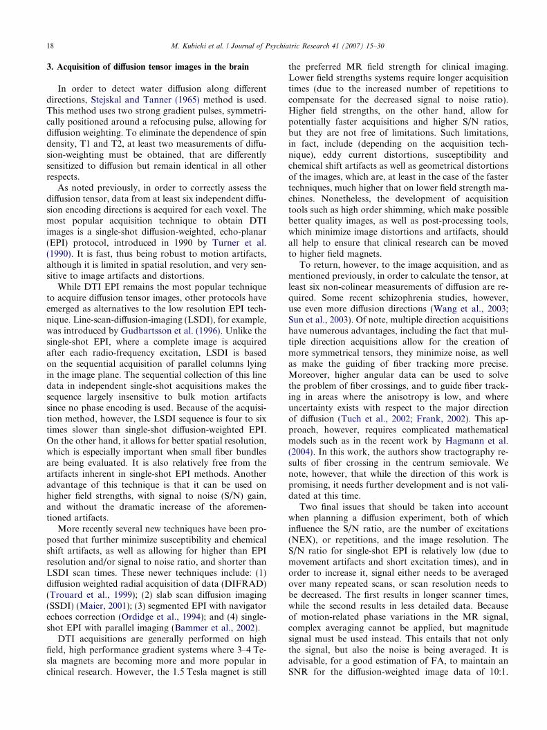
SNR values below 3:1 result in a Rician noise distribu-tion that requires special treatment if magnitude signalaveraging is performed. For very low SNR (<3:1) signalaveraging does not eliminate the overestimation of FAvalues, however, it does improve the estimation of thediffusion tensor eigenvector direction. Of note, in termsof the S/N ratios as well as data reproducibility, LSDIperforms similar to the single-shot EPI with 16 NEX�s(see Kubicki et al., 2004; see also Fig. 2).
The resolution of the images is the other important is-sue that needs to be considered. Fast MR techniquesused for DTI data acquisition do not usually allow forhigh image resolution, or if they do, the increase in res-olution is done at the expense of larger image distor-tions. Since most white matter structures are relativelysmall, the partial volume effect often exceeds the sizeof the fiber bundle. To avoid this problem, one canuse large ROIs, and instead of looking at particular fibertracts focus on the fiber coherence in the larger areas ofthe white matter. An alternative approach is to use im-age acquisitions that are perpendicular to the measuredfiber (see Fig. 3). Unfortunately, this approach is lessefficient, as it can be used only for a very few brain struc-tures (e.g., cingulate bundle, corpus callosum, a portionof the uncinate fasciculus, and the internal capsule). Forall of the reasons mentioned before, the most optimalmethod for data acquisition (when permitted by thescanner software and hardware), is to use as small aspossible, isotropic voxels. This approach is also prefera-ble if fiber tracking is to be applied to the DTI data.
4. Quantitative representation of diffusion tensor
Data from diffusion tensor imaging can be analyzedin several ways. The most general approach is to charac-terize the overall displacement of the molecules (averageellipsoid size) by calculating the mean diffusivity. To doso, the trace (Tr) (Basser, 1995) of the diffusion tensor,which is calculated as the sum of the eigenvalues ofthe tensor, has been introduced. The mean diffusivityis then given by Tr(D)/3.
Fig. 2. Influence of the number of excitations (NEX) on the data noise. 1, 4 athickness 4 mm, axial orientation.
To describe the anisotropy of diffusion, several scalarindices have been introduced. These indices are rotation-ally invariant, i.e., not dependent on the MR gradientsorientation, or absolute orientation of the diffusion ten-sor ellipsoid, and thus provide the ultimate value for aspecific tissue type. The most commonly used indices,proposed by Basser and colleagues (Basser, 1995) are:(1) relative anisotropy (RA), a normalized standarddeviation representing the ratio of the anisotropic partof the tensor to its isotropic part; (2) fractional anisot-ropy (FA), a measure of the fraction of the magnitudeof the tensor that can be ascribed to the anisotropic dif-fusion; and, (3) volume ratio (VR), a measure represent-ing the ratio of the ellipsoid volume to the volume as asphere of radius k. Other anisotropy indices, such asindices measuring intervoxel coherence (Pfefferbaumet al., 2000), and the geometric nature of diffusion andintravoxel coherence (Westin et al., 2002), have alsobeen proposed, and are reviewed in detail by Kubickiet al. (2002b) (for examples of maps representing diffu-sion indices see Fig. 3).
Similar to anisotropy indices, several methods havealso been proposed for visualizing the 3D informationcontained in DT-MRI data. These include: (1) the useof the octahedral icon for each pixel (Reese et al.,1995); (2) color maps, where different intensities of thethree colors indicate the size and the ADC in each ofthe three Cartesian directions (Makris et al., 1997); (3)rendered ellipsoids to visualize diffusion data in a slice,and (Pierpaoli et al., 1996); and (4) the use of lines torepresent the in-plane component of the principal diffu-sion direction, along with a color-coded out-of-planecomponent (Peled et al., 1998) (see Fig. 4).
Finally, multiple methods have been introduced forthe quantitative analysis of diffusion. The most popularapproach to data analysis in clinical studies is to use afixed size (square or circle) ROI method. This approachhas been used to place large ROIs within anatomicallynon-specific white matter regions of the brain (i.e., fron-tal white matter), as well as much smaller ROIs, placedwithin the specific white matter fiber tracts. These twomethods are distinct not only because one is averaging
nd 16 acquisitions of single-shot EPI, and 1 acquisition of LSDI. Slice

Fig. 3. Different indices used to represent diffusion. Upper left – FA (the measure of the fraction of the magnitude of the tensor that can be ascribedto the anisotropic diffusion). Upper right – RA (normalized standard deviation representing the ratio of the anisotropic part of the tensor to itsisotropic part). Lower left – TR (the sum of the eigenvalues of the tensor, or the sum of diffusion in all three principal directions). Lower right – linearmeasure of diffusion (representing the coherence of the fibers for each voxel) and spherical measure of diffusion (visualizing fiber crossings).
Fig. 4. Different methods for visualizing diffusion tensor information – left panel – map of fractional anisotropy with higher intensity representingmore anisotropic diffusion, middle panel – color map, where different intensities of the three colors indicate the size and the ADC in each of the threeCartesian directions; right panel – blue lines representing the in-plane component of the principal diffusion direction, and a color-coded out-of-planecomponent (more intuitive approach, where each line represents the main diffusion direction for local neighborhood, rather than for each voxel).
20 M. Kubicki et al. / Journal of Psychiatric Research 41 (2007) 15–30
over a bigger area, and thus is less sensitive to the noiseand artifacts, and more statistically conservative (Limet al., 1999), while the other is more anatomically spe-cific. Also, in terms of underlying pathology, anisotropy
differences detected by large ROIs, point to the anoma-lies in coherence and/or organization of the white matterstructures within the analyzed region, while differencesdetected by small ROIs, placed in the middle of the fiber
M. Kubicki et al. / Journal of Psychiatric Research 41 (2007) 15–30 21
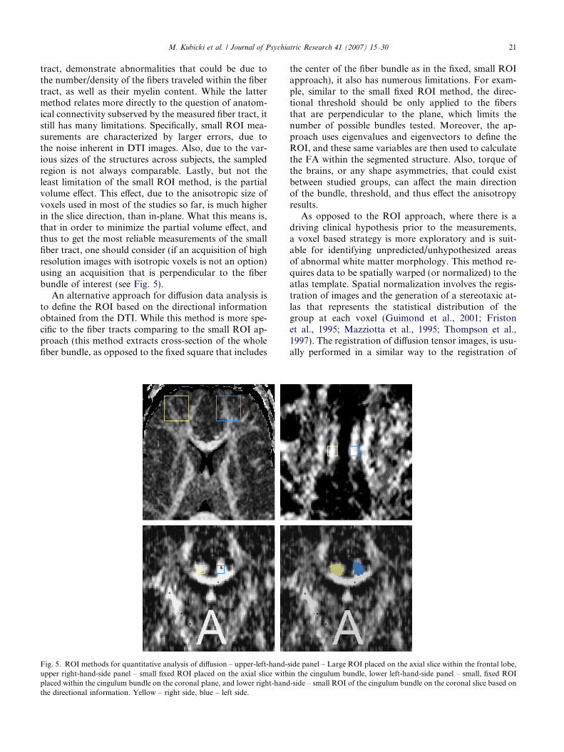
tract, demonstrate abnormalities that could be due tothe number/density of the fibers traveled within the fibertract, as well as their myelin content. While the lattermethod relates more directly to the question of anatom-ical connectivity subserved by the measured fiber tract, itstill has many limitations. Specifically, small ROI mea-surements are characterized by larger errors, due tothe noise inherent in DTI images. Also, due to the var-ious sizes of the structures across subjects, the sampledregion is not always comparable. Lastly, but not theleast limitation of the small ROI method, is the partialvolume effect. This effect, due to the anisotropic size ofvoxels used in most of the studies so far, is much higherin the slice direction, than in-plane. What this means is,that in order to minimize the partial volume effect, andthus to get the most reliable measurements of the smallfiber tract, one should consider (if an acquisition of highresolution images with isotropic voxels is not an option)using an acquisition that is perpendicular to the fiberbundle of interest (see Fig. 5).
An alternative approach for diffusion data analysis isto define the ROI based on the directional informationobtained from the DTI. While this method is more spe-cific to the fiber tracts comparing to the small ROI ap-proach (this method extracts cross-section of the wholefiber bundle, as opposed to the fixed square that includes
Fig. 5. ROI methods for quantitative analysis of diffusion – upper-left-hand-upper right-hand-side panel – small fixed ROI placed on the axial slice withplaced within the cingulum bundle on the coronal plane, and lower right-handthe directional information. Yellow – right side, blue – left side.
the center of the fiber bundle as in the fixed, small ROIapproach), it also has numerous limitations. For exam-ple, similar to the small fixed ROI method, the direc-tional threshold should be only applied to the fibersthat are perpendicular to the plane, which limits thenumber of possible bundles tested. Moreover, the ap-proach uses eigenvalues and eigenvectors to define theROI, and these same variables are then used to calculatethe FA within the segmented structure. Also, torque ofthe brains, or any shape asymmetries, that could existbetween studied groups, can affect the main directionof the bundle, threshold, and thus effect the anisotropyresults.
As opposed to the ROI approach, where there is adriving clinical hypothesis prior to the measurements,a voxel based strategy is more exploratory and is suit-able for identifying unpredicted/unhypothesized areasof abnormal white matter morphology. This method re-quires data to be spatially warped (or normalized) to theatlas template. Spatial normalization involves the regis-tration of images and the generation of a stereotaxic at-las that represents the statistical distribution of thegroup at each voxel (Guimond et al., 2001; Fristonet al., 1995; Mazziotta et al., 1995; Thompson et al.,1997). The registration of diffusion tensor images, is usu-ally performed in a similar way to the registration of
side panel – Large ROI placed on the axial slice within the frontal lobe,in the cingulum bundle, lower left-hand-side panel – small, fixed ROI-side – small ROI of the cingulum bundle on the coronal slice based on
22 M. Kubicki et al. / Journal of Psychiatric Research 41 (2007) 15–30
T1-weighted or SPGR MR images. That is, T2-weightedMR images are used to estimate deformation fields ortransformation functions in order to minimize the inten-sity difference between the template and the normalizedimage. With the estimated transformation function, themorphology of the diffusion images is then deformed tofit a stereotaxic space. Since T2 structural images do notinclude information relevant to the orientation anddirection of the fiber tracts, the registration of thesetypes of images to the atlas template usually performspoorly, with significant residual miss-registration errordue to differences in fiber tracts location, shape and size,as well as anatomical variability that can not be cap-tured with information provided by structural images.For this reason, voxel-based quantification of diffusiontensor images requires more sophisticated spatial nor-malization that takes advantage of the directional infor-mation provided by the full tensor (Park et al., 2003).
5. Applications of DTI to schizophrenia research
As noted previously, based on geometry and the de-gree of anisotropy loss, white matter tract alterations,such as dislocation, disruption or disorganization canbe documented. The cross-sectional size of these path-ways, as well as diffusion anisotropy measured withinthe bundle, yield a quantitative measure of connectivitybetween different brain regions. In fact, the tissue prop-erties of white matter fiber tracts, including the densityof the fibers, the fiber diameter, the thickness of the mye-lin sheaths, and the directionality (or coherence) of thefibers in each voxel, all affect the diffusion of water mol-ecules, thereby providing useful information aboutwhite matter fiber tract abnormalities.
Of note, a number of studies, using a wide spectrumof methods, suggest that disrupted connectivity ofwidely distributed brain circuits is likely characteristicof schizophrenia (Volkow et al., 1988; Friston and Frith,1995; McGuire and Frith, 1996; Weinberger et al., 1992;Fletcher et al., 1999). DTI thus has the potential to pro-vide important new information about links betweenanatomical abnormalities and clinical symptoms ob-served in this disease. To date, 18 studies have examinedwhite matter in schizophrenia using diffusion tensorimaging technique. Despite the consistency of the earli-est DTI findings (first five DTI studies showed diffusionabnormalities in schizophrenia), five more recent studiesdo not show any statistical differences between schizo-phrenics and healthy controls. One reason for this lackof consistency in findings is likely due to the fact thatthere are no set standards for data acquisition and anal-ysis and the methods of post-processing the data are alsovariable. In addition, small sample size, small ROI, low-er S/N ratio due to smaller number of repetitions, acqui-sition angles, scanner gradient performance, greater
partial volume effect due to the lower image resolution,as well as the use of anisotropic voxels, will skew the FAmeasurements, and decrease the statistical power of theDTI analysis. DTI findings in schizophrenia must, there-fore, be understood within the context of the acquisitionparameters used as well as in the context of the differentmethodological strategies adopted by different investiga-tors. These differences are highlighted, below.
6. DTI schizophrenia findings
In order to compare diffusion anisotropy betweencontrol subjects and schizophrenics, most studies re-ported in the literature have used fractional anisotropy(FA). Only one study has used relative anisotropy(RA), (Buchsbaum et al., 1998) and only one studyhas used inter-voxel coherence as an index of diffusion(Kalus et al., 2004). In comparison with FA, RA mapsshow lower visual contrast between white and gray mat-ter areas of the brain, since RA tends to be less sensitiveto anisotropy changes in midrange values than the morepopular FA index (Lim et al., 1999). In areas with higherdiffusion anisotropy (fiber tracts), and lower diffusionanisotropy (gray matter), both indices perform similarly.Of note, an index of inter-voxel coherence for each vox-el, as used by Kalus et al. (2004), takes into account notonly diffusion properties within this particular voxel, butalso in the adjacent voxels. This index tends to reflectbetter the fiber organization in very dense, small fiberbundles (see also Fig. 3). As an additional measure of fi-ber density and organization, mean diffusion along alldirections, or mean diffusivity has also been estimatedin several (n = 7) studies (see Table 1).
Decreased FA, along with increased diffusivity withinprefrontal and temporal lobes, as well as abnormalitieswithin the fiber bundles connecting these regions(including uncinate fasciculus, cingulum bundle andarcuate fasciculus) are the most frequent positive find-ings in schizophrenia studies (see Table 1). For example,uncinate fasciculus abnormalities were detected withsmall ROI defined by a directional threshold method(Kubicki et al., 2002a), and this finding has been con-firmed by a voxel based morphometry analysis (Burnset al., 2003). Cingulum bundle abnormalities have alsobeen reported in schizophrenia using both fixed sizesmall ROI (Sun et al., 2003; Wang et al., 2004), and adirectional threshold method (Kubicki et al., 2003). Inaddition, changes in the genu, but not the splenium ofthe corpus callosum, have been reported in schizophre-nia using the fixed, small ROI method (Foong et al.,2000), and using VBM analysis (Agartz et al., 2001;Ardekani et al., 2003) although this finding was not con-firmed in either another VBM analysis that used thesame sample as the small ROI method (Foong et al.,2002), or in another study using small ROI and a larger
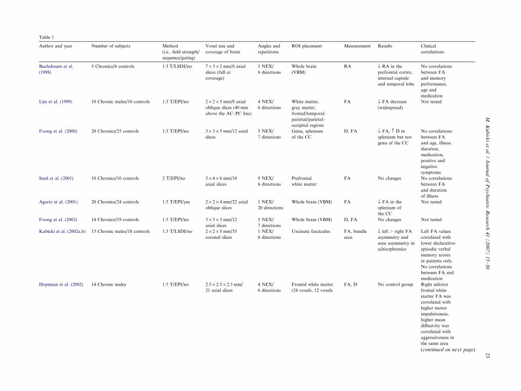
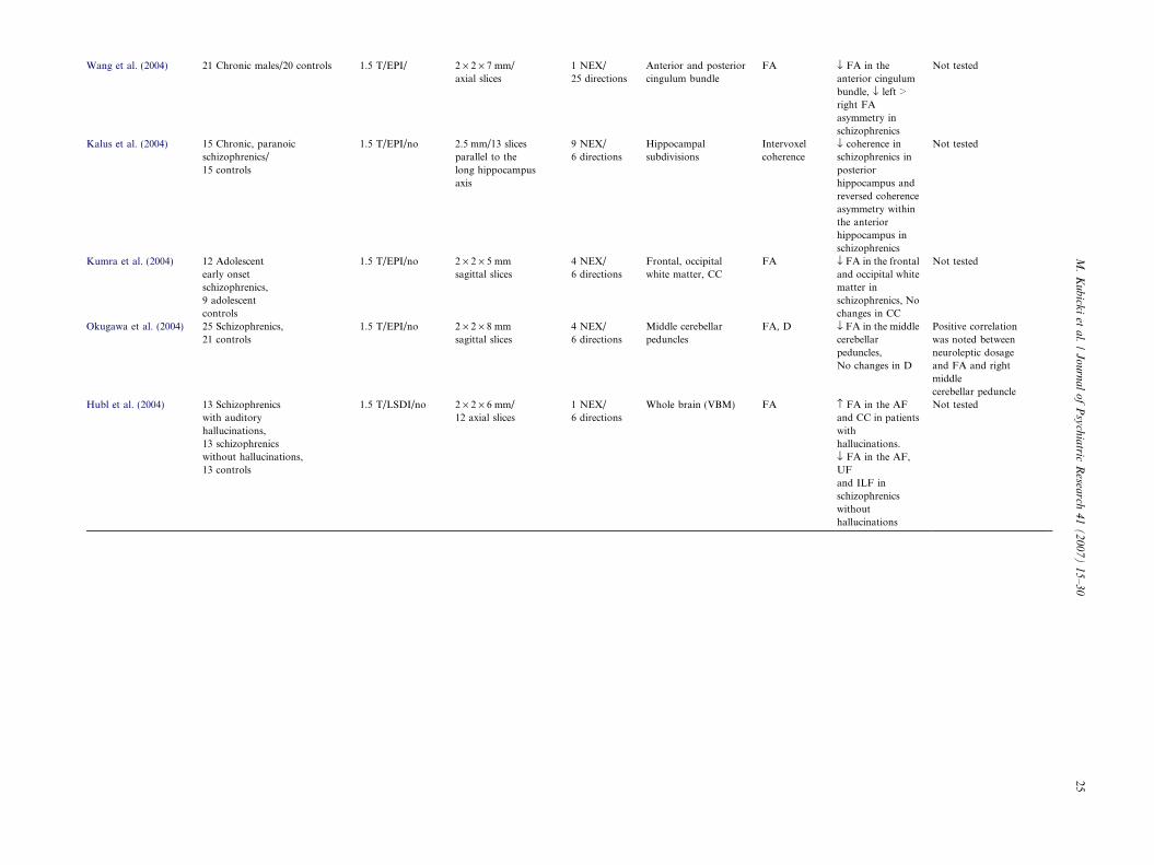
Table 1
Author and year Number of subjects Method
(i.e., field strength/
sequence/gating)
Voxel size and
coverage of brain
Angles and
repetitions
ROI placement Measurement Results Clinical
correlations
Buchsbaum et al.
(1998)
5 Chronics/6 controls 1.5 T/LSDI/no 7 · 3 · 2 mm/6 axial
slices (full cc
coverage)
1 NEX/
6 directions
Whole brain
(VBM)
RA fl RA in the
prefrontal cortex,
internal capsule
and temporal lobe
No correlations
between FA
and memory
performance,
age and
medication
Lim et al. (1999) 10 Chronic males/10 controls 1.5 T/EPI/no 2 · 2 · 5 mm/8 axial
oblique slices (40 mm
above the AC–PC line)
4 NEX/
6 directions
White matter,
gray matter,
frontal/temporal–
parietal/parietal–
occipital regions
FA fl FA decrease
(widespread)
Not tested
Foong et al. (2000) 20 Chronics/25 controls 1.5 T/EPI/no 3 · 3 · 5 mm/12 axial
slices
5 NEX/
7 directions
Genu, splenium
of the CC
D, FA fl FA, › D in
splenium but not
genu of the CC
No correlations
between FA
and age, illness
duration,
medication,
positive and
negative
symptoms
Steel et al. (2001) 10 Chronics/10 controls 2 T/EPI/no 3 · 4 · 6 mm/10
axial slices
9 NEX/
6 directions
Prefrontal
white matter
FA No changes No correlations
between FA
and duration
of illness
Agartz et al. (2001) 20 Chronics/24 controls 1.5 T/EPI/yes 2 · 2 · 4 mm/22 axial
oblique slices
1 NEX/
20 directions
Whole brain (VBM) FA fl FA in the
splenium of
the CC
Not tested
Foong et al. (2002) 14 Chronics/19 controls 1.5 T/EPI/no 3 · 3 · 5 mm/12
axial slices
5 NEX/
7 directions
Whole brain (VBM) D, FA No changes Not tested
Kubicki et al. (2002a,b) 15 Chronic males/18 controls 1.5 T/LSDI/no 2 · 2 · 5 mm/35
coronal slices
1 NEX/
6 directions
Uncinate fasciculus FA, bundle
area
fl left > right FA
asymmetry and
area asymmetry in
schizophrenics
Left FA values
correlated with
lower declarative-
episodic verbal
memory scores
in patients only.
No correlations
between FA and
medication
Hoptman et al. (2002) 14 Chronic males 1.5 T/EPI/no 2.5 · 2.5 · 2.5 mm/
21 axial slices
4 NEX/
6 directions
Frontal white matter
(24 voxels, 12 voxels
FA, D No control group Right inferior
frontal white
matter FA was
correlated with
higher motor
impulsiveness,
higher mean
diffusivity was
correlated with
aggressiveness in
the same area
(continued on next page)
M.
Ku
bick
iet
al.
/J
ou
rna
lo
fP
sych
iatric
Resea
rch4
1(
20
07
)1
5–
30
23
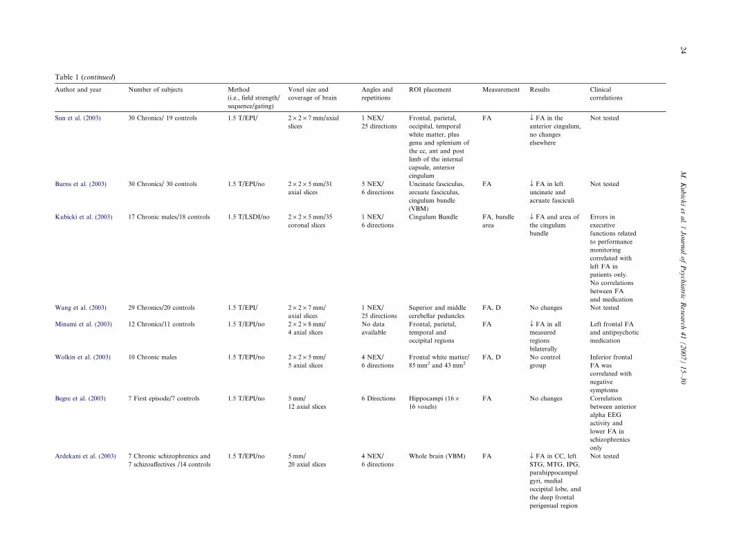
Table 1 (continued)
Author and year Number of subjects Method
(i.e., field strength/
sequence/gating)
Voxel size and
coverage of brain
Angles and
repetitions
ROI placement Measurement Results Clinical
correlations
Sun et al. (2003) 30 Chronics/ 19 controls 1.5 T/EPI/ 2 · 2 · 7 mm/axial
slices
1 NEX/
25 directions
Frontal, parietal,
occipital, temporal
white matter, plus
genu and splenium of
the cc, ant and post
limb of the internal
capsule, anterior
cingulum
FA fl FA in the
anterior cingulum,
no changes
elsewhere
Not tested
Burns et al. (2003) 30 Chronics/ 30 controls 1.5 T/EPI/no 2 · 2 · 5 mm/31
axial slices
5 NEX/
6 directions
Uncinate fasciculus,
arcuate fasciculus,
cingulum bundle
(VBM)
FA fl FA in left
uncinate and
acruate fasciculi
Not tested
Kubicki et al. (2003) 17 Chronic males/18 controls 1.5 T/LSDI/no 2 · 2 · 5 mm/35
coronal slices
1 NEX/
6 directions
Cingulum Bundle FA, bundle
area
fl FA and area of
the cingulum
bundle
Errors in
executive
functions related
to performance
monitoring
correlated with
left FA in
patients only.
No correlations
between FA
and medication
Wang et al. (2003) 29 Chronics/20 controls 1.5 T/EPI/ 2 · 2 · 7 mm/
axial slices
1 NEX/
25 directions
Superior and middle
cerebellar peduncles
FA, D No changes Not tested
Minami et al. (2003) 12 Chronics/11 controls 1.5 T/EPI/no 2 · 2 · 8 mm/
4 axial slices
No data
available
Frontal, parietal,
temporal and
occipital regions
FA fl FA in all
measured
regions
bilaterally
Left frontal FA
and antipsychotic
medication
Wolkin et al. (2003) 10 Chronic males 1.5 T/EPI/no 2 · 2 · 5 mm/
5 axial slices
4 NEX/
6 directions
Frontal white matter/
85 mm2 and 43 mm2FA, D No control
group
Inferior frontal
FA was
correlated with
negative
symptoms
Begre et al. (2003) 7 First episode/7 controls 1.5 T/EPI/no 5 mm/
12 axial slices
6 Directions Hippocampi (16 ·16 voxels)
FA No changes Correlation
between anterior
alpha EEG
activity and
lower FA in
schizophrenics
only
Ardekani et al. (2003) 7 Chronic schizophrenics and
7 schizoaffectives /14 controls
1.5 T/EPI/no 5 mm/
20 axial slices
4 NEX/
6 directions
Whole brain (VBM) FA fl FA in CC, left
STG, MTG, IPG,
parahippocampal
gyri, medial
occipital lobe, and
the deep frontal
perigenual region
Not tested
24M
.K
ub
icki
eta
l./
Jo
urn
al
of
Psy
chia
tricR
esearch
41
(2
00
7)
15
–3
0
Author and year Number of subjects Method
(i.e., field strength/
sequence/gating)
Voxel size and
coverage of brain
Angles and
repetitions
ROI placement Measurement Results Clinical
correlations
Wang et al. (2004) 21 Chronic males/20 controls 1.5 T/EPI/ 2 · 2 · 7 mm/
axial slices
1 NEX/
25 directions
Anterior and posterior
cingulum bundle
FA fl FA in the
anterior cingulum
bundle, fl left >
right FA
asymmetry in
schizophrenics
Not tested
Kalus et al. (2004) 15 Chronic, paranoic
schizophrenics/
15 controls
1.5 T/EPI/no 2.5 mm/13 slices
parallel to the
long hippocampus
axis
9 NEX/
6 directions
Hippocampal
subdivisions
Intervoxel
coherence
fl coherence in
schizophrenics in
posterior
hippocampus and
reversed coherence
asymmetry within
the anterior
hippocampus in
schizophrenics
Not tested
Kumra et al. (2004) 12 Adolescent
early onset
schizophrenics,
9 adolescent
controls
1.5 T/EPI/no 2 · 2 · 5 mm
sagittal slices
4 NEX/
6 directions
Frontal, occipital
white matter, CC
FA fl FA in the frontal
and occipital white
matter in
schizophrenics, No
changes in CC
Not tested
Okugawa et al. (2004) 25 Schizophrenics,
21 controls
1.5 T/EPI/no 2 · 2 · 8 mm
sagittal slices
4 NEX/
6 directions
Middle cerebellar
peduncles
FA, D fl FA in the middle
cerebellar
peduncles,
No changes in D
Positive correlation
was noted between
neuroleptic dosage
and FA and right
middle
cerebellar peduncle
Hubl et al. (2004) 13 Schizophrenics
with auditory
hallucinations,
13 schizophrenics
without hallucinations,
13 controls
1.5 T/LSDI/no 2 · 2 · 6 mm/
12 axial slices
1 NEX/
6 directions
Whole brain (VBM) FA › FA in the AF
and CC in patients
with
hallucinations.
fl FA in the AF,
UF
and ILF in
schizophrenics
without
hallucinations
Not tested
M.
Ku
bick
iet
al.
/J
ou
rna
lo
fP
sych
iatric
Resea
rch4
1(
20
07
)1
5–
30
25
26 M. Kubicki et al. / Journal of Psychiatric Research 41 (2007) 15–30
sample size (Sun et al., 2003; see also Table 1). Addition-ally, internal capsule diffusion abnormalities have beendetected in one VBM study (Buchsbaum et al., 1998),but not in a small ROI study (Sun et al., 2003) nor intwo other VBM studies (Agartz et al., 2001; Foonget al., 2002). Further, two studies have investigated dif-ferences in the cerebellar peduncles in schizophreniacompared to controls (Wang et al., 2003; Okugawaet al., 2004), and one did not show differences, whilethe other demonstrated decrease of the FA in the middlecerebellar peduncles in schizophrenia subjects. In addi-tion, two studies have reported widespread diffusionabnormalities in all tested regions (parietal, temporal,prefrontal and occipital white matter) when a fixed,large ROI method was used (Lim et al., 1999; Minamiet al., 2003; see also Table 1). So far, only one study(Hubl et al., 2004), using VBM and relatively smallnumber of subjects (n = 13), demonstrated increase ofFA in schizophrenics with auditory hallucinations.
Of particular note, all DTI studies of schizophreniathus far, except one with negative findings in hippocam-pus that included 7 first episode schizophrenics (Begreet al., 2003), have examined chronic schizophrenia sam-ples. Furthermore, medication was shown, to be relatedto left frontal white matter anisotropy (Minami et al.,2003), and in another study, to middle cerebellar pedun-cle (Okugawa et al., 2004). These two factors suggestthat in subsequent studies there is a need to study morecarefully the effects of medication, as well as to includefirst episode patients, who are either medication naı̈veor who have limited psychotropic exposure. In addition,the age of patients diagnosed with schizophrenia and theage of controls has not been observed thus far to corre-late with DTI measures. Nonetheless, a recently pub-lished, well-controlled study, that included a largehealthy population, has demonstrated a relationship be-tween FA, diffusivity, and age (Pfefferbaum and Sulli-van, 2003). This latter finding suggests that measuresof diffusion may change with age and it will be usefulto understand white matter changes across the age spanin both normal controls and in pathological popula-tions, as changes in aging that are evinced in normalcontrols may be quite different in pathological popula-tions such as schizophrenia.
Some interesting correlations between diffusion mea-sures and clinical and neuropsychological measures havealso emerged in DTI studies of schizophrenia. Specifi-cally, prefrontal white matter anisotropy has been re-ported to be correlated with negative symptoms,impulsiveness and aggressiveness (Hoptman et al.,2002; Wolkin et al., 2003). Additionally, FA for fron-tal–temporal connections have been reported to be cor-related with measures of errors in executive functionsrelated to performance monitoring (as measured by Wis-consin Card Sorting Test; WCST) – cingulate bundle,and declarative-episodic verbal memory (as measured
by Wechsler Memory Scale-III; WMS-III) – uncinatefasciculus (Kubicki et al., 2002a, 2003). While explor-atory, and not studied frequently in DTI studies(VBM methods so far have not explored clinical correla-tions at all), these findings demonstrate the sensitivity ofthe diffusion measurements to white matter abnormali-ties, and further provide evidence of the potential rolethat DTI-neuropsychological correlates can play inunderstanding disease-related cognitive disturbance.Moreover, the rather strong dissociation of UF-WMS-III and CB-WCST underscores the value of DTI indelineating important and distinct brain networks in pa-tients with schizophrenia.
7. Implications, conclusions and future directions
Preliminary DTI results, data from other MR tech-niques, such as magnetization transfer techniques andMR relaxation measurements (both sensitive to the mye-lin abnormalities), as well as MR spectroscopy (measur-ing neuronal markers), in combination withpathomorphological data, all suggest that white matterpathways play an important role in the neuropathologyof schizophrenia, and are likely related to the myriad ofclinical symptoms observed in this disorder. DTI find-ings to date are interesting, but methodological limita-tions, discussed here as well as elsewhere, limit ourability to conclude, with certainty, the nature of whitematter pathology in schizophrenia. What we do knowis that DTI provides a new window of opportunity toevaluate white matter in a manner not possible with con-ventional MRI. However, we do not as yet knowwhether the abnormalities observed to date reflect a de-crease in number of axons, decreased axonal diameter,thinner myelination sheaths, less coherent fibers, morefiber crossings, or simply more noise in the DTI data.
Despite these limitations, and the many questions thatremain with respect to how best to understand the diffu-sion findings in schizophrenia, we believe that DTI showsmuch promise for illuminating our understanding of howspecific brain regions are connected and how such con-nectivity may be relevant to our understanding of func-tional abnormalities, both cognitive, and clinical,observed in schizophrenia. We further believe that futureresearch, which uses high magnet fields, and fast, low dis-tortion techniques that produce high resolution and highSNR diffusion images, will lead to a further understand-ing of white matter pathology in schizophrenia. Otheradvances that we will likely have over the next few yearsare improved fiber tractography methods that are opti-mized and validated, and then applied to these high res-olution data. Such studies will likely benefit from theacquisition of contiguous, isotropic voxels (so far mostof the clinical studies use anisotropic voxels with gaps be-tween slices, which biases the major diffusion of the ten-

Fig. 6. Example of fiber tracking. High resolution data acquired on 3 Tesla magnet and post-processed using automated tracking procedure. Voxelswithin fiber bundles are color coded according to their FA values (i.e., blue, low anisotropy; and red, high anisotropy).
M. Kubicki et al. / Journal of Psychiatric Research 41 (2007) 15–30 27
sor and affects the contiguity of the fiber tract thus skew-ing the fiber tract direction), and diffusion images sam-pled in multiple directions. With reliable fiber tracking,it should be relatively easy to define brain regions ofinterest along the fiber bundles, and to measure bothmean diffusion anisotropy, as well as cross-sectional areaof these fiber bundles.
Fig. 6 provides an example of work that will likely as-sist us in evaluating fiber bundles in patients with schizo-phrenia. Here, automated fiber tracking procedure(described in detail in Park et al., 2003) has been usedto create major white matter fiber tracts. In brief, allwhite matter voxels have been used as seeding points,and then fiber bundles were created by following thelargest eigenvectors of the diffusion tensor using theRunge-Kutta algorithm (Basser et al., 2000). Next, FAvalues for each voxel within the bundles have been cal-culated, and voxels have been color coded based on theirFA values. This work will need to be validated in post-mortem tissue so that we will have a clearer understand-ing of the pathological processes detectable using DTI.Of further note, with today�s scanning techniques it isclear that results from fiber tractography do not describethe spatial extent of individual axons but instead de-scribe the average diffusion properties in white tissueat the scale of a millimeter. Thus, the diffusion tensorsat this scale should be regarded as estimators of localdiffusion properties and tractography as a visualizationof features in this field. Validation of the features cap-tured in the tensor field is thus very challenging sincethe definition of the tissue properties corresponding tothe diffusion field is not well understood. However, thesimilarities between DT-MRI tractography and realwhite matter fiber architecture in the human brain arecompelling even at today�s limited resolution.
Future studies will also need to include an evaluationof first episode psychotic patients, so as to provide asample of patients who have limited exposure to psycho-
tropic medications. Additionally, samples need to becomprised of both males and females, as there is clearevidence to suggest that the timing, course, and clinicalfeatures of schizophrenia are manifested quite differentlyin males and females, and this may be related to braindifferences (Goldstein et al., 2002). It will also be impor-tant to evaluate a representative sample of normal con-trols across age, as white matter changes with age, andsuch changes may be quite different in pathological pop-ulations such as schizophrenia. Future DTI studies needalso to include other imaging techniques, which high-light white matter pathologies, such as proton spectros-copy, magnetization transfer techniques and relaxationtimes measurements. Moreover, such studies should beconducted in concert with functional MRI and PETimaging in order to characterize and to understand morefully both functional and structural abnormalities inschizophrenia. Voxel based analyses will likely still beperformed in future studies, as this methodological ap-proach remains the simplest way to define possible re-gions of pathology, although it should be accompaniedby ROI analyses, as well as utilize better, more preciseregistration strategies.
Finally, we note that DTI was introduced to clinicalimaging only in 1995, and that such a new imaging toolhas only just begun to be explored with respect to whatit has to offer in documenting white matter abnormali-ties in schizophrenia. We are thus only at the beginningstages of what will likely be further technological ad-vances, as well as new white matter findings inschizophrenia.
Acknowledgments
The authors thank Marie Fairbanks for her adminis-trative assistance. They also gratefully acknowledge thesupport of the National Alliance for Research on

28 M. Kubicki et al. / Journal of Psychiatric Research 41 (2007) 15–30
Schizophrenia and Depression (MK), the NationalInstitute of Health (R03 MH068464-01 to MK, K02MH 01110 and R01 MH 50747 to MES, R01 NS39335 to SEM and R01 MH 40799 to RWM), thePost-doctoral Fellowship Program of Korea Scienceand Engineering Foundation (KOSEF) (HJP), theDepartment of Veterans Affairs Merit Awards (MES,RWM), and the National Center for Research Re-sources (NIH P41 RR13218 to RK).
References
Agartz I, Andersson JL, Skare S. Abnormal brain white matter inschizophrenia: a diffusion tensor imaging study. Neuroreport2001;12(10):2251–4.
Akbarian S, Kim JJ, Potkin SG, Hetrick WP, Bunney Jr WE, JonesEG. Maldistribution of interstitial neurons in prefrontal whitematter of the brains of schizophrenic patients. Archives of GeneralPsychiatry 1996;53(5):425–36.
Ardekani BA, Nierenberg J, Hoptman MJ, Javitt DC, Lim KO. MRIstudy of white matter diffusion anisotropy in schizophrenia.Neuroreport 2003;14(16):2025–9.
Bammer R, Auer M, Keeling SL, Augustin M, Stables LA, ProkeschRW, et al. Diffusion tensor imaging using single-shot SENSE-EPI.Magnetic Resonance in Medicine 2002;48(1):128–36.
Basser PJ. Inferring microstructural features and the physiologicalstate of tissues from diffusion-weighted images. NMR in Biomed-icine 1995;8(7–8):333–44.
Basser PJ, Mattiello J, LeBihan D. MR diffusion tensor spectroscopyand imaging. Biophysical Journal 1994;66(1):259–67.
Basser PJ, Pajevic S, Pierpaoli C, Duda J, Aldroubi A. In vivo fibertractography using DT-MRI data. Magnetic Resonance in Med-icine 2000;44(4):625–32.
Beaulieu C, Allen P. Determinants of anisotropic water diffusion innerves. MRM 1994;31:394–400.
Begre S, Federspiel A, Kiefer C, Schroth G, Dierks T, Strik WK.Reduced hippocampal anisotropy related to anteriorization ofalpha EEG in schizophrenia. Neuroreport 2003;14(5):739–42.
Benes FM. Relationship of cingulate cortex to schizophrenia and otherpsychiatric disorders. In: Vogt BA, Gabriel M, editors. Neurobi-ology of cingulate cortex and limbic thalamus: a comprehensivehandbook. Boston: Birkhauser; 1993. p. 581–605.
Breier A, Buchanan RW, Elkashef A, Munson RC, Kirkpatrick B,Gellad F. Brain morphology and schizophrenia. A magneticresonance imaging study of limbic, prefrontal cortex, and caudatestructures. Archives of General Psychiatry 1992;49(12):921–6 [seecomments].
Buchsbaum MS, Tang CY, Peled S, Gudbjartsson H, Lu D, HazlettEA, et al. MRI white matter diffusion anisotropy and PETmetabolic rate in schizophrenia. Neuroreport 1998;9(3):425–30.
Burns J, Job D, Bastin ME, Whalley H, Macgillivray T, Johnstone EC,et al. Structural disconnectivity in schizophrenia: a diffusion tensormagnetic resonance imaging study. British Journal of Psychiatry2003;182:439–43.
Davis KL, Stewart DG, Friedman JI, Buchsbaum M, Harvey PD, HofPR, et al. White matter changes in schizophrenia: evidence formyelin-related dysfunction. Archives of General Psychiatry2003;60(5):443–56.
Fletcher P, McKenna PJ, Friston KJ, Frith CD, Dolan RJ. Abnormalcingulate modulation of fronto-temporal connectivity in schizo-phrenia. Neuroimage 1999;9(3):337–42.
Foong J, Maier M, Clark CA, Barker GJ, Miller DH, Ron MA.Neuropathological abnormalities of the corpus callosum in schizo-
phrenia: a diffusion tensor imaging study. Journal of NeurologyNeurosurgery and Psychiatry 2000;68(2):242–4.
Foong J, Symms MR, Barker GJ, Maier M, Miller DH, Ron MA.Investigating regional white matter in schizophrenia using diffusiontensor imaging. Neuroreport 2002;13(3):333–6.
Frank LR. Characterization of anisotropy in high angular resolutiondiffusion-weighted MRI. Magnetic Resonance in Medicine2002;47(6):1083–99.
Friston K, Holmes A, Worsley KJ, Poline JP, Frith CD, FrackowiakRSJ. Statistical parametric maps in functional imaging: a generallinear approach. Human Brain Mapping 1995;2:189–210.
Friston KJ, Frith CD. Schizophrenia: a disconnection syndrome?Clinical Neuroscience 1995;3(2):89–97.
Goldstein JM, Seidman LJ, O�Brien LM, Horton NJ, Kennedy DN,Makris N, et al. Impact of normal sexual dimorphisms on sexdifferences in structural brain abnormalities in schizophreniaassessed by magnetic resonance imaging. Archives of GeneralPsychiatry 2002;59(2):154–64.
Gudbartsson H, Maier S, Mulkern R. Line scan diffusion imaging.Magnetic Resonance in Medicine 1996;36:509–19.
Guimond A, Roche A, Ayache N, Meunier J. Three-dimensionalmultimodal brain warping using the demons algorithm andadaptive intensity corrections. IEEE Transactions in MedicalImaging 2001;20(1):58–69.
Hagmann P, Reese TG, Tseng W-YI, Meuli R, Thiran J-P, WedeenVJ. Diffusion spectrum imaging tractography in complex cerebralwhite matter: an investigation of the centrum cemiovale. Proceed-ings of the International Society of Magnetic Resonance inMedicine 2004;11:623.
Hakak Y, Walker JR, Li C, Wong WH, Davis KL, Buxbaum JD,et al. Genome-wide expression analysis reveals dysregulation ofmyelination-related genes in chronic schizophrenia. Proceedings ofthe National Academy of Sciences of United States America2001;98(8):4746–51.
Highley JR, Esiri MM, McDonald B, Cortina-Borja M, Herron BM,Crow TJ. The size and fibre composition of the corpus callosumwith respect to gender and schizophrenia: a post-mortem study.Brain 1999a;122(Pt 1):99–110.
Highley JR, Esiri MM, McDonald B, Roberts HC, Walker MA, CrowTJ. The size and fiber composition of the anterior commissure withrespect to gender and schizophrenia. Biological Psychiatry1999b;45(9):1120–7.
Highley JR, Walker MA, Esiri MM, Crow TJ, Harrison PJ.Asymmetry of the uncinate fasciculus: a post-mortem study ofnormal subjects and patients with schizophrenia. Cerebral Cortex2002;12(11):1218–24.
Hof PR, Haroutunian V, Friedrich Jr VL, Byne W, Buitron C, PerlDP, et al. Loss and altered spatial distribution of oligodendrocytesin the superior frontal gyrus in schizophrenia. Biological Psychiatry2003;53:1075–85.
Hoptman MJ, Volavka J, Johnson G, Weiss E, Bilder RM, Lim KO.Frontal white matter microstructure, aggression, and impulsivity inmen with schizophrenia: a preliminary study. Biological Psychiatry2002;52(1):9–14.
Hubl D, Koenig T, Strik W, Federspiel A, Kreis R, Boesch C,et al. Pathways that make voices: white matter changes inauditory hallucinations. Archives of General Psychiatry 2004;61:658–68.
Huppi PS, Maier SE, Peled S, Zientara GP, Barnes PD, Jolesz FA,et al. Microstructural development of human newborn cerebralwhite matter assessed in vivo by diffusion tensor magneticresonance imaging. Pediatric Research 1998;44(4):584–90.
Kalus P, Buri C, Slotboom J, Gralla J, Remonda L, Dierks T, et al.Volumetry and diffusion tensor imaging of hippocampal subre-gions in schizophrenia. Neuroreport 2004;15(5):867–71.
Kraepelin E. Dementia praecox. New York: Churchill LivingstoneInc.; 1919/1971 (SB E. Barclay, Trans., ed.).
M. Kubicki et al. / Journal of Psychiatric Research 41 (2007) 15–30 29
Kubicki M, Maier SE, Westin CF, Mamata H, Ersner-Hershfield H,Estepar R, et al. Comparison of single-shot echo-planar and linescan protocols for diffusion tensor imaging. Academic Radiology2004;11(2):224–32.
Kubicki M, Westin CF, Maier SE, Frumin M, Nestor PG, SalisburyDF, et al. Uncinate fasciculus findings in schizophrenia: amagnetic resonance diffusion tensor imaging study. AmericanJournal of Psychiatry 2002a;159(5):813–20.
Kubicki M, Westin CF, Maier SE, Mamata H, Frumin M, Ersner-Hershfield H, et al. Diffusion tensor imaging and its application toneuropsychiatric disorders. Harvard Review of Psychiatry2002b;10(6):324–36.
Kubicki M, Westin CF, Nestor PG, Wible CG, Frumin M, Maier SE,et al. Cingulate fasciculus integrity disruption in schizophrenia: amagnetic resonance diffusion tensor imaging study. BiologicalPsychiatry 2003;54(11):1171–80.
Kumra S, Ashtari M, McMeniman M, Vogel J, Augustin R, BeckerDE, et al. Reduced frontal white matter integrity in early-onsetschizophrenia: a preliminary study. Biological Psychiatry2004;55:1138–45.
Le Bihan D, Breton E, Lallemand D, Grenier P, Cabanis E, Laval-Jeantet M. MR imaging of intravoxel incoherent motions: appli-cation to diffusion and perfusion in neurologic disorders. Radiol-ogy 1986;161(2):401–7.
Lim KO, Hedehus M, Moseley M, de Crespigny A, Sullivan EV,Pfefferbaum A. Compromised white matter tract integrity inschizophrenia inferred from diffusion tensor imaging. Archives ofGeneral Psychiatry 1999;56(4):367–74.
Maier SE. Slab scan diffusion imaging. Magnetic Resonance inMedicine 2001;46(6):1136–43.
Makris N, Worth AJ, Sorensen AG, Papadimitriou GM, Wu O, ReeseTG, et al. Morphometry of in vivo human white matter associa-tion pathways with diffusion-weighted magnetic resonance imag-ing. Annals of Neurology 1997;42:951–62.
Mazziotta JC, Toga AW, Evans A, Fox P, Lancaster J. A probabilisticatlas of the human brain: theory and rationale for its development.The International Consortium for Brain Mapping (ICBM). Neu-roimage 1995;2(2):89–101.
McGuire PK, Frith CD. Disordered functional connectivity inschizophrenia [editorial]. Psychological Medicine 1996;26(4):663–7.
Minami T, Nobuhara K, Okugawa G, Takase K, Yoshida T, SawadaS, et al. Diffusion tensor magnetic resonance imaging of disruptionof regional white matter in schizophrenia. Neuropsychobiology2003;47(3):141–5.
Ordidge RJ, Helpern JA, Qing ZX, Knight RA, Nagesh V. Correctionof motional artifacts in diffusion-weighted MR images usingnavigator echoes. Magnetic Resonance in Medicine1994;12(3):455–60.
Okugawa G, Nobuhara K, Minami T, Tamagaki C, Takase K,Sugimoto T, et al. Subtle disruption of the middle cerebellarpeduncles in patients with schizophrenia. Neuropsychobiology2004;50:119–23.
Paillere-Martinot M, Caclin A, Artiges E, Poline JB, Joliot M, MalletL, et al. Cerebral gray and white matter reductions and clinicalcorrelates in patients with early onset schizophrenia. SchizophreniaResearch 2001;50(1–2):19–26.
Park HJ, Kubicki M, Shenton ME, Guimond A, McCarley RW,Maier SE, et al. Spatial normalization of diffusion tensor MRIusing multiple channels. Neuroimage 2003;20(4):1995–2009.
Peled S, Gudbjartsson H, Westin CF, Kikinis R, Jolesz FA. Magneticresonance imaging shows orientation and asymmetry of whitematter fiber tracts. Brain Research 1998;780:27–33.
Pfefferbaum A, Sullivan EV. Increased brain white matter diffusivity innormal adult aging: relationship to anisotropy and partial volum-ing. Magnetic Resonance in Medicine 2003;49(5):953–61.
Pfefferbaum A, Sullivan EV, Hedehus M, Adalsteinsson E, Lim KO,Moseley M. In vivo detection and functional correlates of white
matter microstructural disruption in chronic alcoholism. Alcohol-ism–Clinical and Experimental Research 2000;24(8):1214–21.
Pierpaoli C, Jezzard P, Basser PJ, Barnett A, Di Chiro G. Diffusiontensor MR imaging of the human brain. Radiology1996;201:637–48.
Reese TG, Weisskoff RM, Smith RN, Rosen BR, Dinsmore RE,Wedeen VJ. Imaging myocardial fiber architecture in vivo withmagnetic resonance. Magnetic Resonance in Medicine1995;34:786–91.
Shenton ME, Dickey CC, Frumin M, McCarley RW. A review ofMRI findings in schizophrenia. Schizophrenia Research 2001;49(1–2):1–52.
Sigmudsson T, Suckling J, Maier M, Williams SCR, Bullmore ET,Greenwood KE, et al. Structural abnormalities in frontal, tempo-ral, and limbic regions and interconnecting white matter tracts inschizophrenic patients with prominent negative symptoms. Amer-ican Journal of Psychiatry 2001;158:234–43.
Steel RM, Bastin ME, McConnell S, Marshall I, Cunningham-OwensDG, Lawrie SM, Johnstone EC, Best JJK. Diffusion tensorimaging (DTI) and proton magnetic resonance spectroscopy (HMRS) in schizophrenic subjects and normal controls. PsychiatryResearch: Neuroimaging Section 2001;106:161–70.
Stejskal EO, Tanner JE. Spin diffusion measurements: spin echoes inthe presence of a time-dependent field gradient. Journal ofChemical Physics 1965;42:288–92.
Suddath RL, Christison GW, Torrey EF, Casanova MF, WeinbergerDR. Anatomical abnormalities in the brains of monozygotic twinsdiscordant for schizophrenia. New England Journal of Medicine1990;322(12):789–94.
Sun Z, Wang F, Cui L, Breeze J, Du X, Wang X, et al. Abnormalanterior cingulum in patients with schizophrenia: a diffusion tensorimaging study. Neuroreport 2003;14(14):1833–6.
Thompson PM, MacDonald D, Mega MS, Holmes CJ, Evans AC,Toga AW. Detection and mapping of abnormal brain structurewith a probabilistic atlas of cortical surfaces. Journal of ComputerAssisted Tomography 1997;21(4):567–81.
Trouard TP, Theilmann RJ, Altbach MI, Gmitro AF. High-resolutiondiffusion imaging with DIFRAD-FSE (diffusion-weighted radialacquisition of data with fast spin-echo) MRI. Magnetic Resonancein Medicine 1999;42(1):11–8.
Tuch DS, Reese TG, Wiegell MR, Makris N, Belliveau JW, WedeenVJ. High angular resolution diffusion imaging reveals intravoxelwhite matter fiber heterogeneity. Magnetic Resonance in Medicine2002;48(4):577–82.
Turner R, Le Bihan D, Maier J, Vavrek R, Hedges LK, Pekar J. Echo-planar imaging of intravoxel incoherent motion. Radiology1990;177(2):407–14.
Uranova N, Orlovskaya D, Vikhreva O, Zimina I, Kolomeets N,Vostrikov V, et al. Electron microscopy of oligodendroglia insevere mental illness. Brain Research Bulletin 2001;55(5):597–610.
Uranova NA, Vostrikov VM, Orlovskaya DD, Rachmanova VI.Oligodendroglial density in the prefrontal cortex in schizophreniaand mood disorders: a study from the Stanley NeuropathologyConsortium. Schizophrenia Research 2004;67(2–3):269–75.
Volkow ND, Wolf AP, Brodie JD, Cancro R, Overall JE, et al. Braininteractions in chronic schizophrenics under resting and activationconditions. Schizophrenia Research 1988;1(1):47–53.
Wang F, Sun Z, Cui L, Du X, Wang X, Zhang H, et al. Anteriorcingulum abnormalities in male patients with schizophrenia deter-mined through diffusion tensor imaging. American Journal ofPsychiatry 2004;161(3):573–5.
Wang F, Sun Z, Du X, Wang X, Cong Z, Zhang H, et al. A diffusiontensor imaging study of middle and superior cerebellar peduncle inmale patients with schizophrenia. Neuroscience Letters2003;348(3):135–8.
Weinberger DR, Berman KF, Suddath R, Torrey EF.Evidence of dysfunction of a prefrontal-limbic network

30 M. Kubicki et al. / Journal of Psychiatric Research 41 (2007) 15–30
in schizophrenia: a magnetic resonance imaging andregional cerebral blood flow study of discordant monozy-gotic twins. American Journal of Psychiatry ;149(7):890–7.
Wernicke C. Grundrisse der Psychiatrie. Leipzig: Thieme; 1906.Westin CF, Maier SE, Mamata H, Nabavi A, Jolesz FA, Kikinis R.
Processing and visualization for diffusion tensor MRI. MedicalImage Analysis 2002;6(2):93–108.
Wible CG, Shenton ME, Hokama H, Kikinis R, Jolesz FA, Metcalf D,et al. Prefrontal cortex and schizophrenia. A quantitative magneticresonance imaging study. Archives of General Psychiatry1995;52(4):279–88.
Wolkin A, Choi SJ, Szilagyi S, Sanfilipo M, Rotrosen JP, Lim KO.Inferior frontal white matter anisotropy and negative symptoms ofschizophrenia: a diffusion tensor imaging study. American Journalof Psychiatry 2003;160:572–4.
Related Documents