Diffusion Tensor Imaging for Brain Malformations Does It Help? Thierry A.G.M. Huisman, MD*, Thangamadhan Bosemani, MD, Andrea Poretti, MD INTRODUCTION Diffusion-weighted imaging (DWI) and in particular diffusion tensor imaging (DTI) have propelled our noninvasive exploration and understanding of many common and rare congenital brain abnor- malities, which include malformations as well as disruptions. 1,2 DWI/DTI takes advantage of the preferential mobility and diffusion of water mole- cules in three-dimensional space within biological tissues with a complex microstructural architec- ture like the brain. The mobility of water molecules in the brain is dependent on multiple factors, including the microstructural architecture, which is believed to be predominantly determined by the alignment and diameter of fiber tracts and their myelin sheets as well as the three-dimensional distribution and arrangement of neurons. In addi- tion, multiple other spatially organized flow pat- terns exist within the brain, like blood flow in the arterial and venous networks and the molecular flow of protons, which depend on active and pas- sive transport mechanisms within the brain. Exploring the differential mobility and diffusion of water molecules in the brain gives indirect infor- mation about the normal and abnormal internal ar- chitecture, networks, and integrity of the pediatric brain. The differential, three-dimensional direction and magnitude of water mobility can be measured noninvasively, in vivo by adding diffusion- weighted gradients to standard ultrafast magnetic resonance (MR) sequences. Typically, diffusion Section of Pediatric Neuroradiology, Russell H. Morgan Department of Radiology and Radiological Science, Charlotte R. Bloomberg Children’s Center, The Johns Hopkins University School of Medicine, Sheikh Zayed Tower, Room 4174, 1800 Orleans Street, Baltimore, MD 21287-0842, USA * Corresponding author. E-mail address: [email protected] KEYWORDS Diffusion tensor imaging Fiber tractography Children Neuroimaging Brain malformations Axonal guidance disorders KEY POINTS Diffusion tensor imaging (DTI) is an advanced magnetic resonance (MR) technique that provides qualitative and quantitative information about the microarchitecture of white matter. DTI may show important information about the brain microstructure in brain malformations that may go undetected or remains underestimated on conventional MR sequences. DTI may better categorize various brain malformations that may look similar on conventional MR imaging, but may be caused by different pathomechanisms. Human disorders of axonal guidance result from aberrant axonal wiring and are caused by muta- tions in genes that code for molecules that guide axons within the brain. Much of our knowledge about human disorders of axonal guidance comes from DTI studies of crossing tracts, such as the corpus callosum, optic chiasm, corticospinal tract, and cerebellar peduncles. Neuroimag Clin N Am - (2014) -–- http://dx.doi.org/10.1016/j.nic.2014.07.004 1052-5149/14/$ – see front matter Ó 2014 Elsevier Inc. All rights reserved. neuroimaging.theclinics.com

Welcome message from author
This document is posted to help you gain knowledge. Please leave a comment to let me know what you think about it! Share it to your friends and learn new things together.
Transcript
Diffusion Tensor Imagingfor Brain Malformations
Does It Help?Thierry A.G.M. Huisman, MD*,Thangamadhan Bosemani, MD, Andrea Poretti, MD
KEYWORDS
� Diffusion tensor imaging � Fiber tractography � Children � Neuroimaging � Brain malformations� Axonal guidance disorders
KEY POINTS
� Diffusion tensor imaging (DTI) is an advanced magnetic resonance (MR) technique that providesqualitative and quantitative information about the microarchitecture of white matter.
� DTI may show important information about the brain microstructure in brain malformations that maygo undetected or remains underestimated on conventional MR sequences.
� DTI may better categorize various brain malformations that may look similar on conventional MRimaging, but may be caused by different pathomechanisms.
� Human disorders of axonal guidance result from aberrant axonal wiring and are caused by muta-tions in genes that code for molecules that guide axons within the brain.
� Muchofour knowledgeabout humandisordersof axonal guidancecomes fromDTI studiesofcrossingtracts, such as the corpus callosum, optic chiasm, corticospinal tract, and cerebellar peduncles.
com
INTRODUCTION
Diffusion-weighted imaging (DWI) and in particulardiffusion tensor imaging (DTI) have propelled ournoninvasive exploration and understanding ofmany common and rare congenital brain abnor-malities, which include malformations as well asdisruptions.1,2 DWI/DTI takes advantage of thepreferential mobility and diffusion of water mole-cules in three-dimensional space within biologicaltissues with a complex microstructural architec-ture like the brain. The mobility of water moleculesin the brain is dependent on multiple factors,including the microstructural architecture, whichis believed to be predominantly determined bythe alignment and diameter of fiber tracts and theirmyelin sheets as well as the three-dimensional
Section of Pediatric Neuroradiology, Russell H. MorganCharlotte R. Bloomberg Children’s Center, The Johns HTower, Room 4174, 1800 Orleans Street, Baltimore, MD 2* Corresponding author.E-mail address: [email protected]
Neuroimag Clin N Am - (2014) -–-http://dx.doi.org/10.1016/j.nic.2014.07.0041052-5149/14/$ – see front matter � 2014 Elsevier Inc. All
distribution and arrangement of neurons. In addi-tion, multiple other spatially organized flow pat-terns exist within the brain, like blood flow in thearterial and venous networks and the molecularflow of protons, which depend on active and pas-sive transport mechanisms within the brain.Exploring the differential mobility and diffusion ofwater molecules in the brain gives indirect infor-mation about the normal and abnormal internal ar-chitecture, networks, and integrity of the pediatricbrain.
The differential, three-dimensional direction andmagnitude of water mobility can be measurednoninvasively, in vivo by adding diffusion-weighted gradients to standard ultrafast magneticresonance (MR) sequences. Typically, diffusion
Department of Radiology and Radiological Science,opkins University School of Medicine, Sheikh Zayed1287-0842, USA
rights reserved. neuroimaging.theclinics.
Huisman et al2
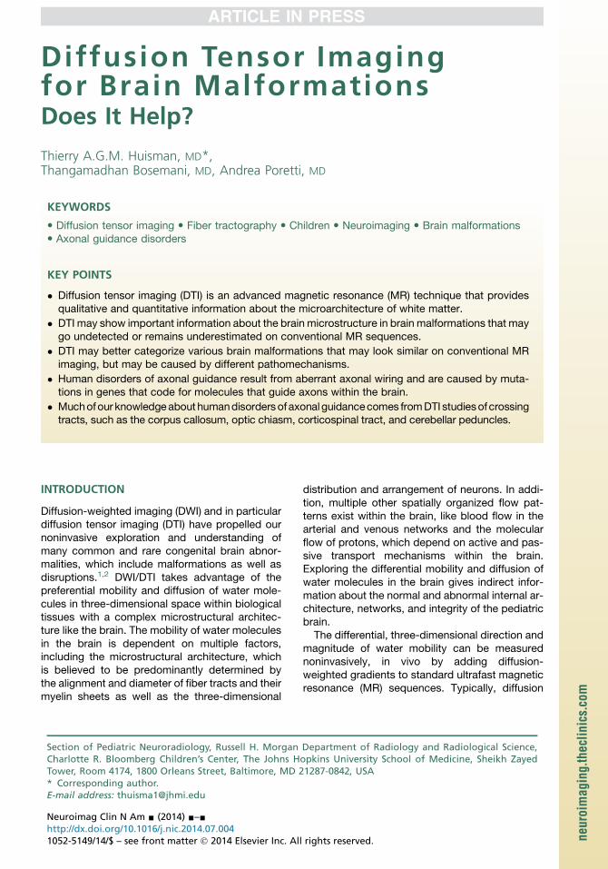
gradients are applied along multiple noncollineardirections in space to resolve the completediffusion tensor. The more diffusion directionsgradients measured, the better is the three-dimensional shape and magnitude of the sampleddiffusion tensor. Powerful postprocessing toolssubsequently allow reconstructing the course ofmajor fiber tracts within the central nervous sys-tem (CNS). DTI has been of invaluable significanceto explore common and uncommon brain malfor-mations. Steve Jobs, one of the most influential in-ventors and entrepreneurs of our time, made aquote that summarizes the goal and value of DTIin brain malformations: “because believing thatthe dots will connect down the road, will give youthe confidence to follow your heart, even when itleads you off the well-worn path.” The DTI acquisi-tions render data sets that have to be postpro-cessed with an open scientific mind, in which thevarious dots or seed points have to be connectedin a nonbiased mathematical approach to confirmthe course of well-known common pathways andnetworks, which should also allow us to exploreand discover unknown or aberrant fiber tractsand anatomic connections. Recognizing theseaberrant pathways may be challenging but helpus to better understand, classify, or group brainmalformations. This approach is similar to high-end brain teasers, in which numbers need to bediscovered using a complex mathematical formula
Fig. 1. Connect the dots puzzles are a formof puzzlesmadeline in the correct order to discover the final picture. Tyascending order. However, in advanced puzzles, they mayexample, the algebraic sequencing of a group of numbers bskills and an openmind to outline and recognize the figure.point of the connect the dots puzzle. Correct analysis of thealgebraic formula that connects the sequence of numbers (connect the numbers, and the outline of an aircraft is discoand fiber tractography in the exploration of normal and a
and then connected to uncover or recognize a hid-den figure (Fig. 1).In this article, the basics of DWI/DTI are dis-
cussed, including a short historical perspective onthe fiber dissection technique, followed by a reviewof selected brain malformations in which DTI andtractography have contributed to a better under-standing of the malformations, and by a clinicalcase in which DTI showed a disorder of the internalneuroarchitecture that could not be correctlyappreciatedbyconventional anatomicMR imaging.
BASICS OF DIFFUSION-WEIGHTED IMAGINGAND DIFFUSION TENSOR IMAGING
DWI generates image contrast based on differ-ences in diffusion characteristics of water mole-cules within the brain.1,2 Diffusion represents therandom thermal movement of molecules, alsoknown as Brownian motion, within tissues. Diffu-sion within the brain is determined by a variety offactors, including the type of molecule underinvestigation, the temperature of the tissue, andmost importantly, the microenvironmental archi-tecture in which the diffusion takes place. Forexample, the rate of diffusion of water moleculeswithin the cerebrospinal fluid (CSF) is higher thanthe rate of diffusion of water molecules withinthe brain tissue. By adding diffusion-encoding gra-dients to standard MR imaging sequences, these
of a sequence of dots that have to be connectedwith apically, they rely on connecting a row of numbers ingo beyond the standard numbering. Identifying, forased on the already connected dots requires analyticalIn this example, the initial figure (A) shows the startingorder of the first 3 numbers allows the discovery of thex 5 21/4y13). Subsequently, the scientist knows how tovered (B). This connect the dots puzzle is similar to DTInomalous white matter tracts in the brain.
DTI for Brain Malformations 3
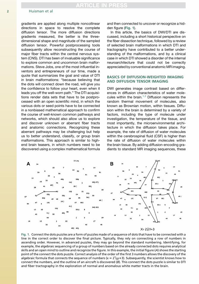
differences in diffusion rates can be translated intoand shown as differences in MR signal. These dif-ferences in MR signal of the individual imagingvoxels can be mapped two-dimensionally as so-
Fig. 2. Axial trace of diffusion (DWI) images (top row) and(lower row) of the brain. CSF with high degree of diffusiotissue with more restricted diffusion is DWI-hyperintense
called DWI images. Typically, isotropic DWI im-ages are generated (Fig. 2). Isotropic DWI imagesincorporate signal related to both the rate of diffu-sion and T2-relaxation phenomena. In addition,
matching apparent diffusion coefficient (ADC) mapsn is DWI-hypointense and ADC-bright, whereas brainand ADC-dark.
Huisman et al4
maps of apparent diffusion can be calculated, alsoknown as apparent diffusion coefficient (ADC). TheADC values are typically displayed as two-dimensional gray-white scale maps, in which thecalculated ADC values are translated into bright-ness maps or so-called ADC maps. Areas withhigh degrees of diffusion or high ADC valuesappear bright (eg, CSF), whereas areas with lowdegrees of diffusion or low ADC values (eg,densely packed white matter tracts) appeardarker. The major advantage of ADC is that theT2 contribution of the DWI maps is canceled out,giving a map of the spatial distribution of diffusionin the brain without contamination by the T2-relaxation phenomena (see Fig. 2).DTI takes DWI to the next level of image-based
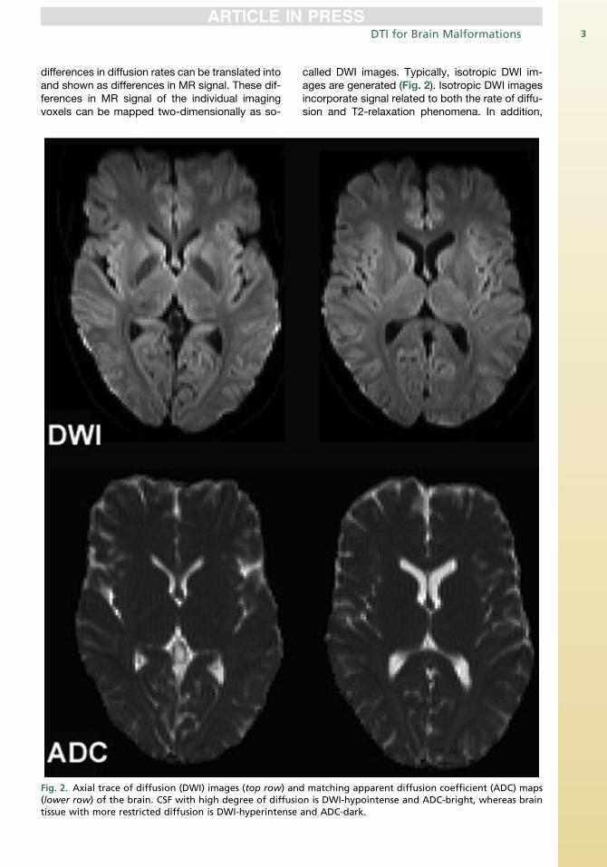
microstructural tissue exploration. The three-dimensional shape and principal direction of diffu-sion as well as the magnitude of diffusion (diffusionrate) within space differ between the various brainstructures. The microstructural architecture aswell as physiologic factors determine the diffusionof water molecules within the brain. The effectivemolecular diffusion in the white matter tracts is,for example, predominantly along the directionparallel to the long axis of white matter tractsand limited in the direction perpendicular to thewhite matter tracts. The three-dimensional shapeof diffusion in white matter tracts resembles anellipsoid, which is also known as anisotropic
Fig. 3. Anisotropic diffusion (A) resembles a three-dimensmolecules along the main axis of the ellipsoid and restricdiffusion (B) can be represented by a sphere with equal dithe motion of individual molecules. (From Huisman TA. Dibrain, made easy. Cancer Imaging 2010;10:S166; with perm
diffusion (Fig. 3A). When the degree of diffusionis equal in all directions such as in CSF, in whichno barriers limit diffusion, the three-dimensionalshape of diffusion resembles a sphere, which isalso known as isotropic diffusion (see Fig. 3B).The three-dimensional shape of diffusion can bestudied by measuring the full tensor of the diffu-sion (DTI), in which diffusion gradients are appliedalong at least 6 noncollinear directions in space.Maps of the spatial distribution and magnitude ofthe anisotropic component of diffusion are gener-ated. Fractional anisotropy (FA) maps are typicallycalculated, in which the assigned signal intensity isrelated to the degree of anisotropic diffusion. FAvalues range between 0 and 1. An FA value of 0 in-dicates complete isotropic diffusion, and an FAvalue of 1 indicates complete anisotropic diffusion(Fig. 4). These FA values can be mapped topo-graphically, in which voxels with an FA value of0 are black and voxels with an FA value of 1 arewhite. In addition, the principal direction of diffu-sion in three-dimensional space can be colorcoded (Fig. 5) or can be shown as a vector foreach voxel (Fig. 6). Blue represents a predomi-nantly craniocaudal diffusion, red represents pre-dominantly left to right diffusion, and greenrepresents predominantly anterior-posterior diffu-sion within the brain. These maps provide informa-tion about the architecture and integrity oforganized tissues. Finally, by combining the
ional ellipsoid in space, with predominant diffusion ofted diffusion perpendicular to the ellipsoid. Isotropicffusion in all directions in space. The arrows representffusion weighted and diffusion tensor imaging of theission.)
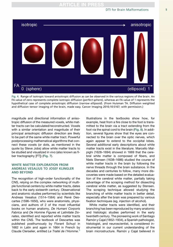
Fig. 4. Range of isotropic toward anisotropic diffusion as can be observed in the various regions of the brain. AnFA value of zero represents complete isotropic diffusion (perfect sphere), whereas an FA value of 1 represents thehypothetical case of complete anisotropic diffusion (narrow ellipsoid). (From Huisman TA. Diffusion weightedand diffusion tensor imaging of the brain, made easy. Cancer Imaging 2010;10:S167; with permission.)
DTI for Brain Malformations 5
magnitude and directional information of aniso-tropic diffusion of the measured voxels, white mat-ter tracts can be calculated/reconstructed. Voxelswith a similar orientation and magnitude of theirprincipal anisotropic diffusion direction are likelyto be part of the same white matter tract. Powerfulpostprocessing mathematical algorithms that con-nect these voxels (or dots, as mentioned in thequote by Steve Jobs) allow white matter tracts tobe studied and visualized in vivo (also known as fi-ber tractography [FT]) (Fig. 7).
WHITE MATTER EXPLORATION FROMANDREAS VESALIUS TO JOSEF KLINGLERAND BEYOND
The recognition of high-order functionality of theCNS, relying on the complex networking of multi-ple functional centers by white matter tracts, datesback to the early sixteenth century. Observationaland anatomic studies performed by scientists likeAndreas Vesalius (1514–1564) and Rene Des-cartes (1596–1650), who were anatomists, physi-cians, and authors of 2 of the most influentialbooks on human anatomy, De Humani CorporisFabrica and De Homine Figures et Latinitate Do-nates, identified and reported white matter tractswithin the CNS. The textbook of Descartes waspublished posthumously by Florant Schuyl in1662 in Latin and again in 1664 in French byClaude Clerselier, entitled Le Traite de l’Homme.3
Illustrations in the textbooks show how, forexample, heat from a fire close to the foot is trans-mitted to the brain via a tract extending from thefoot via the spinal cord to the brain (Fig. 8). In addi-tion, several figures show that the eyes are con-nected to the brain over the optic nerves, whichagain appear to extend to the occipital lobes.Several additional early descriptions about whitematter tracts exist in the literature. Marcello Mal-pighi (1628–1694) showed in 1669 that the cere-bral white matter is composed of fibers, andNiels Stensen (1638–1686) studied the course ofwhite matter tracts in the brain by following thenerve threads through the brain substance. In thedecades and centuries to follow, many more dis-coveries were made based on the detailed evalua-tion of the cerebral white matter, partially takingadvantage of the scraping method for dissectingcerebral white matter, as suggested by Stensen.The scraping technique allowed studying thebranching of white matter tracts in better detail,especially after the brain was prepared by variousfixation techniques (eg, injection of alcohol).
White matter tracts were identified, and theirbranching has been reproduced bymany anatomicand histologic studies performed in the earlytwentieth century. The pioneering work of SantiagoRamon y Cajal (1852–1934), a Spanish pathologist,histologist, and neuroscientist, is considered in-strumental in our current understanding of thebrain microstructure. Ramon y Cajal believed in
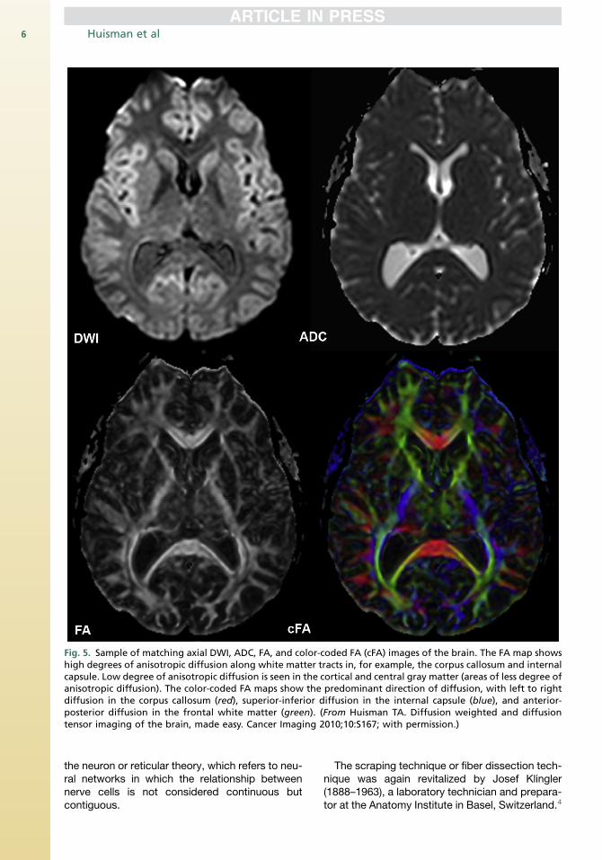
Fig. 5. Sample of matching axial DWI, ADC, FA, and color-coded FA (cFA) images of the brain. The FA map showshigh degrees of anisotropic diffusion along white matter tracts in, for example, the corpus callosum and internalcapsule. Low degree of anisotropic diffusion is seen in the cortical and central gray matter (areas of less degree ofanisotropic diffusion). The color-coded FA maps show the predominant direction of diffusion, with left to rightdiffusion in the corpus callosum (red), superior-inferior diffusion in the internal capsule (blue), and anterior-posterior diffusion in the frontal white matter (green). (From Huisman TA. Diffusion weighted and diffusiontensor imaging of the brain, made easy. Cancer Imaging 2010;10:S167; with permission.)
Huisman et al6
the neuron or reticular theory, which refers to neu-ral networks in which the relationship betweennerve cells is not considered continuous butcontiguous.
The scraping technique or fiber dissection tech-nique was again revitalized by Josef Klingler(1888–1963), a laboratory technician and prepara-tor at the Anatomy Institute in Basel, Switzerland.4
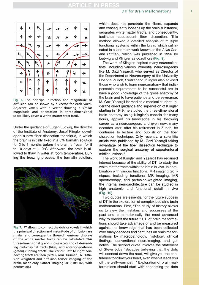
Fig. 6. The principal direction and magnitude ofdiffusion can be shown by a vector for each voxel.Adjacent voxels with a vector showing a similarmagnitude and orientation in three-dimensionalspace likely cover a white matter tract (red).
DTI for Brain Malformations 7
Under the guidance of Eugen Ludwig, the directorof the Institute of Anatomy, Josef Klingler devel-oped a new fiber dissection technique, in whichthe brain is initially fixed in a 5% formalin solutionfor 2 to 3 months before the brain is frozen for 8to 10 days at �10�C. Afterward, the brain is al-lowed to thaw in water at room temperature. Dur-ing the freezing process, the formalin solution,
Fig. 7. FT allows to connect the dots or voxels in whichthe principal direction and magnitude of diffusion aresimilar, and consequently, three-dimensional displaysof the white matter tracts can be calculated. Thisthree-dimensional graph shows a crossing of descend-ing corticospinal tracts (blue) and anterior-posterior(green) running tracts. The various left to right con-necting tracts are seen (red). (From Huisman TA. Diffu-sion weighted and diffusion tensor imaging of thebrain, made easy. Cancer Imaging 2010;10:S168; withpermission.)
which does not penetrate the fibers, expandsand consequently loosens up the brain substance,separates white matter tracts, and consequently,facilitates subsequent fiber dissection. Thismethod allowed a detailed analysis of multiplefunctional systems within the brain, which culmi-nated in a landmark work known as the Atlas Cer-ebri Humani, which was published in 1956 byLudwig and Klingler as coauthors (Fig. 9).
The work of Klingler inspired many neuroscien-tists, including various influential neurosurgeonslike M. Gazi Yasargil, who served as Director ofthe Department of Neurosurgery at the UniversityHospital Zurich, Switzerland. Klingler also advisedthose who wish to learn neuroanatomy that indis-pensable requirements to be successful are tohave a good knowledge of the gross anatomy ofthe brain and to have patience and perseverance.M. Gazi Yasargil learned as a medical student un-der the direct guidance and supervision of Klinglerstarting in 1949; he studied the three-dimensionalbrain anatomy using Klingler’s models for manyhours, applied his knowledge in his followingcareer as a neurosurgeon, and even now, manydecades later, after his retirement in Zurich, hecontinues to lecture and publish on the fiberdissection technique. Only recently, a scientificarticle was published by M. Gazi Yasargil takingadvantage of the fiber dissection technique toexplore the surgical anatomy of supratentorialmidline lesions.5
The work of Klingler and Yasargil has regainedinterest because of the ability of DTI to study thewhite matter tracts within the brain in vivo. In com-bination with various functional MR imaging tech-niques, including functional MR imaging, MRspectroscopy, and perfusion-weighted imaging,the internal neuroarchitecture can be studied inhigh anatomic and functional detail in vivo(Fig. 10).
Two quotes are essential for the future successof DTI in the exploration of complex pediatric brainmalformations. First, “The study of history allowsus to view the mistakes and successes of thepast and is paradoxically the most advancedway to predict the future.” DTI of brain malforma-tions should take advantage of and be measuredagainst the knowledge that has been collectedover many decades and centuries on brain malfor-mations by macropathology, histology, clinicalfindings, conventional neuroimaging, and ge-netics. The second quote involves the statementof Steve Jobs “Because believing that the dotswill connect down the road, will give you the con-fidence to follow your heart, even when it leads youoff the well-worn path.” Exploration of brain mal-formations should start with connecting the dots
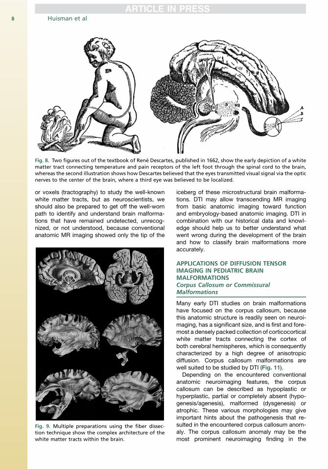
Fig. 8. Two figures out of the textbook of Rene Descartes, published in 1662, show the early depiction of a whitematter tract connecting temperature and pain receptors of the left foot through the spinal cord to the brain,whereas the second illustration shows how Descartes believed that the eyes transmitted visual signal via the opticnerves to the center of the brain, where a third eye was believed to be localized.
Huisman et al8
or voxels (tractography) to study the well-knownwhite matter tracts, but as neuroscientists, weshould also be prepared to get off the well-wornpath to identify and understand brain malforma-tions that have remained undetected, unrecog-nized, or not understood, because conventionalanatomic MR imaging showed only the tip of the
Fig. 9. Multiple preparations using the fiber dissec-tion technique show the complex architecture of thewhite matter tracts within the brain.
iceberg of these microstructural brain malforma-tions. DTI may allow transcending MR imagingfrom basic anatomic imaging toward functionand embryology-based anatomic imaging. DTI incombination with our historical data and knowl-edge should help us to better understand whatwent wrong during the development of the brainand how to classify brain malformations moreaccurately.
APPLICATIONS OF DIFFUSION TENSORIMAGING IN PEDIATRIC BRAINMALFORMATIONSCorpus Callosum or CommissuralMalformations
Many early DTI studies on brain malformationshave focused on the corpus callosum, becausethis anatomic structure is readily seen on neuroi-maging, has a significant size, and is first and fore-most a densely packed collection of corticocorticalwhite matter tracts connecting the cortex ofboth cerebral hemispheres, which is consequentlycharacterized by a high degree of anisotropicdiffusion. Corpus callosum malformations arewell suited to be studied by DTI (Fig. 11).Depending on the encountered conventional
anatomic neuroimaging features, the corpuscallosum can be described as hypoplastic orhyperplastic, partial or completely absent (hypo-genesis/agenesis), malformed (dysgenesis) oratrophic. These various morphologies may giveimportant hints about the pathogenesis that re-sulted in the encountered corpus callosum anom-aly. The corpus callosum anomaly may be themost prominent neuroimaging finding in the
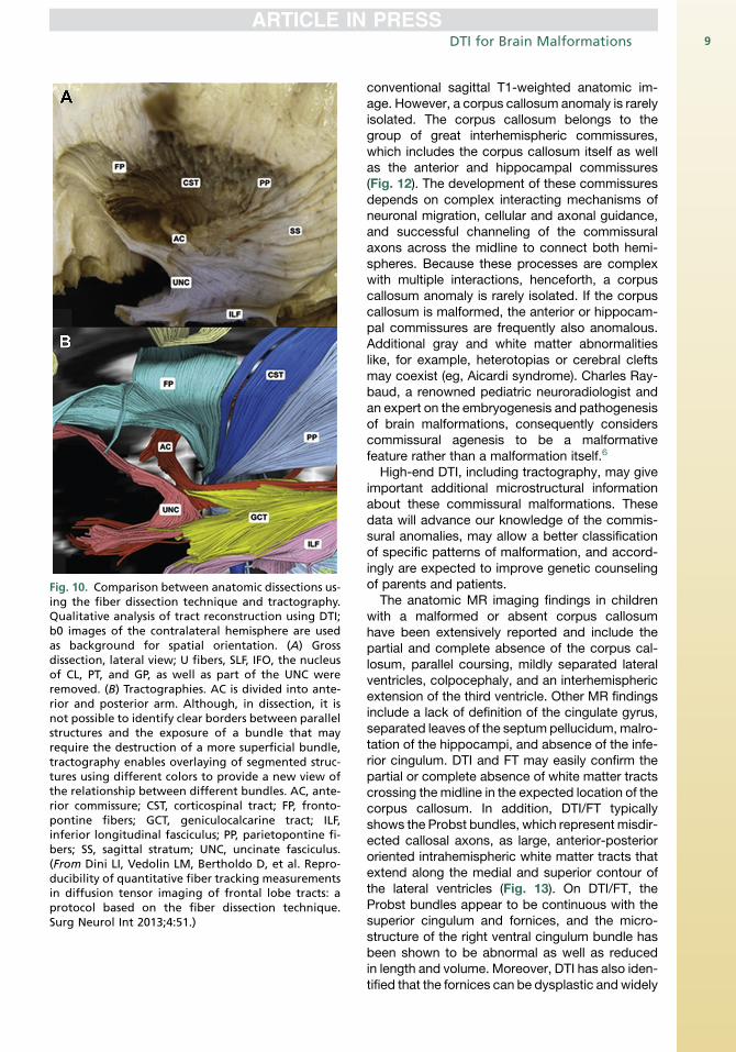
Fig. 10. Comparison between anatomic dissections us-ing the fiber dissection technique and tractography.Qualitative analysis of tract reconstruction using DTI;b0 images of the contralateral hemisphere are usedas background for spatial orientation. (A) Grossdissection, lateral view; U fibers, SLF, IFO, the nucleusof CL, PT, and GP, as well as part of the UNC wereremoved. (B) Tractographies. AC is divided into ante-rior and posterior arm. Although, in dissection, it isnot possible to identify clear borders between parallelstructures and the exposure of a bundle that mayrequire the destruction of a more superficial bundle,tractography enables overlaying of segmented struc-tures using different colors to provide a new view ofthe relationship between different bundles. AC, ante-rior commissure; CST, corticospinal tract; FP, fronto-pontine fibers; GCT, geniculocalcarine tract; ILF,inferior longitudinal fasciculus; PP, parietopontine fi-bers; SS, sagittal stratum; UNC, uncinate fasciculus.(From Dini LI, Vedolin LM, Bertholdo D, et al. Repro-ducibility of quantitative fiber tracking measurementsin diffusion tensor imaging of frontal lobe tracts: aprotocol based on the fiber dissection technique.Surg Neurol Int 2013;4:51.)
DTI for Brain Malformations 9
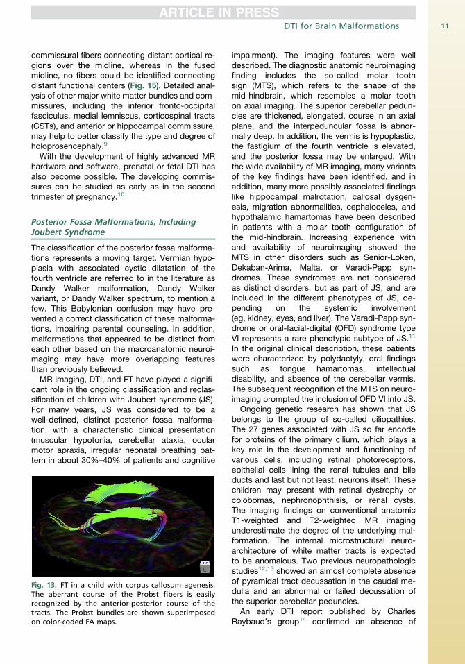
conventional sagittal T1-weighted anatomic im-age. However, a corpus callosum anomaly is rarelyisolated. The corpus callosum belongs to thegroup of great interhemispheric commissures,which includes the corpus callosum itself as wellas the anterior and hippocampal commissures(Fig. 12). The development of these commissuresdepends on complex interacting mechanisms ofneuronal migration, cellular and axonal guidance,and successful channeling of the commissuralaxons across the midline to connect both hemi-spheres. Because these processes are complexwith multiple interactions, henceforth, a corpuscallosum anomaly is rarely isolated. If the corpuscallosum is malformed, the anterior or hippocam-pal commissures are frequently also anomalous.Additional gray and white matter abnormalitieslike, for example, heterotopias or cerebral cleftsmay coexist (eg, Aicardi syndrome). Charles Ray-baud, a renowned pediatric neuroradiologist andan expert on the embryogenesis and pathogenesisof brain malformations, consequently considerscommissural agenesis to be a malformativefeature rather than a malformation itself.6
High-end DTI, including tractography, may giveimportant additional microstructural informationabout these commissural malformations. Thesedata will advance our knowledge of the commis-sural anomalies, may allow a better classificationof specific patterns of malformation, and accord-ingly are expected to improve genetic counselingof parents and patients.
The anatomic MR imaging findings in childrenwith a malformed or absent corpus callosumhave been extensively reported and include thepartial and complete absence of the corpus cal-losum, parallel coursing, mildly separated lateralventricles, colpocephaly, and an interhemisphericextension of the third ventricle. Other MR findingsinclude a lack of definition of the cingulate gyrus,separated leaves of the septum pellucidum, malro-tation of the hippocampi, and absence of the infe-rior cingulum. DTI and FT may easily confirm thepartial or complete absence of white matter tractscrossing the midline in the expected location of thecorpus callosum. In addition, DTI/FT typicallyshows the Probst bundles, which represent misdir-ected callosal axons, as large, anterior-posteriororiented intrahemispheric white matter tracts thatextend along the medial and superior contour ofthe lateral ventricles (Fig. 13). On DTI/FT, theProbst bundles appear to be continuous with thesuperior cingulum and fornices, and the micro-structure of the right ventral cingulum bundle hasbeen shown to be abnormal as well as reducedin length and volume. Moreover, DTI has also iden-tified that the fornices can be dysplastic and widely
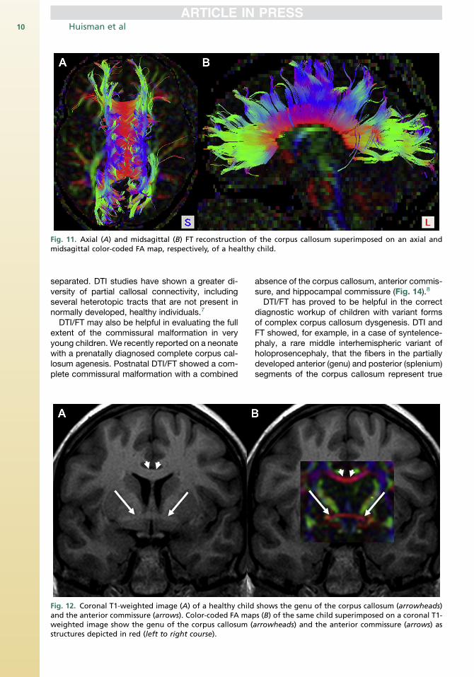
Fig. 11. Axial (A) and midsagittal (B) FT reconstruction of the corpus callosum superimposed on an axial andmidsagittal color-coded FA map, respectively, of a healthy child.
Huisman et al10
separated. DTI studies have shown a greater di-versity of partial callosal connectivity, includingseveral heterotopic tracts that are not present innormally developed, healthy individuals.7
DTI/FT may also be helpful in evaluating the fullextent of the commissural malformation in veryyoung children. We recently reported on a neonatewith a prenatally diagnosed complete corpus cal-losum agenesis. Postnatal DTI/FT showed a com-plete commissural malformation with a combined
Fig. 12. Coronal T1-weighted image (A) of a healthy childand the anterior commissure (arrows). Color-coded FA mapweighted image show the genu of the corpus callosum (structures depicted in red (left to right course).
absence of the corpus callosum, anterior commis-sure, and hippocampal commissure (Fig. 14).8
DTI/FT has proved to be helpful in the correctdiagnostic workup of children with variant formsof complex corpus callosum dysgenesis. DTI andFT showed, for example, in a case of syntelence-phaly, a rare middle interhemispheric variant ofholoprosencephaly, that the fibers in the partiallydeveloped anterior (genu) and posterior (splenium)segments of the corpus callosum represent true
shows the genu of the corpus callosum (arrowheads)s (B) of the same child superimposed on a coronal T1-arrowheads) and the anterior commissure (arrows) as
DTI for Brain Malformations 11
commissural fibers connecting distant cortical re-gions over the midline, whereas in the fusedmidline, no fibers could be identified connectingdistant functional centers (Fig. 15). Detailed anal-ysis of other major white matter bundles and com-missures, including the inferior fronto-occipitalfasciculus, medial lemniscus, corticospinal tracts(CSTs), and anterior or hippocampal commissure,may help to better classify the type and degree ofholoprosencephaly.9
With the development of highly advanced MRhardware and software, prenatal or fetal DTI hasalso become possible. The developing commis-sures can be studied as early as in the secondtrimester of pregnancy.10
Posterior Fossa Malformations, IncludingJoubert Syndrome
The classification of the posterior fossa malforma-tions represents a moving target. Vermian hypo-plasia with associated cystic dilatation of thefourth ventricle are referred to in the literature asDandy Walker malformation, Dandy Walkervariant, or Dandy Walker spectrum, to mention afew. This Babylonian confusion may have pre-vented a correct classification of these malforma-tions, impairing parental counseling. In addition,malformations that appeared to be distinct fromeach other based on the macroanatomic neuroi-maging may have more overlapping featuresthan previously believed.
MR imaging, DTI, and FT have played a signifi-cant role in the ongoing classification and reclas-sification of children with Joubert syndrome (JS).For many years, JS was considered to be awell-defined, distinct posterior fossa malforma-tion, with a characteristic clinical presentation(muscular hypotonia, cerebellar ataxia, ocularmotor apraxia, irregular neonatal breathing pat-tern in about 30%–40% of patients and cognitive
Fig. 13. FT in a child with corpus callosum agenesis.The aberrant course of the Probst fibers is easilyrecognized by the anterior-posterior course of thetracts. The Probst bundles are shown superimposedon color-coded FA maps.
impairment). The imaging features were welldescribed. The diagnostic anatomic neuroimagingfinding includes the so-called molar toothsign (MTS), which refers to the shape of themid-hindbrain, which resembles a molar toothon axial imaging. The superior cerebellar pedun-cles are thickened, elongated, course in an axialplane, and the interpeduncular fossa is abnor-mally deep. In addition, the vermis is hypoplastic,the fastigium of the fourth ventricle is elevated,and the posterior fossa may be enlarged. Withthe wide availability of MR imaging, many variantsof the key findings have been identified, and inaddition, many more possibly associated findingslike hippocampal malrotation, callosal dysgen-esis, migration abnormalities, cephaloceles, andhypothalamic hamartomas have been describedin patients with a molar tooth configuration ofthe mid-hindbrain. Increasing experience withand availability of neuroimaging showed theMTS in other disorders such as Senior-Loken,Dekaban-Arima, Malta, or Varadi-Papp syn-dromes. These syndromes are not consideredas distinct disorders, but as part of JS, and areincluded in the different phenotypes of JS, de-pending on the systemic involvement(eg, kidney, eyes, and liver). The Varadi-Papp syn-drome or oral-facial-digital (OFD) syndrome typeVI represents a rare phenotypic subtype of JS.11
In the original clinical description, these patientswere characterized by polydactyly, oral findingssuch as tongue hamartomas, intellectualdisability, and absence of the cerebellar vermis.The subsequent recognition of the MTS on neuro-imaging prompted the inclusion of OFD VI into JS.
Ongoing genetic research has shown that JSbelongs to the group of so-called ciliopathies.The 27 genes associated with JS so far encodefor proteins of the primary cilium, which plays akey role in the development and functioning ofvarious cells, including retinal photoreceptors,epithelial cells lining the renal tubules and bileducts and last but not least, neurons itself. Thesechildren may present with retinal dystrophy orcolobomas, nephronophthisis, or renal cysts.The imaging findings on conventional anatomicT1-weighted and T2-weighted MR imagingunderestimate the degree of the underlying mal-formation. The internal microstructural neuro-architecture of white matter tracts is expectedto be anomalous. Two previous neuropathologicstudies12,13 showed an almost complete absenceof pyramidal tract decussation in the caudal me-dulla and an abnormal or failed decussation ofthe superior cerebellar peduncles.
An early DTI report published by CharlesRaybaud’s group14 confirmed an absence of
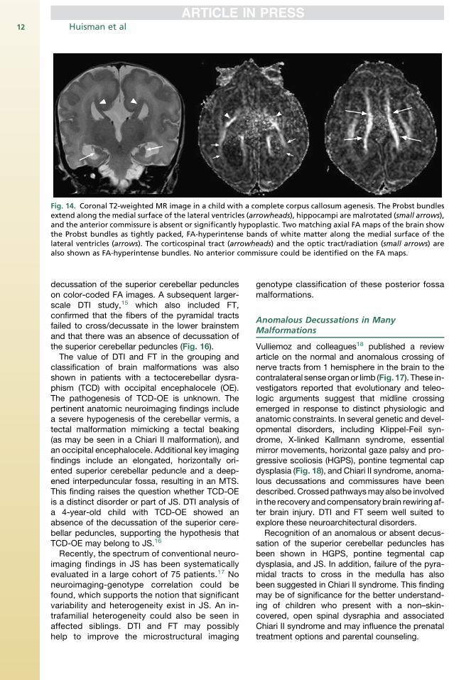
Fig. 14. Coronal T2-weighted MR image in a child with a complete corpus callosum agenesis. The Probst bundlesextend along the medial surface of the lateral ventricles (arrowheads), hippocampi are malrotated (small arrows),and the anterior commissure is absent or significantly hypoplastic. Two matching axial FA maps of the brain showthe Probst bundles as tightly packed, FA-hyperintense bands of white matter along the medial surface of thelateral ventricles (arrows). The corticospinal tract (arrowheads) and the optic tract/radiation (small arrows) arealso shown as FA-hyperintense bundles. No anterior commissure could be identified on the FA maps.
Huisman et al12
decussation of the superior cerebellar peduncleson color-coded FA images. A subsequent larger-scale DTI study,15 which also included FT,confirmed that the fibers of the pyramidal tractsfailed to cross/decussate in the lower brainstemand that there was an absence of decussation ofthe superior cerebellar peduncles (Fig. 16).The value of DTI and FT in the grouping and
classification of brain malformations was alsoshown in patients with a tectocerebellar dysra-phism (TCD) with occipital encephalocele (OE).The pathogenesis of TCD-OE is unknown. Thepertinent anatomic neuroimaging findings includea severe hypogenesis of the cerebellar vermis, atectal malformation mimicking a tectal beaking(as may be seen in a Chiari II malformation), andan occipital encephalocele. Additional key imagingfindings include an elongated, horizontally ori-ented superior cerebellar peduncle and a deep-ened interpeduncular fossa, resulting in an MTS.This finding raises the question whether TCD-OEis a distinct disorder or part of JS. DTI analysis ofa 4-year-old child with TCD-OE showed anabsence of the decussation of the superior cere-bellar peduncles, supporting the hypothesis thatTCD-OE may belong to JS.16
Recently, the spectrum of conventional neuro-imaging findings in JS has been systematicallyevaluated in a large cohort of 75 patients.17 Noneuroimaging-genotype correlation could befound, which supports the notion that significantvariability and heterogeneity exist in JS. An in-trafamilial heterogeneity could also be seen inaffected siblings. DTI and FT may possiblyhelp to improve the microstructural imaging
genotype classification of these posterior fossamalformations.
Anomalous Decussations in ManyMalformations
Vulliemoz and colleagues18 published a reviewarticle on the normal and anomalous crossing ofnerve tracts from 1 hemisphere in the brain to thecontralateral senseorganor limb (Fig. 17). These in-vestigators reported that evolutionary and teleo-logic arguments suggest that midline crossingemerged in response to distinct physiologic andanatomic constraints. In several genetic and devel-opmental disorders, including Klippel-Feil syn-drome, X-linked Kallmann syndrome, essentialmirror movements, horizontal gaze palsy and pro-gressive scoliosis (HGPS), pontine tegmental capdysplasia (Fig. 18), and Chiari II syndrome, anoma-lous decussations and commissures have beendescribed.Crossedpathwaysmayalso be involvedin the recovery and compensatory brain rewiring af-ter brain injury. DTI and FT seem well suited toexplore these neuroarchitectural disorders.Recognition of an anomalous or absent decus-
sation of the superior cerebellar peduncles hasbeen shown in HGPS, pontine tegmental capdysplasia, and JS. In addition, failure of the pyra-midal tracts to cross in the medulla has alsobeen suggested in Chiari II syndrome. This findingmay be of significance for the better understand-ing of children who present with a non–skin-covered, open spinal dysraphia and associatedChiari II syndrome and may influence the prenataltreatment options and parental counseling.
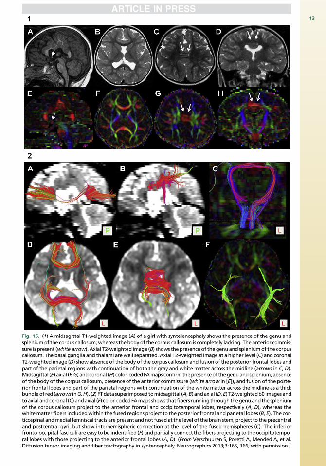
Fig. 15. (1) A midsagittal T1-weighted image (A) of a girl with syntelencephaly shows the presence of the genu andspleniumof the corpus callosum,whereas thebody of the corpus callosum is completely lacking. Theanterior commis-sure is present (white arrow). Axial T2-weighted image (B) shows the presenceof thegenuand spleniumof the corpuscallosum. The basal ganglia and thalami arewell separated. Axial T2-weighted image at a higher level (C) and coronalT2-weighted image (D) show absence of the bodyof the corpus callosum and fusion of theposterior frontal lobes andpart of the parietal regions with continuation of both the gray and white matter across the midline (arrows in C, D).Midsagittal (E) axial (F,G) andcoronal (H) color-codedFAmapsconfirmthepresenceofthegenuandsplenium,absenceof the body of the corpus callosum, presence of the anterior commissure (white arrow in [E]), and fusion of the poste-rior frontal lobes and part of the parietal regions with continuation of the white matter across the midline as a thickbundleof red(arrows inG,H). (2) FTdata superimposedtomidsagittal (A,B) andaxial (D,E) T2-weightedb0 imagesandtoaxialandcoronal (C) andaxial (F) color-codedFAmapsshows that fibers running throughthegenuandthe spleniumof the corpus callosum project to the anterior frontal and occipitotemporal lobes, respectively (A, D), whereas thewhitematter fibers includedwithin the fused regions project to theposterior frontal and parietal lobes (B, E). The cor-ticospinal andmedial lemniscal tracts are present andnot fusedat the level of thebrain stem, project to theprecentraland postcentral gyri, but show interhemispheric connection at the level of the fused hemispheres (C). The inferiorfronto-occipital fasciculi are easy tobe indentified (F) andpartially connect the fibers projecting to theoccipitotempo-ral lobes with those projecting to the anterior frontal lobes (A, D). (From Verschuuren S, Poretti A, Meoded A, et al.Diffusion tensor imaging and fiber tractography in syntencephaly. Neurographics 2013;3:165, 166; with permission.)
13
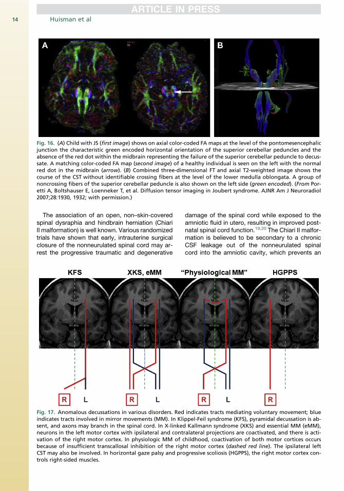
Fig. 16. (A) Child with JS (first image) shows on axial color-coded FA maps at the level of the pontomesencephalicjunction the characteristic green encoded horizontal orientation of the superior cerebellar peduncles and theabsence of the red dot within the midbrain representing the failure of the superior cerebellar peduncle to decus-sate. A matching color-coded FA map (second image) of a healthy individual is seen on the left with the normalred dot in the midbrain (arrow). (B) Combined three-dimensional FT and axial T2-weighted image shows thecourse of the CST without identifiable crossing fibers at the level of the lower medulla oblongata. A group ofnoncrossing fibers of the superior cerebellar peduncle is also shown on the left side (green encoded). (From Por-etti A, Boltshauser E, Loenneker T, et al. Diffusion tensor imaging in Joubert syndrome. AJNR Am J Neuroradiol2007;28:1930, 1932; with permission.)
Huisman et al14
The association of an open, non–skin-coveredspinal dysraphia and hindbrain herniation (ChiariII malformation) is well known. Various randomizedtrials have shown that early, intrauterine surgicalclosure of the nonneurulated spinal cord may ar-rest the progressive traumatic and degenerative
Fig. 17. Anomalous decussations in various disorders. Redindicates tracts involved in mirror movements (MM). In Klisent, and axons may branch in the spinal cord. In X-linkedneurons in the left motor cortex with ipsilateral and contvation of the right motor cortex. In physiologic MM of cbecause of insufficient transcallosal inhibition of the righCST may also be involved. In horizontal gaze palsy and protrols right-sided muscles.
damage of the spinal cord while exposed to theamniotic fluid in utero, resulting in improved post-natal spinal cord function.19,20 The Chiari II malfor-mation is believed to be secondary to a chronicCSF leakage out of the nonneurulated spinalcord into the amniotic cavity, which prevents an
indicates tracts mediating voluntary movement; blueppel-Feil syndrome (KFS), pyramidal decussation is ab-Kallmann syndrome (XKS) and essential MM (eMM),
ralateral projections are coactivated, and there is acti-hildhood, coactivation of both motor cortices occurst motor cortex (dashed red line). The ipsilateral leftgressive scoliosis (HGPPS), the right motor cortex con-
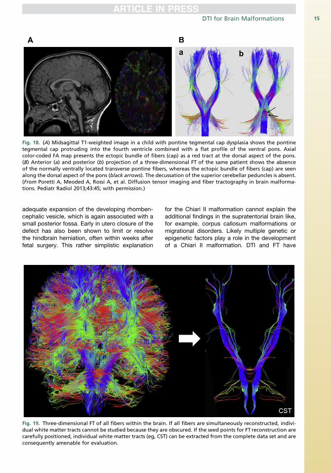
Fig. 18. (A) Midsagittal T1-weighted image in a child with pontine tegmental cap dysplasia shows the pontinetegmental cap protruding into the fourth ventricle combined with a flat profile of the ventral pons. Axialcolor-coded FA map presents the ectopic bundle of fibers (cap) as a red tract at the dorsal aspect of the pons.(B) Anterior (a) and posterior (b) projection of a three-dimensional FT of the same patient shows the absenceof the normally ventrally located transverse pontine fibers, whereas the ectopic bundle of fibers (cap) are seenalong the dorsal aspect of the pons (black arrows). The decussation of the superior cerebellar peduncles is absent.(From Poretti A, Meoded A, Rossi A, et al. Diffusion tensor imaging and fiber tractography in brain malforma-tions. Pediatr Radiol 2013;43:45; with permission.)
DTI for Brain Malformations 15
adequate expansion of the developing rhomben-cephalic vesicle, which is again associated with asmall posterior fossa. Early in utero closure of thedefect has also been shown to limit or resolvethe hindbrain herniation, often within weeks afterfetal surgery. This rather simplistic explanation
Fig. 19. Three-dimensional FT of all fibers within the braindual white matter tracts cannot be studied because they arcarefully positioned, individual white matter tracts (eg, CSTconsequently amenable for evaluation.
for the Chiari II malformation cannot explain theadditional findings in the supratentorial brain like,for example, corpus callosum malformations ormigrational disorders. Likely multiple genetic orepigenetic factors play a role in the developmentof a Chiari II malformation. DTI and FT have
. If all fibers are simultaneously reconstructed, indivi-e obscured. If the seed points for FT reconstruction are) can be extracted from the complete data set and are
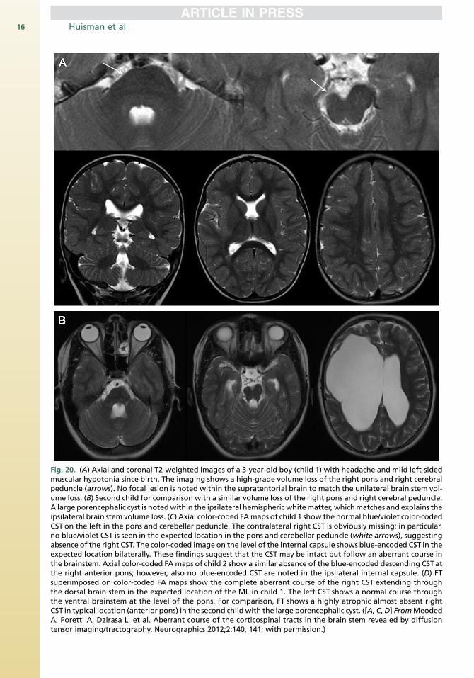
Fig. 20. (A) Axial and coronal T2-weighted images of a 3-year-old boy (child 1) with headache and mild left-sidedmuscular hypotonia since birth. The imaging shows a high-grade volume loss of the right pons and right cerebralpeduncle (arrows). No focal lesion is noted within the supratentorial brain to match the unilateral brain stem vol-ume loss. (B) Second child for comparison with a similar volume loss of the right pons and right cerebral peduncle.A large porencephalic cyst is notedwithin the ipsilateral hemispheric whitematter, whichmatches and explains theipsilateral brain stem volume loss. (C) Axial color-coded FAmaps of child 1 show the normal blue/violet color-codedCST on the left in the pons and cerebellar peduncle. The contralateral right CST is obviously missing; in particular,no blue/violet CST is seen in the expected location in the pons and cerebellar peduncle (white arrows), suggestingabsence of the right CST. The color-coded image on the level of the internal capsule shows blue-encoded CST in theexpected location bilaterally. These findings suggest that the CST may be intact but follow an aberrant course inthe brainstem. Axial color-coded FA maps of child 2 show a similar absence of the blue-encoded descending CSTatthe right anterior pons; however, also no blue-encoded CST are noted in the ipsilateral internal capsule. (D) FTsuperimposed on color-coded FA maps show the complete aberrant course of the right CST extending throughthe dorsal brain stem in the expected location of the ML in child 1. The left CST shows a normal course throughthe ventral brainstem at the level of the pons. For comparison, FT shows a highly atrophic almost absent rightCST in typical location (anterior pons) in the second child with the large porencephalic cyst. ([A, C,D] FromMeodedA, Poretti A, Dzirasa L, et al. Aberrant course of the corticospinal tracts in the brain stem revealed by diffusiontensor imaging/tractography. Neurographics 2012;2:140, 141; with permission.)
Huisman et al16
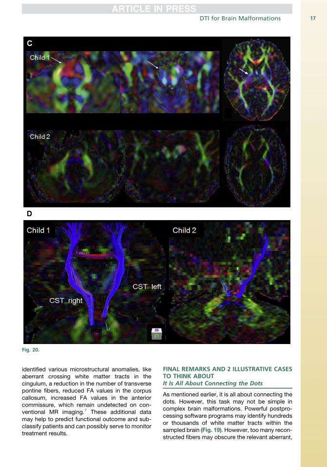
Fig. 20.
DTI for Brain Malformations 17
identified various microstructural anomalies, likeaberrant crossing white matter tracts in thecingulum, a reduction in the number of transversepontine fibers, reduced FA values in the corpuscallosum, increased FA values in the anteriorcommissure, which remain undetected on con-ventional MR imaging.7 These additional datamay help to predict functional outcome and sub-classify patients and can possibly serve to monitortreatment results.
FINAL REMARKS AND 2 ILLUSTRATIVE CASESTO THINK ABOUTIt Is All About Connecting the Dots
As mentioned earlier, it is all about connecting thedots. However, this task may not be simple incomplex brain malformations. Powerful postpro-cessing software programs may identify hundredsor thousands of white matter tracts within thesampled brain (Fig. 19). However, toomany recon-structed fibers may obscure the relevant aberrant,
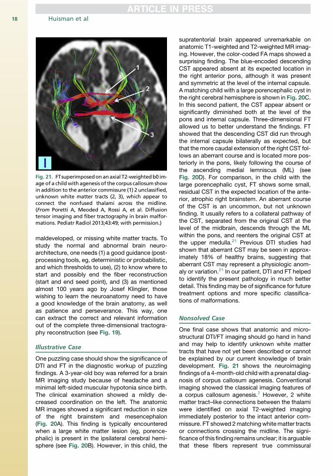
Fig. 21. FTsuperimposedonanaxial T2-weightedb0 im-age of a childwith agenesis of the corpus callosum showin addition to the anterior commissure (1) 2 unclassified,unknown white matter tracts (2, 3), which appear toconnect the nonfused thalami across the midline.(From Poretti A, Meoded A, Rossi A, et al. Diffusiontensor imaging and fiber tractography in brain malfor-mations. Pediatr Radiol 2013;43:49; with permission.)
Huisman et al18
maldeveloped, or missing white matter tracts. Tostudy the normal and abnormal brain neuro-architecture, one needs (1) a good guidance (post-processing tools, eg, deterministic or probabilistic,and which thresholds to use), (2) to know where tostart and possibly end the fiber reconstruction(start and end seed point), and (3) as mentionedalmost 100 years ago by Josef Klingler, thosewishing to learn the neuroanatomy need to havea good knowledge of the brain anatomy, as wellas patience and perseverance. This way, onecan extract the correct and relevant informationout of the complete three-dimensional tractogra-phy reconstruction (see Fig. 19).
Illustrative Case
One puzzling case should show the significance ofDTI and FT in the diagnostic workup of puzzlingfindings. A 3-year-old boy was referred for a brainMR imaging study because of headache and aminimal left-sided muscular hypotonia since birth.The clinical examination showed a mildly de-creased coordination on the left. The anatomicMR images showed a significant reduction in sizeof the right brainstem and mesencephalon(Fig. 20A). This finding is typically encounteredwhen a large white matter lesion (eg, porence-phalic) is present in the ipsilateral cerebral hemi-sphere (see Fig. 20B). However, in this child, the
supratentorial brain appeared unremarkable onanatomic T1-weighted and T2-weighted MR imag-ing. However, the color-coded FA maps showed asurprising finding. The blue-encoded descendingCST appeared absent at its expected location inthe right anterior pons, although it was presentand symmetric at the level of the internal capsule.A matching child with a large porencephalic cyst inthe right cerebral hemisphere is shown in Fig. 20C.In this second patient, the CST appear absent orsignificantly diminished both at the level of thepons and internal capsule. Three-dimensional FTallowed us to better understand the findings. FTshowed that the descending CST did run throughthe internal capsule bilaterally as expected, butthat the more caudal extension of the right CST fol-lows an aberrant course and is located more pos-teriorly in the pons, likely following the course ofthe ascending medial lemniscus (ML) (seeFig. 20D). For comparison, in the child with thelarge porencephalic cyst, FT shows some small,residual CST in the expected location of the ante-rior, atrophic right brainstem. An aberrant courseof the CST is an uncommon, but not unknownfinding. It usually refers to a collateral pathway ofthe CST, separated from the original CST at thelevel of the midbrain, descends through the MLwithin the pons, and reenters the original CST atthe upper medulla.21 Previous DTI studies hadshown that aberrant CST may be seen in approx-imately 18% of healthy brains, suggesting thataberrant CST may represent a physiologic anom-aly or variation.21 In our patient, DTI and FT helpedto identify the present pathology in much betterdetail. This finding may be of significance for futuretreatment options and more specific classifica-tions of malformations.
Nonsolved Case
One final case shows that anatomic and micro-structural DTI/FT imaging should go hand in handand may help to identify unknown white mattertracts that have not yet been described or cannotbe explained by our current knowledge of braindevelopment. Fig. 21 shows the neuroimagingfindings of a 4-month-old childwith aprenatal diag-nosis of corpus callosum agenesis. Conventionalimaging showed the classical imaging features ofa corpus callosum agenesis.7 However, 2 whitematter tract–like connections between the thalamiwere identified on axial T2-weighted imagingimmediately posterior to the intact anterior com-missure. FT showed 2matchingwhitematter tractsor connections crossing the midline. The signi-ficanceof this finding remains unclear; it is arguablethat these fibers represent true commissural
DTI for Brain Malformations 19
fibers, which may represent a confusing artifact;future research may show that this is real.
SUMMARY
DTI and FT allow us to study the microstructure ofthe CNS in vivo and are consequently a valuabletool for the better understanding of the normaland abnormal brain development. The collecteddata will help to better classify malformationsand may give important hints to the genetic basesof the encountered findings. To answer the ques-tion of the title of this article: DTI and FT are helpfulfor brain malformations.
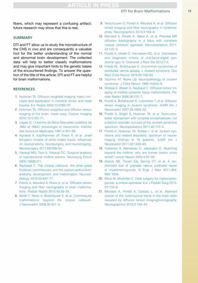
REFERENCES
1. Huisman TA. Diffusion weighted imaging: basic con-
cepts and application in cerebral stroke and head
trauma. Eur Radiol 2003;13:2283–97.
2. Huisman TA. Diffusion weighted and diffusion tensor
imaging of the brain, made easy. Cancer Imaging
2010;10:S163–71.
3. Legee G. L’Homme de Rene Descartes (editions de
1662 et 1664): physiologie et macansime. Histoire
des Sciences Medicales 1987;4:381–98.
4. Agrawal A, Kapfhammer JP, Kress A, et al. Josef
Klingler’s models of white matter tracts: influences
on neuroanatomy, neurosurgery, and neuroimaging.
Neurosurgery 2011;69:238–54.
5. Yasargil MG, Ture U, Yasargil DC. Surgical anatomy
of supratentorial midline lesions. Neurosurg Focus
2005;18(6B):E1.
6. Raybaud C. The corpus callosum, the other great
forebrain commissures, and the septum pellucidum:
anatomy, development, and malformation. Neurora-
diology 2010;52:447–77.
7. Poretti A, Meoded A, Rossi A, et al. Diffusion tensor
imaging and fiber tractography in brain malforma-
tions. Pediatr Radiol 2013;43:28–54.
8. Smith T, Tekes A, Boltshauser E, et al. Commissural
malformations: beyond the corpus callosum.
J Neuroradiol 2008;35:301–3.
9. Verschuuren S, Poretti A, Meoded A, et al. Diffusion
tensor imaging and fiber tractography in syntence-
phaly. Neurographics 2013;3:164–8.
10. Meoded A, Poretti A, Tekes A, et al. Prenatal MR
diffusion tractography in a fetus with complete
corpus callosum agenesis. Neuropediatrics 2011;
42:122–3.
11. Poretti A, Vitiello G, Hennekam RC, et al. Delineation
and diagnostic criteria of oral-facial-digital syn-
drome type IV. Orphanet J Rare Dis 2012;7:4.
12. Friede RL, Boltshauser E. Uncommon syndromes of
cerebellar vermis aplasia. I: Joubert syndrome. Dev
Med Child Neurol 1978;20:758–63.
13. Yachmis AT, Rorke LB. Neuropathology of Joubert
syndrome. J Child Neurol 1999;14:655–9.
14. Widjaja E, Blaser S, Raybaud C. Diffusion tensor im-
aging of midline posterior fossa malformations. Pe-
diatr Radiol 2006;36:510–7.
15. Poretti A, Boltshauser E, Loenneker T, et al. Diffusion
tensor imaging in Joubert syndrome. AJNR Am J
Neuroradiol 2007;28:1929–33.
16. Poretti A, Singhi S, Huisman TA, et al. Tecto-cere-
bellar dysraphism with occipital encephalocele: not
a distinct disorder, but part of the Joubert syndrome
spectrum. Neuropediatrics 2011;42:170–4.
17. Poretti A, Huisman TA, Scheer I, et al. Joubert syn-
drome and related disorders: spectrum of neuroi-
maging findings in 75 patients. AJNR Am J
Neuroradiol 2011;32:1459–63.
18. Vulliemoz S, Raineteau O, Jabaudon D. Reaching
beyond the midline: why are human brains cross
wired? Lancet Neurol 2005;4:87–99.
19. Adzick NS, Thoam EA, Spong CY, et al. A ran-
domized trial of prenatal versus postnatal repair
of myelomeningocele. N Engl J Med 2011;364:
993–1004.
20. Meuli M, Moehrlen E. Fetal surgery for myelomenin-
gocele: a critical appraisal. Eur J Pediatr Surg 2013;
23:103–9.
21. Meoded A, Poretti A, Dzirasa L, et al. Aberrant
course of the corticospinal tracts in the brain stem
revealed by diffusion tensor imaging/tractography.
Neurographics 2012;2:139–43.
Related Documents