materials Article Histologic and Histomorphometric Evaluation of a New Bioactive Liquid BBL on Implant Surface: A Preclinical Study in Foxhound Dogs Eduard Ferrés-Amat 1,† , Ashraf Al Madhoun 2,† , Elvira Ferrés-Amat 1,3 , Saddam Al Demour 4 , Mera A. Ababneh 5 , Eduard Ferrés-Padró 1,6 , Carles Marti 6,7 , Neus Carrio 3 , Miguel Barajas 6,8 and Maher Atari 6,9, * Citation: Ferrés-Amat, E.; Al Madhoun, A.; Ferrés-Amat, E.; Al Demour, S.; Ababneh, M.A.; Ferrés-Padró, E.; Marti, C.; Carrio, N.; Barajas, M.; Atari, M. Histologic and Histomorphometric Evaluation of a New Bioactive Liquid BBL on Implant Surface: A Preclinical Study in Foxhound Dogs. Materials 2021, 14, 6217. https://doi.org/10.3390/ ma14206217 Academic Editors: Gianmario Schierano and Giuliana Muzio Received: 15 September 2021 Accepted: 16 October 2021 Published: 19 October 2021 Publisher’s Note: MDPI stays neutral with regard to jurisdictional claims in published maps and institutional affil- iations. Copyright: © 2021 by the authors. Licensee MDPI, Basel, Switzerland. This article is an open access article distributed under the terms and conditions of the Creative Commons Attribution (CC BY) license (https:// creativecommons.org/licenses/by/ 4.0/). 1 Oral and Maxillofacial Surgery Service, Hospital HM Nens, HM Hospitales, 08009 Barcelona, Spain; [email protected] (E.F.-A.); [email protected] (E.F.-A.); [email protected] (E.F.-P.) 2 Department of Animal and Imaging Core Facilities, Dasman Diabetes Institute, Dasman 15462, Kuwait; [email protected] 3 Oral and Maxillofacial Surgery Department, Universitat Internacional de Catalunya, St Josep Trueta s/n, Sant Cugat del Vallès, 08195 Barcelona, Spain; [email protected] 4 Department of Special Surgery/Division of Urology, School of Medicine, The University of Jordan, Amman 11942, Jordan; [email protected] 5 Department of Clinical Pharmacy, Faculty of Pharmacy, Jordan University of Science and Technology, Irbid 22110, Jordan; [email protected] 6 Biointelligent Technology Systems SL, Diputaccion 316, 3D, 08009 Barcelona, Spain; [email protected] (C.M.); [email protected] (M.B.) 7 Oral and Maxillofacial Surgery Department, Hospital Clinic de Barcelona, 08036 Barcelona, Spain 8 Biochemistry Area, Department of Health Science, Public University of Navarre, 31008 Pamplona, Spain 9 Ziacom Medical SL, C. Buhos, 2, 28320 Madrid, Spain * Correspondence: [email protected] † Eduard Ferrés-Amat and Ashraf Al Madhoun should be considered joint first author. Abstract: Background: Bioactive chemical surface modifications improve the wettability and os- seointegration properties of titanium implants in both animals and humans. The objective of this animal study was to investigate and compare the bioreactivity characteristics of titanium implants (BLT) pre-treated with a novel bone bioactive liquid (BBL) and the commercially available BLT-SLA active. Methods: Forty BLT-SLA titanium implants were placed in in four foxhound dogs. Animals were divided into two groups (n = 20): test (BLT-SLA pre-treated with BBL) and control (BLT-SLA active) implants. The implants were inserted in the post extraction sockets. After 8 and 12 weeks, the animals were sacrificed, and mandibles were extracted, containing the implants and the surrounding soft and hard tissues. Bone-to-implant contact (BIC), inter-thread bone area percentage (ITBA), soft tissue, and crestal bone loss were evaluated by histology and histomorphometry. Results: All animals were healthy with no implant loss or inflammation symptoms. All implants were clinically and histologically osseo-integrated. Relative to control groups, test implants demonstrated a significant 1.5- and 1.7-fold increase in BIC and ITBA values, respectively, at both assessment intervals. Crestal bone loss was also significantly reduced in the test group, as compared with controls, at week 8 in both the buccal crests (0.47 ± 0.32 vs 0.98 ± 0.51 mm, p < 0.05) and lingual crests (0.39* ± 0.3 vs. 0.89 ± 0.41 mm, p < 0.05). At week 12, a pronounced crestal bone loss improvement was observed in the test group (buccal, 0.41 ± 0.29 mm and lingual, 0.54 ± 0.23 mm). Tissue thickness showed com- parable values at both the buccal and lingual regions and was significantly improved in the studied groups (0.82–0.92 mm vs. 33–48 mm in the control group). Conclusions: Relative to the commercially available BLT-SLA active implants, BLT-SLA pre-treated with BBL showed improved histological and histomorphometric characteristics indicating a reduced titanium surface roughness and improved wettability, promoting healing and soft and hard tissue regeneration at the implant site. Keywords: BLT-SLA active; BBL; dental implant; osseo integration dental implantation Materials 2021, 14, 6217. https://doi.org/10.3390/ma14206217 https://www.mdpi.com/journal/materials

Welcome message from author
This document is posted to help you gain knowledge. Please leave a comment to let me know what you think about it! Share it to your friends and learn new things together.
Transcript

materials
Article
Histologic and Histomorphometric Evaluation of a NewBioactive Liquid BBL on Implant Surface: A Preclinical Studyin Foxhound Dogs
Eduard Ferrés-Amat 1,†, Ashraf Al Madhoun 2,† , Elvira Ferrés-Amat 1,3 , Saddam Al Demour 4,Mera A. Ababneh 5, Eduard Ferrés-Padró 1,6 , Carles Marti 6,7, Neus Carrio 3, Miguel Barajas 6,8
and Maher Atari 6,9,*
�����������������
Citation: Ferrés-Amat, E.;
Al Madhoun, A.; Ferrés-Amat, E.;
Al Demour, S.; Ababneh, M.A.;
Ferrés-Padró, E.; Marti, C.; Carrio, N.;
Barajas, M.; Atari, M. Histologic and
Histomorphometric Evaluation of a
New Bioactive Liquid BBL on
Implant Surface: A Preclinical Study
in Foxhound Dogs. Materials 2021, 14,
6217. https://doi.org/10.3390/
ma14206217
Academic Editors: Gianmario
Schierano and Giuliana Muzio
Received: 15 September 2021
Accepted: 16 October 2021
Published: 19 October 2021
Publisher’s Note: MDPI stays neutral
with regard to jurisdictional claims in
published maps and institutional affil-
iations.
Copyright: © 2021 by the authors.
Licensee MDPI, Basel, Switzerland.
This article is an open access article
distributed under the terms and
conditions of the Creative Commons
Attribution (CC BY) license (https://
creativecommons.org/licenses/by/
4.0/).
1 Oral and Maxillofacial Surgery Service, Hospital HM Nens, HM Hospitales, 08009 Barcelona, Spain;[email protected] (E.F.-A.); [email protected] (E.F.-A.);[email protected] (E.F.-P.)
2 Department of Animal and Imaging Core Facilities, Dasman Diabetes Institute, Dasman 15462, Kuwait;[email protected]
3 Oral and Maxillofacial Surgery Department, Universitat Internacional de Catalunya, St Josep Trueta s/n,Sant Cugat del Vallès, 08195 Barcelona, Spain; [email protected]
4 Department of Special Surgery/Division of Urology, School of Medicine, The University of Jordan,Amman 11942, Jordan; [email protected]
5 Department of Clinical Pharmacy, Faculty of Pharmacy, Jordan University of Science and Technology,Irbid 22110, Jordan; [email protected]
6 Biointelligent Technology Systems SL, Diputaccion 316, 3D, 08009 Barcelona, Spain;[email protected] (C.M.); [email protected] (M.B.)
7 Oral and Maxillofacial Surgery Department, Hospital Clinic de Barcelona, 08036 Barcelona, Spain8 Biochemistry Area, Department of Health Science, Public University of Navarre, 31008 Pamplona, Spain9 Ziacom Medical SL, C. Buhos, 2, 28320 Madrid, Spain* Correspondence: [email protected]† Eduard Ferrés-Amat and Ashraf Al Madhoun should be considered joint first author.
Abstract: Background: Bioactive chemical surface modifications improve the wettability and os-seointegration properties of titanium implants in both animals and humans. The objective of thisanimal study was to investigate and compare the bioreactivity characteristics of titanium implants(BLT) pre-treated with a novel bone bioactive liquid (BBL) and the commercially available BLT-SLAactive. Methods: Forty BLT-SLA titanium implants were placed in in four foxhound dogs. Animalswere divided into two groups (n = 20): test (BLT-SLA pre-treated with BBL) and control (BLT-SLAactive) implants. The implants were inserted in the post extraction sockets. After 8 and 12 weeks, theanimals were sacrificed, and mandibles were extracted, containing the implants and the surroundingsoft and hard tissues. Bone-to-implant contact (BIC), inter-thread bone area percentage (ITBA), softtissue, and crestal bone loss were evaluated by histology and histomorphometry. Results: All animalswere healthy with no implant loss or inflammation symptoms. All implants were clinically andhistologically osseo-integrated. Relative to control groups, test implants demonstrated a significant1.5- and 1.7-fold increase in BIC and ITBA values, respectively, at both assessment intervals. Crestalbone loss was also significantly reduced in the test group, as compared with controls, at week 8 inboth the buccal crests (0.47 ± 0.32 vs 0.98 ± 0.51 mm, p < 0.05) and lingual crests (0.39* ± 0.3 vs.0.89 ± 0.41 mm, p < 0.05). At week 12, a pronounced crestal bone loss improvement was observed inthe test group (buccal, 0.41 ± 0.29 mm and lingual, 0.54 ± 0.23 mm). Tissue thickness showed com-parable values at both the buccal and lingual regions and was significantly improved in the studiedgroups (0.82–0.92 mm vs. 33–48 mm in the control group). Conclusions: Relative to the commerciallyavailable BLT-SLA active implants, BLT-SLA pre-treated with BBL showed improved histological andhistomorphometric characteristics indicating a reduced titanium surface roughness and improvedwettability, promoting healing and soft and hard tissue regeneration at the implant site.
Keywords: BLT-SLA active; BBL; dental implant; osseo integration dental implantation
Materials 2021, 14, 6217. https://doi.org/10.3390/ma14206217 https://www.mdpi.com/journal/materials
Materials 2021, 14, 6217 2 of 15
1. Introduction
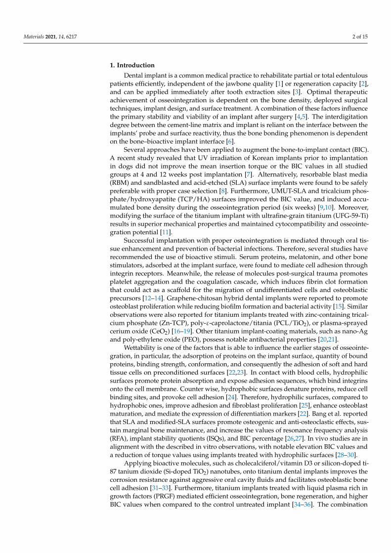
Dental implant is a common medical practice to rehabilitate partial or total edentulouspatients efficiently, independent of the jawbone quality [1] or regeneration capacity [2],and can be applied immediately after tooth extraction sites [3]. Optimal therapeuticachievement of osseointegration is dependent on the bone density, deployed surgicaltechniques, implant design, and surface treatment. A combination of these factors influencethe primary stability and viability of an implant after surgery [4,5]. The interdigitationdegree between the cement-line matrix and implant is reliant on the interface between theimplants’ probe and surface reactivity, thus the bone bonding phenomenon is dependenton the bone–bioactive implant interface [6].
Several approaches have been applied to augment the bone-to-implant contact (BIC).A recent study revealed that UV irradiation of Korean implants prior to implantationin dogs did not improve the mean insertion torque or the BIC values in all studiedgroups at 4 and 12 weeks post implantation [7]. Alternatively, resorbable blast media(RBM) and sandblasted and acid-etched (SLA) surface implants were found to be safelypreferable with proper case selection [8]. Furthermore, UMUT-SLA and tricalcium phos-phate/hydroxyapatite (TCP/HA) surfaces improved the BIC value, and induced accu-mulated bone density during the osseointegration period (six weeks) [9,10]. Moreover,modifying the surface of the titanium implant with ultrafine-grain titanium (UFG-59-Ti)results in superior mechanical properties and maintained cytocompatibility and osseointe-gration potential [11].
Successful implantation with proper osteointegration is mediated through oral tis-sue enhancement and prevention of bacterial infections. Therefore, several studies haverecommended the use of bioactive stimuli. Serum proteins, melatonin, and other bonestimulators, adsorbed at the implant surface, were found to mediate cell adhesion throughintegrin receptors. Meanwhile, the release of molecules post-surgical trauma promotesplatelet aggregation and the coagulation cascade, which induces fibrin clot formationthat could act as a scaffold for the migration of undifferentiated cells and osteoblasticprecursors [12–14]. Graphene-chitosan hybrid dental implants were reported to promoteosteoblast proliferation while reducing biofilm formation and bacterial activity [15]. Similarobservations were also reported for titanium implants treated with zinc-containing trical-cium phosphate (Zn-TCP), poly-ε-caprolactone/titania (PCL/TiO2), or plasma-sprayedcerium oxide (CeO2) [16–19]. Other titanium implant-coating materials, such as nano-Agand poly-ethylene oxide (PEO), possess notable antibacterial properties [20,21].
Wettability is one of the factors that is able to influence the earlier stages of osseointe-gration, in particular, the adsorption of proteins on the implant surface, quantity of boundproteins, binding strength, conformation, and consequently the adhesion of soft and hardtissue cells on preconditioned surfaces [22,23]. In contact with blood cells, hydrophilicsurfaces promote protein absorption and expose adhesion sequences, which bind integrinsonto the cell membrane. Counter wise, hydrophobic surfaces denature proteins, reduce cellbinding sites, and provoke cell adhesion [24]. Therefore, hydrophilic surfaces, compared tohydrophobic ones, improve adhesion and fibroblast proliferation [25], enhance osteoblastmaturation, and mediate the expression of differentiation markers [22]. Bang et al. reportedthat SLA and modified-SLA surfaces promote osteogenic and anti-osteoclastic effects, sus-tain marginal bone maintenance, and increase the values of resonance frequency analysis(RFA), implant stability quotients (ISQs), and BIC percentage [26,27]. In vivo studies are inalignment with the described in vitro observations, with notable elevation BIC values anda reduction of torque values using implants treated with hydrophilic surfaces [28–30].
Applying bioactive molecules, such as cholecalciferol/vitamin D3 or silicon-doped ti-87 tanium dioxide (Si-doped TiO2) nanotubes, onto titanium dental implants improves thecorrosion resistance against aggressive oral cavity fluids and facilitates osteoblastic bonecell adhesion [31–33]. Furthermore, titanium implants treated with liquid plasma rich ingrowth factors (PRGF) mediated efficient osseointegration, bone regeneration, and higherBIC values when compared to the control untreated implant [34–36]. The combination
Materials 2021, 14, 6217 3 of 15
of calcium ion surfaces with PRGF enhanced bone regeneration within two weeks ofimplantation [37,38].
The aim of this study was to compare the crestal bone loss, bone formation, andsoft tissue width at 8- and 12-week assessment intervals for two different SLA implantbioactive surfaces: bone level tapered (BLT) implants with SLA active (Straumann, Basel,Switzerland, control group) and with BLT-SLA-BBL (test group) (BBL, patent # EP353211,US 16/344,322).
2. Material and Methods2.1. Statement of Compliance and Declaration of the Research Ethics
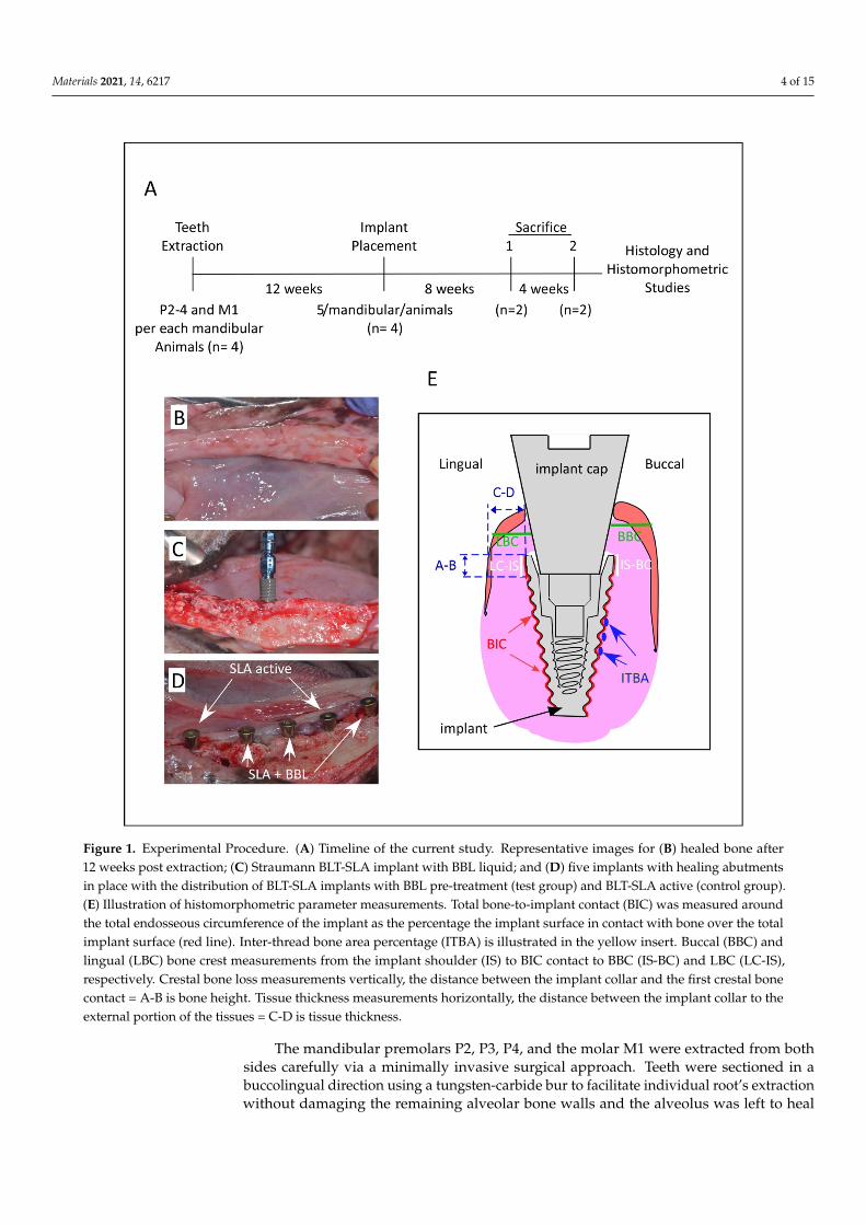
The local Experimental Animal Research Ethics Committee (Comite Etico de Exper-imentacion Animal, CEEA) at The University of Murcia, Murcia, Spain, approved thepresent study protocol using dog animal model article number 34- RD 53/2013, projectnumber A1320141102. In addition, the research project was conducted and adhered tothe guidelines of the Animal Research: Reporting of In vivo Experiments (ARRIVE). Theauthors declare adherence to the proper institutional and national guidelines related tothe care and use of the animals in this study including ethics initiative, design, analysis,and reporting of research using animals. Furthermore, the timeline of the present study isdescribed in Figure 1A. During the entire treatment period, the animals’ behavior, posture,reactivity, and appearance were monitored by a professional veterinary doctor.
2.2. Bone Bioactive Liquid and Implant Design
Here, we describe a new bone bioactive liquid (BBL, patent # EP353211, US 16/344,322)comprising a saline solution (PBS) containing calcium chloride (CaCl2) and magnesiumchloride (MgCl2-6H2O) with a net negative charge that creates an ideal condition forcellular attraction to the trauma area at the bone–implant contact. Apart from that, BBLconsiderably increases the density of hydroxyl groups on the wound implant surface andimproves their hydration significantly. BBL induces surface hydrophilicity, and allowsits’ active ionic interaction with blood plasma and bone progenitor cells on the implant’ssurface. As a result, it leads to coordination and communication between the cells and tobetter contact between all tissues in the wound for “new tissue formation”.
A total of 40 Straumann BLT implants with SLA alone or SLA active (Straumann,Basel, Switzerland) were utilized in this study. The implants had an endosteal diameter of3.3 mm, length of 10 mm, and were designed to be placed at bone level in healed sites asdescribed by the manufacturer. Implants were divided into two groups (20 each). The firstgroup of SLA implants were treated with BBL for 24 h (test group) and the second controlgroup were SLA active implants.
2.3. Tooth Extraction, Surgical Procedure, and Animal Care
Four American foxhound adult male dogs were used in the study, in accordance withthe permit from Experimental Animal Research Ethics Committee, University of Murcia,Murcia, Spain. Animals were anesthetized by femoral quadriceps intramuscular injectionswith a cocktail containing acepromazine (0.12–0.25 mg/kg), buprenorphine (0.01 mg/kg),and medetomidine (35 mg/kg). Animals were then transferred to the operating room.General anesthesia was sustained by a continuous propofol (0.4 mg/kg/min) infusion intothe cephalic vein though an intravenous catheter insertion. In addition, conventional localdental anesthesia, 4% Articaine-hydrochloride and 0.001% epinephrine, was injected at theintraoral surgical sites as described by [39]. The operation was performed by one dentalsurgeon and under the supervision of a veterinary specialist.
Materials 2021, 14, 6217 4 of 15Materials 2021, 14, x FOR PEER REVIEW 4 of 15
Figure 1. Experimental Procedure. (A). Timeline of the current study. Representative images for (B) healed bone after 12 weeks post extraction; (C) Straumann BLT-SLA implant with BBL liquid; and (D) five implants with healing abutments in place with the distribution of BLT-SLA implants with BBL pre-treatment (test group) and BLT-SLA active (control group). (E). Illustration of histomorphometric parameter measurements. Total bone-to-implant contact (BIC) was measured around the total endosseous circumference of the implant as the percentage the implant surface in contact with bone over the total implant surface (red line). Inter-thread bone area percentage (ITBA) is illustrated in the yellow insert. Buccal (BBC) and lingual (LBC) bone crest measurements from the implant shoulder (IS) to BIC contact to BBC (IS-BC) and LBC (LC-IS), respectively. Crestal bone loss measurements vertically, the distance between the implant collar and the first crestal bone contact = A-B is bone height. Tissue thickness measurements horizontally, the distance between the implant collar to the external portion of the tissues = C-D is tissue thickness.
2.3. Tooth Extraction, Surgical Procedure, and Animal Care Four American foxhound adult male dogs were used in the study, in accordance with
the permit from Experimental Animal Research Ethics Committee, University of Murcia, Murcia, Spain. Animals were anesthetized by femoral quadriceps intramuscular injections with a cocktail containing acepromazine (0.12–0.25 mg/kg), buprenorphine (0.01 mg/kg), and medetomidine (35 mg/kg). Animals were then transferred to the operating room.
Figure 1. Experimental Procedure. (A) Timeline of the current study. Representative images for (B) healed bone after12 weeks post extraction; (C) Straumann BLT-SLA implant with BBL liquid; and (D) five implants with healing abutmentsin place with the distribution of BLT-SLA implants with BBL pre-treatment (test group) and BLT-SLA active (control group).(E) Illustration of histomorphometric parameter measurements. Total bone-to-implant contact (BIC) was measured aroundthe total endosseous circumference of the implant as the percentage the implant surface in contact with bone over the totalimplant surface (red line). Inter-thread bone area percentage (ITBA) is illustrated in the yellow insert. Buccal (BBC) andlingual (LBC) bone crest measurements from the implant shoulder (IS) to BIC contact to BBC (IS-BC) and LBC (LC-IS),respectively. Crestal bone loss measurements vertically, the distance between the implant collar and the first crestal bonecontact = A-B is bone height. Tissue thickness measurements horizontally, the distance between the implant collar to theexternal portion of the tissues = C-D is tissue thickness.
The mandibular premolars P2, P3, P4, and the molar M1 were extracted from bothsides carefully via a minimally invasive surgical approach. Teeth were sectioned in abuccolingual direction using a tungsten-carbide bur to facilitate individual root’s extractionwithout damaging the remaining alveolar bone walls and the alveolus was left to heal

Materials 2021, 14, 6217 5 of 15
as previously described [40]. After a healing period of 12 weeks, for implant placement,full-thickness mucoperiosteal flaps were raised in the hole mandible (Figure 1B).
Each dog received 5 implants inserted at the healed sites of each hemi-mandibleusing a motorized hand piece and were subsequently equipped with healing abutments,which were placed 0.5 mm below the occlusal contact with the corresponding antagonisttooth. The implants were arranged into two experimental groups: control group: BLT-SLA active and test group: BLT-SLA +BBL (Figure 1C,D). No graft materials were placedbetween implants and bony plates. Mucoperiosteal flaps were stitched with Silk 4-0 non-absorbable sutures (Lorca Marin, Lorca, Murcia, Spain). To prevent infections, antibioticswere administered for seven days, 500 mg Amoxicillin, and 600 mg Ibuprofen, two andthree times a day, respectively. For a period of seven days, animals were fed a soft diet.To prevent plaque formation, animal received seawater-based mouth wash rinse withSEA4-Encias (Blue Sea Laboratories, Alicante, Spain). Wound healing was monitored dailyto exclude possible clinical complications. Sutures were removed two weeks post-surgery.
2.4. Termination
At a two-time interval, 8 and 12 weeks post-implantation, two dogs were sacrificed bycarotid arteries perfusion with fixative solution containing Pentothal Natrium (Abbot Lab-oratories, Madrid, Spain), 5% glutaraldehyde, and 5% formaldehyde. The hemi-mandibleswere dissected, washed in phosphate-buffered saline (PBA), fixed in 10% formalin (Sigma-Aldrich, Germany), and processed at BioTecnos Laboratories (Santa Maria, Brazil).
2.5. Histologic Processing
Hemi-mandible block sections were dehydrated in a series of ascending concerta-tion’s of graded ethanol up to 100%, and then infiltrated and embedded using Technovit-7200 VLC system (Kulzer Technique, Wehrheim, Germany) in accordance with the manufac-turer’s protocol. After chemical polymerization, samples were cut in buccolingual directionto sections at 2.5 mm and then refined to 35–50 µm in thickness, using IsoMet-1000 highprecision diamond disk (Buehler, Illinois, IL, USA). Slides were preserved for future use.For each implant, two slides were incubated for 1 h. at 25 ◦C in Picrosirius Red Staining(PSR) solution (Polysciences, Warrington, PA, USA).
2.6. Histomorphometric Analysis and Examination
Histomorphometric analysis were performed using Image J software, version 1.38e.Measurements were performed at the region of interest (ROI), defined as the peri-implant,located between 3 and 6 mm below the implant shoulder at the central mesiodistal sections.Measurements were performed for the inter-thread bone area percentage (ITBA%), the boneto implant contact (BIC%), and the crestal bone loss (CBL) for the lingual and buccal boneareas (Figure 1E). Linear measurements were made at the buccal (BBC) and lingual (LBC)bone crest starting from the implant shoulder (IS) to the first point of BIC contact to BBC(IS-BC) or to the LBC (LC-IS). The BIC percentage of native bone was measured throughoutthe implant surface as described in [41] (Figure 1E). The gingival tissue restorations, inthe vertical and lateral direction, were measured at the implant neck throughout thethickness (points A-B) and at the abutment to the bone crest (points C-D) (Figure 1E).Measurements were performed by an expert histology examiner (JLCG). Metric evaluationof the predetermined parameters was carried out using a light microscope (Nikon, Tokyo,Japan) connected to a high-resolution video camera (3CCD, JVC KY-F55B, Yokohama,Japan). After digitizing the phase of each specimen under the light microscope, all thepredetermined distances were measured on images using the program Image Tool version5.02 for Microsoft Windows (UT Health Science Center School of Dentistry, San Antonio,TX, USA).

Materials 2021, 14, 6217 6 of 15
2.7. Statistical Analysis
To test for differences between the paired data within animals, Wilcoxon signed ranktest was used. The associations between the measured outcomes and test were evaluatedusing mixed linear regression models, considering the factor position of the test andthe random animal effect. To evaluate if the measured outcomes correlated with eachother at the time points, the Pearson correlation coefficients were calculated. Means andstandard deviations of the crestal bone height and tissue thickness were calculated forall groups. The Mann–Whitney normality test was applied for all measurements. Afterapplying the Levene test for equality of variances, one-way ANOVA tests were usedto identify significant differences in the IS-LC and BC-IS parameters among groups atthe buccal and lingual aspects. The Student–Newman–Keuls test was applied to makepairwise comparisons. All analyses were performed with specialist software (MedCalcStatistical Software version 15.8; MedCalc Software bvba, Ostend, Belgium). The statisticalsignificance was set at 5% (p < 0.05).
3. Results
In general, no implants were lost during the study, but notably, surgical sites healeddifferentially and inconsistently. All animals presented an appropriate healing duringthe first two week after the surgical procedure, with no pathogen infections or tissueinflammation. All implants presented osseointegration after the 8- and 12-week period andwere available for histological analysis. Accordingly, data were collected from all implantswith no exclusions.
In all studied groups, at week 8 of implant assessment, direct contact was observedbetween implants and living bone in the absence of soft tissues. At implantation week 8,histological assessment of the BLT-SLA active implants revealed a notable integration andbone remodeling around the implant, slight resorption in the buccal wall, and soft tissuestability at both sides surrounding the implant abutment (Figure 2A). On the other hand,the BLT-SLA pretreated with BBL displayed an improved soft tissue and bone remodelingaround the implant surface at both the buccal and lingual bone crests and above the implantneck (Figure 2B).
At 12 weeks post implantation, the BLT-SLA active implants presented with pro-nounced stable soft and hard tissue generation at both the buccal and lingual crestal bone,which was also sustained above the implant neck (Figure 2C). At the buccal wall, the SLAimplants pretreated with BBL presented with bone enrichment and soft tissue eradication;in contrast, bone that kept the implant neck and soft tissues was enhanced at the lingualwall crest (Figure 2D).
3.1. Bone Integration and Density
Bone integration was measured by BIC progression, which showed a prominentimprovement (>60%) over the course of the healing range between. Statistically significantelevated values in BIC were observed for the BLT-SLA + BBL test group, relative to that ofthe control BLT-SLA active group at both assessment week 8 and week 12 post implantation(Figure 3).
Bone density was measured as ITBA percentage values. Like BIC values, we observeda notable improvement in ITBA values during the healing process. Interestingly, the testgroup implants showed a significant increase, 1.5 to 1.7-fold, in the ITBA bone regen-eration values relative to the control BLT-SLA active group (Figure 3 and elucidated byrepresentative Figure 4A,B, respectively).

Materials 2021, 14, 6217 7 of 15Materials 2021, 14, x FOR PEER REVIEW 7 of 15
Figure 2. Representative image Illustrating the implants. (A). Week 8, BLT-SLA active implant showed moderate bone remodeling with slight resorption at the buccal wall. (B). Week 8, BLT-SLA + BBL liquid represented enriched bone remodeling and increased bone crest above the neck of the implant at both the buccal and lingual wall crest. (C). Week 12, BLT-SLA active showed stable gen-eration of soft and hard tissues flanking the buccal and lingual crests. (D). Week 12, BLT-SLA + BBL liquid showed an enrichment of bone at the buccal wall, whereas soft tissue enrichment was ob-served at the lingual wall. All images were taken at magnification 12.5×.
3.1. Bone Integration and Density Bone integration was measured by BIC progression, which showed a prominent im-
provement (>60%) over the course of the healing range between. Statistically significant elevated values in BIC were observed for the BLT-SLA + BBL test group, relative to that of the control BLT-SLA active group at both assessment week 8 and week 12 post implan-tation (Figure 3).
Bone density was measured as ITBA percentage values. Like BIC values, we observed a notable improvement in ITBA values during the healing process. Interestingly, the test
Figure 2. Representative image Illustrating the implants. (A) Week 8, BLT-SLA active implant showedmoderate bone remodeling with slight resorption at the buccal wall. (B) Week 8, BLT-SLA + BBLliquid represented enriched bone remodeling and increased bone crest above the neck of the implantat both the buccal and lingual wall crest. (C) Week 12, BLT-SLA active showed stable generationof soft and hard tissues flanking the buccal and lingual crests. (D) Week 12, BLT-SLA + BBL liquidshowed an enrichment of bone at the buccal wall, whereas soft tissue enrichment was observed atthe lingual wall. All images were taken at magnification 12.5×.

Materials 2021, 14, 6217 8 of 15
Materials 2021, 14, x FOR PEER REVIEW 8 of 15
group implants showed a significant increase, 1.5 to 1.7-fold, in the ITBA bone regenera-tion values relative to the control BLT-SLA active group (Figure 3 and elucidated by rep-resentative Figure 4A,B, respectively).
Figure 3. The bone-to-implant contact (BIC) and inter-thread bone area (ITBA) measurements at the 8- and 12-week interval post-implantation. (* p < 0.05).
Figure 4. Representative image illustrating the inter-thread bone area (ITBA) generated at week 12 post-implantation. (A). Control group, BLT-SLA active implant. (B). Test group, BLT-SLA pre-treated with BBL. At the week-12 assessment, new ITBA is generated in the test group, but SLA active showed sustained bone remodeling around the threads’ pitch and valley. IM = implant. Mag-nification 24×.
3.2. Crestal Bones Loss and Tissue Thickness Next, we evaluated the crestal bone regeneration and loss in both the control and test
groups. At assessment week 8, the control group possessed new bone formation at the lingual but not the buccal bone crest, which showed slight bone reportion. However, the soft tissues were rigid at both crests (Figure 5A,B, respectively). At this interval, the BLT-SLA pre-treated BBL, the test group, showed active crestal bone regeneration exceeding
Figure 3. The bone-to-implant contact (BIC) and inter-thread bone area (ITBA) measurements at the8- and 12-week interval post-implantation. (* p < 0.05).
Materials 2021, 14, x FOR PEER REVIEW 8 of 15
group implants showed a significant increase, 1.5 to 1.7-fold, in the ITBA bone regenera-tion values relative to the control BLT-SLA active group (Figure 3 and elucidated by rep-resentative Figure 4A,B, respectively).
Figure 3. The bone-to-implant contact (BIC) and inter-thread bone area (ITBA) measurements at the 8- and 12-week interval post-implantation. (* p < 0.05).
Figure 4. Representative image illustrating the inter-thread bone area (ITBA) generated at week 12 post-implantation. (A). Control group, BLT-SLA active implant. (B). Test group, BLT-SLA pre-treated with BBL. At the week-12 assessment, new ITBA is generated in the test group, but SLA active showed sustained bone remodeling around the threads’ pitch and valley. IM = implant. Mag-nification 24×.
3.2. Crestal Bones Loss and Tissue Thickness Next, we evaluated the crestal bone regeneration and loss in both the control and test
groups. At assessment week 8, the control group possessed new bone formation at the lingual but not the buccal bone crest, which showed slight bone reportion. However, the soft tissues were rigid at both crests (Figure 5A,B, respectively). At this interval, the BLT-SLA pre-treated BBL, the test group, showed active crestal bone regeneration exceeding
Figure 4. Representative image illustrating the inter-thread bone area (ITBA) generated at week12 post-implantation. (A) Control group, BLT-SLA active implant. (B) Test group, BLT-SLA pre-treated with BBL. At the week-12 assessment, new ITBA is generated in the test group, but SLAactive showed sustained bone remodeling around the threads’ pitch and valley. IM = implant.Magnification 24×.
3.2. Crestal Bones Loss and Tissue Thickness
Next, we evaluated the crestal bone regeneration and loss in both the control and testgroups. At assessment week 8, the control group possessed new bone formation at thelingual but not the buccal bone crest, which showed slight bone reportion. However, thesoft tissues were rigid at both crests (Figure 5A,B, respectively). At this interval, the BLT-SLA pre-treated BBL, the test group, showed active crestal bone regeneration exceedingthe level of the implant neck, a process that was associated with enrichment of soft tissuesat both the lingual and buccal aspects (Figure 5C,D, respectively).

Materials 2021, 14, 6217 9 of 15
Materials 2021, 14, x FOR PEER REVIEW 9 of 15
the level of the implant neck, a process that was associated with enrichment of soft tissues at both the lingual and buccal aspects (Figure 5C,D, respectively).
At assessment week 12 post-implant, an increase in bone remodeling and maturation was observed at both the lingua and buccal crest of the control BLT-SLA active group flanking the implant neck (Figure 5E,F, respectively). At this group, the soft tissue showed no signs of inflammation and remained stable above the ridge with adequate thickness. On the other hand, ameliorated bone maturation was detected at the both buccal and lin-gual walls of the test SLA + BBL group (Figure 5G,H). Mature bone extended along the ridge to the implant neck and the gingival tissue was stable with a semi-normal thickness. In general, greater bone preservation was noticed at the crests of BLT-SLA pretreated with BBL compared to control BLT-SLA active implants at both assessment periods.
Figure 5. Representative image illustrating the crestal bone loss at week 8 (A–D) and 12 (E–H) post-implantation assessment. Control group, BLT-SLA active implant A and E lingual (LBC) and B and F buccal (BBC) bone crest. Test group, BLT-SLA pre-treated with BBL. C and G lingual (LBC) and D and H buccal (BBC) bone crest. Pronounced bone and soft tissue regeneration was observed in the test group at week 8. Whereas, at week 12, significant bone maturation and soft tissue amelioration was observed in the test group in comparison to the control group. Magnification 16×.
Figure 5. Representative image illustrating the crestal bone loss at week 8 (A–D) and 12 (E–H)post-implantation assessment. Control group, BLT-SLA active implant A and E lingual (LBC) and Band F buccal (BBC) bone crest. Test group, BLT-SLA pre-treated with BBL. C and G lingual (LBC) andD and H buccal (BBC) bone crest. Pronounced bone and soft tissue regeneration was observed in thetest group at week 8. Whereas, at week 12, significant bone maturation and soft tissue ameliorationwas observed in the test group in comparison to the control group. Magnification 16×.
At assessment week 12 post-implant, an increase in bone remodeling and maturationwas observed at both the lingua and buccal crest of the control BLT-SLA active groupflanking the implant neck (Figure 5E,F, respectively). At this group, the soft tissue showedno signs of inflammation and remained stable above the ridge with adequate thickness. Onthe other hand, ameliorated bone maturation was detected at the both buccal and lingualwalls of the test SLA + BBL group (Figure 5G,H). Mature bone extended along the ridgeto the implant neck and the gingival tissue was stable with a semi-normal thickness. Ingeneral, greater bone preservation was noticed at the crests of BLT-SLA pretreated withBBL compared to control BLT-SLA active implants at both assessment periods.
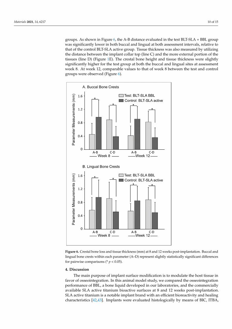
Crestal bone loss was determined by measurements of the distance between theimplant collar top (line A) and the point of contact with bone (line B) as indicated inFigure 1E. Interestingly, both the buccal and lingual dimensions showed statisticallysignificant differences at weeks 8 and 12 post-implantation between the test and control
Materials 2021, 14, 6217 10 of 15
groups. As shown in Figure 6, the A-B distance evaluated in the test BLT-SLA + BBL groupwas significantly lower in both buccal and lingual at both assessment intervals, relative tothat of the control BLT-SLA active group. Tissue thickness was also measured by utilizingthe distance between the implant collar top (line C) and the more external portion of thetissues (line D) (Figure 1E). The crestal bone height and tissue thickness were slightlysignificantly higher for the test group at both the buccal and lingual sites at assessmentweek 8. At week 12, comparable values to that of week 8 between the test and controlgroups were observed (Figure 6).
Materials 2021, 14, x FOR PEER REVIEW 10 of 15
Crestal bone loss was determined by measurements of the distance between the im-plant collar top (line A) and the point of contact with bone (line B) as indicated in Figure 1E. Interestingly, both the buccal and lingual dimensions showed statistically significant differences at weeks 8 and 12 post-implantation between the test and control groups. As shown in Figure 6, the A-B distance evaluated in the test BLT-SLA + BBL group was sig-nificantly lower in both buccal and lingual at both assessment intervals, relative to that of the control BLT-SLA active group. Tissue thickness was also measured by utilizing the distance between the implant collar top (line C) and the more external portion of the tis-sues (line D) (Figure 1E). The crestal bone height and tissue thickness were slightly signif-icantly higher for the test group at both the buccal and lingual sites at assessment week 8. At week 12, comparable values to that of week 8 between the test and control groups were observed (Figure 6).
Figure 6. Crestal bone loss and tissue thickness (mm) at 8 and 12 weeks post-implantation. Buccal and lingual bone crests within each parameter (A–D) represent slightly statistically significant dif-ferences for pairwise comparisons (* p < 0.05).
4. Discussion The main purpose of implant surface modification is to modulate the host tissue in
favor of osseointegration. In this animal model study, we compared the osseointegration performance of BBL, a bone liquid developed in our laboratories, and the commercially
Figure 6. Crestal bone loss and tissue thickness (mm) at 8 and 12 weeks post-implantation. Buccal andlingual bone crests within each parameter (A–D) represent slightly statistically significant differencesfor pairwise comparisons (* p < 0.05).
4. Discussion
The main purpose of implant surface modification is to modulate the host tissue infavor of osseointegration. In this animal model study, we compared the osseointegrationperformance of BBL, a bone liquid developed in our laboratories, and the commerciallyavailable SLA active titanium bioactive surfaces at 8 and 12 weeks post-implantation.SLA active titanium is a notable implant brand with an efficient bioreactivity and healingcharacteristics [42,43]. Implants were evaluated histologically by means of BIC, ITBA,

Materials 2021, 14, 6217 11 of 15
crestal bone loss, tissue thickness, and soft tissue regeneration, requisite parameters fornew bone regeneration and implant stability and indicative of successful osteogenesis [44].
BBL is a saline solution mainly composed CaCl2 and MgCl2-6H2O and adjusted witha negatively charged electrolytes that is suitable for enhancing biological reactivity at theimplant site. Interestingly, histomorphometric analysis revealed that the BIC and ITBAvalues of BLT-SLA pre-treated with BBL are significantly higher than that of the SLA storedin sodium chloride solution BLT-SLA active implants, indicating an improvement in fibrinnetwork organization, new bone formation, and implant stability. Moreover, the elevatedBIC values in the test group are an indication of pronounced hydrophilic properties, afinding that is consistent with previous studies [30,45]. Due to the liquid nature of BBLand SLA-active, the data also indicates an improvement in the surface wettability androughness of the treated titanium implants.
Animal studies indicate that the best results are obtained with implants/bioactivewith combined mechanical and chemical treatment methods, thus obtaining better bone-to-implant contact for implants and improving osseointegration mediated by bioactivesurfaces, relative to untreated controls (best described in the recent systematic review [46]).In accordance, the current study evaluated BLT-SLA pretreated with the bioactive BBL. BLT-SLA is a titanium implant that is sandblasted with long-grit corundum and refined with acidtreatments, a process that provides sufficient mechanical roughness. BLT-SLA pretreatmentwith the bioactive BBL provided the second condition for implant improvements, asobserved with the notable minimization in crestal bone loss and enhancement of softand hard tissue generation and thickness, particularly at week 12 post-implantation. Asimilar bioreactivity was observed for the control BLT-SLA active implants but albeit at asignificant lower competency. These results suggest that the salt combination and negativeionic charge of BBL were sufficient to induce tissue remodeling, osteoblast activity, and/orprospective recruitment of circulating cells rich with growth-inducing factors.
Several other studies reported improvements on SLA surfaces using bioactive ma-terials, indicating that the SLA surface roughness is ideal for implantation, yet there isstill room to improve its bioreactivity by modifying the surface with ingredients that aresufficient to enhance osseointegration and bone healing. In an earlier animal study, Buseret al. observed an enhancement in bone apposition during the early stages of bone gen-eration using a SLA titanium surface treated with isotonic NaCl solution (SLA active),relative to the control untreated implants. Similarly, SLA pretreated with hydroxide ions(conSF) showed an increase in mineralized bone-implant contact after a short healingprocess, relative to untreated controls [45]. In a human randomized controlled clinical trial,Oates et al. concluded that chemically modified SLA active surface implants may alterbiologic events during the osseointegration process, which in turn enhances the healingprocess, and therefore could lead to adjustments in clinical loading protocols for dentalimplant therapy [47]. In a comparative study, Lee et al. evaluated the effectiveness ofbone healing and remodeling of three commercially available implants: a hydrophobicSLA-IS-III active, a hydrophilic SLA surface implant with HA nanocoating (IS-III Bioac-tive), and a hydrophilic SLA active [43]. These authors observed higher and a faster newbone generation associated with the use of hydrophilic surface implants in comparison tohydrophobic surface implants, whereas the use of HA-coated surface implants facilitatedosteoblast activity [43]. Taken together, these observations reveal the importance of theimplant surface’s wettability, which most likely facilitates bone healing dynamics aroundimplants and may be attributed to the improvement of early osseointegration.
The use of liquid bioactive material improves implant wettability and influences theearly stages of osteointegration. However, the reactivity of liquid bioactive surfaces isdependent on their hydrophilic competency, salt composition and concertation, electrody-namic charge, lack of solid components, and proteins, factors that may influence viscosity,stiffness, and roughness [29,48,49]. Anitua et al.’s innovative work using titanium im-plants pre-treated with liquid PRGF proved that the implants accelerate bone regeneration,reduce post-extraction defects, and thus, shorten the time between tooth extraction and

Materials 2021, 14, 6217 12 of 15
implant insertion [34–36,38,50,51]. Liquid PRGF is rich in anti-inflammatory and growthfactors that modulate the immune response, enhance circulating cell recruitment, andmediate cellular proliferation and differentiation at the implant healing site [52]. Utilizinga similar rational, Scarano et al. developed autologous platelet liquid (APL) for implantsite irrigation, implant immersion, or to be mixed with biomaterials during regenerativeprocedures [53].
Several studies have reported that Ca2+- and Mg2+-containing bioactive implants,such as hydroxyapatite (HA), improve material porosity and crystallite and upregulate theexpression of genes associated with bone formation [54,55]. Ratanyake et al. observed asubstantial improvement in the bioactivity of stoichiometric HA pot treated with cationicand anionic sublattices [56]. Similar observations were also reported for other HA mod-ifications including cations, such as Sr2+, Pb2+, or Mg2+, and anions, such as F− or Cl−
(best reviewed in [57]). Interestingly, there modifications are sufficient to provide effec-tive barriers against metal corrosion progress and could possess biocompatibility andosteoconductive properties [58].
5. Conclusions
This study examined and compared the biological parameters of a novel bioactivesurface implant relative to that of a commercially available surface material that is widelyused in dental clinics and known for its favorable characteristics post-transplantation.Our data indicate that implants pre-treated with BBL possess comparable and superiorbioreactivity characteristics when compared to SLA active. Within the limitations of thisstudy, our data revealed that BLT-SLA pre-treated with BBL reduced the titanium surfaceroughness and improved wettability, sufficient characteristics to promote healing, eradicateinflammation, and promote bone and gingiva regeneration at the implant site.
The main limitation of this study is the sample size, as due to ethical restrictions, it isrelatively small. More studies are required with an increase in sample size to confirm thecurrent research outcome and BBL application with a variety of rough titanium implantsfrom different brands. Hopefully, randomized controlled clinical trials will also provideevidence of the efficacy of BBL implant treatment in bone regeneration, inflammation andpathologic resistance, and implant stability.
Author Contributions: E.F.-A. (Eduard Ferrés-Amat) and A.A.M. data analysis, outcome assessment,interpretation, and manuscript writing; E.F.-A. (Elvira Ferrés-Amat), Supervised and conducted theanimal experiments, sample collection and assembly of microscope images data; S.A.D., E.F.-P. andC.M., Data analysis and interpretation; S.A.D., N.C., M.B. and M.A.A., Statistic analysis and wereblinded for the outcome assessment and measurements; M.A. data analysis, outcome assessment,interpretation, manuscript writing and Final approval. All authors participate in revision andapproval of the final version of the Manuscript. All authors have read and agreed to the publishedversion of the manuscript.
Funding: This research was funded by; Biointelligent Technology Systems SL, Diputaccion 316, 3D,08009 Barcelona, Spain.
Institutional Review Board Statement: The study was conducted according to the guidelines ofthe Declaration of Helsinki, and approved by the Experimental Animal Research Ethics Committee,CEEA:(12/02/2015) University of Murcia, Murcia, Spain.
Informed Consent Statement: Not applicable.
Data Availability Statement: Data available on request due to restrictions eg privacy or ethical. Thedata presented in this study are available on request from the corresponding author.
Acknowledgments: This study was funded by Biointelligent Technology Systems S.L and we wouldlike to thank Ziacom Medical SL for the facility and the coordination among the researchers to carryout this study.

Materials 2021, 14, 6217 13 of 15
Conflicts of Interest: The authors declare no conflict of interest. E.F.-P., C.M., M.B., M.A. declare thatthey are associated researchers in Biointelligent Technology Systems S.L. Bone Bioactive compositionand uses thereof. European patents: EP353211, US 16/344,322.
References1. Fugazzotto, P.A. Success and Failure Rates of 1,344 6- to 9-mm-Length Rough-Surface Implants Placed at the Time of Transalveolar
Sinus Elevations, Restored with Single Crowns, and Followed for 60 to 229 Months in Function. Int. J. Oral Maxillofac. Implant.2017, 32, 1359–1363. [CrossRef]
2. Scarano, A.; Carinci, F.; Assenza, B.; Piattelli, M.; Murmura, G.; Piattelli, A. Vertical ridge augmentation of atrophic posteriormandible using an inlay technique with a xenograft without miniscrews and miniplates: Case series. Clin. Oral Implant. Res. 2011,22, 1125–1130. [CrossRef] [PubMed]
3. Bonfante, E.A.; Janal, M.N.; Granato, R.; Marin, C.; Suzuki, M.; Tovar, N.; Coelho, P.G. Buccal and lingual bone level alterationsafter immediate implantation of four implant surfaces: A study in dogs. Clin. Oral Implant. Res. 2012, 24, 1375–1380. [CrossRef][PubMed]
4. Fernández-Domínguez, M.; Ortega-Asensio, V.; Fuentes-Numancia, E.; Aragoneses, J.M.; Barbu, H.M.; Ramírez-Fernández, M.P.;Delgado-Ruiz, R.A.; Calvo-Guirado, J.L.; Samet, N.; Gehrke, S.A. Can the Macrogeometry of Dental Implants Influence GuidedBone Regeneration in Buccal Bone Defects? Histomorphometric and Biomechanical Analysis in Beagle Dogs. J. Clin. Med. 2019,8, 618. [CrossRef]
5. Gehrke, S.A.; Treichel, T.L.E.; Pérez-Díaz, L.; Calvo-Guirado, J.L.; Júnior, J.A.; Mazón, P.; De Aza, P.N. Impact of Different TitaniumImplant Thread Designs on Bone Healing: A Biomechanical and Histometric Study with an Animal Model. J. Clin. Med. 2019,8, 777. [CrossRef] [PubMed]
6. Davies, J.E.; Baldan, N. Scanning electron microscopy of the bone-bioactive implant interface. J. Biomed. Mater. Res. 1997, 36,429–440. [CrossRef]
7. Lee, C.; Jeong, S.-M.; Yang, H.-W.; Choi, B.-H. Effect of Ultraviolet Irradiation on Osseointegration of Dental Implants: AComparative Histomorphometric Study in Canine Models. Appl. Sci. 2020, 10, 4216. [CrossRef]
8. Yoon, W.-J.; Kim, S.-G.; Oh, J.-S.; You, J.-S.; Jeong, K.-I.; Lim, S.-C.; Jeong, M.-A. Comparative study on the osseointegration ofimplants in dog mandibles according to the implant surface treatment. J. Korean Assoc. Oral Maxillofac. Surg. 2016, 42, 345–351.[CrossRef]
9. Szmukler-Moncler, S.; Blus, C.; Schwarz, D.M.; Orrù, G. Characterization of a Macro- and Micro-Textured Titanium Grade 5 AlloySurface Obtained by Etching Only without Sandblasting. Materials 2020, 13, 5074. [CrossRef]
10. Rizo-Gorrita, M.; Fernandez-Asian, I.; Garcia-De-Frenza, A.; Vazquez-Pachon, C.; Serrera-Figallo, M.-A.; Torres-Lagares, D.;Gutierrez-Perez, J.-L. Influence of Three Dental Implant Surfaces on Cell Viability and Bone Behavior. An In Vitro and aHistometric Study in a Rabbit Model. Appl. Sci. 2020, 10, 4790. [CrossRef]
11. Pippenger, B.; Rottmar, M.; Kopf, B.S.; Stübinger, S.; Torre, F.H.D.; Berner, S.; Maniura-Weber, K. Surface modification of ultrafine-grained titanium: Influence on mechanical properties, cytocompatibility, and osseointegration potential. Clin. Oral Implant. Res.2018, 30, 99–110. [CrossRef] [PubMed]
12. Salomó-Coll, O.; De Val, J.E.M.-S.; Ramírez-Fernandez, M.P.; Satorres-Nieto, M.; Gargallo-Albiol, J.; Calvo-Guirado, J.L. Osseoin-ductive elements for promoting osseointegration around immediate implants: A pilot study in the foxhound dog. Clin. OralImplant. Res. 2015, 27, e167–e175. [CrossRef] [PubMed]
13. López-Valverde, N.; López-Valverde, A.; Aragoneses, J.; de Sousa, B.M.; Rodrigues, M.; Ramírez, J. Systematic Review andMeta-Analysis of the Effectiveness of Calcium-Phosphate Coating on the Osseointegration of Titanium Implants. Materials 2021,14, 3015. [CrossRef] [PubMed]
14. López-Valverde, N.; Pardal-Peláez, B.; López-Valverde, A.; Ramírez, J. Role of Melatonin in Bone Remodeling around TitaniumDental Implants: Meta-Analysis. Coatings 2021, 11, 271. [CrossRef]
15. Park, S.; Kim, H.; Choi, K.S.; Ji, M.-K.; Kim, S.; Gwon, Y.; Park, C.; Kim, J.; Lim, H.-P. Graphene–Chitosan Hybrid Dental Implantswith Enhanced Antibacterial and Cell-Proliferation Properties. Appl. Sci. 2020, 10, 4888. [CrossRef]
16. Kiran, A.S.K.; Kumar, T.S.; Sanghavi, R.; Doble, M.; Ramakrishna, S. Antibacterial and Bioactive Surface Modifications of TitaniumImplants by PCL/TiO2 Nanocomposite Coatings. Nanomaterials 2018, 8, 860. [CrossRef]
17. Ito, A.; Senda, K.; Sogo, Y.; Oyane, A.; Yamazaki, A.; Legeros, R.Z. Dissolution rate of zinc-containing beta-tricalcium phosphateceramics. Biomed. Mater. 2006, 1, 134–139. [CrossRef]
18. Kulkarni Aranya, A.; Pushalkar, S.; Zhao, M.; LeGeros, R.Z.; Zhang, Y.; Saxena, D. Antibacterial and bioactive coatings ontitanium implant surfaces. J. Biomed. Mater. Res. A 2017, 105, 2218–2227. [CrossRef]
19. Yue, J.; Jin, Z.; Poon, H.L.E.; Shang, G.; Liu, H.; Wang, D.; Qi, S.; Chen, F.; Xu, Y. Osteogenic and Antibacterial Activity of aPlasma-Sprayed CeO2 Coating on a Titanium (Ti)-Based Dental Implant. Coatings 2020, 10, 1007. [CrossRef]
20. Odatsu, T.; Kuroshima, S.; Sato, M.; Takase, K.; Valanezhad, A.; Naito, M.; Sawase, T. Antibacterial Properties of Nano-Ag Coatingon Healing Abutment: An In Vitro and Clinical Study. Antibiotics 2020, 9, 347. [CrossRef] [PubMed]
21. Simsek, M.; Aldemir, S.D.; Gümüsderelioglu, M.; Anticellular, P.E.O. Coatings on titanium surfaces by sequential electrospinningand crosslinking processes. Emergent Mater. 2019, 2, 169–179. [CrossRef]

Materials 2021, 14, 6217 14 of 15
22. Gittens, R.A.; Scheideler, L.; Rupp, F.; Hyzy, S.L.; Geis-Gerstorfer, J.; Schwartz, Z.; Boyan, B.D. A review on the wettability ofdental implant surfaces II: Biological and clinical aspects. Acta Biomater. 2014, 10, 2907–2918. [CrossRef] [PubMed]
23. Scarano, A.; Lorusso, F.; Orsini, T.; Morra, M.; Iviglia, G.; Valbonetti, L. Biomimetic Surfaces Coated with Covalently ImmobilizedCollagen Type I: An X-Ray Photoelectron Spectroscopy, Atomic Force Microscopy, Micro-CT and Histomorphometrical Study inRabbits. Int. J. Mol. Sci. 2019, 20, 724. [CrossRef]
24. Kulkarni, M.; Patil-Sen, Y.; Junkar, I.; Kulkarni, C.V.; Lorenzetti, M.; Iglic, A. Wettability studies of topologically distinct titaniumsurfaces. Colloids Surf. B Biointerfaces 2015, 129, 47–53. [CrossRef]
25. Ponsonnet, L.; Reybier, K.; Jaffrezic, N.; Comte, V.; Lagneau, C.; Lissac, M.; Martelet, C. Relationship between surface properties(roughness, wettability) of titanium and titanium alloys and cell behaviour. Mater. Sci. Eng. C 2003, 23, 551–560. [CrossRef]
26. Bang, S.-M.; Moon, H.-J.; Kwon, Y.-D.; Yoo, J.-Y.; Pae, A.; Kwon, I.K. Osteoblastic and osteoclastic differentiation on SLA andhydrophilic modified SLA titanium surfaces. Clin. Oral Implant. Res. 2013, 25, 831–837. [CrossRef]
27. Romero-Ruiz, M.M.; Gil-Mur, F.J.; Ríos-Santos, J.V.; Lázaro-Calvo, P.; Ríos-Carrasco, B.; Herrero-Climent, M. Influence of a NovelSurface of Bioactive Implants on Osseointegration: A Comparative and Histomorfometric Correlation and Implant StabilityStudy in Minipigs. Int. J. Mol. Sci. 2019, 20, 2307. [CrossRef]
28. Lee, S.Y.; Yang, D.J.; Yeo, S.; An, H.W.; Ryoo, K.H.; Park, K.B. The cytocompatibility and osseointegration of the Ti implants withXPEED(R) surfaces. Clin. Oral Implant. Res. 2012, 23, 1283–1289. [CrossRef]
29. Elias, C.N.; Oshida, Y.; Lima, J.H.C.; Muller, C.A. Relationship between surface properties (roughness, wettability and morphol-ogy) of titanium and dental implant removal torque. J. Mech. Behav. Biomed. Mater. 2008, 1, 234–242. [CrossRef]
30. Lang, N.P.; Salvi, G.E.; Huynh-Ba, G.; Ivanovski, S.; Donos, N.; Bosshardt, D.D. Early osseointegration to hydrophilic andhydrophobic implant surfaces in humans. Clin. Oral Implant. Res. 2011, 22, 349–356. [CrossRef]
31. Zhao, X.; Wang, T.; Qian, S.; Liu, X.; Sun, J.; Li, B. Silicon-Doped Titanium Dioxide Nanotubes Promoted Bone Formation onTitanium Implants. Int. J. Mol. Sci. 2016, 17, 292. [CrossRef]
32. Katic, J.; Šaric, A.; Despotovic, I.; Matijakovic, N.; Petkovic, M.; Petrovic, Z. Bioactive Coating on Titanium Dental Implants forImproved Anticorrosion Protection: A Combined Experimental and Theoretical Study. Coatings 2019, 9, 612. [CrossRef]
33. Salomó-Coll, O.; De Val, J.E.M.-S.; Ramírez-Fernández, M.P.; Hernández-Alfaro, F.; Gargallo-Albiol, J.; Calvo-Guirado, J.L. Topicalapplications of vitamin D on implant surface for bone-to-implant contact enhance: A pilot study in dogs part II. Clin. Oral Implant.Res. 2015, 27, 896–903. [CrossRef] [PubMed]
34. Anitua, E.; Orive, G.; Pla, R.; Roman, P.; Serrano, V.; Andía, I. The effects of PRGF on bone regeneration and on titanium implantosseointegration in goats: A histologic and histomorphometric study. J. Biomed. Mater. Res. Part A 2008, 91A, 158–165. [CrossRef]
35. Anitua, E. Plasma rich in growth factors: Preliminary results of use in the preparation of future sites for implants. Int. J. OralMaxillofac. Implant. 1999, 14, 529–535.
36. Anitua, E.; Muruzabal, F.J.; Alcalde, I.; Merayo-Lloves, J.; Orive, G. Plasma rich in growth factors (PRGF-Endoret) stimulatescorneal wound healing and reduces haze formation after PRK surgery. Exp. Eye Res. 2013, 115, 153–161. [CrossRef]
37. Anitua, E.; Prado, R.; Orive, G.; Tejero, R. Effects of calcium-modified titanium implant surfaces on platelet activation, clotformation, and osseointegration. J. Biomed. Mater. Res. Part A 2014, 103, 969–980. [CrossRef]
38. Anitua, E.; Cerqueira, A.; Romero-Gavilán, F.; García-Arnáez, I.; Martinez-Ramos, C.; Ozturan, S.; Azkargorta, M.; Elortza, F.;Gurruchaga, M.; Goñi, I.; et al. Influence of calcium ion-modified implant surfaces in protein adsorption and implant integration.Int. J. Implant. Dent. 2021, 7, 1–11. [CrossRef] [PubMed]
39. Gehrke, S.A.; Bragança, L.K.; Velasco-Ortega, E.; Calvo-Guirado, J.L. Evaluation of dimensional behavior of peri-implant tissuesin implants immediately exposed or submerged in fresh extraction and healed sites: A histological study in dogs. Int. J. Implant.Dent. 2018, 4, 1–9. [CrossRef]
40. Discepoli, N.; Vignoletti, F.; Laino, L.; de Sanctis, M.; Muñoz, F.; Sanz, M. Early healing of the alveolar process after toothextraction: An experimental study in the beagle dog. J. Clin. Periodontol. 2013, 40, 638–644. [CrossRef] [PubMed]
41. Calvo-Guirado, J.L.; Benítez-García, J.A.; de Val, J.E.M.S.; Martínez, C.P.-A.; Gehrke, S.A.; Delgado-Ruiz, R.; Moses, O. Socket-shield technique: The influence of the length of the remaining buccal segment of healthy tooth structure on peri-implant boneand socket preservation. A study in dogs. Ann. Anat. Anat. Anz. 2018, 221, 84–92. [CrossRef] [PubMed]
42. Buser, D.; Broggini, N.; Wieland, M.; Schenk, R.K.; Denzer, A.J.; Cochran, D.L.; Hoffmann, B.; Lussi, A.; Steinemann, S.G.Enhanced Bone Apposition to a Chemically Modified SLA Titanium Surface. J. Dent. Res. 2004, 83, 529–533. [CrossRef]
43. Lee, J.; Yoo, J.M.; Amara, H.B.; Lee, Y.-M.; Lim, Y.-J.; Kim, H.; Koo, K.-T. Bone healing dynamics associated with 3 implants withdifferent surfaces: Histologic and histomorphometric analyses in dogs. J. Periodontal Implant. Sci. 2019, 49, 25–38. [CrossRef]
44. Park, Y.-S.; Yi, K.-Y.; Lee, I.-S.; Jung, Y.-C. Correlation between microtomography and histomorphometry for assessment ofimplant osseointegration. Clin. Oral Implant. Res. 2005, 16, 156–160. [CrossRef]
45. Stadlinger, B.; Lode, A.T.; Eckelt, U.; Range, U.; Schlottig, F.; Hefti, T.; Mai, R. Surface-conditioned dental implants: An animalstudy on bone formation. J. Clin. Periodontol. 2009, 36, 882–891. [CrossRef] [PubMed]
46. López-Valverde, N.; Flores-Fraile, J.; Ramírez, J.M.; Sousa, B.M.d.; Herrero-Hernández, S.; López-Valverde, A. Bioactive Surfacesvs. Conventional Surfaces in Titanium Dental Implants: A Comparative Systematic Review. J. Clin. Med. 2020, 9, 2047. [CrossRef]
47. Oates, T.W.; Valderrama, P.; Bischof, M.; Nedir, R.; Jones, A.; Simpson, J.; Toutenburg, H.; Cochran, D.L. Enhanced implantstability with a chemically modified SLA surface: A randomized pilot study. Int. J. Oral Maxillofac. Implant. 2007, 22, 755–760.

Materials 2021, 14, 6217 15 of 15
48. Lech, A.; Butruk-Raszeja, B.A.; Ciach, T.; Lawniczak-Jablonska, K.; Kuzmiuk, P.; Bartnik, A.; Wachulak, P.; Fiedorowicz, H.Surface Modification of PLLA, PTFE and PVDF with Extreme Ultraviolet (EUV) to Enhance Cell Adhesion. Int. J. Mol. Sci. 2020,21, 9679. [CrossRef] [PubMed]
49. Peng, Z.; Ni, J.; Zheng, K.; Shen, Y.; Wang, X.; He, G.; Jin, S.; Tang, T. Dual effects and mechanism of TiO2 nanotube arrays inreducing bacterial colonization and enhancing C3H10T1/2 cell adhesion. Int. J. NanoMed. 2013, 8, 3093–3105.
50. Anitua, E.; Muruzabal, F.J.; De la Fuente, M.; Merayo-Lloves, J.; Orive, G. Effects of heat-treatment on plasma rich in growthfactors-derived autologous eye drop. Exp. Eye Res. 2014, 119, 27–34. [CrossRef] [PubMed]
51. Anitua, E.; Tejero, R.; Zalduendo, M.M.; Orive, G. Plasma Rich in Growth Factors Promotes Bone Tissue Regeneration byStimulating Proliferation, Migration, and Autocrine Secretion in Primary Human Osteoblasts. J. Periodontol. 2013, 84, 1180–1190.[CrossRef]
52. Mussano, F.; Genova, T.; Munaron, L.; Petrillo, S.; Erovigni, F.; Carossa, S. Cytokine, chemokine, and growth factor profile ofplatelet-rich plasma. Platelets 2016, 27, 467–471. [CrossRef] [PubMed]
53. Scarano, A.; Inchingolo, F.; Murmura, G.; Traini, T.; Piattelli, A.; Lorusso, F. Three-Dimensional Architecture and MechanicalProperties of Bovine Bone Mixed with Autologous Platelet Liquid, Blood, or Physiological Water: An In Vitro Study. Int. J. Mol.Sci. 2018, 19, 1230. [CrossRef] [PubMed]
54. Sato, M.; Aslani, A.; Sambito, M.A.; Kalkhoran, N.M.; Slamovich, E.B.; Webster, T.J. Nanocrystalline hydroxyapatite/titaniacoatings on titanium improves osteoblast adhesion. J. Biomed. Mater. Res. Part A 2007, 84A, 265–272. [CrossRef] [PubMed]
55. Yu, J.-M.; Choe, H.-C. Mg-containing hydroxyapatite coatings on Ti-6Al-4V alloy for dental materials. Appl. Surf. Sci. 2018, 432,294–299. [CrossRef]
56. Ratnayake, J.T.B.; Mucalo, M.; Dias, G.J. Substituted hydroxyapatites for bone regeneration: A review of current trends. J. Biomed.Mater. Res. Part B Appl. Biomater. 2017, 105, 1285–1299. [CrossRef] [PubMed]
57. Arcos, D.; Vallet-Regi, M. Substituted hydroxyapatite coatings of bone implants. J. Mater. Chem. B 2020, 8, 1781–1800. [CrossRef][PubMed]
58. Cesarz-Andraczke, K.; Nowosielski, R.; Basiaga, M.; Babilas, R. Study of the Morphology and Properties of Biocompatible Ca-PCoatings on Mg Alloy. Materials 2019, 13, 2. [CrossRef] [PubMed]
Related Documents