- 1 - Title Page: A Pre-targeting Strategy for MR Imaging of Functional Molecules Using Dendritic Gd-Based Contrast Agents Kohei Sano 1 , Takashi Temma 1 , Takashi Azuma 2 , Ryusuke Nakai 2 , Michiko Narazaki 3 , Yuji Kuge 1,4 , Hideo Saji 1 1 Department of Patho-Functional Bioanalysis, Graduate School of Pharmaceutical Sciences, Kyoto University, Kyoto 606-8501, Japan 2 Department of Medical Simulation Engineering Research Center for Nano Medical Engineering Institute for Frontier Medical Sciences, Kyoto University, Kyoto 606-8501, Japan 3 Department of Systems Science, Graduate School of Informatics, Kyoto University, Kyoto 606-8501, Japan 4 Central Institute of Isotope Science, Hokkaido University, Sapporo 060-8638, Japan Corresponding author: Hideo Saji, PhD Department of Patho-Functional Bioanalysis

Welcome message from author
This document is posted to help you gain knowledge. Please leave a comment to let me know what you think about it! Share it to your friends and learn new things together.
Transcript

- 1 -
Title Page:
A Pre-targeting Strategy for MR Imaging of Functional Molecules Using Dendritic Gd-Based
Contrast Agents
Kohei Sano1, Takashi Temma1, Takashi Azuma2, Ryusuke Nakai2, Michiko Narazaki 3, Yuji
Kuge1,4, Hideo Saji1
1Department of Patho-Functional Bioanalysis, Graduate School of Pharmaceutical Sciences,
Kyoto University, Kyoto 606-8501, Japan
2Department of Medical Simulation Engineering Research Center for Nano Medical
Engineering Institute for Frontier Medical Sciences, Kyoto University, Kyoto 606-8501,
Japan
3Department of Systems Science, Graduate School of Informatics, Kyoto University, Kyoto
606-8501, Japan
4Central Institute of Isotope Science, Hokkaido University, Sapporo 060-8638, Japan
Corresponding author:
Hideo Saji, PhD
Department of Patho-Functional Bioanalysis

- 2 -
Graduate School of Pharmaceutical Sciences, Kyoto University
46-29 Yoshida Shimoadachi-cho, Sakyo-ku, Kyoto 606-8501, Japan
Tel; (+81/0)-75-753-4556
Fax; (+81/0)-75-753-4568
E-mail; [email protected]
Running Title:
MRI protocol for molecular imaging by a pre-targeting method
Manuscript category
Article
- 3 -
ABSTRACT (~ 150 words)
Purpose
We aimed to establish a magnetic resonance imaging (MRI) protocol for the sensitive and
specific imaging of functional molecules with a pre-targeting strategy utilizing the
streptavidin-biotin interaction. Membrane type-1 matrix metalloproteinase (MT1-MMP) was
selected as the target molecule.
Procedures
The biotinylated polyamidoamine dendrimer (PAMAM)-based contrast agent
(Bt-PAMAM-DTPA(Gd)) was prepared, and its proton relaxivity (r1) and affinity to
streptavidin were evaluated. Tumor-bearing mice were pre-targeted with
streptavidin-conjugated anti-MT1-MMP monoclonal antibody (mAb),
streptavidin-conjugated negative control IgG, or saline and 3 days later were injected with
Bt-PAMAM-DTPA(Gd) followed immediately by MRI for a period of 3 h.
Results
High r1 (15.5 L mmol-1 s-1) and 1.9-fold higher affinity than D-biotin were obtained.
Significantly higher relative tumor signals were observed in mice pre-targeted with
streptavidin-conjugated anti-MT1-MMP mAb (165% at 3 h vs. pre-administration) than with
saline or streptavidin-conjugated negative control IgG (P < 0.0001).
Conclusions

- 4 -
This pre-targeting approach can accomplish sensitive and specific in vivo MRI of functional
molecules.
Key Words
Pre-targeting, Polyamidoamine dendrimer (PAMAM), Membrane type-1 matrix
metalloproteinase, Magnetic Resonance Imaging

- 5 -
INTRODUCTION
Magnetic resonance imaging (MRI), characterized by a remarkable spatial resolution,
is a powerful tool for noninvasive morphologic diagnosis of diseases including cancer.
Recently, the application of MRI to functional molecular imaging coupled with anatomical
information has been explored. To realize functional molecular imaging by MRI, contrast
agents are required that possess a high relaxation to produce high MR signals to compensate
for the low intrinsic sensitivity of MRI [1] in addition to selectively accumulating in the
targeting site.
Gadolinium (Gd) chelates conjugated to macromolecules such as liposomes, micelles
and dendrimers can give rise to enhanced proton relaxivities in comparison with simple,
small molecule contrast agents such as Gd-diethylenetriamine pentaacetic acid (Gd-DTPA)
[2-4]. This effect is due to a restriction in thermal flexibility leading to increased interactions
between the Gd atom and surrounding water molecules [5-7]. Some groups have developed
monoclonal antibody (mAb)-conjugated macromolecular contrast agents for imaging integrin
vor human epidermal growth factor receptor type 2 (HER2) in tumors which
successfully increased the tumor signal intensity by 15-30% [8, 9]. However, many
researchers have failed to demonstrate in vivo functional molecular imaging using
macromolecules conjugated with several Gd chelates and targeting moieties like antibodies
and peptides because the pharmacokinetics of the targeting moieties were significantly altered

- 6 -
by the introduction of macromolecular contrast agents, which resulted in low target
recognition and high accumulation of the labeling agent in non-targeted tissues [10, 11].
Furthermore, when antibody-conjugated macromolecular contrast agents are injected at a Gd
dose (0.1 mmol Gd/kg) necessary for adequate imaging, excess antibody (on the order of
milligrams per mouse) are typically administered, which leads to major limitations of cost
and in vivo toxicity.
Thus, in this study, to overcome these problems and to realize functional molecular
MRI by a macromolecule-based contrast agent, we aimed to use a pre-targeting strategy that
utilizes the high affinity interaction between streptavidin and biotin (Kd=10-15 M) [12]. In this
pre-targeting method, the first step is to administer a streptavidin-conjugated target-specific
antibody. In the second step, after selective accumulation of streptavidin-conjugated antibody
in the targeted tissue and clearance of unbound targeting agent from the circulation, a
biotin-bound imaging probe is injected. As the post-administration contrast agent,
polyamideamine dendrimer (PAMAM) was selected as the base structure since it is
structurally well-defined and functional moieties including biotins and Gd chelates for both
targeting and signal emission functions can easily be attached to the large number of its
surface amino groups. The pre-targeting strategy is expected to provide selective and
effective accumulation of the PAMAM-based contrast agent to the targeted site and a high
S/N ratio during the first hours following administration as has been observed in

- 7 -
radioimmunotherapy and radioimmunodetection [13-16], and to potentially lead to lower in
vivo toxicity [17, 18].
Thus, in this paper we describe our efforts to establish a sensitive and specific in vivo
MRI protocol for imaging functional molecules utilizing a pre-targeting strategy that
combines a streptavidin-conjugated antibody with a PAMAM based contrast agent modified
with biotins. As the targeted biomolecule, membrane type-1 matrix metalloproteinase
(MT1-MMP) was selected. Since MT1-MMP is exclusively expressed in tumors and is
closely associated with metastasis [19] and invasion [20], MT1-MMP is a potential imaging
target for evaluation of tumor malignancy.

- 8 -
MATERIALS AND METHODS
Synthesis of streptavidin-conjugated anti-MT1-MMP mAb
Streptavidin-conjugated anti-MT1-MMP mAb and streptavidin-conjugated negative
control IgG were synthesized according to a previously described method [14]. Briefly,
EZ-Link® sulfosuccinimidyl-6-(biotinamido) hexanoate (sulfo-NHS-LC-biotin) (Pierce, Inc.)
was added to a solution of anti-MT1-MMP mAb (113-5B7, Daiichi Fine Chemical Co.) in a
molar ratio of 12:1. The mixture was gently stirred for 30 min at room temperature and then
was purified with a diafiltration membrane (Amicon Ultra 4 (MWCO 30,000), Millipore Co.).
A solution of biotinylated anti-MT1-MMP mAb was added to a solution of streptavidin (SAv;
Pierce, Inc.) in a 1:3 molar ratio. The mixture was incubated for 1 h at 37 ˚C followed by
purification twice by affinity chromatography using a HiTrap rProtein A column (GE
Healthcare Bioscience). The eluate containing anti-MT1-MMP mAb-SAv was concentrated
with a diafiltration membrane (MWCO 30,000), and the protein concentration was
determined by the bicinchoninate (BCA) method. The purification was monitored by a size
exclusion chromatograph using a 300×4.6-mm i.d. TSK-Gel Super SW 3000 column (Tosoh
Co., Japan) eluted with phosphate buffer (0.1 M, pH 6.8) at a flow rate of 0.1 mL/min.
Comparison of molecular mass standards (Oriental Yeast Co., Japan) of the absorbance at
280 nm indicated that a peak at 30.3 min was consistent with the presence of the 210 kDa
streptavidin-conjugated anti-MT1-MMP mAb. Furthermore, streptavidin-conjugated

- 9 -
anti-MT1-MMP mAb retained 81.3% immunoreactivity of the anti-MT1-MMP mAb, which
was confirmed by flow cytometry.
Synthesis of Bt-PAMAM-DTPA(Gd)
EZ-Link® sulfo-NHS-LC-biotin was added to a solution of PAMAM (generation 4 (G4))
(Sigma Aldrich) in a molar ratio of 20:1. The mixture was stirred for 30 min at room
temperature and then was applied to a diafiltration membrane (MWCO 10,000) to remove
unbound biotins as well as to change the buffer to phosphate buffer (0.1 M, pH 9.0). After
purification, the incorporation ratio of biotins conjugated to each dendrimer was measured
using an EZTM Biotin Quantitation Kit (Pierce, Inc.). The biotinylated PAMAM was reacted
with a 64-fold molar excess of 2-(p-isothiocyanatobenzyl)-diethylenetriaminepentaacetic acid
(p-SCN-Bz-DTPA) (Macrocyclics) at 40˚C for 24 h. During the reaction, the pH was
maintained at 9.0 with 1 N NaOH. An additional equal amount of p-SCN-Bz-DTPA was
added after 24 h and the reaction was incubated for another 24 h at 40˚C. The resulting
preparation was purified by diafiltration membrane (MWCO 10,000). After purification, the
number of DTPAs incorporated into each G4 dendrimer was checked by chelate titration
using ZnSO4 (indicator: 4-(2-pyridylazo)resorcinol, NH3/NH4+, pH 10) according to a
previously described method with some modification [21]. Purified Bt-PAMAM-DTPA was
mixed with GdCl3 (Sigma Aldrich) in citrate buffer (0.3 M, pH 5.0) for 2 h at 40˚C. The

- 10 -
excess Gd was removed by diafiltration membrane (MWCO 10,000) while simultaneously
changing the buffer to PBS (0.1 M, pH 7.4). The number of Gd incorporated into a dendrimer
was checked by separating the free Gd and Bt-PAMAM-DTPA(Gd) with diafiltration filter
after labeling the Bt-PAMAM-DTPA with 153Gd and nonradioactive Gd. For comparison
purposes, PAMAM-DTPA(Gd) containing one biotin (Bt1-PAMAM-DTPA(Gd)) was also
prepared in a similar manner.
Stability of Bt-PAMAM-DTPA(Gd) in mouse plasma
153Gd-labeled Bt-PAMAM-DTPA(Gd) was prepared by reacting
Bt-PAMAM-DTPA(Gd) with 153Gd (1 Ci, PerkinElmer Japan Co., Osaka, Japan) and
non-radioactive Gd in 0.3 M citrate buffer at pH 5.0 for 2 h at 40˚C. 153Gd labeled
Bt-PAMAM-DTPA(Gd) (30 L) was added to mouse plasma collected from female C3H/He
mice (270 L), and the plasma samples were incubated at 37˚C for 0, 3, and 24 h. After
incubation, aliquots of the samples were drawn, and radioactivity was analyzed by
size-exclusion chromatography with a PD-10 column (GE Healthcare Bioscience) using
saline as eluent.
Affinity of Bt-PAMAM-DTPA(Gd) for streptavidin
Competition assays of 125I-(3-iodobenzoyl)norbiotinamide (125I-IBB), a radiolabeled
- 11 -
biotin derivative synthesized as reported previously [22], were performed by incubating
streptavidin (Pierce, Inc.; 400 L, 2 g/mL), 125I-IBB (50 L, 5 Ci), and various
concentrations of Bt-PAMAM-DTPA(Gd), Bt1-PAMAM-DTPA(Gd), and D-biotin (Nacalai
Tesque, Kyoto, Japan) (50 L, 10-8~10-4 M) in PBS (0.1 M, pH 7.4) for 60 min at 37˚C. At
the end of the incubation, the mixture was applied to a size exclusion column with a
Sephadex G-50 Fine (GE Healthcare Bioscience), followed by measurement of the
radioactivity from the column eluent (containing macromolecules) with a NaI well-type
scintillation counter (1470WIZARD, PerkinElmer Japan Co.). Nonspecific binding was
determined in the presence of 10 mg/mL D-biotin. The 50% inhibitory concentrations (IC50s)
were determined from displacement curves of the percent inhibition of 125I-IBB binding vs.
the inhibitor concentration.
Preparation of tumor-bearing animals
Female C3H/He mice (5 weeks old), supplied by Japan SLC Co. (Hamamatsu, Japan),
were housed under a 12-h light/12-h dark cycle and were given free access to food and water.
The animal experiments in this study were conducted in accordance with institutional
guidelines and were approved by the Kyoto University Animal Care Committee, Japan.
FM3A mouse breast carcinoma cells were supplied by the Health Science Research
Resources Bank (Osaka, Japan). They were cultured in DMEM medium (Nissui

- 12 -
Pharmaceutical Co.) supplemented with 10% fetal bovine serum at 37˚C in a humidified
atmosphere containing 5% CO2 and 95% air and had a 10.6-h doubling time.
FM3A cells were suspended in 0.01 M PBS (pH 7.4) followed by subcutaneous
inoculation into the right hind leg of the mouse (5 × 10⁶ cells/100 L/mouse) [23]. The tumor
volume was estimated by (length)×(width)2/2 [24] over a 10-day tumor growth period. The
average size of the tumors was 213 ± 82 mm3 on the MR imaging study day. The expression
of MT1-MMP in FM3A cells and tumor tissues was confirmed by western blotting and
immunohistochemistry [25].
Magnetic Resonance Imaging
MRI was performed using a clinical 1.5 Tesla MR scanner (MAGNETOM Symphony
Sonata, Siemens). All T1-weighted MR images were acquired with a multislice spin-echo
pulse sequence. MRI data were analyzed using the ImageJ software.
Phantom study
Solutions of Gd-DTPA (Sigma Aldrich) and Bt-PAMAM-DTPA(Gd) were prepared
with a Gd concentration in the range of 10 to 500 M in vials with an inner diameter of 15
mm followed by the MR scan using a knee coil (20.5 cm in diameter) at 20˚C. To obtain
proton relaxivity (r1) for samples, spin-echo images were obtained using a sequence with
TR=500, 1000, 1500, and 2000 msec and with TE=15 msec. The imaging parameters were as

- 13 -
follows: field-of-view, 256×128 mm; matrix, 256×128; slice thickness, 7 mm; number of
average, 3.
In vivo study
Mice (n=4) bearing FM3A tumors in the right thigh received streptavidin-conjugated
anti-MT1-MMP mAb (50 g/100 L in saline) via tail vein. Three days later,
Bt-PAMAM-DTPA(Gd) (0.1 mmolGd/kg, 100 L in PBS (0.1 M, pH 7.4), i.v.) was injected
followed by data acquisition by MRI at several time points over a 3-h post-injection period
under sodium pentobarbital (50 mg/kg, i.p.) anesthesia. All MR images were obtained using a
hand-made round surface coil (5.5 cm in diameter) fixed by a custom constructed coil holder.
The imaging parameters were as follows: TR/TE, 300/5.2 msec; field-of-view, 128×96 mm;
matrix, 256×192; slice thickness, 1.5 mm; number of average, 3. MRI studies were also
conducted as above on FM3A tumor bearing mice (n=3) pre-treated with saline (100 L) or
streptavidin-conjugated negative control IgG (50 g/100 L in saline). The signal intensity
was calculated by drawing a region of interest around the tumor, muscle in the contralateral
hind limb, and kidneys. The relative signal intensity in each tissue was defined as the signal
intensity after administration of Bt-PAMAM-DTPA(Gd) divided by the signal intensity
before administration.
Statistical Analysis

- 14 -
Unpaired Student’s t test was used to evaluate the significance of differences of r1
between Bt-PAMAM-DTPA(Gd) and Gd-DTPA. To compare the time courses of relative
signal intensity in the tumor and kidneys and tumor/muscle (T/M) signal ratios among
Bt-PAMAM-DTPA(Gd) pre-targeted by streptavidin-conjugated anti-MT1-MMP mAb,
streptavidin-conjugated negative control IgG, and saline, two-way repeated measures ANOVA
with post-hoc analysis by the Tukey-Kramer test was performed. Differences at the 95%
confidence level (P < 0.05) were considered significant.

- 15 -
RESULTS
Characterization of Bt-PAMAM-DTPA(Gd)
Bt-PAMAM-DTPA(Gd) was synthesized in four steps from PAMAM in a yield of
63%. PAMAM was conjugated to 9.9 ± 1.3 biotins and 43.6 ± 1.9 DTPAs, which were
quantitatively coordinated to Gd. Bt1-PAMAM-DTPA(Gd) containing 1.0 ± 0.1 biotin and
47.6 ± 2.2 DTPAs on PAMAM dendrimer was also synthesized. The 153Gd-labeled
Bt-PAMAM-DTPA(Gd), which was incubated with mouse plasma for 24 h, did not release
any low molecular weight metabolites or free radiometals (Fig. 1).
The competitive binding assay revealed that all of the contrast agents inhibited the
binding of 125I-IBB to streptavidin in a dose-dependent manner (Fig. 2). The IC50s for
Bt-PAMAM-DTPA(Gd), Bt1-PAMAM-DTPA(Gd), and D-biotin were 32 ± 31, 1390 ± 1220,
and 60 ± 45 nM, respectively, demonstrating that Bt-PAMAM-DTPA(Gd) had about 1.9- and
43.2-fold higher affinity to streptavidin than D-biotin and Bt1-PAMAM-DTPA(Gd).
MR imaging study (Phantom study)
The in vitro T1-weighted MR images with Bt-PAMAM-DTPA(Gd) and Gd-DTPA are
shown in Fig. 3a. Water and PBS were used as baselines. With the same Gd concentration,
the signals with Bt-PAMAM-DTPA(Gd) were higher compared to Gd-DTPA. The
longitudinal relaxation rate (1/T1) vs. the concentration of Gd for both contrast agents are
- 16 -
shown in Fig. 3b with good linear fits (R2 = 1.00 and 0.99 for Bt-PAMAM-DTPA(Gd) and
Gd-DTPA, respectively). Calculated r1 values (L mmol-1 s-1) for Bt-PAMAM-DTPA(Gd) and
Gd-DTPA were 15.5 ± 1.1 and 3.6 ± 0.1, respectively, which shows that the proton relaxivity
of Bt-PAMAM-DTPA(Gd) was 4.3-fold higher than that of Gd-DTPA (P < 0.0001).
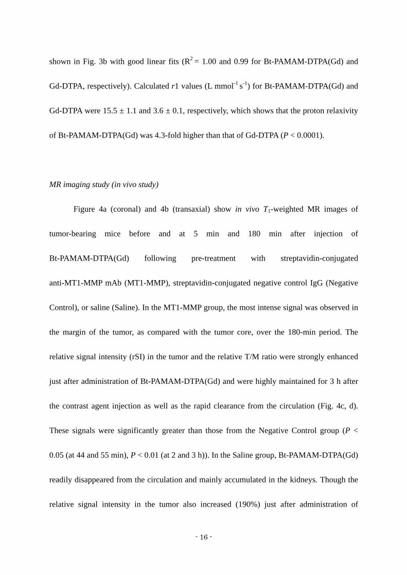
MR imaging study (in vivo study)
Figure 4a (coronal) and 4b (transaxial) show in vivo T1-weighted MR images of
tumor-bearing mice before and at 5 min and 180 min after injection of
Bt-PAMAM-DTPA(Gd) following pre-treatment with streptavidin-conjugated
anti-MT1-MMP mAb (MT1-MMP), streptavidin-conjugated negative control IgG (Negative
Control), or saline (Saline). In the MT1-MMP group, the most intense signal was observed in
the margin of the tumor, as compared with the tumor core, over the 180-min period. The
relative signal intensity (rSI) in the tumor and the relative T/M ratio were strongly enhanced
just after administration of Bt-PAMAM-DTPA(Gd) and were highly maintained for 3 h after
the contrast agent injection as well as the rapid clearance from the circulation (Fig. 4c, d).
These signals were significantly greater than those from the Negative Control group (P <
0.05 (at 44 and 55 min), P < 0.01 (at 2 and 3 h)). In the Saline group, Bt-PAMAM-DTPA(Gd)
readily disappeared from the circulation and mainly accumulated in the kidneys. Though the
relative signal intensity in the tumor also increased (190%) just after administration of

- 17 -
Bt-PAMAM-DTPA(Gd), it decreased to the basal level (115%) within 3 h (Fig. 4c). A slightly
higher tumor signal was obtained in the Negative Control group than in the Saline group only
3 h after injection of Bt-PAMAM-DTPA(Gd) (P < 0.01). The time-dependent change of
relative T/M ratios was similar to that of the relative signal intensity in the tumor (Fig. 4d).
The time-dependent change of relative signal intensity in the kidneys was very similar in all
three groups (Fig. 4e).

- 18 -
DISCUSSION
In this study, we accomplished visualization of MT1-MMP by MRI using a
pre-targeting method with a PAMAM-based contrast agent (Bt-PAMAM-DTPA(Gd)) which
possesses high proton relaxivity and high affinity to streptavidin. For future applications, this
pre-targeting method based on the interaction between biotin and streptavidin is promising
for the detection of functional molecules, such as biomarkers in tumors like MT1-MMP, by in
vivo MRI.
Although several macromolecular contrast agents have been developed for functional
molecular imaging with MRI using a mAb or peptide as the targeting moiety, these attempts
have been largely unsuccessful because the macromolecular contrast agents, such as an
antibody attached to a dendrimer, have a poorer targeting ability and slower pharmacokinetics
in the circulation than the targeting moiety alone, which leads to an inadequately low S/N
ratio for several days post-injection [10, 11]. Thus, we focused on a pre-targeting strategy
whose effectiveness in elevating the S/N ratio shortly after injection has been well
documented in the field of radioimmunotherapy [13, 26]. In the pre-targeting strategy, high
affinity between the pre- and post-administered agents is required; thus, the affinity of a
post-administered biotinylated contrast agent to streptavidin needed to be evaluated. In this
study, Bt-PAMAM-DTPA(Gd) containing approximately 10 biotins in the structure showed
43.2- and 1.9-fold higher affinity to streptavidin compared with Bt1-PAMAM-DTPA(Gd)

- 19 -
containing only one biotin per dendrimer and D-biotin, respectively, which suggests a
multivalent effect of Bt-PAMAM-DTPA(Gd) binding to streptavidin. Zhu et al. recently
reported the MRI of functional molecules by a pre-targeting approach [27]; however, the
authors failed to show a significant tumor image probably because of the small number of
biotins per dendrimer (~4 biotins per dendrimer). Therefore, in a pre-targeting method where
a macromolecule is used as the post-administered agent, it is essential that an optimal number
of biotins on the macromolecule is evaluated.
In the streptavidin-conjugated anti-MT1-MMP mAb-treated group, MR signals in the
tumor and T/M ratios were highly maintained following Bt-PAMAM-DTPA(Gd)
administration compared with the saline-treated group, which suggests that the tumor
accumulation of Bt-PAMAM-DTPA(Gd) depended on the pre-targeted
streptavidin-conjugated anti-MT1-MMP mAb. Furthermore, MR signals in the tumor and
T/M ratios were also significantly higher in the streptavidin-conjugated anti-MT1-MMP
mAb-treated group than those in the negative control, which suggests that the accumulation
of Bt-PAMAM-DTPA(Gd) was primarily specific for MT1-MMP. The slightly significant
difference in the relative tumor signal was shown between negative control and saline group
is probably caused in part by passive accumulation of the pre-targeted
streptavidin-conjugated antibody as a macromolecule due to an enhanced permeability and
retention effect [28].

- 20 -
Previously, to determine the optimal interval between injections of
streptavidin-conjugated anti-MT1-MMP mAb and Bt-PAMAM-DTPA(Gd), the
biodistribution of 125I-labeled streptavidin-conjugated anti-MT1-MMP mAb was evaluated in
C3H/He mice bearing FM3A mouse breast carcinoma [14]. From consideration of the high
accumulation of streptavidin-conjugated antibody in the tumor and the high tumor to blood
ratio at 72 h, we adopted this time as the interval between pre- and post-administrations in
this study. Recently, some reports have shown that clearing agents (e.g. galactosylated
biotin-albumin conjugate) can readily (within a few hours) clear surplus streptavidin
conjugated antibody in the circulation to the liver where the complex is metabolized and
excreted without loss of the biotin binding sites in the tumor [29-31], thereby shortening the
interval between injections. In the future, by taking advantage of this type of strategy, we can
establish an optimal protocol for MT1-MMP imaging for clinical applications.
Dendrimers are a class of highly branched spherical polymers, with a variety of
properties, such as chemical structure, size, molecular weight and functional groups that can
be easily manipulated at the molecular level through their synthesis. The pharmacokinetics of
the dendrimer is susceptible to control with its generation number such that it may be highly
bioavailable, an important consideration for a variety of applications, especially in the
biomedical field [32]. Here, PAMAM dendrimer was chosen as the base structure of the
contrast agent for post-administration. PAMAM dendrimer (G4) with an ethylene diamine

- 21 -
core has a molecular weight of 14,215 Da and possesses 64 amino groups on the surface of
the molecule [33]. In this study, 10 biotins and 44 Gds for specific targeting and sensitive
imaging were introduced onto the dendrimer. An in vitro MR study showed that the relaxivity
of Bt-PAMAM-DTPA(Gd) was 4.3-fold higher than Gd-DTPA, which indicates the
effectiveness of Bt-PAMAM-DTPA(Gd) as a contrast agent with high proton relaxivity as
expected because of slow tumbling rates and a short water residence time [2, 5].
The post-administered contrast agent in a pre-targeting study should satisfy the
following two requirements besides specific affinity to pre-administered streptavidin: rapid
blood clearance and low nonspecific accumulation in the tumor. It has been reported that a
PAMAM (G4) dendrimer is quickly excreted via glomerular filtration primarily during the
first pass (the blood phase half-life: 2.5 min, phase half-life: 35 min [34]), and not via the
bile pathway. In addition, these dendrimers exhibit no measurable leakage from normal blood
vessels because of their moderate size (ca 6 nm) [2, 34-37], which leads to low nonspecific
accumulation in the tumor caused by passive accumulation based on an enhanced
permeability and retention effect. PAMAM (G4)-based MR contrast agents can be effective
as imaging probes, as supported by the experimental data that showed low MR signals
observed in the tumors of the saline pre-targeted group while intense signals were observed in
the kidneys after the acute disappearance of Bt-PAMAM-DTPA(Gd) from the circulation.
As mentioned above, in the case of antibody-conjugated dendrimer-based contrast

- 22 -
agents, excess antibodies (on the order of milligrams per mouse) are typically administered
when injected at a Gd dose (0.1 mmol Gd/kg) necessary for adequate imaging, which leads to
major limitations of cost and toxicity. On the other hand, our pre-targeting strategy could
control the amount of injected streptavidin-conjugated antibody by corresponding to the
targeted molecule (about 50 g per mouse for MT1-MMP), which would be useful for
reducing the cost and toxicity of the imaging process.
In the application of dendrimers in vivo, cytotoxicity is often a major issue. To date, as
has been widely demonstrated for other polycations, dendrimers bearing amino termini
display concentration- and commonly generation-dependent cytotoxicity [38] and potent
hemolytic activity [39]. These effects could be attributable to the electrostatic interactions of
the positively charged dendrimer with the negatively charged cell membrane under
physiological pH. Nevertheless, Bt-PAMAM-DTPA(Gd) used in this study was negatively
charged due to modifications of the amino termini to bind biotin and DTPA such that it could
be acceptable in vivo. This assertion is supported by a report that PAMAM dendrimers
bearing carboxylate termini display dramatically lower toxicity to cells [40]. We also plan to
acetylate or succinylate the free amino groups to further reduce the positive charge of the
complexes if needed to decrease toxicity and hemolytic activity. The rapid excretion of
Bt-PAMAM-DTPA(Gd) via glomerular filtration should alleviate adverse effects such as
nephrogenic systemic fibrosis [41] derived from released Gd, as compared with

- 23 -
macromolecular contrast agents which have slow elimination pharmacokinetics [11, 42],
although further analysis of the cytotoxicity is needed.

- 24 -
CONCLUSIONS
The pre-targeting method utilizing the specific interaction between streptavidin and
biotin enabled the visualization of MT1-MMP expressing tumors by 1.5 T MRI with high
S/N ratios during the first hours following administration of a contrast agent,
Bt-PAMAM-DTPA(Gd). The results suggest that this method may be beneficial to diagnose
tumor malignancy in a clinical setting. In future work, this method could be applied to the
imaging of a variety of pathologic functional molecules expressed on cell surface.

- 25 -
ACKNOWLEDGMENTS
This study was supported by Grants-in-Aid for Scientific Research and by the 21st
Century Center of Excellence Programs at Kyoto University “Knowledge Information
Infrastructure for Genome Science” from the Ministry of Education, Culture, Sports, Science
and Technology, Japan. A part of this study was conducted as a part of the project, “R&D of
Molecular Imaging Equipment for Malignant Tumor Therapy Support”, supported by the
New Energy and Industrial Technology Development Organization (NEDO), Japan.

- 26 -
Conflict of interest
The authors have no conflict of interest.

- 27 -
REFERENCES
1. Caravan P (2006) Strategies for increasing the sensitivity of gadolinium based MRI
contrast agents. Chem Soc Rev 35:512-523
2. Kobayashi H,Brechbiel MW (2003) Dendrimer-based macromolecular MRI contrast
agents: characteristics and application. Mol Imaging 2:1-10
3. Accardo A, Tesauro D, Roscigno P et al (2004) Physicochemical properties of mixed
micellar aggregates containing CCK peptides and Gd complexes designed as tumor specific
contrast agents in MRI. J Am Chem Soc 126:3097-3107
4. Mulder WJ, Strijkers GJ, van Tilborg GA, Griffioen AW,Nicolay K (2006) Lipid-based
nanoparticles for contrast-enhanced MRI and molecular imaging. NMR Biomed 19:142-164
5. Nicolle GM, Toth E, Schmitt-Willich H, Raduchel B,Merbach AE (2002) The impact of
rigidity and water exchange on the relaxivity of a dendritic MRI contrast agent. Chemistry
8:1040-1048
6. Toth EE, Vauthey S, Pubanz D,Merbach AE (1996) Water Exchange and Rotational
Dynamics of the Dimeric Gadolinium(III) Complex [BO{Gd(DO3A)(H(2)O)}(2)]: A
Variable-Temperature and -Pressure (17)O NMR Study(1). Inorg Chem 35:3375-3379
7. Toth E,Merbach AE (1998) Water exchange dynamics: The key for high relaxivity contrast
agents in medical magnetic resonance imaging. Ach-Models in Chemistry 135:873-884
8. Sipkins DA, Cheresh DA, Kazemi MR et al (1998) Detection of tumor angiogenesis in
- 28 -
vivo by alphaVbeta3-targeted magnetic resonance imaging. Nat Med 4:623-626
9. Lee JH, Huh YM, Jun YW et al (2007) Artificially engineered magnetic nanoparticles for
ultra-sensitive molecular imaging. Nat Med 13:95-99
10. Boswell CA, Eck PK, Regino CA et al (2008) Synthesis, characterization, and biological
evaluation of integrin alphavbeta3-targeted PAMAM dendrimers. Mol Pharm 5:527-539
11. Kobayashi H, Sato N, Saga T et al (2000) Monoclonal antibody-dendrimer conjugates
enable radiolabeling of antibody with markedly high specific activity with minimal loss of
immunoreactivity. Eur J Nucl Med 27:1334-1339
12. Green NM (1990) Avidin and streptavidin. Methods Enzymol 184:51-67
13. Boerman OC, van Schaijk FG, Oyen WJ,Corstens FH (2003) Pretargeted
radioimmunotherapy of cancer: progress step by step. J Nucl Med 44:400-411
14. Sano K, Temma T, Kuge Y et al (2010) Radioimmunodetection of MT1-MMP relevant to
tumor malignancy with pre-targeting method. Biol Pharm Bull 32:1589-1595
15. Axworthy DB, Reno JM, Hylarides MD et al (2000) Cure of human carcinoma xenografts
by a single dose of pretargeted yttrium-90 with negligible toxicity. Proc Natl Acad Sci U S A
97:1802-1807
16. Paganelli G, Malcovati M,Fazio F (1991) Monoclonal antibody pretargetting techniques
for tumour localization: the avidin-biotin system. International Workshop on Techniques for
Amplification of Tumour Targetting. Nucl Med Commun 12:211-234

- 29 -
17. Goldenberg DM, Sharkey RM, Paganelli G, Barbet J,Chatal JF (2006) Antibody
pretargeting advances cancer radioimmunodetection and radioimmunotherapy. J Clin Oncol
24:823-834
18. Sharkey RM, Karacay H, Cardillo TM et al (2005) Improving the delivery of
radionuclides for imaging and therapy of cancer using pretargeting methods. Clin Cancer Res
11:7109s-7121s
19. Deryugina EI,Quigley JP (2006) Matrix metalloproteinases and tumor metastasis. Cancer
Metastasis Rev 25:9-34
20. Shiomi T,Okada Y (2003) MT1-MMP and MMP-7 in invasion and metastasis of human
cancers. Cancer Metastasis Rev 22:145-152
21. Laus S, Sour A, Ruloff R, Toth E,Merbach AE (2005) Rotational dynamics account for
pH-dependent relaxivities of PAMAM dendrimeric, Gd-based potential MRI contrast agents.
Chemistry 11:3064-3076
22. Foulon CF, Alston KL,Zalutsky MR (1997) Synthesis and preliminary biological
evaluation of (3-iodobenzoyl)norbiotinamide and
((5-iodo-3-pyridinyl)carbonyl)norbiotinamide: two radioiodinated biotin conjugates with
improved stability. Bioconjug Chem 8:179-186
23. Kudo T, Ueda M, Kuge Y et al (2009) Imaging of HIF-1-active tumor hypoxia using a
protein effectively delivered to and specifically stabilized in HIF-1-active tumor cells. J Nucl
- 30 -
Med 50:942-949
24. Zhang Y, Wang C, Zhang Y,Sun M (2004) C6 glioma cells retrovirally engineered to
express IL-18 and Fas exert FasL-dependent cytotoxicity against glioma formation. Biochem
Biophys Res Commun 325:1240-1245
25. Temma T, Sano K, Kuge Y et al (2009) Achievement of MT1-MMP imaging shortly after
radioligand administration by pretargeting strategy with SPECT. J Nucl Med 50(suppl):337P
26. Kraeber-Bodere F, Rousseau C, Bodet-Milin C et al (2006) Targeting, toxicity, and
efficacy of 2-step, pretargeted radioimmunotherapy using a chimeric bispecific antibody and
131I-labeled bivalent hapten in a phase I optimization clinical trial. J Nucl Med 47:247-255
27. Zhu W, Okollie B, Bhujwalla ZM,Artemov D (2008) PAMAM dendrimer-based contrast
agents for MR imaging of Her-2/neu receptors by a three-step pretargeting approach. Magn
Reson Med 59:679-685
28. Iyer AK, Khaled G, Fang J,Maeda H (2006) Exploiting the enhanced permeability and
retention effect for tumor targeting. Drug Discov Today 11:812-818
29. Pantelias A, Pagel JM, Hedin N et al (2007) Comparative biodistributions of pretargeted
radioimmunoconjugates targeting CD20, CD22, and DR molecules on human B-cell
lymphomas. Blood 109:4980-4987
30. Sharkey RM, Karacay H, Griffiths GL et al (1997) Development of a
streptavidin-anti-carcinoembryonic antigen antibody, radiolabeled biotin pretargeting method
- 31 -
for radioimmunotherapy of colorectal cancer. Studies in a human colon cancer xenograft
model. Bioconjug Chem 8:595-604
31. Lin Y, Pagel JM, Axworthy D et al (2006) A genetically engineered anti-CD45
single-chain antibody-streptavidin fusion protein for pretargeted radioimmunotherapy of
hematologic malignancies. Cancer Res 66:3884-3892
32. Tomalia DA, Reyna LA,Svenson S (2007) Dendrimers as multi-purpose nanodevices for
oncology drug delivery and diagnostic imaging. Biochem Soc Trans 35:61-67
33. Tomalia DA, Naylor AM,Goddard WA (1990) Starburst Dendrimers - Molecular-Level
Control of Size, Shape, Surface-Chemistry, Topology, and Flexibility from Atoms to
Macroscopic Matter. Angewandte Chemie-International Edition in English 29:138-175
34. Kobayashi H, Sato N, Hiraga A et al (2001) 3D-micro-MR angiography of mice using
macromolecular MR contrast agents with polyamidoamine dendrimer core with reference to
their pharmacokinetic properties. Magn Reson Med 45:454-460
35. Sato N, Kobayashi H, Hiraga A et al (2001) Pharmacokinetics and enhancement patterns
of macromolecular MR contrast agents with various sizes of polyamidoamine dendrimer
cores. Magn Reson Med 46:1169-1173
36. Choyke PL, Kobayashi H (2006) Functional magnetic resonance imaging of the kidney
using macromolecular contrast agents. Abdom Imaging 31:224-231
37. Kobayashi H, Brechbiel MW (2005) Nano-sized MRI contrast agents with dendrimer

- 32 -
cores. Adv Drug Deliv Rev 57:2271-2286
38. Roberts JC, Bhalgat MK,Zera RT (1996) Preliminary biological evaluation of
polyamidoamine (PAMAM) Starburst dendrimers. J Biomed Mater Res 30:53-65
39. Malik N, Wiwattanapatapee R, Klopsch R et al (2000) Dendrimers: relationship between
structure and biocompatibility in vitro, and preliminary studies on the biodistribution of
125I-labelled polyamidoamine dendrimers in vivo. J Control Release 65:133-148
40. Jevprasesphant R, Penny J, Jalal R et al (2003) The influence of surface modification on
the cytotoxicity of PAMAM dendrimers. Int J Pharm 252:263-266
41. Buhaescu I,Izzedine H (2008) Gadolinium-induced nephrotoxicity. Int J Clin Pract
62:1113-1118
42. Kobayashi H, Kawamoto S, Jo SK et al (2003) Macromolecular MRI contrast agents with
small dendrimers: pharmacokinetic differences between sizes and cores. Bioconjug Chem
14:388-394

- 33 -
Figure Captions
Figure 1
Size exclusion analysis of 153Gd-labeled Bt-PAMAM-DTPA(Gd) radioactivity after
incubation at 37˚C in mouse plasma. The error bars represent standard deviations.
Figure 2
Inhibition of 125I-IBB binding to streptavidin by D-biotin, Bt-PAMAM-DTPA(Gd), or
Bt1-PAMAM-DTPA(Gd).
Figure 3
(a) In vitro T1-weighted MR measurements of different concentrations of Gd (micromolar)
from Gd-DTPA and Bt-PAMAM-DTPA(Gd) in PBS at 1.5 T. PBS and water were used as
references. These images show that at all concentrations, the signals are greater for
Bt-PAMAM-DTPA(Gd) than for Gd-DTPA.
(b) Longitudinal relaxation rate (1/T1) vs. the concentration of Gd from Gd-DTPA (crosses)
and Bt-PAMAM-DTPA(Gd) (circles) in PBS at 1.5 T are presented with good linear fits
(R2>0.99). The r1 value for Bt-PAMAM-DTPA(Gd) was 4.3-fold higher than for
Gd-DTPA.

- 34 -
Figure 4
(a, b) In vivo T1-weighted MR images of C3H/He mice before and at 5 min and 180 min after
injection of Bt-PAMAM-DTPA(Gd) following pre-treatment with streptavidin-conjugated
anti-MT1-MMP mAb (MT1-MMP), streptavidin-conjugated negative control IgG (Negative
control), or saline (Saline). The coronal (a) and transaxial (b) images are shown. Arrows or
dotted squares indicate the tumor site. Enlarged images of the dotted square regions are also
shown.
(c-e) The dynamic change of signal intensity in the tumor by Bt-PAMAM-DTPA(Gd) (c) and
relative tumor to muscle ratios (d) following pre-treatment with streptavidin-conjugated
anti-MT1-MMP mAb (MT1-MMP, circles), streptavidin-conjugated negative control IgG
(Negative control, squares), or saline (Saline, crosses). (e) The dynamic change of signal
intensity in the kidney for each animal group. *P < 0.05, §P < 0.01 vs. Negative Control; #P
< 0.01 vs. Saline; †P < 0.01 Negative Control vs. Saline


Related Documents