A Novel Wounding Device Suitable for Quantitative Biochemical Analysis of Wound Healing and Regeneration of Cultured Epithelium Rongpei Lan, MD, PhD 1 , Hui Geng, MD, PhD 1 , Yoon Hwang, PhD 1 , Pramod Mishra, MS 2 , Wayne L. Skloss 3 , Eugene A. Sprague, PhD 4 , Pothana Saikumar, PhD 1 , and Manjeri Venkatachalam, MBBS 1,5,6 1 Department of Pathology, University of Texas Health Science Center At San Antonio, San Antonio, TX 2 Department of Microbiology and Immunology, University of Texas Health Science Center At San Antonio, San Antonio, TX 3 Instrumentation of Academic Informatics Service, University of Texas Health Science Center At San Antonio, San Antonio, TX 4 Department of Radiology, University of Texas Health Science Center At San Antonio, San Antonio, TX 5 Department of Biochemistry, University of Texas Health Science Center At San Antonio, San Antonio, TX 6 Department of Medicine, University of Texas Health Science Center At San Antonio, San Antonio, TX Abstract We describe the fabrication and use of an in vitro wounding device that denudes cultured epithelium in patterns designed to leave behind strips or islands of cells sufficiently narrow or small to ensure that all remaining cells become rapidly activated and then migrate, dedifferentiate and proliferate in near synchrony. The design ensures that signals specific to regenerating cells do not become diluted by quiescent differentiated cells that are not affected by wound induced activation. The device consists of a flat circular disk of rubber engraved to produce alternating ridges and grooves in patterns of concentric circles or parallel lines. The disk is mounted at the end of a pneumatically controlled piston assembly. Application of controlled pressure and circular or linear movement of the disk on cultures produced highly reproducible wounding patterns. The near synchronous regenerative activity of cell bands or islands permitted the collection of samples large enough for biochemical studies to sensitively detect alterations involving mRNA for several early response genes and protein phosphorylation in major signaling pathways. The method is versatile, easy to use and reproducible, and should facilitate biochemical, proteomic and genomic studies of wound induced regeneration of cultured epithelium. Keywords wound healing; wounding device; regeneration; epithelium Correspondence to: Rongpei Lan, Department of Pathology, The University of Texas Health Science Center at San Antonio, 7703 Floyd Curl Drive, San Antonio, TX, 78229, Tel: (210) 567-4107;, Fax: (210) 567-2367; [email protected] (RL). Dept of Pathology, University of Texas Health Science Center at San Antonio, TX NIH Public Access Author Manuscript Wound Repair Regen. Author manuscript; available in PMC 2011 March 4. Published in final edited form as: Wound Repair Regen. 2010 ; 18(2): 159–167. doi:10.1111/j.1524-475X.2010.00576.x. NIH-PA Author Manuscript NIH-PA Author Manuscript NIH-PA Author Manuscript

Welcome message from author
This document is posted to help you gain knowledge. Please leave a comment to let me know what you think about it! Share it to your friends and learn new things together.
Transcript
A Novel Wounding Device Suitable for Quantitative BiochemicalAnalysis of Wound Healing and Regeneration of CulturedEpithelium
Rongpei Lan, MD, PhD1, Hui Geng, MD, PhD1, Yoon Hwang, PhD1, Pramod Mishra, MS2,Wayne L. Skloss3, Eugene A. Sprague, PhD4, Pothana Saikumar, PhD1, and ManjeriVenkatachalam, MBBS1,5,6
1 Department of Pathology, University of Texas Health Science Center At San Antonio, SanAntonio, TX2 Department of Microbiology and Immunology, University of Texas Health Science Center At SanAntonio, San Antonio, TX3 Instrumentation of Academic Informatics Service, University of Texas Health Science Center AtSan Antonio, San Antonio, TX4 Department of Radiology, University of Texas Health Science Center At San Antonio, SanAntonio, TX5 Department of Biochemistry, University of Texas Health Science Center At San Antonio, SanAntonio, TX6 Department of Medicine, University of Texas Health Science Center At San Antonio, SanAntonio, TX
AbstractWe describe the fabrication and use of an in vitro wounding device that denudes culturedepithelium in patterns designed to leave behind strips or islands of cells sufficiently narrow orsmall to ensure that all remaining cells become rapidly activated and then migrate, dedifferentiateand proliferate in near synchrony. The design ensures that signals specific to regenerating cells donot become diluted by quiescent differentiated cells that are not affected by wound inducedactivation. The device consists of a flat circular disk of rubber engraved to produce alternatingridges and grooves in patterns of concentric circles or parallel lines. The disk is mounted at the endof a pneumatically controlled piston assembly. Application of controlled pressure and circular orlinear movement of the disk on cultures produced highly reproducible wounding patterns. Thenear synchronous regenerative activity of cell bands or islands permitted the collection of sampleslarge enough for biochemical studies to sensitively detect alterations involving mRNA for severalearly response genes and protein phosphorylation in major signaling pathways. The method isversatile, easy to use and reproducible, and should facilitate biochemical, proteomic and genomicstudies of wound induced regeneration of cultured epithelium.
Keywordswound healing; wounding device; regeneration; epithelium
Correspondence to: Rongpei Lan, Department of Pathology, The University of Texas Health Science Center at San Antonio, 7703Floyd Curl Drive, San Antonio, TX, 78229, Tel: (210) 567-4107;, Fax: (210) 567-2367; [email protected] (RL).Dept of Pathology, University of Texas Health Science Center at San Antonio, TX
NIH Public AccessAuthor ManuscriptWound Repair Regen. Author manuscript; available in PMC 2011 March 4.
Published in final edited form as:Wound Repair Regen. 2010 ; 18(2): 159–167. doi:10.1111/j.1524-475X.2010.00576.x.
NIH
-PA Author Manuscript
NIH
-PA Author Manuscript
NIH
-PA Author Manuscript
INTRODUCTIONLoss of epithelium by wounding or cell death in tissues such as skin, intestine, airways andkidney tubules is followed by a series of healing responses: migration and proliferation ofepithelial and stromal cells, inflammation, tissue remodeling and fibrosis. These complexevents have been studied extensively using models of wound healing in experimentalanimals (1,2). Research on epithelial regeneration requires in vivo models of wounding andinjury to provide a framework of relevance to repair in the living organism. Regardless, suchin vivo studies fail to yield information on the unique roles played by specific cell types inthe repair process. This type of information becomes necessary for the design of rationaltreatment strategies directed at important targets – for example, the signaling pathways thatcontrol the migration, proliferation and differentiation of epithelial cells. The identificationand characterization of epithelial specific responses during in vivo repair is made difficult byconfusion caused by the simultaneous activation of multiple signaling pathways in severalcell types. This has necessitated the use of in vitro “wound” models to remove cells fromdesignated areas of cultured confluent and contact inhibited epithelial monolayers. Cells thatborder the “wounds” are then studied to characterize the epithelial specific healingresponses.
Conventionally used in vitro wound-healing models employ plastic pipette tips (3), needlepoints (4), scalpels (5), scrapers (6), floating pin arrays (7), or plastic hair combs (8–10) todislodge narrow strips of cell monolayers from the culture substratum. Such techniques yieldvisible wounds bounded by large areas of intact epithelium. At wound edges, cells becomeactivated, migrate into denuded areas and proliferate. Epithelial responses at the edges ofwounds produced by these methods can be evaluated by microscopy, immunocytochemistryand in situ hybridization. Laser capture microdissection can also be used to remove activatedcells from wound edges and unperturbed cells from distant areas to extract RNA foranalysis. However, due to the small size of samples obtained by laser capturemicrodissection, even RT-PCR based techniques to study gene expression are demanding,tedious and require specially prepared expensive proprietary reagents. Moreover, theversatility of analysis that is possible with large sample sizes is compromised. Several othertechnical issues make these wound models restrictive for analysis by biochemical andmolecular biological techniques. Most conventional wounding methods are unsuitable forhigh throughput sample handling and the wound areas are too inconsistent for standardizedexperimental analysis. Compared to non-activated cells distant from wounds, the mass ofactivated, migrating and proliferating cells adjacent to wound edges is very small, resultingin high noise to signal ratios. Consequently, there has been a need for simple, quick,reproducible and uniform wounding methods that yield experimental samples sufficient forbiochemical studies on regenerating cells. Such methods need to fulfill the followingcriteria. First, the wounding pattern should be such that the remaining islands of viable cellsare of dimensions sufficiently small to ensure nearly synchronous activation of the entirepopulation. Although activation occurs immediately at wound edges and involves a narrowband only a few cells deep at first, this requirement would assure sequential activation of allremaining cells rapidly. Second, it would be important that damaged cells not remain on theculture substratum to prevent the dilution and contamination of signals specific to viableactivated cells by degenerative changes that occur in damaged and dead cells.
Other investigators have attempted to develop wounding devices suitable for biochemicalstudies. To minimize noise to signal ratio, a scraping device was designed to produce aspiral curvilinear wound that is continuous from the centers of culture dishes to theirperiphery (11). However, even with this improvement, only 40% of the remaining cells wereinvolved in the wound-healing process. Shark’s tooth gel sequencing combs were used towound cultured cell monolayers along multiple axes and study the biochemical aspects of
Lan et al. Page 2
Wound Repair Regen. Author manuscript; available in PMC 2011 March 4.
NIH
-PA Author Manuscript
NIH
-PA Author Manuscript
NIH
-PA Author Manuscript
epithelial regeneration (9). In view of the relatively large widths of cell islands that areexpected to be left behind with this technique, we surmised that there would be room forimproving the noise to signal ratios. Wounding with electrical current was also tried (12).Although novel in concept with the potential to yield reproducible patterns of dead andviable cells, use of this technique leaves behind electrically scorched cell debris on theculture substratum. The presence of coagulated cells in “wound” areas could hamperuniform migration of remaining cells as well as complicate the biochemical analysis ofremaining activated cells. These problems have been largely responsible for the infrequentuse of wound healing models as paradigms for the biochemical and molecular biologicalanalysis of regenerating epithelium. This type of information is critically required to beplaced in the context of other data from in vivo models of epithelial regeneration so that wemay arrive at a better understanding of epithelium-specific processes during repair andregeneration.
In this study, we describe a new wound healing method using a unique stamping device,which would fulfill most of the above stated requirements for in vitro wound healingresearch. We provide details regarding the design, construction and use of the device andsignaling data with documentation of regeneration associated alterations of gene expressionto validate the merits of the new method.
MATERIALS AND METHODSCell Culture
Boston University mouse proximal tubule cells (BUMPT-Clone 306; from Drs. W.Lieberthal and J. Schwartz) were grown at 37° in DMEM (Gibco) with 10% fetal bovineserum or in serum free DMEM supplemented with insulin (10μg/ml), epidermal growthfactor (10 ng/ml), transferrin (5μg/ml), Na selenite (6.7μg/ml) and dexamethasone (4 μg/ml).
Design and use of new wounding deviceTo obtain reproducible wounding of epithelial monolayers in culture dishes, we designedflat circular disks (“stamps”) of Neoprene spring rubber (McMaster-Carr, 8629K181) thatwere machine engraved with alternating ridges and grooves cut in concentric circles or in aparallel, linear pattern (Fig. 1a–c). We have constructed stamps designed to produce variablysized wounds (200–1000 μm) leaving behind cell bands of widths between 50–200 μm.Stamps were designed to fit 35 mm, 60 mm or 100 mm culture dishes. Our hypothesis wasthat application of the stamps on cultured cells under conditions of controlled pressurefollowed by rotation or horizontal translatory movement of stamps would result inreproducibly uniform shearing and removal of cells in desired patterns. To operate thedevice, the stamp was mounted on an aluminum alloy block at the bottom end of a metalpiston which could be moved vertically up and down through a snug fitting vertical tunnel ina heavy aluminum alloy metal stand (Fig. 2). The speed of piston movement and thepressure at which the stamp was applied to the cell monolayer were controlled by means of apneumatic actuator (Parker Hannifin Corp, WD408801G) with attached pressure regulatorproviding a controllable pressure range up to 250 psi. The actuator was pneumaticallyconnected to either the house compressed air system or to a compressed air tank. Air flow tothe actuator could be controlled by miniature flow controls connected to a 4 way air valve(Mead Fluid Dynamics, LTV-130). This permitted the operator to move the piston up ordown at reproducibly controlled speeds and apply the stamp on the culture monolayer at astandardized pressure as well as withdraw the stamp at a controlled speed to avoid creatingundue turbulence over the remaining cells. Stamps with concentric patterns were furtherengraved radially (Fig. 1b) to create channels that would ensure smooth flow of culturemedium through them and avoid the creation of vacuum between the remaining cells and the
Lan et al. Page 3
Wound Repair Regen. Author manuscript; available in PMC 2011 March 4.
NIH
-PA Author Manuscript
NIH
-PA Author Manuscript
NIH
-PA Author Manuscript
stamp as it was withdrawn. A heavy plastic machine ground culture dish holder was tightlymounted to fit on metal pins fixed to the bottom plate of the machine assembly such that thedish and the piston axis were concentric (Fig. 2). Stamps with parallel patterns of ridges andgrooves were mounted on the bottom of a precision sliding platform that permitted stampmovement along one axis using a screw controlled mechanism (Fig. 3). Stamps with parallelpatterns were slightly elliptical in configuration to allow 0.3–0.7 cm sliding movementswithin the confines of circular culture dishes.
To operate the wounding device, the stamp was quickly sterilized with 75% alcohol, rinsedwith phosphate buffered saline (PBS) and then primed with culture medium in a dish. For aconcentric wounding pattern, the stamp was locked snugly at the bottom of the piston usinga set screw on the side of the stamp holder. The culture dish was mounted on a siliconerubber pad placed on the dish holder after removal of most of the culture medium, keepingjust enough medium to keep the cells hydrated through the operation. The stamp was thenlowered onto the culture dish by the actuator connected to the house air supply or a tank ofcompressed air. A control button was used to operate pneumatic valves (Fig. 2) and regulatethe pressure within the actuator, drive the piston downwards and contact the culture surfaceat a standardized pressure. By trial and error, we optimized the pressure to be ~40–50 psi for35mm/60mm Petri dishes, and ~140 psi for 100mm Petri dishes. Using a handle fixed to theside of the stamp (Fig. 2), the stamp was then manually rotated through an arc of 30–60degrees to shear the cells away from the culture substratum, thus wounding the monolayer ina concentric pattern (Fig. 4a, b). The arc of rotation was empirically determined for a givenexperiment by examination of the wounding pattern, the widths of wounds and survivingcell bands. Due to imperceptible variations of width in ridges on the stamp, increasing thearcs of rotation tended to make the wounds wider and therefore, the cell bands becamenarrower. For this reason we found it advisable not to increase the arc of rotation beyond 60degrees. Surviving cell bands with a parallel pattern (Fig. 4a, c) were obtained by horizontaltranslation (0.3–0.7 cm) of the corresponding stamp mounted on a precision sliding platformusing a screw controlled mechanism (Fig. 3). To get surviving islands with a square orrectangular configuration, the stamp was lifted after the first wounding step, rotated 90degrees, applied on to the cells again, and wounding was done once more (Fig. 4a, c, d). Forrepeated use, the stamp (still mounted on the piston) was washed and primed with freshculture medium. After wounding, culture surfaces with growing cells not contacted by thestamp were denuded of cells using an eyebrow brush. This step was found to be importantbecause large numbers of cells were found to be spared from wounding along the peripheralflange areas and lateral walls of the dish. Furthermore, all wounded dishes were checkedvisually against a black background and microscopically to identify unwounded areas or cellbands that were unacceptably wide. These areas were manually scraped with a brush toremove the cells. If the acceptable areas were less than 60% of the total, the dish wasrejected. The remaining wounded cells were rinsed gently with culture medium severaltimes and returned to the incubator. After use, stamps were rinsed clean with strong streamsof de-ionized water.
Quantitative Real-time PCR (qPCR) for RNATo measure mRNA for c-Fos, c-Jun and JunB by qPCR, we designed oligonucleotideprimers using Primer Express® version 3.0 software. The following primers were used: 5′-TGCGGACGGTTTTGTCAA-3′ and 5′-GCGTCACGTGGTTCATCTTG -3′ for JunB; 5′-AATGGGCACATCACCACTACAC-3′ and 5′-TGCTCGTCGGTCACGTTCT -3′ for cJun;5′-TTCTTGTTTCCGGCATCATCT-3′ and 5′-GCTCCCAGTCTGCTGCATAGA -3′ forcFos. Total RNA was prepared from cells using RNeasy reagents according to themanufacturer’s instructions (Qiagen) and reverse transcribed. Real time PCR was doneusing a SYBR Green PCR mixture (Applied Biosystems) in a 7900HT Fast Real-Time PCR
Lan et al. Page 4
Wound Repair Regen. Author manuscript; available in PMC 2011 March 4.
NIH
-PA Author Manuscript
NIH
-PA Author Manuscript
NIH
-PA Author Manuscript
System (Applied Biosystems). RNA abundance was normalized to that for 18S ribosomalRNA and expressed as the relative fold increase over the value before wounding.
Time-lapse Video MicroscopyWe performed time lapse video microscopy using an inverted microscope (LTC0450,Philips) in a temperature controlled, 5% CO2 incubator (Model 3956, Forma Scientific,Inc.). Images were captured at 1 frame/6 minutes using a digital camera (AxioCam MR, CarlZeiss, Inc.) controlled by AxioVision software (version 3.1, Carl Zeiss, Inc.).
BrdU incorporation into DNAWounded or unwounded confluent BUMPT cell monolayers were incubated with 10 μM 5-bromo-2′-deoxyuridine (BrdU, Sigma) during the last 1 hour prior to fixation with 4%paraformaldehyde for 30 minutes. After fixation, the cells were washed 3X with PBS andincubated with 0.1M glycine-PBS for 5 minutes. After permeabilization of cells with 1%SDS in PBS for 5 min. and PBS washes (X3), DNA was denatured at 70°C with 9:1formamide:0.1M NaPi, pH 7.4 for 40 min. Dishes with cells were then incubatedsequentially with BrdU antibody and Cy3 labelled secondary antibody, and mounted inProLong Gold antifade reagent with DAPI (Molecular Probes).
Western blot analysis of proteinsCells were washed 2X with ice-cold phosphate-buffered saline (PBS) and extracted withLaemmli buffer, reduced and boiled. Proteins were separated by SDS-PAGE on 10% bis-trisor 8% tris-glycine gels (Invitrogen, Carlsbad, CA) and transferred to nitrocellulosemembranes (Whatman Inc., Piscataway, NJ). Membranes were blocked with 5% non-fatmilk or BSA in PBS-0.2% Tween-20 (PBST) and incubated with primary antibodies inblocking buffer or in 5% Bovine Serum Albumin (BSA)-PBST overnight at 4°C. Afterincubation with affinity purified secondary antibodies conjugated with horseradishperoxidase (Jackson ImmunoResearch, West Grove, PA), proteins were visualized byEnhanced Chemiluminescence (ECL) substrates (Pierce, Rockford, IL). The followingprimary antibodies were used: Akt, phospho-Akt (S473), S6 Ribosomal Protein andPhospho-S6 Ribosomal Protein (S235/236), Phospho-p44/42 MAP Kinase (Thr202/Tyr204),phospho-Smad2 (S463/465), and phospho-Smad1 (S463/465) (Cell signaling, Danvers,MA); p44/42 MAP Kinase and Smad1 (Santa Cruz Biotechnology Inc., Santa Cruz, CA);Smad2 (BD Biosciences, San Jose, CA); Ksp-Cadherin Clone 4H6/F9 (Zymed, SanFrancisco, CA); Paxillin (BD Biosciences, San Jose, CA); phosphotyrosine Clone 4G10(Millipore, Billerica, M); BrdU Clone G3G4 (DSHB, Iowa City, IA); Phospho-Paxillin(Y31) and Phospho-Paxillin (Y118) (Invitrogen, Carlsbad, CA); and GAPDH (RDI,Concord, MA).
RESULTSThe new device permits easy, rapid and reproducible wounding
With relatively little practice, the instrument was found to be easy to use. The time requiredfor the entire wounding operation varied between 3–5 minutes. For stamps that yielded ~170μm wide cell bands with ~800 μm wounds, nearly 100% of attempts yielded uniformwounding over most of the culture area. On the other hand, with stamps designed to yieldcell band widths of 100–150 μm with wound widths of 350–1000 μm, most dishes woundedby the new technique showed uniform patterns of wounding, but also tended to exhibit someareas with unacceptably narrow or wide cell bands or complete denudation. However, withpractice and consistent use, such areas were small enough to be ignored. As outlined under“Methods”, non-wounded areas were easily removed with a brush by direct or microscopic
Lan et al. Page 5
Wound Repair Regen. Author manuscript; available in PMC 2011 March 4.
NIH
-PA Author Manuscript
NIH
-PA Author Manuscript
NIH
-PA Author Manuscript
examination and increased denudation could be ignored unless it was excessive, in whichcase, the dish was rejected for use. Data presented in this paper were obtained mostly usinga stamp that yielded concentric cell bands of ~130 μm width alternating with ~750 μm widewounds.
Cells in surviving cell bands adopt an activated, migratory and proliferative phenotypeStamps with a concentric pattern produced correspondingly arranged concentric circular cellbands separated by wounds (Fig. 5); uniform cell bands of 50–70 μm could be obtained withexperience, but wounding patterns yielding cell bands ~130 μm wide or greater covering theentire dish were easier to produce, even by inexperienced operators. Sequential wounding intwo axes 90 degrees apart using a stamp with parallel pattern was used to produce square orrectangular islands of surviving cells surrounded on all sides by denuded substratum (Fig.6). Notably, the denuded areas were almost free of scratches permitting unimpededmigration of activated cells into the wounds (Fig. 5, Fig. 6). To ensure that scratches wouldnot occur during the wounding process, we carefully selected the grade of rubber that wasused to fabricate the stamp (see Methods). Within a few minutes of wounding, cells at theedges of wounds displayed prominent lamellipodia (Fig. 5), indicating a normal and healthyresponse by remaining cells. Thereafter, the activation process spread rapidly towards thecenter of the bands or islands, accompanied by sheet migration into the wounds (Fig. 5, Fig.6). This was accompanied by separation of cell-cell junctions and eventually by migration ofmost of the cells as individual units (Fig. 5, Fig. 6). In particular, very rapid cell activationand complete breakdown of intercellular adhesion was observed to occur as early as 6 hoursfollowing wounding in the “island” pattern (Fig. 6).
The proliferative capacity of surviving bands of cells was monitored by BrdU incorporationand immunofluorescence. Cells with increased nuclear BrdU incorporation were detectedafter 6 hours (not shown), became numerous by 12 hours after wounding and weredistributed across the entire band width (Fig. 7). These results indicated DNA synthesis,consistent with a vigorous proliferative response. Of interest, there were no differences inthe frequency of BrdU positive cells between the center and periphery of the bands (Fig. 7)suggesting that proliferative stimuli had spread rapidly inwards into the bands.
Validation of the wounding system for biochemical and signaling studiesWe measured the expression level of mRNA for several early response genes (c-Fos, c-Junand JunB) at serial time intervals after wounding. Confluent BUMPT cells were wounded asdescribed in Materials and Methods and cells were harvested after 0, 0.5, 1, 3, 6, 12, 24 and72h. The mRNA expression level of c-Fos, c-Jun and JunB was measured by qPCR. ThemRNA expression levels of all three early response genes were increased many foldbetween 30–60 minutes after wounding (Fig. 8), consistent with early activation ofproliferative signaling. The data shown in Fig. 8 were generated using a concentricallyengraved stamp that produced circular islands that were ~130 μm wide. However, we expectthat expression of early response genes such as c-Fos, c-Jun and JunB and subsequentproliferation would occur with any type of wounding pattern as long as the dimensions ofislands are small enough to permit rapid cell activation. Thus, we may expect similar resultswith islands resulting from the wounding pattern shown in Fig. 6.
Within a few minutes to hours after wounding, cells in the contact-inhibited anddifferentiated epithelium became de-differentiated, migratory and proliferative. Cellactivation, de-differentiation, migration and proliferation were reflected by the results ofwestern blotting for several signaling proteins, a focal adhesion protein and an intercellularjunction protein. Consistent with wound induced cell activation and stimulation of tyrosinekinase dependent signaling pathways, samples obtained from wounded cells showed
Lan et al. Page 6
Wound Repair Regen. Author manuscript; available in PMC 2011 March 4.
NIH
-PA Author Manuscript
NIH
-PA Author Manuscript
NIH
-PA Author Manuscript
increased tyrosine phosphorylation of several proteins (Fig. 9a). Proteins with increasedphosphotyrosine included paxillin; in wounded cells, paxillin showed increasedphosphorylation at Y31 and Y118, consistent with the role played by this focal adhesionprotein in cellular migration (Fig. 9a). Wounding was also associated with increasedtyrosine phosphorylation of the epidermal growth factor receptor (EGFR), that we showedby immunoprecipitation of the receptor and immunoblotting with phosphotyrosine antibody(data not shown). We investigated the activity of several other signaling pathways as well.Western blotting showed that signaling through the phosphatidylinositol 3-kinase (PI3K)and p44/42 mitogen activated protein kinase (ERK-MAPK) pathways was increased asshown by enhanced phosphorylation of Akt and p44/42 ERK respectively (Fig. 9b).Consistent with the effectiveness of the wound stimulated PI3K and ERK-MAPK signalingin eliciting downstream responses in protein synthetic machinery, wounding resulted in thephosphorylation of ribosomal S6 kinase as well (Fig. 9b). In addition, there was activation ofautocrine signaling through Bone Morphogenetic Protein Receptor(s) shown by markedlyincreased phosphorylation of Smad1, and the transforming growth factor-beta (TGFβ)pathway evidenced by Smad2 phosphorylation, as we have reported (13) (Fig. 9b). Theseintense signaling activities were accompanied by steep declines of the proximal tubuleintercellular adherens junction protein ksp-Cadherin consistent with the breakdown ofjunctions and de-differentiation of epithelial phenotype that occurred during migration (Fig.9c). Together, these wound-elicited events indicated vigorous activation of cells at thebiochemical level, responses that would be required for the complete transformation of thecontact-inhibited and differentiated epithelial monolayer to a de-differentiated, migratoryand proliferative phenotype.
DiscussionThe new wounding device described here presents several advantages over methods that arecurrently used to study the regeneration of cultured epithelial cells. It is simple and rapid touse and reproducible. The dimensions of wounds and remaining cell bands are easilychanged by employing stamps engraved with different wounding patterns. Most important,the wounds and cell bands are of uniform size without significant variation between dish todish or experiment to experiment. The technique yields clear and smooth wounding beds forcells to migrate on and cell bands can be made narrow enough to induce rapid activation ofthe entire population of remaining cells. The mass of cells that is left behind after woundingis also sufficiently large for biochemical analysis. Typically, depending on the stamp design,~5–20% of the original cell mass remains and our data clearly show that this was sufficientfor the analysis of cellular proteins and RNA for signaling studies. Although we did notattempt to measure lipids, carbohydrates and smaller molecules such as ions, nucleotides etc,we anticipate that suitably prepared extracts of cells should be more than sufficient to permitbiochemical analysis of a wide variety of metabolites, ions and other cellular components.Thus, the technique presented here is versatile enough for comprehensive biochemicalprofiling of epithelial regenerative behavior. Importantly, the technique would lend itselfreadily for investigation of proteomics by 2-D electrophoresis and mass spectrometry andgenomics by microarrays.
Our results show that the speed of activation of cells that remain after wounding depends onthe dimensions of the cell bands. Within several minutes of wounding by any technique, anarrow band of cells several rows wide inwards of the wound edge become ‘activated’ asvisualized by microscopy. Activation commences in cells immediately at the wound edge,spreads inwards and corresponds to increased cytosolic Ca++ and increased expression ofearly response genes such as c-Fos (14–17). As we alluded to earlier, the large mass ofwounded contact-inhibited epithelium left behind by conventional wounding methodsensures that cell activation by wounding remains restricted to a narrow band near the edge.
Lan et al. Page 7
Wound Repair Regen. Author manuscript; available in PMC 2011 March 4.
NIH
-PA Author Manuscript
NIH
-PA Author Manuscript
NIH
-PA Author Manuscript
Regenerative signaling becomes progressively diluted as it goes inwards and encounterspowerful inhibitory signaling from contact-inhibited cells. This difficulty is completelyavoided by wounding patterns that leave behind bands that are ~10–20 cell layers wide,allowing regenerative signaling to proceed inwards from both wound edges to meet in themiddle. Thus, using our new method, cell activation is technically not synchronous, i.e., theprocess occurs over many minutes, but it is virtually complete by this period, after which allcells can be expected to be participating in the regenerative response. The duration for theactivation process to spread completely across depends on the width of the cell band.Certainly, the activity is always more at the wound edges, but the overall extent of activationand the involvement of the entire population within relatively short periods of time isunprecedented. As such, serial sampling of cells would permit reliable study of the temporalsequence of signaling events in cells as they migrate and regenerate. With stamps designedto produce parallel wounding patterns, repeat wounding at right angles to the originalwounds produces small square islands of remaining cells (see Fig. 6) that become activatedwith a rapidity that should enable the identification of crucial early events.
In view of the considerations discussed above, we believe that stamps designed to produceconcentric wounding patterns with islands that are ~130 μm wide are suitable for mostapplications in the investigation of epithelial regeneration including comprehensive genomicand proteomic studies and analysis of signaling pathways. Stamps that produced concentricwounds and left behind ~130 μm wide islands were sufficient to generate reliableinformation on DNA synthesis (Fig. 7), expression of early response genes (Fig. 8) andaltered expression and phosphorylation of several signaling proteins (Fig. 9). As we havediscussed earlier, use of stamps producing islands much less than 130 μm wide morefrequently results in irregular wounding patterns and undue loss of cells. We recommendstamps with parallel ridges and sequential wounding at right angles to produce “square”islands (Fig. 6) if extremely rapid and simultaneous activation of all remaining cells isdesired. This would result in less dilution of a small but important response from activatedcells by other cells that do get activated sequentially, but only later. We have used thismethod to study signaling proteins that are somewhat slow to respond to the woundingstimulus (unpublished observations). Similarly, we recommend that wound widths bemodified according to the expected duration of the experiment based on the speed ofmigration and proliferation of the individual cell type and the age of the regeneratedpopulation at which the investigator desires to study the cells.
Our results show clearly that cells remaining after wounding are healthy as evaluated byseveral criteria - they become activated, migrate and proliferate; moreover, they exhibitsignaling events that require the efficient operation of energy dependent processes. We haveobserved some variations in the response of different cell lines and cell types to woundingby the new method (data not shown). This device works best for cells that are firmlyattached to the culture substrate. Cell types that are weakly attached to the substratum showa tendency to lift off the dish during the wounding procedure, particularly with stamps thatproduce the narrower cell bands. However, these problems could be addressed by varyingthe arc of rotation of the stamp during wounding, increasing the width of remaining cellislands by designing suitably engraved stamps and more careful attention of the procedureitself. A cautionary note is that following the wounding procedure, microscopic examinationof the dish is advisable to ensure that wounds and cell bands are uniform. If regions withunusually wide bands are identified, they are easily denuded using a cosmetic brush such asan eyeliner or eyebrow brush. Such brushes should also be routinely used to removeunwounded cells from outmost areas of the dish at and near the flange area. Similarly, theinside surfaces of the vertical rim, especially the corners of the dish, should be wiped cleanof cells growing upwards from the flange area. Staining of the culture dishes with confluentcell populations showed that large number of cells grow upwards on these surfaces (not
Lan et al. Page 8
Wound Repair Regen. Author manuscript; available in PMC 2011 March 4.
NIH
-PA Author Manuscript
NIH
-PA Author Manuscript
NIH
-PA Author Manuscript
shown). Attention to these precautions results in a remaining population that is uniformlywounded and therefore activated in a uniform manner. As a corollary, it would be advisablefor beginners in this wounding technique to stain several dishes after wounding to identifyimperfectly wounded areas as well as cells that are not accessed by the stamp, such as theflange areas and inner surfaces of the raised rims.
In conclusion, we present what we believe is an advance in the technique of in vitrowounding of cultured cells, one that will permit reliable biochemical, proteomic andgenomic analysis of large homogeneous populations of regenerating cells in culture. Theinformation gleaned from these studies, placed in the context of in vivo studies, shouldresult in a better understanding of how epithelia regenerate and heal.
AcknowledgmentsThese studies were supported by National Institutes of Health grants DK37139 (M.A.V.) and DK54472 (P.S.).
References1. Martin P. Wound healing--aiming for perfect skin regeneration. Science. 1997; 276:75–81.
[PubMed: 9082989]2. Werner S, Grose R. Regulation of wound healing by growth factors and cytokines. Physiol Rev.
2003; 83:835–870. [PubMed: 12843410]3. Providence KM, Kutz SM, Staiano-Coico L, Higgins PJ. PAI-1 gene expression is regionally
induced in wounded epithelial cell monolayers and required for injury repair. J Cell Physiol. 2000;182:269–280. [PubMed: 10623891]
4. Sung YJ, Sung Z, Ho CL, Lin MT, Wang JS, Yang SC, Chen YJ, Lin CH. Intercellular calciumwaves mediate preferential cell growth toward the wound edge in polarized hepatic cells. Exp CellRes. 2003; 287:209–218. [PubMed: 12837277]
5. Xu KP, Ding Y, Ling J, Dong Z, Yu FS. Wound-induced HB-EGF ectodomain shedding and EGFRactivation in corneal epithelial cells. Invest Ophthalmol Vis Sci. 2004; 45:813–820. [PubMed:14985295]
6. Hoang AM, Oates TW, Cochran DL. In vitro wound healing responses to enamel matrix derivative.J Periodontol. 2000; 71:1270–1277. [PubMed: 10972642]
7. Yarrow JC, Feng Y, Perlman ZE, Kirchhausen T, Mitchison TJ. Phenotypic screening of smallmolecule libraries by high throughput cell imaging. Comb Chem High Throughput Screen. 2003;6:279–286. [PubMed: 12769670]
8. Ellis PD, Hadfield KM, Pascall JC, Brown KD. Heparin-binding epidermal-growth-factor-likegrowth factor gene expression is induced by scrape-wounding epithelial cell monolayers:involvement of mitogen-activated protein kinase cascades. Biochem J. 2001; 354:99–106.[PubMed: 11171084]
9. Xu KP, Yu FS. Cross talk between c-Met and epidermal growth factor receptor during retinalpigment epithelial wound healing. Invest Ophthalmol Vis Sci. 2007; 48:2242–2248. [PubMed:17460286]
10. Yin J, Xu K, Zhang J, Kumar A, Yu FS. Wound-induced ATP release and EGF receptor activationin epithelial cells. J Cell Sci. 2007; 120:815–825. [PubMed: 17284517]
11. Turchi L, Chassot AA, Rezzonico R, Yeow K, Loubat A, Ferrua B, Lenegrate G, Ortonne JP,Ponzio G. Dynamic characterization of the molecular events during in vitro epidermal woundhealing. J Invest Dermatol. 2002; 119:56–63. [PubMed: 12164925]
12. Keese CR, Wegener J, Walker SR, Giaever I. Electrical wound-healing assay for cells in vitro.Proc Natl Acad Sci U S A. 2004; 101:1554–1559. [PubMed: 14747654]
13. Geng H, Lan R, Wang G, Siddiqi AR, Naski MC, Brooks AI, Barnes JL, Saikumar P, WeinbergJM, Venkatachalam MA. Inhibition of autoregulated TGFbeta signaling simultaneously enhancesproliferation and differentiation of kidney epithelium and promotes repair following renalischemia. Am J Pathol. 2009; 174:1291–1308. [PubMed: 19342372]
Lan et al. Page 9
Wound Repair Regen. Author manuscript; available in PMC 2011 March 4.
NIH
-PA Author Manuscript
NIH
-PA Author Manuscript
NIH
-PA Author Manuscript
14. Jacinto A, Martinez-Arias A, Martin P. Mechanisms of epithelial fusion and repair. Nat Cell Biol.2001; 3:E117–123. [PubMed: 11331897]
15. Klepeis VE, Cornell-Bell A, Trinkaus-Randall V. Growth factors but not gap junctions play a rolein injury-induced Ca2+ waves in epithelial cells. J Cell Sci. 2001; 114:4185–4195. [PubMed:11739651]
16. Martin P, Parkhurst SM. Parallels between tissue repair and embryo morphogenesis. Development.2004; 131:3021–3034. [PubMed: 15197160]
17. Woolley K, Martin P. Conserved mechanisms of repair: from damaged single cells to wounds inmulticellular tissues. Bioessays. 2000; 22:911–919. [PubMed: 10984717]
Lan et al. Page 10
Wound Repair Regen. Author manuscript; available in PMC 2011 March 4.
NIH
-PA Author Manuscript
NIH
-PA Author Manuscript
NIH
-PA Author Manuscript
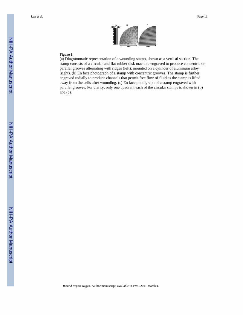
Figure 1.(a) Diagrammatic representation of a wounding stamp, shown as a vertical section. Thestamp consists of a circular and flat rubber disk machine engraved to produce concentric orparallel grooves alternating with ridges (left), mounted on a cylinder of aluminum alloy(right). (b) En face photograph of a stamp with concentric grooves. The stamp is furtherengraved radially to produce channels that permit free flow of fluid as the stamp is liftedaway from the cells after wounding. (c) En face photograph of a stamp engraved withparallel grooves. For clarity, only one quadrant each of the circular stamps is shown in (b)and (c).
Lan et al. Page 11
Wound Repair Regen. Author manuscript; available in PMC 2011 March 4.
NIH
-PA Author Manuscript
NIH
-PA Author Manuscript
NIH
-PA Author Manuscript
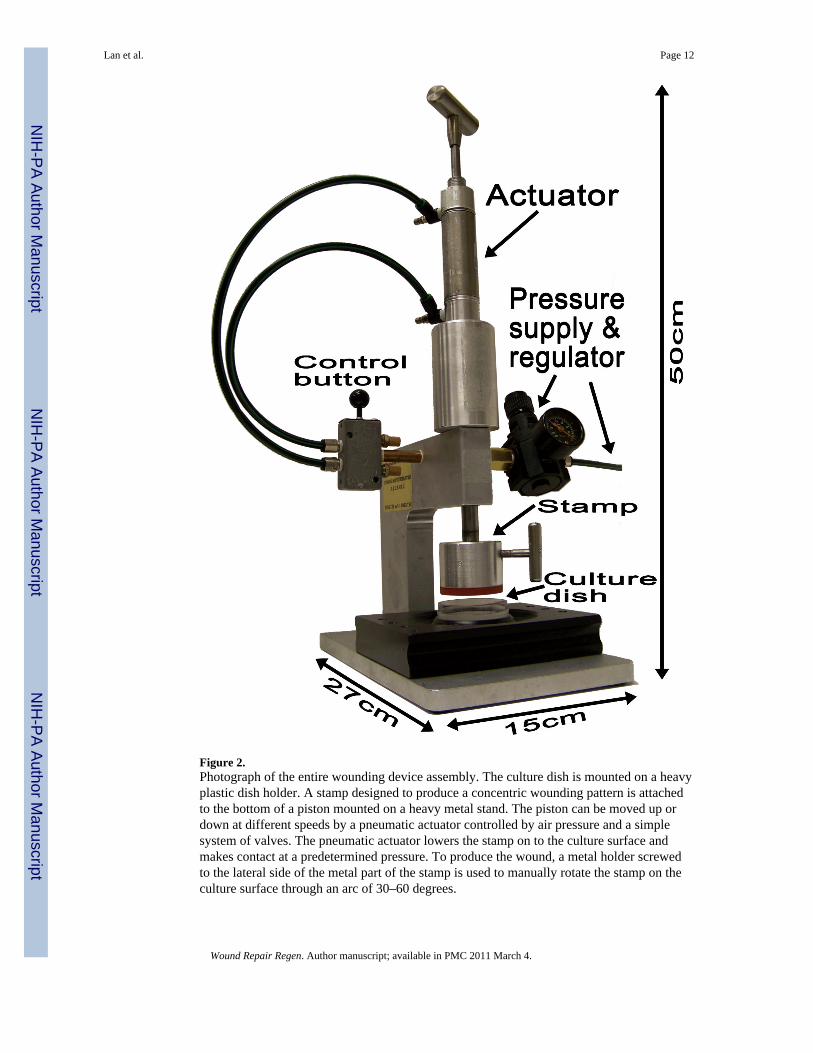
Figure 2.Photograph of the entire wounding device assembly. The culture dish is mounted on a heavyplastic dish holder. A stamp designed to produce a concentric wounding pattern is attachedto the bottom of a piston mounted on a heavy metal stand. The piston can be moved up ordown at different speeds by a pneumatic actuator controlled by air pressure and a simplesystem of valves. The pneumatic actuator lowers the stamp on to the culture surface andmakes contact at a predetermined pressure. To produce the wound, a metal holder screwedto the lateral side of the metal part of the stamp is used to manually rotate the stamp on theculture surface through an arc of 30–60 degrees.
Lan et al. Page 12
Wound Repair Regen. Author manuscript; available in PMC 2011 March 4.
NIH
-PA Author Manuscript
NIH
-PA Author Manuscript
NIH
-PA Author Manuscript
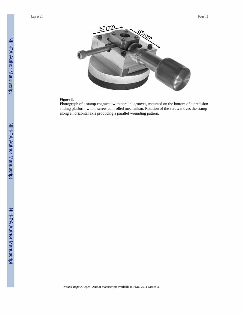
Figure 3.Photograph of a stamp engraved with parallel grooves, mounted on the bottom of a precisionsliding platform with a screw controlled mechanism. Rotation of the screw moves the stampalong a horizontal axis producing a parallel wounding pattern.
Lan et al. Page 13
Wound Repair Regen. Author manuscript; available in PMC 2011 March 4.
NIH
-PA Author Manuscript
NIH
-PA Author Manuscript
NIH
-PA Author Manuscript
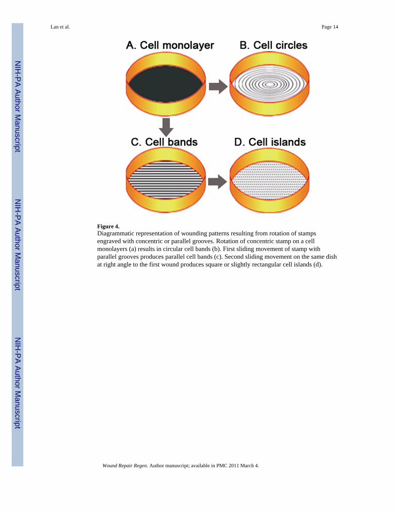
Figure 4.Diagrammatic representation of wounding patterns resulting from rotation of stampsengraved with concentric or parallel grooves. Rotation of concentric stamp on a cellmonolayers (a) results in circular cell bands (b). First sliding movement of stamp withparallel grooves produces parallel cell bands (c). Second sliding movement on the same dishat right angle to the first wound produces square or slightly rectangular cell islands (d).
Lan et al. Page 14
Wound Repair Regen. Author manuscript; available in PMC 2011 March 4.
NIH
-PA Author Manuscript
NIH
-PA Author Manuscript
NIH
-PA Author Manuscript

Figure 5.Still frames from time-lapse video microscopy of cells wounded in a concentric circularpattern. Confluent monolayers of proximal tubule (BUMPT) cells were wounded using astamp that yielded concentric cell bands of ~130 μm width. The right panel at “0 hour”depicts a low power scanned picture of a wounded dish fixed and stained with alcoholictoluidine blue/basic fuchsin.
Lan et al. Page 15
Wound Repair Regen. Author manuscript; available in PMC 2011 March 4.
NIH
-PA Author Manuscript
NIH
-PA Author Manuscript
NIH
-PA Author Manuscript
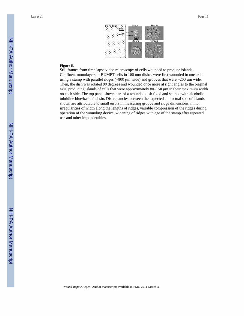
Figure 6.Still frames from time lapse video microscopy of cells wounded to produce islands.Confluent monolayers of BUMPT cells in 100 mm dishes were first wounded in one axisusing a stamp with parallel ridges (~800 μm wide) and grooves that were ~200 μm wide.Then, the dish was rotated 90 degrees and wounded once more at right angles to the originalaxis, producing islands of cells that were approximately 80–150 μm in their maximum widthon each side. The top panel shows part of a wounded dish fixed and stained with alcoholictoluidine blue/basic fuchsin. Discrepancies between the expected and actual size of islandsshown are attributable to small errors in measuring groove and ridge dimensions, minorirregularities of width along the lengths of ridges, variable compression of the ridges duringoperation of the wounding device, widening of ridges with age of the stamp after repeateduse and other imponderables.
Lan et al. Page 16
Wound Repair Regen. Author manuscript; available in PMC 2011 March 4.
NIH
-PA Author Manuscript
NIH
-PA Author Manuscript
NIH
-PA Author Manuscript
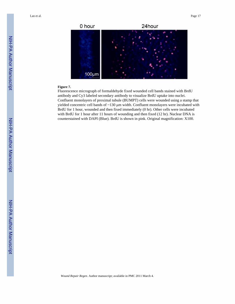
Figure 7.Fluorescence micrograph of formaldehyde fixed wounded cell bands stained with BrdUantibody and Cy3 labeled secondary antibody to visualize BrdU uptake into nuclei.Confluent monolayers of proximal tubule (BUMPT) cells were wounded using a stamp thatyielded concentric cell bands of ~130 μm width. Confluent monolayers were incubated withBrdU for 1 hour, wounded and then fixed immediately (0 hr). Other cells were incubatedwith BrdU for 1 hour after 11 hours of wounding and then fixed (12 hr). Nuclear DNA iscounterstained with DAPI (Blue). BrdU is shown in pink. Original magnification: X100.
Lan et al. Page 17
Wound Repair Regen. Author manuscript; available in PMC 2011 March 4.
NIH
-PA Author Manuscript
NIH
-PA Author Manuscript
NIH
-PA Author Manuscript
Figure 8.Cellular concentration of mRNA for c-Fos, c-Jun and Jun B measured by Q-PCR at serialtime intervals after wounding. Confluent monolayers of proximal tubule (BUMPT) cellswere wounded using a stamp that yielded concentric cell bands of ~130 μm width. RNA wasextracted from wounded cultures and corresponding non-wounded time controls at serialtime intervals. N=3.
Lan et al. Page 18
Wound Repair Regen. Author manuscript; available in PMC 2011 March 4.
NIH
-PA Author Manuscript
NIH
-PA Author Manuscript
NIH
-PA Author Manuscript
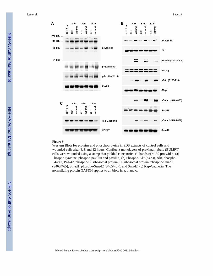
Figure 9.Western Blots for proteins and phosphoproteins in SDS extracts of control cells andwounded cells after 4, 8 and 12 hours. Confluent monolayers of proximal tubule (BUMPT)cells were wounded using a stamp that yielded concentric cell bands of ~130 μm width. (a)Phospho-tyrosine, phospho-paxillin and paxillin; (b) Phospho-Akt (S473), Akt, phospho-P44/42, P44/42, phospho-S6 ribosomal protein, S6 ribosomal protein, phospho-Smad1(S463/465), Smad1, phospho-Smad2 (S465/467), and Smad2. (c) Ksp-Cadherin. Thenormalizing protein GAPDH applies to all blots in a, b and c.
Lan et al. Page 19
Wound Repair Regen. Author manuscript; available in PMC 2011 March 4.
NIH
-PA Author Manuscript
NIH
-PA Author Manuscript
NIH
-PA Author Manuscript
Related Documents