ALTEX 25, 3/08 191 A Newly Developed In Vitro Model of the Human Epithelial Airway Barrier to Study the Toxic Potential of Nanoparticles Barbara Rothen-Rutishauser 1 , Loretta Müller 1 , Fabian Blank 2 , Christina Brandenberger 1 , Christian Mühlfeld 1 and Peter Gehr 1 1 Institute of Anatomy, Division of Histology, University of Bern, Bern, Switzerland; 2 Telethon Institute for Child Health Research, Subiaco, WA, Australia Summary The potential health effects of inhaled engineered nanoparticles are almost unknown. To avoid and replace toxicity studies with animals, a triple cell co-culture system composed of epithelial cells, macrophages and dendritic cells was established, which simulates the most important barrier functions of the epithelial airway. Using this model, the toxic potential of titanium dioxide was assessed by measuring the production of reactive oxygen species and the release of tumour necrosis factor alpha. The intracellular localisation of titanium dioxide nanoparticles was analyzed by energy filtering transmission electron microscopy. Titanium dioxide nanoparticles were detected as single parti- cles without membranes and in membrane-bound agglomer- ates. Cells incubated with titanium dioxide particles showed an elevated production of reactive oxygen species but no increase of the release of tumour necrosis factor alpha. Our in vitro model of the epithelial airway barrier offers a valu- able tool to study the interaction of particles with lung cells at a nanostructural level and to investigate the toxic potential of nanoparticles. Zusammenfassung: Ein neu entwickeltes in vitro Modell für die menschliche epitheliale Lungenbarriere zur Prüfung des tox- ischen Potentials von Nanopartikeln Die möglichen gesundheitlichen Auswirkungen von einge- atmeten Nanopartikeln sind bisher weitgehend unbekannt. Wir haben ein Zellkulturmodell der epithelialen Lungenbarri- ere, bestehend aus Epithelzellen, Makrophagen und dendriti- schen Zellen, aufgebaut, um die Interaktion von Partikeln mit Zellen und die Partikel-Toxizität zu studieren. Dieses Modell kann Toxizitätsversuche mit Tieren reduzieren und allenfalls ersetzen. Die allfällige Auslösung von oxidativem Stress und Entzün- dungsreaktionen durch Titandioxid Nanopartikel wurde mit diesem Kulturmodel untersucht. Die intrazelluläre Lokalisation dieser Partikel konnte mittels Elektronen-Energie-Verlust-Spek- troskopie nachgewiesen werden. In den verschiedenen Zelltypen des Modells konnten Titandi- oxid Nanopartikel membranumhüllt wie auch frei im Zyto- plasma detektiert werden. Die Inkubation mit Titandioxid Nanopartikeln löste in den Zellen oxidativen Stress aber keine Entzündungsreaktion aus. Mit unserem in vitro Modell der epithelialen Lungenbarrie- re steht uns ein Testssystem zur Verfügung, um Partikel-Zell- Interaktionen und das toxische Potential von Nanopartikeln zu stu- dieren, sowie die Zell-Zell-Interaktion nach Partikelex-position. Keywords: epithelial airway barrier, nanoparticles, toxicity, oxidative stress, pro-inflammatory reactions 1 Introduction During the last years there has been a substantial increase in the debate on the potential harmful effects of nanomateri- als (Donaldson et al., 2005; Oberdörster et al., 2005a, Maynard and Michelson, 2006; Mühlfeld et al., 2008; Nel et al., 2006; Poland et al., 2008; Takagi et al., 2008). Among these nanomaterials, consisting of particles ≤100 nm, concern is expressed about their possible toxicity (Borm et al., 2006a). An important basis for the cur- rent concerns about the possible adverse health effects of NP has been provided by research in the field of inhalation toxicol- ogy (Oberdörster et al., 2005b). It has been observed that in rodent inhalation studies a number of different nanoparticulate ma- terials show considerably stronger pul- monary toxicity when compared at equal mass dose with larger particles (Ober- dörster et al., 2005a; Stoeger et al., 2006). Recent studies indicate the importance of exposed surface area as the most appropri- ate dose metric for NP since it has been found that particle surface area shows the closest correlation with the inflammatory response (Stoeger et al., 2006). Received 22 th May 2008; received in final form and accepted for publication 17 th June 2008

Welcome message from author
This document is posted to help you gain knowledge. Please leave a comment to let me know what you think about it! Share it to your friends and learn new things together.
Transcript
Altex 25, 3/08 191
A Newly Developed In Vitro Model of the Human Epithelial Airway Barrier to Study the Toxic Potential of NanoparticlesBarbara Rothen-Rutishauser1, Loretta Müller1, Fabian Blank2, Christina Brandenberger1, Christian Mühlfeld1 and Peter Gehr11 Institute of Anatomy, Division of Histology, University of Bern, Bern, Switzerland; 2 telethon Institute for Child Health Research, Subiaco, WA, Australia
SummaryThe potential health effects of inhaled engineered nanoparticles are almost unknown. To avoid and replace toxicity studies with animals, a triple cell co-culture system composed of epithelial cells, macrophages and dendritic cells was established, which simulates the most important barrier functions of the epithelial airway.Using this model, the toxic potential of titanium dioxide was assessed by measuring the production of reactive oxygen species and the release of tumour necrosis factor alpha. The intracellular localisation of titanium dioxide nanoparticles was analyzed by energy filtering transmission electron microscopy. Titanium dioxide nanoparticles were detected as single parti-cles without membranes and in membrane-bound agglomer-ates. Cells incubated with titanium dioxide particles showed an elevated production of reactive oxygen species but no increase of the release of tumour necrosis factor alpha.Our in vitro model of the epithelial airway barrier offers a valu-able tool to study the interaction of particles with lung cells at a nanostructural level and to investigate the toxic potential of nanoparticles.
Zusammenfassung: ein neu entwickeltes in vitro Modell für die menschliche epitheliale lungenbarriere zur Prüfung des tox-ischen Potentials von NanopartikelnDie möglichen gesundheitlichen Auswirkungen von einge- atmeten Nanopartikeln sind bisher weitgehend unbekannt. Wir haben ein Zellkulturmodell der epithelialen Lungenbarri-ere, bestehend aus Epithelzellen, Makrophagen und dendriti-schen Zellen, aufgebaut, um die Interaktion von Partikeln mit Zellen und die Partikel-Toxizität zu studieren. Dieses Modell kann Toxizitätsversuche mit Tieren reduzieren und allenfalls ersetzen.Die allfällige Auslösung von oxidativem Stress und Entzün-dungsreaktionen durch Titandioxid Nanopartikel wurde mit diesem Kulturmodel untersucht. Die intrazelluläre Lokalisation dieser Partikel konnte mittels Elektronen-Energie-Verlust-Spek-troskopie nachgewiesen werden.In den verschiedenen Zelltypen des Modells konnten Titandi-oxid Nanopartikel membranumhüllt wie auch frei im Zyto-plasma detektiert werden. Die Inkubation mit Titandioxid Nanopartikeln löste in den Zellen oxidativen Stress aber keine Entzündungsreaktion aus.Mit unserem in vitro Modell der epithelialen Lungenbarrie-re steht uns ein Testssystem zur Verfügung, um Partikel-Zell- Interaktionen und das toxische Potential von Nanopartikeln zu stu-dieren, sowie die Zell-Zell-Interaktion nach Partikelex-position.
Keywords: epithelial airway barrier, nanoparticles, toxicity, oxidative stress, pro-inflammatory reactions
1 Introduction
During the last years there has been a substantial increase in the debate on the potential harmful effects of nanomateri-als (Donaldson et al., 2005; Oberdörster et al., 2005a, Maynard and Michelson, 2006; Mühlfeld et al., 2008; Nel et al., 2006; Poland et al., 2008; takagi et al., 2008).
Among these nanomaterials, consisting of particles ≤100 nm, concern is expressed about their possible toxicity (Borm et al., 2006a). An important basis for the cur-rent concerns about the possible adverse health effects of NP has been provided by research in the field of inhalation toxicol-ogy (Oberdörster et al., 2005b). It has been observed that in rodent inhalation studies
a number of different nanoparticulate ma-terials show considerably stronger pul-monary toxicity when compared at equal mass dose with larger particles (Ober-dörster et al., 2005a; Stoeger et al., 2006). Recent studies indicate the importance of exposed surface area as the most appropri-ate dose metric for NP since it has been found that particle surface area shows the closest correlation with the inflammatory response (Stoeger et al., 2006).
Received 22th May 2008; received in final form and accepted for publication 17th June 2008
Rothen-RutishauseR et al.
Altex 25, 3/08192
described in detail in Rothen-Rutishauser et al. (2007). Briefly, the particle suspen-sions were sonicated for 2 min prior to incubation with cells. A stock solution of tiO2 particles in millipore water (2.5 mg/ml) was diluted in RPMI 1640 to a final concentration of 5 µg/ml. One ml of this suspension was then added to cell cul-tures. Incubations were performed for 24 h before analysis of the cells.
Visualisation and quantification of carboxy-dichlorodihydrofluorescein dia-cetate (H2DCFD) oxidation in cells by laser scanning microscopy Following particle incubation, the Im-age-it™ lIVe detection kit for reac-tive oxygen species (Molecular Probes, Invitrogen AG, Basel, Switzerland) was used for the detection of ROS in live cells according to the user manual. Briefly, cells were incubated with 25 µM carboxy-H2DCFDA and the cell nuclei were stained with Hoechst. After 30 min incubation in the dark, the cells were mounted in warm buffer and imaged im-mediately. The fluorescence in cells was visualised by conventional fluorescence microscopy.
Cell labelling and fixationthe cells were labelled as described in detail in Rothen-Rutishauser et al. (2005). Briefly, the cultures were fixed, permeabilised and incubated with the first and second antibodies. Preparations were mounted in Mowiol on microscopic slides. Antibodies were diluted in phos-phate buffered saline (PBS) as follows: mouse anti-human CD14 1:20 (Clone UCHM-1, C 7673, Sigma), mouse an-ti-human CD86 1:20 (Clone HB15e, 36931A, PharMingen, BD Biosciences,), goat anti-mouse cyanine 5 1:50 (AP124S, Chemicon, VWR International AG, life Sciences, lucerne, Switzerland) and phalloidin rhodamine 1:100 (R-415, Mo-lecular Probes, Invitrogen AG, Basel, Switzerland).
Laser scanning microscopy and image restorationA Zeiss lSM 510 Meta with an inverted Zeiss microscope (Axiovert 200M, la-sers: HeNe 633 nm, HeNe 543 nm and Ar 488 nm) was used. Image processing and visualisation was performed using IMA-
(tiO2) NP was studied using energy fil-tering transmission electron microscopy (eFteM). tiO2 NP were among the ear-liest industrially produced and applied NP in everyday applications (Maynard and Michelson, 2006). the potential of these particles to induce a toxic reaction was investigated by measurement of re-active oxygen species (ROS) and tumour necrosis factor alpha (TNF-α).
2 Material and methods
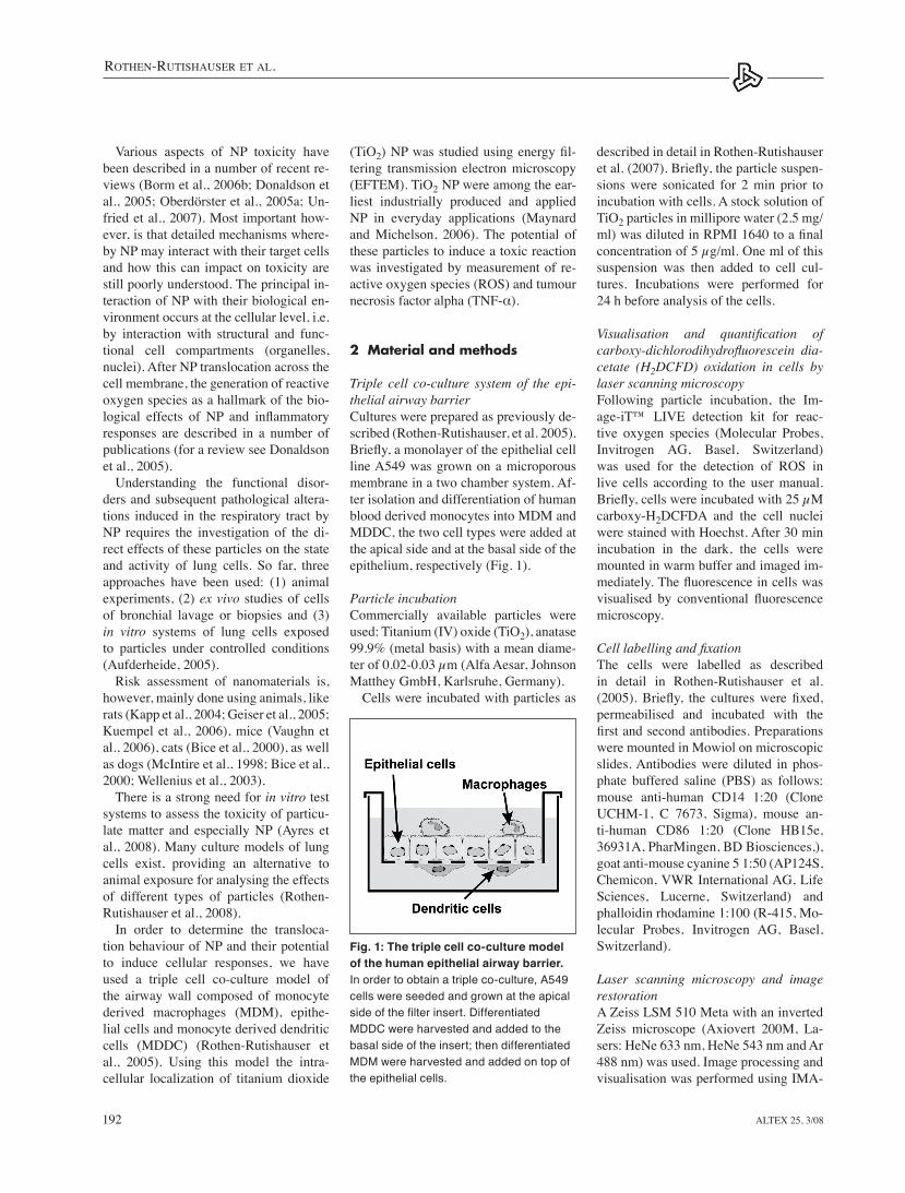
Triple cell co-culture system of the epi-thelial airway barrierCultures were prepared as previously de-scribed (Rothen-Rutishauser, et al. 2005). Briefly, a monolayer of the epithelial cell line A549 was grown on a microporous membrane in a two chamber system. Af-ter isolation and differentiation of human blood derived monocytes into MDM and MDDC, the two cell types were added at the apical side and at the basal side of the epithelium, respectively (Fig. 1).
Particle incubationCommercially available particles were used: titanium (IV) oxide (tiO2), anatase 99.9% (metal basis) with a mean diame-ter of 0.02-0.03 µm (Alfa Aesar, Johnson Matthey GmbH, Karlsruhe, Germany).
Cells were incubated with particles as
Various aspects of NP toxicity have been described in a number of recent re-views (Borm et al., 2006b; Donaldson et al., 2005; Oberdörster et al., 2005a; Un-fried et al., 2007). Most important how-ever, is that detailed mechanisms where-by NP may interact with their target cells and how this can impact on toxicity are still poorly understood. the principal in-teraction of NP with their biological en-vironment occurs at the cellular level, i.e. by interaction with structural and func-tional cell compartments (organelles, nuclei). After NP translocation across the cell membrane, the generation of reactive oxygen species as a hallmark of the bio-logical effects of NP and inflammatory responses are described in a number of publications (for a review see Donaldson et al., 2005).
Understanding the functional disor-ders and subsequent pathological altera-tions induced in the respiratory tract by NP requires the investigation of the di-rect effects of these particles on the state and activity of lung cells. So far, three approaches have been used: (1) animal experiments, (2) ex vivo studies of cells of bronchial lavage or biopsies and (3) in vitro systems of lung cells exposed to particles under controlled conditions (Aufderheide, 2005).
Risk assessment of nanomaterials is, however, mainly done using animals, like rats (Kapp et al., 2004; Geiser et al., 2005; Kuempel et al., 2006), mice (Vaughn et al., 2006), cats (Bice et al., 2000), as well as dogs (McIntire et al., 1998; Bice et al., 2000; Wellenius et al., 2003).
there is a strong need for in vitro test systems to assess the toxicity of particu-late matter and especially NP (Ayres et al., 2008). Many culture models of lung cells exist, providing an alternative to animal exposure for analysing the effects of different types of particles (Rothen-Rutishauser et al., 2008).
In order to determine the transloca-tion behaviour of NP and their potential to induce cellular responses, we have used a triple cell co-culture model of the airway wall composed of monocyte derived macrophages (MDM), epithe-lial cells and monocyte derived dendritic cells (MDDC) (Rothen-Rutishauser et al., 2005). Using this model the intra-cellular localization of titanium dioxide
Fig. 1: The triple cell co-culture model of the human epithelial airway barrier. In order to obtain a triple co-culture, A549 cells were seeded and grown at the apical side of the filter insert. Differentiated MDDC were harvested and added to the basal side of the insert; then differentiated MDM were harvested and added on top of the epithelial cells.
Rothen-RutishauseR et al.
Altex 25, 3/08 193
model was evaluated carefully and com-pared with in vivo data and we found that the morphological (Rothen-Rutishauser et al., 2008) as well as the quantitative occurrence of macrophages and dendritic cells resembled very closely the in vivo situation (Blank et al., 2007).
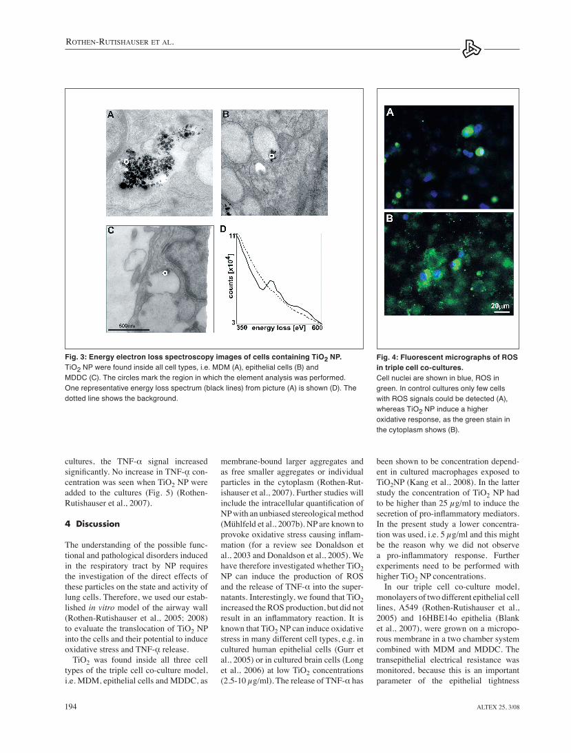
Intracellular TiO2 localisationAfter its thorough evaluation, this model was exposed to tiO2 NP (mean diameter of 0.032 µm) and the particles were visu-alised and analysed in cells using eFteM (Rothen-Rutishauser et al., 2006). Bigger membrane-bound aggregates (>0.2 µm) of tiO2 NP were identified in all cell types (Fig. 3). In addition we found sin-gle particles and smaller (<0.2 µm) ag-gregates that were not membrane bound (data not shown) (Rothen-Rutishauser et al., 2007).
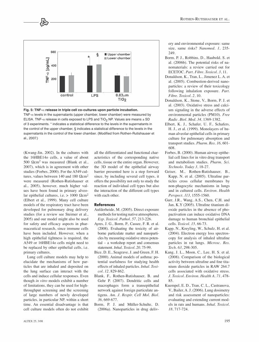
Production of ROS upon particle expo-sureAfter particle incubation, the production of ROS was visualised using carboxy-DCFH, a reliable fluorogenic marker for ROS in live cells. tiO2 NP can induce a higher oxidative stress when compared with control cultures (Fig. 4).
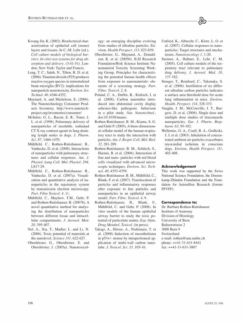
TNF-a release into supernatants after particle incubationThe pro-inflammatory cytokine TNF-α released into the culture supernatants after incubation with particles for 24 h was determined. In the control cultures, minor tNF-α concentrations were meas-ured. When lPS (lipopolysaccaride), a positive control, was added to the cell
Systems, Catalogue Number: DY 210, Oxon, UK) according to the manufac-turer’s recommendations.
3 Results
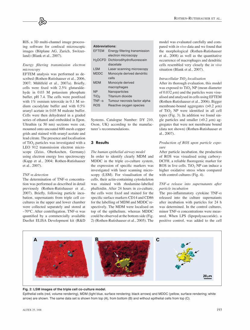
The human epithelial airway modelIn order to identify clearly MDM and MDDC in the triple co-culture system, the expression of specific markers was investigated with laser scanning micro-scopy (lSM). For visualisation of the cells, their actin-containing cytoskeleton was stained with rhodamine-labelled phalloidin. After 24 hours in co-culture, the cells were fixed and stained for the specific surface markers CD14 and CD86 for the labelling of MDM and MDDC re-spectively. the MDM were localised on top of the epithelium, whereas MDDC could be observed at the bottom side (Fig. 2) (Rothen-Rutishauser et al., 2005). the
RIS, a 3D multi-channel image process-ing software for confocal microscopic images (Bitplane AG, Zurich, Switzer-land) (Blank et al., 2007).
Energy filtering transmission electron microscopyeFteM analysis was performed as de-scribed (Rothen-Rutishauser et al., 2006, 2007; Mühlfeld et al., 2007a). Briefly, cells were fixed with 2.5% glutaralde-hyde in 0.03 M potassium phosphate buffer, pH 7.4. The cells were postfixed with 1% osmium tetroxide in 0.1 M so-dium cacodylate buffer and with 0.5% uranyl acetate in 0.05 M maleate buffer. Cells were then dehydrated in a graded series of ethanol and embedded in epon. Ultrathin (≤ 50 nm) sections were cut, mounted onto uncoated 600-mesh copper grids and stained with uranyl acetate and lead citrate. the presence and localisation of tiO2 particles was investigated with a leO 912 transmission electron micro-scope (Zeiss, Oberkochen, Germany) using electron energy loss spectroscopy (Kapp et al., 2004; Rothen-Rutishauser et al., 2007).
TNF-α detectionThe determination of TNF-α concentra-tion was performed as described in detail previously (Rothen-Rutishauser et al., 2007). Briefly, following particle incu-bation, supernatants from triple cell co-cultures in the upper and lower chamber were collected separately and stored at –70°C. After centrifugation, TNF-α was quantified by a commercially available DuoSet elISA Development kit (R&D
Fig. 2: LSM images of the triple cell co-culture model. Epithelial cells (red, volume rendering), MDM (light blue, surface rendering; black arrows) and MDDC (yellow, surface rendering; white arrow) are shown. The same data set is shown from top (A), from bottom (B) and without epithelial cells from top (C).
Abbreviations: EFTEM Energy filtering transmission electron microscopyH2DCFD Dichlorodihydrofluorescein diacetateLSM Laser scanning microscopyMDDC Monocyte derived dendritic cellsMDM Monocyte derived macrophagesNP NanoparticlesTiO2 Titanium dioxideTNF- α Tumour necrosis factor alphaROS Reactive oxygen species
Rothen-RutishauseR et al.
Altex 25, 3/08194
been shown to be concentration depend-ent in cultured macrophages exposed to tiO2NP (Kang et al., 2008). In the latter study the concentration of tiO2 NP had to be higher than 25 µg/ml to induce the secretion of pro-inflammatory mediators. In the present study a lower concentra-tion was used, i.e. 5 µg/ml and this might be the reason why we did not observe a pro-inflammatory response. Further experiments need to be performed with higher tiO2 NP concentrations.
In our triple cell co-culture model, monolayers of two different epithelial cell lines, A549 (Rothen-Rutishauser et al., 2005) and 16HBe14o epithelia (Blank et al., 2007), were grown on a micropo-rous membrane in a two chamber system combined with MDM and MDDC. the transepithelial electrical resistance was monitored, because this is an important parameter of the epithelial tightness
membrane-bound larger aggregates and as free smaller aggregates or individual particles in the cytoplasm (Rothen-Rut-ishauser et al., 2007). Further studies will include the intracellular quantification of NP with an unbiased stereological method (Mühlfeld et al., 2007b). NP are known to provoke oxidative stress causing inflam-mation (for a review see Donaldson et al., 2003 and Donaldson et al., 2005). We have therefore investigated whether tiO2 NP can induce the production of ROS and the release of TNF-α into the super-natants. Interestingly, we found that tiO2 increased the ROS production, but did not result in an inflammatory reaction. It is known that tiO2 NP can induce oxidative stress in many different cell types, e.g. in cultured human epithelial cells (Gurr et al., 2005) or in cultured brain cells (long et al., 2006) at low tiO2 concentrations (2.5-10 µg/ml). The release of TNF-α has
cultures, the tNF-α signal increased significantly. No increase in TNF-ᾳ con-centration was seen when tiO2 NP were added to the cultures (Fig. 5) (Rothen-Rutishauser et al., 2007).
4 Discussion
the understanding of the possible func-tional and pathological disorders induced in the respiratory tract by NP requires the investigation of the direct effects of these particles on the state and activity of lung cells. therefore, we used our estab-lished in vitro model of the airway wall (Rothen-Rutishauser et al., 2005; 2008) to evaluate the translocation of tiO2 NP into the cells and their potential to induce oxidative stress and TNF-ᾳ release.
tiO2 was found inside all three cell types of the triple cell co-culture model, i.e. MDM, epithelial cells and MDDC, as
Fig. 4: Fluorescent micrographs of ROS in triple cell co-cultures. Cell nuclei are shown in blue, ROS in green. In control cultures only few cells with ROS signals could be detected (A), whereas TiO2 NP induce a higher oxidative response, as the green stain in the cytoplasm shows (B).
Fig. 3: Energy electron loss spectroscopy images of cells containing TiO2 NP. TiO2 NP were found inside all cell types, i.e. MDM (A), epithelial cells (B) and MDDC (C). The circles mark the region in which the element analysis was performed. One representative energy loss spectrum (black lines) from picture (A) is shown (D). The dotted line shows the background.
Rothen-RutishauseR et al.
Altex 25, 3/08 195
ery and environmental exposure: same size, same risks? Nanomed. 1, 235-249.
Borm, P. J., Robbins, D., Haubold, S. et al. (2006b). the potential risks of na-nomaterials: a review carried out for eCetOC. Part. Fibre. Toxicol. 3, 11.
Donaldson, K., tran, l., Jimenez l. A. et al. (2005). Combustion-derived nano-particles: a review of their toxicology following inhalation exposure. Part. Fibre. Toxicol. 2, 10.
Donaldson, K., Stone, V., Borm, P. J. et al. (2003). Oxidative stress and calci-um signaling in the adverse effects of environmental particles (PM10). Free Radic. Biol. Med. 34, 1369-1382.
elbert, K. J., Schafer, U. F., Schafers, H. J., et al. (1999). Monolayers of hu-man alveolar epithelial cells in primary culture for pulmonary absorption and transport studies. Pharm. Res. 16, 601-608.
Forbes, B. (2000). Human airway epithe-lial cell lines for in vitro drug transport and metabolism studies. Pharm. Sci. Technolo. Today 3, 18-27.
Geiser, M., Rothen-Rutishauser, B., Kapp, N. et al. (2005). Ultrafine par-ticles cross cellular membranes by non-phagocytic mechanisms in lungs and in cultured cells. Environ. Health Perspect. 113, 1555-1560.
Gurr, J.R., Wang, A.S., Chen, C.H. and Jan, K.Y (2005). Ultrafine titanium di-oxide particles in the absence of pho-pactivation can induce oxidative DNA damage to human bronchial epithelial cells. Toxicol. 15, 66-73.
Kapp, N., Kreyling, W., Schulz, H. et al. (2004). electron energy loss spectros-copy for analysis of inhaled ultrafine particles in rat lungs. Microsc. Res. Tech. 63, 298-305.
Kang, J. l., Moon, C., lee, H. S. et al. (2008). Comparison of the biological activity between ultrafine and fine tita-nium dioxide particles in RAW 264.7 cells associated with oxidative stress. J. Toxicol. Environ. Health A. 71, 478-85.
Kuempel, e. D., tran, C. l., Castranova, V., Bailer, A. J. (2006). lung dosimetry and risk assessment of nanoparticles: evaluating and extending current mod-els in rats and humans. Inhal. Toxicol. 18, 717-724.
all the differentiated and functional char-acteristics of the corresponding native cells, tissue or the entire organ. However, the 3D model of the epithelial airway barrier presented here is a step forward since, by including several cell types, it offers the possibility not only to study the reaction of individual cell types but also the interaction of the different cell types with each other.
ReferencesAufderheide, M. (2005). Direct exposure
methods for testing native atmospheres. Exp. Toxicol. Pathol. 57, 213-226.
Ayres, J. G., Borm, P., Cassee, F. R. et al. (2008). evaluating the toxicity of air-borne particulate matter and nanoparti-cles by measuring oxidative stress poten-tial – a workshop report and consensus statement. Inhal. Toxicol. 20, 75-99.
Bice, D. e., Seagrave, J. and Green, F. H. (2000). Animal models of asthma: po-tential usefulness for studying health effects of inhaled particles. Inhal. Toxi-col. 12, 829-862.
Blank, F., Rothen-Rutishauser, B. and Gehr P. (2007). Dendritic cells and macrophages form a transepithelial network against foreign particulate an-tigens. Am. J. Respir. Cell Mol. Biol. 36, 669-677.
Borm, P. J. and Müller-Schulte, D. (2006a). Nanoparticles in drug deliv-
(Kwang-Jin, 2002). In the cultures with the 16HBe14o cells, a value of about 500 Ωcm2
was measured (Blank et al., 2007), which is in agreement with other studies (Forbes, 2000). For the A549 cul-tures, values between 140 and 180 Ωcm2 were measured (Rothen-Rutishauser et al., 2005), however, much higher val-ues have been found in primary alveo-lar epithelial cultures, i.e. > 1000 Ωcm2 (elbert et al., 1999). Many cell culture models of the respiratory tract have been developed for pulmonary drug delivery studies (for a review see Steimer et al., 2005) and our model might also be used for safety and efficacy aspects in phar-maceutical research, since immune cells have been included. However, when a high epithelial tightness is required, the A549 or 16HBe14o cells might need to be replaced by other epithelial cells, i.e. primary cultures.
lung cell culture models may help to elucidate the mechanisms of how par-ticles that are inhaled and deposited on the lung surface can interact with the cells and induce cellular responses. even though in vitro models exhibit a number of limitations, they can be used for high-throughput screening and the screening of large numbers of newly developed particles, in particular NP, within a short time. An essential disadvantage is that cell culture models often do not exhibit
Fig. 5: TNF-α release in triple cell co-cultures upon particle incubation. TNF-α levels in the supernatants (upper chamber, lower chamber) were measured by ELISA. TNF-α release in cells exposed to LPS and TiO2 NP. Values are means ± SD of 3 experiments. * indicates a statistical difference to the levels in the supernatants in the control of the upper chamber, § indicates a statistical difference to the levels in the supernatants in the control of the lower chamber. (Modified from Rothen-Rutishauser et al., 2007.)
Rothen-RutishauseR et al.
Altex 25, 3/08196
ogy: an emerging discipline evolving from studies of ultrafine particles. En-viron. Health Perspect. 113, 823-839.
Oberdörster, G., Maynard, A., Donald-son, K. et al. (2005b). IlSI Research Foundation/Risk Science Institute Na-nomaterial toxicity Screening Work-ing Group, Principles for characteriz-ing the potential human health effects from exposure to nanomaterials: ele-ments of a screening strategy. Part. Fibre. Toxicol. 2, 8.
Poland, C. A., Duffin, R., Kinloch, I. et al. (2008). Carbon nanotubes intro-duced into abdominal cavity display asbestos-like pathogenic behaviour in a pilot study. Nat. Nanotechnol., doi:10.1038/nnano.
Rothen-Rutishauser, B. M., Kiama, S. G. and Gehr P. (2005). A three-dimension-al cellular model of the human respira-tory tract to study the interaction with particles. Am. J. Respir. Cell. Mol. Biol. 32, 281-289.
Rothen-Rutishauser, B. M., Schürch, S., Haenni, B. et al. (2006). Interaction of fine and nano- particles with red blood cells visualized with advanced micro-scopic techniques. Environ. Sci. Tech-nol. 40, 4353-4359.
Rothen-Rutishauser, B. M., Mühlfeld, C., Blank, F. et al. (2007). translocation of particles and inflammatory responses after exposure to fine particles and nanoparticles in an epithelial airway model. Part. Fibre. Toxicol. 4, 9.
Rothen-Rutishauser, B., Blank, F., Mühlfeld, C. and Gehr, P. (2008). In vitro models of the human epithelial airway barrier to study the toxic po-tential of particulate matter. Exp. Opin. Drug Metabol. Toxicol. (in press).
takagi, A., Hirose, A., Nishimura, t. et al. (2008) Induction of mesothelioma in p53+/- mouse by intraperitoneal ap-plication of multi-wall carbon nano-tube. J. Toxicol. Sci. 33, 105-16.
Kwang-Jin, K. (2002). Bioelectrical char-acterization of epithelial cell (mono)layers and tissues. In C.-M. lehr (ed.), Cell culture models of biological bar-riers. In-vitro test systems for drug ab-sorption and delivery. (3:41-51). lon-don, New York: taylor and Francis.
long, t. C., Saleh, N., tilton, R. D. et al. (2006). titanium dioxide (P25) produces reactive oxygen species in immortalized brain microglia (BV2): implications for nanoparticle neurotoxicity. Environ. Sci. Technol. 40, 4346-4352.
Maynard, A. and Michelson, e. (2006). the Nanotechnology Consumer Prod-ucts Inventory. http://www.nanotech-project.org/inventories/consumer/
McIntire, G. l., Bacon, e. R., toner, J. l. et al. (1998). Pulmonary delivery of nanoparticles of insoluble, iodinated Ct x-ray contrast agents to lung drain-ing lymph nodes in dogs. J. Pharm. Sci. 87, 1466-1470.
Mühlfeld, C., Rothen-Rutishauser, B., Vanhecke, D. et al. (2008). Interactions of nanoparticles with pulmonary struc-tures and cellular responses. Am. J. Physiol. Lung Cell. Mol. Physiol. 294, l817-29.
Mühlfeld, C., Rothen-Rutishauser, B., Vanhecke, D. et al. (2007a). Visuali-zation and quantitative analysis of na-noparticles in the repsiratory system by transmission electron microscopy. Part. Fibre Toxicol. 4, 11.
Mühlfeld, C., Mayhew, t.M., Gehr, P. and Rothen-Rutishauser, B. (2007b). A novel quantitative method for analys-ing the distribution of nanoparticles between different tissue and intracel-lular compartments. J. Aerosol. Med. 20, 395-407.
Nel, A., xia, t., Madler, l. and li, N. (2006). toxic potential of materials at the nanolevel. Science 311, 622-627.
Oberdörster, G., Oberdörster, e. and Oberdörster, J. (2005a). Nanotoxicol-
Unfried, K., Albrecht, C., Klotz, l. O. et al. (2007). Cellular responses to nano-particles: target structures and mecha-nisms. Nanotoxicology 1, 1-20.
Steimer, A., Haltner, e., lehr, C. M. (2005). Cell culture models of the res-piratory tract relevant to pulmonary drug delivery. J. Aerosol. Med. 18, 137-182.
Stoeger, t., Reinhard, C., takenaka, S. et al. (2006). Instillation of six differ-ent ultrafine carbon particles indicates a surface area threshold dose for acute lung inflammation in mice. Environ. Health. Perspect. 114, 328-333.
Vaughn, J. M., McConville, J. t., Bur-gess, D. et al. (2006). Single dose and multiple dose studies of itraconazole nanoparticles. Eur. J. Pharm. Biop-harm. 63, 95-102.
Wellenius, G. A., Coull, B. A., Godleski, J. J. et al. (2003). Inhalation of concen-trated ambient air particles exacerbates myocardial ischemia in conscious dogs. Environ. Health Perspect. 111, 402-408.
Acknowledgementthis work was supported by the Swiss National Science Foundation, the Doeren-kamp-Zbinden Foundation and the Foun-dation for Animalfree Research (former FFVFF).
Correspondence to:Dr. Barbara Rothen-RutishauserInstitute of AnatomyDivision of HistologyUniversity of BernBaltzerstrasse 23000 Bern 9Switzerlande-mail: [email protected] phone: ++41-31-631-8441fax: ++41-31-631-3807
Related Documents