A Long-Term High-Carbohydrate Diet Causes an Altered Ontogeny of Pancreatic Islets of Langerhans in the Neonatal Rat JAMES PETRIK, MALATHI SRINIVASAN, RAVIKUMAR AALINKEEL, STEPHEN COUKELL, EDITH ARANY, MULCHEND S. PATEL, AND DAVID J. HILL Lawson Research Institute, St. Joseph’s Health Centre, London, Ontario N6A 4V2, Canada [J.P., S.C., E.A., D.J.H.]; Departments of Physiology [J.P., D.J.H.], Medicine [E.A., D.J.H.], and Paediatrics [D.J.H.], University of Western Ontario, London, Ontario, N6A 5A5, Canada; and Department of Biochemistry, School of Medicine and Biomedical Sciences, State University of New York at Buffalo, 3435 Main Street, Buffalo, New York 14214, U.S.A. [M.S., R.A., M.S.P.] Neonatal rats fed a high-carbohydrate (HC) formula by gas- trostomy are hyperinsulinemic but normoglycemic. We deter- mined whether HC formula altered pancreatic islet cell ontogeny. Rats were reared from d 4 on an HC formula or a high-fat formula, or were allowed to suckle naturally, and the pancreata were examined histologically from animals #24 d of age. The mean area of individual islets was reduced, but islet number was increased in HC rats compared with mother-fed or high fat–fed animals, which were similar. Islets from HC animals were rela- tively deficient in a cells and had a greater incidence of islet cells with fragmented DNA, indicative of apoptosis. Ductal epithe- lium, a source of new islets by neogenesis, had a greater inci- dence of cells staining immunopositive for proliferating cell nuclear antigen, a marker of cell replication, and a lower inci- dence of apoptosis. The islet cell mitogen and survival factor, IGF-II, had a reduced mRNA expression in whole pancreas from HC animals. The relative area of islet cells demonstrating IGF-II immunoreactivity was reduced in HC-fed rats versus controls, although a greater percentage of ductal epithelial cells were immunopositive. HC formula alters islet cell ontogeny by affect- ing islet size and number, which may be linked to an altered IGF-II expression. (Pediatr Res 49: 84–92, 2001) Abbreviations HC, high carbohydrate MF, mother fed HF, high fat PCNA, proliferating cell nuclear antigen Hyperinsulinemia in obesity is associated with pancreatic islet hyperplasia and hypertrophy (1, 2). Additionally, both genetic and nutritionally induced obesity in animal models cause impaired glucose tolerance and increased insulin resis- tance (1–3). Such predisposition to obesity and diabetes may be related to nutritional experiences early in fetal life or childhood during critical windows of endocrine pancreatic development. We have previously reported that feeding an HC formula to rat pups by an artificial rearing technique induces hyperinsulinemia, and in the long-term affects lipogenesis, leading to obesity (4). When HC formula was commenced on postnatal d 4, b-cell mass and pancreatic insulin content were increased compared with MF control rats by 12 d of age (5). The hyperinsulinemia, obesity, and glucose intolerance that were observed in the first generation of rats fed an HC diet during their early postnatal life were transmitted, via the females, into the next generation, who were fed a normal diet, leading to hyperinsulinemia, obesity, and impaired glucose tolerance in the next generation of animals also (6, 7). The rapid increase in islet cell mass that occurs in late fetal and neonatal life in the rat may explain why pancreatic mor- phology is so sensitive to nutritional insult at this time. In the 2 d before term, the cellular area staining immunopositive for insulin increases 2-fold because of both b-cell replication and the neogenesis of new b cells from the pancreatic ductal epithelium (8, 9). In the adult animal, the b-cell replication rate is low, approximately 3% per day (10). Development of the endocrine pancreas is associated with a balance between islet cell replication and neogenesis and programmed cell death. We and others have shown previously that a transient wave of apoptosis occurs in the developing rat islets between 1 and 2 wk of postnatal age (10 –12). The mechanisms controlling islet cell neogenesis and apoptosis are not well understood, but there is considerable evidence that IGFs are major contributors Received March 23, 2000; accepted August 22, 2000. Correspondence and reprint requests: Dr. D.J. Hill, Lawson Research Institute, St. Joseph’s Health Centre, 268 Grosvenor Street, London, Ontario N6A 4V2, Canada. Supported by the Juvenile Diabetes Foundation, the Canadian Diabetes Association, the Medical Research Council of Canada, and the National Institutes of Health (Grants HD-11089 and DK-51601). 0031-3998/01/4901-0084 PEDIATRIC RESEARCH Vol. 49, No. 1, 2001 Copyright © 2001 International Pediatric Research Foundation, Inc. Printed in U.S.A. ABSTRACT 84

Welcome message from author
This document is posted to help you gain knowledge. Please leave a comment to let me know what you think about it! Share it to your friends and learn new things together.
Transcript

A Long-Term High-Carbohydrate Diet Causes anAltered Ontogeny of Pancreatic Islets of
Langerhans in the Neonatal RatJAMES PETRIK, MALATHI SRINIVASAN, RAVIKUMAR AALINKEEL, STEPHEN COUKELL,
EDITH ARANY, MULCHEND S. PATEL, AND DAVID J. HILL
Lawson Research Institute, St. Joseph’s Health Centre, London, Ontario N6A 4V2, Canada [J.P., S.C.,E.A., D.J.H.]; Departments of Physiology [J.P., D.J.H.], Medicine [E.A., D.J.H.], and Paediatrics[D.J.H.], University of Western Ontario, London, Ontario, N6A 5A5, Canada; and Department of
Biochemistry, School of Medicine and Biomedical Sciences, State University of New York at Buffalo,3435 Main Street, Buffalo, New York 14214, U.S.A. [M.S., R.A., M.S.P.]
Neonatal rats fed a high-carbohydrate (HC) formula by gas-trostomy are hyperinsulinemic but normoglycemic. We deter-mined whether HC formula altered pancreatic islet cell ontogeny.Rats were reared from d 4 on an HCformula or a high-fatformula, or were allowed to suckle naturally, and the pancreatawere examined histologically from animals#24 d of age. Themean area of individual islets was reduced, but islet number wasincreased in HC rats compared with mother-fed or high fat–fedanimals, which were similar. Islets from HC animals were rela-tively deficient ina cells and had a greater incidence of islet cellswith fragmented DNA, indicative of apoptosis. Ductal epithe-lium, a source of new islets by neogenesis, had a greater inci-dence of cells staining immunopositive for proliferating cellnuclear antigen, a marker of cell replication, and a lower inci-
dence of apoptosis. The islet cell mitogen and survival factor,IGF-II, had a reduced mRNA expression in whole pancreas fromHC animals. The relative area of islet cells demonstrating IGF-IIimmunoreactivity was reduced in HC-fed ratsversuscontrols,although a greater percentage of ductal epithelial cells wereimmunopositive. HC formula alters islet cell ontogeny by affect-ing islet size and number, which may be linked to an alteredIGF-II expression.(Pediatr Res49: 84–92, 2001)
AbbreviationsHC, high carbohydrateMF, mother fedHF, high fatPCNA, proliferating cell nuclear antigen
Hyperinsulinemia in obesity is associated with pancreaticislet hyperplasia and hypertrophy (1, 2). Additionally, bothgenetic and nutritionally induced obesity in animal modelscause impaired glucose tolerance and increased insulin resis-tance (1–3). Such predisposition to obesity and diabetes maybe related to nutritional experiences early in fetal life orchildhood during critical windows of endocrine pancreaticdevelopment. We have previously reported that feeding an HCformula to rat pups by an artificial rearing technique induceshyperinsulinemia, and in the long-term affects lipogenesis,leading to obesity (4). When HC formula was commenced onpostnatal d 4,b-cell mass and pancreatic insulin content wereincreased compared with MF control rats by 12 d of age (5).The hyperinsulinemia, obesity, and glucose intolerance that
were observed in the first generation of rats fed an HC dietduring their early postnatal life were transmitted,via thefemales, into the next generation, who were fed a normal diet,leading to hyperinsulinemia, obesity, and impaired glucosetolerance in the next generation of animals also (6, 7).
The rapid increase in islet cell mass that occurs in late fetaland neonatal life in the rat may explain why pancreatic mor-phology is so sensitive to nutritional insult at this time. In the2 d before term, the cellular area staining immunopositive forinsulin increases 2-fold because of bothb-cell replication andthe neogenesis of newb cells from the pancreatic ductalepithelium (8, 9). In the adult animal, theb-cell replication rateis low, approximately 3% per day (10). Development of theendocrine pancreas is associated with a balance between isletcell replication and neogenesis and programmed cell death. Weand others have shown previously that a transient wave ofapoptosis occurs in the developing rat islets between 1 and 2wk of postnatal age (10–12). The mechanisms controlling isletcell neogenesis and apoptosis are not well understood, butthere is considerable evidence that IGFs are major contributors
Received March 23, 2000; accepted August 22, 2000.Correspondence and reprint requests: Dr. D.J. Hill, Lawson Research Institute, St.
Joseph’s Health Centre, 268 Grosvenor Street, London, Ontario N6A 4V2, Canada.Supported by the Juvenile Diabetes Foundation, the Canadian Diabetes Association, the
Medical Research Council of Canada, and the National Institutes of Health (GrantsHD-11089 and DK-51601).
0031-3998/01/4901-0084PEDIATRIC RESEARCH Vol. 49, No. 1, 2001Copyright © 2001 International Pediatric Research Foundation, Inc. Printed in U.S.A.
ABSTRACT
84

to b-cell growth, maturation, and function, and are expressedby b cells throughout life. We have shown previously that thelevels of IGF-II peptide and mRNA are relatively high in fetaland early neonatal life and then decline sharply toward adult-hood (11, 13). We recently demonstrated that the wave ofb-cell apoptosis during rat pancreatic development is tempo-rally associated with a loss of expression of IGF-II withinpancreatic islets and that the IGFs can act as survival factors,protectingb cells against cytokine-induced apoptosis (11).
The purpose of this study was to examine the impact offeeding an HC formula during early postnatal life on pancreaticislet cell ontogeny, and to determine whether the long-termchanges seen in insulin release could result from an alteredneonatal pancreatic development. A second objective was todetermine whether any ontological changes seen were relatedto an altered local expression of IGF-II.
METHODS
Animal studies.All animal protocols were approved by theInstitutional Animal Care and Use Committee. Timed pregnantSprague-Dawley rats (Zivic Miller Labs., Zellenople, PA,U.S.A.) were given free access to a stock laboratory diet(Formulab 5008, Purina Mills, St. Louis, MO, U.S.A.) andwaterad libitum. Offspring of both sexes were removed fromtheir mothers on postnatal d 4 and assigned randomly to eithercontrol or experimental diet groups. Pups were either assignedto the MF control group, where they were allowed to nursefrom their foster mother as normal, or to an HC or HF formula.Nutrient composition (percent caloric content) of the HC for-mula was 24% protein, 56% carbohydrates, and 20% fat asdescribed in detail previously (4). The HF formula was de-signed to be similar in composition to rat milk to isolate theeffects of the artificial rearing techniqueper se, and consistedof 24% protein, 8% carbohydrate, and 68% fat (4). The for-mulae were prepared as described previously (4) and frozen forseveral days before use. The animals in this study were artifi-cially reared using the technique developed by Hall (14) inwhich a chronic intragastric cannula was insertedvia the mouthunder mild anesthesia. The pups were kept in styrofoam cupsat a constant temperature of 37°C, and were fed the HC or HFformula at a rate of 0.45 kcal/g body wt/d. Pups from the threegroups were killed at postnatal d 6, 12, 14, 18, and 24. Onpostnatal d 18, the pups from the HC and HF groups had thecannulae cut and were allowed free access to their formulauntil postnatal d 24. Pancreata were then removed and eitherfixed in ice-cold 0.2% glutaraldehyde/4% paraformaldehydebuffered with 70 mM phosphate buffer (pH 7.4) or immediatelyimmersed in liquid nitrogen and stored at280°C. After fixa-tion, the pancreata were washed in four changes of PBS duringa 48-h period before dehydration in 70% (vol/vol) ethanol andembedding in paraffin. Blood (300mL) was collected fromeach pup for RIA of circulating levels of insulin, IGF-I, andIGF-II.
Northern blot analysis.Total RNA was extracted frompancreata as described previously (13). Briefly, frozen pancre-ata (approximately 0.25 g) were homogenized in 4 M guani-dine isothiocyanate (GIBCO BRL, Burlington, Ontario, Can-
ada) and RNA was separated by centrifugation at 36,000 rpmovernight over a cushion of 5.7 M cesium chloride (SigmaChemical Co., St. Louis, MO, U.S.A.). The RNA was precip-itated and redissolved in diethyl pyrocarbonate (DEPC)-treatedwater (Sigma Chemical Co.). Before hybridization, the integ-rity and relative amounts of RNA from each pancreas wereassessed by size separation on 2% agarose TAE (Tris HCl–EDTA) gels with ethidium bromide. Pancreata in which ribo-somal RNA showed degradation were not used. Northern blothybridization was performed as described previously (13) us-ing 20mg of total RNA after separation by size on 1% agarosegels with 2.2 M deionized formaldehyde. The separated RNAwas transferred to Zetaprobe membranes (BioRad Inc., Rich-mond, CA, U.S.A.) by capillary transfer. The blots wereprehybridized for 4 h in hybridization buffer containing 13SSPE (150 mM NaCl, 10 mM sodium phosphate monobasic, 1mM EDTA), 7% (vol/vol) SDS, 100mg of salmon spermDNA, and 50% deionized formamide. The blots were thenhybridized in a change of the same buffer at 42°C overnight ina hybridization oven (TEK-star, BioCan Scientific, Missis-sauga, Ontario, Canada) with 23 106 cpm/mL radiolabeledcDNA probe for IGF-II or 1 3 106 cpm/mL for cDNAencoding 18S ribosomal RNA. After hybridization overnight,blots were washed with 13 standard saline citrate (SSC, 0.75M NaCl and 0.075 M sodium citrate) with 0.1% SDS (wt/vol)for 30 min at 42°C, followed by two final 30-min washes at42°C in 0.13 SSC with 0.1% (wt/vol) SDS in a shaking waterbath. Blots were air-dried and exposed to x-ray film (KodakXAR-5, Eastman Kodak Inc., Rochester, NY, U.S.A.) at280°C with intensifying screens for#7 d. Blots were hybrid-ized consecutively with cDNAs for IGF-II followed by 18Sribosomal RNA. Between hybridizations, the blots werestripped with two changes of 0.013 SSC with 0.5% (wt/vol)SDS at 90°C for 30 min each wash to remove the previouslabeled cDNA probe.
Radiolabeling of cDNA probes with [32P]dCTP (ICN Bio-medicals Inc., Irvine, CA, U.S.A.) was performed by randompriming using a Pharmacia oligolabelling kit (Pharmacia LKBBiotechnology, Uppsala, Sweden), to specific radioactivities of1–2 3 109 dpm/mg. Separation of radiolabeled cDNA fromunincorporated [32P]dCTP was performed using ProbeQuantG-50 microcolumns (Pharmacia). Complementary DNAprobes were kindly provided by the following investigators: an807-bp mouse IGF-II in pGEM 4z by Dr. G. Bell, Universityof Chicago, Chicago, IL, U.S.A.; and a rat 18S ribosomal RNAcDNA by Dr. D. Denhardt, Rutgers University, Piscataway,NJ, U.S.A. The 18S ribosomal RNA cDNA was used todetermine equality of RNA loading and transfer.
Immunohistochemistry.Sections of pancreas (5mm) werecut from paraffin blocks with a rotary microtome and mountedon Superfrost Plus glass slides (Fisher Scientific, Nepeon,Ontario, Canada). Immunohistochemistry was performed tolocalize IGF-II, PCNA, insulin, glucagon, and somatostatinwithin islets by a modified avidin-biotin peroxidase method(15) as described by us previously for pancreas (11). Sectionswere deparaffinized in xylene, rehydrated in a descendingalcohol series (100%, 90%, 70%, vol/vol), and washed in PBS.Sections were then incubated in 1% (vol/vol) hydrogen perox-
85PANCREATIC ONTOGENY AND HIGH-CARBOHYDRATE DIET

ide to block endogenous peroxidase activity, followed by a15-min incubation in 5% (wt/vol) BSA, 0.02% (wt/vol) sodiumazide in PBS to reduce nonspecific binding. Slides were incu-bated with either rabbit anti-human IGF-II (1:300 dilution;GroPep Ltd., Adelaide, Australia); mouse anti-PCNA (1:750dilution; Sigma Chemical Co.); guinea pig anti-insulin (1:15dilution; provided by Dr. T.J. McDonald, University of West-ern Ontario, London, Ontario, Canada); rabbit anti-porcineglucagon (1:100 dilution; C-terminal–specific O4A antiserumkindly provided by Dr. R. Ungar, Dallas, TX, U.S.A.); or rabbitanti-rat somatostatin (1:100 dilution; Dako Laboratories, Mis-sissauga, Ontario, Canada). All antisera were diluted in 0.01 MPBS (pH 7.5) containing 1% (wt/vol) BSA and 0.02% (wt/vol)sodium azide (100mL per slide). All subsequent incubationswere performed at room temperature. Biotinylated goat anti-rabbit IgG (1:30 dilution), goat anti-mouse IgG (1:100 dilu-tion), or mouse anti-guinea pig IgG (1:500 dilution; all fromVector Laboratories, Burlingame, CA, U.S.A.), were diluted inthe same buffer and applied to the tissue for 2 h in ahumidifiedchamber. The slides were then washed in PBS and incubatedwith avidin and horseradish peroxidase for 1 h. Peptide immu-noreactivity was visualized by incubation with fresh 1.89 mMdiaminobenzidine tetrahydrochloride (Fast DAB tablets, SigmaChemical Co.) for 2 min. Tissue sections were counterstainedwith Carrazi’s hematoxylin, dehydrated in ascending series ofalcohols (50%, 70%, 90%,. 100%, vol/vol), cleared in xylene,and mounted under glass coverslip with Permount (FisherScientific).
To establish specificity of staining, the primary antisera forIGF-II was preadsorbed overnight at 4°C with excess homol-ogous antigen before application to the sections, resulting in anabolition of staining. Further controls included substitution ofprimary antisera with nonimmune serum and omission of thesecondary antiserum. Dual staining for PCNA and insulin, orPCNA and glucagon, was performed by first performing im-munohistochemistry for PCNA as described above using dia-minobenzidine as the chromogen. Before counterstaining anddehydration, the sections were then subjected to immunohis-tochemistry for insulin or glucagon as described above, usingalkaline phosphatase (blue) (alkaline phosphatase substrate kitIII, Vector) as the chromogen. After incubation with antiseraagainst insulin or glucagon, anti-guinea pig or anti-porcinealkaline phosphatase conjugate (Sigma Chemical Co.) wasapplied to the sections for 1 h, followed by incubation withalkaline phosphatase substrate for 20 min before washing andcounterstaining with Mayer’s hemalum. Sections weremounted under glass coverslips with an aqueous mountingsolution (Aquamount, Polysciences, Warmington, PA,U.S.A.).
To demonstrate that pancreatic ductal tissue and acinartissue could be appropriately identified for morphometric anal-ysis, immunohistochemistry was performed with mouse anti-human cytokeratin 20 (1:50; Dako Corporation, Santa Barbara,CA, U.S.A.), or rabbit anti-humana amylase (1:2000; SigmaChemical Co.), respectively. For the visualization of cytoker-atin, tissues were first incubated with Bacto-Trypsin (0.015%wt/vol in Trizma buffer, pH 7.6; Difco Laboratories, Detroit,MI, U.S.A.) for 45 min at 37°C.
Visualization of apoptosis.Immunohistochemistry was per-formed to localize apoptotic nuclei within histologic sectionsof pancreas (16) using the Apoptagin situ apoptosis detectionkit (Oncor Inc., Gaithersburg, MD, U.S.A.), as described by usin detail (11). Staining was performed according to the man-ufacturer’s protocol. Histologic sections (5mL) were deparaf-finized in xylene, rehydrated in a descending alcohol series(100%, 90%, 70%, vol/vol), and washed in PBS before incu-bation with proteinase K (20mg/mL; Boehringer-Mannheim,Dorval, Quebec, Canada) for 15 min at room temperature.After proteinase K digestion, sections were incubated with 2%(vol/vol) hydrogen peroxide for 5 min to quench endogenousperoxidase activity, followed by application of the terminaldeoxynucleotidyl transferase (TdT) enzyme for 1 h at 37°C.Apoptotic nuclei were visualized with 1.89 mM diaminoben-zidine (Sigma Chemical Co.) as described for immunohisto-chemistry, followed by counterstaining with methyl green for 1min. Tissues were dehydrated in butanol, cleared in xylene,and mounted under glass coverslips with aqueous mountingsolution (Aquamount, BDH, Poole, Dorset, U.K.).
Radioimmunoassays.The insulin, IGF-I, and IGF-II con-tents of serum obtained from MF, HF, and HC animals weremeasured by RIAs. Circulating levels of insulin were measuredwith a rat insulin RIA Kit (Linco Research Inc., St. Louis, MO,U.S.A.) with rat insulin used for the standard curve. Sensitivityof 0.1 ng/mL was achieved with overnight equilibrium using a100-mL serum sample. IGF-I and -II RIAs were also performedas previously described (17) after separation from bindingproteins by incubation with 8 M formic acid in a 1 MTris base.
Morphometric and statistical analysis.Morphometric anal-ysis was performed using a Zeiss transmitted light microscopeat a magnification of3250 or3400. Analyses were performedwith Northern Eclipse (v2.0) morphometric analysis software(Empix Imaging Co., Mississauga, Ontario, Canada). An isletwas defined as being a unit of six insulin-positive immunore-active cells or greater, thus excluding the newly formed endo-crine cells resulting from neogenesis at the ductal epithelium.The percentage of islet, ductal, or exocrine cells stainingimmunopositive for IGF-II or PCNA, or demonstrating apo-ptotic nuclei, was calculated for each dietary condition at eachage from five tissue sections obtained from each pancreas.Sections were taken every 50mm representing predominantlythe head region of the pancreas. Pancreata from five animalswere examined for each age and variable. To calculate isletarea, or the percentage islet area staining immunopositive forinsulin, glucagon, or somatostatin, individual islets were cir-cled for image analysis and selected by RGB threshold. ForNorthern blot analysis, the ratio of hybridization signal forIGF-II compared with 18S rRNA was calculated at each agefor each of three separate pancreata after scanning densitom-etry. Differences among mean values for variables withinindividual experiments were compared statistically by two-wayANOVA followed by a Scheffe’s test. No differences betweenMF and HF groups of animals were found for each of thevariables measured. Data for the MF group are thereforepresented as a control groupversusHC.
86 PETRIK ET AL.
RESULTS
Pancreata from offspring chronically fed HC formula con-tained islets that were significantly (p , 0.05) smaller thanage-matched MF and HF controls. Islets in the MF group hada mean area of 8,9656 1,326mm2 (mean6 SD) at postnatald 6, rising to a mean area of 17,0586 2,652mm2 at d 24. At6 d of age, the mean islet area for HF-fed animals was 8,4826966mm2, and at 24 d was 16,2506 2,827mm2, similar to thatin MF rats and showing that intragastric feeding did not alterislet cell area by the time of weaning. The HC animals,however, had islets with a mean area of 4,0146 856 mm2 atpostnatal d 6, increasing to 8,3096 2,119mm2 at postnatal d24; a reduction in area of approximately 50 to 60% comparedwith controls. Although the mean islet size in the HC groupwas significantly smaller than controls, there was significant (p, 0.05) increase in the number of islets per unit area ofpancreas in HC compared with MF controls at each agebetween d 12 and 24 (Table 1). Further analysis of the sizedistribution of small (,10,000mm2) or larger (.10,000mm2)islets showed that rats fed HC formula demonstrated a muchgreater percent distribution of small islets compared with MFanimals, many of these being small endocrine cell clusters withbetween 6 and 20b cells (Table 2). The mean area of individ-ual islet cells did not differ among diet groups, or with age(data not shown).
Immunohistochemistry was used to determine the number ofislet cells staining immunopositive for the pancreatic hormonesinsulin, glucagon, and somatostatin. Islets from HC animalshad a significantly reduced relative area occupied by glucagon-positivea cells at ages 12–18 d, whereas the area occupied byinsulin-positiveb cells was significantly greater at 18 d, but notat other ages. Theb/a cell ratio, assessed by these criteria, wassignificantly greater at each age in HC rats (Table 3). The mean
(6 SD) percentage area of islets demonstrating immunoreac-tive somatostatin was 3.36 1.0, and did not differ with age ordiet. More than 94% of islet cells could be accounted for, thebalance probably reflecting pancreatic polypeptide-containingcells and endothelial cells, which were not analyzed. There-fore, pancreata from animals fed HC formula containedsmaller, but more numerous, islets that contained a relativelyhigher proportion ofb cells relative toa cells through earlydevelopment.
As an indicator of islet cell replication, sections of pancreatawere examined for the presence of PCNA by immunohisto-chemistry. The percentage of islet cells that stained for PCNAdid not significantly change in MF rats with age (Table 4), butwas greater in HC rats than in control animals on d 12 and 18.When insulin or glucagon were each colocalized immunohis-tochemically with PCNA, it was found that approximately 70to 80% of the islet cells staining for PCNA wereb cells,whereas between 16 and 26% werea cells (Fig. 1). This didnot change significantly with age or diet. The histomorpho-logical identity of pancreatic ductal epithelium was confirmedfrom the immunohistochemical localization of cytokeratin 20.In MF controls the PCNA labeling index within ductal epithe-lial cells was approximately 10% on d 6 and did not change atsubsequent ages (Fig. 1 and Table 4). However, in HC animalssignificantly more ductal epithelial cells contained immunore-active PCNA as early as d 6, and on d 18 and 24. The identityof acinar tissue was confirmed by immunohistochemical stain-ing for amylase. The PCNA labeling of exocrine acinar cellsvaried between 4 and 8% but did not differ with age or diet.
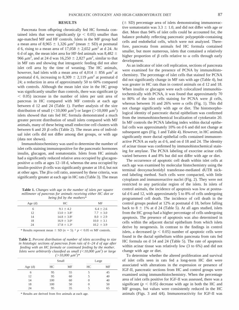
The occurrence of apoptotic cell death within islet cells ateach age was examined by molecular histochemistry using theterminal deoxynucleotidyl transferase-mediated dUTR nick-end labeling method. Such cells were compacted, with littlecytoplasm and immunoreactive nuclei (Fig. 2). They were notrestricted to any particular region of the islets. In islets ofcontrol animals, the incidence of apoptosis was low at postna-tal d 6 and 12, with approximately 5 to 8% of cells undergoingprogrammed cell death. The incidence of cell death in thecontrol groups peaked at 12% at postnatal d 18, before fallingback to 86 1% at d 24 (Table 5). At all ages studied, isletsfrom the HC group had a higher percentage of cells undergoingapoptosis. The presence of apoptosis was also determined incells within the adjacent ductal epithelium from which isletsderive by neogenesis. In contrast to the findings in controlislets, a decreased (p , 0.05) number of apoptotic cells werefound in the ductal epithelium within pancreata from rats fedHC formula on d 14 and 24 (Table 5). The rate of apoptosiswithin acinar tissue was relatively low (3 to 6%) and did notchange with age or diet.
To determine whether the altered proliferation and survivalof islet cells seen in rats fed a long-term HC diet wereassociated with alterations in the expression or presence ofIGF-II, pancreatic sections from HC and control groups wereexamined using immunohistochemistry. When the percentagearea of islet cells positive for IGF-II was assessed, there was asignificant (p , 0.05) decrease with age in both the HC andMF groups, but values were consistently reduced in the HCanimals (Figs. 3 and 4A). Immunoreactivity for IGF-II was
Table 1. Changes with age in the number of islets per squaremillimeter of pancreas for animals receiving either HC diet or
being fed by the mothers*
Age (d) HC MF
6 9.16 4.2 6.46 2.612 13.06 3.8† 7.76 3.014 14.86 3.8† 8.06 2.918 16.96 3.0† 10.06 3.124 17.86 1.3† 10.26 3.9
* Results represent mean6 SD (n 5 5). †p , 0.05vs MF controls.
Table 2. Percent distribution of number of islets according to sizein histologic sections of pancreas from rats of 6–24 d of age afterfeeding with an HC formula or continued feeding by the mother.Islets were arbitrarily classified as small (,10,000mm2) or large
(.10,000mm2)*
Age (d)
Small Large
HC MF HC MF
6 95 55 5 4512 95 60 5 4014 100 60 0 4018 100 50 0 5024 95 35 5 65
* Results are derived from five animals at each age.
87PANCREATIC ONTOGENY AND HIGH-CARBOHYDRATE DIET
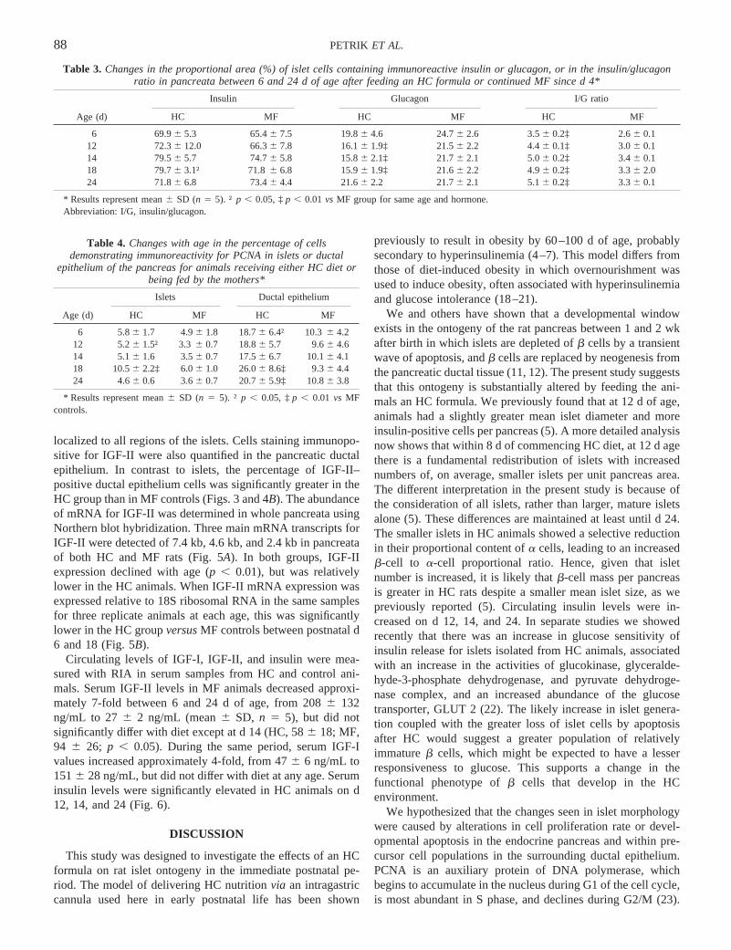
localized to all regions of the islets. Cells staining immunopo-sitive for IGF-II were also quantified in the pancreatic ductalepithelium. In contrast to islets, the percentage of IGF-II–positive ductal epithelium cells was significantly greater in theHC group than in MF controls (Figs. 3 and 4B). The abundanceof mRNA for IGF-II was determined in whole pancreata usingNorthern blot hybridization. Three main mRNA transcripts forIGF-II were detected of 7.4 kb, 4.6 kb, and 2.4 kb in pancreataof both HC and MF rats (Fig. 5A). In both groups, IGF-IIexpression declined with age (p , 0.01), but was relativelylower in the HC animals. When IGF-II mRNA expression wasexpressed relative to 18S ribosomal RNA in the same samplesfor three replicate animals at each age, this was significantlylower in the HC groupversusMF controls between postnatal d6 and 18 (Fig. 5B).
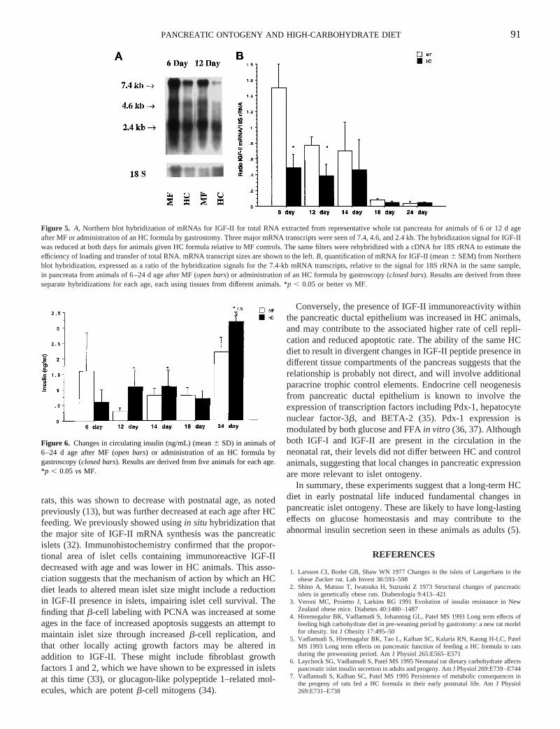
Circulating levels of IGF-I, IGF-II, and insulin were mea-sured with RIA in serum samples from HC and control ani-mals. Serum IGF-II levels in MF animals decreased approxi-mately 7-fold between 6 and 24 d of age, from 2086 132ng/mL to 276 2 ng/mL (mean6 SD, n 5 5), but did notsignificantly differ with diet except at d 14 (HC, 586 18; MF,94 6 26; p , 0.05). During the same period, serum IGF-Ivalues increased approximately 4-fold, from 476 6 ng/mL to1516 28 ng/mL, but did not differ with diet at any age. Seruminsulin levels were significantly elevated in HC animals on d12, 14, and 24 (Fig. 6).
DISCUSSION
This study was designed to investigate the effects of an HCformula on rat islet ontogeny in the immediate postnatal pe-riod. The model of delivering HC nutritionvia an intragastriccannula used here in early postnatal life has been shown
previously to result in obesity by 60–100 d of age, probablysecondary to hyperinsulinemia (4–7). This model differs fromthose of diet-induced obesity in which overnourishment wasused to induce obesity, often associated with hyperinsulinemiaand glucose intolerance (18–21).
We and others have shown that a developmental windowexists in the ontogeny of the rat pancreas between 1 and 2 wkafter birth in which islets are depleted ofb cells by a transientwave of apoptosis, andb cells are replaced by neogenesis fromthe pancreatic ductal tissue (11, 12). The present study suggeststhat this ontogeny is substantially altered by feeding the ani-mals an HC formula. We previously found that at 12 d of age,animals had a slightly greater mean islet diameter and moreinsulin-positive cells per pancreas (5). A more detailed analysisnow shows that within 8 d ofcommencing HC diet, at 12 d agethere is a fundamental redistribution of islets with increasednumbers of, on average, smaller islets per unit pancreas area.The different interpretation in the present study is because ofthe consideration of all islets, rather than larger, mature isletsalone (5). These differences are maintained at least until d 24.The smaller islets in HC animals showed a selective reductionin their proportional content ofa cells, leading to an increasedb-cell to a-cell proportional ratio. Hence, given that isletnumber is increased, it is likely thatb-cell mass per pancreasis greater in HC rats despite a smaller mean islet size, as wepreviously reported (5). Circulating insulin levels were in-creased on d 12, 14, and 24. In separate studies we showedrecently that there was an increase in glucose sensitivity ofinsulin release for islets isolated from HC animals, associatedwith an increase in the activities of glucokinase, glyceralde-hyde-3-phosphate dehydrogenase, and pyruvate dehydroge-nase complex, and an increased abundance of the glucosetransporter, GLUT 2 (22). The likely increase in islet genera-tion coupled with the greater loss of islet cells by apoptosisafter HC would suggest a greater population of relativelyimmatureb cells, which might be expected to have a lesserresponsiveness to glucose. This supports a change in thefunctional phenotype ofb cells that develop in the HCenvironment.
We hypothesized that the changes seen in islet morphologywere caused by alterations in cell proliferation rate or devel-opmental apoptosis in the endocrine pancreas and within pre-cursor cell populations in the surrounding ductal epithelium.PCNA is an auxiliary protein of DNA polymerase, whichbegins to accumulate in the nucleus during G1 of the cell cycle,is most abundant in S phase, and declines during G2/M (23).
Table 3. Changes in the proportional area (%) of islet cells containing immunoreactive insulin or glucagon, or in the insulin/glucagonratio in pancreata between 6 and 24 d of age after feeding an HC formula or continued MF since d 4*
Age (d)
Insulin Glucagon I/G ratio
HC MF HC MF HC MF
6 69.96 5.3 65.46 7.5 19.86 4.6 24.76 2.6 3.56 0.2‡ 2.66 0.112 72.36 12.0 66.36 7.8 16.16 1.9‡ 21.56 2.2 4.46 0.1‡ 3.06 0.114 79.56 5.7 74.76 5.8 15.86 2.1‡ 21.76 2.1 5.06 0.2‡ 3.46 0.118 79.76 3.1† 71.86 6.8 15.96 1.9‡ 21.66 2.2 4.96 0.2‡ 3.36 2.024 71.86 6.8 73.46 4.4 21.66 2.2 21.76 2.1 5.16 0.2‡ 3.36 0.1
* Results represent mean6 SD (n 5 5). †p , 0.05, ‡p , 0.01vs MF group for same age and hormone.Abbreviation: I/G, insulin/glucagon.
Table 4. Changes with age in the percentage of cellsdemonstrating immunoreactivity for PCNA in islets or ductal
epithelium of the pancreas for animals receiving either HC diet orbeing fed by the mothers*
Age (d)
Islets Ductal epithelium
HC MF HC MF
6 5.86 1.7 4.96 1.8 18.76 6.4† 10.36 4.212 5.26 1.5† 3.36 0.7 18.86 5.7 9.66 4.614 5.16 1.6 3.56 0.7 17.56 6.7 10.16 4.118 10.56 2.2‡ 6.06 1.0 26.06 8.6‡ 9.36 4.424 4.66 0.6 3.66 0.7 20.76 5.9‡ 10.86 3.8
* Results represent mean6 SD (n 5 5). †p , 0.05, ‡p , 0.01 vs MFcontrols.
88 PETRIK ET AL.

As reported previously (24), we found that most islet cells inthe neonatal rat pancreas containing immunoreactive PCNAwere b cells. Despite the mean islet size being smaller, theproliferative rate of islet cells was higher in islets from HCanimals at d 12 and 18. This was mainly because ofb-cellproliferation as the relative proportion ofa cells was reducedin HC-fed rats. However, the rate of islet cell apoptosis wasalso increased at every age examined in pancreata from HC
versusMF rats, suggesting that the net effect was to limit isletcell number, and thereby mean islet growth rate. A transientpeak of islet cell apoptosis occurred at postnatal d 14–18, asshown previously (11). The timing of this event was not alteredin HC rats, although the amplitude of the apoptosis wasincreased. It was difficult to identify from which endocrine cellpopulation apoptotic cells were predominantly derived frombecause, by definition, they were condensed with little cyto-
Figure 1. Dual chromogen immunohistochemistry for PCNA (brown) and either insulin (A andB, blue), or glucagon (C andD, blue) in islets within sectionsof rat pancreas from animals at 14 d postnatal age. Animals either continued to be suckled by the mothers (A andC) or were fed an HC formula by gastrostomy(B andD). Staining for PCNA is seen within islet (i) cells (arrows), within the ductal epithelium (d), and within acinar tissue (a). Magnification bar 10mm.
89PANCREATIC ONTOGENY AND HIGH-CARBOHYDRATE DIET

plasm and no longer stained for any hormone by immunocy-tochemistry. However, apoptotic cells were located in all re-gions of the islets and likely represented botha andb cells.
The increased number of smaller islets within HC ratssuggests that the rate of endocrine cell neogenesis from theductal epithelium may have been higher. This would be sup-ported by a greater percentage of ductal epithelial cells show-ing immunoreactivity for PCNA in HC animals, with a lowerincidence of apoptosis. In MF animals 8 to 12% of ductalepithelial cells showed evidence of apoptosis at any age, whichsuggests that substantial remodelling occurs in this tissue inearly postnatal life. The changes in ductal and islet cell mor-phology that accompany an HC diet were not accompanied bysimilar changes in acinar tissue, in which the incidence ofPCNA immunoreactivity and apoptosis were unaltered by diet.An increasedb-cell neogenesis from ductal epithelium, ratherthan islet cell replication, was recently shown to be the majormechanism responsible for an increase inb-cell mass in ratsafter infusion of glucose to normal animals or those depleted ofb cells by the administration of low-dose streptozotocin (25).
The IGFs are mitogens for islet endocrine cells in fetal andneonatal life (26–28). Exogenous IGFs have been shown toprevent apoptosis in a variety of cell types (29, 30), and wehave shown previously that IGF-I and IGF-II can protect isletcells against apoptosis induced by cytotoxic cytokines (11).
The transient increase in islet cell apoptosis in the neonatal ratis temporally associated with a rapid loss of expression ofIGF-II mRNA from pancreatic islet cells (11, 13). We haveshown that IGF-II transgenic mice have a dramatic increase inislet size at birth related to both an increased islet cell prolif-eration and a reduction in islet cell apoptosis (31). When weexamined the pancreatic expression of IGF-II mRNA in HC
Figure 2. Immunohistochemical localization of apoptotic cells (arrows) usingmolecular histochemistry in a pancreatic islet from an HC formula-fed rat of14 d age. Apoptotic cells have condensed nuclei with little cytoplasm. Mag-nification bar 10mm.
Figure 3. Immunohistochemical localization of IGF-II in islets within sectionsof rat pancreas from animals at 12 d postnatal age. Animals either continuedto be suckled by the mothers (A) or were fed an HC formula by gastrostomy(B). Staining for IGF-II is seen within islet (i) cells (arrows) and within theductal epithelium (d). Magnification bar 10mm.
Figure 4. Quantification of IGF-II immunoreactivitity (% cell number, mean6 SD) in (A) islets of Langerhans and (B) ductal epithelia from animals of6–24 d age after MF (open bars) or administration of an HC formula bygastroscopy (closed bars). Results are derived from five animals for each age.*p , 0.05, **p , 0.01, †p , 0.001vs MF.
Table 5. Changes with age in the percentage of cellsdemonstrating apoptosis in islets or ductal epithelium of the
pancreas for animals receiving either HC diet or being fed bythe mothers*
Age (d)
Islets Ductal epithelium
HC MF HC MF
6 9.26 2.4† 5.76 1.2 6.66 3.1 8.56 3.112 8.96 1.9† 6.86 2.1 7.66 2.8 8.66 3.214 13.06 2.5‡ 9.16 0.7 8.56 3.6† 11.56 2.718 14.76 2.4† 12.16 1.1 8.36 3.6 10.96 2.424 9.26 1.1† 7.96 0.8 6.86 2.1† 11.96 2.4
* Results represent mean6 SD (n 5 5). †p , 0.05, ‡p , 0.01 vs MFcontrols.
90 PETRIK ET AL.
rats, this was shown to decrease with postnatal age, as notedpreviously (13), but was further decreased at each age after HCfeeding. We previously showed usingin situ hybridization thatthe major site of IGF-II mRNA synthesis was the pancreaticislets (32). Immunohistochemistry confirmed that the propor-tional area of islet cells containing immunoreactive IGF-IIdecreased with age and was lower in HC animals. This asso-ciation suggests that the mechanism of action by which an HCdiet leads to altered mean islet size might include a reductionin IGF-II presence in islets, impairing islet cell survival. Thefinding thatb-cell labeling with PCNA was increased at someages in the face of increased apoptosis suggests an attempt tomaintain islet size through increasedb-cell replication, andthat other locally acting growth factors may be altered inaddition to IGF-II. These might include fibroblast growthfactors 1 and 2, which we have shown to be expressed in isletsat this time (33), or glucagon-like polypeptide 1–related mol-ecules, which are potentb-cell mitogens (34).
Conversely, the presence of IGF-II immunoreactivity withinthe pancreatic ductal epithelium was increased in HC animals,and may contribute to the associated higher rate of cell repli-cation and reduced apoptotic rate. The ability of the same HCdiet to result in divergent changes in IGF-II peptide presence indifferent tissue compartments of the pancreas suggests that therelationship is probably not direct, and will involve additionalparacrine trophic control elements. Endocrine cell neogenesisfrom pancreatic ductal epithelium is known to involve theexpression of transcription factors including Pdx-1, hepatocytenuclear factor-3b, and BETA-2 (35). Pdx-1 expression ismodulated by both glucose and FFAin vitro (36, 37). Althoughboth IGF-I and IGF-II are present in the circulation in theneonatal rat, their levels did not differ between HC and controlanimals, suggesting that local changes in pancreatic expressionare more relevant to islet ontogeny.
In summary, these experiments suggest that a long-term HCdiet in early postnatal life induced fundamental changes inpancreatic islet ontogeny. These are likely to have long-lastingeffects on glucose homeostasis and may contribute to theabnormal insulin secretion seen in these animals as adults (5).
REFERENCES
1. Larsson CI, Boder GB, Shaw WN 1977 Changes in the islets of Langerhans in theobese Zucker rat. Lab Invest 36:593–598
2. Shino A, Matsuo T, Iwatsuka H, Suzuoki Z 1973 Structural changes of pancreaticislets in genetically obese rats. Diabetologia 9:413–421
3. Veroni MC, Proietto J, Larkins RG 1991 Evolution of insulin resistance in NewZealand obese mice. Diabetes 40:1480–1487
4. Hiremegalur BK, Vadlamudi S, Johanning GL, Patel MS 1993 Long term effects offeeding high carbohydrate diet in pre-weaning period by gastrotomy: a new rat modelfor obesity. Int J Obesity 17:495–50
5. Vadlamudi S, Hiremagalur BK, Tao L, Kalhan SC, Kalaria RN, Kaung H-LC, PatelMS 1993 Long term effects on pancreatic function of feeding a HC formula to ratsduring the preweaning period. Am J Physiol 265:E565–E571
6. Laychock SG, Vadlamudi S, Patel MS 1995 Neonatal rat dietary carbohydrate affectspancreatic islet insulin secretion in adults and progeny. Am J Physiol 269:E739–E744
7. Vadlamudi S, Kalhan SC, Patel MS 1995 Persistence of metabolic consequences inthe progeny of rats fed a HC formula in their early postnatal life. Am J Physiol269:E731–E738
Figure 5. A, Northern blot hybridization of mRNAs for IGF-II for total RNA extracted from representative whole rat pancreata for animals of 6 or 12 d ageafter MF or administration of an HC formula by gastrostomy. Three major mRNA transcripts were seen of 7.4, 4.6, and 2.4 kb. The hybridization signal forIGF-IIwas reduced at both days for animals given HC formula relative to MF controls. The same filters were rehybridized with a cDNA for 18S rRNA to estimate theefficiency of loading and transfer of total RNA. mRNA transcript sizes are shown to the left.B, quantification of mRNA for IGF-II (mean6 SEM) from Northernblot hybridization, expressed as a ratio of the hybridization signals for the 7.4-kb mRNA transcripts, relative to the signal for 18S rRNA in the same sample,in pancreata from animals of 6–24 d age after MF (open bars) or administration of an HC formula by gastroscopy (closed bars). Results are derived from threeseparate hybridizations for each age, each using tissues from different animals. *p , 0.05 or bettervs MF.
Figure 6. Changes in circulating insulin (ng/mL) (mean6 SD) in animals of6–24 d age after MF (open bars) or administration of an HC formula bygastroscopy (closed bars). Results are derived from five animals for each age.*p , 0.05vs MF.
91PANCREATIC ONTOGENY AND HIGH-CARBOHYDRATE DIET

8. Hill DJ, Hogg J 1991 Growth factor control of pancreaticb cell hyperplasia. In:Herington A (ed) Clinical Endocrinology and Metabolism. Balliere Tindall, London,pp 689–698
9. Kaung HL 1994 Growth dynamics of pancreatic islet cell populations during fetal andneonatal development of the rat. Dev Dyn 200:163–175
10. Finegood DT, Scaglia L, Bonner-Weir S 1995 Dynamics ofb-cell mass in thegrowing rat pancreas. Diabetes 44:249–256
11. Petrik J, Arany E, McDonald TJ, Hill DJ 1998 Apoptosis in the pancreatic islet cellsof the neonatal rat is associated with a reduced expression of insulin-like growthfactor II that may act as a survival factor. Endocrinology 139:2994–3004
12. Scaglia L, Cahill CJ, Finegood DT, Bonner-Weir S 1997 Apoptosis participates in theremodelling of the endocrine pancreas in the neonatal rat. Endocrinology 138:1736–1741
13. Hogg J, Hill DJ, Han VKM 1994 The ontogeny of insulin-like growth factor (IGF)and IGF binding protein gene expression in the rat pancreas. J Mol Endocrinol13:49–58
14. Hall WG 1975 Weaning and growth of artificially reared rats. Science 190:1313–131515. Hsu SM, Raine L, Fanger H 1981 Use of avidin-biotin peroxidase complex (ABC) in
immunoperoxidase techniques: a comparison between ABC and unlabelled antibody(PAP) procedures. J Histochem Cytochem 29:577–580
16. Wijsman JH, Jonker RR, Keijzer R, Van de Velde CJ, Cornelisse CJ, Van Dieren-donck JH 1993 A new method to detect apoptosis in paraffin sections:in situ endlabelling of fragmented DNA. J Histochem Cytochem 41:7–12
17. Hill DJ 1990 Relative abundance and molecular size of immunoreactive insulin-likegrowth factors I and II in human fetal tissues. Early Hum Dev 21:49–58
18. Aubert R, Suquet JP, Lemonnier D 1980 Long term morphological and metaboliceffects of early under- and over-nutrition in mice. J Nutr 110:649–661
19. Oscai LB 1982 Dietary-induced severe obesity: a rat model. Am J Physiol 242:R212–R215
20. West DB, Diaz J, Roddy S, Woods SC 1987 Long-term effects on adiposity afterpreweaning nutritional manipulations in the gastrostomy-reared rat. J Nutr 117:1259–1264
21. Lemonnier D, Suquet JP, Aubert R, Rosselin G 1973 Long term effect of mouseneonate food intake on adult body composition, insulin and glucose serum levels.Horm Metab Res 5:223–224
22. Ravikumar A, Srinivasan M, Kalhan SC, Laychock SG, Patel MS 2000 A dietaryintervention (high carbohydrate) during the neonatal period causes islet dysfunctionin rats. Am J Physiol (in press)
23. Prosperi E, Scouvassi AI, Stivala LA, Bianhi L 1994 Proliferating cell nuclear antigenbound to DNA synthesis sites: phosphorylation and association with cyclin D1 andcyclin A. Exp Cell Res 215:257–262
24. Petrik J, Reusens B, Arany E, Remacle C, Hoet JJ, Hill DJ 1999 A low protein dietalters the balance of islet cell replication and apoptosis in the fetal and neonatal rat,and is associated with a reduced pancreatic expression of insulin-like growth factor-II.Endocrinology 140:4861–4873
25. Bernard C, Berthault MF, Saulnier C, Ktorza A 1999 Neogenesis vs apoptosis as maincomponents of pancreatic beta cell changes in glucose-infused normal and mildlydiabetic adult rats. FASEB J 13:1195–1205
26. Bryson JM, Tuch GI, Baxter RC 1989 Production of insulin-like growth factor-II byhuman fetal pancreas in culture. J Endocrinol 121:367–373
27. Scharfmann R, Corvol M, Czernichow P 1989 Characterization of IGF-I produced byfetal rat pancreatic cells. Diabetes 38:686–690
28. Hill DJ, Frazer A, Swenne I, Wiedemann PK, Milner RGD 1987 Somatomedin-C inthe human fetal pancreas. Cellular localization and release during organ culture.Diabetes 36:465–471
29. Stewart CE, Rotwein P 1996 Insulin-like growth factor II is an autocrine survivalfactor for differentiating myoblasts. J Biol Chem 271:11330–11338
30. Grier A, Hamishon M, Beery R, Lunenfeld B 1992 Insulin-like growth factor-Iinhibits cell death induced by cycloheximide in MCF-7 cells—a model system foranalyzing control of cell death. In Vitro Cell Dev Biol Anim 28A:725–729
31. Petrik J, Pell JM, Arany E, McDonald TJ, Dean WL, Reik W, Hill DJ 1999Over-expression of insulin-like growth factor-II in transgenic mice is associated withpancreatic islet cell hyperplasia. Endocrinology 140:2353–2363
32. Hill DJ, Hogg J, Petrik J, Arany E, Han VKM 1999 Cellular distribution and ontogenyof insulin-like growth factors (IGFs) and IGF binding protein messenger RNAs andpeptides in developing rat pancreas. J Endocrinol 160:305–317
33. Arany E, Jones A, Petrik J, Hill DJ 1997 Fibroblast growth factors in the earlymaturation of rat pancreata. J Endocrinol 152(suppl):P88
34. Xu G, Stoffers DA, Habener JF, Bonner Weir S 1999 Exendin-4 stimulates bothbeta-cell replication and neogenesis, resulting in increased beta-cell mass and im-proved glucose tolerance in diabetic rats. Diabetes 48:2270–2276
35. Sander M, German S 1997 Theb cell transcription factors and development of thepancreas. J Mol Med 75:327–340
36. Gremlich S, Bonny C, Waeber G, Thorens B 1997 Fatty acids induce a decrease inislet/duodenum homeobox-1 (IDX-1) expression in adult rat pancreatic islets, and aparallel decrease in GLUT2, glucokinase, insulin and somatostatin gene expression.Exp Clin Endocrinol Diabetes 105:A22
37. Jonas J, Sharma A, Ilkova H, Patane G, Bonner-Weir S, Weir G 1998 Pdx-1 and HNFmRNA levels are reduced in islets from hyperglycemic 90% pancreatectomized rats(abstr). Diabetes 47(Suppl1):1004;A258
92 PETRIK ET AL.
Related Documents