EXCLI Journal 2003;2:6-21 – ISSN 1611-2156 received: 29. January 2003, accepted: 10. February 2003, published: 10. February 2003 Original article: Cryopreservation of islets of Langerhans: Optimization of protocols using rat pancreatic tissue Marc-Alexander von Mach *1,2 , Josef Schlosser 1 , Marcus Weiland 1 , Peter J. Feilen 1 , Michael Ringel 3 , Jan G. Hengstler 3 , L. Sacha Weilemann 2 , Jürgen Beyer 1 , Peter Kann 1 , M. M. Weber 1 , Stephan Schneider *1 1 I. Medical Department, Division of Endocrinology and Metabolic Disease, 2 II. Medical Department, 3 Institute of Toxicology, University of Mainz, Langenbeckstrasse 1, 55131 Mainz, Germany Telephone: +49 6131 174154, Fax: +49 6131 232468, E-mail: [email protected] (*corresponding authors) SUMMARY Banking of pancreatic islets following cryopreservation has been shown to be the most effective method facilitating sufficient availability for transplantation to reverse type 1 diabetes. As success of transplantation depends on number of functioning islets the aim of this study was to determine crucial variables of cryopreservation to increase postthaw viability. Isolated rat pancreatic islets were cryopreserved using a protocol systematically changed in several variables: (i) freezing media (RPMI-1640 + 10% FCS, 100% FCS, hepatocyte suspension buffer); (ii) final concentration of cryoprotectant (Me 2 SO 5%, 10%, 14.4%, 18.3%, 21.7%); (iii) conditions of equilibration (15 min vs 45 min, 22°C vs on ice); (iv) postthaw removal of cryoprotectant (15 vs 30 min; stepwise vs one-step). Concerning freezing media RPMI showed highest viability (60.7±1.5% vs FCS 45.3±1.8%, p<0.05 and hepatocyte suspension buffer 56.3±1.4%, ns). Optimal Me 2 SO concentration revealed to be 14.4% resulting in 60.5±1.0% viability compared to 22.0±0.9% with 5% Me 2 SO (p<0.05), 49.5±1.3% with 10% (p<0.05), 19.6±1.3% with 18.3% (p<0.05) and 8.6±0.8% with 21.7% (p<0.05). An equilibration with cryoprotectant on ice showed significantly higher viability than at 22°C (66.7±1.1% vs 60.7±1.1%; p<0.05), whereas more rapid equlibration within 15 min instead of 45 min showed no improvement. For postthaw removal of cryoprotectant stepwise dilution within 30 min (68.9±1.1%; 15 min: 68.7±1.2%) was found to be superior to a one-step technique (65.6±1.2%; p<0.05). Our study determined freezing media, cryoprotectant concentration, conditions of equilibration and postthaw removal of cryoprotectant as important parameters giving the opportunity to further advances of low temperature banking of isolated islets. Keywords: pancreatic islets; cryopreservation; dimethyl sulfoxide; viability; Sprague-Dawley rat 6

Welcome message from author
This document is posted to help you gain knowledge. Please leave a comment to let me know what you think about it! Share it to your friends and learn new things together.
Transcript
EXCLI Journal 2003;2:6-21 – ISSN 1611-2156 received: 29. January 2003, accepted: 10. February 2003, published: 10. February 2003
Original article:
Cryopreservation of islets of Langerhans:
Optimization of protocols using rat pancreatic tissue Marc-Alexander von Mach*1,2, Josef Schlosser1, Marcus Weiland1, Peter J. Feilen1, Michael
Ringel3, Jan G. Hengstler3, L. Sacha Weilemann2, Jürgen Beyer1, Peter Kann1, M. M. Weber1, Stephan Schneider*1
1I. Medical Department, Division of Endocrinology and Metabolic Disease, 2II. Medical Department, 3Institute of Toxicology, University of Mainz, Langenbeckstrasse 1, 55131 Mainz, Germany Telephone: +49 6131 174154, Fax: +49 6131 232468, E-mail: [email protected] (*corresponding authors)
SUMMARY Banking of pancreatic islets following cryopreservation has been shown to be the most effective method facilitating sufficient availability for transplantation to reverse type 1 diabetes. As success of transplantation depends on number of functioning islets the aim of this study was to determine crucial variables of cryopreservation to increase postthaw viability. Isolated rat pancreatic islets were cryopreserved using a protocol systematically changed in several variables: (i) freezing media (RPMI-1640 + 10% FCS, 100% FCS, hepatocyte suspension buffer); (ii) final concentration of cryoprotectant (Me2SO 5%, 10%, 14.4%, 18.3%, 21.7%); (iii) conditions of equilibration (15 min vs 45 min, 22°C vs on ice); (iv) postthaw removal of cryoprotectant (15 vs 30 min; stepwise vs one-step). Concerning freezing media RPMI showed highest viability (60.7±1.5% vs FCS 45.3±1.8%, p<0.05 and hepatocyte suspension buffer 56.3±1.4%, ns). Optimal Me2SO concentration revealed to be 14.4% resulting in 60.5±1.0% viability compared to 22.0±0.9% with 5% Me2SO (p<0.05), 49.5±1.3% with 10% (p<0.05), 19.6±1.3% with 18.3% (p<0.05) and 8.6±0.8% with 21.7% (p<0.05). An equilibration with cryoprotectant on ice showed significantly higher viability than at 22°C (66.7±1.1% vs 60.7±1.1%; p<0.05), whereas more rapid equlibration within 15 min instead of 45 min showed no improvement. For postthaw removal of cryoprotectant stepwise dilution within 30 min (68.9±1.1%; 15 min: 68.7±1.2%) was found to be superior to a one-step technique (65.6±1.2%; p<0.05). Our study determined freezing media, cryoprotectant concentration, conditions of equilibration and postthaw removal of cryoprotectant as important parameters giving the opportunity to further advances of low temperature banking of isolated islets. Keywords: pancreatic islets; cryopreservation; dimethyl sulfoxide; viability; Sprague-Dawley rat
6
INTRODUCTION Insulin therapy of type 1 diabetes still fails to mimic the physiological response of insulin secretion (von Mach et al., 2002). Since islet transplantation has become an option for the treatment of type 1 diabetes (Shapiro et al., 2000), several methods of isolation, purification, and cryopreservation of islets of Langerhans have been developed (Kneteman et al., 1989; Lakey et al., 1996; Marchetti et al., 1994; Miyamoto et al., 1994). Intraportal transplantation of isolated islets of Langerhans has recently been demonstrated to facilitate good glycemic control without major surgical risks (Ryan et al., 2001). The recent advances were achieved by the introduction of a glucocorticoid-free immunosuppressive regimen resulting in insulin independence in more than 90% of type 1 diabetic patients after islet transplantation (Shapiro et al., 2000). However, in these encouraging studies an amount of 9,000 islet equivalents per kilogram body weight were required for intraportal injection. These high numbers outline one of the major problems in islet transplantation being the insufficient availability of human insulin producing tissue. To allow the unlimited collection and long-term storage of isolated islets low temperature banking after cryopreservation has become an important objective in investigations concerning islet transplantation (Rajotte et al., 1984). Thus, cryopreserved isolated human islets have successfully been used to increase the amount of islets for transplantation in type 1 diabetic patients (Warnock et al., 1991, 1992, 1995). Additionally, creating a low-temperature tissue bank with tissues labeled for their type of human leukocyte antigen (HLA) would give the opportunity to select islet preparations based on the matching of type of HLA (Warnock et al., 1994). Before transplantation low-temperature banking of islets would enable investigators to assess islet function and microbiological sterility (Lakey et al., 1995).
Unfortunately, reduced recovery, function, and viability of islets after thawing has been described (Di Carlo et al., 1994; Rich et al., 1994). As success of transplantation depends on the number of functioning islets several different protocols for cryopreservation have been developed to minimize damage of islet cells (Cattral et al., 1998; Lakey et al., 1997; Lakey et al., 2001). The aim of this study was to determine crucial variables for cryopreservation of islets of Langerhans to increase postthaw viability.
MATERIALS AND METHODS Pancreatic islets were isolated from male Sprague-Dawley rats with a body weight of 250 – 270 g and 6 – 8 weeks of age (Central Animal Facility, University of Mainz). Animals were kept under SPF conditions and fed ad libitum with Sniff (Soest, Germany) standard diet. Anesthesia was performed by intraperitoneal injection of pentobarbital at a dose of 60 mg/kg, as previously described (Jastrow et al., 1997a, b). The peritoneal cavity was opened by a midline incision and perfusion of the pancreas was performed via the pancreatic duct using Hanks’ balanced salt solution (HBSS, Gibco BRL, Long Island, NY, USA) with a concentration of collagenase of 1.7 mg/ml (Serva PanPlus, Heidelberg, Germany). The pancreatic organ was quickly removed and incubated for 20 minutes at 37°C in collagenase solution. Mechanical disruption of the tissue was achieved by intermittent shaking. Enzymatic digestion process was finished by adding HBSS supplemented with 10% fetal calf serum in excess with a temperature of 4°C. Pancreatic digest containing endocrine and exocrine material was purified using discontinuous gradients of Ficoll at densities of 1.090, 1.077 and 1.040. Before cryopreservation islets were cultured overnight in RPMI-1640 medium (Biochrom KG, Berlin, Germany) at 37°C in a humidified atmosphere of 95% air and 5% CO2. The medium was supplemented with
7
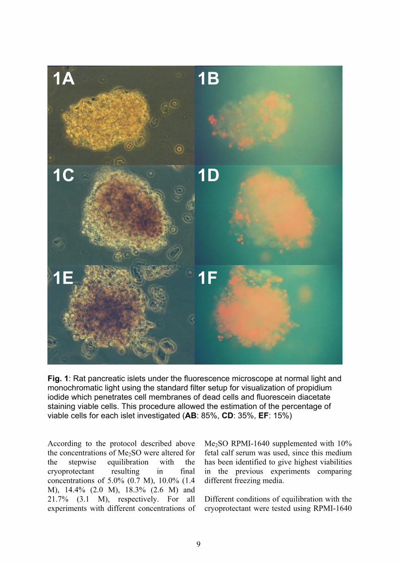
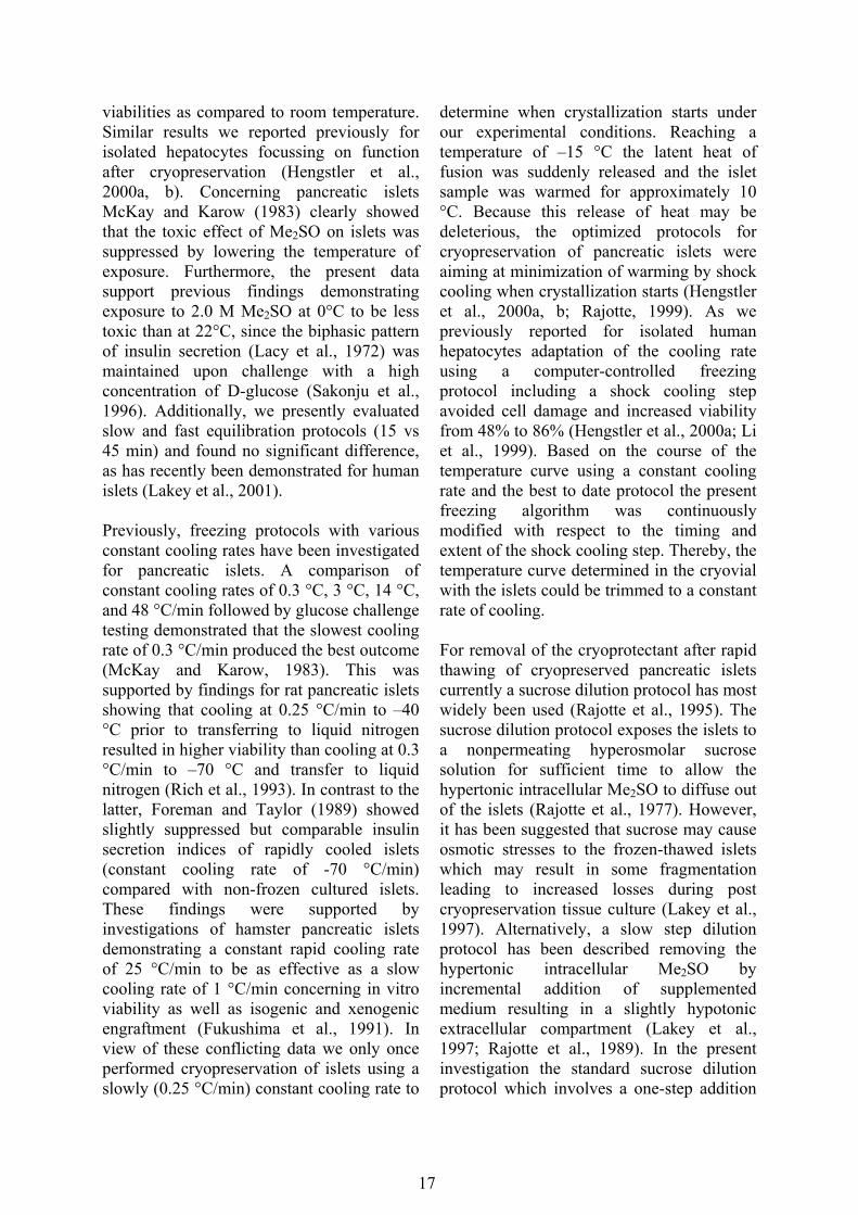
5.1 mmol/l glucose, 25 mmol/l HEPES, 10% fetal calf serum (Greiner Laboratories, Frickenhausen, Germany), 0.2 g/l Glutamax, 200 U/ml penicillin, 200 µg/ml streptomycin (Gibco BRL, Paisley, Scotland, GB) and 50 µg/ml ciprofloxacin (Bayer, Leverkusen, Germany). Cultured islets were cryopreserved following the same protocol being changed systematically in several variables as described below. Before cryopreservation viability assessment was performed using propidium iodide staining according to the staining protocol of the supplier (Sigma, St. Louis, USA). Specimens were scrutinized under the fluorescence microscope (Axiovert 35, XR-X3000, Zeiss, Oberkochem, Germany) at normal light and monochromatic light using the standard filter setup (Beerheide et al., 2002) for visualization of propidium iodide which penetrates cell membranes of dead cells (filter no. 15, BP 546, FT 580, LP 590). This procedure allowed the calculation of the percentage of viable cells for each islet investigated. For example representative islets were co-stained using propidium iodide marking the dead cells red and fluorescein diacetate marking the living cells green as previously described (Schneider et al., 2003; Zimmermann et al., 2003; Fig. 1). Additionally, for n = 16 islets viability assessment was performed as well as the ATP content (pg/ml) was measured by the luciferin/luciferase-technique (Hengstler et al., 1999). For the latter experiment islets were systematically damaged by heating as described previously (Gray and Morris, 1987; London et al., 1989) to obtain the entire range of viabilities between 0% an 100%. After viability assessment an average number of 500 islets was transferred into each cryovial (2ml). All viability assessments before cryopreservation as well as at 0 h, 24 h and 48 h post thawing were performed for approximately 10% of islets (n = 50). During the first set of experiments different freezing media were tested. Three cryovials
containing 500 pancreatic islets each suspended in culture medium were centrifuged for 3 min at 1200 rpm and 4°C. The supernatant was discarded and the pellet of islets was resuspended in 0.2 ml of one of the following solutions: (i) 100% fetal calf serum; (ii) hepatocyte suspension buffer (HSB; 620 ml glucose solution [9 g D-glucose/l], 100 ml KH buffer [60 g NaCl/l, 1.75 g KCl/l and 1.6 g KH2PO4/l], 100 ml HEPES buffer [60 g HEPES/l], 150 ml amino acid solution [0.27 g/l L-alanine, 0.14 g/l L-aspartic acid, 0.4 g/l L-asparagine, 0.27 g/l L-citrulline, 0.14 g/l L-cysteine, 1.0 g/l L-histidine, 1.0 g/l L-glutamic acid, 1.0 g/l L-glycin, 0.40 g/l L-isoleucine, 0.8 g/l L-leucine, 1.30 g/l L-lysine, 0.55 g/l L-proline, 0.65 g/l L-serine, 1.35 g/l L-threonine, 0.65 g/l L-tryptophan, 0.55 g/l L-tyrosine, 0.80 g/l L-valine], 10 ml glutamine solution [7g L-glutamine/l], 5 ml insulin solution [2 g insulin/l], 8 ml CaCl2 solution [19 g CaCl2 x 2H2O/l], 4 ml MgSO4 solution [24.6 g/l MgSO4 x 7H2O] and 2 g bovine serum albumin/l); (iii) RPMI-1640 supplemented with 10% fetal calf serum. To the 0.2 ml of resuspended islets cryoprotectants were added to the resuspended islet pellets in a stepwise protocol with at first 0.1 ml of 14.4% Me2SO (using the respective cryopreservation buffer as solvent) was added incubating for 5 min at 22 °C (step one). After that again 0.1 ml of 14.4% Me2SO was added and incubated for 25 min at 22 °C resuspending the islet pellets every 5 min by shaking gently (step two). Then 0.4 ml 21.7% Me2SO was added resulting in a final concentration of cryoprotectant of 14.4% and incubated for 15 min at 0 °C resuspending pellets every 5 min (step three). When this prefreezing phase was finished the cryovials were transferred into the chamber of a computer-controlled freezing machine (BV-8, Consarctic, Heidelberg, Germany). Freezing included several steps of cooling as described below. Immediately after the freezing program had been completed the cryovials were transferred into liquid nitrogen.
8
1A 1B
1C 1D
1E 1F
Fig. 1: Rat pancreatic islets under the fluorescence microscope at normal light and monochromatic light using the standard filter setup for visualization of propidium iodide which penetrates cell membranes of dead cells and fluorescein diacetate staining viable cells. This procedure allowed the estimation of the percentage of viable cells for each islet investigated (AB: 85%, CD: 35%, EF: 15%) According to the protocol described above the concentrations of Me2SO were altered for the stepwise equilibration with the cryoprotectant resulting in final concentrations of 5.0% (0.7 M), 10.0% (1.4 M), 14.4% (2.0 M), 18.3% (2.6 M) and 21.7% (3.1 M), respectively. For all experiments with different concentrations of
Me2SO RPMI-1640 supplemented with 10% fetal calf serum was used, since this medium has been identified to give highest viabilities in the previous experiments comparing different freezing media. Different conditions of equilibration with the cryoprotectant were tested using RPMI-1640
9
supplemented with 10% fetal calf serum as freezing medium and 14.4 % Me2SO as final cryoprotectant concentration for all cryovials. Pancreatic islets were equilibrated with Me2SO using three different protocols: (i) islets were treated as described above with step one including 5 min of incubation at 22 °C, step two with 25 min of incubation at 22 °C, and step three with 15 min of incubation at 0 °C; (ii) step one, two and three were all performed at 0 °C with the same periods of time of incubation; (iii) again each step was performed at 0 °C with 5 min of incubation for each step resulting in a shortened time of equilibration with the cryoprotectant from 45 to 15 min. As release of the latent heat of fusion due to crystallization during cryopreservation causes major damage to cells (Hengstler et al., 2000a, b; Steinberg et al., 1999) the algorithm of the computer controlled-freezing machine was optimized towards a complete compensation for this effect. For each algorithm the temperature in the freezing chamber as well as in the islet suspension of the cryovial were determined and monitored by a chart recorder. At first, the critical temperature for occurrence of crystallization during freezing of pancreatic islets was determined using an almost continuous rate of cooling (step 1: duration [D] 5 min / final temperature [FT] 0°C; step 2: D 5 min / FT –5 °C; step 3: D 5 min / FT –5 °C; step 4: D 140 min / FT –40 °C and rapid cooling to –196 °C). Additionally, an algorithm resembling the best to date protocol for islet cryopreservation was applied (step 1: D 5 min / FT 0 °C; step 2: D 2 min / FT 0 °C; step 3: D 2 min / FT -7.2 °C; step 4: D 6 min / FT –7.2 °C; step 5: D 0.5 min / FT –37 °C; step 6: D 0.5 min / FT –9 °C; step 7: D 5 min / FT –9 °C; step 8: D 120 min / FT –40 °C and rapid cooling to –196 °C, for review see Rajotte, 1999). Based on these data the algorithm was systematically optimized to eliminate the peak temperature increase caused by heat of fusion using an initial shock cooling step (step 1: D 5 min / FT 0 °C; step 2: D 5 min /
FT -7.2 °C; step 3: D 10 min / FT -7.2 °C; step 4: D 0.1 min / FT –33 °C; step 5: D 0.3 min / FT –9 °C; step 6: D 120 min / FT –40 °C and rapid cooling to –196 °C). Finally, timing and peak of the shock cooling period was continuously adapted to prevent a rapid loss of temperature in the cryovial (step 1: D 5 min / FT 0 °C; step 2: D 2 min / FT -4.5 °C; step 3: D 10 min / FT -4.5 °C; step 4: D 0.1 min / FT –20 °C; step 5: D 0.1 min / FT –50 °C; step 6: D 0.2 min / FT –80 °C; step 7: D 0.2 min / FT –80 °C; step 8: D 0.5 min / FT –5 °C; step 9: D 1 min / FT –7 °C; step 10: D 5 min / FT –9 °C; step 11: D 120 min / FT –40 °C and rapid cooling to –196 °C). After storage in liquid nitrogen for at least 7 days, cryovials with frozen islets were plunged into a water bath maintained at 37 °C and rapidly thawed to 0 °C at a rate of approximately 200 °C/min. After centrifugation at 1200 rpm for 3 min supernatant was discarded and the intracellular cryoprotectant was removed by the addition of 1 ml 0.75 M sucrose solution (dissolved in RPMI-1640 supplemented with 10% fetal calf serum) to the cell pellet and incubation for 30 min at 0 °C resuspending the cell pellet every 5 min by shaking gently. This incubation was followed by a period of 20 min with repeated addition of 1 ml ice-cold culture medium (RPMI-1640 supplemented with 10% fetal calf serum) incubating 5 min and resuspension of islets. The final suspension was centrifuged at 1200 rpm for 3 min, resuspended in culture medium at 37 °C and transferred to tissue culture plates. Viability was determined immediately postthawing and after maintaining islets in culture for 24 and 48 hours in a humidified atmosphere as described above. Three different protocols for the removal of cryoprotectant were tested for islets cryopreserved with RPMI-1640 supplemented with 10% fetal calf serum used as freezing medium, a final concentration of cryoprotectant of 14.4% and an equilibration with cryoprotectant for 15 min at 0 °C as described above: (i) after rapid thawing and centrifugation the first islet pellet was
10
incubated with 1 ml of 0.75 M sucrose solution for 30 min (one-step, 30 min) followed by repeated additions of culture medium as described above; (ii) incubation with 0.75 M sucrose solution in a stepwise manner adding 0.17 ml every 5 min for a period of 30 min (stepwise, 30 min); (iii) the period of incubation was shortened to 15 min with a stepwise addition of 0.2 ml 0.75 M sucrose solution every 3 min (stepwise, 15 min). To assess the in vitro function of the frozen-thawed pancreatic islets using the protocol which resulted in the highest viability in the previous experiments the basal and stimulated glucose-induced insulin response was estimated. After thawing islets were cultured for 48 h and washed subsequently. Viability was assessed as described above using propidium iodide staining. Additionally, 18 aliquots of 10 islets were incubated in multiwell plates for 60 min in RPMI-1640 supplemented with 10% fetal calf serum and 2.8 mM of glucose concentration. The supernatant was collected. Islets were washed and incubated for a second period of 60 min in the same media with 20 mM of glucose concentration and the
supernatant was collected. The supernatants were frozen (-20 °C) and subsequently assayed for insulin content by enzyme immunoassay (Mercodia High Range Rat Insulin ELISA, Mercodia AB, Uppsala, Sweden). The insulin secretion capabilities of the islets were expressed as pmol/islet/h and compared to 18 aliquots of non-frozen controls. The stimulatory response of the islets was estimated to be: stimulatory response (%) = (stimulation value / basal value) * 100 as described previously (Schneider et al., 2001). Results are expressed as mean ± standard error of the mean (SEM). The correlation of Spearman was used to correlate islet viabilities determined by fluorescent staining with ATP measurements. As the determined viabilities failed testing for equal variance a Kruskal-Wallis one way analysis of variance (ANOVA) on ranks was used to make comparisons between the different groups. If an interaction was significant, pairwise comparisons of the mean responses to the different treatment groups (Dunn´s method) were performed. A p-value of smaller than 0.05 was considered to be statistically significant.
11
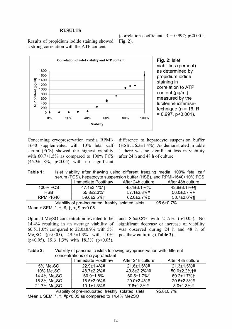
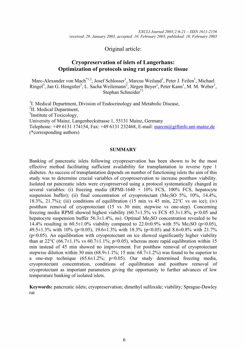
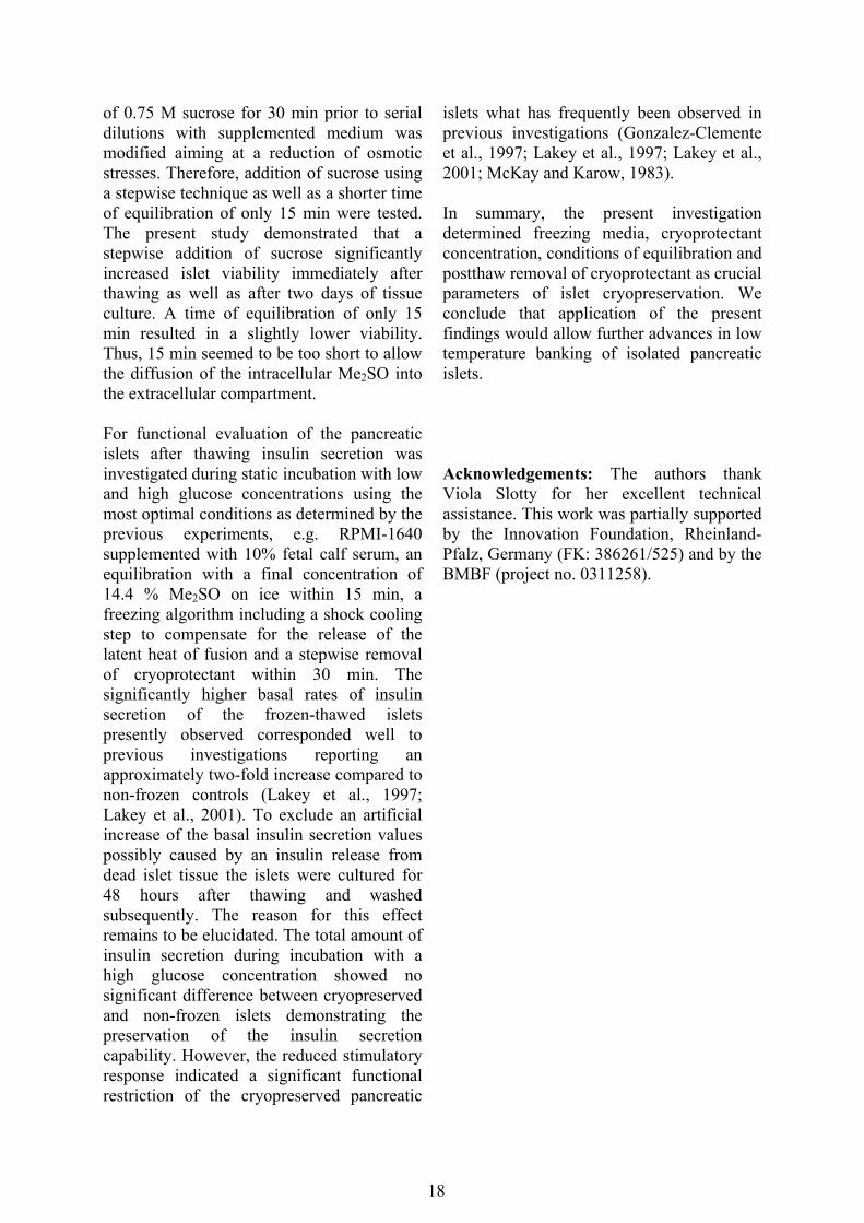
RESULTS Results of propidium iodide staining showed a strong correlation with the ATP content
(correlation coefficient: R = 0.997; p<0.001; Fig. 2).
Correlation of islet viability and ATP content
0200400600800
10001200140016001800
0% 20% 40% 60% 80% 100%
Viability
ATP
con
tent
(pg/
ml)
Fig. 2: Islet viabilities (percent) as determined by propidium iodide staining in correlation to ATP content (pg/ml) measured by the luciferin/luciferase-technique (n = 16, R = 0.997, p<0.001).
Concerning cryopreservation media RPMI-1640 supplemented with 10% fetal calf serum (FCS) showed the highest viability with 60.7±1.5% as compared to 100% FCS (45.3±1.8%, p<0.05) with no significant
difference to hepatocyte suspension buffer (HSB; 56.3±1.4%). As demonstrated in table 1 there was no significant loss in viability after 24 h and 48 h of culture.
Table 1: Islet viability after thawing using different freezing media: 100% fetal calf
serum (FCS), hepatocyte suspension buffer (HSB), and RPMI-1640+10% FCS Immediate Postthaw After 24h culture After 48h culture
100% FCS 47.1±3.1%*† 45.1±3.1%#‡ 43.8±3.1%+¶ HSB 55.8±2.3%* 57.1±2.3%# 56.0±2.7%+
RPMI-1640 59.6±2.5%† 62.0±2.7%‡ 58.7±2.6%¶ Viability of pre-incubated, freshly isolated islets 95.6±0.7%
Mean ± SEM; *, †, #, ‡, +, ¶ p<0.05 Optimal Me2SO concentration revealed to be 14.4% resulting in an average viability of 60.5±1.0% compared to 22.0±0.9% with 5% Me2SO (p<0.05), 49.5±1.3% with 10% (p<0.05), 19.6±1.3% with 18.3% (p<0.05),
and 8.6±0.8% with 21.7% (p<0.05). No significant decrease or increase of viability was observed during 24 h and 48 h of postthaw culturing (Table 2).
Table 2: Viability of pancreatic islets following cryopreservation with different
concentrations of cryoprotectant Immediate Postthaw After 24h culture After 48h culture
5% Me2SO 22.9±1.4%# 21.6±1.6%# 21.3±1.5%# 10% Me2SO 48.7±2.2%# 49.8±2.2%*# 50.0±2.2%†#
14.4% Me2SO 60.9±1.8% 60.5±1.7%* 60.2±1.7%† 18.3% Me2SO 18.5±2.0%# 20.0±2.4%# 20.5±2.3%# 21.7% Me2SO 10.1±1.3%# 7.8±1.3%# 8.0±1.3%#
Viability of pre-incubated, freshly isolated islets 95.8±0.7% Mean ± SEM; *, †, #p<0.05 as compared to 14.4% Me2SO
12
Equilibration with cryoprotectant on ice showed significantly higher postthaw viability than at 22°C (66.7±1.1% vs 60.7±1.1%; p<0.05), whereas more rapid equilibration within 15 min instead of 45 min
showed no significant improvement (67.1±1.1%). Determination of viabilities 24 h and 48 h after thawing revealed no significant changes as compared to viabilities immediately measured (Table 3).
Table 3: Postthaw viability of islets using different conditions of equilibration with
cryoprotectant Immediate Postthaw After 24h culture After 48h culture
45 min, 22°C 60.9±1.8%*† 60.0±1.9%#‡ 61.1±1.8%+¶ 45 min, 4°C 66.4±1.9%* 66.4±1.8%# 67.1±2.1%+ 15 min, 4°C 67.6±1.9%† 66.9±1.8%‡ 66.9±2.1%¶
Viability of pre-incubated, freshly isolated islets 95.6±0.7% Mean ± SEM; *, †, #, ‡, +, ¶p<0.05 A stepwise postthaw removal of cryoprotectant within 30 min was found to produce the highest viability with 68.9±1.1% as compared to a one-step technique within 30 min (65.6±1.2%; p<0.05), whereas a
shortened incubation with sucrose solution of only 15 min showed an insignificantly decreased viability of 68.7±1.2%. During postthaw tissue culture only insignificant changes in viability were observed (Table 4).
Table 4: Viability assessment of pancreatic islets after removal of cryoprotectant using
different protocols Immediate Postthaw After 24h culture After 48h culture
One-step - 30 min 65.6±2.0% 65.8±2.1% 65.3±2.2% Stepwise - 30 min 69.1±1.9% 68.7±2.0% 68.9±2.0% Stepwise - 15 min 68.4±2.0% 68.7±2.0% 69.1±2.2%
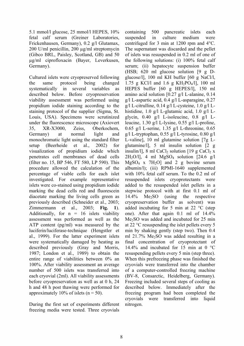
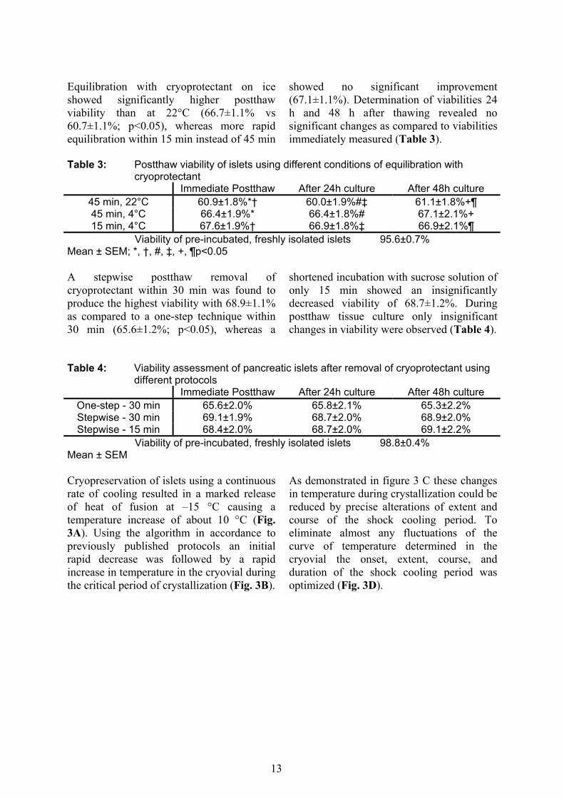
Viability of pre-incubated, freshly isolated islets 98.8±0.4% Mean ± SEM Cryopreservation of islets using a continuous rate of cooling resulted in a marked release of heat of fusion at –15 °C causing a temperature increase of about 10 °C (Fig. 3A). Using the algorithm in accordance to previously published protocols an initial rapid decrease was followed by a rapid increase in temperature in the cryovial during the critical period of crystallization (Fig. 3B).
As demonstrated in figure 3 C these changes in temperature during crystallization could be reduced by precise alterations of extent and course of the shock cooling period. To eliminate almost any fluctuations of the curve of temperature determined in the cryovial the onset, extent, course, and duration of the shock cooling period was optimized (Fig. 3D).
13
Temperature (°C) A B C D
↓ ↓ ↓ ↓ ↓ ↓ ↓ ↓
Time (min) Fig. 3: Influence of the freezing protocol on temperature curves (0 – 50 min) inside the cryovials as indicated (↓): 1A - linear freezing protocol; 1B - common freezing protocol used for pancreatic islets with shock cooling step; 1C - modification of the shock cooling step; 1D - final freezing protocol with only minimal excursions of the temperature curve inside the cryovial. - The lower curve demonstrates the temperature within the cooling chamber. Investigation of insulin secretion of cryopreserved islets using the presently best protocol revealed a significantly increased basal level of insulin secretion as compared to non-frozen controls (Table 5). During static incubation with a high glucose
concentration similar stimulated insulin secretion values were observed resulting in a reduced stimulatory response of the cryopreserved islets compared to non-frozen controls.
Table 5: Assessment of viability and insulin secretion capability of pancreatic islets after
cryopreservation using the optimized protocol Viability Basal insulin secretion
(pmol/islet/h) Stimulated insulin
secretion (pmol/islet/h) Stimulatory response
Cryopreserved islets
72.0±2.0% 0.27±0.05 0.88±0.16 353±13%
Non-frozen controls
98.2±0.5% 0.12±0.01 0.70±0.06 568±15%
p<0.05 p<0.05 ns P<0.05 Mean ± SEM
14
DISCUSSION As the number of pancreatic islet allografts performed annually and the number of islet transplantation centers has markedly grown throughout recent years current methods of islet isolation and cryopreservation were focused on aiming at successful clinical islet transplantation (Brendel et al., 1999). Although a number of different protocols have been described for the cryopreservation of pancreatic islets of Langerhans, most involve stepwise equilibration with 2 M Me2SO followed by slow cooling and fast warming (Lakey et al., 1999; Rajotte, 1999). Recently, some key cryobiological parameters of islet cells isolated from the pancreas of newborn pigs, the osmotically inactive fraction of cell volume, the permeability coefficients of Me2SO at varying temperatures, and the activation energies of these transport processes have been examined (Fedorow et al., 2001). These parameters would permit the mathematical modeling of osmotic responses of intact islets during the cryopreservation process, which may lead to further improvements in the low temperature storage of islets from newborn pigs. However, current cryopreservation protocols are empirically derived. The aim of the present investigation was to determine the influence of four parameters on islet viability after cryopreservation: (i) freezing media; (ii) concentration of cryoprotectant; (iii) conditions of equilibration with the cryoprotectant; (iv) conditions of removal of the cryoprotectant. Among membrane integrity assays most commonly trypan blue staining has been used for determination of viability in studies concerning cryopreservation of pancreatic islets. As this method does not guarantee reliable data on islet viability usually additional tests like glucose challenging followed by measurement of insulin concentrations or transplantation into streptozotocin-induced diabetic animals have
been performed (Hardikar et al., 2001; Stegemann et al., 1998). In the present study we applied the technique of fluorescence staining with propidium iodide for islet viability assessment. Microfluorometric counting, for instance, with fluorescein diacetate or ethidium bromide have been shown to correlate highly significantly on low-temperature (24 °C) culture and on 37 °C culture with the insulin concentrations of the same islets (Miyamoto et al., 2000). Additionally, we compared propidium iodide staining with the luciferin/luciferase-technique for measurement of ATP content, which previously has been used to determine the proportion of viable versus dead cells (Hengstler et al., 1999). This experiment demonstrated a high correlation particular for viabilities greater than 40%. As success of islet cryopreservation also depends on a period of culturing prior to the freezing procedure all experiments performed presently were done after 24 hours of incubation. Culture of 2 days after isolation of pancreatic islets in medium RPMI 1640 before freezing has been shown to be superior with respect to insulin synthesis and release as compared to freshly isolated tissue (McKay and Karow, 1983; Sandler and Andersson, 1984). This effect was speculated to be due to cell damage induced by the collagenase digestion. During culture the islets may recover and become more resistant to stress associated with freezing and thawing. Similarly, different degrees of islet viability have been reported after thawing during the first days of culture. The beneficial effect of culture after thawing was supposed to reflect recovery of cells with minor freeze-damage or the loss of damaged cells, which otherwise would influence the results of the viability tests (Sandler and Andersson, 1984). For detailed registration of such possible influences viability assessments in the present study were performed immediately, and 24 as well as 48 hours after thawing. Nevertheless, the present
15
results showed no significant changes in viability during this period of time post thawing indicating that most pancreatic islets immediately and entirely recovered after cryopreservation. Cryopreservation of pancreatic islets is most commonly performed using RPMI-1640 or TCM-199 supplemented with 10% fetal calf serum as freezing media (Rajotte, 1999; Sandler and Andersson, 1984). The addition of serum has been shown to have beneficial effects on postthaw outcome (Sandler et al., 1987). For hepatocytes of various species we previously demonstrated increased postthaw viability and a better conservation of differentiated functions such as drug metabolism and enzyme induction using a newly developed hepatocyte suspension buffer or a solution of 100% fetal calf serum (Hengstler et al., 2000a, b). However, the present results showed a significantly lower viability using 100% fetal calf serum in comparison to RPMI-1640 and hepatocyte suspension buffer. Since hepatocyte suspension buffer resulted in slightly decreased viabilities as compared to RPMI-1640 the following experiments were performed with the latter solution. The difference between pancreatic islets and hepatocytes was probably due to the fact that pancreatic islets are micro-organs consisting of about 1000 cells in contrast to single hepatic cells which may lead to different physico-chemical properties during freezing. Further improvement of the present cryopreservation protocol was achieved by the adjustment of the concentration of cryoprotectant. One of the mechanisms by which addition of cryoprotectants may improve survival after cryopreservation is through effective dilution of residual salts and thereby, avoidance of solution effects injury (Mazur, 1970). However, a number of studies have shown that cryoprotectants exert a wide range of toxic effects on the biological systems of various tissues and pancreatic islets are no exception (Shlafer, 1981). For cryopreservation of pancreatic
islets Me2SO has widely been used (Rajotte et al., 1984; Rajotte, 1999). As an alternative cryoprotectant ethylene glycol has been suggested to be sufficiently useful for rat pancreatic islets (Lakex et al., 1995; Sakonju et al., 1995). However, recent studies revealed higher recovery of human pancreatic islets after cryopreservation using Me2SO compared to ethylene glycol (Lakey et al., 2001). In the present study we compared five different Me2SO concentrations from 5.0% to 21.7% as most frequently concentrations of Me2SO of 7% - 15% have been applied for cryopreservation of pancreatic islets in previous studies (Fukushima et al., 1991; Lakey et al., 2001; Rich et al., 1993). We observed that 14.4 % (2.0 M) resulted in significantly higher viability compared to 10.0% and 5.0% as well as to 18.3% and 21.7%. In contrast to the present findings Lakey et al. (2001) demonstrated better results using 1.5 M Me2SO in comparison to 2.0 M. However, the study of Lakey et al. was performed with human pancreatic tissue and no additional concentrations of Me2SO were tested (2001). Low concentrations of cryoprotectant may lead to increased freeze-damage whereas high concentrations of cryoprotectant probably exert the toxic effects of Me2SO on pancreatic islet cells. RPMI-1640 with a concentration of 14.4% Me2SO seemed to be a solution balancing both effects and therefore, was applied in the following experiments. Taylor and Brons (1982) investigated insulin secretion and found a two-step technique of equilibration with cryoprotectant to produce better results as compared to a one-step technique. In view of the literature most commonly the cryoprotectant was added in a stepwise fashion with time allowed between the steps for equilibration with the cryoprotectant to occur (1995). Therefore, presently equilibration with cryoprotectant was routinely performed in a stepwise manner. Investigating the influence of the temperature during equilibration an equilibration on ice clearly showed higher
16
viabilities as compared to room temperature. Similar results we reported previously for isolated hepatocytes focussing on function after cryopreservation (Hengstler et al., 2000a, b). Concerning pancreatic islets McKay and Karow (1983) clearly showed that the toxic effect of Me2SO on islets was suppressed by lowering the temperature of exposure. Furthermore, the present data support previous findings demonstrating exposure to 2.0 M Me2SO at 0°C to be less toxic than at 22°C, since the biphasic pattern of insulin secretion (Lacy et al., 1972) was maintained upon challenge with a high concentration of D-glucose (Sakonju et al., 1996). Additionally, we presently evaluated slow and fast equilibration protocols (15 vs 45 min) and found no significant difference, as has recently been demonstrated for human islets (Lakey et al., 2001). Previously, freezing protocols with various constant cooling rates have been investigated for pancreatic islets. A comparison of constant cooling rates of 0.3 °C, 3 °C, 14 °C, and 48 °C/min followed by glucose challenge testing demonstrated that the slowest cooling rate of 0.3 °C/min produced the best outcome (McKay and Karow, 1983). This was supported by findings for rat pancreatic islets showing that cooling at 0.25 °C/min to –40 °C prior to transferring to liquid nitrogen resulted in higher viability than cooling at 0.3 °C/min to –70 °C and transfer to liquid nitrogen (Rich et al., 1993). In contrast to the latter, Foreman and Taylor (1989) showed slightly suppressed but comparable insulin secretion indices of rapidly cooled islets (constant cooling rate of -70 °C/min) compared with non-frozen cultured islets. These findings were supported by investigations of hamster pancreatic islets demonstrating a constant rapid cooling rate of 25 °C/min to be as effective as a slow cooling rate of 1 °C/min concerning in vitro viability as well as isogenic and xenogenic engraftment (Fukushima et al., 1991). In view of these conflicting data we only once performed cryopreservation of islets using a slowly (0.25 °C/min) constant cooling rate to
determine when crystallization starts under our experimental conditions. Reaching a temperature of –15 °C the latent heat of fusion was suddenly released and the islet sample was warmed for approximately 10 °C. Because this release of heat may be deleterious, the optimized protocols for cryopreservation of pancreatic islets were aiming at minimization of warming by shock cooling when crystallization starts (Hengstler et al., 2000a, b; Rajotte, 1999). As we previously reported for isolated human hepatocytes adaptation of the cooling rate using a computer-controlled freezing protocol including a shock cooling step avoided cell damage and increased viability from 48% to 86% (Hengstler et al., 2000a; Li et al., 1999). Based on the course of the temperature curve using a constant cooling rate and the best to date protocol the present freezing algorithm was continuously modified with respect to the timing and extent of the shock cooling step. Thereby, the temperature curve determined in the cryovial with the islets could be trimmed to a constant rate of cooling. For removal of the cryoprotectant after rapid thawing of cryopreserved pancreatic islets currently a sucrose dilution protocol has most widely been used (Rajotte et al., 1995). The sucrose dilution protocol exposes the islets to a nonpermeating hyperosmolar sucrose solution for sufficient time to allow the hypertonic intracellular Me2SO to diffuse out of the islets (Rajotte et al., 1977). However, it has been suggested that sucrose may cause osmotic stresses to the frozen-thawed islets which may result in some fragmentation leading to increased losses during post cryopreservation tissue culture (Lakey et al., 1997). Alternatively, a slow step dilution protocol has been described removing the hypertonic intracellular Me2SO by incremental addition of supplemented medium resulting in a slightly hypotonic extracellular compartment (Lakey et al., 1997; Rajotte et al., 1989). In the present investigation the standard sucrose dilution protocol which involves a one-step addition
17
of 0.75 M sucrose for 30 min prior to serial dilutions with supplemented medium was modified aiming at a reduction of osmotic stresses. Therefore, addition of sucrose using a stepwise technique as well as a shorter time of equilibration of only 15 min were tested. The present study demonstrated that a stepwise addition of sucrose significantly increased islet viability immediately after thawing as well as after two days of tissue culture. A time of equilibration of only 15 min resulted in a slightly lower viability. Thus, 15 min seemed to be too short to allow the diffusion of the intracellular Me2SO into the extracellular compartment. For functional evaluation of the pancreatic islets after thawing insulin secretion was investigated during static incubation with low and high glucose concentrations using the most optimal conditions as determined by the previous experiments, e.g. RPMI-1640 supplemented with 10% fetal calf serum, an equilibration with a final concentration of 14.4 % Me2SO on ice within 15 min, a freezing algorithm including a shock cooling step to compensate for the release of the latent heat of fusion and a stepwise removal of cryoprotectant within 30 min. The significantly higher basal rates of insulin secretion of the frozen-thawed islets presently observed corresponded well to previous investigations reporting an approximately two-fold increase compared to non-frozen controls (Lakey et al., 1997; Lakey et al., 2001). To exclude an artificial increase of the basal insulin secretion values possibly caused by an insulin release from dead islet tissue the islets were cultured for 48 hours after thawing and washed subsequently. The reason for this effect remains to be elucidated. The total amount of insulin secretion during incubation with a high glucose concentration showed no significant difference between cryopreserved and non-frozen islets demonstrating the preservation of the insulin secretion capability. However, the reduced stimulatory response indicated a significant functional restriction of the cryopreserved pancreatic
islets what has frequently been observed in previous investigations (Gonzalez-Clemente et al., 1997; Lakey et al., 1997; Lakey et al., 2001; McKay and Karow, 1983). In summary, the present investigation determined freezing media, cryoprotectant concentration, conditions of equilibration and postthaw removal of cryoprotectant as crucial parameters of islet cryopreservation. We conclude that application of the present findings would allow further advances in low temperature banking of isolated pancreatic islets. Acknowledgements: The authors thank Viola Slotty for her excellent technical assistance. This work was partially supported by the Innovation Foundation, Rheinland-Pfalz, Germany (FK: 386261/525) and by the BMBF (project no. 0311258).
18
REFERENCES
Beerheide W, von Mach MA, Ringel M, Fleckenstein C, Schumann S, Renzing N, Hildebrandt A, Brenner W, Jensen O, Bockamp EO, Gebhard S, Reifenberg K, Oesch F, Wernecke, Bender J, Wernet P, Hengstler JG. Downregulation of beta2-microglobulin in human cord blood stem cells after transplantation into livers of SCID-mice: an escape mechanism of stem cells? Biochem Biophys Res Commun 2002;294:1052-63 Brendel MD, Hering BJ, Schultz AO & Bretzel RG (1999) International islet Transplant Registry Newsletter #8. Cattral MS, Lakey JRT, Warnock GL, Kneteman NN, Rajotte RV. Effect of cryopreservation on the survival and function of murine islet isografts and allografts. Cell Transplant 1998;7:373-9 Di Carlo A, Scharp DW, Gingerich RL, Giannarelli R, Ansara MF, Olack BJ, Navalesi R. Insulin and glucagon release from isolated, perifused human islets following low temperature culture and cryopreservation. Transplant Proc 1994;26:821-2 Fedorow C, McGann LE, Korbutt GS, Rayat GR, Rajotte RV, Lakey JR. Osmotic and cryoprotectant permeation characteristics of islet cells isolated from the newborn pig pancreas. Cell Transplant 2001;10:651-9 Foreman J, Taylor MJ. The effect of rapid cooling and culture on in vitro insulin release in cryopreserved rat islets of Langerhans. Diabetes Res 1989;11:149-54 Fukushima W, Note M, Kojima Y, Nakagawara G. Cryopreservation of hamster pancreatic islets using a rapid cooling rate. Jpn J Surg 1991;21:547-55 Gonzalez-Clemente JM, Goyti C, Mendola J, Conget JI, Casamitjana R, Gomis R, Vilardell, E. Cryopreservation: in vitro results in rat pancreatic islets. Metabolism 1997;46:644-9 Gray DWR, Morris PJ. The use of fluorescein diacetate and ethidium bromide as a viability stain for isolated islets of Langerhans. Stain Technol 1987;62:373-81 Hardikar AA, Risbud MV, Remacle C, Reusens B, Hoet JJ, Bhonde RR. Islet cryopreservation: improved recovery following taurine pretreatment. Cell Transplant 2001;10:247-53 Hengstler JG, Lange J, Kett A, Dornhöfer N, Meinert R, Arand M, Knapstein PG, Becker R, Oesch F & Tanner B (1999) Cancer Res 59, 3206-3214.
Hengstler JG, Utesch D, Steinberg P, Platt KL, Diener B, Ringel M, Swales N, Fischer T, Biefang K, Gerl M, Böttger T, Oesch F. Cryopreserved primary hepatocytes as a constantly available in vitro model for the evaluation of human and animal drug metabolism and enzyme induction. Drug Metab Rev 2000a;32:81-118 Hengstler JG, Ringel M, Biefang K, Hammel S, Milbert U, Gerl M, Klebach M, Diener B, Platt KL, Böttger T, Steinberg P, Oesch F. Cultures with cryopreserved hepatocytes: applicability for studies of enzyme induction. Chem-Biolog Interact 2000b;125:51-73 Jastrow H, von Mach MA, Vollrath L. The shape of synaptic ribbons in the rat pineal gland. Cell Tiss Res 1997a;287:255-61 Jastrow H, von Mach MA, Vollrath L. Adaptation of the disector method to rare small organelles in TEM sections exemplified by counting synaptic bodies in the rat pineal gland. J Anat 1997b;191:399-405 Kneteman NM, Alderson D, Scharp DW, Lacy PE. Long term cryogenic storage of purified adult islets of Langerhans. Diabetes 1989;38:386-96 Lacy PE, Walker MM, Fink CJ. Perifusion of solated rat islets in vitro: participation of the microtubular system in the biphasic release of insulin release. Diabetes 1972;21:987-98 Lakey JRT, Rajotte RV, Warnock GL. Microbial surveillance of human islet isolation, in vitro culture, and cryopreservation. Clin Invest Med 1995;18:168-76 Lakey JRT, Warnock GL, Ao Z, Rajotte RV. Bulk cryopreservation of isolated islets of Langerhans. Cell Transplant 1996;5:395-404 Lakey JRT, Warnock GL, Rajotte RV. Comparison of methods used for the removal of DMSO following cryopreservation and the development of an automated protocol. Cell Transplant 1997;6:163-72 Lakey JRT, Aspinwall CA, Cavanagh TJ, Kennedy RT. Secretion from islets and single islet cells following cryopreservation. Cell Transplant 1999;8:691-698 Lakey JRT, Anderson TJ, Rajotte RV. Novel approaches to cryopreservation of human pancreatic islets. Transplantation 2001;72:1005-11 Li AP, Gorycki PD, Hengstler JG, Kedderis GL, Koebe HG, Rahmani R, de Sousas G, Silva JM, Skett
19
P. Present status of the application of cryopreserved hepatocytes in the evaluation of xenobiotics: consensus of an international expert panel. Chem-Biolog Interact 1999;121:117-23 London NJM, Contractor H, Lake SP, Aucott GC, Bell PRF, James RFL. A microfluorometric viability assay for isolated human and rat islets of Langerhans. Diabetes Res 1989;12:141-9 Marchetti P, Scharp DW, Pfiffner K, Swanson CJ, Finke EH, Olack BJ, Gerasimidi-Vazeou A, McLear M, Lacy PE. Cryogenic storage of isolated, purified porcine pancreatic islets. Transplantation 1994;57:340-5 Mazur P. Cryobiology: the freezing of biological systems. Science 1970;168 :939-49 McKay DB, Karow AM. A functional analysis on isolated rat islets of Langerhans: effects of dimethylsulfoxide and low-temperature preservation. Cryobiology 1983;20:41-50 Miyamoto M, Mullen Y, Stein E, Watanabe Y, Kenmochi T, Watt PC, Behnamou PY, Brunicardi FC. Cryopreservation of human islets by a fully automated cryo-unit. Transplant Proc 1994;26:832 Miyamoto M, Morimoto Y, Nozawa Y, Balamurugan AN, Xu B, Inoue K. Establishment of fluorescein diacetate and ethidium bromide (FDAEB) assay for quality assessment of isolated islets. Cell Transplant 2000;9:681-6 Rajotte RV, Stewart HL, Voss WAG, Shnitka TK. Viability studies on frozen-thawed rat islets of Langerhans. Cryobiology 1977;14:116-20 Rajotte RV, Warnock GL, Kneteman NN. Cryopreservation of insulin-producing tissue in rats and dogs. World Journal of Surgery 1984;8:179-86 Rajotte RV, Warnock GL, Kneteman NM, Erickson C, Ellis DK. Optimizing cryopreservation of isolated islets. Transplant Proc 1989;21:2638-40 Rajotte RV, Lakey JRT, Warnock GL (1995) in Methods in cell transplantation, (ed) C Ricordi, RG Landes Co, pp 517-524 Rajotte RV. Islet cryopreservation protocols. Ann N Y A Sci 1999;875 :200-7 Rich SJ, Swift S, Thirdborough SM, Rumford G, James RF, London NJ. Cryopreservation of rat islets of Langerhans: A comparison of two techniques. Cryobiology 1993;30:407-12
Rich SJ, Swift S, Thirborough SM, James RFL, Bell PRF, London NJM. Islet cryopreservation: a detailed study of total functional losses. Transplant Proc 1994;26:823-4 Ryan EA, Lakey JR, Rajotte RV, Korbutt GS, Kin T, Imes S, Rabinovitch A, Elliott JF, Bigam D, Kneteman NM, Warnock GL, Larsen I, Shapiro AM. Clinical outcomes and insulin secretion after islet transplantation with the Edmonton protocol. Diabetes 2001;50:710-9 Sakonju I, Taura Y, Takagi M, Suzuki T, Takimoto K, Nakaichi M, Nakama S. Cryopreservation of rat pancreatic islets in the presence of ethylene glycol. CryoLetters 1995;16,21-30 Sakonju I, Taura Y, Inayoshi Y, Suzuki T, Takimoto K, Nakaichi M, Nakama S. Cryopreservation of isolated rat islets of Langerhans in the presence of ethylene glycol or dimethyl sulfoxide: evaluation of toxicity and the dynamic pattern of subsequent insulin release in vitro. Cryobiology 1996;33:354-62 Sandler S, Andersson A. The significance of culture for successful cryopreservation of isolated pancreatic islets of Langerhans. Cryobiology 1984;21:503-10 Sandler S, Nilsson B, Andersson A. Cryopreservation of mouse pancreatic islets: effects of human serum on islet survival. Ups J Med Sci 1987;92:177-84 Schneider S, Feilen PJ, Slotty V, Kampfner D, Preuss S, Berger S, Beyer J, Pommersheim R. Multilayer capsules: a promising microencapsulation system for transplantation of pancreatic islets. Biomaterials 2001;22:1961-70 Schneider S, von Mach MA, Kraus O, Kann P, Feilen P. Intraportal transplantation of allogenic pancreatic islets encapsulated in barium alginate beads in diabetic rats. Artificial Organs 2003 (in press) Shapiro AMJ, Lakey JRT, Ryan EA, Korbutt GS, Toth E, Warnock GL, Kneteman NM, Rajotte RV. Islet transplantation in seven patients with type 1 diabetes mellitus using a glucocorticoid-free immunosuppressive regimen. N Engl J Med 2000;343:230-38 Shlafer M (1981) in Organ Preservation for Transplantation, (ed) AM Karow, DE Pegg, 2nd ed, Dekker Inc, New York, pp 177-212 Stegemann JP, O`Neil JJ, Nicholson DT, Mullon CJP. Improved assessment of isolated islet tissue volume using digital image analysis. Cell Transplant 1998;7:469-78
20

Steinberg P, Fischer T, Kiulies S, Biefang K, Platt KL, Oesch F, Böttger T, Bulitta C, Kempf P, Hengstler JG. Drug metabolizing capacity of cryopreserved human, rat, and mouse liver parenchymal cells in suspension. Drug Metab Disp 1999;27:1415-22 Taylor MJ, Brons G. Exposure of isolated rat islets of Langerhans to dimethyl sulfoxide at 0°C: effect upon the static release of insulin. CryoLetters 1982;3:158-63 von Mach MA, Brinkmann C, Hansen T, Weilemann LS, Beyer J. Differences in pharmacokinetics and pharmacodynamics of insulin lispro and aspart in healthy volunteers. Exp Clin Endocrinol Diabetes 2002;110:416-9 Warnock GL, Kneteman NM, Ryan E, Seelis RE, Rabinovitch A, Rajotte RV. Normogylcemia after transplantation of freshly and cryopreserved pancreatic islets into patients with type 1 (insulin dependent) diabetes mellitus. Diabetologia 1991;34:55-8 Warnock GL, Kneteman NM, Ryan E, Seelis RE, Rabinovitch A, Rajotte RV. Long-term follow-up after transplantation of insulin.producing pancreatic islets into patients with type 1 (insulin dependent) diabetes mellitus. Diabetologia 1992;35:89-95 Warnock GL, Lakey JRT, Ao Z. Establishment of a low temperature tissue bank of purified cryopreserved human islets for clinical islet transplantation. Transplant Proc 1994;26:3438 Warnock GL, Tsapogas P, Ryan EA. Natural history of insulin independence after transplantation of multidonor cryopreserved pancreatic islets in type 1 diabetic humans. Transplant Proc 1995;27:3159-60 Zimmermann H, Hillgärtner M, Manz B, Feilen P, Brunnenmeier F, Leinfelder U, Weber M, Cramer H, Schneider S, Hendrich C, Volke F, Zimmermann U. Fabrication of homogeneously cross-linked, functional alginate microcapsules validated by NMR-, CLSM- and AFM-imaging. Biomaterials 2003 (in press)
21
Related Documents