1 A Kidney Problem? The Case Ten years ago, your patient was diagnosed with Type 2 diabetes. She has been careless about following the treatment needed to keep her blood glucose levels regulated. Now she is experiencing fatigue, muscle cramps, swollen legs, nausea and back pain. She explains that her urine is pinkish and cloudy. You suspect that your patient’s kidneys may not be functioning normally. Part 1: Are the patient’s kidneys functioning normally? You will test a sample of the patient’s urine to determine if her kidneys are functioning normally. 1. Test the patient’s urine sample. Use the “Instructions for Urine Testing”, the urine test strip, and the tube of patient urine. Record the results of the tests on Table 1: Results of Patient’s Urine Test. Table 1: Results of Patient’s Urine Test Urine Tests Patient’s Results Normal Urine Blood Not Present Protein Not Present Glucose Not Present 2. Are the patient’s kidneys functioning normally? State two evidences to support your answer. _________________________________________________________________________ _________________________________________________________________________ STO-118 Copyright © 2009 by University of Rochester. This document may be copied for use only with Science Take-Out educational materials. This document may not be reproduced or distributed for any other purpose without written consent from Science Take-Out.

Welcome message from author
This document is posted to help you gain knowledge. Please leave a comment to let me know what you think about it! Share it to your friends and learn new things together.
Transcript

Copyright © 2009 by University of Rochester. All rights reserved. www.sciencetakeout.com
1
A Kidney Problem?
The Case Ten years ago, your patient was diagnosed with Type 2 diabetes. She has been careless about following the treatment needed to keep her blood glucose levels regulated. Now she is experiencing fatigue, muscle cramps, swollen legs, nausea and back pain. She explains that her urine is pinkish and cloudy. You suspect that your patient’s kidneys may not be functioning normally. Part 1: Are the patient’s kidneys functioning normally?
You will test a sample of the patient’s urine to determine if her kidneys are functioning normally.
1. Test the patient’s urine sample. Use the “Instructions for Urine Testing”, the urine test strip, and the tube of patient
urine. Record the results of the tests on Table 1: Results of Patient’s Urine Test.
Table 1: Results of Patient’s Urine Test
Urine Tests Patient’s Results Normal Urine
Blood Not Present
Protein Not Present Glucose Not Present
2. Are the patient’s kidneys functioning normally? State two evidences to support your
answer. _________________________________________________________________________
_________________________________________________________________________
STO-118
Copyright © 2009 by University of Rochester. This document may be copied for use only with Science Take-Out educational materials. This document may not be reproduced or distributed for any other purpose without written consent from Science Take-Out.

Copyright © 2009 by University of Rochester. All rights reserved. www.sciencetakeout.com
2
Figure 1: A Kidney
Blood containing wastes enters through the Renal Artery
Urine carrying wastes leaves through the ureter
Clean blood leaves through the Renal Vein
Part 2: How do normal kidney’s work?
Your patient doesn’t understand that kidneys play a critical role in removing wastes and maintaining homeostasis by keeping blood composition stable—within normal limits. You would like to use a model to explain how normal kidneys work and what happens when kidneys are damaged. In this activity, you will use a model to illustrate how healthy kidneys work to keep the levels of substances in the blood within normal ranges.
A. Kidneys Regulate the Composition of Blood Your kidneys play a vital role in maintaining homeostasis. The kidneys
Excrete (remove) urea and other wastes. Regulate the amount of water in the blood. Adjust the concentration of various substances in the blood.
1. Observe Figure 1 (to the right).
What blood vessel carries blood containing wastes into the kidney? ___________________________
What blood vessel carries cleaned blood out
of the kidney? ___________________________
What structure carries urine that contains the
wastes out of the kidney? ___________________________
Important Note: The diagrams in these lab instructions are black and white. It is much easier to understand these diagrams if you can look at them in color. Your lab kit contains a sheet of colored diagrams Kidneys, Nephrons, and the Urinary System. Set this colored diagram sheet out on your desk so that you can look at it as you work on this lab activity.

Copyright © 2009 by University of Rochester. All rights reserved. www.sciencetakeout.com
3
You will use a model to help you understand how the kidneys work to maintain the proper concentrations of substances in the blood. 1. Remove the bag labeled “Blood Components” from
your kit. The beads in this bag represent substances in the blood entering the kidney. The key (on the right) indicates what blood components are represented by each type of bead.
2. Blood enters the kidney through the renal artery.
Add the contents of the bag labeled “Blood Components” to the cup labeled “Renal Artery.”
Blood also contains water. Add enough water to fill the cup containing the beads about three quarters full of water.
3. Use the information in the “Kidney Function” box and in the Key to complete the following
table that summarizes the substances present in the blood that enters the kidney.
Substance Represented in model by: Should be Removed, Balanced, or Kept?
Urea Small yellow beads
Glucose
Amino Acids
Red Blood Cells
White Blood Cells
Proteins
Salt
Water Water in cup
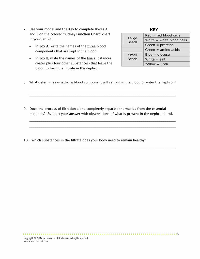
KEY
Large Beads
Red = red blood cells White = white blood cells Green = proteins
Small Beads
Green = amino acids Blue = glucose White = salt Yellow = urea
Kidney Function Kidneys regulate the concentrations of substances in the blood. As blood travels through the kidney, some blood components need to be:
Kept in the blood because they are essential. Red blood cells, white blood cells, protein, glucose and amino acids should be kept in the blood. These components should not be present in urine.
Removed from the blood and excreted in the urine because they are toxic (poisonous). Urea is a toxic substance that should be removed from the blood.
Balanced so they are present in the correct concentration in the blood. A certain amount of water and salt is needed by the body and will remain in the blood. If excess water and excess salt are present in the blood, they will be excreted in the urine.

Copyright © 2009 by University of Rochester. All rights reserved. www.sciencetakeout.com
4
B. Kidneys Filter Blood
4. Prepare a model of a glomerulus and a nephron
by placing the screen (to represent the glomerulus) over the large bowl (to represent the nephron) See diagram on the right.
5. Model the process of filtration that occurs in the glomerulus. Pour the contents of the
“Renal Artery” cup onto the screen (the glomerulus) to form a single layer. 6. The materials trapped on top of the screen remain in the blood. Pour the materials that stay
on top of the screen into the cup labeled “Renal Vein.” Note: some of the small beads may remain on top of the screen. This is OK. In fact, this actually occurs in the kidneys. Most, but not all, of the small substances leave the blood.
Glomerulus Nephron
Each kidney contains over 1 million microscopic blood-cleaning structures called nephrons. Blood enters the kidney through renal arteries. The renal arteries branch to supply blood to the form tiny balls of capillaries called glomeruli. The walls of the glomerulus capillaries are porous. They act like filters to allow small molecules to move from the blood into a cup-like part of the nephron. The movement of materials out of the glomerulus capillaries and into the nephron is known as filtration. The fluid that collects in the nephron is called the filtrate. Figure 2: Filtration allows small molecules to enter the nephron
Glomerulus Nephron
Urine
Glomerulus Renal Vein
Renal Artery
Copyright © 2009 by University of Rochester. All rights reserved. www.sciencetakeout.com
5
7. Use your model and the Key to complete Boxes A and B on the colored “Kidney Function Chart” chart in your lab kit.
In Box A, write the names of the three blood components that are kept in the blood.
In Box B, write the names of the five substances (water plus four other substances) that leave the blood to form the filtrate in the nephron.
8. What determines whether a blood component will remain in the blood or enter the nephron?
_________________________________________________________________________
_________________________________________________________________________
9. Does the process of filtration alone completely separate the wastes from the essential
materials? Support your answer with observations of what is present in the nephron bowl. _________________________________________________________________________
_________________________________________________________________________
10. Which substances in the filtrate does your body need to remain healthy?
_________________________________________________________________________
KEY
Large Beads
Red = red blood cells White = white blood cells Green = proteins
Small Beads
Green = amino acids Blue = glucose White = salt Yellow = urea

Copyright © 2009 by University of Rochester. All rights reserved. www.sciencetakeout.com
6
C. Kidneys Reabsorb Needed Substances Obviously you can’t afford to lose large amounts of water, salt, glucose, and amino acids in your urine! So a second process, called reabsorption, moves essential materials from the nephron back into the blood. Reabsorption occurs when transport proteins molecules in the walls of the nephron return essential substances such as glucose, amino acids, water, and salt to the capillaries that surround the nephron.
Complete Reabsorption Some essential molecules, such as glucose and amino acids, are kept by being completely reabsorbed. These molecules should be completely returned to the blood and should not end up in the urine produced by the kidney. Specific transport proteins in the nephron use energy to move these molecules from the nephron into the capillaries that surround the nephron. 11. What two substances in the filtrate are essential and need to be completely reabsorbed?
_______________________ and _______________________
Renal Artery
Figure 3: Reabsorption returns essential materials to the blood
Nephron
Urine
Glomerulus Renal Vein
KEY
Large Beads
Red = red blood cells White = white blood cells Green = proteins
Small Beads
Green = amino acids Blue = glucose White = salt Yellow = urea

Copyright © 2009 by University of Rochester. All rights reserved. www.sciencetakeout.com
7
12. Model the complete reabsorption of these substances. Use the specific “transport proteins” (these are represented by colored spoons that match the color of the beads) to pick up and move ALL of the completely reabsorbed substances from the “Nephron” bowl to the “Renal Vein” cup.
Selective Reabsorption Other molecules, such as water and salt, are balanced by being selectively reabsorbed to maintain the proper salt and water balance in the body. Selective reabsorption is regulated so that these substances are:
returned to the blood if needed excreted in the urine if present in excess amounts
Specific transport proteins in the nephron use energy to move these molecules from the nephron into the capillaries that surround the nephron.
13. What two substances should be balanced by being selectively reabsorbed?
_______________________ and _______________________
14. Model how selective reabsorption is used to keep the proper amounts of these substances
in the blood.
To maintain homeostasis, the blood needs to contain the proper amount of salt. The “Renal Vein” cup should contain 5 white beads representing salt.
o Use the specific “transport protein” (represented by the colored spoon that matches the color of the beads) to pick up and move white beads so that there are 5 white beads in the “Renal Vein” cup.
o The white beads remaining in the nephron bowl represent excess salt that will be excreted in the urine.
To maintain homeostasis, the blood needs to contain the proper amount of water. The “Renal Vein” cup should be about one-half full of water.
o Pour enough of the water from the “Nephron” bowl to fill the “Renal Vein” cup approximately one-half full.
o The water remaining in the “Nephron” bowl represents excess water that will be excreted in the urine.
Copyright © 2009 by University of Rochester. All rights reserved. www.sciencetakeout.com
8
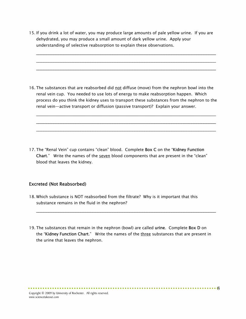
15. If you drink a lot of water, you may produce large amounts of pale yellow urine. If you are dehydrated, you may produce a small amount of dark yellow urine. Apply your understanding of selective reabsorption to explain these observations. _________________________________________________________________________
_________________________________________________________________________
_________________________________________________________________________
16. The substances that are reabsorbed did not diffuse (move) from the nephron bowl into the renal vein cup. You needed to use lots of energy to make reabsorption happen. Which process do you think the kidney uses to transport these substances from the nephron to the renal vein—active transport or diffusion (passive transport)? Explain your answer. _________________________________________________________________________
_________________________________________________________________________
_________________________________________________________________________
17. The “Renal Vein” cup contains “clean” blood. Complete Box C on the “Kidney Function Chart.” Write the names of the seven blood components that are present in the “clean” blood that leaves the kidney.
Excreted (Not Reabsorbed)
18. Which substance is NOT reabsorbed from the filtrate? Why is it important that this substance remains in the fluid in the nephron? _________________________________________________________________________
19. The substances that remain in the nephron (bowl) are called urine. Complete Box D on
the “Kidney Function Chart.” Write the names of the three substances that are present in the urine that leaves the nephron.

Copyright © 2009 by University of Rochester. All rights reserved. www.sciencetakeout.com
9
20. The urine produced by the millions of nephrons collects in the hollow center of the kidney
and then flows out of the body. List the structures of the urinary system (shown in Figure 4, above) that urine must pass through to exit from the body. _________________________________________________________________________
Figure 4: Structures in the Urinary System

Copyright © 2009 by University of Rochester. All rights reserved. www.sciencetakeout.com
10
Part 3: What is wrong with the patient’s kidneys?
So far, you have modeled the function of normal kidneys. Now you will consider what might happen when kidney structure is damaged and the kidney does not function properly. Kidney damage may occur as a result of diabetes, high blood pressure, damage by viruses or bacteria, or by an auto-immune disease in which antibodies attack the kidneys. 1. Your patient’s diabetes has caused kidney disease. What substances in the patient’s urine
indicate that her kidneys are not functioning properly? (Refer to Part 1, question 2 on page 1) _________________________________________________________________________
2. Your patient reported pinkish and cloudy urine.
What substance might cause her urine to be pink? __________________
What substance might cause her urine to be cloudy? __________________
3. Explain how you could change the beads, screen, cup, and bowl model that you used to illustrate how kidney damage caused your patient to have blood cells and protein in her urine.
What part of the model should be changed?
_______________________________________________
How should you change this part?
_______________________________________________
_______________________________________________
What kidney structure was represented by this part of the model?
_______________________________________________
4. What process (filtration or reabsorption) was not working properly in your patient? Explain
how you know. _________________________________________________________________________
_________________________________________________________________________

Copyright © 2009 by University of Rochester. All rights reserved. www.sciencetakeout.com
11
Part 4: Reviewing and Applying What You Learned 1. Label the diagram, above using the following terms: renal artery, renal vein, nephron,
glomerulus, and urine entering the ureter. 2. Draw a labeled arrow on the diagram to represent the process of filtration. In your own
words, explain the process of filtration. _________________________________________________________________________
_________________________________________________________________________
3. Draw a labeled arrow on the diagram to represent the process of reabsorption. In your own
words, explain the process of reabsorption. _________________________________________________________________________
_________________________________________________________________________
4. Excretion involves an interaction between the circulatory system and the excretory system.
On the diagram above:
Put an X in front of the labels for structures that are part of the circulatory system.
Put an O in front of the labels for structures that are part of the excretory system.
Copyright © 2009 by University of Rochester. All rights reserved. www.sciencetakeout.com
12
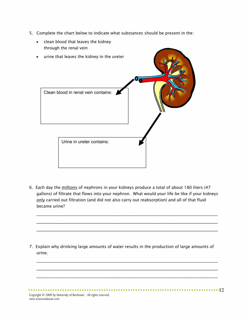
5. Complete the chart below to indicate what substances should be present in the:
clean blood that leaves the kidney through the renal vein
urine that leaves the kidney in the ureter
6. Each day the millions of nephrons in your kidneys produce a total of about 180 liters (47
gallons) of filtrate that flows into your nephron. What would your life be like if your kidneys only carried out filtration (and did not also carry out reabsorption) and all of that fluid became urine? _________________________________________________________________________
_________________________________________________________________________
_________________________________________________________________________
7. Explain why drinking large amounts of water results in the production of large amounts of
urine. _________________________________________________________________________
_________________________________________________________________________
_________________________________________________________________________
Clean blood in renal vein contains:
Urine in ureter contains:
Copyright © 2009 by University of Rochester. All rights reserved. www.sciencetakeout.com
13
8. Explain why eating large amounts of salty foods increases the amount of salt in the urine? _________________________________________________________________________
_________________________________________________________________________
_________________________________________________________________________
9. In addition to diabetes, list three other things may cause kidney disease?
_________________________________________________________________________
_________________________________________________________________________
_________________________________________________________________________
10. Why is kidney disease a serious health risk? What would happen to a person if their
kidneys did not function properly? _________________________________________________________________________
_________________________________________________________________________
_________________________________________________________________________
Optional: If you have access to a computer, try the interactive animation of kidney function at http://www.biologymad.com/resources/kidney.swf.Substitute Click on each of the substances on the left and watch what happens to each different kind as they go through the kidney nephron.
Related Documents


