METHODS A high-capacity RNA affinity column for the purification of human IRP1 and IRP2 overexpressed in Pichia pastoris CHARLES R. ALLERSON, 1 ALAN MARTINEZ, EMINE YIKILMAZ, and TRACEY A. ROUAULT Cell Biology and Metabolism Branch, National Institute of Child Health and Human Development, National Institutes of Health, Bethesda, Maryland 20892, USA ABSTRACT Regulated expression of proteins involved in mammalian iron metabolism is achieved in part through the interaction of the iron regulatory proteins IRP1 and IRP2 with highly conserved RNA stem-loop structures, known as iron-responsive elements (IREs), that are located within the 5 or 3 untranslated regions of regulated transcripts. As part of an effort to determine the structures of the IRP–IRE complexes using crystallographic methods, we have developed an efficient process for obtaining functionally pure IRP1 and IRP2 that relies upon the improved overexpression (>10 mg of soluble IRP per liter of culture) of each human IRP in the yeast Pichia pastoris and large-scale purification using RNA affinity chromatography. Despite the utility of RNA affinity chromatography in the isolation of RNA-binding proteins, current methods for preparing RNA affinity matrices produce columns of low capacity and limited stability. To address these limitations, we have devised a simple method for preparing stable, reusable, high-capacity RNA affinity columns. This method utilizes a bifunctional linker to covalently join a 5-amino tethered RNA with a thiol-modified Sepharose, and can be used to load 150 nmole or more of RNA per milliliter of solid support. We demonstrate here the use of an IRE affinity column in the large-scale purification of IRP1 and IRP2, and suggest that the convenience of this approach will prove attractive in the analysis of other RNA-binding proteins. Keywords: 5-aminoalkyl tether; affinity chromatography; iron regulatory proteins; iron-responsive element (IRE); protein purification; RNA-binding proteins; thiol-modified sepharose INTRODUCTION The isolation and characterization of many RNA-binding proteins has been greatly facilitated by the use of RNA affinity chromatography (Rouault et al. 1989; Neupert et al. 1990; Copeland and Driscoll 1999; Sela-Brown et al. 2000). Although numerous strategies have been employed in the preparation of RNA affinity matrices (for review, see Ka- minski et al. 1998), these methods generally involve the covalent or noncovalent immobilization of the RNA on a solid support. Typically, noncovalent immobilization meth- ods rely upon either the hybridization of poly-A sequences within the target RNA to anchored oligo-U or oligo-dT, or the association of biotinylated RNAs with streptavidin- bearing matrices. Two recent innovations have used RNA aptamers with affinities for streptavidin (Srisawat and En- gelke 2001) or streptomycin (Bachler et al. 1999) to immo- bilize the target RNA. Covalent attachment of RNA has most typically been achieved through either the reaction of unmodifed RNAs with cyanogen bromide activated Sepharose, or the cou- pling of periodate-oxidized RNA with adipic acid dihydra- zide-agarose beads (Caputi et al. 1999). In the former, re- action between the RNA and the matrix happens at random locations, which presumably limits the number of accessible protein binding sites. The latter method requires the treat- ment of the RNA with an oxidant and limits attachment to the 5 end of the RNA. Despite the obvious advantages of RNA affinity chroma- tography, it is rarely used for the routine purification of overexpressed RNA-binding proteins. For large-scale puri- fication, the benefits of RNA affinity chromatography are often discarded in favor of more traditional methodologies. Practical preparative RNA affinity chromatography would require an RNA affinity column that has both high capacity and long-term stability. To the best of our knowledge, none of the above methods, covalent or noncovalent, has ever succeeded in loading more than a few nanomoles of RNA per milliliter of solid support. Furthermore, the intrinsic Reprint requests to: Tracey A. Rouault, Cell Biology and Metabolism Branch, National Institute of Child Health and Human Development, National Institutes of Health, Bethesda, MD 20892, USA; e-mail: trou@ helix.nih.gov; fax: (301) 402-0078. Present address: 1 Isis Pharmaceuticals, 2292 Faraday Avenue, Carlsbad, CA 92008, USA Article and publication are at http://www.rnajournal.org/cgi/doi/ 10.1261/rna.2143303. RNA (2003), 9:364–374. Published by Cold Spring Harbor Laboratory Press. Copyright © 2003 RNA Society. 364

Welcome message from author
This document is posted to help you gain knowledge. Please leave a comment to let me know what you think about it! Share it to your friends and learn new things together.
Transcript
METHODS
A high-capacity RNA affinity column for the purificationof human IRP1 and IRP2 overexpressed in Pichia pastoris
CHARLES R. ALLERSON,1 ALAN MARTINEZ, EMINE YIKILMAZ, and TRACEY A. ROUAULTCell Biology and Metabolism Branch, National Institute of Child Health and Human Development, National Institutes of Health,Bethesda, Maryland 20892, USA
ABSTRACT
Regulated expression of proteins involved in mammalian iron metabolism is achieved in part through the interaction of the ironregulatory proteins IRP1 and IRP2 with highly conserved RNA stem-loop structures, known as iron-responsive elements (IREs),that are located within the 5� or 3� untranslated regions of regulated transcripts. As part of an effort to determine the structuresof the IRP–IRE complexes using crystallographic methods, we have developed an efficient process for obtaining functionallypure IRP1 and IRP2 that relies upon the improved overexpression (>10 mg of soluble IRP per liter of culture) of each humanIRP in the yeast Pichia pastoris and large-scale purification using RNA affinity chromatography. Despite the utility of RNAaffinity chromatography in the isolation of RNA-binding proteins, current methods for preparing RNA affinity matrices producecolumns of low capacity and limited stability. To address these limitations, we have devised a simple method for preparingstable, reusable, high-capacity RNA affinity columns. This method utilizes a bifunctional linker to covalently join a 5�-aminotethered RNA with a thiol-modified Sepharose, and can be used to load 150 nmole or more of RNA per milliliter of solid support.We demonstrate here the use of an IRE affinity column in the large-scale purification of IRP1 and IRP2, and suggest that theconvenience of this approach will prove attractive in the analysis of other RNA-binding proteins.
Keywords: 5�-aminoalkyl tether; affinity chromatography; iron regulatory proteins; iron-responsive element (IRE); proteinpurification; RNA-binding proteins; thiol-modified sepharose
INTRODUCTION
The isolation and characterization of many RNA-binding
proteins has been greatly facilitated by the use of RNA
affinity chromatography (Rouault et al. 1989; Neupert et al.
1990; Copeland and Driscoll 1999; Sela-Brown et al. 2000).
Although numerous strategies have been employed in the
preparation of RNA affinity matrices (for review, see Ka-
minski et al. 1998), these methods generally involve the
covalent or noncovalent immobilization of the RNA on a
solid support. Typically, noncovalent immobilization meth-
ods rely upon either the hybridization of poly-A sequences
within the target RNA to anchored oligo-U or oligo-dT, or
the association of biotinylated RNAs with streptavidin-
bearing matrices. Two recent innovations have used RNA
aptamers with affinities for streptavidin (Srisawat and En-
gelke 2001) or streptomycin (Bachler et al. 1999) to immo-
bilize the target RNA.
Covalent attachment of RNA has most typically been
achieved through either the reaction of unmodifed RNAs
with cyanogen bromide activated Sepharose, or the cou-
pling of periodate-oxidized RNA with adipic acid dihydra-
zide-agarose beads (Caputi et al. 1999). In the former, re-
action between the RNA and the matrix happens at random
locations, which presumably limits the number of accessible
protein binding sites. The latter method requires the treat-
ment of the RNA with an oxidant and limits attachment to
the 5� end of the RNA.
Despite the obvious advantages of RNA affinity chroma-
tography, it is rarely used for the routine purification of
overexpressed RNA-binding proteins. For large-scale puri-
fication, the benefits of RNA affinity chromatography are
often discarded in favor of more traditional methodologies.
Practical preparative RNA affinity chromatography would
require an RNA affinity column that has both high capacity
and long-term stability. To the best of our knowledge, none
of the above methods, covalent or noncovalent, has ever
succeeded in loading more than a few nanomoles of RNA
per milliliter of solid support. Furthermore, the intrinsic
Reprint requests to: Tracey A. Rouault, Cell Biology and MetabolismBranch, National Institute of Child Health and Human Development,National Institutes of Health, Bethesda, MD 20892, USA; e-mail: [email protected]; fax: (301) 402-0078.
Present address: 1Isis Pharmaceuticals, 2292 Faraday Avenue, Carlsbad,CA 92008, USA
Article and publication are at http://www.rnajournal.org/cgi/doi/10.1261/rna.2143303.
RNA (2003), 9:364–374. Published by Cold Spring Harbor Laboratory Press. Copyright © 2003 RNA Society.364
reversibility of noncovalent associations limits the lifetime
and reusability of columns prepared by those methods.
Two extensively studied mammalian mRNA-binding
proteins are the iron regulatory proteins IRP1 and IRP2.
These cytosolic proteins regulate the expression of proteins
involved in iron metabolism through their interaction with
conserved RNA stem-loop structures, referred to as iron-
responsive elements (IREs), that are located within the 5� or3� untranslated regions (UTR) of regulated transcripts (for
review, see Hentze and Kuhn 1996; Harford and Rouault
1998). Under conditions of intracellular iron depletion,
both IRP1 and IRP2 are capable of binding IREs with high
affinity, resulting in either repression of translation or sta-
bilization of the transcript. When iron levels rise, the two
IRPs respond via distinct mechanisms, both of which result
in the loss of IRE-binding activity. Whereas IRP2 is targeted
for degradation by the proteasome (Guo et al. 1995; Iwai et
al. 1995, 1998), IRP1 is loaded with a [4Fe-4S] cluster that
impedes its ability to bind IREs while converting it into a
functional cytosolic aconitase (for review, see Beinert et al.
1996; Hentze and Kuhn 1996; Rouault and Klausner 1996;
Schneider and Leibold 2000).
Efforts to understand the manner in which IRP1 and
IRP2 bind to their IRE substrates have been hindered by
both a lack of structural information and the absence of any
recognizable RNA recognition motif within the IRP se-
quences. However, the similarity between IRP1 and other
aconitases has provided a useful framework for analyzing
IRP structure (Rouault et al. 1991, 1992). Crystal structures
of mitochondrial aconitases reveal a common folding motif
in which the aconitase active site lies in a cleft formed
between the N-terminal globular domains 1–3 and the C-
terminal domain 4 (Robbins and Stout 1989). Several lines
of evidence suggest that both IRP1 and IRP2 adopt similar
conformations, and that the IRE-binding site lies within the
active site cleft (Basilion et al. 1994b; Hirling et al. 1994;
Philpott et al. 1994; Swenson and Walden 1994; Butt et al.
1996). NMR studies (Laing and Hall 1996; Addess et al.
1997) and analysis of IRE mutations (Allerson et al. 1999;
Meehan and Connell 2001) have led to the identification of
nucleotides that are likely to be involved in specific contacts
with the IRPs. A recent study of IRP mutants has further
identified short stretches of amino acids that are likely to
interact with these critical nucleotides (Kaldy et al. 1999).
However, a complete understanding of the IRP–IRE com-
plexes awaits their analysis by X-ray crystallographic meth-
ods.
As part of our effort to determine the structures of the
IRP–IRE complexes, we set out to develop an optimized
strategy for overexpressing and purifying both human IRPs.
We have developed an overexpression system using the
methylotrophic yeast Pichia pastoris, in which yields greatly
exceed those of a previously developed baculovirus expres-
sion system (Basilion et al. 1994a; Samaniego et al. 1994).
Use of the P. pastoris system provides the combined benefits
of high-level expression, easy scale-up, and the ability to
express larger eukaryotic proteins (Cregg et al. 2000).
The eventual crystallization of the IRP–IRE complexes
will require functionally pure IRPs that are fully capable of
being bound to IREs. However, the IRE-binding activities of
expressed IRP1 and IRP2 have been shown to be sensitive to
oxidation in vitro (Phillips et al. 1996), giving rise to the
possibility of functional isomers. To eliminate the nonbind-
ing isomers and ensure functional homogeneity, we have
adopted a purification strategy that employs RNA affinity
chromatography as a final step. To circumvent the limita-
tions of existing methodologies, we have developed a novel
procedure for covalently linking an amine-modified RNA
to a Sepharose support that should provide the capacity and
stability required of a truly reusable column. We report here
the overexpression of human IRP1 and IRP2, the prepara-
tion of an RNA affinity column with an estimated capacity
of ∼15 mg of IRP, and its use in the isolation of functionally
enriched IRPs.
RESULTS AND DISCUSSION
Expression of human IRP1 and IRP2
To obtain the quantities of protein required for future crys-
tallographic studies, we overexpressed human IRP1 and
IRP2 in the yeast P. pastoris. Our laboratory had previously
reported good expression of IRPs in insect cells using a
baculovirus system (Basilion et al. 1994a; Samaniego et al.
1994), but the yields of this system were variable and in-
sufficient for our purposes. Although the expression of rat
IRPs in Saccharomyces cerevisiae has achieved modest levels
(1–2 mg/L of culture; Phillips et al. 1996), the methylotro-
phic yeast P. pastoris has been known to occasionally reach
grams per liter protein yields (Sreekrishna et al. 1988). This
system, which takes advantage of the strong alcohol oxidase
(AOX1) promoter that is activated when the optimal car-
bon source of glucose is replaced with methanol, seemed
ideally suited for IRP expression.
Of the many vectors available for expression in P. pasto-
ris, we used the intracellular expression vector pHIL-D2,
which was included with the original expression kit from
Invitrogen. We selected intracellular expression in an effort
to minimize posttranslational modification of the IRPs and
to protect the expressed protein (especially IRP2) from un-
chelated metals within the expression media. Based on a
report that expression levels can be dramatically improved
by placing the start codon immediately adjacent to the end
of the AOX1 5� UTR (Sreekrishna et al. 1997), we made
several modifications to pHIL-D2. In that report, a modi-
fied expression plasmid was designed to take advantage of a
BstBI (TTCGAA) site located in the last 9 bp of the AOX1
5� UTR. By appending the coding region insert with the
sequence TTCGAAACG ATG . . . (start codon in italics),
the BstBI site can be used to obtain the optimal 5� UTR
RNA affinity chromatography in IRP purification
www.rnajournal.org 365
sequence. To employ a similar strategy with pHIL-D2, we
removed a second BstBI site from the 3� AOX1 region of the
plasmid, and introduced a simple multiple cloning site
(containing unique SacII, BamHI, and XhoI restriction
sites) to create the expression vector pCA10.3.
From plasmids containing the coding sequence of either
human IRP1 (pGEM-hIRF; Hirling et al. 1992) or a myc
epitope-tagged form of human IRP2 (Samaniego et al.
1994), coding regions were isolated and subcloned into
pBluescript II SK(+) to facilitate the introduction of the
sequence TTCGAAACG upstream of each start codon, the
elimination of a preexisting BstBI site in IRP2, and the
removal of the myc-epitope tag from IRP2. Subsequent sub-
cloning into pCA10.3 created the IRP1 and IRP2 expression
constructs pCA19.8 and pCA22.1, respectively.
The P. pastoris strain GS115 was transformed with
pCA19.8, pCA22.1, or control plasmid pCA10.3. After
screening of the transformants, small-scale expression tests
led to the identification of the optimal IRP1-expressing
clone CA1302 (Fig. 1A, lane 5), IRP2-expressing clone
CA1506 (Fig. 1B, lane 5), and a control nonexpressing clone
CA1103 (Fig. 1A,B, lane 3). Each of the optimal expression
clones appeared to contain multiple copies of integrated
insert (data not shown). Both IRP1 and IRP2 were opti-
mally expressed after 36 h of growth in methanol-contain-
ing media and were readily visualized in unpurified total
lysates.
Initial purification of IRP1 and IRP2
Yeast lysates were subjected to one or more rounds of chro-
matographic purification, then analyzed by both Coo-
massie-stained and silver-stained SDS-PAGE. An initial pass
through a Heparin-Sepharose column resulted in a substan-
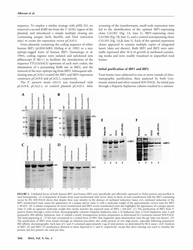
FIGURE 1. Unlabeled forms of both human IRP1 and human IRP2 were specifically and efficiently expressed in Pichia pastoris and purified tonear homogeneity. (A) Comparison of lysates from yeast transformed with vector alone to those of yeast transformed with the IRP1-containingvector by 8% SDS-PAGE shows that despite their near identity in the absence of methanol induction (lanes 2,4), methanol induction of theIRP1-transformed yeast causes the appearance of a unique species (lane 5) with a molecular weight of the approximately correct mass for IRP1(∼98 kD). (B) A similar comparison of vector-transformed and IRP2-vector transformed yeast also highlights the appearance of a unique species(lane 5) with an apparent molecular weight that closely matches the expected mass of IRP2 (∼104 kD). (C) The purification of IRP1 from P.pastoris lysates through a series of four chromatographic columns (Heparin Sepharose, lane 1; Q Sepharose, lane 2; Octyl Sepharose, lane 3; andpreparative IRE-affinity Sepharose, lane 4) yielded a nearly homogeneous protein preparation as determined by Coomassie-stained SDS-PAGE.The band appearing at ∼75 kD may correspond to a nicked form of IRP1 that fragments upon denaturation onto the gel (data not shown). (D)The purification of IRP2 from lysates through an identical procedure also yields protein of very high purity, especially following preparativeIRE-affinity chromatography. Each lane of C and D contained approximately 4 µg of total protein (as determined by BCA assay). (E,F) Analysesof IRP1 (E) and IRP2 (F) purification identical to those depicted in C and D, respectively, except that silver staining was used to visualize theprotein and less protein was used per lane.
Allerson et al.
366 RNA, Vol. 9, No. 3
tial enrichment of both IRPs relative to the initial lysates
(Fig. 1C,D, lane 1). Subsequent fractionation through a Q-
Sepharose column (Fig. 1C,D, lane 2), followed by an octyl-
Sepharose column (Fig. 1C,D, lane 3) yielded IRPs of very
high purity. The efficiency of this stepwise purification was
confirmed using the enhanced sensitivity of silver-staining
methods (Fig. 1E,F, lanes 1–3). Typically, the lysis and pu-
rification of 0.5 L of yeast culture yielded 7–9 mg of purified
IRP after these three chromatographic steps.
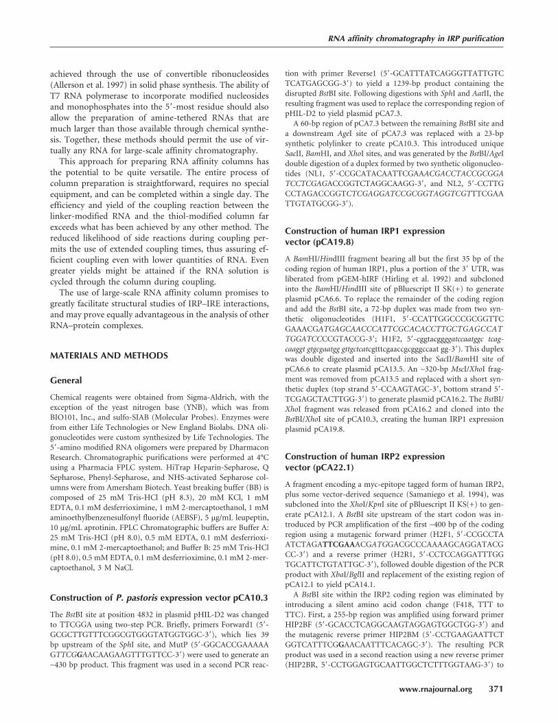
To determine the specific IRE-binding activity of the
IRPs after these three chromatographic steps, each prepa-
ration was used in band-shift RNA-binding titration experi-
ments, as illustrated for IRP1 in Figure 5A (see below).
Despite their relative purity, the amount of IRE-binding
activity of the IRPs varied considerably between batches of
purified protein, and was always much less than expected
based on determinations of protein concentration. In one
batch of IRP1 (Fig. 5A, see below), the specific activity was
measured as approximately 30% of the total protein. Al-
though some of the deviation from the expected maximum
is likely to arise from the inaccuracies of determining both
protein concentration (calibrated against bovine serum al-
bumin standards) and RNA concentration (calculated from
absorbance at 260 nm), the batch-wise variation seemed to
be a separate phenomenon linked to the IRPs themselves. In
several instances, the specific activity was lower than 10% of
maximum, and was consistently lower for IRP2 (data not
shown) than for IRP1. Although not rigorously investi-
gated, there also appeared to be a correlation between the
elapsed time of purification and measured activity, where
longer purification times reflected lower activities. These
observations are in general agreement with previous reports
of diminished IRP2 activity during purification (Phillips et
al. 1996). Although some of the loss of IRE-binding activity
can be reversed by the addition of reducing agents, there is
no assurance that this reversion is complete.
Preparation of a high-capacity RNA affinity column
The variable specific activity and residual impurities in the
purified IRP1 and IRP2 highlighted the need for RNA af-
finity purification. High-capacity DNA affinity columns
have previously been prepared for use in purification of
DNA-binding proteins through the covalent coupling of
alkylamine-tethered DNA with NHS-activated Sepharose
columns (Larson and Verdine 1992). This method was used
to obtain a column with a loading of 250 nmole of DNA per
milliliter of matrix and uses prepacked HiTrap columns
(Amersham Pharmacia) that simplify column preparation
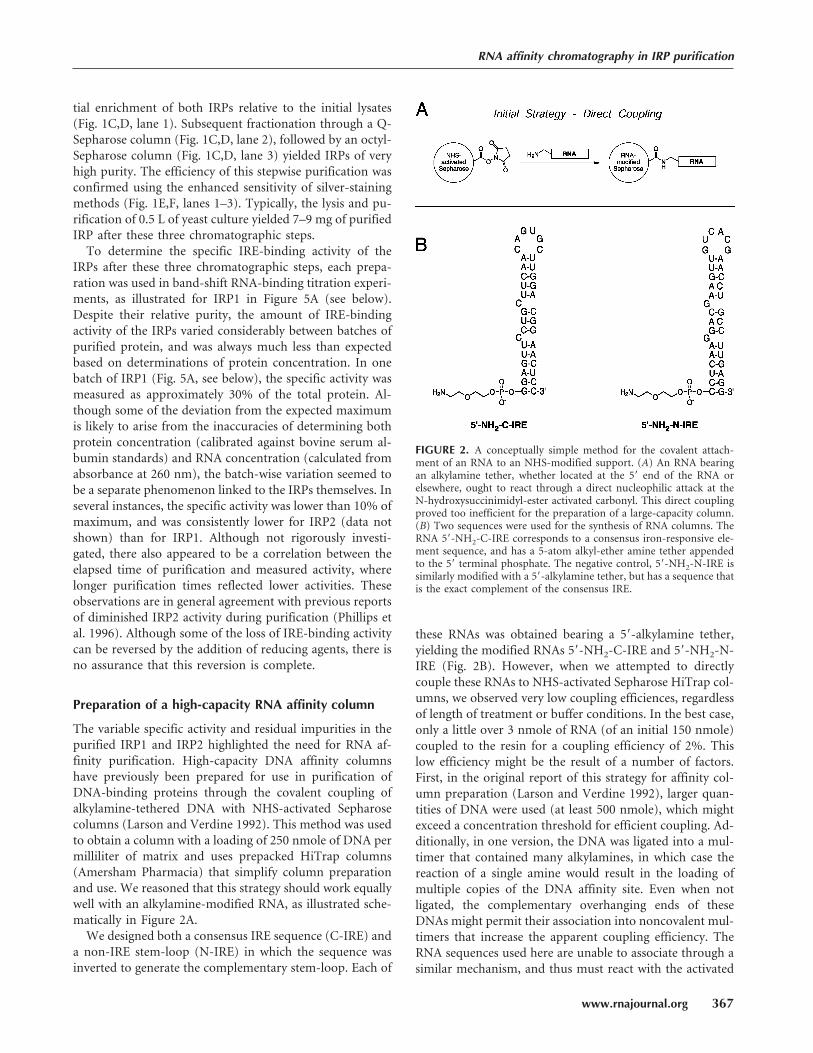
and use. We reasoned that this strategy should work equally
well with an alkylamine-modified RNA, as illustrated sche-
matically in Figure 2A.
We designed both a consensus IRE sequence (C-IRE) and
a non-IRE stem-loop (N-IRE) in which the sequence was
inverted to generate the complementary stem-loop. Each of
these RNAs was obtained bearing a 5�-alkylamine tether,
yielding the modified RNAs 5�-NH2-C-IRE and 5�-NH2-N-
IRE (Fig. 2B). However, when we attempted to directly
couple these RNAs to NHS-activated Sepharose HiTrap col-
umns, we observed very low coupling efficiences, regardless
of length of treatment or buffer conditions. In the best case,
only a little over 3 nmole of RNA (of an initial 150 nmole)
coupled to the resin for a coupling efficiency of 2%. This
low efficiency might be the result of a number of factors.
First, in the original report of this strategy for affinity col-
umn preparation (Larson and Verdine 1992), larger quan-
tities of DNA were used (at least 500 nmole), which might
exceed a concentration threshold for efficient coupling. Ad-
ditionally, in one version, the DNA was ligated into a mul-
timer that contained many alkylamines, in which case the
reaction of a single amine would result in the loading of
multiple copies of the DNA affinity site. Even when not
ligated, the complementary overhanging ends of these
DNAs might permit their association into noncovalent mul-
timers that increase the apparent coupling efficiency. The
RNA sequences used here are unable to associate through a
similar mechanism, and thus must react with the activated
FIGURE 2. A conceptually simple method for the covalent attach-ment of an RNA to an NHS-modified support. (A) An RNA bearingan alkylamine tether, whether located at the 5� end of the RNA orelsewhere, ought to react through a direct nucleophilic attack at theN-hydroxysuccinimidyl-ester activated carbonyl. This direct couplingproved too inefficient for the preparation of a large-capacity column.(B) Two sequences were used for the synthesis of RNA columns. TheRNA 5�-NH2-C-IRE corresponds to a consensus iron-responsive ele-ment sequence, and has a 5-atom alkyl-ether amine tether appendedto the 5� terminal phosphate. The negative control, 5�-NH2-N-IRE issimilarly modified with a 5�-alkylamine tether, but has a sequence thatis the exact complement of the consensus IRE.
RNA affinity chromatography in IRP purification
www.rnajournal.org 367
support as true monomers. It is also possible that the reac-
tivity of the alkylamine tether in 5�-NH2-C-IRE and 5�-NH2-N-IRE is somehow diminished, either sterically or
electrostatically, by its local environment. Regardless, the
direct coupling of the singly tethered IREs proved to be too
inefficient for large-scale use.
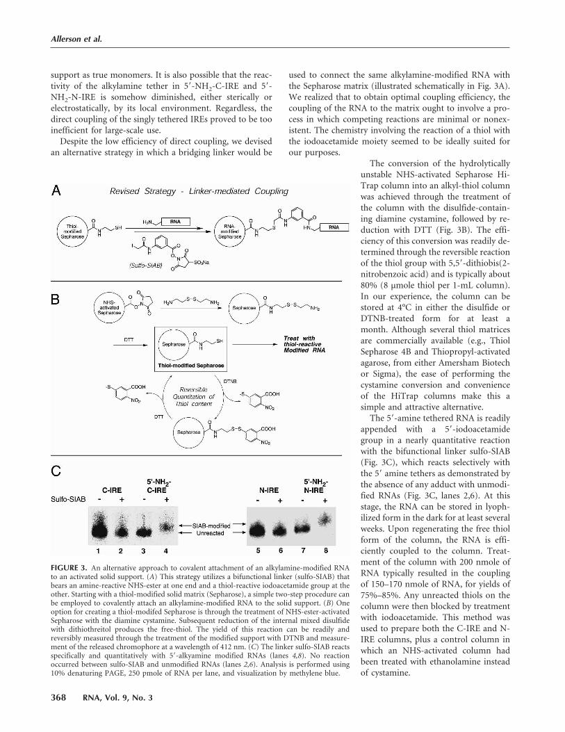
Despite the low efficiency of direct coupling, we devised
an alternative strategy in which a bridging linker would be
used to connect the same alkylamine-modified RNA with
the Sepharose matrix (illustrated schematically in Fig. 3A).
We realized that to obtain optimal coupling efficiency, the
coupling of the RNA to the matrix ought to involve a pro-
cess in which competing reactions are minimal or nonex-
istent. The chemistry involving the reaction of a thiol with
the iodoacetamide moiety seemed to be ideally suited for
our purposes.
The conversion of the hydrolytically
unstable NHS-activated Sepharose Hi-
Trap column into an alkyl-thiol column
was achieved through the treatment of
the column with the disulfide-contain-
ing diamine cystamine, followed by re-
duction with DTT (Fig. 3B). The effi-
ciency of this conversion was readily de-
termined through the reversible reaction
of the thiol group with 5,5�-dithiobis(2-nitrobenzoic acid) and is typically about
80% (8 µmole thiol per 1-mL column).
In our experience, the column can be
stored at 4°C in either the disulfide or
DTNB-treated form for at least a
month. Although several thiol matrices
are commercially available (e.g., Thiol
Sepharose 4B and Thiopropyl-activated
agarose, from either Amersham Biotech
or Sigma), the ease of performing the
cystamine conversion and convenience
of the HiTrap columns make this a
simple and attractive alternative.
The 5�-amine tethered RNA is readily
appended with a 5�-iodoacetamide
group in a nearly quantitative reaction
with the bifunctional linker sulfo-SIAB
(Fig. 3C), which reacts selectively with
the 5� amine tethers as demonstrated by
the absence of any adduct with unmodi-
fied RNAs (Fig. 3C, lanes 2,6). At this
stage, the RNA can be stored in lyoph-
ilized form in the dark for at least several
weeks. Upon regenerating the free thiol
form of the column, the RNA is effi-
ciently coupled to the column. Treat-
ment of the column with 200 nmole of
RNA typically resulted in the coupling
of 150–170 nmole of RNA, for yields of
75%–85%. Any unreacted thiols on the
column were then blocked by treatment
with iodoacetamide. This method was
used to prepare both the C-IRE and N-
IRE columns, plus a control column in
which an NHS-activated column had
been treated with ethanolamine instead
of cystamine.
FIGURE 3. An alternative approach to covalent attachment of an alkylamine-modified RNAto an activated solid support. (A) This strategy utilizes a bifunctional linker (sulfo-SIAB) thatbears an amine-reactive NHS-ester at one end and a thiol-reactive iodoacetamide group at theother. Starting with a thiol-modified solid matrix (Sepharose), a simple two-step procedure canbe employed to covalently attach an alkylamine-modified RNA to the solid support. (B) Oneoption for creating a thiol-modifed Sepharose is through the treatment of NHS-ester-activatedSepharose with the diamine cystamine. Subsequent reduction of the internal mixed disulfidewith dithiothreitol produces the free-thiol. The yield of this reaction can be readily andreversibly measured through the treatment of the modified support with DTNB and measure-ment of the released chromophore at a wavelength of 412 nm. (C) The linker sulfo-SIAB reactsspecifically and quantitatively with 5�-alkyamine modified RNAs (lanes 4,8). No reactionoccurred between sulfo-SIAB and unmodified RNAs (lanes 2,6). Analysis is performed using10% denaturing PAGE, 250 pmole of RNA per lane, and visualization by methylene blue.
Allerson et al.
368 RNA, Vol. 9, No. 3
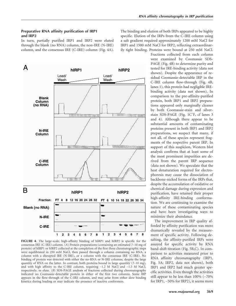
Preparative RNA affinity purification of IRP1and IRP2In turn, partially purified IRP1 and IRP2 were eluted
through the blank (no RNA) column, the non-IRE (N-IRE)
column, and the consensus IRE (C-IRE) column (Fig. 4A).
The binding and elution of both IRPs appeared to be highly
specific. Elution of the IRPs from the C-IRE column using
a salt gradient required approximately 1200 mM NaCl for
IRP1 and 1500 mM NaCl for IRP2, reflecting extraordinar-
ily tight binding. Proteins were bound at 250 mM NaCl.
Fractions collected from each column
were examined by Coomassie SDS-
PAGE (Fig. 4B) to determine purity and
tested for IRE-binding activity (data not
shown). Despite the appearance of re-
sidual Coomassie-detectable IRP in the
C-IRE column flow-through (Fig. 4B,
lanes 1), this protein had negligible IRE-
binding activity (data not shown). In
comparison to the pre-affinity-purified
protein, both IRP1 and IRP2 prepara-
tions appeared only marginally cleaner
by both Coomassie-stain and silver-
stain SDS-PAGE (Fig. 1C�F, cf lanes 3
and 4). Although there appear to be
substantial amounts of contaminating
proteins present in both IRP1 and IRP2
preparations, we suspect that many, if
not all, of these species represent frag-
ments of the respective parent IRP. In
support of this suspicion, Western blot
analysis confirms that at least some of
the most prominent impurities are de-
rived from the parent IRP sequence
(data not shown). We speculate that the
heat denaturation required for electro-
phoresis may cause the dissociation of
backbone-nicked forms of the IRPs that,
despite the accumulation of oxidative or
chemical damage during expression and
purification, have retained their proper
high-affinity IRE-binding conforma-
tion. We are continuing to examine the
nature of these contaminating species
and have been investigating ways to
minimize their abundance.
The improvement in IRP quality af-
forded by affinity purification was more
dramatically revealed by the measure-
ment of specific activity. Following de-
salting, the affinity-purified IRPs were
assayed for specific activity by RNA
band-shift titration (Fig. 5B,C). In com-
parison to activities measured prior to
RNA affinity chromatography (IRP1,
Fig. 5A; IRP2, data not shown), both
IRP1 and IRP2 had much greater spe-
cific activities. Even though the activities
still appear to be less than 100% (∼70%for IRP1, ∼50% for IRP2), it seems more
FIGURE 4. The large-scale, high-affinity binding of hIRP1 and hIRP2 is specific for theconsensus IRE (C-IRE) column. (A) Protein preparations (containing an estimated 7–10 mg ofprotein) of hIRP1 or hIRP2 collected at the completion of the first three chromatographic stepswere equilibrated in 250 mM NaCl, then passed through a column containing no RNA, acolumn with a disrupted IRE (N-IRE), or a column with the consensus IRE (C-IRE). Nobinding of protein was detected with either the no-RNA or N-IRE columns, despite the largequantity of RNA on the latter. In contrast, both proteins bound in large quantity (7–10 mg),and with high affinity to the C-IRE column, requiring ∼1.2 M NaCl and ∼1.5 M NaCl,respectively, to elute. (B) SDS-PAGE analysis of fractions collected during chromatographyindicated no Coomassie-detectable protein in either of the first two columns. Some IRPappears in the flow-through of the C-IRE column, and may arise from either slow bindingkinetics during loading or may indicate the presence of inactive conformers.
RNA affinity chromatography in IRP purification
www.rnajournal.org 369
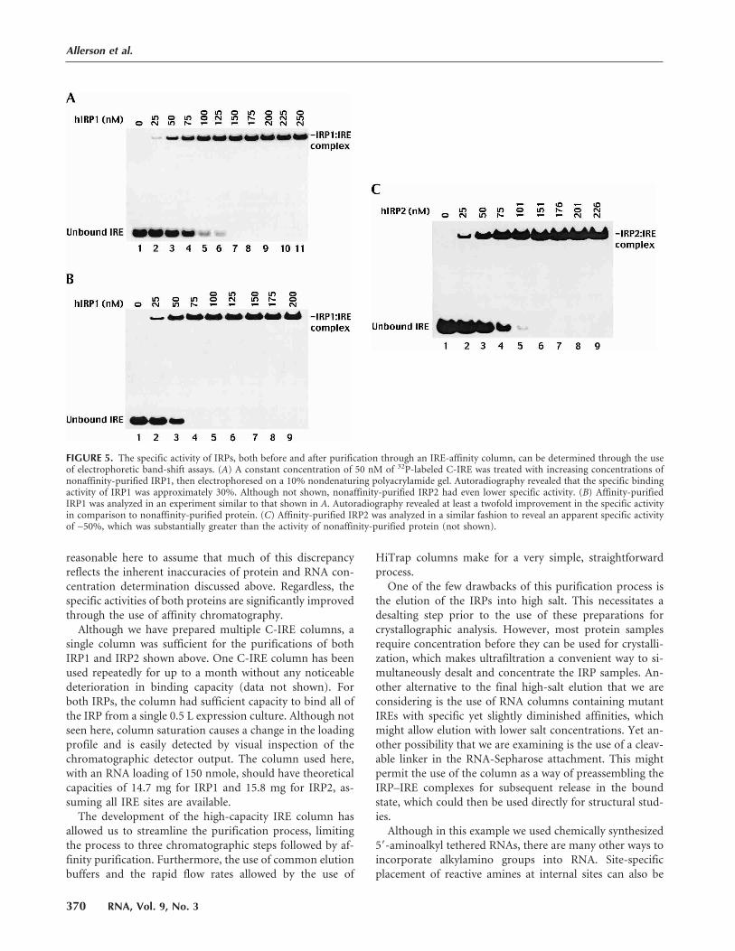
reasonable here to assume that much of this discrepancy
reflects the inherent inaccuracies of protein and RNA con-
centration determination discussed above. Regardless, the
specific activities of both proteins are significantly improved
through the use of affinity chromatography.
Although we have prepared multiple C-IRE columns, a
single column was sufficient for the purifications of both
IRP1 and IRP2 shown above. One C-IRE column has been
used repeatedly for up to a month without any noticeable
deterioration in binding capacity (data not shown). For
both IRPs, the column had sufficient capacity to bind all of
the IRP from a single 0.5 L expression culture. Although not
seen here, column saturation causes a change in the loading
profile and is easily detected by visual inspection of the
chromatographic detector output. The column used here,
with an RNA loading of 150 nmole, should have theoretical
capacities of 14.7 mg for IRP1 and 15.8 mg for IRP2, as-
suming all IRE sites are available.
The development of the high-capacity IRE column has
allowed us to streamline the purification process, limiting
the process to three chromatographic steps followed by af-
finity purification. Furthermore, the use of common elution
buffers and the rapid flow rates allowed by the use of
HiTrap columns make for a very simple, straightforward
process.
One of the few drawbacks of this purification process is
the elution of the IRPs into high salt. This necessitates a
desalting step prior to the use of these preparations for
crystallographic analysis. However, most protein samples
require concentration before they can be used for crystalli-
zation, which makes ultrafiltration a convenient way to si-
multaneously desalt and concentrate the IRP samples. An-
other alternative to the final high-salt elution that we are
considering is the use of RNA columns containing mutant
IREs with specific yet slightly diminished affinities, which
might allow elution with lower salt concentrations. Yet an-
other possibility that we are examining is the use of a cleav-
able linker in the RNA-Sepharose attachment. This might
permit the use of the column as a way of preassembling the
IRP–IRE complexes for subsequent release in the bound
state, which could then be used directly for structural stud-
ies.
Although in this example we used chemically synthesized
5�-aminoalkyl tethered RNAs, there are many other ways to
incorporate alkylamino groups into RNA. Site-specific
placement of reactive amines at internal sites can also be
FIGURE 5. The specific activity of IRPs, both before and after purification through an IRE-affinity column, can be determined through the useof electrophoretic band-shift assays. (A) A constant concentration of 50 nM of 32P-labeled C-IRE was treated with increasing concentrations ofnonaffinity-purified IRP1, then electrophoresed on a 10% nondenaturing polyacrylamide gel. Autoradiography revealed that the specific bindingactivity of IRP1 was approximately 30%. Although not shown, nonaffinity-purified IRP2 had even lower specific activity. (B) Affinity-purifiedIRP1 was analyzed in an experiment similar to that shown in A. Autoradiography revealed at least a twofold improvement in the specific activityin comparison to nonaffinity-purified protein. (C) Affinity-purified IRP2 was analyzed in a similar fashion to reveal an apparent specific activityof ∼50%, which was substantially greater than the activity of nonaffinity-purified protein (not shown).
Allerson et al.
370 RNA, Vol. 9, No. 3
achieved through the use of convertible ribonucleosides
(Allerson et al. 1997) in solid phase synthesis. The ability of
T7 RNA polymerase to incorporate modified nucleosides
and monophosphates into the 5�-most residue should also
allow the preparation of amine-tethered RNAs that are
much larger than those available through chemical synthe-
sis. Together, these methods should permit the use of vir-
tually any RNA for large-scale affinity chromatography.
This approach for preparing RNA affinity columns has
the potential to be quite versatile. The entire process of
column preparation is straightforward, requires no special
equipment, and can be completed within a single day. The
efficiency and yield of the coupling reaction between the
linker-modified RNA and the thiol-modified column far
exceeds what has been achieved by any other method. The
reduced likelihood of side reactions during coupling per-
mits the use of extended coupling times, thus assuring ef-
ficient coupling even with lower quantities of RNA. Even
greater yields might be attained if the RNA solution is
cycled through the column during coupling.
The use of large-scale RNA affinity column promises to
greatly facilitate structural studies of IRP–IRE interactions,
and may prove equally advantageous in the analysis of other
RNA–protein complexes.
MATERIALS AND METHODS
General
Chemical reagents were obtained from Sigma-Aldrich, with the
exception of the yeast nitrogen base (YNB), which was from
BIO101, Inc., and sulfo-SIAB (Molecular Probes). Enzymes were
from either Life Technologies or New England Biolabs. DNA oli-
gonucleotides were custom synthesized by Life Technologies. The
5�-amino modified RNA oligomers were prepared by Dharmacon
Research. Chromatographic purifications were performed at 4°C
using a Pharmacia FPLC system. HiTrap Heparin-Sepharose, Q
Sepharose, Phenyl-Sepharose, and NHS-activated Sepharose col-
umns were from Amersham Biotech. Yeast breaking buffer (BB) is
composed of 25 mM Tris-HCl (pH 8.3), 20 mM KCl, 1 mM
EDTA, 0.1 mM desferrioximine, 1 mM 2-mercaptoethanol, 1 mM
aminoethylbenzenesulfonyl fluoride (AEBSF), 5 µg/mL leupeptin,
10 µg/mL aprotinin. FPLC Chromatographic buffers are Buffer A:
25 mM Tris-HCl (pH 8.0), 0.5 mM EDTA, 0.1 mM desferrioxi-
mine, 0.1 mM 2-mercaptoethanol; and Buffer B: 25 mM Tris-HCl
(pH 8.0), 0.5 mM EDTA, 0.1 mM desferrioximine, 0.1 mM 2-mer-
captoethanol, 3 M NaCl.
Construction of P. pastoris expression vector pCA10.3
The BstBI site at position 4832 in plasmid pHIL-D2 was changed
to TTCGGA using two-step PCR. Briefly, primers Forward1 (5�-GCGCTTGTTTCGGCGTGGGTATGGTGGC-3�), which lies 39
bp upstream of the SphI site, and MutP (5�-GGCACCGAAAAAGTTCGGAACAAGAAGTTTGTTCC-3�) were used to generate an
∼430 bp product. This fragment was used in a second PCR reac-
tion with primer Reverse1 (5�-GCATTTATCAGGGTTATTGTCTCATGAGCGG-3�) to yield a 1239-bp product containing the
disrupted BstBI site. Following digestions with SphI and AatII, the
resulting fragment was used to replace the corresponding region of
pHIL-D2 to yield plasmid pCA7.3.
A 60-bp region of pCA7.3 between the remaining BstBI site and
a downstream AgeI site of pCA7.3 was replaced with a 23-bp
synthetic polylinker to create pCA10.3. This introduced unique
SacII, BamHI, and XhoI sites, and was generated by the BstBI/AgeI
double digestion of a duplex formed by two synthetic oligonucleo-
tides (NL1, 5�-CCGCATACAATTCGAAACGACCTACCGCGGATCCTCGAGACCGGTCTAGGCAAGG-3�, and NL2, 5�-CCTTGCCTAGACCGGTCTCGAGGATCCGCGGTAGGTCGTTTCGAA
TTGTATGCGG-3�).
Construction of human IRP1 expressionvector (pCA19.8)
A BamHI/HindIII fragment bearing all but the first 35 bp of the
coding region of human IRP1, plus a portion of the 3� UTR, was
liberated from pGEM-hIRF (Hirling et al. 1992) and subcloned
into the BamHI/HindIII site of pBluescript II SK(+) to generate
plasmid pCA6.6. To replace the remainder of the coding region
and add the BstBI site, a 72-bp duplex was made from two syn-
thetic oligonucleotides (H1F1, 5�-CCATTGGCCCGCGGTTCGAAACGATGAGCAACCCATTCGCACACCTTGCTGAGCCAT
TGGATCCCCGTACCG-3�; H1F2, 5�-cggtacggggatccaatggc tcag-caaggt gtgcgaatgg gttgctcatcgtttcgaaccgcgggccaat gg-3�). This duplexwas double digested and inserted into the SacII/BamHI site of
pCA6.6 to create plasmid pCA13.5. An ∼320-bp MscI/XhoI frag-ment was removed from pCA13.5 and replaced with a short syn-
thetic duplex (top strand 5�-CCAAGTAGC-3�, bottom strand 5�-TCGAGCTACTTGG-3�) to generate plasmid pCA16.2. The BstBI/
XhoI fragment was released from pCA16.2 and cloned into the
BstBI/XhoI site of pCA10.3, creating the human IRP1 expression
plasmid pCA19.8.
Construction of human IRP2 expressionvector (pCA22.1)
A fragment encoding a myc-epitope tagged form of human IRP2,
plus some vector-derived sequence (Samaniego et al. 1994), was
subcloned into the XhoI/KpnI site of pBluescript II KS(+) to gen-
erate pCA12.1. A BstBI site upstream of the start codon was in-
troduced by PCR amplification of the first ∼400 bp of the coding
region using a mutagenic forward primer (H2F1, 5�-CCGCCTAATCTAGATTCGAAACGATGGACGCCCAAAAGCAGGATACG
CC-3�) and a reverse primer (H2R1, 5�-CCTCCAGGATTTGGTGCATTCTGTATTGC-3�), followed double digestion of the PCR
product with XbaI/BglII and replacement of the existing region of
pCA12.1 to yield pCA14.1.
A BstBI site within the IRP2 coding region was eliminated by
introducing a silent amino acid codon change (F418, TTT to
TTC). First, a 255-bp region was amplified using forward primer
HIP2BF (5�-GCACCTCAGGCAAGTAGGAGTGGCTGG-3�) and
the mutagenic reverse primer HIP2BM (5�-CCTGAAGAATTCTGGTCATTTCGGAACAATTTCACAGC-3�). The resulting PCR
product was used in a second reaction using a new reverse primer
(HIP2BR, 5�-CCTGGAGTGCAATTGGCTCTTTGGTAAG-3�) to
RNA affinity chromatography in IRP purification
www.rnajournal.org 371
generate an 1184-bp product, which was digested with BspEI and
NcoI and substituted into pCA14.1 to create pCA17.7. The myc-
epitope tag was removed and the wild-type stop codon reestab-
lished by amplifying the terminal portion of the coding region
with forward primer HIP2EF (5�-CGAGTAGAGGAAGAACATGTTATACTATCC-3�) and a reverse primer HIP2ER (5�GGCGTGTATTGGATCCTATGAGAATTTTCGTGCCACAAAGTTTAAT
AATCCTCC-3�) that adds a BamHI site adjacent to the new stop
codon, digesting this product with NcoI/BamHI, and replacing the
existing segment of pCA17.7 to make the new plasmid pCA21.2.
The BstBI/BamHI fragment was subsequently released and cloned
into the corresponding site of pCA10.3, creating the human IRP2
expression plasmid pCA22.1.
Transformation of P. pastoris and selection ofoptimally expressing clones
The IRP1 expression plasmid (pCA19.8) and the IRP2 expression
plasmid (pCA22.1), along with empty pCA10.3 plasmid as a con-
trol, were linearized by digestion with SalI, then transformed into
strain GS115 (his4, Mut+) of P. pastoris. Transformations were
performed using the spheroplast method as described in the sup-
plier’s handbook (Invitrogen). From these transformations, 20
(control), 30 (IRP1), and 12 (IRP2) colonies were selected for
further screening. Genomic DNA for each was collected and ex-
amined by PCR for the presence of expression insert using the
primer set NHA5 (5�-CAGAAGGAAGCTGCCCTGTCTTAAACC-3�) and NHA3 (5�-GCGAGATAGGCTGATCAGGAGCAAGCTCGTACG-3�). These primers should generate products of
∼2200 bp, corresponding to the wild-type AOX1 gene, 338 bp for
the CA11 (control) clones, 2994 bp for CA13 (IRP1) clones, and
3154 bp for CA15 (IRP2) clones. By this analysis, 10 of 20 CA11,
22 of 30 CA13, and 6 of 12 CA15 contained the appropriate insert.
To screen for optimal protein expression, single colonies were
used to inoculate 10 mL of minimal glycerol media (1.34% YNB,
1% glycerol, 4 × 10⁻⁵% biotin) and were subsequently cultured at
30°C until they reached an O.D.600 of 2–3 (∼2 days), at which
point they were gently pelleted and resuspended in minimal
methanol media (1.34% YNB, 0.5% methanol, 4 × 10⁻⁵% biotin)
at an O.D.600 of 1.0. These were incubated for an additional 2 days
at 30°C (cultures were supplemented with methanol at 24 h to
maintain 0.5% concentration). The yeast were then pelleted and
resuspended in breaking buffer and lysed using glass beads (425–
600 µm, acid washed, Sigma). Lysates were examined by 8% SDS-
PAGE and stained with Coomassie brilliant blue. From this analy-
sis, CA13-02 and CA13-17 showed the highest levels of IRP1 ex-
pression, whereas CA15-06 and CA15-10 showed the highest levels
of IRP2 expression. Time course analysis found optimal expression
after 36 h of induction with methanol.
Large scale expression of IRP1 and IRP2
Glycerol stocks of IRP1 expression clone CA13-02 and IRP2 ex-
pression clone CA15-06 were prepared and maintained at −80°C.
Periodically, portions of these stocks were streaked onto minimal
2% dextrose plates, grown at 30°C for 2 days, then stored at 4°C.
For optimal expression, colonies were picked within 2 wk of plat-
ing. Single colonies were used to inoculate 10 mL of minimal
glycerol media, followed by incubation for 20 h at 30°C and 275
rpm. A 4-mL aliquot of this culture was used to inoculate 250 mL
of minimal glycerol media, which was then incubated at 30°C and
275 rpm until the cell density had reached O.D.600 ∼6–8 (18–22 h).Yeast were pelleted by centrifugation (500g), then resuspended in
mimimal methanol media to a density of O.D,600 = 1. Culture
volumes of 250 mL were incubated in 1 L baffled flasks for 36 h.
At 24 h, additional methanol was added to maintain 0.5% con-
centration. To harvest, yeast were collected at 6500 rpm (7500g)
and stored at −80°C until ready for purification.
Lysis and initial purification of IRP1 and IRP2
Yeast pelleted from two 250-mL cultures were thawed on ice and
resuspended in 60 mL of degassed ice-cold breaking buffer. Lysis
was performed either with glass beads or using a French pressure
cell, followed by centrifugation at 9500 rpm (12,500g), for 12 min
at 4°C. The supernatant was collected, transferred to a fresh tube,
and immediately subjected to chromatographic purification.
Chromatographic buffers were continually sparged with Argon
to minimize the presence of oxygen. Elution of protein from each
column was monitored by absorbance at 280 nm as 1-mL fractions
were collected. Flow rates were typically 1 mL/min for loading, 0.5
mL/min for elution. Crude lysates were first loaded onto a 2 × 5
mL HiTrap Heparin Sepharose column (two 5-mL columns con-
nected in series). The column was washed with 10 mM NaCl, then
eluted with an gradient to 2400 mM NaCl. Typically, IRP1 eluted
at ∼390 mM NaCl, IRP2 at ∼435 mM NaCl. The appropriate
fractions (as determined by SDS-PAGE) were pooled, diluted to 40
mM NaCl, then loaded onto a 2 × 5 mL HiTrap Q Sepharose
column. After washing with 40 mM NaCl, protein was eluted with
a gradient up to 1400 mM NaCl, with IRP1 eluting at ∼225 mM
NaCl and IRP2 eluting at ∼255 mM NaCl. Again, the appropriate
fractions were pooled, brought to 3 M NaCl, then loaded onto a 4
× 1 mL HiTrap Octyl Sepharose column and eluted with a de-
creasing NaCl gradient. IRP1 eluted at ∼0 mMNaCl, whereas IRP2
eluted at ∼500 mM NaCl. Protein concentrations were determined
using the Pierce/Endogen BCA assay kit.
RNA affinity column preparation
The 36-mer modified RNAs (5�-NH2-C-IRE, 5�-aminotether-
GGAGUUCCUGCUUCAACAGUGCUUGGACGGAACUCC-3�; 5�-NH2-N-IRE, 5�-aminotether-GGAGUUCCUGAUUCAAUACACCUU
GGACGGAACUCC-3�), were received as lyophilized pellets and depro-
tected as recommended by the supplier. For direct coupling to the Hi-
Trap NHS-activated Sepharose column (Amersham Biotech), 150
nmole of RNAwere dissolved in 1.2 mL of 0.2MNaHCO3, 0.5MNaCl
(pH 8.3) and loaded onto the column as recommended by the manu-
facturer. After elution through a sizing column to remove release N-
hydroxysuccinimide, RNA was quantified by measuring the absorbance
at 260 nm.
To prepare a thiol-modified column for the linker approach, a
1-mL HiTrap-NHS Sepharose column was washed with 5 mL of
ice-cold 1-mM HCl (slowly, to avoid irreversible compression of
the column matrix), then with 100 mM cystamine (pH 8.0) in
three 2-mL portions. An additional column was treated instead
with 100 mM ethanolamine as a control. All columns were sub-
sequently washed with 50 mM Tris-HCl.
Allerson et al.
372 RNA, Vol. 9, No. 3
The amount of cystamine on the column was determined by
measuring the release of 4-thio-2-nitrobenzoic acid upon treat-
ment of the reduced columns with 2,2�-dithiobisnitrobenzoic acid(DTNB). Each column was reduced with 50 mM DTT then
washed with 40 mM Tris-HCl (pH 7.5), 20 mM NaCl. Treatment
with 5 mL of 30 mM DTNB in 200 mM Tris-HCl (pH 7.5) was
achieved by passing 2 mL of the solution through the column,
waiting 4 min, then repeating twice more with 1.5-mL portions.
The combined DTNB eluents were pooled, diluted, and assayed
for absorbance at 412 nm.
Both RNAs were modified with sulfo-SIAB by treating with 4.5
mM sulfo-SIAB in 200 mM sodium phosphate (pH 8) for 6 h at
room temperature in the dark. The RNA was then ethanol pre-
cipitated, dried, and redissolved in 1.1 mL 180 mM sodium phos-
phate (pH 8), 5 mM NaCl. Folding of the IRE structure was
promoted by heating to 85°C for 3 min, followed by rapid cooling
on ice. After washing the thiol column first with 20 mM solution
of DTT in 200 mM Tris-HCl (pH 7.5)/20 mM NaCl, then with 200
mM Tris-HCl (pH 7.5)/20 mM NaCl, the RNA solution was care-
fully loaded into the column. The column was sealed and incu-
bated in the dark at room temperature for 12–16 h, after which it
was washed with 4 mL of 200 mM Tris-HCl (pH 7.5). The RNA
loading of the column was determined by comparing the A260 of
the RNA solution from before and after the coupling reaction.
Unreacted thiol groups were blocked in a subsequent treatment of
the column with 10 mM iodoacetamide in 200 mM Tris-HCl (pH
7.5).
Elution of IRP1 and IRP2 through RNA columns
Solutions of IRP1 or IRP2 were loaded through the control, N-
IRE, and C-IRE columns at 250 mM NaCl and 0.5 mL/min. Elu-
tion from the C-IRE column was achieved with a gradient of 250
mM to 3 M NaCl. Protein-containing fractions were desalted by
ultrafiltration (Amicon).
Band-shift titrations
A 32P-labeled RNA corresponding to the C-IRE sequence (5�-GGAGUUCCUGCUUCAACAGUGCUUGGACGGAACUCC-3�) was
generated using methods previously described (Allerson et al.
1999). For each mixture, 50 nM of 32P-labeled C-IRE was mixed
with increasing concentrations of either IRP1 or IRP2 (from 0 to
225 nM) in the presence of 5% glycerol, 0.025 units/µL RNase
Inhibitor (5 Prime 3 Prime, Inc.), 0.15 mg/mL yeast tRNA, 2 mM
DTT, 25 mM Tris-HCl (pH 7.5), and 40 mM KCl. These reactions
were incubated for 20 minutes at room temperature, then eletro-
phoresed on 10% nondenaturing polyacrylamide gels at 130 V for
4 h. After drying, the gels were exposed to either Kodak BioMax
MR film, or a Molecular Dynamics Phosphorimaging screen.
Quantitation was performed using the ImageQuant software pack-
aged (Molecular Dynamics).
ACKNOWLEDGMENTS
We thank Lukas Kuhn for providing the plasmid pGEM-hIRF.
The publication costs of this article were defrayed in part by
payment of page charges. This article must therefore be hereby
marked “advertisement” in accordance with 18 USC section 1734
solely to indicate this fact.
Received September 19, 2002; accepted December 2, 2002.
REFERENCES
Addess, K.J., Basilion, J.P., Klausner, R.D., Rouault, T.A., and Pardi. A.1997. Structure and dynamics of the iron responsive element RNA:Implications for binding of the RNA by iron regulatory bindingproteins. J. Mol. Biol. 274: 72–83.
Allerson, C.R., Chen, S.L., and Verdine, G.L. 1997. A chemical methodfor site-specific modification of RNA: The convertible nucleosideapproach. J. Am. Chem. Soc. 119: 7423–7433.
Allerson, C.R., Cazzola, M., and Rouault, T.A. 1999. Clinical severityand thermodynamic effects of iron-responsive element mutationsin hereditary hyperferritinemia-cataract syndrome. J. Biol. Chem.274: 26439–26447.
Bachler, M., Schroeder, R., and Ahsen, U.V. 1999. StreptoTag: A novelmethod for the isolation of RNA-binding proteins. RNA 5: 1509–1516.
Basilion, J.P., Kennedy, M.C., Beinert, H., Massinople, C.M., Klausner,R.D., and Rouault, T.A. 1994a. Overexpression of iron-responsiveelement-binding protein and its analytical characterization as theRNA-binding form, devoid of an iron-sulfur cluster. Arch. Bio-chem. Biophys. 311: 517–522.
Basilion, J.P., Rouault, T.A., Massinople, C.M., Klausner, R.D., andBurgess, W.H. 1994b. The iron-responsive element-binding pro-tein: Localization of the RNA-binding site to the aconitase active-site cleft. Proc. Natl. Acad. Sci. 91: 574–578.
Beinert, H., Kennedy, M.C., and Stout, D.C. 1996. Aconitase as iron-sulfur protein, enzyme, and iron-regulatory protein. Chem. Rev.96: 2335–2373.
Butt, J., Kim, H.-Y., Basilion, J.P., Cohen, S., Iwai, K., Philpott, C.C.,Altschul, S., Klausner, R.D., and Rouault, T.A. 1996. Differences inthe RNA binding sites of iron regulatory proteins and potentialtarget diversity. Proc. Natl. Acad. Sci. 93: 4345–4349.
Caputi, M., Mayeda, A., Krainer, A.R., and Zahler, A.M. 1999. hnRNPA/B proteins are required for inhibition of HIV-1 pre-mRNA splic-ing. EMBO J. 18: 4060–4067.
Copeland, P.R. and Driscoll, D.M. 1999. Purification, redox sensitiv-ity, and RNA binding properties of SECIS-binding protein 2, aprotein involved in selenoprotein biosynthesis. J. Biol. Chem.274: 25447–25454.
Cregg, J.M., Cereghino, J.L., Shi, J., and Higgins, D.R. 2000. Recom-binant protein expression in Pichia pastoris.Mol. Biotechnol. 6: 23–52.
Guo, B., Phillips, J.D, Yu, Y., and Leibold, E.A. 1995. Iron regulates theintracellular degradation of iron regulatory protein 2 by the pro-teasome. J. Biol. Chem. 270: 21645–21651.
Harford, J.B. and Rouault, T.A. 1998. RNA structure and function incellular iron homeostasis. In RNA structure and function (eds. R.W.Simons and M. Grunberg-Manago), pp. 575–602. Cold SpringHarbor Laboratory Press, Cold Spring Harbor, NY.
Hentze, M.W. and Kuhn, L.C. 1996. Molecular control of vertebrateiron metabolism: mRNA-based regulatory circuits operated byiron, nitric oxide, and oxidative stress. Proc. Natl. Acad. Sci.93: 8175–8182.
Hirling, H., Emery-Goodman, A., Thompson, N., Neupert, B., Seiser,C., and Kuhn, L.C. 1992. Expression of active iron regulatory fac-tor from a full-length human cDNA by in vitro transcription/translation. Nuc. Acids Res. 20: 33–39.
Hirling, H., Henderson, B.R., and Kuhn, L.C. 1994. Mutational analy-sis of the [4Fe-4S]-cluster converting iron regulatory factor fromits RNA-binding form to cytoplasmic aconitase. EMBO J. 13: 453–461.
Iwai, K., Klausner, R.D., and Rouault, T.A. 1995. Requirements for
RNA affinity chromatography in IRP purification
www.rnajournal.org 373
iron-regulated degradation of the RNA binding protein, iron regu-latory protein 2. EMBO J. 14: 5350–5357.
Iwai, K., Drake, S.K., Wehr, N.B., Weissman, A.M., LaVaute, T., Mi-nato, N., Klausner, R.D., Levine, R.L., and Rouault, T.A. 1998.Iron-dependent oxidation, ubiquitination, and degradation of ironregulatory protein 2: Implications for degradation of oxidized pro-teins. Proc. Natl. Acad. Sci. 95: 4924–4928.
Kaldy, P., Menotti, E., Moret, R., and Kuhn, L.C. 1999. Identificationof RNA-binding surfaces in iron regulatory protein-1. EMBO J.18: 6073–6083.
Kaminski, A., Ostareck, D.H., Standart, N.M., and Jackson, R.J. 1998.Affinity methods for isolating RNA binding proteins. In RNA:pro-tein interactions, a practical approach (ed. C.W.J. Smith), pp. 137–160. Oxford University Press, New York.
Laing, L.G. and Hall, K.B. 1996. A model of the iron responsive ele-ment RNA hairpin loop structure determined from NMR andthermodynamic data. Biochemistry 35: 13586–13596.
Larson, C.J. and Verdine, G.L. 1992. A high-capacity column for af-finity purification of sequence-specific DNA-binding proteins.Nucleic Acids Res. 20: 3525.
Meehan, H.A. and Connell, G.J. 2001. The hairpin loop but not thebulged C of the iron responsive element is essential for high affinitybinding to iron regulatory protein-1. J. Biol. Chem. 276: 14791–14796.
Neupert, B., Thompson, N.A., Meyer, C., and Kuhn, L.C. 1990. A highyield affinity purification method for specific RNA-binding pro-teins: Isolation of the iron regulatory factor from human placenta.Nucleic Acids Res. 18: 51–55.
Phillips, J.D., Guo, B., Yu, Y., Brown, F.M., and Leibold, E.A. 1996.Expression and biochemical characterization of iron regulatoryproteins 1 and 2 in Saccharomyces cerevisiae. Biochemistry35: 15704–15714.
Philpott, C.C., Klausner, R.D., and Rouault, T.A. 1994. The bifunc-tional iron-responsive element binding protein/cytosolic aconitase:The role of active-site residues in ligand binding and regulation.Proc. Natl. Acad. Sci. 91: 7321–7325.
Robbins, A.H. and Stout, C.D. 1989. The structure of aconitase. Pro-teins 5: 289–312.
Rouault, T.A. and Klausner, R.D. 1996. Post-transcriptional regulationof genes of iron metabolism in mammalian cells. J. Bioinorg. Chem.
1: 494–499.Rouault, T.A., Hentze, M.W., Haile, D.J., Harford, J.B., and Klausner,
R.D. 1989. The iron-responsive element binding protein: Amethod for the affinity purification of a regulatory RNA-bindingprotein. Proc. Natl. Acad. Sci. 86: 5768–5772.
Rouault, T.A., Stout, C.D., Kaptain, S., Harford, J.B., and Klausner,R.D. 1991. Structural relationship between an iron-regulated RNA-binding protein (IRE-BP) and aconitase: Functional implications.Cell 64: 881–883.
Rouault, T.A., Haile, D.J., Downey, W.E., Philpott, C.C., Tang, C.,Samaniego, F., Chin, J., Paul, I., Orloff. D., Harford, J.B., et al.1992. An iron-sulfur cluster plays a novel regulatory role in theiron-responsive element binding protein. BioMetals 5: 131–140.
Samaniego, F., Chin, J., Iwai, K., Rouault, T.A., and Klausner, R.D.1994. Molecular characterization of a second iron-responsive ele-ment binding protein, iron regulatory protein 2. J. Biol. Chem.269: 30904–30910.
Schneider, B.D. and Leibold, E.A. 2000. Regulation of mammalianiron homeostasis. Curr. Opin. Clin. Nutr. Metab. Care 3: 267–273.
Sela-Brown, A., Silver, J., Brewer, G., and Naveh-Many, T. 2000. Iden-tification of AUF1 as a parathyroid hormone mRNA 3�-untrans-lated region-binding protein that determines parathyroid hormonemRNA stability. J. Biol. Chem. 275: 7424–7429.
Sreekrishna, K., Potenz, R.H.B., Cruze, J.A., McCombie, W.R., Parker,K.A., Nelles, L., Mazzaferro, P.K., Holden, K.A., Harrison, R.G.,Wood, P.J., et al. 1988. High level expression of heterologous pro-teins in methylotrophic yeast Pichia pastoris. J. Basic Microbiol.28: 265–278.
Sreekrishna, K., Brankamp, R.G., Kropp, K.E., Blankenship, D.T.,Tsay, J.-T., Smith, P.L., Wierschke, J.D., Subramaniam, A., andBirkenberger, L.A. 1997. Strategies for optimal synthesis and se-cretion of heterologous proteins in the methylotrophic yeast Pichiapastoris. Gene 190: 55–62.
Srisawat, C. and Engelke, D.R. 2001. Streptavidin aptamers: Affinitytags for the study of RNAs and ribonucleoproteins. RNA 7: 632–641.
Swenson, G.R. and Walden, W.E. 1994. Localization of an RNA bind-ing element of the iron responsive element binding protein withina proteolytic fragment containing iron coordination ligands.Nucleic Acids Res. 22: 2627–2633.
Allerson et al.
374 RNA, Vol. 9, No. 3
Related Documents