A different function for a member of an ancient and highly conserved cytochrome P450 family: From essential sterols to plant defense Xiaoquan Qi* † , Saleha Bakht*, Bo Qin*, Mike Leggett ‡ , Andrew Hemmings § , Fred Mellon ¶ , John Eagles ¶ , Daniele Werck-Reichhart , Hubert Schaller , Agnes Lesot , Rachel Melton*, and Anne Osbourn* , ** *Department of Metabolic Biology, John Innes Centre, Norwich NR4 7UH, United Kingdom; ‡ Institute of Grassland and Environmental Research, Aberystwyth SY23 3EB, Wales, United Kingdom; § School of Biological Sciences and School of Chemical Sciences and Pharmacy, University of East Anglia, Norwich NR4 7TJ, United Kingdom; ¶ Institute of Food Research, Norwich NR4 7UA, United Kingdom; Institute of Plant Molecular Biology, Centre National de la Recherche ´ Scientifique–Unite Propre de Recherche ´ 2357, Universite Louis Pasteur, 67000 Strasbourg, France; and † Laboratory of Photosynthesis and Environmental Molecular Physiology, Institute of Botany, Chinese Academy of Sciences, Nanxincun 20, Fragrance Hill, Beijing 100093, China Edited by Klaus Hahlbrock, Max Planck Institute for Plant Breeding Research, Cologne, Germany, and approved October 10, 2006 (received for review September 7, 2006) CYP51 sterol demethylases are the only cytochrome P450 enzymes with a conserved function across the animal, fungal, and plant kingdoms (in the synthesis of essential sterols). These highly conserved enzymes, which are important targets for cholesterol- lowering drugs, antifungal agents, and herbicides, are regarded as the most ancient member cytochrome P450 family. Here we present a report of a CYP51 enzyme that has acquired a different function. We show that the plant enzyme AsCYP51H10 is dispens- able for synthesis of essential sterols and has been recruited for the production of antimicrobial compounds (avenacins) that confer disease resistance in oats. The AsCyp51H10 gene is synonymous with Sad2, a gene that we previously had defined by mutation as being required for avenacin synthesis. In earlier work, we showed that Sad1, the gene encoding the first committed enzyme in the avenacin pathway (-amyrin synthase), had arisen by duplication and divergence of a cycloartenol synthase-like gene. Together these data indicate an intimate evolutionary connection between the sterol and avenacin pathways. Sad1 and Sad2 lie within 70 kb of each other and are expressed specifically in the epidermal cells of the root tip, the site of accumulation of avenacins. These findings raise intriguing questions about the recruitment, coevo- lution, and regulation of the components of this specialized de- fense-related metabolic pathway. Avena disease resistance oat metabolic diversity gene duplication P lants synthesize a diverse range of natural products. Many of these compounds are specialized metabolites that are pro- duced only by certain taxonomic groups (1). Plant-derived natural products have important ecological functions, often serving as attractants or deterrents in interactions with other organisms (1, 2). The ability to synthesize particular natural products therefore is likely to be a consequence of niche colonization and adaptive evolution (2, 3). Currently, we know very little about how metabolic pathways arise. A better under- standing of the origin and nature of the genes and enzymes that comprise natural product pathways will enable us to probe the mechanisms underpinning the generation of metabolic diversity. Avenacins are antimicrobial triterpene glycosides (saponins) that accumulate in the roots of oats (Avena spp.) (4, 5). The ability to synthesize avenacins is restricted to members of the genus Avena (4) and has arisen relatively recently, since the divergence of oats from other cereals and grasses (6). The major avenacin, A-1, contains the fluorophore N-methyl anthranilic acid and so confers a bright blue f luorescence on the roots of oat seedlings under ultraviolet illumination (Fig. 1). In previous work we have exploited this fluorescence as a screen to isolate saponin-deficient (sad) mutants of diploid oat (Avena strigosa) after chemical (sodium azide) mutagenesis (5). sad mutants are compromised in disease resistance to a range of fungal patho- gens, demonstrating that avenacins confer broad-spectrum pro- tection against microbial attack (5). These experiments have provided direct evidence for a role for preformed antimicrobial compounds in plant defense. Avenacins are synthesized from the isoprenoid pathway and share a common biogenetic origin with sterols, the two pathways diverging after 2,3-oxidosqualene (Fig. 1) (4, 7–9). In primary sterol biosynthesis, 2,3-oxidosqualene is cyclized to cycloartenol by cycloartenol synthase. Cycloartenol then is converted to other sterols via a series of intermediates that includes obtusifoliol. The first committed step in the avenacin pathway is the cycliza- tion of 2,3-oxidosqualene to the triterpene precursor -amyrin, catalyzed by the oxidosqualene cyclase enzyme -amyrin syn- thase (7–9). -Amyrin is not antimicrobial but is converted to the biologically active avenacins by a series of uncharacterized modifications that are predicted to involve oxidation, glycosyl- ation, and acylation (9). From genetic analysis of our mutant collection, we originally defined eight loci for avenacin synthesis (Sad1–8). We have cloned Sad1, the gene encoding -amyrin synthase (Fig. 1) (8). Our data indicate that Sad1 is likely to have been recruited from sterol metabolism by duplication and di- vergence of a plant cycloartenol-synthase like gene and that this is a relatively recent evolutionary event (6, 8). Remarkably, six of the seven other Sad loci that we have defined by mutation (Sad2, Sad3, Sad5, Sad6, Sad7, and Sad8) cosegregate with Sad1, indicating that the genes for avenacin biosynthesis are clustered (5, 6). Although many examples of clustered genes for natural product pathways have been reported in microbes, gene clusters of this kind are not a common phenomenon in plants (2, 6). The reason for clustering of avenacin biosynthetic genes is not yet known. In this article, we report the cloning and characterization of a second gene in the avenacin pathway, Sad2 (AsCyp51H10), which encodes a cytochrome P450 enzyme belonging to the CYP51 sterol demethylase family. The CYP51 sterol demethylases are regarded as the most ancient cytochrome P450 family. They are Author contributions: X.Q., F.M., D.W.-R., H.S., and A.O. designed research; X.Q., S.B., B.Q., M.L., A.H., F.M., J.E., H.S., A.L., and R.M. performed research; X.Q. and S.B. contributed new reagents/analytic tools; X.Q., S.B., B.Q., A.H., F.M., J.E., D.W.-R., H.S., A.L., R.M., and A.O. analyzed data; and X.Q. and A.O. wrote the paper. The authors declare no conflict of interest. This article is a PNAS direct submission. Freely available online through the PNAS open access option. Data deposition: The sequences reported in this paper have been deposited in the GenBank database (accession nos. DQ680849 –DQ680854). **To whom correspondence should be addressed. E-mail: [email protected]. © 2006 by The National Academy of Sciences of the USA 18848 –18853 PNAS December 5, 2006 vol. 103 no. 49 www.pnas.orgcgidoi10.1073pnas.0607849103

Welcome message from author
This document is posted to help you gain knowledge. Please leave a comment to let me know what you think about it! Share it to your friends and learn new things together.
Transcript

A different function for a member of an ancientand highly conserved cytochrome P450 family:From essential sterols to plant defenseXiaoquan Qi*†, Saleha Bakht*, Bo Qin*, Mike Leggett‡, Andrew Hemmings§, Fred Mellon¶, John Eagles¶,Daniele Werck-Reichhart�, Hubert Schaller�, Agnes Lesot�, Rachel Melton*, and Anne Osbourn*,**
*Department of Metabolic Biology, John Innes Centre, Norwich NR4 7UH, United Kingdom; ‡Institute of Grassland and Environmental Research,Aberystwyth SY23 3EB, Wales, United Kingdom; §School of Biological Sciences and School of Chemical Sciences and Pharmacy, University of East Anglia,Norwich NR4 7TJ, United Kingdom; ¶Institute of Food Research, Norwich NR4 7UA, United Kingdom; �Institute of Plant Molecular Biology, Centre Nationalde la Recherche Scientifique–Unite Propre de Recherche 2357, Universite Louis Pasteur, 67000 Strasbourg, France; and †Laboratory of Photosynthesis andEnvironmental Molecular Physiology, Institute of Botany, Chinese Academy of Sciences, Nanxincun 20, Fragrance Hill, Beijing 100093, China
Edited by Klaus Hahlbrock, Max Planck Institute for Plant Breeding Research, Cologne, Germany, and approved October 10, 2006 (received for reviewSeptember 7, 2006)
CYP51 sterol demethylases are the only cytochrome P450 enzymeswith a conserved function across the animal, fungal, and plantkingdoms (in the synthesis of essential sterols). These highlyconserved enzymes, which are important targets for cholesterol-lowering drugs, antifungal agents, and herbicides, are regarded asthe most ancient member cytochrome P450 family. Here wepresent a report of a CYP51 enzyme that has acquired a differentfunction. We show that the plant enzyme AsCYP51H10 is dispens-able for synthesis of essential sterols and has been recruited for theproduction of antimicrobial compounds (avenacins) that conferdisease resistance in oats. The AsCyp51H10 gene is synonymouswith Sad2, a gene that we previously had defined by mutation asbeing required for avenacin synthesis. In earlier work, we showedthat Sad1, the gene encoding the first committed enzyme in theavenacin pathway (�-amyrin synthase), had arisen by duplicationand divergence of a cycloartenol synthase-like gene. Togetherthese data indicate an intimate evolutionary connection betweenthe sterol and avenacin pathways. Sad1 and Sad2 lie within 70 kbof each other and are expressed specifically in the epidermal cellsof the root tip, the site of accumulation of avenacins. Thesefindings raise intriguing questions about the recruitment, coevo-lution, and regulation of the components of this specialized de-fense-related metabolic pathway.
Avena � disease resistance � oat � metabolic diversity � gene duplication
P lants synthesize a diverse range of natural products. Many ofthese compounds are specialized metabolites that are pro-
duced only by certain taxonomic groups (1). Plant-derivednatural products have important ecological functions, oftenserving as attractants or deterrents in interactions with otherorganisms (1, 2). The ability to synthesize particular naturalproducts therefore is likely to be a consequence of nichecolonization and adaptive evolution (2, 3). Currently, we knowvery little about how metabolic pathways arise. A better under-standing of the origin and nature of the genes and enzymes thatcomprise natural product pathways will enable us to probe themechanisms underpinning the generation of metabolic diversity.
Avenacins are antimicrobial triterpene glycosides (saponins)that accumulate in the roots of oats (Avena spp.) (4, 5). Theability to synthesize avenacins is restricted to members ofthe genus Avena (4) and has arisen relatively recently, since thedivergence of oats from other cereals and grasses (6). The majoravenacin, A-1, contains the fluorophore N-methyl anthranilicacid and so confers a bright blue fluorescence on the roots of oatseedlings under ultraviolet illumination (Fig. 1). In previouswork we have exploited this f luorescence as a screen to isolatesaponin-deficient (sad) mutants of diploid oat (Avena strigosa)after chemical (sodium azide) mutagenesis (5). sad mutants are
compromised in disease resistance to a range of fungal patho-gens, demonstrating that avenacins confer broad-spectrum pro-tection against microbial attack (5). These experiments haveprovided direct evidence for a role for preformed antimicrobialcompounds in plant defense.
Avenacins are synthesized from the isoprenoid pathway andshare a common biogenetic origin with sterols, the two pathwaysdiverging after 2,3-oxidosqualene (Fig. 1) (4, 7–9). In primarysterol biosynthesis, 2,3-oxidosqualene is cyclized to cycloartenolby cycloartenol synthase. Cycloartenol then is converted to othersterols via a series of intermediates that includes obtusifoliol.The first committed step in the avenacin pathway is the cycliza-tion of 2,3-oxidosqualene to the triterpene precursor �-amyrin,catalyzed by the oxidosqualene cyclase enzyme �-amyrin syn-thase (7–9). �-Amyrin is not antimicrobial but is converted to thebiologically active avenacins by a series of uncharacterizedmodifications that are predicted to involve oxidation, glycosyl-ation, and acylation (9). From genetic analysis of our mutantcollection, we originally defined eight loci for avenacin synthesis(Sad1–8). We have cloned Sad1, the gene encoding �-amyrinsynthase (Fig. 1) (8). Our data indicate that Sad1 is likely to havebeen recruited from sterol metabolism by duplication and di-vergence of a plant cycloartenol-synthase like gene and that thisis a relatively recent evolutionary event (6, 8). Remarkably, sixof the seven other Sad loci that we have defined by mutation(Sad2, Sad3, Sad5, Sad6, Sad7, and Sad8) cosegregate with Sad1,indicating that the genes for avenacin biosynthesis are clustered(5, 6). Although many examples of clustered genes for naturalproduct pathways have been reported in microbes, gene clustersof this kind are not a common phenomenon in plants (2, 6). Thereason for clustering of avenacin biosynthetic genes is not yetknown.
In this article, we report the cloning and characterization of asecond gene in the avenacin pathway, Sad2 (AsCyp51H10), whichencodes a cytochrome P450 enzyme belonging to the CYP51sterol demethylase family. The CYP51 sterol demethylases areregarded as the most ancient cytochrome P450 family. They are
Author contributions: X.Q., F.M., D.W.-R., H.S., and A.O. designed research; X.Q., S.B., B.Q.,M.L., A.H., F.M., J.E., H.S., A.L., and R.M. performed research; X.Q. and S.B. contributed newreagents/analytic tools; X.Q., S.B., B.Q., A.H., F.M., J.E., D.W.-R., H.S., A.L., R.M., and A.O.analyzed data; and X.Q. and A.O. wrote the paper.
The authors declare no conflict of interest.
This article is a PNAS direct submission.
Freely available online through the PNAS open access option.
Data deposition: The sequences reported in this paper have been deposited in the GenBankdatabase (accession nos. DQ680849–DQ680854).
**To whom correspondence should be addressed. E-mail: [email protected].
© 2006 by The National Academy of Sciences of the USA
18848–18853 � PNAS � December 5, 2006 � vol. 103 � no. 49 www.pnas.org�cgi�doi�10.1073�pnas.0607849103
highly conserved across the animal, fungal, and plant kingdomsand are known only to have a single strictly conserved function,in the synthesis of essential sterols (10–13). AsCYP51H10belongs to a unique subfamily of divergent plant CYP51 enzymes(CYP51H) that until now has been defined only by rice se-quences of unknown function (11). This subfamily is not repre-sented in Arabidopsis or other dicots. Our data indicate thatAsCYP51H10 has undergone neofunctionalization and is re-quired for the synthesis of defense-related antimicrobial triter-pene glycosides (avenacins) but is dispensable for primary sterolbiosynthesis. Thus, this is a report of a CYP51 enzyme that hasacquired a unique function. Our demonstration that both Sad1(6, 8) and Sad2 (AsCyp51H10) (this work) have been recruitedfrom plant primary sterol metabolism indicates an intimateevolutionary connection between the sterol and avenacin path-ways. However, the expression patterns of Sad1 and Sad2 havebeen refined. While their sterol biosynthesis counterparts (thecycloartenol synthase and obtusifoliol 14�-demethylase genes,respectively) are expressed constitutively throughout the plant,expression of Sad1 and Sad2 (which are 70 kb apart) is tightlyregulated and is restricted to the epidermal cells of the root tip,the site of accumulation of avenacins.
Results and DiscussionCloning of AsCyp51H10. The avenacin gene cluster maps to thedistal part of linkage group AswC of diploid oat in a region of thegenome that is not conserved in other cereals (6). We haveshown that the uncharacterized restriction fragment lengthpolymorphism (RFLP) probe isu441, which is derived from ahexaploid oat cDNA library, maps within the avenacin genecluster (6). We sequenced this 480-nt cDNA and found homol-ogy with cytochrome P450 monoxygenases, the closest matchbeing with wheat obtusifoliol 14�-demethylase (CYP51) (52%amino acid sequence identity) (14). Because cytochrome P450sare implicated in avenacin biosynthesis (9), this gene became acandidate pathway gene. We isolated and sequenced the full-length cDNA and gene corresponding to isu441 from the diploidoat accession A. strigosa S75 (the wild-type parent of the sadmutants). The gene was designated AsCyp51H10.
The AsCyp51H10 cDNA was used as a probe to screen abacterial artificial chromosome (BAC) library that we con-structed for A. strigosa S75. Six BAC clones spanning the Sad1region were identified. Sequence analysis established thatAsCyp51H10 is 66,828 bp from Sad1 and that the gap betweenthese two genes contains repetitive sequences but no otherobvious ORFs (Fig. 2A). A seventh BAC clone mapped to adifferent linkage group (AswG) and contained a homologue ofAsCyp51H10 (designated AsCyp51H11). AsCyp51H10 andAsCyp51H11 share 74% nucleotide sequence identity. The pre-sumed oat obtusifoliol 14�-demethylase gene was not detected
in our BAC screen. However, we were able to identify sequencescorresponding to this in an expressed sequence tag (EST)database of �16,000 sequences that we previously had generatedfrom oat roots (8). We then cloned and sequenced the full-length
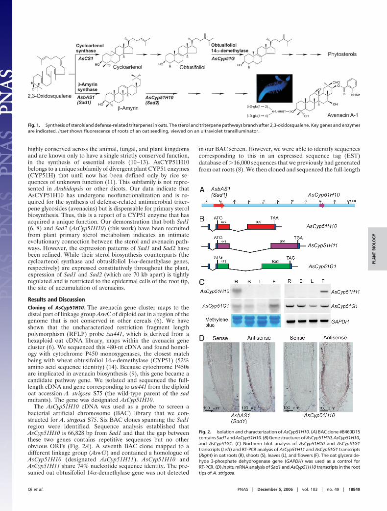
Fig. 1. Synthesis of sterols and defense-related triterpenes in oats. The sterol and triterpene pathways branch after 2,3-oxidosqualene. Key genes and enzymesare indicated. Inset shows fluorescence of roots of an oat seedling, viewed on an ultraviolet transilluminator.
Fig. 2. Isolation and characterization of AsCyp51H10. (A) BAC clone #B460D15containsSad1andAsCyp51H10. (B)GenestructuresofAsCyp51H10,AsCyp51H10,and AsCyp51G1. (C) Northern blot analysis of AsCyp51H10 and AsCyp51G1transcripts (Left) and RT-PCR analysis of AsCyp51H11 and AsCyp51G1 transcripts(Right) in oat roots (R), shoots (S), leaves (L), and flowers (F). The oat glyceralde-hyde 3-phosphate dehydrogenase gene (GAPDH) was used as a control forRT-PCR. (D) In situ mRNA analysis of Sad1 and AsCyp51H10 transcripts in the roottips of A. strigosa.
Qi et al. PNAS � December 5, 2006 � vol. 103 � no. 49 � 18849
PLA
NT
BIO
LOG
Y

cDNA and the corresponding gene (designated AsCyp51G1).AsCyp51H10 and AsCyp51H11 have 53% and 54% nucleotidesequence identity with AsCYP51G, respectively. AsCyp51G1maps to a third linkage group, AswB.
Expression of AsCyp51H10 Is Restricted to the Root Epidermis. Thegene structures of AsCyp51H10, AsCyp51H11, and AsCyp51G1are very similar (Fig. 2B). All three genes are predicted toencode products 490 aa in length. The exon sizes of AsCyp51H10and AsCyp51H11 are identical but differ from those of theobtusifoliol 14�-demethylase gene AsCyp51G, suggesting acloser evolutionarily relationship between the former two genes(Fig. 2B). The obtusifoliol 14�-demethylase gene AsCyp51G1 isexpressed in all plant organs examined, consistent with a role inprimary sterol metabolism (Fig. 2C). In contrast, AsCyp51H10 isexpressed specifically in the roots, whereas AsCyp51H11 tran-scripts were detected only in the flowers (Fig. 2C). Synthesis ofavenacin A-1 is under tight regulation and is restricted to theepidermal cells of the root tip (8). Previously we have shown bymRNA in situ hybridization that expression of Sad1 is restrictedto this cell type (8). AsCyp51H10 showed a very similar patternof expression (Fig. 2D).
AsCYP51H10 Is Synonymous with Sad2. The above data are sugges-tive of a role for AsCYP51H10 in avenacin biosynthesis. Wetherefore sequenced the AsCyp51H10 gene in our originalcollection of ten sad mutants (5) to establish whether this genewas likely to correspond to any of the loci that we had definedby mutation. As expected, we found no differences in thesequence of AsCyp51H10 in the two characterized sad1 mutantswithin the collection. The sequence of AsCyp51H10 in six othermutants (single mutants for each of the loci, Sad3–Sad8) also wasunaffected. However, nonsynonymous point mutations werefound in the coding region of AsCyp51H10 in the two indepen-dent sad2 mutants within the collection (#791 and #1027)(Table 1). Preliminary experiments suggested that the sad2mutants #791 and #1027 accumulate �-amyrin, whereas mu-tants affected at other Sad loci do not (15). This finding wasconfirmed by quantitative GC/MS analysis (Fig. 3A and Table 1).These data are consistent with a block in a cytochrome P450-mediated step early in the pathway and suggest that AsCyp51H10is synonymous with Sad2.
We then screened an extended collection of 92 reduced rootfluorescence mutants, with the objective of isolating more sad2mutants, by using TLC analysis as a preliminary screen. Thisscreen identified six more candidate sad2 mutants (#283, #500,#638, #698, #1325, and #1412). Allelism tests confirmed thatthese were indeed unique mutant alleles of Sad2. Five of these
mutants had nonsynonymous point mutations in theAsCyp51H10 gene, whereas the sixth mutant (#500) had a pointmutation in an exon–intron boundary (Table 1). Significantly,AsCyp51H10 transcript levels were substantially reduced inmutant #500 (Fig. 3B). Quantitative GC/MS confirmed that,like #791 and #1027, these newly isolated sad2 mutants hadelevated levels of �-amyrin (Table 1). These data providecompelling evidence that AsCyp51H10 corresponds to Sad2.
AsCYP51H10 Is a Divergent Member of the CYP51 Family. Compari-sons of the amino acid sequences of sterol 14�-demethylase(CYP51) sequences from diverse organisms indicate 34 con-served amino acid residues across bacteria, protozoa, fungi,animals, and plants. Six of these residues are not conserved in theoat AsCYP51H10 protein (Fig. 5, which is published as support-ing information on the PNAS web site). The predicted amino
Table 1. Characterization of sad2 mutants
MutantMutation
eventPredicted amino
acid change Region of proteinMean �-amyrin content
(�g/g of fresh roots)
Wild typeS75 — — — 1.4 � 0.1
Original sad2791 C23603 T Pro4633 Ser Near SRS6 40.2 � 1.9
1027 C3713 T Ala1243 Val SRS1 50.4 � 1.2New sad2
283 G22773 A Gly4353 Asp Heme binding 47.4 � 8.5500 G4753 A Splicing error — 41.3 � 0.6638 G19223 A Glu3173 Lys Conserved amino acid in �J helix 48.2 � 2.9698 G16703 A Ala2333 Thr SRS3 ND1325 C18663 T Ser2983 Phe SRS4 37.1 � 1.11412 C3383 T Thr1133 Ile SRS1 41.5 � 1.3
SRSs, predicted substrate recognition sites (16). ND, not determined.
Fig. 3. Identification and characterization of sad2 mutants. (A) GC analysisof root extracts from S75 (WT) and sad1 and sad2 mutants. The vertical bar onthe bottom left indicates relative mass abundance. sad2 mutants accumulate�-amyrin, whereas sad1 mutants (such as mutant #109) accumulate 2,3-oxidosqualene. The identity of the accumulated intermediates was deter-mined by MS. (B) Northern blot analysis of AsCyp51H10 transcript levels in sad2mutants and the WT S75.
18850 � www.pnas.org�cgi�doi�10.1073�pnas.0607849103 Qi et al.

acid changes in the seven sad2 mutants with normal levels ofAsCYP51H10 transcript were all within conserved substraterecognition sites or in other regions that are likely to be critical
for structure and/or activity (Table 1). An alignment of selectedregions of 36 representative CYP51 amino acid sequences acrosssubstrate recognition sites 1, 4, 5, and 6 (16) is shown in Fig. 4A.
Fig. 4. Sequence and structure of AsCYP51H10. (A) Alignment of selected regions of 36 representative CYP51 sequences from diverse organisms. The predictedsubstrate recognition sites (SRS) (16) are framed. Completely conserved amino acids are shown on a black background and those that are conserved in all exceptAsCYP51H10 are shown on a gray background. Mutations in sad2 mutants in these regions are shown (mutant number preceded by ‘‘#’’; changes marked withblack dots). Residues that line the active site cavity are indicated by triangles. The filled triangles denote the subset of these that are likely to be key determinantsin modulating the size and shape of the cavity in AsCYP51H10. (B) Modeling of the active site cavity of AsCYP51H10 (Bottom) and the oat sterol 14�-demethylaseAsCYP51G1 (Middle) based on the Mycobacterium tuberculosis MtCYP51B1 crystal structure (Top). (C) Phylogenetic analysis of CYP51 amino acid sequences. Thenumbers indicate the percentage of bootstrap replicates (out of 1,000) in which the given branching was observed. Accession nos. for the sequences used inalignments, modeling, and phylogenetic analysis are given in Table 2.
Qi et al. PNAS � December 5, 2006 � vol. 103 � no. 49 � 18851
PLA
NT
BIO
LOG
Y
Modeling of the 3D structures of AsCYP51H10 and the oatsterol 14�-demethylase AsCYP51G1 by using the Mycobacte-rium tuberculosis MtCYP51B1 crystal structure (16) as a tem-plate predicted the shapes and sizes of the active site cavities ofMtCYP51B1 and AsCYP51G1 to be very similar, whereas thatof AsCYP51H10 is quite different (Fig. 4B). Residues that arepredicted to significantly affect the size and shape of the activesite cavity are shown in Fig. 4B. The ensemble-averaged activesite volume in the model of AsCYP51H10 is 568 � 96 Å3,whereas that of AsCYP51G1 is 346 � 108 Å3, very similar to theactive site volume determined from the crystal structure ofMtCYP51B1 (343 � 62 Å3). These observations are consistentwith acquisition of a different function by AsCYP51H10.
Fig. 4C shows conserved subfamilies of CYP51 sterol 14�-demethylases from animals (CYP51A; pink), bacteria (CYP51B;light blue), protozoa (CYP51E; turquoise), fungi (CYP51F;orange), and plants (CYP51G; dark green). AsCyp51G1, thepredicted obtusifoliol 14�-demethylase from oat, falls within theCYP51G (dark green) subfamily. Yeast expression experimentshave confirmed that this gene does indeed encode functionalobtusifoliol 14�-demethylase (data not shown). However, wewere unable to express AsCYP51H10 and AsCYP51H11 inactive form with the standard yeast expression system used forconserved plant CYP51G enzymes (17). The CYP51H subfamilyappears to be restricted to oats and rice and is not representedin Arabidopsis.
The position of Chlamydomonas reinhardtii CYP51G(CrCYP51G1) in the phylogenetic tree (Fig. 4C) implies that theCYP51H family was derived from an ancient CYP51G-likesequence during the evolution of green plants. Tajima’s relativerate test (18) using C. reinhardtii CYP51G as an outgroupindicates that the AsCYP51H10 and AsCYP51H11 branches aresignificantly longer than that of AsCYP51G1 (�2 � 39.68 and41.67, respectively; P � 0.0001). The branches of the ricesequences within the CYP51H subgroup also are significantlylonger than that of rice CYP51G1 (data not shown). There isgreater mean diversity in the rice and oat CYP51H subfamilythan in the monocot CYP51G subfamily (0.653 � 0.028 versus0.088 � 0.009, respectively). Collectively, these results indicatethat the CYP51H subfamily is evolving at a much higher ratethan the conserved CYP51G subfamily, consistent with acqui-sition of a different function(s), as suggested by Nelson et al. (11).Our data confirm that AsCYP51H10 has indeed acquired adifferent function in the synthesis of secondary metabolitesrequired for plant defense. This finding has broad significancefor understanding the mechanisms of action and potentialevolutionary plasticity of the CYP51 family as a whole.
Coevolution of Sad1 and Sad2. Previously we reported that Sad1 hasarisen by duplication and divergence of a cycloartenol synthase-likegene (6, 8). The data presented here indicate that a second gene inthe avenacin pathway, Sad2 (AsCyp51H10), has been recruited froman ancient CYP51G-like sequence. These results indicate an inti-mate evolutionary connection between sterol and triterpene bio-synthesis. The first step in the pathway for the synthesis of adifferent group of defense-related compounds produced by maize(benzoxazinoids) also has been shown to be recruited from primarymetabolism, in this case from tryptophan biosynthesis (19, 20), andthere is an increasing body of evidence to indicate that geneduplication, neofunctionalization, and positive selection drive met-abolic diversification in plants (e.g., refs. 21 and 22). Unlike theirsterol pathway counterparts AsCS1 (cycloartenol synthase) andAsCyp51G1 (obtusifoliol 14�-demethylase), which are expressedthroughout the plant, expression of Sad1 and Sad2 is highlytissue-specific and is restricted to the epidermal cells of the root tips.AsCS1 and AsCyp51G1 are not genetically linked to each other orto the Sad gene cluster. Sad1 and Sad2 are physically linked andcosegregate with other genetically defined loci in the pathway that
are required for clearly distinct biochemical functions (6). Thebiochemical function of AsCYP51H10 is as yet unknown. Conver-sion of �-amyrin to avenacin A-1 will require oxidation at fivedifferent sites (Fig. 1), and all of these conversions potentially couldinvolve cytochrome P450 enzymes. AsCYP51H10 therefore may berequired for hydroxylation of �-amyrin (or a modified derivative of�-amyrin) at one or more positions. Elucidation of the precisebiochemical function of AsCYP51H10, coupled with further inves-tigation of the nature and origin of the avenacin gene cluster, willshed light on mechanisms underpinning the evolution of metabolicdiversity in plants and on the selective pressures that drive thisprocess.
Materials and MethodsPlant Material. Wild-type and mutant A. strigosa lines are asdescribed previously (5).
AsCyp51H10, AsCyp51H11, and AsCyp51G1 cDNA and Gene Isolation.Full-length cDNAs were defined by 5� and 3� RACE with aGeneRacer kit (Invitrogen, Carlsbad, CA), amplified by PCRand cloned into the pCR4-TOPO plasmid (Invitrogen). Geneswere characterized by direct sequencing of PCR products gen-erated from genomic DNA and/or by sequencing of BAC clones(see below).
BAC Library Construction and Screening. A BAC library of A.strigosa accession no. S75 was constructed by using establishedmethods (23). Approximately 150,000 colonies with an averageinsert size of �110 Kb (ca. 4.2� genome coverage) were storedin 384-well microtiter plates and gridded onto high-densityfilters. Filters were screened with 32P-labeled cDNA probes.Hybridization and washing were conducted at stringencies ofeither 60°C or 65°C following standard methods (24). BACfingerprinting was conducted by digestion of BAC DNA withHindIII and BamHI, and manual comparison of the restrictionfragments was performed after agarose gel electrophoresis.Subcloning of BAC inserts and sequencing was carried out byusing standard methods (24).
Transcript Analysis. For Northern blot analysis, total RNA wasextracted by using TRI-REAGENT (Sigma, St. Louis, MO).Hybridizations with biotin-labeled (Biotin-16-dUTP; Roche,Indianapolis, IN) antisense RNA probes for AsCyp51H10 werecarried out at high stringency (68°C) with signal detection byusing BrightStar BioDetect (Ambion, Austin, TX). For RT-PCR, first-strand cDNA synthesis was carried out by usingSuperScript II Reverse Transcriptase (Invitrogen) according tothe manufacturer’s instructions, and cDNA was amplified bystandard PCR with 30 cycles. For mRNA in situ analysis,biotin-labeled sense and antisense RNA probes from AsbAS1and AsCyp51H10 were used. Tissue preparation and hybridiza-tion was carried out as described in ref. 25.
Metabolite Analysis. Roots from individual 6-day-old seedlingswere harvested, freeze-dried, ground in liquid nitrogen, andextracted with methanol. Extracts were centrifuged, and thesupernatant was removed and dried down before extraction with100 �l of CHCl3/MeOH (7:3 vol/vol). Extracts and a �-amyrinstandard were spotted onto silica gel 60 TLC plates (Merck &Co., Whitehouse Station, NJ), and the TLC was developed withhexane:acetone (80:20 vol/vol). �-Amyrin and other compoundswere detected with iodine vapor. Qualitative and quantitativeGC/MS was conducted by using an Agilent (Santa Clara , CA)5973 Electron Ionization mass selective detector coupled to anAgilent 6890 gas chromatograph. Trimethylsilyl (TMS) deriva-tives of the extracted samples were separated on a J&W DB-5MScapillary column (30-m long, 0.25-mm i.d., 0.25-�m film thick-ness; Agilent). The GC oven temperature was maintained at
18852 � www.pnas.org�cgi�doi�10.1073�pnas.0607849103 Qi et al.
250°C for 1 min after injection, then programmed to 325°C at arate of 5°/min and held for 10 min at the final temperature.Helium carrier gas was used at a flow of 1.0 ml/min, and 2-�lsamples were injected in hexane in split mode (10:1) at aninjector temperature of 250°C. The mass spectrometer ionsource was maintained at 250°C. �-Amyrin was supplied by ApinChemicals Ltd. (Oxon, U.K.), and 2,3-oxidosqualene and 5�-cholestan-3�-ol standards were supplied by Sigma-Aldrich.
Sequence Comparisons and Homology Modeling. Protein sequences(Table 2, which is published as supporting information on thePNAS web site) were aligned by using CLUSTAL X, Version 1.8(http://bips.u-strasbg.fr), manually adjusted according to Lepe-sheva et al. (26), and displayed by using ESPript (version 2.1)(27). MEGA3.1 software (28) was used for phylogenetic analysis,assessment of sequence diversity, and Tajima’s relative rate test(18). Gaps in the alignment were excluded from our analysis(complete-deletion option). The neighbor-joining method wasused to construct the phylogenetic tree.
For homology modeling, sequence alignments ofAsCYP51H10 and AsCYP51G1 with MtCYP51B1 utilized thestructural information available in the Protein Data Bank entry1EA1 and were generated by using Fugue (29). Modeler (version8.2) (30) was used to generate homology models of the two
enzymes based on the MtCYP51B1 crystal structure. The mod-els were subjected to stereochemical validation by using appro-priate routines in Modeler (30). To more fully explore alterna-tive active site residue conformations in our models other thanthose delivered directly by Modeler, we used the non-Newtonianensemble generator CONCOORD (31). The pocket definitionsincluded residues 72–85, 95–103, 253–263, 319–324, and 433–435 in MtCYP51B1. Similar residues were included in simula-tions of AsCYP51H10 and AsCYP51G1. This approach allowsprediction of the range of likely configurations adopted by theresidues of the substrate-binding pocket. An energy-basedmethod QsiteFinder (www.bioinformatics.leeds.ac.uk) then wasused to characterize the active site cavities in the ensembles ofmodeled structures (32).
We thank Mike Lee (Iowa State University, Ames, IA) for restrictionfragment length polymorphism (RFLP) probes, Stephane Deschampsand Karlene Butler (DuPont Agricultural Products, Wilmington, DE)for BAC sequence analysis, David Nelson (University of Tennessee,Memphis, TN) for guidance on cytochrome P450 nomenclature, andGuo Jiemao (John Innes Centre, Norwich, U.K.) for technical assistancewith in situ analysis. We also thank Enno Krebbers (DuPont/PioneerCrop Genetics) for constructive comments throughout the project andthe Biotechnology and Biological Sciences Research Council U.K. andthe Gatsby Charitable Foundation for funding.
1. Dixon RA (2001) Nature 411:843–847.2. Field B, Jordan F, Osbourn A (2006) New Phytologist 172:193–207.3. Pichersky E, Gang DR (2000) Trends Plants Sci 5:439–445.4. Hostettmann V, Marston A (1995) Saponins (Cambridge Univ Press, Cam-
bridge, UK).5. Papadopoulou K, Melton RE, Leggett M, Daniels MJ, Osbourn AE (1999)
Proc Natl Acad Sci USA 96:12923–12928.6. Qi X, Bakht S, Leggett M, Maxwell C, Melton R, Osbourn A (2004) Proc Natl
Acad Sci USA 101:8233–8238.7. Chappell J (2002) Curr Opin Plant Biol 5:151–157.8. Haralampidis K, Bryan G, Qi X, Papadopoulou K, Bakht S, Melton R, Osbourn
A (2001) Proc Natl Acad Sci USA 98:13431–13436.9. Haralampidis K, Trojanowska M, Osbourn AE (2002) Adv Biochem Eng
Biotechnol 75:31–49.10. Aoyama Y, Noshiro M, Gotoh O, Imaoka S, Funae Y, Kurosawa N, Horiuchi
T, Yoshida Y (1996) J Biochem 119:926–933.11. Nelson DR, Schuler MA, Paquette SM, Werck-Reichhart D, Bak S (2004) Plant
Physiol 135:756–772.12. Aoyama Y (2005) Front Biosci 10:1546–1557.13. Waterman R, Lepesheva GI (2005) Biochem Biophys Res Commun 338:418–
422.14. Cabello-Hurtado F, Zimmerlin A, Rahier A, Taton M, DeRose R, Nedelkina
S, Batard Y, Durst F, Pallett KE, Werck-Reichhart D (1997) Biochem BiophysRes Commun 230:381–385.
15. Trojanowska MR, Osbourn AE, Daniels MJ, Threlfall DR (2001) Phytochem-istry 56:121–129.
16. Podust LM, Poulos TL, Waterman MR (2001) Proc Natl Acad Sci USA98:3068–3073.
17. Cabello-Hurtado F, Taton M, Forthoffer N, Kahn R, Bak S, Rahier A,Werck-Reichhart D (1999) Eur J Biochem 262:435–446.
18. Tajima F (1993) Genetics 135:599–607.19. Gierl A, Frey M (2001) Planta 213:493–498.20. Osbourn AE, Qi X, Townsend B, Qin B (2003) New Phytologist 159:101–108.21. Hartmann T, Kutchan TM, Strack D (2005) Phytochemistry 1198–1199.22. Benderoth M, Textor S, Windsor AJ, Mitchell-Olds T, Gershenzon J, Kroy-
mann J (2006) Proc Natl Acad Sci USA 103:9118–9123.23. Allouis S, Moore G, Bellec A, Sharp R, Faivre Rampant P, Mortimer K,
Pateyron S, Foote TN, Griffiths S, Caboche M, Chalhoub B (2003) Cereal ResCommun 31:331–338.
24. Sambrook J, Russell DW (2001) Molecular Cloning: A Laboratory Manual (ColdSpring Harbor Lab Press, New York), 3rd Ed.
25. Mao G, Buschmann H, Doonan JH, Lloyd CW (2006) J Cell Sci119:753–758.
26. Lepesheva GI, Virus C, Waterman MR (2003) Biochemistry 42:9091–9101.27. Gouet P, Courcelle E, Stuart DI, Metoz F (1999) Bioinformatics 15:305–308.28. Kumar S, Tamura K, Nei M (2004) Brief Bioinform 5:150–163.29. Shi J, Blundell TL, Mizuguchi K (2001) J Mol Biol 310:243–257.30. Marti-Renom MA, Stuart AC, Fiser A, Sanchez R, Melo F, Sali A (2000) Annu
Rev Biophys Biomol Struct 29:291–325.31. de Groot BL (1997) Proteins 29:240–251.32. Laurie AT, Jackson RM (2005) Bioinformatics 21:1908–1916.
Qi et al. PNAS � December 5, 2006 � vol. 103 � no. 49 � 18853
PLA
NT
BIO
LOG
Y
Related Documents