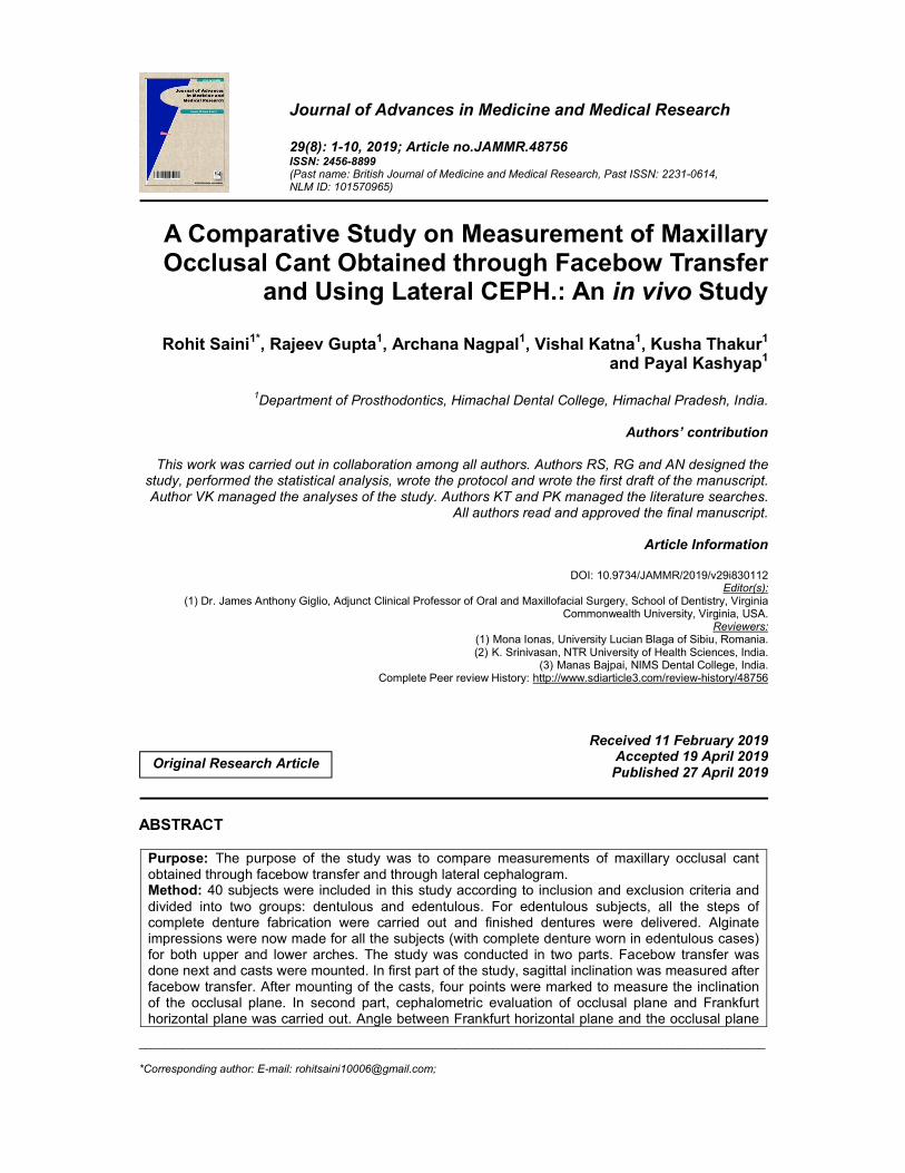
_____________________________________________________________________________________________________ *Corresponding author: E-mail: [email protected]; Journal of Advances in Medicine and Medical Research 29(8): 1-10, 2019; Article no.JAMMR.48756 ISSN: 2456-8899 (Past name: British Journal of Medicine and Medical Research, Past ISSN: 2231-0614, NLM ID: 101570965) A Comparative Study on Measurement of Maxillary Occlusal Cant Obtained through Facebow Transfer and Using Lateral CEPH.: An in vivo Study Rohit Saini 1* , Rajeev Gupta 1 , Archana Nagpal 1 , Vishal Katna 1 , Kusha Thakur 1 and Payal Kashyap 1 1 Department of Prosthodontics, Himachal Dental College, Himachal Pradesh, India. Authors’ contribution This work was carried out in collaboration among all authors. Authors RS, RG and AN designed the study, performed the statistical analysis, wrote the protocol and wrote the first draft of the manuscript. Author VK managed the analyses of the study. Authors KT and PK managed the literature searches. All authors read and approved the final manuscript. Article Information DOI: 10.9734/JAMMR/2019/v29i830112 Editor(s): (1) Dr. James Anthony Giglio, Adjunct Clinical Professor of Oral and Maxillofacial Surgery, School of Dentistry, Virginia Commonwealth University, Virginia, USA. Reviewers: (1) Mona Ionas, University Lucian Blaga of Sibiu, Romania. (2) K. Srinivasan, NTR University of Health Sciences, India. (3) Manas Bajpai, NIMS Dental College, India. Complete Peer review History: http://www.sdiarticle3.com/review-history/48756 Received 11 February 2019 Accepted 19 April 2019 Published 27 April 2019 ABSTRACT Purpose: The purpose of the study was to compare measurements of maxillary occlusal cant obtained through facebow transfer and through lateral cephalogram. Method: 40 subjects were included in this study according to inclusion and exclusion criteria and divided into two groups: dentulous and edentulous. For edentulous subjects, all the steps of complete denture fabrication were carried out and finished dentures were delivered. Alginate impressions were now made for all the subjects (with complete denture worn in edentulous cases) for both upper and lower arches. The study was conducted in two parts. Facebow transfer was done next and casts were mounted. In first part of the study, sagittal inclination was measured after facebow transfer. After mounting of the casts, four points were marked to measure the inclination of the occlusal plane. In second part, cephalometric evaluation of occlusal plane and Frankfurt horizontal plane was carried out. Angle between Frankfurt horizontal plane and the occlusal plane Original Research Article

Welcome message from author
This document is posted to help you gain knowledge. Please leave a comment to let me know what you think about it! Share it to your friends and learn new things together.
Transcript
_____________________________________________________________________________________________________ *Corresponding author: E-mail: [email protected];
Journal of Advances in Medicine and Medical Research 29(8): 1-10, 2019; Article no.JAMMR.48756 ISSN: 2456-8899 (Past name: British Journal of Medicine and Medical Research, Past ISSN: 2231-0614, NLM ID: 101570965)
A Comparative Study on Measurement of Maxillary Occlusal Cant Obtained through Facebow Transfer
and Using Lateral CEPH.: An in vivo Study
Rohit Saini1*, Rajeev Gupta1, Archana Nagpal1, Vishal Katna1, Kusha Thakur1 and Payal Kashyap1
1Department of Prosthodontics, Himachal Dental College, Himachal Pradesh, India.
Authors’ contribution
This work was carried out in collaboration among all authors. Authors RS, RG and AN designed the
study, performed the statistical analysis, wrote the protocol and wrote the first draft of the manuscript. Author VK managed the analyses of the study. Authors KT and PK managed the literature searches.
All authors read and approved the final manuscript.
Article Information
DOI: 10.9734/JAMMR/2019/v29i830112 Editor(s):
(1) Dr. James Anthony Giglio, Adjunct Clinical Professor of Oral and Maxillofacial Surgery, School of Dentistry, Virginia Commonwealth University, Virginia, USA.
Reviewers: (1) Mona Ionas, University Lucian Blaga of Sibiu, Romania. (2) K. Srinivasan, NTR University of Health Sciences, India.
(3) Manas Bajpai, NIMS Dental College, India. Complete Peer review History: http://www.sdiarticle3.com/review-history/48756
Received 11 February 2019 Accepted 19 April 2019 Published 27 April 2019
ABSTRACT
Purpose: The purpose of the study was to compare measurements of maxillary occlusal cant obtained through facebow transfer and through lateral cephalogram. Method: 40 subjects were included in this study according to inclusion and exclusion criteria and divided into two groups: dentulous and edentulous. For edentulous subjects, all the steps of complete denture fabrication were carried out and finished dentures were delivered. Alginate impressions were now made for all the subjects (with complete denture worn in edentulous cases) for both upper and lower arches. The study was conducted in two parts. Facebow transfer was done next and casts were mounted. In first part of the study, sagittal inclination was measured after facebow transfer. After mounting of the casts, four points were marked to measure the inclination of the occlusal plane. In second part, cephalometric evaluation of occlusal plane and Frankfurt horizontal plane was carried out. Angle between Frankfurt horizontal plane and the occlusal plane
Original Research Article
Saini et al.; JAMMR, 29(8): 1-10, 2019; Article no.JAMMR.48756
2
was maxillary occlusal cant. which was evaluated by tracing. Paired t test was used to compare mean facebow values and lateral ceph values in edentulous subjects. Intergroup comparison between lateral ceph and mean facebow values between dentulous and edentulous subjects was evaluated using independent t test. Results: Facebow measurements gave comparatively higher values in both dentulous and edentulous patients and are subjected to less variation as compared to the lateral cephalogram values p<0.0001. Conclusion: The occlusal plane angle of lateral cephalogram was found to be significantly different from angle obtained through facebow transfer.
Keywords: Occlusal cant; facebow; frankfurt horizontal plane; lateral cephalogram.
1. INTRODUCTION In complete denture construction, the Prosthodontist is responsible for restoring the natural esthetics of the patient and for developing an occlusion that is compatible with functional movements of the mandible [1]. One of the salient factor that help us in developing occlusion which is compatible with the functional movement of the stomatognathic system is the orientation of occlusal plane [2]. Occlusal plane orientation is one of the most important clinical procedure in removable prosthodontic treatment for edentulous patients [3].
Ideally the occlusal plane should be located in a direction perpendicular to the occlusal bite force. This position provides stability to dentures supported by underlying resilient tissue. Functionally the occlusal table is a milling surface that is designed in such a manner so that the tongue and the buccinator muscle are able to position the food bolus onto it and hold it there during the process of mastication.
To orient the maxillary arch and dentition using a facebow, involves a plane of reference, ie, the Frankfurt horizontal plane (porion‑orbitale), which appears horizontal when the head is placed in the natural head position [2]. A facebow is used to record the antero-posterior and vertical relationship of the maxilla to the hinge axis of the temporomandibular joints and to transfer this relationship to the opening axis of an articulator [4]. The proper use of an anatomic articulator is dependent upon an accurate facebow transfer [5]. The third point of reference recommended for the Hanau Wide-Vue model 183-2 semiadjustable articulator is, orbitale [6].
A lateral cephalogram reveals areas in a cranial base that are not subjected to alteration, it is used in identifying predictable relationships between the teeth and other cranial landmarks,
henceforth it is considered as the gold standard [2]. Cephalometric analysis is an important diagnostic tool in dentistry, in prosthodontics, the significance of cephalometrics lies in the ability to re-establish the spatial position of lost structures (such as the teeth) [7]. In complete denture fabrication, recording a correct jaw relationship is of utmost importance and occlusal plane record is a part of the same. Hence, the purpose of the study was to compare measurements of maxillary occlusal cant obtained through facebow transfer and through lateral cephalogram. 2. MATERIALS AND METHODS
The study included 20 dentulous and 20 edentulous subjects comprising both males and females randomly selected who visited the out-patient department of Prosthodontics. All the procedures were carried out in Department of Prosthodontics. All the subjects were informed about the study and institutional ethical clearance was also obtained.
Inclusion Criteria (dentulous patients):
Age group: 18-30 years with completed facial growth
Full complement of healthy and natural teeth
No history of orthodontic treatment
Exclusion Criteria (dentulous patients): Periodontally compromised teeth Teeth grossly attrited or abraded Presence of fixed or removable partial
dentures Gross malalignment of teeth
Inclusion Criteria (edentulous patients):
Normal ridge relationship Well-formed ridge
Saini et al.; JAMMR, 29(8): 1-10, 2019; Article no.JAMMR.48756
3
Exclusion Criteria (edentulous patients): Resorbed ridge
Reference Planes: Frankfurt horizontal plane. Occlusal plane: Plane touching
mesiopalatal cusp of left maxillary first molar and left mesioincisal edge of central incisor.
2.1 Methodology Subjects, both dentulous as well as edentulous, were selected randomly keeping in mind the specified inclusion criteria. For edentulous subjects, all the steps of complete denture fabrication were carried out and finished dentures were delivered.
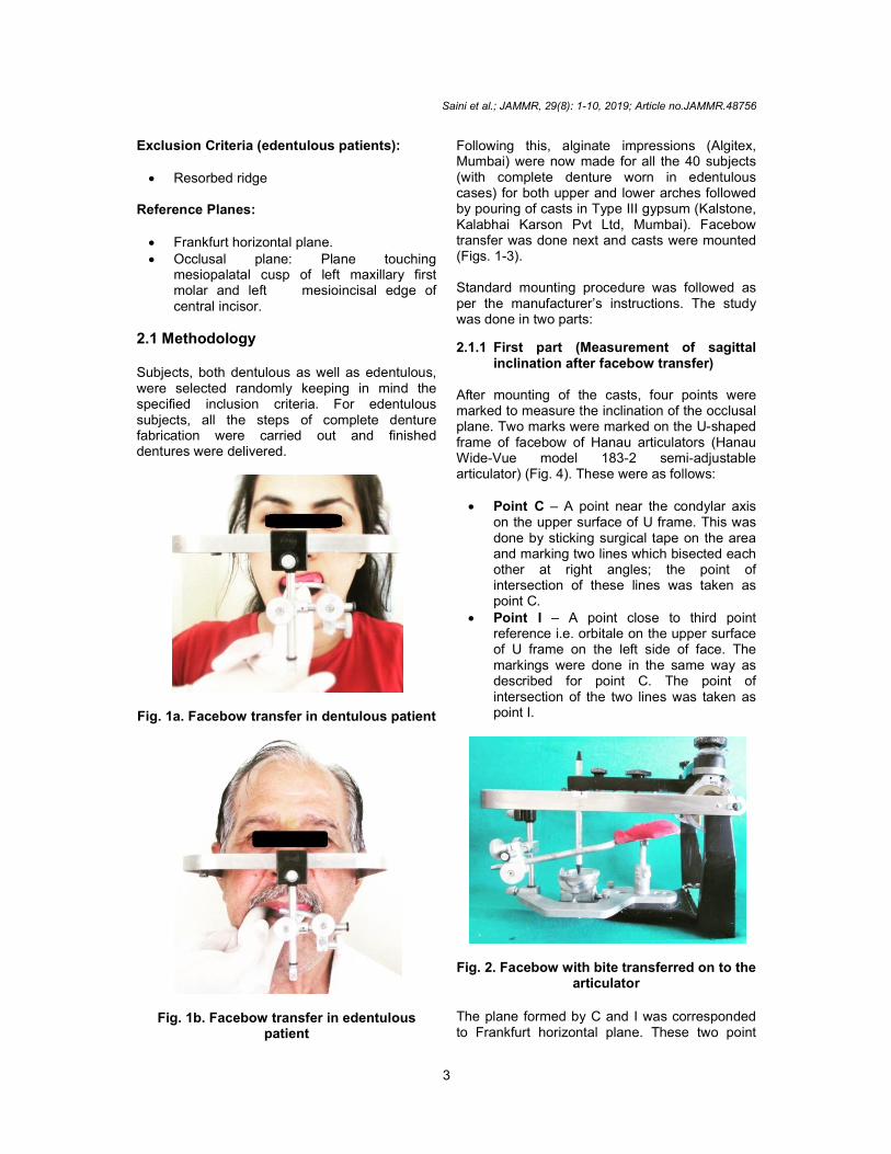
Fig. 1a. Facebow transfer in dentulous patient
Fig. 1b. Facebow transfer in edentulous patient
Following this, alginate impressions (Algitex, Mumbai) were now made for all the 40 subjects (with complete denture worn in edentulous cases) for both upper and lower arches followed by pouring of casts in Type III gypsum (Kalstone, Kalabhai Karson Pvt Ltd, Mumbai). Facebow transfer was done next and casts were mounted (Figs. 1-3).
Standard mounting procedure was followed as per the manufacturer’s instructions. The study was done in two parts:
2.1.1 First part (Measurement of sagittal inclination after facebow transfer)
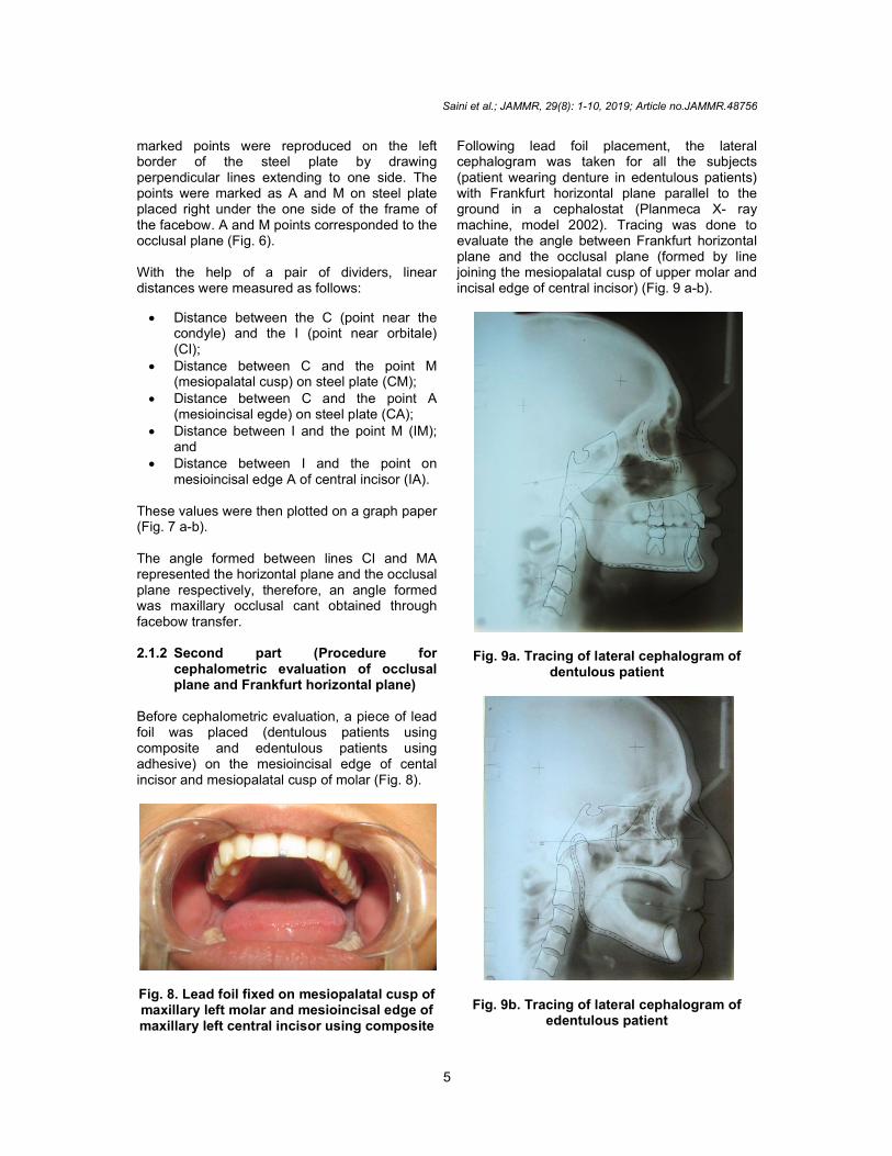
After mounting of the casts, four points were marked to measure the inclination of the occlusal plane. Two marks were marked on the U-shaped frame of facebow of Hanau articulators (Hanau Wide-Vue model 183-2 semi-adjustable articulator) (Fig. 4). These were as follows: Point C – A point near the condylar axis
on the upper surface of U frame. This was done by sticking surgical tape on the area and marking two lines which bisected each other at right angles; the point of intersection of these lines was taken as point C.
Point I – A point close to third point reference i.e. orbitale on the upper surface of U frame on the left side of face. The markings were done in the same way as described for point C. The point of intersection of the two lines was taken as point I.
Fig. 2. Facebow with bite transferred on to the articulator
The plane formed by C and I was corresponded to Frankfurt horizontal plane. These two point
Saini et al.; JAMMR, 29(8): 1-10, 2019; Article no.JAMMR.48756
4
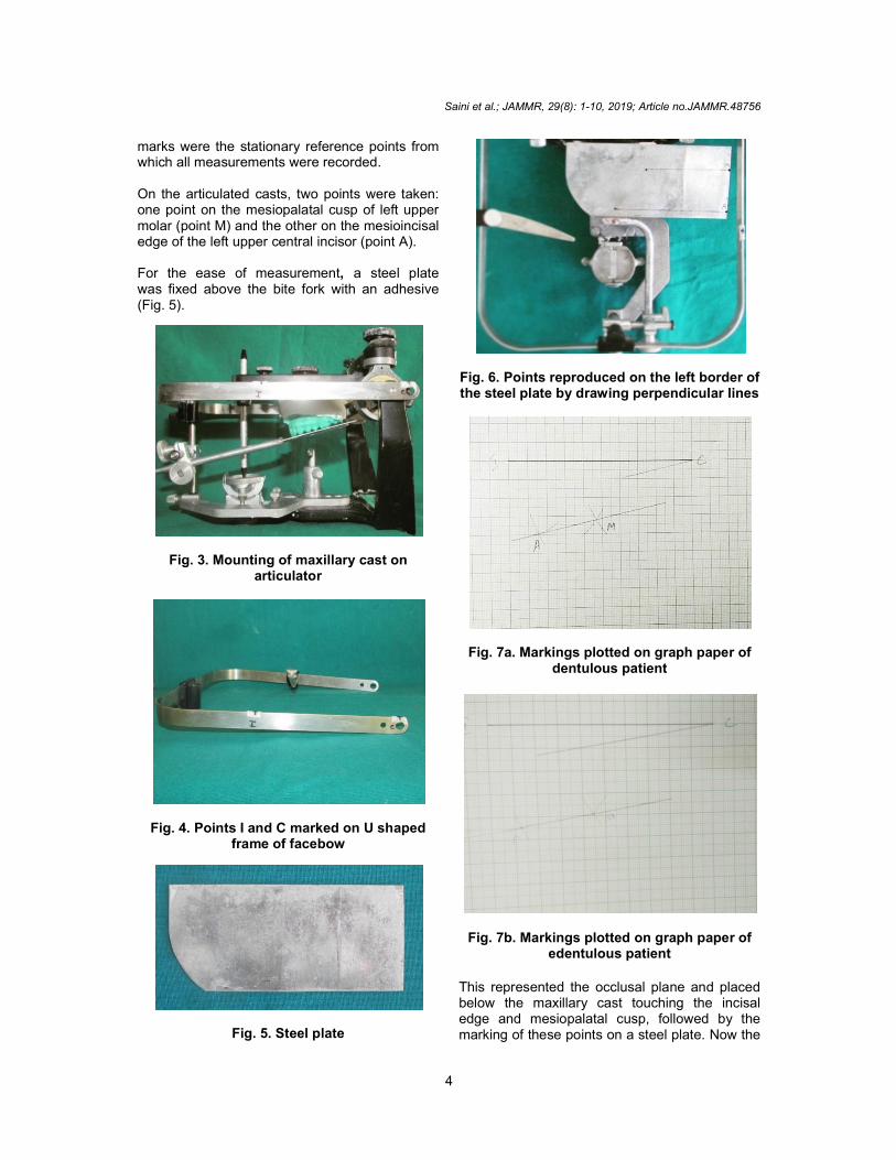
marks were the stationary reference points from which all measurements were recorded. On the articulated casts, two points were taken: one point on the mesiopalatal cusp of left upper molar (point M) and the other on the mesioincisal edge of the left upper central incisor (point A). For the ease of measurement, a steel plate was fixed above the bite fork with an adhesive (Fig. 5).
Fig. 3. Mounting of maxillary cast on articulator
Fig. 4. Points I and C marked on U shaped frame of facebow
Fig. 5. Steel plate
Fig. 6. Points reproduced on the left border of the steel plate by drawing perpendicular lines
Fig. 7a. Markings plotted on graph paper of dentulous patient
Fig. 7b. Markings plotted on graph paper of edentulous patient
This represented the occlusal plane and placed below the maxillary cast touching the incisal edge and mesiopalatal cusp, followed by the marking of these points on a steel plate. Now the
Saini et al.; JAMMR, 29(8): 1-10, 2019; Article no.JAMMR.48756
5
marked points were reproduced on the left border of the steel plate by drawing perpendicular lines extending to one side. The points were marked as A and M on steel plate placed right under the one side of the frame of the facebow. A and M points corresponded to the occlusal plane (Fig. 6). With the help of a pair of dividers, linear distances were measured as follows:
Distance between the C (point near the condyle) and the I (point near orbitale) (CI);
Distance between C and the point M (mesiopalatal cusp) on steel plate (CM);
Distance between C and the point A (mesioincisal egde) on steel plate (CA);
Distance between I and the point M (IM); and
Distance between I and the point on mesioincisal edge A of central incisor (IA).
These values were then plotted on a graph paper (Fig. 7 a-b). The angle formed between lines CI and MA represented the horizontal plane and the occlusal plane respectively, therefore, an angle formed was maxillary occlusal cant obtained through facebow transfer. 2.1.2 Second part (Procedure for
cephalometric evaluation of occlusal plane and Frankfurt horizontal plane)
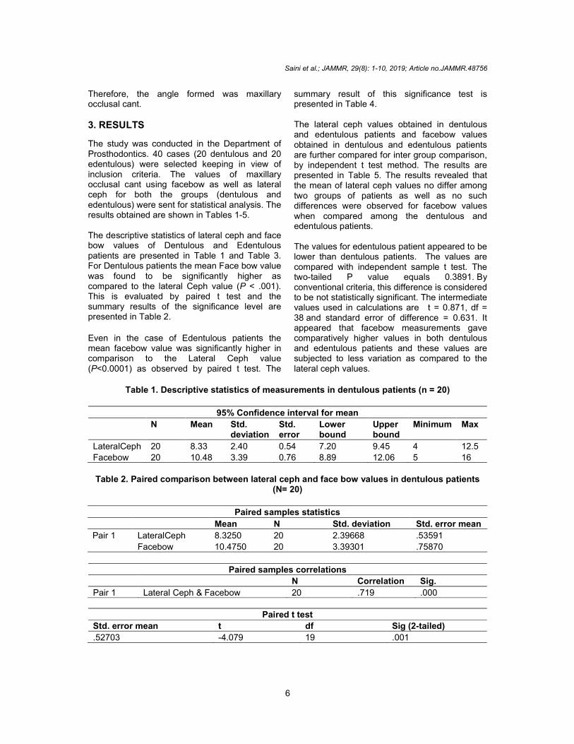
Before cephalometric evaluation, a piece of lead foil was placed (dentulous patients using composite and edentulous patients using adhesive) on the mesioincisal edge of cental incisor and mesiopalatal cusp of molar (Fig. 8).
Fig. 8. Lead foil fixed on mesiopalatal cusp of maxillary left molar and mesioincisal edge of maxillary left central incisor using composite
Following lead foil placement, the lateral cephalogram was taken for all the subjects (patient wearing denture in edentulous patients) with Frankfurt horizontal plane parallel to the ground in a cephalostat (Planmeca X- ray machine, model 2002). Tracing was done to evaluate the angle between Frankfurt horizontal plane and the occlusal plane (formed by line joining the mesiopalatal cusp of upper molar and incisal edge of central incisor) (Fig. 9 a-b).
Fig. 9a. Tracing of lateral cephalogram of dentulous patient
Fig. 9b. Tracing of lateral cephalogram of edentulous patient
Saini et al.; JAMMR, 29(8): 1-10, 2019; Article no.JAMMR.48756
6
Therefore, the angle formed was maxillary occlusal cant.
3. RESULTS
The study was conducted in the Department of Prosthodontics. 40 cases (20 dentulous and 20 edentulous) were selected keeping in view of inclusion criteria. The values of maxillary occlusal cant using facebow as well as lateral ceph for both the groups (dentulous and edentulous) were sent for statistical analysis. The results obtained are shown in Tables 1-5. The descriptive statistics of lateral ceph and face bow values of Dentulous and Edentulous patients are presented in Table 1 and Table 3. For Dentulous patients the mean Face bow value was found to be significantly higher as compared to the lateral Ceph value (P < .001). This is evaluated by paired t test and the summary results of the significance level are presented in Table 2.
Even in the case of Edentulous patients the mean facebow value was significantly higher in comparison to the Lateral Ceph value (P<0.0001) as observed by paired t test. The
summary result of this significance test is presented in Table 4. The lateral ceph values obtained in dentulous and edentulous patients and facebow values obtained in dentulous and edentulous patients are further compared for inter group comparison, by independent t test method. The results are presented in Table 5. The results revealed that the mean of lateral ceph values no differ among two groups of patients as well as no such differences were observed for facebow values when compared among the dentulous and edentulous patients. The values for edentulous patient appeared to be lower than dentulous patients. The values are compared with independent sample t test. The two-tailed P value equals 0.3891. By conventional criteria, this difference is considered to be not statistically significant. The intermediate values used in calculations are t = 0.871, df = 38 and standard error of difference = 0.631. It appeared that facebow measurements gave comparatively higher values in both dentulous and edentulous patients and these values are subjected to less variation as compared to the lateral ceph values.
Table 1. Descriptive statistics of measurements in dentulous patients (n = 20)
95% Confidence interval for mean
N Mean Std. deviation
Std. error
Lower bound
Upper bound
Minimum Max
LateralCeph 20 8.33 2.40 0.54 7.20 9.45 4 12.5
Facebow 20 10.48 3.39 0.76 8.89 12.06 5 16
Table 2. Paired comparison between lateral ceph and face bow values in dentulous patients
(N= 20)
Paired samples statistics
Mean N Std. deviation Std. error mean
Pair 1 LateralCeph 8.3250 20 2.39668 .53591
Facebow 10.4750 20 3.39301 .75870
Paired samples correlations
N Correlation Sig.
Pair 1 Lateral Ceph & Facebow 20 .719 .000
Paired t test
Std. error mean t df Sig (2-tailed)
.52703 -4.079 19 .001
Saini et al.; JAMMR, 29(8): 1-10, 2019; Article no.JAMMR.48756
7
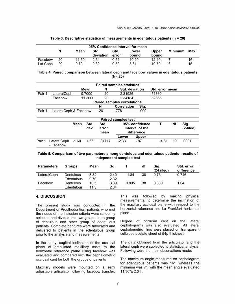
Table 3. Descriptive statistics of measurements in edentulous patients (n = 20)
95% Confidence interval for mean N Mean Std.
deviation Std. error
Lower bound
Upper bound
Minimum Max
Facebow 20 11.30 2.34 0.52 10.20 12.40 7 16 Lat Ceph 20 9.70 2.32 0.52 8.61 10.79 6 15
Table 4. Paired comparison between lateral ceph and face bow values in edentulous patients
(N= 20)
Paired samples statistics Mean N Std. deviation Std. error mean Pair 1 LateralCeph 9.7000 20 2.31926 .51860
Facebow 11.3000 20 2.34184 .52365 Paired samples correlations
N Correlation Sig. Pair 1 LateralCeph & Facebow 20 .778 .000
Paired samples test
Mean Std. dev
Std. error mean
95% confidence interval of the
difference
T df Sig (2-tiled)
Lower Upper Pair 1 LateralCeph
- Facebow -1.60 1.55 .34717 -2.33 -.87 -4.61 19 .0001
Table 5. Comparison of two parameters among dentulous and edentulous patients- results of
independent sample t test
Parameters Groups Mean Sd t df Sig. (2-tailed)
Std. error difference
LateralCeph Dentulous 8.32 2.40 -1.84 38 0.73 0.746 Edentulous 9.70 2.32 Facebow Dentulous 10.5 3.39 0.895 38 0.380 1.04 Edentulous 11.3 2.34
4. DISCUSSION The present study was conducted in the Department of Prosthodontics, patients who met the needs of the inclusion criteria were randomly selected and divided into two groups i.e. a group of dentulous and other group of edentulous patients. Complete dentures were fabricated and delivered to patients in the edentulous group prior to the analysis and measurements. In the study, sagittal inclination of the occlusal plane of articulated maxillary casts to the horizontal reference plane using facebow was evaluated and compared with the cephalometric occlusal cant for both the groups of patients
Maxillary models were mounted on a semi adjustable articulator following facebow transfer.
This was followed by making physical measurements, to determine the inclination of the maxillary occlusal plane with respect to the horizontal reference line i.e Frankfurt horizontal plane. Degree of occlusal cant on the lateral cephalograms was also evaluated. All lateral cephalometric films were placed on transparent cellulose acetate sheet of 54μ thickness The data obtained from the articulator and the lateral ceph were subjected to statistical analysis. Following were the main observations made: The maximum angle measured on cephalogram for edentulous patients was 16°, whereas the minimum was 7°, with the mean angle evaluated 11.30°± 2.34°.
Saini et al.; JAMMR, 29(8): 1-10, 2019; Article no.JAMMR.48756
8
The maximum angle measured on the articulated cast using facebow 15°, whereas the minimum angle was 6°, with the mean angle calculated was 9.70°± 2.32°. The maximum angle measured on cephalogram for dentulous patients was 12.5°, whereas the minimum was 4°, with the mean angle being 8.33°± 2.40° for this study. In the study carried out by Shetty et al., (2016) [2], the Frankfurt horizontal plane‑occlusal plane angle for lateral cephalogram varied from a maximum of 13.3° to a minimum of 3.5° with a mean of 8.7° ± 2.24° thereby showing similar results as shown in the current study.
According to the study by Rupal J Shah et al., (2013) [8], minimum angle value for lateral ceph was 3° and maximum was 17° mean value was 9.13° ± 3.77.
In another study conducted by Nazir et al. [9], the maximum angle measured on cephalogram was 15°, whereas the minimum was 6°, with the mean angle being 9.61° ± 2.55.
The mean occlusal plane angle in cephalogram was 10.4° ± 4.3, which was slightly higher in the study by Kyung Suk Seo. [10] as compared to the present study.
On the casts that were mounted on hanau wide vue articulator using facebow for dentulous patients, the maximum angle measured was 16° and the minimum was 5°. The mean angle was calculated to be 10.48° ± 3.39. This result is in accordance with the study carried out by Shetty et al. [2], in which the Frankfurt horizontal plane ‑ Occlusal plane angle using
Hanau Wide‑Vue group, varied from a maximum of 15° to a minimum of 5.1° with a mean of 10.69° ± 2.44°. The study by Nazier et al., [9] also yielded similar result showing maximum angle of 15° and minimum of 6°. The average angle of sagittal inclination was calculated to be 10.77° ± 2.60°.
The mean angle of sagittal inclination of maxillary cast mounted on Hanau Wide‑vue articulator was, however, higher in the study conducted by Mohammad Abdullah and Sherfudhin. [4] and a study by Kyung Suk Seo. [10] who got a mean angle of 13.77° and 13.5° ± 5.4 respectively. On the other hand, Rupal J Shah et al. [8], in their study, got a mean angle of
8.57° ± 3.45 which was lower than the values in the current study. The mean difference between the facebow and lateral ceph for dentulous patients in this study is 2.15°. This study showed a mean difference 2.15° between the sagittal inclination of maxillary cast mounted on Hanau wide Vue articulator and the value obtained using lateral ceph. This result was similar to the results given by Shetty et al. [2], who after reported a mean difference of 1.9° between the occlusal cant measured on Hanau wide Vue articulator and lateral ceph. Nazir et al. [9] also showed a mean difference of 1.16° in their study. Kyung Suk Seo. [10] in his study, found a mean difference of 3.3° ± 4.6 which was higher as compared to this study. On the contrary, a mean difference of -0.567° was found in a study conducted by Rupal J Shah et al., (2013) [8]. The results showed that the angle formed between the Frankfurt horizontal plane- Occlusal plane in a lateral ceph could be considered more reliable as compared to the measurements done with facebow transfer using articulator. A lateral ceph is considered as the gold standard as it unveils hard tissue areas in a cranial base. It is used in assessing predictable relationships between the teeth and other cranial landmarks that remain unaffected even post extraction of teeth.
In reality, the Frankfurt horizontal plane is not transferred to the articulator by the use of orbitale pointer. This is because only the anterior point of reference for this plane is used; the orbitale. Porion does not come into play during the face-bow transfer [6]. As the facebow transfer on articulator is an arbitrary process, there could be chances of errors due to soft tissue involvement, position of anterior reference, mounting of maxillary casts. If there are errors during the facebow transfer using Hanau Wide-Vue articulator, it can further leave an impact on the procedures to follow and
Saini et al.; JAMMR, 29(8): 1-10, 2019; Article no.JAMMR.48756
9
consequently lead to unreliable result after delivery of the prosthesis.
The various procedures that can get adversely affected due to these errors may range from full mouth rehabilitation procedures and fixed partial dentures to balanced complete denture prosthesis.
Thus, the present study confirms the importance of cephalometry in the field of Prosthodontics to establish plane of occlusion for proper functions of chewing, mastication and also to restore the esthetics of an individual. [11].
5. CONCLUSION
The present study comprised of 40 patients, 20 dentulous and 20 edentulous who visited the out-patient department of Prosthodontics. The maxillary occlusal cant was evaluated through facebow transfer on semi adjustable articulator and through cephalometrically. Study was divided into following groups: Occlusal cant of dentulous patients
through facebow transfer. Occlusal cant of dentulous patients
through lateral cephalogram. Occlusal cant of edentulous patients
through facebow transfer. Occlusal cant of edentulous patients
through lateral cephalogram.
After statistical analysis, the following conclusions were made: Within the limitations of this study, it was
seen that reproducibility of the occlusal cant on an articulator by a facebow was not exact.
The sagittal inclination of the mounted maxillary casts on the Hanau Wide-Vue semi adjustable articulator was closer to the individual’s occlusal cant as measured on the cephalogram.
The correlation value (Pearson’s value) obtained between maxillary cast mounted on Hanau Wide-Vue articulator was greater as compared to the lateral cephalogram.
Thus, it could be concluded that the occlusal plane angle of lateral cephalogram was
significantly different from angle obtained through facebow transfer.
Based on the results of this study, the lateral cephalogram is more reliable as compared to the facebow transfer in determining the maxillary occlusal cant.
CONSENT Informed consent to participate was obtained from each patient prior to their enrollment in the study.
ETHICAL APPROVAL Ethical approval was obtained from institutional and university ethical research cell committee.
ACKNOWLEDGEMENT I express my heartful gratitude to my guides, mentor and my fellow colleagues for their invaluable guidance and assistance through the course of this study.
COMPETING INTERESTS Authors have declared that no competing interests exist.
REFERENCES 1. Souza DNL, Bhargva K. A cephalometric
study comparing the occlusal plane in dentulous and edentulous subjects in relation to the maxillomandibular space. J Prosthet Dent 1996;75:177-82. Available:https://doi.org/10.1016/S0022-3913(96)90096-7
2. Shetty S, Shenoy KK, Sabu A. Evaluation of accuracy of transfer of maxillary occlusal cant of two articulators using two facebow/ semi-adjustable articulator system: An in vivo study. J Indian Prosthodont Soc. 2016;16:248-52. Available:https://doi.org/ 10.4103/0972-4052.176525
3. Quran FAM, AL Quran, Hazza’a A, Nahass NAl. The Position of the occlusal plane in natural and artificial dentitions as related to other craniofacial planes. Journal of Prosthodontics. 2010;601–5. Available:https://doi.org/10.1111/j.1532-849X.2010.00643

Saini et al.; JAMMR, 29(8): 1-10, 2019; Article no.JAMMR.48756
10
4. Abdullah MA, Sherfudhin H. A comparative study of Facebow transfer n Hanau and Whip-Mix articulators. Saudi Dent J. 1994; 6:8-12.
5. Teteruck WR, Lundeen HC. The accuracy of an ear face-bow. J Prosthet Dent. 1966;16:1039-46. Available:https://doi.org/10.1016/0022-3913(66)90169-7
6. Bailey JO. Evaluation of the third point of reference for mounting maxillary casts on the Hanau articulator. J Prosthet dent 1984;51:199-201. Available:https://doi.org/10.1016/0022-3913(84)90260-9
7. Hindocha AD, Vartak VN, Bhandari AJ, Dudani MT. A cephalometric study to determine the plane of occlusion in completely edentulous patients. Indian Journal of Dental Research. 2013;24;669-73. Available:https://dx.doi.org/10.1007%2Fs13191-011-0049-x
8. Shah RJ, Sharma S, Agrawal P. EvaluatIon of two facebow transfer systems in measuring occlusal cant. Guident Research. 2013;64-68.
9. Nazir N, Sujesh M, Kumar R, Sreenivas P. Accuracy of two face-bow/semi-adjustable articulator systems in transferring the maxillary occlusal cant. Indian Journal of Dental Research. 2012;23:437-42. Available:https://doi.org/10.4103/0970-9290.104945.
10. Seo KS, Park MH, Lee JH, Kim CH, Chae JH. The comparative study for occlusal plane between articulated cast model and cephalogram in orthoganthic surgery patients. J. Kor. Oral Maxillofac. Surg. 2003;29:239-44
11. Mittal R. Comparison of the occlusal plane in dentulous and edentulous patients: A cephalometric study. The Journal of Indian Prosthodontic Society. 2008;8:195-200. Available:https://doi.org/ 10.4103/0972-4052.49182
_________________________________________________________________________________ © 2019 Saini et al.; This is an Open Access article distributed under the terms of the Creative Commons Attribution License (http://creativecommons.org/licenses/by/4.0), which permits unrestricted use, distribution, and reproduction in any medium, provided the original work is properly cited.
Peer-review history: The peer review history for this paper can be accessed here:
http://www.sdiarticle3.com/review-history/48756
Related Documents