Copyright © 2011 Korean Society of Otorhinolaryngology-Head and Neck Surgery 791 Case Report Korean J Otorhinolaryngol-Head Neck Surg 2011;54:791-3 / pISSN 2092-5859 / eISSN 2092-6529 http://dx.doi.org/10.3342/kjorl-hns.2011.54.11.791 A Case of Tracheobronchopathia Osteochondroplastica Associated with Atrophic Rhinitis Tae Jung Park, Jung Uk Han, Do hyun Kim and Bo-Yoing Kim Department of Otorhinolaryngology, Maryknoll Hospital, Busan, Korea 위축성 비염과 동반된 기관기관지 골연골형성증 1예 박태정·한정욱·김도현·김보영 메리놀병원 이비인후과 Received June 19, 2011 Revised August 17, 2011 Accepted August 29, 2011 Address for correspondence Bo-Yoing Kim, MD Department of Otorhinolaryngology, Maryknoll Hospital, 12 Daecheong-dong 4-ga, Jung-gu, Busan 600-730, Korea Tel +82-51-465-2205 Fax +82-51-461-0297 E-mail [email protected] Tracheobronchopathia osteochondroplastica (TO) is a rare benign disease of trachea character- ized by numerous osseocartilaginous nodules protruding into the tracheobronchial lumen. The etiology of TO is unknown; however, an association with upper respiratory diseases such as atrophic rhinitis has been suggested. The authors report a case of TO with atrophic rhinitis with related literatures. Korean J Otorhinolaryngol-Head Neck Surg 2011;54:791-3 Key WordsZZTracheobronchopathia osteochondroplasticaㆍAtrophic rhinitis. 서 론 기관기관지 골연골형성증(Tracheobronchopathia osteo- chondroplastica, TO)은 기관과 기관지 내로 골화된 소결절들 이 돌출되어 있는 드문 질환으로 원인과 병태생리에 대해서 는 아직까지 정확하게 밝혀지지 않았다. 1) 병태생리에 대한 여 러 가지 가설 중 상부호흡기계의 반복적인 감염이 기관의 탄 성조직(elastic tissue) 의 연골과 골로 화생(metaplasia)을 야기 한다는 것이 널리 받아들여지고 있다. 2) 특히 위축성 비염과 동반된 기관기관지 골연골형성증 증례가 여러 차례 보고되면 서, 위축성 비염이 화생의 자극원이 될 수 있음을 제시하고 있 다. 이에 저자는 수십 년간 지속된 위축성 비염환자에서 동반 된 기관기관지 골연골형성증 1 예를 경험하였기에 문헌고찰과 함께 보고하는 바이다. 증 례 69세 여자 환자로 수십 년간 지속된 비강 내 악취 및 가피 를 주소로 이비인후과를 내원하였다. 이학적 검사상 외비는 안비를 보였고, 비내시경상 비중격은 후방 골부까지 큰 천공 을 보였으며 악취 나는 가피가 비강전체와 비인강을 가득 채 우고 있었으며, 양측 이개 변형은 없었다(Fig. 1A). 비강 내 두 껍고 악취 나는 가피를 제거하였을 때 가피 아래 점막에서 특 별한 출혈 경향은 없었으며, 비중격 천공의 가장자리는 비교 적 깨끗하였고, 추가적인 육아조직 형성은 관찰되지 않았다 (Fig. 1B). 환자는 수십 년 전 개인 이비인후과에서 비중격 천공을 진 단받았으나, 병력청취상 비내 수술, 방사선 조사, 매독, 결핵, 외상병력 및 약물 흡인력은 없었으며, 다른 내분비 장애 및 유전적 질환도 없었다. 환자는 수 년 전 만성 기침으로 본원 호흡기내과를 방문하 여 시행한 단순흉부촬영(chest X-ray), 흉부전산화단층촬영 (computed tomography, CT), 기관지 내시경 및 기관병변 조 직검사상 기관기관지 골연골형성증으로 진단되어 보존적 약 물치료를 하면서 외래 추적 관찰 중인 것 이외에 다른 병력 은 발견되지 않았다(Fig. 2). online © ML Comm

Welcome message from author
This document is posted to help you gain knowledge. Please leave a comment to let me know what you think about it! Share it to your friends and learn new things together.
Transcript
Copyright © 2011 Korean Society of Otorhinolaryngology-Head and Neck Surgery 791
Case Report Korean J Otorhinolaryngol-Head Neck Surg 2011;54:791-3 / pISSN 2092-5859 / eISSN 2092-6529
http://dx.doi.org/10.3342/kjorl-hns.2011.54.11.791
A Case of Tracheobronchopathia Osteochondroplastica Associated with Atrophic Rhinitis
Tae Jung Park, Jung Uk Han, Do hyun Kim and Bo-Yoing KimDepartment of Otorhinolaryngology, Maryknoll Hospital, Busan, Korea
위축성 비염과 동반된 기관기관지 골연골형성증 1예
박태정 ·한정욱 ·김도현 ·김보영
메리놀병원 이비인후과
Received June 19, 2011Revised August 17, 2011Accepted August 29, 2011AddressforcorrespondenceBo-Yoing Kim, MDDepartment of Otorhinolaryngology, Maryknoll Hospital, 12 Daecheong-dong 4-ga, Jung-gu, Busan 600-730, KoreaTel +82-51-465-2205Fax +82-51-461-0297E-mail [email protected]
Tracheobronchopathia osteochondroplastica (TO) is a rare benign disease of trachea character-ized by numerous osseocartilaginous nodules protruding into the tracheobronchial lumen. The etiology of TO is unknown; however, an association with upper respiratory diseases such as atrophic rhinitis has been suggested. The authors report a case of TO with atrophic rhinitis with related literatures. Korean J Otorhinolaryngol-Head Neck Surg 2011;54:791-3
Key WordsZZTracheobronchopathia osteochondroplastica ㆍAtrophic rhinitis.
서 론
기관기관지 골연골형성증(Tracheobronchopathia osteo-chondroplastica, TO)은 기관과 기관지 내로 골화된 소결절들
이 돌출되어 있는 드문 질환으로 원인과 병태생리에 대해서
는 아직까지 정확하게 밝혀지지 않았다.1) 병태생리에 대한 여
러 가지 가설 중 상부호흡기계의 반복적인 감염이 기관의 탄
성조직(elastic tissue)의 연골과 골로 화생(metaplasia)을 야기
한다는 것이 널리 받아들여지고 있다.2) 특히 위축성 비염과
동반된 기관기관지 골연골형성증 증례가 여러 차례 보고되면
서, 위축성 비염이 화생의 자극원이 될 수 있음을 제시하고 있
다. 이에 저자는 수십 년간 지속된 위축성 비염환자에서 동반
된 기관기관지 골연골형성증 1예를 경험하였기에 문헌고찰과
함께 보고하는 바이다.
증 례
69세 여자 환자로 수십 년간 지속된 비강 내 악취 및 가피
를 주소로 이비인후과를 내원하였다. 이학적 검사상 외비는
안비를 보였고, 비내시경상 비중격은 후방 골부까지 큰 천공
을 보였으며 악취 나는 가피가 비강전체와 비인강을 가득 채
우고 있었으며, 양측 이개 변형은 없었다(Fig. 1A). 비강 내 두
껍고 악취 나는 가피를 제거하였을 때 가피 아래 점막에서 특
별한 출혈 경향은 없었으며, 비중격 천공의 가장자리는 비교
적 깨끗하였고, 추가적인 육아조직 형성은 관찰되지 않았다
(Fig. 1B).
환자는 수십 년 전 개인 이비인후과에서 비중격 천공을 진
단받았으나, 병력청취상 비내 수술, 방사선 조사, 매독, 결핵,
외상병력 및 약물 흡인력은 없었으며, 다른 내분비 장애 및
유전적 질환도 없었다.
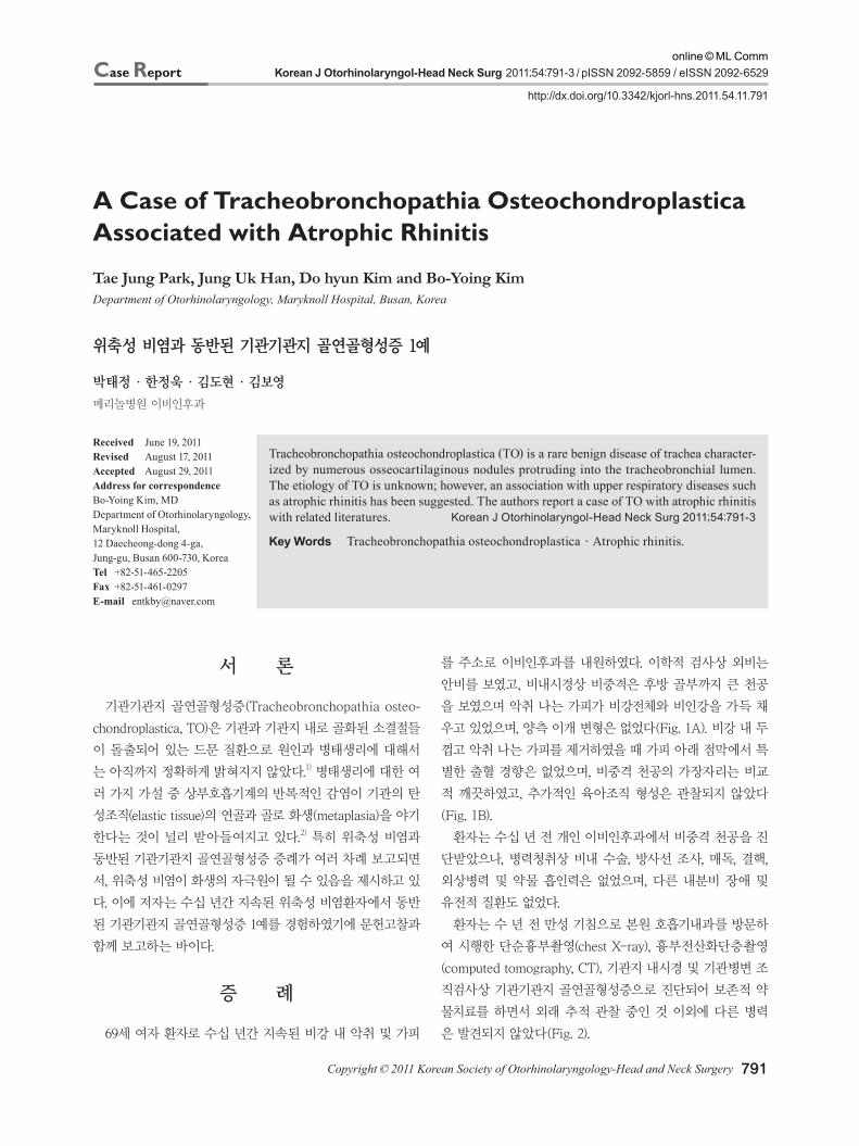
환자는 수 년 전 만성 기침으로 본원 호흡기내과를 방문하
여 시행한 단순흉부촬영(chest X-ray), 흉부전산화단층촬영
(computed tomography, CT), 기관지 내시경 및 기관병변 조
직검사상 기관기관지 골연골형성증으로 진단되어 보존적 약
물치료를 하면서 외래 추적 관찰 중인 것 이외에 다른 병력
은 발견되지 않았다(Fig. 2).
online © ML Comm
Korean J Otorhinolaryngol-Head Neck Surg █ 2011;54:791-3
792
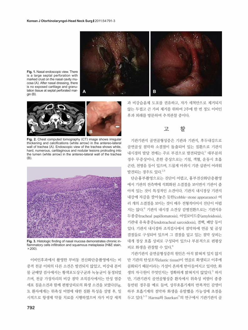
이비인후과에서 촬영한 부비동 전산화단층촬영에서는 비
중격 천공 이외의 다른 소견은 발견되지 않았고, 비강내 분비
물 균배양 검사에서는 황색포도상구균과 녹농균이 동정되었
으며, 천공 가장자리의 비강 점막 조직검사에서는 만성 염증
세포 침윤소견과 함께 편평상피로의 화생 소견을 보였다(Fig.
3). 환자에게는 위축성 비염에 대한 질환 특성을 설명 후, 일
시적으로 항생제 약물 치료를 시행하였으며 자가 비강 세척
과 비강습윤제 도포를 권유하고, 자가 세척만으로 제거되지
않는 두껍고 큰 가피 제거를 위하여 2주에 한 번 정도 이비인
후과 외래를 방문하며 추적관찰 중이다.
고 찰
기관기관지 골연골형성증은 기관과 기관지, 후두내강으로
골연골성 점막하 소결절이 돌출되어 있는 질환으로 기관지
내시경의 발달 전에는 주로 부검으로 발견되었다.1) 대부분의
경우 무증상이나, 흔한 증상으로는 기침, 객혈, 운동시 호흡
곤란, 천명음 등이 있으며, 드물게 마취시 기관 삽관이 어려워
발견되는 경우도 있다.2,3)
단순흉부촬영으로는 진단이 어렵고, 흉부전산화단층촬영
에서 기관의 전측벽에 석회화된 소결절을 보이면서 기관이 좁
아져 있는 것이 특징적인 소견이다. 기관지 내시경상 기관지
내강에 자갈을 깔아놓은 듯한(cobble-stone appearance) 여
러 개의 소결절을 보이는 것이 매우 전형적이어서 진단이 어렵
지는 않다.4) 기관지 내시경 소견상 감별진환으로는 기관지유
두종증(tracheal papillomatosis), 아밀로이드증(amyloidosis),
기관내 유육종증(endotracheal sarcoidosis), 결핵, 폐암 등이
있다. 기관지 내시경하 조직검사에서 점막하에 연골 및 골성
결절들로 구성되어 있으며 그 결절을 덮고 있는 점막 상피는
대개 정상 호흡 상피로 구성되어 있으나 부분적으로 편평상
피로 화생을 관찰할 수 있다.5)
기관기관지 골연골형성증의 원인은 아직 밝혀져 있지 않지
만 기관의 탄성조직(elastic tissue)이 연골로 화생되고 이후에
골화되기 때문이라는 가설이 흔하게 받아들여지고 있지만, 화
생의 자극원이 무엇인지는 정확하게 밝혀지지 않았다.2) 하지
만, 기관기관지 골연골형성증 환자에서 위축성 비염이 종종
동반된 경우를 예로 들며, 상부호흡기계의 반복적인 감염이
하부 호흡기계의 점막하 화생을 유발했을 가능성에 초점을
두고 있다.5-7) Harma와 Suurkari7)의 연구에서 기관기관지 골
A B
Fig. 1. Nasal endoscopic view. There is a large septal perforation with marked crust on the nasal cavity mu-cosa (A). After nasal dressing, there is no exposed cartilage and granu-lation tissue at septal perforated mar-gin (B).
A BFig. 2. Chest computed tomography (CT) image shows irregular thickening and calcifications (white arrow) in the antereo-lateral wall of trachea (A). Endoscopic view of the trachea shows white, hard, numerous, cartilaginous and nodular lesions protruding into the lumen (white arrow) in the antereo-lateral wall of the trachea (B).
Fig. 3. Histologic finding of nasal mucosa demonstrates chronic in-flammatory cells infiltration and squamous metaplasia (H&E stain, ×200).
Tracheobronchopathia Osteochondroplastica Associated with Atrophic Rhinitis █ Park TJ, et al.
www.jkorl.org 793
연골형성증 환자 30명 중 23명에서 위축성 비염이 동반되어
있었으며, 이들 중 많은 수에서 기관지 악취증(tracheo-ozena)
을 가지고 있었다고 하였다. 그리고 이들 환자 중 위축성 비
염 증상이나 농성의 기관지염 소견이 줄어들수록 기관지 내
결절이 어느 정도 흡수된다고 보고하였다.7) 그리고 Los 등3)의
보고에서도 위축성 비염과 동반된 기관기관지 골연골형성증
환자에서 비강과 기관지 내시경상에서 보인 화농성의 점액의
배양에서 Klesiella ozena가 같이 동정되었음을 밝히고, 이 두
질환의 상관관계에 대해 서술하였다. Jepsen과 Sorensen5)은
기관기관지 골연골형성증이 있는 기관지의 점막과 위축성 비
염이 있는 비강점막의 조직검사에서 부분적으로 호흡점막 상
피가 편평상피세포로 화생을 보이고 있는 것을 지적하며 이 두
질환의 상관관계의 가능성에 대해 서술하면서, 반복적인 자
극과 감염이 선행하는 유전적 경향이 있을 때 기관기관지 골
연골형성증을 유발할 수 있다고 주장하였다. 하지만, 기관기
관지 골연골형성증 환자 대부분에서 위축성 비염을 동반하
는 것이 아니고 위축성 비염을 오래 앓은 환자에서 모두 기관
기관지 골연골형성증이 발생하는 것이 아니기 때문에, 위축성
비염이 기관점막하의 화생을 유발시키는 자극원과 반복적인
감염원이라는 직접적인 인과관계를 밝힐 수는 없다.
위축성 비염 이외에도 일부 저자는 아밀로이드증과 연관관
계가 있다고 하였고, 일부에서는 암, 결핵과 연관관계가 있다
고 했으나 정확한 인과관계를 밝히지는 못했다.8-10) 동반된 질
환이 이처럼 다양하지만, 이들 질환 모두가 기관지 점막의 화
생을 유발할 수 있는 것들이기 때문에, 기관기관지 골연골형
성증 환자의 원인을 규명하기 위해서는 특히 호흡 상피에 자
극원이나 감염원으로 작용할 수 있는 원인에 초점을 맞추어
체계적이고 전반적인 조사가 필요할 것이다.7)
이 환자에서 위축성 비염이 일차성인지 이차성인지 알 수는
없다. 환자와의 병력청취 과정에서 어린 시절 심하게 수두를
앓은 뒤 비강 내에서 농성 분비물이 많이 나온 뒤 비증상이 시
작되었다는 것으로 보아 수두로 인한 급성 감염으로 비중격
천공이 발생하여 이차적으로 위축성 비염이 발생했을 가능성
도 있으며, 이와 상관없이 환자 자체의 문제로 일차성 위축성
비염이 심하여 이차적으로 비중격 천공이 발생했을 수도 있
다. 비록 일차성이든 이차성 위축성 비염이든 환자는 수십 년
동안 위축성 비염 증상을 호소하였고, 50대가 넘어서 발생한
만성적인 호흡기계 증상으로 기관기관지 골연골형성증을 진단
받았다. 본 증례에서도 이전의 다른 증례들처럼 위축성 비염
이 기관기관지 골연골형성증의 직접적인 원인임을 증명할 수
는 없지만, 충분한 상관관계는 있을 것으로 생각된다.
기관기관지 골연골형성증은 대부분에서 특별한 치료가 필
요 없지만 감염, 출혈 등의 합병증이 발생하거나, 기관의 폐쇄
가 발생한 경우는 기관절개술이나 기관의 부분적 절제 등의
수술적 치료가 필요하다.11) 유의한 기관의 폐쇄 소견이 발견될
경우 경기관지 내시경적 제거술이 가장 좋은 치료법이다. 그리
고 본 증례처럼 위축성 비염이 동반된 경우는 주기적으로 비
강 세척을 하며 비강 내 악취 나는 가피를 제거함으로써 자
극원의 반복적인 흡인을 막아주며, 하부기계 감염으로 진행
하는 것을 막아주는 것이 기관기관지 골연골형성증의 증상
악화 및 질병 진행 억제에 도움을 줄 것으로 생각된다.
REFERENCES1)Jabbardarjani HR, Radpey B, Kharabian S, Masjedi MR. Tracheo-
bronchopathia osteochondroplastica: presentation of ten cases and review of the literature. Lung 2008;186(5):293-7.
2)Thomas D, Stonell C, Hasan K. Tracheobronchopathia osteoplastica: incidental finding at tracheal intubation. Br J Anaesth 2001;87(3): 515-7.
3)Los H, Schramel FM, van der Harten JJ, Golding RP, Postmus PE. An unusual cause of recurrent fever. Eur Respir J 1997;10(2):504-7.
4)Kim YG, Lee HG, Kim TI, Kim MK, Choi YS, Gwak CH, et al. A case of tracheobronchopathia osteochondroplastica with upper air-way obstruction. Korean J Med 1998;54(1):131-4.
5)Jepsen O, Sorensen H. Tracheopathia osteoplastica and ozaena. Acta Otolaryngol 1960;51(1-2):79-83.
6)Vaheri E, Vaheri E. Tracheopathia osteoplastica. Acta Otolaryngol 1967;64(3):251-5.
7)Härmä RA, Suurkari S. Tracheopathia chondro-osteoplastica. A clinical study of thirty cases. Acta Otolaryngol 1977;84(1-2):118-23.
8)Sakula A. Tracheobronchopathia osteoplastica: its relationship to primary tracheobronchial amyloidosis. Thorax 1968;23(1):105-10.
9)Leske V, Lazor R, Coetmeur D, Crestani B, Chatté G, Cordier JF. Tracheobronchopathia osteochondroplastica: a study of 41 patients. Medicine (Baltimore) 2001;80(6):378-90.
10)Baugnee PE, Delaunois LM. Mycobacterium avium-intracellulare associated with tracheobronchopathia osteochondroplastica. Eur Respir J 1995;8(1):180-2.
11)Tibesar RJ, Edell ES. Tracheopathia osteoplastica: effective long-term management. Otolaryngol Head Neck Surg 2003;129(3):303-4.
Related Documents