Mechanisms behind postspinal headache and brain stem compression following lumbar dural puncture - a physiological approach. Grände, Per-Olof Published in: Acta Anaesthesiologica Scandinavica DOI: 10.1111/j.1399-6576.2004.00601.x 2005 Link to publication Citation for published version (APA): Grände, P-O. (2005). Mechanisms behind postspinal headache and brain stem compression following lumbar dural puncture - a physiological approach. Acta Anaesthesiologica Scandinavica, 49(5), 619-626. https://doi.org/10.1111/j.1399-6576.2004.00601.x Total number of authors: 1 General rights Unless other specific re-use rights are stated the following general rights apply: Copyright and moral rights for the publications made accessible in the public portal are retained by the authors and/or other copyright owners and it is a condition of accessing publications that users recognise and abide by the legal requirements associated with these rights. • Users may download and print one copy of any publication from the public portal for the purpose of private study or research. • You may not further distribute the material or use it for any profit-making activity or commercial gain • You may freely distribute the URL identifying the publication in the public portal Read more about Creative commons licenses: https://creativecommons.org/licenses/ Take down policy If you believe that this document breaches copyright please contact us providing details, and we will remove access to the work immediately and investigate your claim.

Welcome message from author
This document is posted to help you gain knowledge. Please leave a comment to let me know what you think about it! Share it to your friends and learn new things together.
Transcript
LUND UNIVERSITY
PO Box 117221 00 Lund+46 46-222 00 00
Mechanisms behind postspinal headache and brain stem compression followinglumbar dural puncture - a physiological approach.
Grände, Per-Olof
Published in:Acta Anaesthesiologica Scandinavica
DOI:10.1111/j.1399-6576.2004.00601.x
2005
Link to publication
Citation for published version (APA):Grände, P-O. (2005). Mechanisms behind postspinal headache and brain stem compression following lumbardural puncture - a physiological approach. Acta Anaesthesiologica Scandinavica, 49(5), 619-626.https://doi.org/10.1111/j.1399-6576.2004.00601.x
Total number of authors:1
General rightsUnless other specific re-use rights are stated the following general rights apply:Copyright and moral rights for the publications made accessible in the public portal are retained by the authorsand/or other copyright owners and it is a condition of accessing publications that users recognise and abide by thelegal requirements associated with these rights. • Users may download and print one copy of any publication from the public portal for the purpose of private studyor research. • You may not further distribute the material or use it for any profit-making activity or commercial gain • You may freely distribute the URL identifying the publication in the public portal
Read more about Creative commons licenses: https://creativecommons.org/licenses/Take down policyIf you believe that this document breaches copyright please contact us providing details, and we will removeaccess to the work immediately and investigate your claim.
1
The following pages constitute the final, accepted and revised manuscript of the article:
Mechanisms behind post-spinal headache and brain stem compression following lumbar dural puncture – a
physiological approach
Per-Olof Grände
Published in: Acta Anaesthesiol Scand. 2005 May;49(5):619-26
Publisher: Blackwell Use of alternative location of the published article requires journal subscription.
Alternative location: http://dx.doi.org/ 10.1111/j.1399-6576.2004.00601.x
Departments of Anaesthesia and Intensive Care, and Physiological Sciences,
University Hospital of Lund and University of Lund, Sweden
Correspondence: PO Grände, Department of Anaesthesia and Intensive Care,
University Hospital of Lund, SE-221 85 Lund, Sweden
Tel: +4646 2227753 Fax: +4646 2224546 e-mail: [email protected]
2
Abstract
Background: The cause of post-spinal headache and its specific characteristics are unknown, and
whether lumbar dural puncture (LP) may trigger brain stem compression in patients with brain
oedema is still controversial.
Methods: Hydrostatic effects of distal opening of the dural sac towards the atmosphere are
described and applied to the normal brain and the brain with disrupted BBB. Analogue analyses
from an isolated skeletal muscle enclosed in a rigid shell were applied to the brain in an attempt to
simulate and verify haemodynamic effects of distal opening of the spinal canal.
Results: The theoretical considerations and the experimental results are compatible with the
hypothesis that hydrostatic effects of distal opening of the fluid-filled spinal canal may obliterate
the normal subdural venous collapse after change from horizontal to vertical position, which may
be compatible with postural post-spinal headache as occurring close to pain-sensitive meningeal
regions. The hydrostatic forces may also initiate transcapillary filtration and aggravate oedema
when permeability is increased, which may cause a more narrow situation in the brain stem region,
perhaps aggravated by venous stasis and a Cushing reflex-induced increase in blood pressure. An
MR picture illustrates how this scenario may separate the subdural space into an upper high- and a
lower low-pressure cavity, pressing the brain downwards with sagging of the brain. A life-
threatening positive feed back situation for brain stem compression may develop.
Conclusion: The present study strongly suggest that post-spinal headache and brain stem
compression and other LP-related effects are predictable following LP, without involving CSF
leakage, and can be explained by hydrostatic effects triggered by distal opening of the normally
closed dural space to the atmosphere.
Key words: brain edema, brain stem herniation, blood-brain barrier, cushing reflex, lumbar dural
puncture, intracranial pressure, meningitis, post spinal headache
3
Introduction
Opening of the spinal canal via lumbar dural puncture (LP) is used clinically for infusion of local
anaesthetics or for CSF sampling, or may be a complication of epidural anaesthesia. It is sometimes
associated with postural headache, which appears momentarily when rising from supine to
upright/sitting position, and disappears when back in supine position (1-3). The headache may be
intolerable (1-4) and can continue for days and even for weeks. In most cases it is of temporary
nature, but specific treatment such as application of epidural blood patch is sometimes necessary for
its disappearance (4,5). The mechanisms behind post-spinal headache are still unknown.
It has been under discussion for decades, originating from anecdotic cases, whether LP is associated
with brain stem compression precipitating a life-threatening situation when performed in patients
with increased ICP (6-10). An outcome study from our hospital of severe meningitis showed that in
most patients developing brain stem symptoms, these appeared shortly after LP (11). It has been
argued that the suspected temporal relation between signs of brain stem compression and LP may
be a coincidence, as LP often is performed at a critical time point, and often shortly after start of
antibiotic treatment with subsequent release of toxic substances (12). An important reason why no
consensus has been reached regarding the use of LP in patients with suspected raised ICP is that no
reliable scientific studies on potential adverse effects with LP have been made. As a matter of
precaution, some authors recommend that LP should be avoided in patients with clear signs of
raised ICP, such as progressive loss of consciousness, agitation and motor anxiety, increasing blood
pressure, pupil abnormalities and other focal neurological signs (8, 9, 11). LP is, however,
frequently used in clinical practice also in risk patients (13).
It is generally believed that post-spinal headache is an effect of sagging of the brain and traction on
pain-sensitive structures due to leakage of CSF (1, 3, 4, 14-17). Leakage of CSF, which must be

4
relatively slow, however, is not consistent with the fact that post-spinal headache appears
momentarily when changing from supine to upright position and disappears immediately after
returning back to supine position. No data on CSF leakage in vivo are available, but normal
production of CSF in all likelihood is large enough to compensate for the CSF volume leaking
through the small dural tear. The fact that brain stem symptoms have not been observed following
drainage of CSF from the more distal ventricular system (if not collapsed) is another argument
supporting the view that loss of CSF volume per se is not a triggering mechanism behind brain stem
compression.
There is a great need to find the truth regarding the risk entailed in LP, since LP is used more or less
routinely in clinical practice in spite of its potentially serious adverse effects in states of raised ICP.
However, it is very difficult or impossible to perform an ethically acceptable study analysing the
hypothesis that LP precipitates brain stem compression, especially since LP often has to be
performed in a critical situation, or shortly after start of antibiotic treatment.
The complicated anatomy of the brain with impediment for effective recording and evaluation of
important cerebral circulatory variables (18), is one explanation of why we still lack a reliable
analysis of cerebral effects of distal opening of the closed dural sac. An attempt to overcome this
problem has been made in some previous studies from our laboratory by analysing the general
haemodynamic effects on an organ of being enclosed in a rigid shell and the consequences of
opening of the closed space to the atmospheric pressure (19, 20, 22). These analyses were
performed on a skeletal muscle placed in a fluid-filled and closed plethysmograph with increased
tissue pressure.
5
Based on expected hydrostatic pressure effects of distal opening of the closed and fluid-filled dura
sac combined with data from the skeletal muscle experiments, this study is an attempt to reveal the
mechanisms behind post-spinal headache, and to show whether LP may precipitate brain stem
compression. The analysis gave reliable explanations of post-spinal headache, and supported the
view that LP may trigger brain stem symptoms if the blood-brain barrier (BBB) is disrupted.
General physiological and physical principles
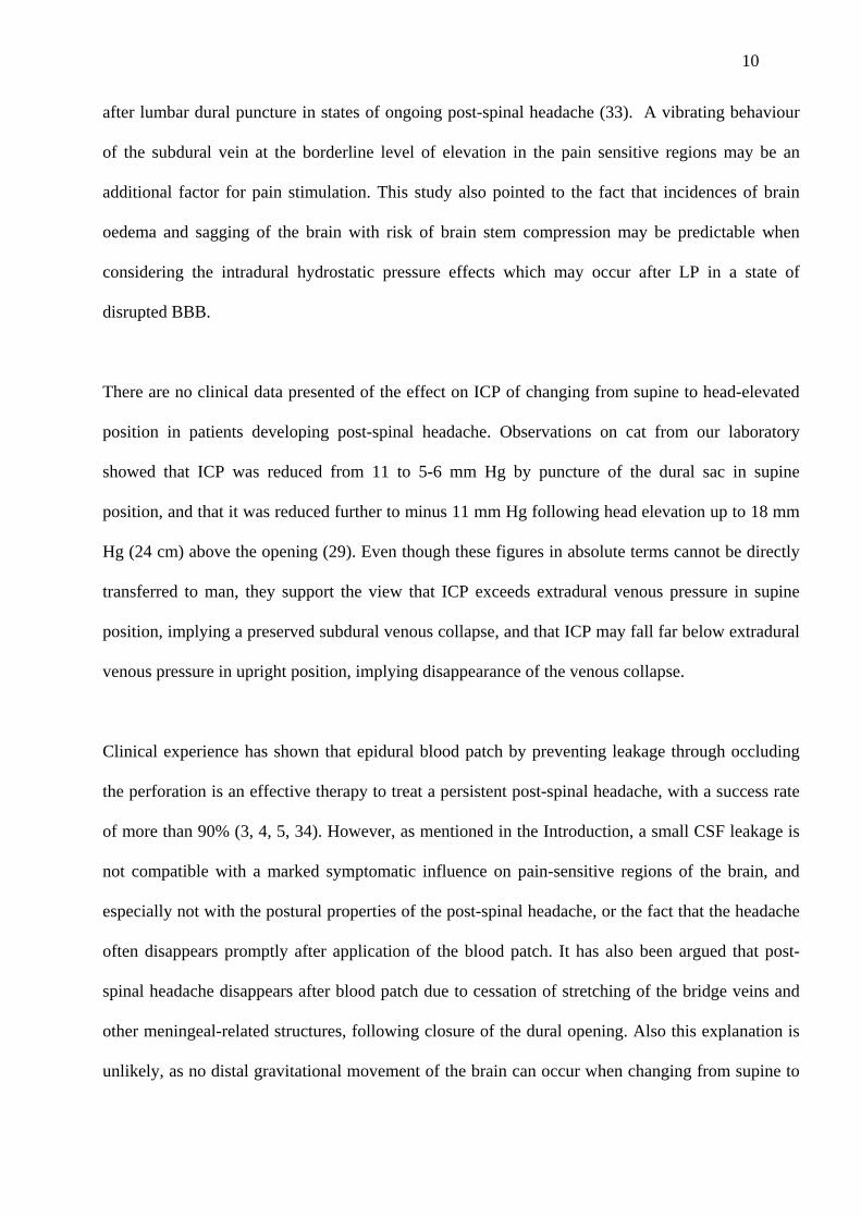
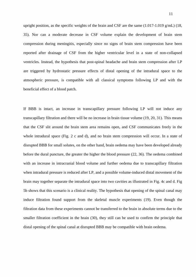
The normal brain with brain stem and spinal canal enclosed in the fluid-filled intradural space is
illustrated in supine and upright position in Fig. 1 a and b, respectively. As the cavity is a closed
and fairly rigid system, the intradural hydrostatic pressure of normally 9-12 mm Hg (19) is of about
the same magnitude in all parts of the closed intradural cavity, and will be only moderately lowered
when changing from horizontal to vertical position (23). Thus, the tissue pressure of the brain is
higher than that of other organs of the body, in which it is close to the atmospheric pressure (24),
and the epidural hydrostatic pressure may even be slightly negative (25). This means that the
intracranial pressure does not only exceed the atmospheric pressure, but also the venous pressure
outside the dura of 0-5 mm Hg in both supine and upright position. The pressure fall between the
intra- and extra-dural space will cause a passive venous collapse of the subdural draining veins at a
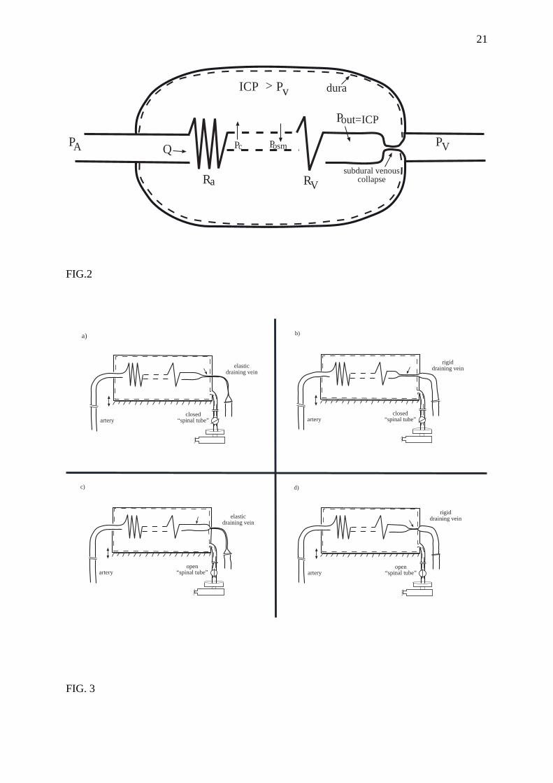
limited length just inside the dura as illustrated schematically in Fig. 2. The existence of a subdural
venous collapse was demonstrated already in 1928 (26), but its physiological role or its role in
various pathophysiological conditions for brain circulation has not until recently received any
scientific attention (20,27,28).
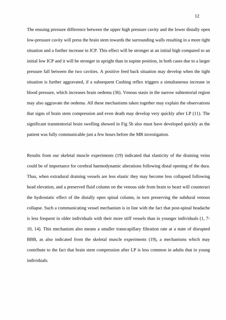
The skeletal muscle model enclosed in a plethysmograph is shown schematically in Fig. 3 a-d,
simulating head elevation at a closed and distally open spinal canal. The experiments were
performed with an elastic or a rigid draining vein to compare the situation in younger individuals

6
with that in older individuals with a more rigid vascular system. In these experiments we confirmed
the existence of the venous collapse when the tissue pressure exceeded the venous pressure, and
showed that the venous collapse acts functionally as a variable passive venous resistance, the
strength of which is determined by the difference between the tissue pressure and the venous
pressure (20, 22). When using a passive draining vein from the plethysmograph, to simulate a more
normal physiological condition, this vein showed a passive venous collapse at vertical levels at
which the venous pressure was below the atmospheric pressure at elevation of the plethysmograph
(Fig. 3a) (19). By using a more rigid draining vein to simulate the less elastic situation in older
individuals, there was an increased collapse inside the plethysmograph at elevation as shown in Fig.
3 b.
Since the degree of subdural venous collapse is determined by the difference between intracranial
and extradural pressure, it varies not only with ICP, but also with extradural venous pressure
variations. An important consequence of this effect is that venous pressure variations will not be
transferred retrogradely to the brain (20). Thus, the normal brain is protected not only from arterial
pressure variations via the well-established active arterial autoregulatory mechanism (18, 24), but
also from venous pressure variations via a passive venous collapse function.
Effects of lumbar dural puncture under normal conditions
If the dural space after LP remains open to the atmospheric pressure, there will be a pressure fall
across the dural opening. The fluid resistance in the dural opening is dependent on the size of the
dural opening, which means that the reduction in intradural pressure following LP will vary
between patients. In supine position, the intradural pressure most likely will stay above the venous
pressure, if a normal thin spinal needle is used (Fig. 1c) and, as CSF communicates freely around
7
the brain, the brain stem and the spinal cord, the pressure inside the dura will be equal in the whole
cavity.
Distal opening of the dura means that ICP must be reduced in upright position due to hydrostatic
effects (Fig. 1d), as also confirmed experimentally on cat (29). The magnitude of the reduction
depends on the vertical distance between the brain and the dural opening. ICP may even be reduced
to negative values. The decrease in ICP will increase transcapillary forces across the cerebral
capillaries, but this increase will not induce any filtration resulting in oedema, as cerebral capillaries
are impermeable for small solutes in the normal brain (intact BBB) (30-32). However, a decrease in
ICP will have other haemodynamic effects in terms of disappearance of the subdural venous
collapse if ICP falls below the extradural pressure (p<pv in Fig. 1d).
Simulation of this situation with the skeletal muscle model also showed a reduction in tissue
pressure during the elevation, and that the venous collapse disappeared above a certain vertical level
at which the tissue pressure and the venous pressure were equal (Fig. 3c). At this borderline level,
the veins showed an unstable high-frequency opening and closing behaviour. The disappearance of
the venous collapse above this level resulted in a prompt and significant venous blood volume
increase (19, 20, 22). The model experiments also showed that the draining passive vein outside the
plethysmograph remained collapsed (Fig. 3c). If the collapse was prevented by using a non-elastic
draining vein, the hydrostatic effect of the distally open “spinal tube” was partly outbalanced,
resulting in a preserved venous collapse of the draining vein as shown in Fig. 3 d. It will be
discussed below whether such an effect can explain why post-spinal headache is much less common
in older individuals, in whom the vessel elasticity is reduced, than in younger individuals with more
elastic veins. The dura is insensitive to pain except in the region of the traversing blood vessels,
8
which means that the subdural volume effects in patients occur close to the pain-sensitive
meningeal regions.
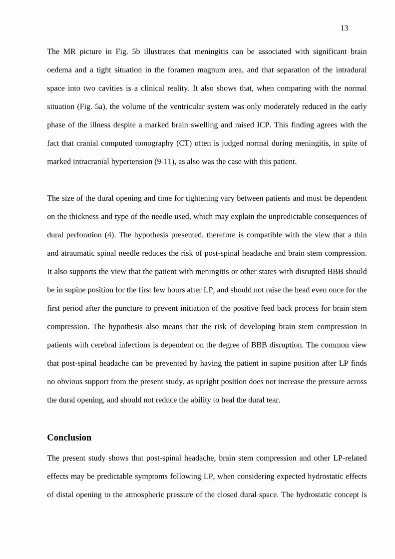
Effects of lumbar dural puncture in patients with severe cerebral infection
If there is a significant oedema and raised ICP, and the BBB is disrupted, another scenario than that
described above may develop. Before LP, there will be no limitations for CSF to pass the foramen
magnum area if no brain stem compression has occurred, and the intradural pressure will be of
about the same size in the whole subdural space both in supine and upright position (Fig. 4 a and b).
We assumed an ICP of 25 mm Hg in supine position and a slight reduction in upright position
(mainly an effect of reduced intracranial blood volume, and the fact that the dura is not a
completely rigid shell). If the dural tear remains open after LP, there will be a reduction in
interstitial pressure of the brain in turn increasing cerebral transcapillary pressure, and this increase
will be larger after head elevation. As BBB is permeable for small solutes, the increased
transcapillary pressure will induce filtration and aggravate the oedema. An increase in oedema,
combined with a small increase in intracranial blood volume and a small distal movement of the
brain, will narrow the width of the split for CSF to pass around the brain stem, and the intradural
space may be separated into a high- and a low-pressure cavity (Fig. 4 c-d). The upper cavity
comprises a closed high-pressure space, and the lower cavity a distally open low-pressure space. In
supine position, the pressure in the low-pressure cavity will be equal in the whole lower space and
below the pressure in the brain (Fig. 4 c), and in upright position the pressure in the low-pressure
space will be further reduced in its upper parts in relation to the vertical distance to the dural
opening, and it may even be below zero just distal to the brain stem (Fig. 4 d). This means that there
is a pressure fall across the foramen magnum area creating a force moving the brain distally
aggravating compression of the brain stem, a force which must be larger in upright than in supine
position. The initial compression of the brain stem may also induce venous stasis, which will

9
further increase the oedema. Altogether, a positive feed back scenario for brain stem compression
may develop.
The skeletal muscle corresponds closely to the injured brain in the sense that its capillaries are
permeable for small solutes. The skeletal muscle experiments showed a significant transcapillary
filtration following simulation of distal opening of the spinal canal, and the filtration was more
prominent with organ elevation (Fig. 3c) (19). These experiments also showed that the filtration rate
was dependent on elasticity of the draining veins in the sense that it was larger at low than at high
elasticity. It will be discussed, whether vein elasticity is a factor, which may explain the higher
frequency of brain stem compression after LP in children and young individuals than in adults.
Fig. 5b shows an MR picture of the brain taken a few hours after LP, on a 14-years-old girl
suffering from bacterial meningitis. The girl became unconscious shortly after LP, and ICP
measurement about an hour afterwards showed a value above 60 mm Hg. A CT scan a few hours
earlier was interpreted as normal. The MR picture illustrates that the scenario described above of a
tight subtentorial situation with abolished CSF slit and a swollen brain is a clinical reality. Fig. 5a
shows that 5 weeks later when the clinical situation has been normalised, there is free passage of
CSF in the foramen magnum region.
Discussion
The present study points at the possibility that post-spinal headache and its postural characteristics
may be explained by changes in venous blood volume just subdurally via intradural hydrostatic
pressure effects following LP. This conclusion is supported by the fact that the volume alterations
mainly occur close to the pain sensitive meningeal areas in the regions of transversing blood
vessels, and the fact that MRI has demonstrated what is described as “meningeal enhancement”
10
after lumbar dural puncture in states of ongoing post-spinal headache (33). A vibrating behaviour
of the subdural vein at the borderline level of elevation in the pain sensitive regions may be an
additional factor for pain stimulation. This study also pointed to the fact that incidences of brain
oedema and sagging of the brain with risk of brain stem compression may be predictable when
considering the intradural hydrostatic pressure effects which may occur after LP in a state of
disrupted BBB.
There are no clinical data presented of the effect on ICP of changing from supine to head-elevated
position in patients developing post-spinal headache. Observations on cat from our laboratory
showed that ICP was reduced from 11 to 5-6 mm Hg by puncture of the dural sac in supine
position, and that it was reduced further to minus 11 mm Hg following head elevation up to 18 mm
Hg (24 cm) above the opening (29). Even though these figures in absolute terms cannot be directly
transferred to man, they support the view that ICP exceeds extradural venous pressure in supine
position, implying a preserved subdural venous collapse, and that ICP may fall far below extradural
venous pressure in upright position, implying disappearance of the venous collapse.
Clinical experience has shown that epidural blood patch by preventing leakage through occluding
the perforation is an effective therapy to treat a persistent post-spinal headache, with a success rate
of more than 90% (3, 4, 5, 34). However, as mentioned in the Introduction, a small CSF leakage is
not compatible with a marked symptomatic influence on pain-sensitive regions of the brain, and
especially not with the postural properties of the post-spinal headache, or the fact that the headache
often disappears promptly after application of the blood patch. It has also been argued that post-
spinal headache disappears after blood patch due to cessation of stretching of the bridge veins and
other meningeal-related structures, following closure of the dural opening. Also this explanation is
unlikely, as no distal gravitational movement of the brain can occur when changing from supine to
11
upright position, as the specific weights of the brain and CSF are the same (1.017-1.019 g/mL) (18,
35). Nor can a moderate decrease in CSF volume explain the development of brain stem
compression during meningitis, especially since no signs of brain stem compression have been
reported after drainage of CSF from the higher ventricular level in a state of non-collapsed
ventricles. Instead, the hypothesis that post-spinal headache and brain stem compression after LP
are triggered by hydrostatic pressure effects of distal opening of the intradural space to the
atmospheric pressure, is compatible with all classical symptoms following LP and with the
beneficial effect of a blood patch.
If BBB is intact, an increase in transcapillary pressure following LP will not induce any
transcapillary filtration and there will be no increase in brain tissue volume (19, 20, 31). This means
that the CSF slit around the brain stem area remains open, and CSF communicates freely in the
whole intradural space (Fig. 2 c and d), and no brain stem compression will occur. In a state of
disrupted BBB for small solutes, on the other hand, brain oedema may have been developed already
before the dural puncture, the greater the higher the blood pressure (22, 36). The oedema combined
with an increase in intracranial blood volume and further oedema due to transcapillary filtration
when intradural pressure is reduced after LP, and a possible volume-induced distal movement of the
brain may together separate the intradural space into two cavities as illustrated in Fig. 4c and d. Fig
5b shows that this scenario is a clinical reality. The hypothesis that opening of the spinal canal may
induce filtration found support from the skeletal muscle experiments (19). Even though the
filtration data from these experiments cannot be transferred to the brain in absolute terms due to the
smaller filtration coefficient in the brain (30), they still can be used to confirm the principle that
distal opening of the spinal canal at disrupted BBB may be compatible with brain oedema.
12
The ensuing pressure difference between the upper high pressure cavity and the lower distally open
low-pressure cavity will press the brain stem towards the surrounding walls resulting in a more tight
situation and a further increase in ICP. This effect will be stronger at an initial high compared to an
initial low ICP and it will be stronger in upright than in supine position, in both cases due to a larger
pressure fall between the two cavities. A positive feed back situation may develop when the tight
situation is further aggravated, if a subsequent Cushing reflex triggers a simultaneous increase in
blood pressure, which increases brain oedema (36). Venous stasis in the narrow subtentorial region
may also aggravate the oedema. All these mechanisms taken together may explain the observations
that signs of brain stem compression and even death may develop very quickly after LP (11). The
significant transtentorial brain swelling showed in Fig 5b also must have developed quickly as the
patient was fully communicable just a few hours before the MR investigation.
Results from our skeletal muscle experiments (19) indicated that elasticity of the draining veins
could be of importance for cerebral haemodynamic alterations following distal opening of the dura.
Thus, when extradural draining vessels are less elastic they may become less collapsed following
head elevation, and a preserved fluid column on the venous side from brain to heart will counteract
the hydrostatic effect of the distally open spinal column, in turn preserving the subdural venous
collapse. Such a communicating vessel mechanism is in line with the fact that post-spinal headache
is less frequent in older individuals with their more stiff vessels than in younger individuals (1, 7-
10, 14). This mechanism also means a smaller transcapillary filtration rate at a state of disrupted
BBB, as also indicated from the skeletal muscle experiments (19), a mechanisms which may
contribute to the fact that brain stem compression after LP is less common in adults that in young
individuals.
13
The MR picture in Fig. 5b illustrates that meningitis can be associated with significant brain
oedema and a tight situation in the foramen magnum area, and that separation of the intradural
space into two cavities is a clinical reality. It also shows that, when comparing with the normal
situation (Fig. 5a), the volume of the ventricular system was only moderately reduced in the early
phase of the illness despite a marked brain swelling and raised ICP. This finding agrees with the
fact that cranial computed tomography (CT) often is judged normal during meningitis, in spite of
marked intracranial hypertension (9-11), as also was the case with this patient.
The size of the dural opening and time for tightening vary between patients and must be dependent
on the thickness and type of the needle used, which may explain the unpredictable consequences of
dural perforation (4). The hypothesis presented, therefore is compatible with the view that a thin
and atraumatic spinal needle reduces the risk of post-spinal headache and brain stem compression.
It also supports the view that the patient with meningitis or other states with disrupted BBB should
be in supine position for the first few hours after LP, and should not raise the head even once for the
first period after the puncture to prevent initiation of the positive feed back process for brain stem
compression. The hypothesis also means that the risk of developing brain stem compression in
patients with cerebral infections is dependent on the degree of BBB disruption. The common view
that post-spinal headache can be prevented by having the patient in supine position after LP finds
no obvious support from the present study, as upright position does not increase the pressure across
the dural opening, and should not reduce the ability to heal the dural tear.
Conclusion The present study shows that post-spinal headache, brain stem compression and other LP-related
effects may be predictable symptoms following LP, when considering expected hydrostatic effects
of distal opening to the atmospheric pressure of the closed dural space. The hydrostatic concept is
14
compatible with venous blood volume variations close to the pain-sensitive meningeal regions,
independent of CSF leakage. It is also compatible with brain oedema and brain sagging in a state of
disrupted BBB, initiating venous stasis and brain stem compression, and there is no need of CSF
leakage to explain these symptoms following LP. Hopefully, this study may contribute to a more
respectful attitude to the often uncritically use of diagnostic LP in severely ill brain injured patients,
and increased awareness of the fact that refraining from LP in selected cases with clinical signs of
raised ICP or brain stem symptoms may be lifesaving.

15
References
1) Gielen M. Post dural headache (PDPH): A Review. Reg Anesth 1989; 14: 101-106.
2) Raskin NH. Lumbar puncture headache. A review. Headache 1990; 30: 197-200.
3) Candido KD, Stevens RA. Post-dural puncture headache: pathophysiology, prevention and
treatment. Best Pract Res Clin Anaesthesiol 2003; 17: 451-69.
4) Turnbull DK, Shepherd DB. Post-dural puncture headache: pathogenesis, prevention and
treatment. Brit J Anaesthesia 2003; 91: 718-729.
5) Ylonen P, Kokki H. Epidural blood patch for management of postdural puncture headache in
adolescents. Acta Anaesthesiol Scand 2002; 46: 794-798.
6) Duffy GP. Lumbar puncture in the presence of raised intracranial pressure. BMJ 1969; 1: 407-
409.
7) Horwitz SJ, Boxerbaum B, O�Bell J. Cerebral herniation in bacterial meningitis in childhood.
Ann Neurol 1980; 7: 524-528.
8) Rennick G, Shann F, de Campo J. Cerebral herniation during bacterial meningitis in children.
BMJ 1993; 306: 953-955.
9) Riordan FAI, Cant AJ. When to do a lumbar puncture? Arch Dis Child 2002; 87: 235-237.
10) Shetty AK, Desselle BC, Craver RD. Fatal cerebral herniation after lumbar puncture in a
patient with a normal computed tomography scan. Pediatrics 1999; 103: 1284-1287.
11) Grände PO, Myhre EB, Nordström CH, Schliamser S. Treatment of intracranial hypertension
and aspects of lumbar dural puncture in severe bacterial meningitis. Acta Anaesthesiol Scand
2002; 46: 264-70.
12) Nau R, Eiffert H. Modulation of release of proinflammatory bacterial compounds by
antibacterials: potential impact on course of inflammation and outcome in sepsis and meningitis.
Clin Microbiol Rev 2002; 15: 95-110.
13) Spielman FJ. Post-lumbar puncture. Headache 1982; 22: 280-283.

16
14) Lybecker H, Möller JT, May O, Nielsen HK. Incidence and prediction of postural puncture
headache. A prospective study of 1021 spinal anaesthesias. Anesth Analg 1990: 70: 389-394.
15) Bownridge P. The management of headache following accidental dural puncture in obstetric
patients. Anesth Intensive Care 1983; 11: 4-15.
16) Gerard KW, Fagraeus L. Postspinal headache. Semin Anesth 1990; 9: 69-74
17) The Research Committee of the BSSI. Bacterial meningitis: causes for concern. J Infect
1995; 30: 89-94.
18) Heistad DD, Kontos HA. Cerebral circulation. In: Shepard JT, Abboud FM, eds.
Handbook of Physiology 3. Baltimore: William & Wilkins, 1983; 137-182.
19) Kongstad L, Grände PO. Local vascular response during organ elevation. A model for
cerebral effects of upright position and dural puncture. Acta Anaesthiol scand 1999; 43: 438-
446.
20) Asgeirsson B, Grände PO. Effects of arterial and venous pressure alterations on
transcapillary fluid exchange during raised tissue pressure. Intensive Care Med 1994; 20: 567-
572.
22) Kongstad L, Grände PO. The role of arterial and venous pressure for volume regulation of
an organ enclosed in a rigid compartment with application to the injured brain. Acta
Anaesthesiol Scand 1999, 43: 501-508.
23) Magnes B. Body position and cerebrospinal fluid pressure. J Neurosurg 1976; 44: 687-697.
24) Guyton AC, Hall JE. Textbook of Medical Physiology. Ninth edition. WB Saunders
Company 1996; 188-189
25) Harrisson GR. Epidural pressure. Anaesthesia 1990; 45: 336-7.
26) Wolf HG, Forbes HS. The cerebral circulation. V. Observations of the pial circulation
during changes in intracranial pressure. Arch Neurol Psychiat 1928; 20: 1035-1047.
17
27) Yada K, Nakagawa Y, Tsuru. Circulatory disturbances of the venous system during
experimental intracranial hypertension. J Neurosurg 1973, 39: 723-729.
28) Luce JM, Huseby JS, Kirk W, Butler J. A Starling resistor regulates cerebral venous outflow
in dogs. J Appl Physiol: Respirat Environ Exercise Physiol 1982; 53: 1496-1503.
29. Kongstad L, Grände PO. Effects on intracranial pressure of dural puncture in supine and head
elevated position. A study on cat. Acta Anaesthesiol Scand 2005; Submitted
30) Fenstermacher JD. Volume regulation of the central nervous system. In Staub NC, Taylor
AK, eds. Edema. New-York: Raven Press, 1984; 383-404.
31) Grände PO, Asgeirsson B, Nordström CH. Physiologic principles for volume regulation
of a tissue enclosed in a rigid shell with application to the injured brain. J Trauma 1997; 42:
S23-31.
32) Grände PO, Asgeirsson B, Nordström CH. Volume-targeted therapy of increased
intracranial pressure: the Lund concept unifies surgical and non-surgical treatments. Review.
Acta Anaesthesiol Scand 2002; 46: 929-41.
33) Hannerz J, Ericsson K, Bro Skejo HP. MR imaging with gadolinium in patients with and
without post-lumbar puncture headache. Acta Radiol 1999; 40: 135-141
34) Safa-Tisseront V, Thormann F, Malassiné P, Henry M, Riou B, Coriat P, Seebacher J.
Effectiveness of Epidural Blood Patch in the Management of Post-Dural Puncture Headache.
Anesthesiology 2001; 95: 334-339.
35) Blatter DD, Bigler ED, et al A normative database from magnetic resonance imaging.
Human brain function. Neuroimaging 1: Basic science. By Biegler ED. New York, Plenum,
1996; 79-95.
36) Kongstad L, Grände PO. Arterial hypertension increases intracranial pressure in cat after
opening of the blood-brain barrier. J Trauma 2001; 51: 490-6.

18
Legends
Fig. 1. The brain, the brain stem and the spinal cord enclosed in the fluid-filled dural space in a
normal state without oedema before LP in supine (a) and upright (b) position, and after LP in
supine (c) and upright (d) position. The assumed lowering of ICP from 10 to 6-8 mmHg when
changing from supine (a) to upright position (b) is predicted from a reduced blood volume and the
fact that the dura is not a completely rigid shell.
Fig. 2. A schematic illustration of brain circulation surrounded by the dura and the rigid cranium.
The tissue pressure (ICP) is higher than the extradural venous pressure (PV), causing a passive
venous collapse just subdurally (ROUT). Q: blood flow, PA: arterial inflow pressure, PC:
hydrostatic capillary pressure, POSM: colloid osmotic pressure, POUT: venous pressure just
proximal to ROUT, RA: arterial resistance, and RV: venular resistance. POUT = ICP.
Fig. 3. Schematic illustrations of experiments with a skeletal muscle in a fluid-filled
plethysmograph, simulating the haemodynamic situation on the venous side of the brain and
effects of the spinal canal before and after LP in upright position. The situation in upright position
before “LP” using an elastic draining vein is shown in (a), and a rigid draining vein in (b), and the
situation also in upright position after “LP” using an elastic draining vein is shown in (c), and
using a rigid draining vein in (d). The arrows point at the status of the venous collapse in upright
position before (a, b) and after (c, d) opening of the “spinal tube” , and elastic or rigid draining
veins (for details, see text).
Fig. 4. The brain, the brain stem and the spinal cord enclosed in the fluid-filled dural space at a
state of disrupted BBB and oedema before LP in supine (a) and upright (b) position, and after LP
in supine (c) and upright (d) position. The assumed lowering of ICP from 25 to 22 mmHg when

19
changing from supine (a) to upright position (b) is predicted from a reduced blood volume and the
fact that the dura is not a completely rigid shell.
Fig. 5. An MR picture of the brain of a 14 years old girl suffering meningitis with raised ICP a
few hours after LP (b), and 5 weeks later when ICP was normalised (a).

20
p = 10 mm Hg
p = 10 m
mH
g
Normal brain, before spinalpuncture supine position
b
0 mm
Hg
Normal brain, after spinalpuncture supine position
d
p =
10-1
2 m
mH
g
Normal brain, before spinalpuncture upright position
a c
0 mmHg
Normal brain, after spinalpuncture upright position
p = 6-8 mmHgp < pv
p > pv
Pv < p < 10
Fig.1
21
Q
Pout=ICP
PA PVPc
RVRa
PICP v>
Posm
subdural venouscollapse
dura
FIG.2
elasticdraining vein
closed“spinal tube”artery
a) b)
rigiddraining vein
c)
rigiddraining vein
d)
elasticdraining vein
closed“spinal tube”artery
open“spinal tube”artery
open“spinal tube”artery
FIG. 3

22
p = 25 mmHg
Meningitis, before spinalpuncture upright position
p =
26
mm
Hg
aMeningitis, before spinalpuncture supine position
b
p < 0 mmHg
0 mmHg
Meningitis, after spinalpuncture upright position
cMeningitis, after spinalpuncture supine position
d
p < 25 mmHg
0 mm
Hg
p = 22 mmHg
p = 25 m
mH
g p < 25 m
mH
g
p = 25 m
mH
g
p = 25 mmHg
FIG. 4

23
Veronica
FI G. 5 a and b
Related Documents