55 Basilar-Predominant Disease

55 basilar predominant disease
Aug 15, 2015
Welcome message from author
This document is posted to help you gain knowledge. Please leave a comment to let me know what you think about it! Share it to your friends and learn new things together.
Transcript
55 Basilar-Predominant Disease
CLINICAL IMAGAGINGAN ATLAS OF DIFFERENTIAL DAIGNOSIS
EISENBERG
DR. Muhammad Bin Zulfiqar PGR-FCPS III SIMS/SHL
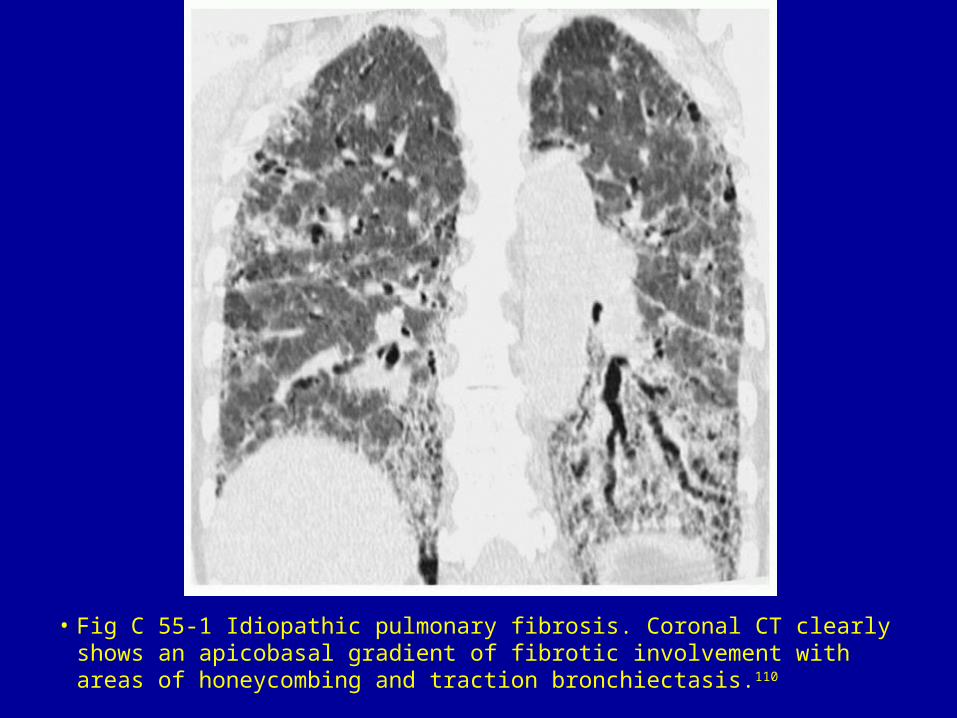
• Fig C 55-1 Idiopathic pulmonary fibrosis. Coronal CT clearly shows an apicobasal gradient of fibrotic involvement with areas of honeycombing and traction bronchiectasis.110
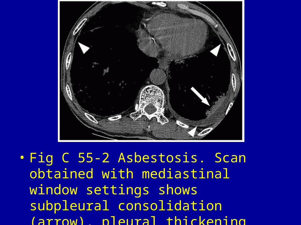
• Fig C 55-2 Asbestosis. Scan obtained with mediastinal window settings shows subpleural consolidation (arrow), pleural thickening (arrowheads), and effusion.105
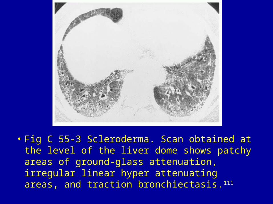
• Fig C 55-3 Scleroderma. Scan obtained at the level of the liver dome shows patchy areas of ground-glass attenuation, irregular linear hyper attenuating areas, and traction bronchiectasis.111
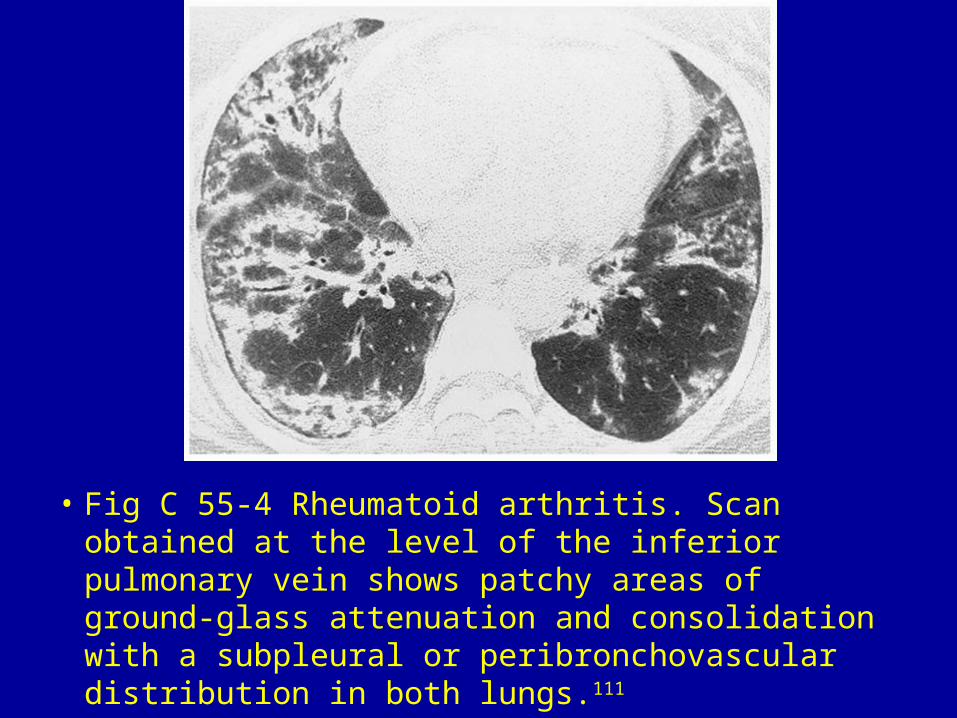
• Fig C 55-4 Rheumatoid arthritis. Scan obtained at the level of the inferior pulmonary vein shows patchy areas of ground-glass attenuation and consolidation with a subpleural or peribronchovascular distribution in both lungs.111
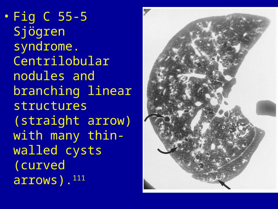
• Fig C 55-5 Sjögren syndrome. Centrilobular nodules and branching linear structures (straight arrow) with many thin-walled cysts (curved arrows).111
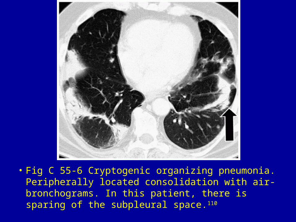
• Fig C 55-6 Cryptogenic organizing pneumonia. Peripherally located consolidation with air-bronchograms. In this patient, there is sparing of the subpleural space.110
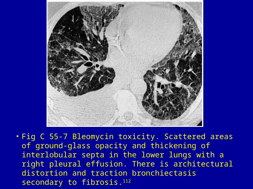
• Fig C 55-7 Bleomycin toxicity. Scattered areas of ground-glass opacity and thickening of interlobular septa in the lower lungs with a right pleural effusion. There is architectural distortion and traction bronchiectasis secondary to fibrosis.112
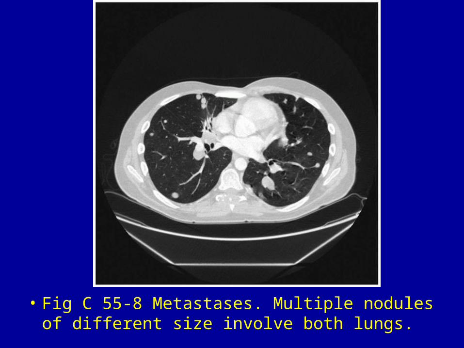
• Fig C 55-8 Metastases. Multiple nodules of different size involve both lungs.

Related Documents