Recurrent Streptococcal Pharyngotonsillitis Studies on etiology and treatment Orrling, Arne 2006 Link to publication Citation for published version (APA): Orrling, A. (2006). Recurrent Streptococcal Pharyngotonsillitis Studies on etiology and treatment. Lund University. Total number of authors: 1 General rights Unless other specific re-use rights are stated the following general rights apply: Copyright and moral rights for the publications made accessible in the public portal are retained by the authors and/or other copyright owners and it is a condition of accessing publications that users recognise and abide by the legal requirements associated with these rights. • Users may download and print one copy of any publication from the public portal for the purpose of private study or research. • You may not further distribute the material or use it for any profit-making activity or commercial gain • You may freely distribute the URL identifying the publication in the public portal Read more about Creative commons licenses: https://creativecommons.org/licenses/ Take down policy If you believe that this document breaches copyright please contact us providing details, and we will remove access to the work immediately and investigate your claim. Download date: 26. Aug. 2022

Welcome message from author
This document is posted to help you gain knowledge. Please leave a comment to let me know what you think about it! Share it to your friends and learn new things together.
Transcript
LUND UNIVERSITY
PO Box 117221 00 Lund+46 46-222 00 00
Recurrent Streptococcal Pharyngotonsillitis Studies on etiology and treatment
Orrling, Arne
2006
Link to publication
Citation for published version (APA):Orrling, A. (2006). Recurrent Streptococcal Pharyngotonsillitis Studies on etiology and treatment. LundUniversity.
Total number of authors:1
General rightsUnless other specific re-use rights are stated the following general rights apply:Copyright and moral rights for the publications made accessible in the public portal are retained by the authorsand/or other copyright owners and it is a condition of accessing publications that users recognise and abide by thelegal requirements associated with these rights. • Users may download and print one copy of any publication from the public portal for the purpose of private studyor research. • You may not further distribute the material or use it for any profit-making activity or commercial gain • You may freely distribute the URL identifying the publication in the public portal
Read more about Creative commons licenses: https://creativecommons.org/licenses/Take down policyIf you believe that this document breaches copyright please contact us providing details, and we will removeaccess to the work immediately and investigate your claim.
Download date: 26. Aug. 2022
Department of Otorhinolaryngology, Head and Neck Surgery
Clinical Sciences, Lund
Lund University, Sweden
Recurrent Streptococcal Pharyngotonsillitis
Studies on Etiology and Treatment
Arne Orrling
The Faculty of Medicine Lund University Lund 2006


To my three sons
Albert, Henrik and Filip
and to
Gunilla my love

TABLE OF CONTENTS
ABBREVIATIONS AND DEFINITIONS 7
LIST OF PUBLICATIONS 8
THE THERAPEUTIC DILEMMA 9
INTRODUCTION 11 Pharyngotonsillitis 11
-haemolytic GAS 13Antibiotics in the treatment of GAS pharyngotonsillitis 15
The carrier state 16Reasons to treat GAS pharyngotonsillitis 17
Possible reasons for failure in penicillin treatment of GAS pharyngotonsillitis 18
AIMS OF THE PRESENT STUDY 20
THE PRESENT INVESTIGATION 21Comparison of penicillin and clindamycin in bacterial failureafter pcV treated GAS pharyngotonsillitis.A one-year follow up study (I, II) 21
An attempt to identify penicillin tolerant GAS (III) 24
Genetic profiles in GAS isolates from failures and nonfailures. An investigation using AP-PCR technique (IV) 27
Penicillin V, loracarbef and clindamycin in TSF during and after treatment of GAS pharyngotonsillitis (V) 30
GENERAL DISCUSSION 34 Treatment failure 34 Reinfection 34 Genetic profiles 35 Penicillin tolerance 35 Antibiotic concentrations in TSF 36 CRP and orosomucoid 37 Penicillin for ten days 37 Loracarbef 38 Clindamycin 38 Conclusion 39
CONCLUSIONS 40
SUMMARY 41
SUMMARY in SWEDISH 44
ACKNOWLEDGEMENTS 49
REFERENCES 51
APPENDIX 62 Paper I Paper II Paper III Paper IV Paper V
ABBREVIATIONS AND DEFINITIONS
AP-PCR Arbitrarily Primed Polymerase Chain Reaction
CFU Colony Forming Unit
GAS -haemolytic Group A Streptococci
MBC Minimum Bactericidal Concentration
MIC Minimum Inhibitory Concentration
NF Necrotizing Fasciitis
N.S. Non Significant
STSS Streptococcal Toxic Shock Syndrome
TSF Tonsillar Surface Fluid
PcV Phenoxymethyl penicillin
Bacterial treatment failure presence of GAS of the same T-type as that of the pre-
treatment strain, within two weeks after completing therapy.
Clinical and bacterial failure as above in combination with clinical symptoms and signs of
pharyngotonsillitis.
Reinfection occurrence of a new GAS strain, irrespective of T-type, after
successfull eradication of the primary GAS.
Penicillin tolerance MBC/MIC 32 and survival rate 1% in "Time killing test".
7
LIST OF PUBLICATIONS
This thesis is based on studies reported in the following papers, referred to in the text by their respective Roman numerals (I –V).
I Orrling A, Stjernquist-Desatnik A, Schalén C, Kamme C. Clindamycin in persisting streptococcal pharyngotonsillitis after penicillin treatment. Scand J Inf Dis 26:535-41, 1994.
II Orrling A, Stjernquist-Desatnik A, Schalén C, Kamme C. Clindamycin in recurrent group A streptococcal pharyngotonsillitis - An alternative to tonsillectomy. Acta Otolaryngol (Stockh) 117:618-22, 1997.
III Orrling A, Stjernquist-Desatnik A, Schalén C, Kamme C: Treatment failure in streptococcal pharyngotonsillitis. An attempt to identify penicillin tolerant streptococcus pyogenes. Scand J Infect Dis 28:143-7, 1996.
IV Orrling A, Karlsson E, Melhus Å, Stjernquist-Desatnik A. Penicillin treatment failure in group A streptococcal tonsillopharyngitis: No genetic difference found between strains isolated from failures and nonfailures. Ann Otology Rhinol Laryngol 110:690- 5, 2001.
V Orrling A, Kamme C, Stjernquist-Desatnik A. Penicillin V, loracarbef and clindamycin in tonsillar surface fluid during acute group A streptococcal pharyngotonsillitis. Scand J Inf Dis 37: 429-35, 2005.
Papers I, III and V are reproduced with permission from Scandinavian Journal of Infectious Diseases.
Paper II are reproduced with permission from Acta Otolaryngologica.
Paper IV is reproduced with permission from Annals of ORL.
8

�
������������������������
������������������������������������������������������������������������
The physician: If this does not help, then please come back – and I will prescribe another medicine.The little woman: Couldn´t I as well get that other medicine at once!The physician: If this does not help, then please come back – and I will prescribe another medicine.The little woman: Couldn´t I as well get that other medicine at once!
�
������������������������
������������������������������������������������������������������������
The physician: If this does not help, then please come back – and I will prescribe another medicine.The little woman: Couldn´t I as well get that other medicine at once!
�
������������������������
������������������������������������������������������������������������
The physician: If this does not help, then please come back – and I will prescribe another medicine.The little woman: Couldn´t I as well get that other medicine at once!

10

INTRODUCTION
Acute pharyngotonsillitis is a common infection with an annual incidence in Sweden of
approximately 300.000 cases (Tierpsprojektet) and group A streptococci (GAS) is the
etiologic agent in 30-50% of cases (Wannamaker 1972; Roos 1985; Stjernquist-Desatnik et al
1987). GAS pharyngotonsillitis results in a high degree of absence from day-care, school and
work, and it is agreed that antibiotic treatment is indicated. Phenoxymethylpenicillin (pcV) is
the drug of choice in Sweden. In spite of exposure to -lactams for decades and in contrast to
several other common pathogens, GAS has over the years retained unchanged high
susceptibility to these drugs. However, failure rates in pcV treated GAS pharyngotonsillitis
are as high as 5-25% (Schwartz et al 1981; Strömberg et al 1988). A second course of pcV
treatment is followed by still higher failure rates (Kaplan & Johnson 1988), and repeated
failure in some cases necessitates tonsillectomy, one of the most common surgical procedures
in the western world, although the benefit of operation in recurrent pharyngotonsillitis, lasts
for only two to three years (Paradise et al 1984).
The background of failure remains largely elusive. Several factors possibly contributing to the
recurrences have been mentioned: low compliance, reinfection from the environment,
eradication of -streptococci with inhibitory effect on GAS, increase in -lactamase
producing bacteria inactivating the drug, penicillin tolerant streptococci, low antibiotic
concentration at site of infection and finally intracellular GAS surviving therapy.
PHARYNGOTONSILLITIS
Microbial etiology
Pharyngotonsillitis can be caused by a wide variety of pathogens. When symptoms are mainly
restricted to the throat, however, a majority are of bacterial origin. GAS is the causative agent
in about 50 % of cases. Group C and G streptococci cause 5-10% whereas other bacteria such
as Arcanobacterium hemolyticum, Chlamydia pneumoniae, Mycoplasma pneumonie,
Borrelia vincenti, Corynebacterium diphteriae and Neisseria gonorrhoeae are more seldomly
seen (Hill et al 1969 ; Benjamin & Perriello 1976; Woodruff 1980; Telian 1986; Banck &
Nyman 1986 ). Viruses account for 20-30 % of cases (Glezen et al 1967; Moffet et al 1968),
and in the remaining 10-20% the causative agent is unknown (Ross et al 1971; Nordenfelt 1981).
11

Diagnosis
Clinical diagnosis
Signs and symptoms of acute pharyngotonsillitis include fever, throat angina, redness of
tonsils and pharynx, tonsillar exudate, enlarged and tender cervical lymph nodes and
dysphagia. Concurrent symptoms from the respiratory tract, e.g. cough or rhinorrhea, indicate
viral origin. Established GAS throat and skin infection in the close surroundings like family,
school or day care increases the probability of GAS origin. Certain symptoms could be more
pronounced in pharyngotonsillitis of GAS origin than of other etiology. Thus the degree of
redness in the throat (Roos 1985), fever (Hansen et al 1983), and a shorter duration of
symptoms before seeking medical care (Stjernquist-Desatnik et al 1987) were reported to
correlate significantly to recovery of GAS. The findings are, however, inconsistent and
although the clinical picture could be of some guidance it is seldom sufficient for a reliable
etiological diagnosis.
Microbiological diagnosis of GAS pharyngotonsillitis
Since there is no pathognomonic sign or combination of symptoms and signs in this
condition, the definite diagnosis of GAS pharyngotonsillitis depends on identification of the
bacteria. This might be done by a rapid antigen detection test or by a throat culture. A good
view of the pharynx and correct sampling technique is essential to achieve a representative
sample. The specimen should be obtained from the tonsillar surface, since in GAS
pharyngotonsillitis the streptococci are predominantly localized on the tonsils and on the
posterior oropharyngeal wall (Lilja et al 1997). A certain amount of bacteria, larger than for
culture, is needed for a positive rapid antigen test. Quality of sampling may therefore
influence on sensitivity and specificity of the rapid tests, currently reported as 74 % - 97 %
and 89 % -95 % respectively (Nerbrant 2002; Lindbaek et al 2004).
Laboratory findings
A correlation between leucocytosis (Roos 1985; Hjortdahl & Melbye 1994) as well as
increased levels of CRP and GAS pharyngotonsillitis has been reported (Kaplan &
Wannamaker 1977) while other investigators failed to verify this (Putto et al 1986; Sun et al
2002).
12
� HAEMOLYTIC GROUP A STREPTOCOCCI
Grouping of streptococci
A basic tool for epidemiological investigations, as well as studies on treatment failure vs.
reinfection of pharyngotonsillitis is the accurate identification of bacterial strains . The
streptococci are classified into Lancefield´s serological groups A –U according to
carbohydrate antigen in the cell wall (Lancefield 1933). Based on the presence of T-antigen,
GAS are divided into approximately 30 T-types (Lancefield 1928).
Further subdivision is made on basis of the M-protein (Lancefield 1928). Advances in DNA-
sequencing technology in the late twentieth century resulted in the development of methods
for determining the M type of GAS from the sequence of the corresponding gene emm, and up
to now more than 120 emm-types are identified (Facklam et al 2002).
Virulence factors
GAS exhibit a multitude of extracellular and cell-bound virulence factors with probable
impact on different disease manifestations, and at various stages of the invasive process.
The cell wall M-protein, an extended �-helical protein with anticomplementary and
antiphagocytic properties, is considered as a main factor determining virulence of GAS.
Many M-proteins, by interacting with plasma proteins, such as IgG, fibrinogen and C4-
binding protein, exhibit mechanisms specifically blocking the innate or acquired immune
systems (Carlsson et al 2005). As established long ago, only type-specific antibodies directed
to the N-terminal part of M-protein will be opsonic, and protect against GAS disease
(Fischetti 1989)
The hyaluronic capsule, though poorly expressed in vitro, is a second, antiphagocytic part of
GAS (Wessels et al 1991). The T-protein, previously not implicated as biologically important,
was recently shown to mediate formation of fimbriae-like structures in GAS, of possible role
for tissue adhesion (Mora et al 2005). Pyrogenic exotoxins (erythrogenic toxins), are now
established as so-called superantigens, viz highly active toxins triggering T cells to massive
cytokine and interleukin release, thereby generating severe symptoms, such as fever, the
scarlatiniform rash, tissue necrosis, hypotension and organ failure (Bisno et al 2003). The
cysteine protease (identical to exotoxin SpeB) according to experimental work may be
essential for severe clinical manifestations, such as circulatory shock and lung damage
(Herwald et al 1998; Herwald et al 2004). This enzyme, and a second cysteine protease of
GAS, may also cleave IgG, thereby interfering with immune opsonization of GAS (von
13
Penicillin tolerance
Tolerance to �-lactam antibiotics is a known phenomenon in some medically important
species, such as Enterococcus faecalis, Streptococcus pneumoniae and various �- haemolytic
streptococci (Tuomanen et al 1986) and it appears to account for failure of penicillin therapy
of Arcanobacter haemolyticum infections (Nyman et al 1990). Penicillin tolerance in GAS has
been suggested to promote failure in pcV treatment of GAS pharyngotonsillitis, but reports
have been contradictory, conceivably due to the variability in the definition of “tolerance” as
well as technical pitfalls of methods used (Kim & Kaplan 1985; Krasinski et al 1986; Grahn
et al 1987; Smith et al 1987; Stjernquist-Desatnik et al 1992). The existence of penicillin
tolerance in GAS has also been questioned (Woolfrey 1988).
Low antibiotic concentration at site of infection
In acute GAS pharyngotonsillitis the causative bacteria are mainly present in the secretion on
surface and in crypts, rather than in the tonsillar parenchyma (Ebenfelt et al 1998; Lilja et al
1997). PcV was detected in the TSF in a majority of patients on the first day of treatment of
acute GAS pharyngotonsillitis, but despite high concentrations in serum, rarely on the tenth
day or in healthy treated subjects (Stjernquist-Desatnik et al 1993). Insufficient concentrations
of antibiotics in TSF might contribute to treatment failure in GAS pharyngotonsillitis.
Intracellular GAS surviving therapy
As shown in vitro, internalized GAS in human respiratory epithelial cells, in the absence of
extracellular antibiotics, were mobilized and established infection. (Österlund & Engstrand
1995). In analogy to these findings the respiratory epithelial cells may act as a reservoir where
internalized GAS with potential to cause infection can survive pcV treatment and account for
recurrent pharyngotonsillitis after pcV treatment.
19

Pawel-Rammingen et al 2003). Streptolysins S and O are capable of lysing erythrocytes as
well as leucocytes and platelets (Sierig 2003, Fontaine 2003). Streptokinase, which converts
plasminogen to plasmin, may significantly contribute to rapid spread of GAS in infected
tissue, i.a. by lysing blood clots (Lottenberg 1994).
Internalization
The ability of GAS, especially in the stationary phase, to invade respiratory epithelial cells
has been demonstrated in recent years (LaPenta et al 1994; Österlund & Engstrand 1995).
GAS are mainly found extracellulary, but by specifically binding fibronectin, a
protein that exists in human blood plasma and in the extracellular matrix GAS may be
efficiently internalized into human mucosal cells. The fibronectin bound to the bacterial
surface thereby acts like a bridging molecule towards host cell integrins, which in turn initiate
the uptake process that leads to internalization (Kreikemeyer et al 2004).
Sela and Barziali (1999) found that GAS strains were able to survive for 4–7 days inside
cultured epithelial cells, and also that GAS strains from patients with eradication failure
harboured an internalization-associated gene in higher prevalence than strains recovered from
patients with successful eradication. Internalized GAS have been found in asymptomatic
carriers as well as in patients with pharyngotonsillitis (Österlund et al 1997) and various
strains of streptococci have different capacity to internalize (LaPenta et al 1994; Österlund &
Engstrand 1995). Interestingly strains from cases of eradication failure showed significantly
increased intracellular survival compared to strains from non failures (Marouni et al 2004)
Whether internalization into host cells may influence on the severity of GAS infections is not
known; however, in an animal model, a GAS strain able to internalize was less prone to cause
serious disease than GAS without that capacity (Nyberg et al 2004).
Disease manifestations
GAS are strict human pathogens giving rise to a wide range of infections. Impetigo,
pharyngotonsillitis and erysipelas may be comparatively mild and are effectively treated with
antibiotics. However, since late eighties a rising number of life threatening, invasive GAS
infections such as necrotizing fasciitis (NF) and streptococcal toxic shock syndrome (STSS)
have been encountered (Cone et al 1987; Hoge et al 1993). Streptococci in these cases are
often restricted to certain M-types, in particular M1, and produce powerful superantigens,
such as pyrogenic exotoxin A – SpeA. Surgical intervention is often needed in the case of NF.
However, in spite of antibiotics and intensive care the mortality in both NF and STSS is high.
14
(Davies et al 1996; Eriksson et al 1998). Acute rheumatic fever, the most serious non-
suppurative complication to GAS pharyngotonsillitis, is the leading cause of acquired heart
disease among children in developing countries (Bisno 1991). Although no longer a
significant health problem in most socioeconomically advanced countries, limited outbreaks
of acute rheumatic fever have occurred in the US in the eighties (Veasy et al 1987).
Acute post-streptococcal glomerulonephritis, a major cause of child renal failure occurs after
throat as well as skin infections with GAS (Wannamaker 1970). Large epidemics are still
noted in the developing countries, as compared to sporadic cases in our part of the world.
ANTIBIOTICS IN TREATMENT OF GAS PHARYNGOTONSILLITIS
-lactam antibiotics
PcV and cephalosporins act on bacteria by inhibiting synthesis of the cell wall and are thus
only active against growing bacteria, while organisms in lag phase and stationary phase, since
they are not replicating, are not susceptible to these substances. The -lactam antibiotics have
low or no intracellular accessibility. Although GAS have been exposed to -lactams for
decades, there has been no development of resistance to these drugs. A possible explanation
may be, that penicillin resistance in this species is not compatible with a virulent phenotype
(Gutman & Tomasz 1982).
Penicillin is inactivated by -lactamase produced by Staphylococci, Bacteroides and
Fusobacteria spp in the throat.
In pcV treatment of GAS pharyngotonsillitis the importance of no less than 10 days treatment
to achieve acceptably low recurrence rate, has been well documented (Schwartz et al 1981;
Gerber et al 1987; Strömberg et al 1988; Zwart et al 2000). In a meta-analysis Lan and
colleagues (2000) found the current recommended dosing frequency of 2 times daily for 10
days to be as efficacious as more frequent dosing regimens in treatment of GAS
pharyngotonsillitis.
In primary GAS pharyngotonsillitis cephalosporins have been shown to be more effective
than pcV (Pichichero et al 1987; Holm et al 1991; Milatovic & Knauer 1989). Cephalosporins
may enable shorter treatment regimens than pcV in GAS pharyngotonsillitis, and some may
be dosed once daily (Pichichero et al 1994).
Cephalosporins are less susceptible to the -lactamases produced by the oral bacterial flora
and probably have lesser impact on the bacteriocin producing -haemolytic streptococci in the
throat. Theoretically, lack of effect of cephalosporins on -streptococci not disturbing the
15
bacterial interference could explain the better eradication of GAS by cephalosporins than by
pcV (Holm et al. 1991; Roos et al. 1993).
Macrolides
Macrolides act by interfering with the protein synthesis and are mainly bacteriostatic.
Stjernquist-Desatnik and colleagues (1993) investigated erythromycin in TSF and found
detectable levels in half of the healthy persons investigated. Even though macrolides act
intracellularly, and therefore may reach internalized GAS, the rate of failure in GAS
pharyngotonsillitis is almost the same as by pcV treatment (Brook & Hirokawa 1985;
Söderström et al 1991; Watkins et al 1997; Cohen et al 2002). In GAS pharyngotonsillitis,
however, macrolides are less suitable because of tendency to induce resistance in GAS.
Outbreaks of erythromycin resistant GAS are known from Japan (Maruyama et al 1979),
Finland (Seppälä et al 1992) and many other countries. Although only 2 % of Swedish GAS
strains are currently resistant to erythromycin, higher figures have been reported in Sweden in
the eighties (Stjernquist-Desatnik et al 1994).
Clindamycin
Clindamycin also blocks protein synthesis and acts intracellularly. Log phase as well as
stationary phase GAS are susceptible to the drug. Low recurrence rates have been achieved in
treatment of pharyngotonsillitis by clindamycin (Brook & Hirokawa 1985; Jensen & Larsen
1991). The rate of GAS isolates resistant to clindamycin is generally low e.g. < 1% in
Sweden in 2005. However clindamycin resistance in GAS may be linked to macrolide
resistance and in areas with a high consumption of macrolides the proportion of GAS strains
resistant to both antibiotics rapidly may reach alarming levels. For example, in the Olomouc
region in the Czech Republic the proportion of clindamycin resistant GAS strains rose from
4% to 28% between 1999 and 2001 (Urbanek et al 2005).
THE CARRIER STATE
Asymptomatic carriage of GAS in the throat is more frequent in children than in adults. The
frequency found in Scandinavian investigations was 2-11% in children <4 years of age, 5-
21% in age group 4-15 years and 1-4% in adults (Hoffmann 1985; Strömberg et al 1988;
Gunnarsson et al 1997). However, in outbreaks of GAS pharyngotonsillitis in for example
day-care or school the carrier rate could be as high as 60 % (Falk & Kjellander 1992).
16
In a four year longitudinal study of school children, 5 -15 years old, the mean time for a
period of carriage, during which the child harboured GAS of the same emm type, was 10.8
weeks (range: 3-34 weeks). Many children, however, experienced several periods of carriage
during the study and frequently exhibited switches in emm-type (Martin et al 2004). The risk
of becoming a carrier or contract disease, is related to the time spent in close contact with a
patient during the week preceding onset of illness (Engelgau et al 1994; Weiss et al 1999).
The background why some individuals become carriers is not known, but the carriership
appears to be a harmless condition, as it probably does not result in clinical infection (Kaplan
et al 1981). In addition, the streptococci are present in low numbers (Roos 1985) and the
carrier probably does not transmit infection (Falk & Kjellander 1992). However, problems
arise when a carrier acquires viral pharyngitis, as positive test for GAS will raise the issue of
antibiotic treatment. This highlights the importance of careful evaluation of symptoms in
order to avoid unnecessary antibiotic treatment.
REASONS TO TREAT GAS PHARYNGOTONSILLITIS
GAS pharyngotonsillitis is a self-limiting disease and the routine of pcV treatment has
therefore been questioned (Flottorp et al 2000). However, GAS is one of the most virulent
human pathogens, and in pharyngotonsillitis the patient can be seriously affected with high
fever, dysphagia and severe pain. Irrespective of treatment, a majority of patients are free of
symptoms within a week, but antibiotic treatment of GAS pharyngotonsillitis was shown to
shorten the duration of symptoms (De-Meyere et al 1992; Dagnelie et al 1996). Treatment
also in some degree reduces the risk of purulent complications, such as peritonsillitis, otitis
and sinusitis (Del Mar et al 2000; Dagnelie et al 1996; Zwart et al 2000). In acute rheumatic
fever, it is claimed that a majority of the patients have a history of pharyngotonsillitis. The
decline of acute rheumatic fever in the western world might be the result of consequent
antibiotic use in GAS pharyngotonsillitis, in strong support of the present principles of
treatment. In NF and STSS, however, the port of entry is seldom reported to be the pharynx
(Davies 1996; Eriksson et al 1998).
Thus the reasons for antibiotic treatment of GAS pharyngotonsillitis are: 1) Faster alleviation
of symptoms; 2) Reducing the spread of GAS; 3) Reducing the risk for suppurative and non
suppurative complications. Hence it is mostly agreed that benefits of antibiotic treatment
outweigh disadvantages (Hoffman & Kolmos 2000; Roos et al 2000; Workshop 2001).
17

POSSIBLE REASONS FOR FAILURE IN PENICILLIN TREATMENT OF GAS
PHARYNGOTONSILLITIS
Low compliance
In GAS pharyngotonsillitis treatment with pcV results in fast recovery (De Meyere et al 1992;
Zwart et al 2000). Since the patient is often free of symptoms already after 2-3 days of
treatment, further medication may appear unnecessary and discontinuation of treatment
probably accounts for failure in many cases.
Reinfection from the environment
Since family members and other close contacts of patients with GAS pharyngotonsillitis are
often infected by the same strain many supposed failures may in fact be due to reinfection
(Falck et al 1997).
Eradication of -streptococci with inhibitory effect on GAS
Some -streptococci produce bacteriocins with inhibitory activity against GAS. Eradication
of -streptococci by pcV will theoretically reduce bacterial interference which could increase
the risk of treatment failure (Sanders et al 1976). However, other investigations failed to show
that lack of bacterial interference was related to bacterial treatment failure in GAS
pharyngotonsillitis (Gerber et al 1999). Interestingly, administration of -streptococci into the
throat following -lactam treatment of GAS pharyngotonsillitis has been shown to reduce
the recurrence rate (Roos et al 1993; Falck et al 1999).
Increase in -lactamase producing bacteria inactivating the drug
Treatment with pcV will promote selection of bacterial species producing -lactamase
conceivably accounting for inactivation of pcV (Brook 1985; Tuner & Nord 1986).
The benefit of - lactamas inhibitors as supplements of penicillin is unclear. (Kaplan &
Johnsson 1988; Tanz et al 1990) Gerber and colleagues 1999 comparing cefadroxil ( stable to
- lactamas) and pcV in treatment of primary GAS pharyngotonsillitis found no evidence that
-lactamases produced by normal pharyngeal flora was related to bacterial treatment failure.
The role of -lactamases in treatment failure, thus remains unclear.
18

Penicillin tolerance
Tolerance to -lactam antibiotics is a known phenomenon in some medically important
species, such as Enterococcus faecalis, Streptococcus pneumoniae and various - haemolytic
streptococci (Tuomanen et al 1986) and it appears to account for failure of penicillin therapy
of Arcanobacter haemolyticum infections (Nyman et al 1990). Penicillin tolerance in GAS has
been suggested to promote failure in pcV treatment of GAS pharyngotonsillitis, but reports
have been contradictory, conceivably due to the variability in the definition of “tolerance” as
well as technical pitfalls of methods used (Kim & Kaplan 1985; Krasinski et al 1986; Grahn
et al 1987; Smith et al 1987; Stjernquist-Desatnik et al 1992). The existence of penicillin
tolerance in GAS has also been questioned (Woolfrey 1988).
Low antibiotic concentration at site of infection
In acute GAS pharyngotonsillitis the causative bacteria are mainly present in the secretion on
surface and in crypts, rather than in the tonsillar parenchyma (Ebenfelt et al 1998; Lilja et al
1997). PcV was detected in the TSF in a majority of patients on the first day of treatment of
acute GAS pharyngotonsillitis, but despite high concentrations in serum, rarely on the tenth
day or in healthy treated subjects (Stjernquist-Desatnik et al 1993). Insufficient concentrations
of antibiotics in TSF might contribute to treatment failure in GAS pharyngotonsillitis.
Intracellular GAS surviving therapy
As shown in vitro, internalized GAS in human respiratory epithelial cells, in the absence of
extracellular antibiotics, were mobilized and established infection. (Österlund & Engstrand
1995). In analogy to these findings the respiratory epithelial cells may act as a reservoir where
internalized GAS with potential to cause infection can survive pcV treatment and account for
recurrent pharyngotonsillitis after pcV treatment.
19

AIMS OF THE PRESENT STUDY
1 To investigate the short- and long-term effect of pcV versus clindamycin in
patients with GAS pharyngotonsillitis who failed on pcV treatment.
2 To examine failure and non-failure GAS strains for possible penicillin tolerance.
3 To compare the DNA-profiles of failure and non-failure GAS strains.
4 To evaluate the kinetics of pcV, loracarbef and clindamycin in the tonsillar
surface fluid during acute GAS pharyngotonsillitis, and to evaluate a possible
correlation to their clinical efficacy.
20

THE PRESENT INVESTIGATION
Comparison of Penicillin and Clindamycin in Bacterial Failure after PcV
Treated GAS Pharyngotonsillitis. A One-Year Follow up Study (I, II)
Patients and Methods
Patients
278 patients with acute GAS pharyngotonsillitis attending a private ENT clinic (Dr. Orrling)
were treated with pcV for ten days. They all had a positive rapid test and a positive throat
culture for GAS. 239 patients fulfilled the inclusion criteria by taking the drug as prescribed
and showing up for scheduled control 4-6 days after completing therapy. At that time 53
patients manifested bacterial treatment failure. Their age range was 2-62 years (median 9,
mean 15.2).
It was declared that the objective of the study was to eradicate the bacteria from the throat in
case of bacterial treatment failure, and thus antibiotics could be given even if the patient was
free from symptoms.
Bacterial failure was defined as presence of GAS of the same T-type as that of the pre-
treatment strain within two weeks after completing therapy. Clinical and bacterial failure was
defined as above in combination with clinical symptoms and signs of pharyngotonsillitis.
Reinfection was defined as occurrence of another T-type within two weeks after completing
therapy.
The study was approved by the Medical Ethics Committee of the University of Lund.
Treatment
The 53 patients with bacterial treatment failure were openly randomized to treatment with
either pcV (n=25) or clindamycin (n=28) for ten days. The patients were followed for one
year with examination and throat culture every third month. They were also told to return for
examination, including a throat culture, in the event of sore throat.
For the rest of the follow up period the patients were treated with pcV in case of a positive
throat culture or a primary GAS pharyngotonsillitis, and failures were treated with the drug to
which the patient was randomized. Treatment was repeated until a negative throat culture was
obtained.
21

However, owing to the poor effect of repeated pcV treatment of bacterial failures, and the
superiority of clindamycin in this situation, 12 patients in the pcV group were crossed over to
clindamycin in case of bacterial failure.
Bacteriological investigation
At inclusion a rapid test for GAS was performed. Culture specimens were obtained from the
throat by rotating a sterile cotton swab along both tonsils. The GAS strains were T-typed and
the growth was classified semi-quantitatively as sparse, moderate or abundant. All clinical
examinations were performed, and all cultures were taken, by the same physician.
Statistical analysis
The ²-test with Yate´s correction was used for statistical analysis of the data p values below
0,05 being considered significant.
Results
Information in detail regarding culture and treatment, from initial treatment and throughout
the 12 month observation period, is given in the flowchart – Figure 1.
At examination within two weeks following the initial pcV treatment. 53 patients (22%) still
harboured GAS of the same T-type as in the pre-treatment culture, 20 patients (8%) also had
signs and symptoms of pharyngotonsillitis, while remaining 14 % were free of symptoms.
After randomization and treatment with either pcV or clindamycin, 48 patients (median 6.0,
mean 9.5) were evaluable 22 in the pcV group and 26 in the clindamycin group. After the
second treatment 14 patients (64%) in the pcV group showed bacterial failure, compared to
0% in the clindamycin group (p< 0,001). Of the 14 bacterial failures in the pcV group four
patients also had signs and symptoms of pharyngotonsillitis (N.S.).
In the first 3-month period after the second treatment, one or more positive throat cultures of
the same T-type were obtained from 15/22 (68%) patients in the pcV group, of which five
also had clinical failure. In the same period there were no bacterial failures (0%) in the
clindamycin group. However in this group three patients had pharyngotonsillitis and yielded a
positive culture but with another T-type and were thus reinfections.
Within the first three months 12/22 patients in the pcV group were switched to treatment with
clindamycin in case of bacterial failure. They were then separately registered as the “switched
to clindamycin group”.
For the rest of the 12 month observation period the differences between the groups were
diminished, and without statistical significance. All positive cultures, except two with sparse
growth, were classified as abundant or moderate.
22
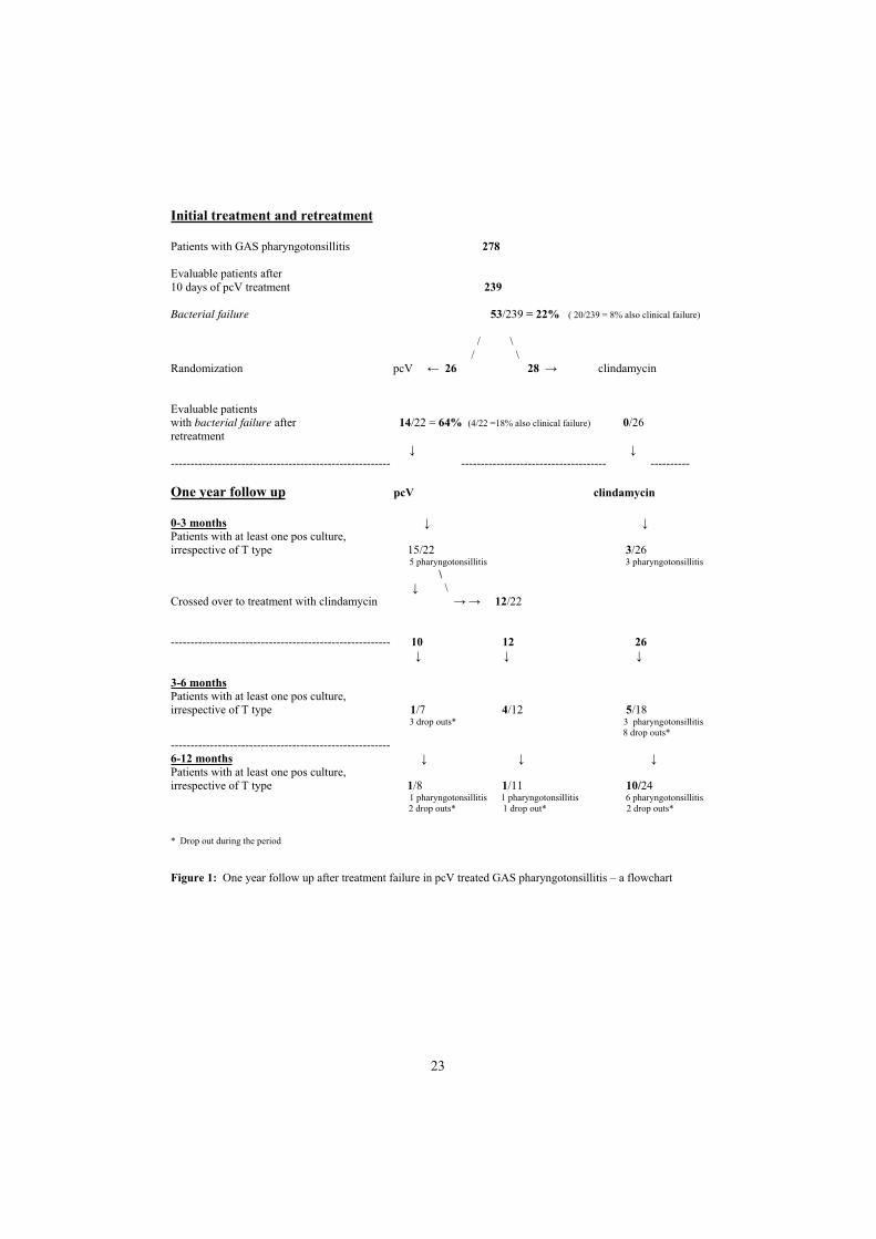
Initial treatment and retreatment
Patients with GAS pharyngotonsillitis 278
Evaluable patients after10 days of pcV treatment 239
Bacterial failure 53/239 = 22% ( 20/239 = 8% also clinical failure)
/ \ / \
Randomization pcV 26 28 clindamycin
Evaluable patientswith bacterial failure after 14/22 = 64% (4/22 =18% also clinical failure) 0/26retreatment
-------------------------------------------------------- ------------------------------------- ----------
One year follow up pcV clindamycin
0-3 monthsPatients with at least one pos culture,irrespective of T type 15/22 3/26
5 pharyngotonsillitis 3 pharyngotonsillitis
\ \
Crossed over to treatment with clindamycin 12/22
-------------------------------------------------------- 10 12 26
3-6 monthsPatients with at least one pos culture,irrespective of T type 1/7 4/12 5/18
3 drop outs* 3 pharyngotonsillitis 8 drop outs*
--------------------------------------------------------6-12 monthsPatients with at least one pos culture,irrespective of T type 1/8 1/11 10/24
1 pharyngotonsillitis 1 pharyngotonsillitis 6 pharyngotonsillitis 2 drop outs* 1 drop out* 2 drop outs*
* Drop out during the period
Figure 1: One year follow up after treatment failure in pcV treated GAS pharyngotonsillitis – a flowchart
23

An Attempt to Identify Penicillin Tolerant GAS (III)
Patients and Methods
Patients and bacterial isolates
GAS strains were selected from the previous study on patients with pharyngotonsillitis
(Orrling et al 1994). Samples were obtained before pcV therapy from patients who healed on
pcV therapy as well as from patients with subsequent failure. The distribution of the isolates
is shown in shown below.
GAS isolates tested for penicillin tolerancen
Before treatment: patients who healed 33Before treatment: patients with bacterial failure: 25After treatment *: patients with bacterial failure: 25After second treatment **:patients with bacterial failure: 7 90 * 13 patients with clinical failure ** 2 patients with clinical failure.
.
Three control strains, one group A representing non-tolerance, and one group A and one
group G, representing different levels of tolerance, were primarily selected from a total of
approximately 150 clinical isolates examined by the disc diffusion test (Slater & Greenwood
1983).
Four streptococcal strains, reported by others as belonging to group A and penicillin tolerant
(van Asselt & Mouton 1993; van Asselt et al 1995), and 16 own isolates from throat
specimens – 12 group G and 4 group C – were also investigated.
Bacteriological investigation
The MBC/MIC ratios were determined by a modified plate dilution method (Kamme &
Petersson 1993), and by broth dilution (Taylor et al. 1983). Survival rates were determined by
the plate screening method and by the time killing kinetic test.
Log phase as well as stationary phase cultures from our previous study were investigated with
the plate screening method. Log phase strains with a survival rate of 0.2-0.5% were subjected
to time killing test. All strains were T-typed. The person performing the in vitro tests of
clinical isolates was not informed of whether the various strains originated from cases of
failure or not.
24
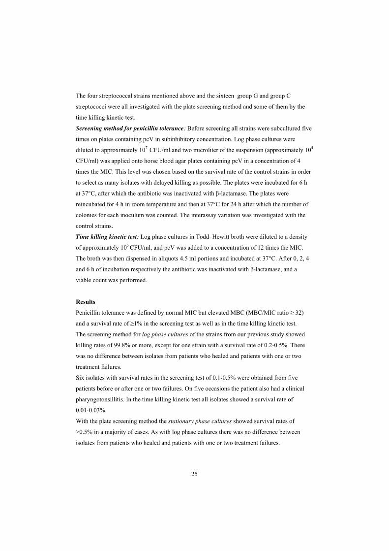
The four streptococcal strains mentioned above and the sixteen group G and group C
streptococci were all investigated with the plate screening method and some of them by the
time killing kinetic test.
Screening method for penicillin tolerance: Before screening all strains were subcultured five
times on plates containing pcV in subinhibitory concentration. Log phase cultures were
diluted to approximately 107 CFU/ml and two microliter of the suspension (approximately 104
CFU/ml) was applied onto horse blood agar plates containing pcV in a concentration of 4
times the MIC. This level was chosen based on the survival rate of the control strains in order
to select as many isolates with delayed killing as possible. The plates were incubated for 6 h
at 37°C, after which the antibiotic was inactivated with -lactamase. The plates were
reincubated for 4 h in room temperature and then at 37°C for 24 h after which the number of
colonies for each inoculum was counted. The interassay variation was investigated with the
control strains.
Time killing kinetic test: Log phase cultures in Todd–Hewitt broth were diluted to a density
of approximately 105 CFU/ml, and pcV was added to a concentration of 12 times the MIC.
The broth was then dispensed in aliquots 4.5 ml portions and incubated at 37°C. After 0, 2, 4
and 6 h of incubation respectively the antibiotic was inactivated with -lactamase, and a
viable count was performed.
Results
Penicillin tolerance was defined by normal MIC but elevated MBC (MBC/MIC ratio 32)
and a survival rate of 1% in the screening test as well as in the time killing kinetic test.
The screening method for log phase cultures of the strains from our previous study showed
killing rates of 99.8% or more, except for one strain with a survival rate of 0.2-0.5%. There
was no difference between isolates from patients who healed and patients with one or two
treatment failures.
Six isolates with survival rates in the screening test of 0.1-0.5% were obtained from five
patients before or after one or two failures. On five occasions the patient also had a clinical
pharyngotonsillitis. In the time killing kinetic test all isolates showed a survival rate of
0.01-0.03%.
With the plate screening method the stationary phase cultures showed survival rates of
>0.5% in a majority of cases. As with log phase cultures there was no difference between
isolates from patients who healed and patients with one or two treatment failures.
25

The 16 group C and G isolates all yielded a survival rate of >1% by the plate screening
method. Two group C isolates that were subjected to the time killing kinetic test yielded
survival rates of 2 and 5% respectively.
The four allegedly tolerant group A strains all turned out to be group G streptococci. They all
yielded >1% survival rates in the plate screening test. One of them, when examined in the
time killing kinetic test, showed a survival rate of approximately 5%.
26
Genetic Profiles in GAS Isolates from Failures and Nonfailures. An
Investigation Using AP-PCR Technique (IV)
Patients and Methods
Patients
Isolates from four patients, selected from the previous study (Orrling et al 1997) with one or
more bacterial treatment failures, were analyzed. They were compared with strains of the
same T-type isolated during the same time period from patients who healed on a single course
of pcV for pharyngotonsillitis and who lived in the same geographical area. All cultures
showed abundant growth.
Case 1: A 6-year –old boy was treated with a total of four pcV courses for growth of GAS of
T-type 4 (Figure 1 – paper IV). On three of the occasions, he displayed signs and symptoms
of pharyngotonsillitis. .
Case 2: A 2-year –old boy with pharyngotonsillitis and growth of GAS of T-type 12 was
treated with four pcV courses (Figure 1 – paper IV). The boy displayed no symptoms or signs
of pharyngotonsillitis at any of the five follow-up visits despite positive cultures.
Cases 3 and 4: A 28-year old mother and her 2-year-old son displayed clinical
pharyngotonsillitis and growth of GAS of T-type R28. They were both treated with pcV for
ten days (Figure 1 – paper IV). At the follow up visit the boy showed bacterial failure, but at
the following controls the cultures were negative. In contrast the mother was prescribed three
additional pcV courses because of repeated growth of the same T-type (twice) followed by a
positive culture for group G streptococci. At this point she was treated with trimethoprim-
sulfamethoxazole for urinary tract infection. After that the culture was still positive for group
G streptococci, but the patient was asymptomatic, and no further treatment was given.
The DNA profiles of the isolates from case 1 -4 were compared with profiles of isolates of the
same T-type respectively from nonfailures.
Bacteriological investigation
A total of 23 primers were initially tested, some of which produced no bands at all. Three
primers giving 2-6 strong bands and 3-8 weak bands were chosen. Figure2.
Arbitrarily primed polymerase chain reaction (AP-PCR) was carried out (Jackson &Cook
1985), and the product was electrophoresed in a 1.5% agarose gel. The DNA bands were
27

visualized under UV-light and photographed. The size of the PCR product was compared
using a DNA ladder. To check the reproducibility the samples were run twice.
Results
The reproducibility of the AP PCR technique was high. The method also demonstrated a high
discriminatory capacity. Among the three types recognized by the conventional T-typing, 11
different DNA profiles could be detected by AP-PCR. The strains of T-type 4, 12 and R28
segregated into 5, 3 and 3 AP-PCR profiles respectively.
Case 1 showed the same DNA profile in 5 of 6 isolates, which were all of T-type 4. The
aberrant isolate was obtained after the fourth pcV course. After the fifth pcV treatment the
original DNA profile reappeared. The same profile was found in 4 of the 7 T-type 4 strains
from non failures. The remaining three nonfailure strains were assigned to two different
clones.
The genetic profiles of all the isolates from case 2 were identical, and this clone was also
represented in 2 of the 7 nonfailure strains. The other five nonfailure strains of T-type 12
exhibited band patterns corresponding to four different clones.
Cases 3 and 4, mother and son, exhibited the same genetic profile in all five isolates,
classified as T-type R28. This profile was also found in 4 of the 6 nonfailure strains, whereas
2 of the 6 had different genetic profiles.
28
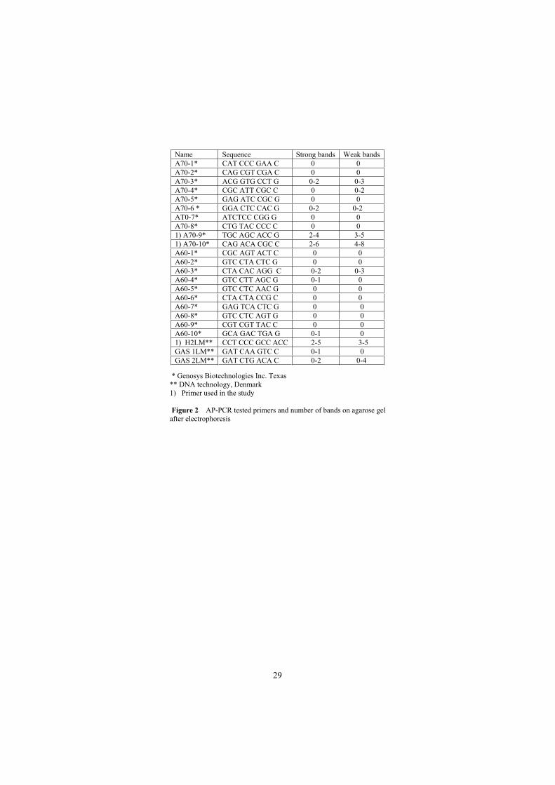
Name Sequence Strong bands Weak bandsA70-1* CAT CCC GAA C 0 0A70-2* CAG CGT CGA C 0 0A70-3* ACG GTG CCT G 0-2 0-3A70-4* CGC ATT CGC C 0 0-2A70-5* GAG ATC CGC G 0 0A70-6 * GGA CTC CAC G 0-2 0-2AT0-7* ATCTCC CGG G 0 0A70-8* CTG TAC CCC C 0 01) A70-9* TGC AGC ACC G 2-4 3-51) A70-10* CAG ACA CGC C 2-6 4-8A60-1* CGC AGT ACT C 0 0 A60-2* GTC CTA CTC G 0 0 A60-3* CTA CAC AGG C 0-2 0-3A60-4* GTC CTT AGC G 0-1 0 A60-5* GTC CTC AAC G 0 0 A60-6* CTA CTA CCG C 0 0 A60-7* GAG TCA CTC G 0 0A60-8* GTC CTC AGT G 0 0A60-9* CGT CGT TAC C 0 0A60-10* GCA GAC TGA G 0-1 01) H2LM** CCT CCC GCC ACC 2-5 3-5GAS 1LM** GAT CAA GTC C 0-1 0GAS 2LM** GAT CTG ACA C 0-2 0-4
* Genosys Biotechnologies Inc. Texas** DNA technology, Denmark1) Primer used in the study
Figure 2 AP-PCR tested primers and number of bands on agarose gel after electrophoresis
29
Penicillin V, Loracarbef and Clindamycin in TSF During and After
Treatment of GAS Pharyngotonsillitis (V)
Patients and Methods
Patients
35 consecutive patients attending a private ENT clinic with a history of no more than two
days of acute pharyngotonsillitis (11 males, 24 females) were included. Their age range was
20 – 77 years (median: 35, mean 38.0), and they all had a normal serum creatinin. All patients
manifested a positive rapid antigen test for GAS as well as a positive culture.
The study was approved by the Medical Ethics Committee of the University of Lund.
Treatment
Patients were randomly assigned to either pcV 12.5 mg/kg bodyweight b.i.d. (n=13),
clindamycin 300 mg t.i.d. (n=11) or loracarbef 200 mg b.i.d. (n=11), all for ten days, which
are the currently recommended dosages for GAS pharyngotonsillitis in Sweden.
Sampling
Sampling of serum, saliva and TSF was performed on four occasions: 1) On the day of
inclusion before start of therapy, in order to exclude any unspecific antibacterial activity.
2) 1.5 h after intake of drug at two randomized days during treatment. 3) On one occasion
within four days after end of therapy. No anaesthesia was used. Blood was drawn for C-
reactive protein and orosomucoid on the same occasions. Three sterile filter paper disks were
placed on the surface of the tonsils and three under the tongue. The disks were left in place for
one minute and were then immediately sealed in plastic tubes and kept at -80° until assayed.
The procedure was similar to that previously described (Strömberg et al. 1987; Stjernquist et
al. 1993). A venous blood sample was obtained at each visit.
Antibiotic assays
The concentration of the drugs in serum was determined by agar-well diffusion. For saliva
and TSF disk diffusion was used. Sterile powder of each drug with known potencies were
used for the preparation of 2-fold standard solutions, of which 10 µl was added to sterile paper
disks.
Sera, standard solutions and impregnated disks were assayed in duplicate. Zones of inhibition
were measured after overnight incubation at 37°C. For the disks with TSF and saliva
respectively the mean value of zones of inhibition for the three disks was given.
30

The detection limit for pcV was 0.03 mg/L, for loracarbef 0.12 mg/L, and for clindamycin
0.12 mg/L. MICs for susceptible GAS were 0.004-0.002, 0.06-0.25 and 0.03-0.12 mg/L
respectively (Kamme & Petersson 1993, Kataja et al 1999).
Results
The results were given in five periods: I = day 1-3, II = day 4-7, III = day 8-10, IV = day 11-
12, V = day 13 -14.
All included patients were examined three times within the first ten days, i.e. at inclusion and
in two periods during treatment. Three patients in the pcV group and two patients in the
loracarbef and clindamycin group respectively were examined twice in period I or II. The
results were shown as the mean value of the two measurements in respective period.
One patient was excluded from further examination owing to zones of inhibition around the
pre-treatment disks with TSF. Two patients in the pcV group, and one in the loracarbef group
did not show up following treatment.
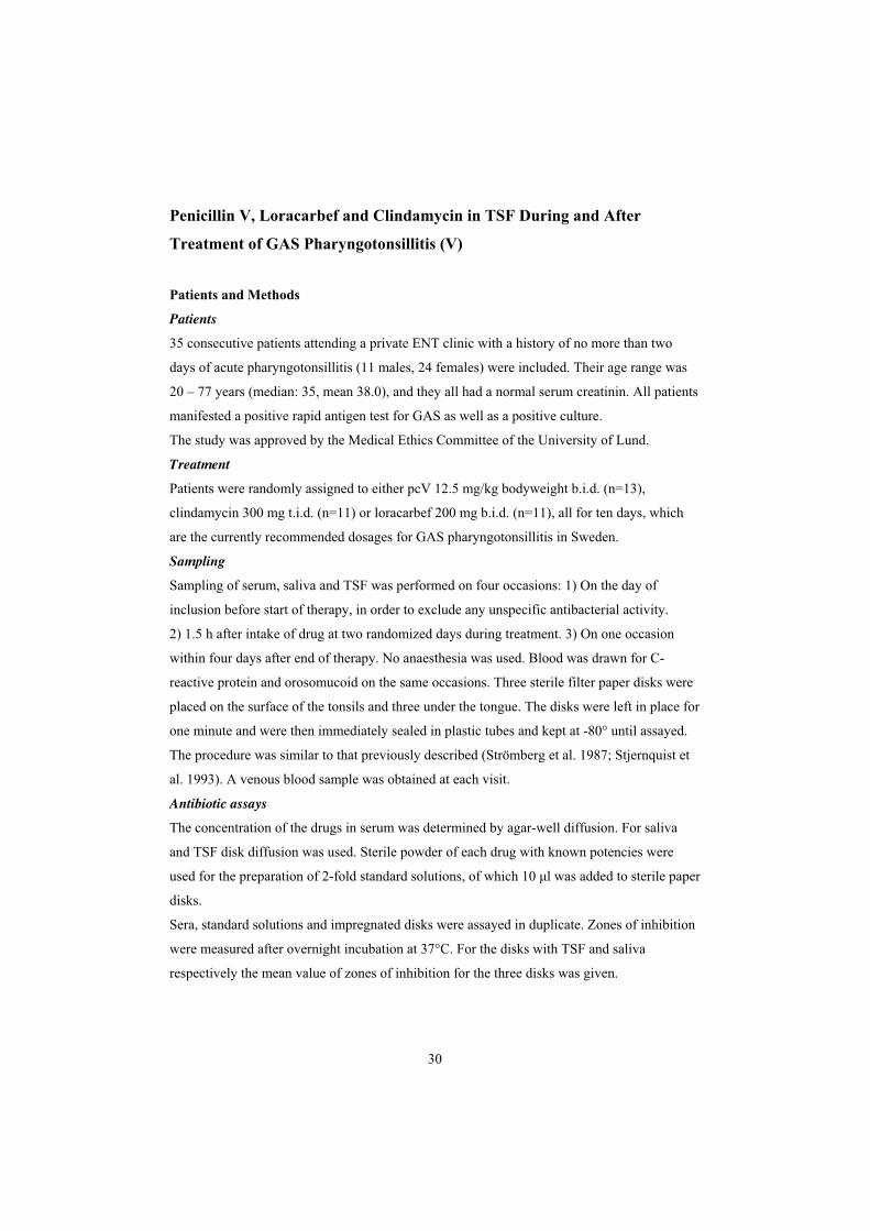
In order to find out the optimal time for sampling, two patients, one in the pcV and one in the
loracarbef group, were repeatedly examined at 30 min intervals after taking the drug. The pcV
concentration in the TSF showed a fast increase with a maximum after 90 min, followed by a
fast decrease. The concentration of loracarbef in TSF showed a similar pattern, although the
maximum concentration was reached 40 min later. (Figure 3)
Antibiotic in serum: Detectable serum levels of all drugs were obtained throughout the
treatment period. After end of therapy pcV and loracarbef did not reach detectable serum
levels. In period IV clindamycin reached measurable concentrations in each of four tested sera
at two days after end of therapy. On the following two days detectable levels of clindamycin
were not obtained in any of six tested serum samples.
31
Concentration in TSF after intake of drug
0
0,2
0,4
0,6
0,8
1
1,2
1,4
1,6
0 50 100 150 200
Minutes after intake
Co
nce
ntr
atio
n in
TS
F [µ
g/m
l]
Loracarbef
Penicillin
Figure 3
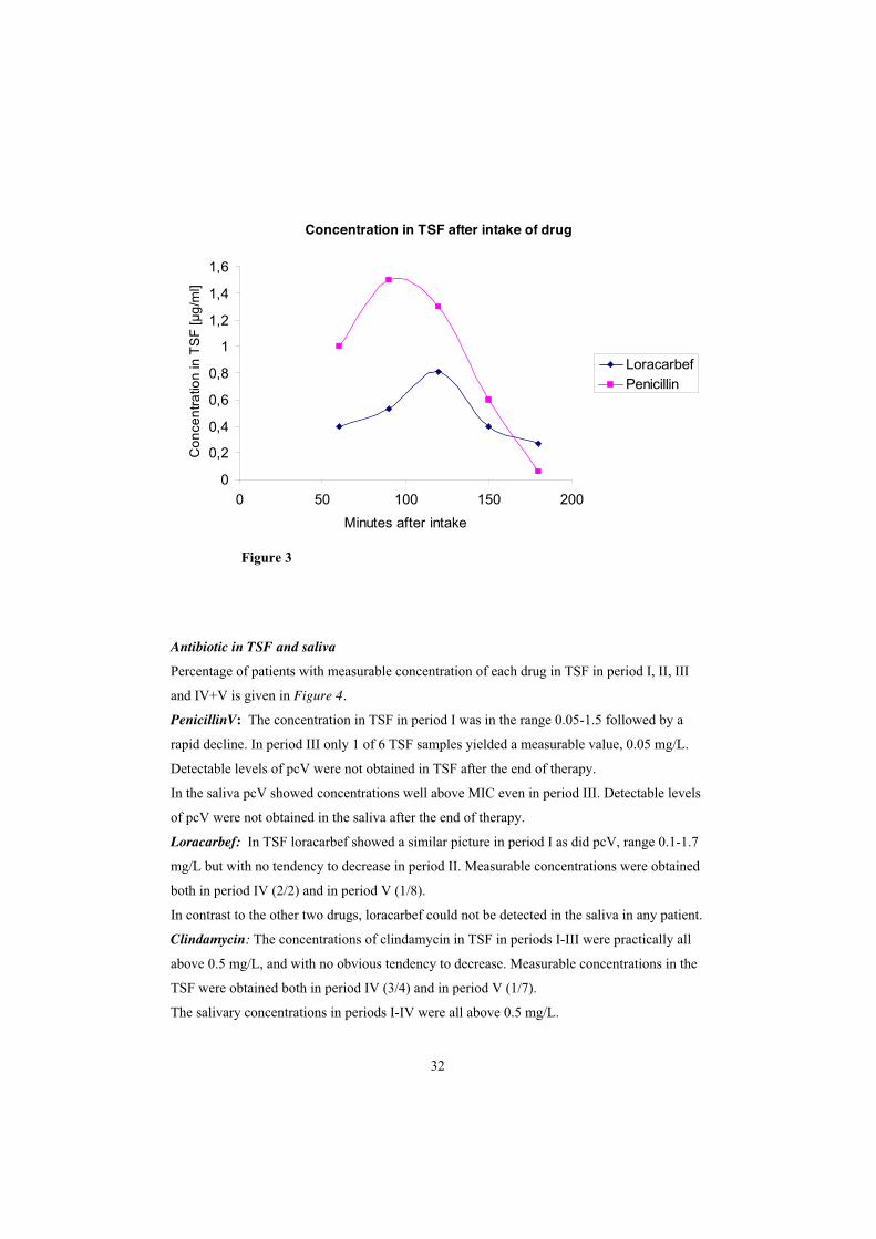
Antibiotic in TSF and saliva
Percentage of patients with measurable concentration of each drug in TSF in period I, II, III
and IV+V is given in Figure 4.
PenicillinV: The concentration in TSF in period I was in the range 0.05-1.5 followed by a
rapid decline. In period III only 1 of 6 TSF samples yielded a measurable value, 0.05 mg/L.
Detectable levels of pcV were not obtained in TSF after the end of therapy.
In the saliva pcV showed concentrations well above MIC even in period III. Detectable levels
of pcV were not obtained in the saliva after the end of therapy.
Loracarbef: In TSF loracarbef showed a similar picture in period I as did pcV, range 0.1-1.7
mg/L but with no tendency to decrease in period II. Measurable concentrations were obtained
both in period IV (2/2) and in period V (1/8).
In contrast to the other two drugs, loracarbef could not be detected in the saliva in any patient.
Clindamycin: The concentrations of clindamycin in TSF in periods I-III were practically all
above 0.5 mg/L, and with no obvious tendency to decrease. Measurable concentrations in the
TSF were obtained both in period IV (3/4) and in period V (1/7).
The salivary concentrations in periods I-IV were all above 0.5 mg/L.
32
Patients with measureable antibiotic concentration in TSF
0
10
20
30
40
50
60
70
80
90
100
I II III IV+V
Period
Perc
enta
ge
Clindamycin
Loracarbef
Penicillin V
Figure 4
CRP and orosomucoid
At day 1 CRP was elevated >30 mg/L in 12/35 patients and >50 mg/L in 7/35 patients. After
five days of treatment the median of CRP was below 10 mg/L.
Orosomucoid was elevated >1.17g/l in 13/35 patients and >1.5 g/l in 3/35 at inclusion. After
five days of treatment the median orosomucoid level was below 1.1 g/l.
There was no difference in the decrease of either CRP or orosomucoid between the three
groups.
33

GENERAL DISCUSSION
Treatment failure
The bacterial treatment failure rate in our study, 22% after the first and 64% after the second
course of pcV, was in accordance with other studies (Kaplan & Johnson 1988; Schwartz et al.
1981). In the clindamycin group, on the other hand, none of the patients exhibited failure.
Furthermore, in the following three month period, three patients in the clindamycin group had
clinical pharyngotonsillitis, but in all cases the GAS strain isolated was of a new T-type thus
representing reinfection. In the pcV group, however, five patients developed one or more
clinical pharyngotonsillitis episodes, in each case due to GAS of the same T-type as in the
primary episode. Thus clindamycin treatment apparently, by eradicating carriage of GAS,
interrupted the vicious circle of recurrent GAS pharyngotonsillitis among patients originally
treated with pcV. Accordingly none of the patients receiving clindamycin had to undergo
tonsillectomy.
The difference between the two groups was reduced later in the follow up period, when
positive cultures for GAS were seen in a few patients left in the pcV group. Owing to the
study design, however, “tonsillitis-prone” patients in the pcV group were switched to
clindamycin treatment in case of bacterial failure. The remaining pcV group, therefore was
selected and consisted of individuals with comparatively high resistance to GAS.
During the 12 month follow up period some patients in the clindamycin group manifested
failure after pcV treated primary GAS pharyngotonsillitis. Our findings partly disagree with
those obtained by Brook & Hirokawa (1985) where only one of 15 patients treated with
clindamycin manifested a single recurrence of pharyngotonsillitis during one year of follow
up. Jensen and Larsen (1991) found the frequency of episodes of acute pharyngotonsillitis to
be significantly reduced among patients treated with one course of clindamycin, as compared
to untreated controls, in a 12 month follow up period. Unfortunately in their study, diagnosis
of pharyngotonsillitis was based on anamnestic data rather than throat cultures.
Reinfection
Since close contacts, such as family members often harbour the same strain as the patient, a
distinction between failure and reinfection is not always possible. Efforts were made to
minimize the risk of including patients with reinfection from the environment in the study.
This was done by obtaining the follow-up throat culture as soon as four days after cessation of
34
therapy. In addition, family members with symptoms and a positive culture for GAS were
treated with pcV.
In contrast to the pcV group, there was no GAS pharyngotonsillitis caused by the original T-
type in the clindamycin group in the first three month follow-up period. Therefore our
findings strongly suggest that the recurrences after pcV treated GAS pharyngotonsillitis
mainly were due to bacterial treatment failure rather than reinfection.
Genetic profiles
When exploring the isolates with the AP-PCR technique, we found that isolates from four
patients with several failures exhibited the same genetic profile as the pre-treatment isolate
from each patient. A minor exception was an extra band found in one of the isolates. Since the
same profile as the original was found in the next isolate, this extra band was probably due to
two different bacterial populations of GAS at that time. Our findings minimize the probability
that the bacterial failures were due to infection with a new GAS strain and were in accordance
with Österlund and Engstrand (1995) and Bingen and colleagues (1992) who also found most
pre-treatment and post-treatment isolates to have the same genetic profile.
Similar genetic profiles were also found in pre-treatment isolates from patients in the same
area who healed on their first pcV course, as in isolates from patients with multiple failures.
Moreover, the isolates from a mother who had repeated clinical and bacterial failures showed
the same profile as those from her son who healed after a second pcV course. We were thus
unable to identify special strains accounting for treatment failures. Analogously Norgren and
colleagues (1992) found the same genetic profile of GAS of T-type 1 in patients with
bacteraemia as in asymptomatic carriers in the same family.
The findings suggest that host factors, such as local or systemic immunity to streptococci, or
local production of peptides with antibacterial properties (Nizet et al 2001; Bessen & Fishetti
1988) may significantly influence the outcome of treatment of GAS.
Penicillin tolerance
In our study, using log phase bacteria, only a minor proportion of strains showed survival
rates above 0.1 % after exposure to pcV, at four times the MIC. When these strains were
subjected to time killing kinetic test, however, they did not show any delayed killing. There
was no difference between isolates obtained before compared to after treatment, and results
were similar for isolates from healed cases and isolates from one or two treatment failures,
respectively.
35

Stationary phase cultures of GAS have been shown to yield a phenotypic response to -lactam
antibiotics affecting the killing rate rather than MIC (Kamme & Petersson 1993). This
property of many GAS strains might be due to the fact that non-replicating bacteria are not
sensitive to the inhibitory effect on cell wall synthesis accomplished by -lactam drugs. As
with log phase cultures however there was no difference in killing rate between isolates from
patients who healed and from those with one or two treatment failures.
One single GAS strain, out of 150 tested clinical isolates, showed delayed killing, with a
survival rate close to tolerance as defined above. This strain, however, showed a MBC/MIC
ratio = 1 in the modified plate dilution test as well as in the broth dilution test. This shows the
difficulties in investigating penicillin tolerance, and emphasizes the need for appropriate
confirmation of positive findings.
Our failure to detect tolerance in GAS was in accordance with Ciftci and colleagues (2002)
who could not identify penicillin tolerant strains among 263 isolates from children with GAS
pharyngotonsillitis. Thus penicillin tolerance seems to be of little or no significance in
accounting for failures after pcV treated GAS pharyngotonsillitis. Furthermore, penicillin
tolerance or resistance, in this species may not be compatible with a virulent phenotype
(Gutman & Tomasz 1982).
Somewhat unexpectedly, our results suggested that penicillin tolerance may be rather
common in group C and G streptococci. However, clinical experience does not indicate, that
penicillin tolerance in these species should represent a therapeutic problem in the context of
pharyngotonsillitis.
Antibiotic concentrations in TSF
The high initial concentrations of pcV in TSF, followed by a rapid decline and measurable
level in only one late sample, was in contrast to results for clindamycin and loracarbef which
showed detectable concentrations in TSF even after end of therapy. Hence early, high
concentrations of the three investigated drugs in TSF, as here found, may account for prompt
clinical recovery. All patients had satisfactory serum levels of antibiotic during treatment, so
the rapid decline of pcV in TSF was not due to low compliance or low resorption of the drug.
The post treatment persistence in TSF of loracarbef and clindamycin may conceivably be
explained by mobilized tissue depots. Levels of all three antibiotics in TSF and saliva were
essentially unrelated, and loracarbef did not reach measurable levels in saliva, thus in contrast
to TSF.
36

The finding of high initial concentrations of pcV in TSF, followed by a rapid decline, was in
agreement with a previous study by Stjernquist – Desatnik and colleagues (1993). These
authors found measurable concentration of pcV in TSF in all patients on the first day of
treatment of GAS pharyngotonsillitis, but in only one of nine patients at the end of treatment.
Furthermore, pcV could not be detected in TSF in healthy individuals. In contrast, Strömberg
and co-workers (1987) found the concentrations in TSF of both pcV and cefadroxil to be
higher than those in tonsillar tissue from patients tonsillectomized because of recurrent
tonsillitis. However, these patients manifested chronic inflammation in the tonsils implying
plasma leakage and exudation of fluid through the epithelium. Furthermore, sampling was
made under general anaesthesia which might have affected the tonsillar blood flow. Our study
comprised patients with ongoing acute GAS pharyngotonsillitis and no local anaesthesia was
used during sampling.
In acute pharyngotonsillitis, GAS are predominantly localized in the secretion on the tonsillar
surface and in the crypts (Ebenfelt et al 1998). The concentration of antibiotics in TSF thus
would most probably be crucial in treatment of GAS pharyngotonsillitis.
CRP and orosomucoid
We also evaluated CRP and orosomucoid as indicators on the degree of inflammation in the
throat and a possible linkage between serum levels of these markers and drug concentrations
in TSF. However, no such correlation was found. Only in a minor proportion of the patients
CRP and orosomucoid showed serum levels indicating a bacterial infection, and there were no
differences between the three groups receiving different antibiotics. Our findings were in
accordance with other studies (Stjernquist et al 1987; Putto et al 1986; Sun et al 2002) and
provided no support for the use of CRP or orosomucoid as tools to separate bacterial from
viral pharyngotonsillitis.
Penicillin for ten days
In pcV treated acute otitis media Ingvarsson and colleagues (1980) recorded a rapid decline in
pcV concentration in the middle ear as the inflammation abated. This resulted in a change in
Swedish treatment praxis from 10 to 5 days of pcV in primary acute otitis media. However, in
pcV treated GAS pharyngotonsillitis, despite fast clinical recovery, the importance of no less
than 10 days therapy, in order to keep the failure rate on acceptable level, has been reported
(Schwartz et al 1981; Strömberg et al 1988; Zwart et al 2000).
37
The need for a 10 days course of pcV in the treatment of GAS pharyngotonsillitis then seems
paradoxical, since the drug concentration in TSF later in therapy often does not reach
detectable levels. A possible reason might be that although the log phase bacteria are killed
early in treatment, GAS in lag phase, as well as intracellulary GAS, are not affected by pcV.
However, the gradual passage to log phase, as well as the externalization and start of
replication of intracellulary GAS, will successively render additional bacteria susceptible to
the drug. PcV is probably transferred to the tonsillar surface by inflammatory exudation,
hence later in therapy at lower levels. The continued, local inflammation caused by the
replicating bacteria may still result in exudation of pcV in sufficient amount to kill the
bacteria. A course long enough to cover the shift from lag phase to log phase for most
bacterial cells, as well as the start of growth of most internalized bacteria, thus might be
important for efficacy of pcV in GAS pharyngotonsillitis. It seems likely that an extended
course, i.e. for more than the arbitrarily chosen 10 days, although it would result in
compliance problems, could result in further reduced failure rate in GAS pharyngotonsillitis.
Loracarbef
In a study of loracarbef versus pcV in recurrent GAS pharyngotonsillitis Roos and Larsson
(1997) found significantly higher bacterial eradication rate among the loracarbef treated
patients (90% versus 66%). The better eradication rates by the cephalosporins, as compared
to pcV, in primary and recurrent GAS pharyngotonsillitis is usually ascribed to a higher
stability of cephalosporins to -lactamases as well as lesser impact on the interfering resident
-streptococci. It seems reasonable that maintained high concentrations of loracarbef, and
perhaps also other cephalosporins, in TSF is another important factor for their lower failure
rates as compared to pcV.
Clindamycin
Since -streptococci are susceptible to clindamycin, bacterial interference does not seem to
contribute to the low failure rate after clindamycin treated GAS pharyngotonsillitis. However,
clindamycin enters and accumulates in human cells. The longstanding concentrations in TSF,
the insusceptibility to -lactamase and the effect on extra- as well as intracellular GAS,
whether resting or actively growing, are all properties that might be of crucial importance for
the superior capacity of clindamycin to eradicate GAS in recurrent pharyngotonsillitis.
38

Conclusion
In primary GAS pharyngotonsillitis there is still support for use of pcV, due to narrow
spectrum and absence of penicillin resistance. However, in treatment failure use of
clindamycin, due to proven ability to eradicate GAS from the throat, can be justified. This
drug may also interrupt the vicious circle of repeated recurrences of GAS pharyngotonsillitis
often leading to tonsillectomy. However, since clindamycin also is of high value in the
treatment of life threatening streptococcal diseases such as streptococcal toxic chock
syndrome and necrotizing fasciitis, it is imperative, in order to avoid development of
resistance, that the drug is used on strict indications.
39
CONCLUSIONS
1 In patients with GAS pharyngotonsillitis who failed on pcV treatment,
clindamycin could prevent further failures for at least the following three
months.
2 Reinfection is of less importance than bacterial treatment failure as an
explanation for recurrent GAS pharyngotonsillitis.
3 Penicillin tolerant GAS could not be identified, and we conclude that penicillin
tolerance seems to be of no significance in failures of pcV treated GAS
pharyngotonsillitis.
4 Failure and nonfailure strains exhibited similar DNA profiles, indicating that
failures are associated with host rather than bacterial factors.
5 The longstanding concentrations of both loracarbef and clindamycin in TSF may
contribute to their capacity to eradicate GAS in patients who failed on pcV
treatment of GAS pharyngotonsillitis.
6 CRP is of no diagnostic value in GAS pharyngotonsillitis.
40

SUMMARY
In acute pharyngotonsillitis group A streptococci (GAS) is the etiological agent in 30-50% of
cases. GAS are virulent human pathogens, and may cause both suppurative and
nonsuppurative complications, and sometimes life threatening diseases such as “streptococcal
toxic shock syndrome” and necrotising fasciitis. GAS pharyngotonsillitis results in a high
degree of absence from day care, school and work, and it is agreed that antibiotic treatment is
indicated in these cases. Phenoxymethylpenicillin (pcV) is the drug of choice in Sweden.
Although penicillin resistance is not recorded in GAS, the failure rate is as high as 5-25%. A
second course of pcV treatment is followed by still higher failure rates, in some cases
necessitating tonsillectomy.
Several factors possibly contributing to the recurrences have been mentioned: low
compliance, reinfection from the environment, eradication of -streptococci with inhibitory
effect on GAS, increase in -lactamase-producing bacteria inactivating the drug, penicillin
tolerant streptococci, low antibiotic concentration at site of infection and finally intracellular
GAS surviving therapy.
Object
The aim of the present studies was:
1 To investigate the short- and long-term efficacy of pcV versus clindamycin in
patients with GAS pharyngotonsillitis who failed on pcV treatment.
2 To examine failure and non-failure strains considering so called penicillin-
tolerance.
3 To compare the DNA-profiles of failure and non-failure strains.
4 To evaluate the kinetics of pcV, loracarbef and clindamycin in tonsillar surface
fluid in order to find a possible correlate to their clinical efficacy.
5 To investigate the diagnostic value of CRP and orosomucoid in GAS
pharyngotonsillitis.
41

Material and methods
We defined bacterial failure as presence of GAS of the same T-type as in pre treatment
samples within two weeks after completing therapy.
239 patients with GAS pharyngotonsillitis were treated with pcV for ten days. At examination
4-6 days after therapy, 53 patients still harboured GAS of the same T-type as in the pre-
treatment culture. These 53 patients were randomized to treatment with either pcV or
clindamycin and were then followed for one year with throat culture every third month and in
case of pharyngotonsillitis. To investigate the role of penicillin tolerance, failure and non-
failure strains were screened for tolerance. Isolates with a high survival rate were subjected to
time killing tests. Using arbitrarily primed polymerase chain reaction (AP-PCR), the DNA-
profiles of failure and non-failure strains were compared. Three different antibiotics - pcV,
loracarbef and clindamycin - were investigated regarding concentration in tonsillar surface
fluid (TSF) during and after ten days treatment of GAS pharyngotonsillitis and CRP and
orosomucoid were analyzed throughout the investigation period.
Results
In the pcV group 64% yielded GAS in the throat culture, compared to 0% in the clindamycin
group. In the first three months 68% in the pcV group yielded one or more positive cultures
for GAS of the same T-type compared to 0% in the clindamycin group. However, for the
remaining investigation period the difference between the groups was reduced.
No penicillin tolerant strains could be identified. The strains were of three different T-types,
and using AP-PCR technique eleven different clones were identified. The same clones were
found in both failures and non-failures.
PcV was found in TSF during the first three day period of treatment, after which the
concentrations declined rapidly. This was in contrast to loracarbef and clindamycin, both of
which showed longstanding concentration in TSF, with measurable values throughout, and
even after, therapy.
Neither CRP nor orosomucoid was of any significance as indicators of GAS as a cause of
pharyngotonsillitis.
42
Conclusions
1 Treatment with clindamycin could prevent further treatment failures for at least
the following three months in patients with GAS pharyngotonsillitis who failed
on pcV treatment.
2 Penicillin tolerance seems to be of no significance in failures of pcV treated
GAS pharyngotonsillitis.
3 Failure and nonfailure strains exhibited similar DNA profiles, indicating that
failures are associated with host rather than bacterial factors.
4 The longstanding concentration of both loracarbef and clindamycin in TSF may
contribute to their capacity to eradicate GAS in patients who failed on pcV
treatment of GAS pharyngotonsillitis.
5 CRP and orosomucoid is of no diagnostic value in GAS pharyngotonsillitis.
43
SVENSK SAMMANFATTNING
BAKGRUND
Akut faryngotonsillit orsakas till ca 40 % av betahaemolytiska grupp A streptokocker (GAS)
och till ca 10% av andra bakterier. Ca 30% av fallen är virusutlösta och i ca 20% är genesen
okänd. GAS är mycket virulenta humanpatogener som förutom svalg- och hudinfektioner
även kan förorsaka livshotande tillstånd som ”streptococcal toxic shock syndrome” och
nekrotiserande fasciit. Tidigare fruktade, men i Sverige numera sällsynta komplikationer är
reumatisk feber och glomerulonefrit.
GAS faryngotonsillit medför hög arbets- och skolfrånvaro. Trots att GAS alltid är känsliga för
penicillinV (pcV), så återfinns GAS i svalget hos 15-25% av patienterna ett par veckor efter
avslutad behandling. Återinsjuknande i GAS faryngotonsillit sker i 5-10% av fallen, och efter
ytterligare pcV kurer stiger återfallsfrekvensen markant. Upprepade recidiv leder ibland till
tonsillektomi.
Som orsak till den höga recidivfrekvensen har bland annat följande faktorer föreslagits:
1. Bristande compliance, dvs. icke fullföljd behandling.
2. Reinfektion från omgivningen,
3. Utradering av normalt skyddande -streptokocker under pcV behandlingen,
4. Tillväxt av penicillinresistenta -laktamasproducerande bakterier med åtföljande
inaktivering av pcV.
5. Penicillintolerans, dvs. fördröjd avdödning vid exponering för pcV.
6. Dålig penetration av penicillin till infektionsfocus, dvs. tonsillyta och kryptor.
7. Intracellulärt belägna streptokocker som inte nås av penicillinet.
MÅLSÄTTNING
Målsättningen med studierna har varit
1 Att på såväl kort som lång sikt jämföra effekten av pcV respektive klindamycin
vid behandling av patienter med bakteriell terapisvikt efter pcV behandling av
GAS faryngotonsillit.
44

2 Att undersöka GAS stammar från såväl patienter som blev bakteriefria efter pcV
behandling som från patienter med bakteriell terapisvikt avseende förekomst av
penicillintolerans och eventuell skillnad i DNA-profil.
3 Att undersöka huruvida koncentrationen av pcV, loracarbef och klindamycin i
sekret på tonsillytan (TSF) under och efter behandling, kan korreleras till deras
kliniska effekt på GAS faryngotonsillit.
4 Att utvärdera den kliniska signifikansen av CRP och orosomucoid vid GAS-
faryngotonsillit.
MATERIAL och METODER
I studierna är bakteriell terapisvikt definierad som förekomst i svalget av GAS av den
ursprungliga T-typen inom två veckor efter avslutad behandling.
Arbete I och II
239 patienter med faryngotonsillit och positiv snabbtest för GAS behandlades med pcV i tio
dagar. Positiv svalgodling verifierade snabbtestfyndet. Kontrollodling utfördes 4-6 dagar efter
avslutad behandling Patienter med bakteriell terapisvikt randomiserades därefter till
behandling antingen med penicillin eller klindamycin i tio dagar. Kontroll med snabbtest och
svalgodling utfördes åter ca fyra dagar efter avslutad behandling.
Patienterna följdes därefter under ett år. Svalgodling togs var tredje månad och dessutom vid
faryngotonsillit.
T-typning och kvantitativ odling utfördes genomgående. Fynd av GAS i svalgodling
föranledde behandling med pcV, och vid bakteriell svikt gavs behandling enligt tidigare
randomisering.
Arbete III
GAS stammar före behandling från patienter som sedan blev streptokockfria (n=33) och från
patienter med kommande terapisvikt (n=25), samt stammar från odling efter första (n=25) och
andra (n=7) terapisvikten undersöktes avseende förekomsten av penicillintolerans. Kulturer i
såväl stationär som i log-fas undersöktes.
Lägsta inhiberande (MIC) och lägsta baktericida (MBC) koncentrationen av pcV bestämdes.
Tolerans definierades som en MBC/MIC kvot 32 och en överlevnad på 1 % i screening
test och i ”time killing kinetic test”. Stammarna screenades med en modifierad
45
plattspädningsmetod där de under 6 timmar exponerades för pcV i en koncentration av 4 x
MIC. Isolat med hög överlevnad undersöktes med s.k. ”time killing test”.
Arbete IV
Med arbitrarily primed polymerase chain reaction (AP-PCR) undersöktes DNA-profilen hos
GAS stammar från fyra patienter med upprepade recidiv och jämfördes med profilen hos
stammar från patienter som blev streptokockfria efter en pcV kur. Odlingarna togs under
samma tidsperiod och i samma geografiska område.
Arbete V
Trettiofem konsekutiva patienter med faryngotonsillit och positivt snabbtest för GAS
randomiserades till behandling med antingen pcV, klindamycin eller loracarbef. Positiv
svalgodling verifierade snabbtestfyndet. Prov på antibiotikakoncentrationen i serum,
tonsillsekret och saliv togs vid två randomiserade tillfällen under pågående behandling
samt 1-3 dagar efter avslutad behandling. Prov på tonsillsekret och saliv togs med
filterpapperslappar som placerades på tonsillytan och under tungan. CRP/s och
orosomucoid/s kontrollerades vid varje besök.
RESULTAT
Arbete I o II
Totalt 53, dvs 22 % av de 239 patienterna hade positiv svalgodling för GAS vid kontroll fyra
dagar efter avslutad behandling. 8% hade dessutom faryngotonsillit, medan 14% var
symptomfria . 43 patienter hade riklig bakterieväxt i odlingen. 25 patienter randomiserades till
pcV - och 28 till klindamycinbehandling. .
Bland patienter med terapisvikt kvarstod 48 evaluerbara fall. Av dessa uppvisade 14/22
(64 %) i penicillingruppen ånyo terapisvikt jämfört med 0/26 (0 %) i klindamycingruppen
(P<0.001).
Under den följande tremånadersperioden förekom en eller flera positiva odlingar med samma
T-typ hos 15 patienter i pcV gruppen, fem av dessa hade klinisk faryngotonsillit.
I klindamycingruppen erhölls under första tremånadsperioden ingen positiv odling med
samma T-typ, men tre patienter hade faryngotonsillit med GAS av annan T-typ.
46

Upprepade recidiv under den första tremånadersperioden föranledde att 12 patienter i
penicillingruppen korsades över till behandling med klindamycin vid terapisvikt, och i den
gruppen förekom i fortsättningen fem positiva odlingar.
Under den resterande uppföljningstiden (3-12 månader) förekom positiva odlingar och klinisk
faryngotonsillit både i den starkt decimerade pcV-gruppen och i klindamycingruppen - men
utan signifikant skillnad. Samtliga odlingar utom en visade riklig eller måttlig växt.
Arbete III
Ingen penicillintolerans kunde konstateras hos log-fas bakterier, medan stammar i stationär
fas uppvisade ett fördröjt avdödande. Ingen skillnad i detta avseende förelåg mellan isolat från
de patienter som läkte på sin första pcV behandling och de som sviktade en eller två gånger.
Arbete IV
Stammarna tillhörde tre T-typer. Med AP-PCR teknik kunde elva olika genetiska profiler
identifieras. Samma kloner fanns hos patienter som läkte på sin första pcV behandling och hos
dem som sviktade en eller flera gånger. Vid bakteriell svikt sågs genomgående samma
genetiska profil hos isolaten från respektive patient.
Arbete V
Under behandlingsdag 1-3 hade alla testade patienter i klindamycin och loracarbefgruppen
samt 7 av 8 patienter i pcV-gruppen antibiotikakoncentrationer över MIC i TSF.
Under resterande behandlingstid sjönk koncentrationen av pcV snabbt, i kontrast till de båda
andra preparaten.
Efter avslutad behandling påvisades mätbara antibiotikakoncentrationer i TSF hos 4 av 11
patienter i klindamycingruppen och hos 3 av10 patienter i loracarbefgruppen, men inte hos
någon patient i pcV-gruppen. Såväl pcV som klindamycin påvisades i saliven i
koncentrationer över MIC under hela, och vad gäller klindamycin även efter, behandlingen.
Detta i kontrast till loracarbef som ej kunde påvisas i saliv.
Initialt sågs CRP värden >50 hos 7/35 (20%) och orosomucoid >1.17 g/l hos 13/35 (37%) av
patienterna.
47
SLUTSATSER
1 Hos patienter med terapisvikt efter pcV behandling av GAS faryngotonsillit
kunde behandling med klindamycin bryta den onda cirkeln med upprepade
insjuknanden och ge ett skydd mot förnyad terapisvikt under minst tre månader.
2 Penicillintolerans var inte en faktor av betydelse vid terapisvikt efter pcV
behandlad GAS faryngotonsillit.
3 Värdrelaterade faktorer var troligen viktigare vid terapisvikt än
bakterierelaterade, eftersom samma DNA profil sågs hos GAS stammar från
patienter som läkte som från patienter med bakteriell terapisvikt efter pcV
behandling.
4 Tillräcklig koncentration i TSF av såväl loracarbef som av klindamycin under,
och även efter avslutad, behandling bidrog sannolikt till att preparaten har bättre
förmåga än penicillin att utplåna GAS från svalget.
5 CRP och orosomucoid var inte användbara som diagnostiska hjälpmedel vid
GAS tonsillit.
48

More than fifteen years have passed from the first study to the complete thesis and I would like to express my gratitude to all colleagues and co-workers who supported and encouraged me during this long journey. Special thanks goes to:
Anna Stjernquist-Desatnik –My tutor and enthusiastic supervisor. It all began a sunny day in Helsinki in the late eighties, when you, Anna, asked me to enrol patients from my private practice for a study of GAS pharyngotonsillitis. For me this was the start of a most stimulating work which has broadened my views and introduced me to the fascinating field of microbiological science. Being a private practitioner it has been a great favour to get the opportunity to do clinical research in collaboration with the ENT clinic in Lund and your supervision has been superb. I am impressed with the purposeful resolution with which you have guided me all the way to a thesis. Looking back I now have a strong feeling, although the first years I did not realize it, that from the very start you aimed at my disputation. Thank you Anna!
Professor emeritus Carl-Magus Eneroth for your constant positive and generous attitude towards the colleague from Eslöv . ”Arne - you are part of the clinic” you used to say. And for your enthusiastic attitude to my research. You never wondered if I should one day defend a thesis, you just asked “when?”.
Claes Schalén, my co-tutor and Carl Kamme for fruitful cooperation and for valuable advice and constructive criticism during the work with my thesis.
Åsa Melhus and the late Eva Karlsson at the Microbiological Department in Malmö for inspiring collaboration regading the PCR paper.
Evy Malm, Marianne Hartell and Solveig Göransson – the staff at my private practice in Eslöv.
Barbro Kahl for invaluable assistance at the Microbiological Laboratory.
Anita Groth - in times of doubt your sparkling enthusiasm put me back on the right track again. Five minutes in your company have always been enough to make me see that writing my thesis was a “must”.
Professor Karin Prellner - You read my papers and told me that it was OK to go on with a thesis. Thanks for your encouragement and for constructive criticism during writing.
Professor Johan Wennerberg for the undisturbed room at my disposal on the ENT-clinic – it was a prerequisite for my work.
Per-Anders Fransson for help with the figures.
Karin Brundell-Freij for help with the statistics.
49
ACKNOWLEDGEMENTS
Birgitta Nilsson and Marita Fryksén for expert secretarial assistance.
My colleagues Olof Kalm, Peter Groth, Gunnar Svensson, Mikael Karlberg and Morgan Andersson for taking care of my patients at “Specialisthuset i Eslöv” while I was working on my dissertation.
Finally and most important Gunilla, my wife and best friend, for your constant support, for your endless patience during the months with a thesis writing husband and for your encouragement when I most needed it.
50
My sister Eva Stenquist- Orrling who, via international contacts (Copenhagen), finally traced the picture.
1. Banck G, Nyman M. Tonsillitis and rash associated with Corynebacterium
haemolyticum. J Infect Dis 154:1037-40, 1986.
2. Benjamin JT, Perriello VA Jr. Pharyngitis due to group C hemolytic streptococci
in children. J Pediatr 89:254-6, 1976.
3. Bessen D, Fischetti VA. Passive acquired mucosal immunity to group A
streptococci by secretory immunoglobulin A. J Exp Med 167:1945-50, 1988.
4. Bingen E, Denamur E, Lambert-Zechovsky N, Braimi N, El Lakany M, Elion J.
DNA restriction fragment length polymorphism differentiates recurrence from
relapse in treatment failures of Streptococcus pyogenes pharyngitis. J Med
Microbiol 37:162-4, 1992.
5. Bisno AL. Group A streptococcal infections and acute rheumatic fever. N Engl J
Med 325:783-93, 1991.
6. Bisno AL, Brito MO, Collins CM. Molecular basis of group A streptococcal
virulence. Lancet Infect Dis 3:191-200, 2003.
7. Brook I. The role of -lactamase-producing bacteria in the persistence of
streptococcal tonsillar infection. Rev Infect Dis 6:601-7, 1984a.
8. Brook I. -lactamase-producing bacteria recovered after clinical failures with
various penicillin therapy. Arch Otolaryngol 110:228-31, 1984b.
9. Brook I. Role of -lactamase-producing bacteria in the failure of penicillin to
eradicate group A streptococci. Pediatr Infect Dis 4:491-5, 1985a.
10. Brook I, Hirokawa R. Treatment of patients with a history of recurrent tonsillitis
due to group A -hemolytic streptococci. A prospective randomized study
comparing penicillin, erythromycin, and clindamycin. Clin Pediatr (Phila) 24:331-
6, 1985b.
11. Carlsson F, Sandin C, Lindahl G. Human fibrinogen bound to Streptococcus
pyogenes M protein inhibits complement deposition via the classical pathway. Mol
Microbiol 56:28-39, 2005.
12. Ciftci E, Dogru U, Guriz H, Aysev AD, Ince E. Penicillin tolerance in group A -
haemolytic streptococci isolated from throat cultures of children with
tonsillopharyngitis. Mikrobiyol Bul 36:147-52, 2002.
13. Cohen R, Reinert P. DeLa Rocque F, Levy C, Boucherat M, Robert M et al.
Comparison of 2 dosages of azithromycin for 3 d vs penicillin V for 10 d in acute
group A streptococcal tonsillopharyngitis,. Pediatr Infect Dis J 21:297-303, 2002.
51
REFERENCES
14. Cone LA, Woodard DR, Schlievert PM, Tomory GS. Clinical and bacteriologic
observations of a toxic shock-like syndrome due to Streptococcus pyogenes. N
Engl J Med 317:146-9, 1987.
15. Dagnelie CF, van der Graaf Y, De Melker RA. Do patients with sore throat benefit
from penicillin? A randomized double-blind placebo-controlled clinical trial with
penicillin V in general practice. Br J Gen Pract 46:589-93, 1996.
16. Davis HD, McGeer A, Schwartz B, Green K, Cann D, Simor AE, Low DE.
Invasive group A streptococcal infections in Ontario, Canada. Ontario Group A
streptococcal Study Group. N Engl J Med 335:547-54, 1996.
17. Del Mar CB, Glasziou PP, Spinks AB. Antibiotics for sore throat. Cochrane
Database Syst Rev (4):CD000023, 2000.
18. De Meyere M, Mervielde Y, Verschraegen G, Bogaert M. Effect of penicillin on
the clinical course of streptococcal pharyngitis in general practice. Eur J Clin
Pharmacol 43:581-5, 1992.
19. Ebenfelt A, Ericson LE, Lundberg C. Acute pharyngotonsillitis is an infection
restricted to the crypt and surface secretion. Acta Otolaryngol (Stockh) 118:264-
71, 1998.
20. Engelgau MM, Woernle CH, Schwartz B, Vance NJ, Horan JM. Invasive group A
streptococcus carriage in a child care centre after a fatal case. Arch Dis Child
71:318-22, 1994.
21. Eriksson BK, Andersson J, Holm SE, Norgren M. Epidemiological and clinical
aspects of invasive group A streptococcal infections and the streptococcal toxic
shock syndrome. Clin Infect Dis 27:1428-36, 1998.
22. Facklam RF, Martin DR, Lovgren M, Johnson DR, Efstratiou A, Thompson TA,
Gowan S, Kriz P, Tyrrell GJ, Kaplan E, Beall B. Extension of the Lancefield
classification for group A streptococci by addition of 22 new M protein gene
sequence types from clinical isolates: emm103 to emm124. Clin Infect Dis 34:28-
38, 2002.
23. Falck, G, Kjellander J. Outbreak of group A streptococcal infection in a day-care
center. Pediatr Infect Dis J 11:914-9, 1992.
24. Falck G, Holm SE, Kjellander J, Norgren M, Schwan Å. The role of household
contacs in the transmission of group A streptococci. Scand J Infect Dis 29:239-44,
1997.
52

25. Falck G, Grahn-Håkansson, E, Holm SE, Roos K, Lagergren L. Tolerance and
efficacy of interfering -streptococci in recurrence of streptococcal
pharyngotonsillitis: a placebo-controlled study. Acta Otolaryngol (Stockh)
119:944-8, 1999.
26. Fischetti VA. Streptococcal M protein: molecular design and biological behaviour.
Clin Microbiol Rev 2:285-314, 1989.
27. Flottorp S, Oxman AD, Cooper JG, Hjortdahl P, Sandberg S, Vorland LH.
Guidelines for diagnosis and treatment of sore throat. Tidskr Nor Laegeforen
10:1754-60, 2000.
28. Fontaine MC, Lee JJ, Kehoe MA. Combined contributions of streptolysin O and
streptolysin S to the virulence of serotype M5 Streptococcus pyogenes strain
Manfredo. Infect Immun 71:3857-65, 2003.
29. Gerber MA, Randolph MF, Chanatry J, Wright LL, De Meo K, Kaplan EL. Five
vs ten days of penicillin V therapy for streptococcal pharyngitis. Am J Dis Child
141:224-7, 1987.
30. Gerber MA, Tanz RR, Kabat W, Bell GL, Siddiqui B, Lerer TJ, Lepow ML,
Kaplan EL, Shulman ST. Potential mechanisms for failure to eradicate group A
streptococci from the pharynx. Pediatrics 104:911-7, 1999.
31. Glezen WP, Clyde WA Jr, Senior RJ, Sheaffer CI, Denny FW. Group A
streptococci, mycoplasmas, and viruses associated with acute pharyngitis. JAMA
202:119-24, 1967.
32. Grahn E, Holm S-E, Roos K. Penicillin tolerance in betastreptococci isolated from
patients with tonsillitis. Scand J Infect Dis 421-6, 1987.
33. Gunnarsson RK, Holm SE, Söderström M. The prevalence of -haemolytic
streptococci in throat specimens from healthy children and adults. Implications for
the clinical value of throat cultures. Scand J Prim Health Care 149-55, 1997.
34. Gutman L, Tomasz A. Penicillin-resistant and penicillin-tolerant mutants of group
A streptococci. Antimicrob Agents Chemother 22:128-36, 1982.
35. Hansen J, Schmidt H, Bitsch N. Sore throat. Principles of diagnosis and treatment.
Practitioner 227:937-48, 1983.
36. Herwald H, Mörgelin M, Olsen A, Rhen M, Dahlbäck B, Muller-Esterl W, Björck
L. Activation of the contact-phase system on bacterial surfaces – a clue to serious
complications in infectious diseases. Nat Med 4:298-302, 1998.
53

37. Herwald H, Cramer H, Mörgelin M, Russel W, Sollenberg U, Norrby-Teglund A,
Flodgaard H, Lindbom L, Björck L. M protein, a classical bacterial virulence
determinant, forms complexes with fibrinogen that induce vascular leakage. Cell
116:367-79, 2004.
38. Hill HR, Caldwell GG, Wilson E, Hager D, Zimmerman RA. Epidemic of
pharyngitis due to streptococci of Lancefield group G. Lancet ii:371-4. 1969.
39. Hjortdahl P, Melbye H. Does near-to-patient testing contribute to the diagnosis of
streptococcal pharyngitis in adults? Scand J Prim Health Care 12:70-6, 1994.
40. Hoge CW, Schwartz B, Talkington DF, Breiman RF, MacNeill EM, Englender SJ.
The changing epidemiology of invasive group A streptococcal infections and the
emergence of streptococcal toxic shock-like syndrome. A retrospective population-
based study. JAMA 269:384-9, 1993.
41. Hoffmann S. The throat carrier rate of group A and other hemolytic streptococci
among patients in general practice. Acta Path Microbiol Immunol Scand sect B.
93:347-51, 1985.
42. Hoffman S, Kolmos HJ. Effect of antibiotics on symptoms and complications of
sore throat. Comments on a meta-analysis from the Cochrane Collaboration.
Läkartidn 97:2730-2, 2000.
43. Holm SE, Roos K, Strömberg A. A randomized study of treatment of streptococcal
pharyngotonsillitis with cefadroxil or phenoxymethylpenicillin (penicillin V).
Pediatr Infect Dis J 10:S68-71, 1991.
44. Ingvarsson L, Kamme C, Lundgren K. Concentration of penicillin V in serum and
middle ear exudate during treatment of acute otitis media. Ann Otol Rhinol
Laryngol Suppl May-Jun 89:275-7, 1980.
45. Jackson DA, Cook PR. A general method for preparing chromatin containing
intact DNA.EMBO J 4:913-8, 1985.
46. Jensen JH, Larsen S: Treatment of recurrent acute tonsillitis with clindamycin. An
alternative tonsillectomy? Clin Otolaryngol 16:498-500, 1991.
47. Kamme C, Petersson A-C. In vitro effect on group A streptococci of loracarbef
versus cefadroxil, cefaclor and penicillin V. Scand J Infect Dis 25:37-42, 1993.
48. Kaplan EL, Wannamaker LW. C-reactive protein in streptococcal pharyngitis.
Pediatrics 60:28-32, 1977.
54

49. Kaplan EL, Gastanaduy AS, Huwe BB. The role of carrier in treatment failures
after antibiotic for group A streptococci in the upper respiratory tract. J Lab Clin
Med 98:326-35, 1981.
50. Kaplan EL, Johnson DR. Eradication of group A streptococci from the upper
respiratory tract by amoxicillin with clavulanate after oral penicillin V treatment
failure. J Pediatr 113:400-3, 1988.
51. Kataja J, Huovinen P, Skurnik M. The Finnish study group for antimicrobial
resistance, Seppälä H. Erythromycin resistance genes in group A streptococci in
Finland. Antimicrob Agents Chemother 43:48-52, 1999.
52. Kim KS, Kaplan EL. Association of penicillin tolerance with failure to eradicate
group A streptococci from patients with pharyngitis. J Pediatr 107:681-4, 1985.
53. Krasinski K, Hanna B, LaRussa P, Desiderio D. Penicillin tolerant group A
streptococci. Diagn Microbiol Infect Dis 4:291-7, 1986.
54. Kreikemeyer B, Klenk M, Podbielski A. The intracellular status of Streptococcus
pyogenes: role of extracellular matrix-binding proteins and their regulation. Int J
Med Microbiol 294:177-88, 2004.
55. Lan AJ, Colford JM, Colford JM Jr. The impact of dosing frequency on the
efficacy of 10-day penicillin or amoxicillin therapy for streptococcal
tonsillopharyngitis: A meta-analysis. Pediatrics 105:E19, 2000.
56. Lancefield RC. The antigenic complex of Streptococcus haemolyticus. I.
Demonstration of a type-specific substance in extracts of Streptococcus
haemolyticus. J Exp Med 47:91-103, 1928.
57. Lancefield RC. A serological differentiation of human and other groups of
hemolytic streptococci. J Exp Med 57:571-95, 1933.
58. La Penta D, Rubens XC, Chi E, Cleary P. Group A streptococci efficiently invade
human respiratory epithelial cells. Proc Natl Acad Sci USA 91:12115-9, 1994.
59. Lilja M, Myklebust R, Raisanen S, Stenfors LE. Selective attachment of -
haemolytic streptococci group A to oropharyngeal epithelium in health and
disease. Acta Otolaryngol (Stockh) 117:744-9, 1997.
60. Lindbaek M, Hoiby EA, Lermark G, Steinsholt IM, Hjortdahl P. Which is the best
method to trace group A streptococci in sore throat patients: culture or GAS
antigen test? Scand J Prim Health Care 22:233-8, 2004.
61. Lottenberg R, Minning-Wenz D, Boyle MD. Capturing host plasmin(ogen):
a common mechanism for invasive pathogens. Trends Microbiol 2:20-4, 1994.
55

62. Marouni MJ, Barzilai A, Keller N, Rubinstein E, Sela S. Intracellular survival of
persistent group A streptococci in cultured epithelial cells. Int J Med Microbiol
294:27-33, 2004.
63. Martin DR. Rheumatogenic and nephritogenic group A streptococci. Myth or
reality? An opening lecture. Adv Exp Med Biol 481:21-7, 1997.
64. Martin JM, Green M, Barbadora KA, Wald ER. Group A streptococci among
school-aged children: clinical characteristics and the carrier state. Pediatrics
114:1212-9, 2004.
65. Maruyama S, Yoshioka H, Fujita K, Takimoto M, Satake Y. Sensitivity of group
A streptococci to antibiotics. Prevalence of resistance to erythromycin in Japan.
Am J Dis Child 133:1143-5, 1979.
66. Milatovic D, Knauer J. Cefadroxil versus penicillin in the treatment of
streptococcal tonsillopharyngitis. Eur J Clin Microbiol Infect Dis 8:282-8, 1989.
67. Moffet HL, Siegel AC, Doyle HK. Nonstreptococcal pharyngitis. J Pediatr 73:51-
60, 1968.
68. Mora M, Bensi G, Capo S, Falugi F, Zingaretti C, Manetti AG, Maggi T, Taddei
AR, Grandi G, Telford JL. Group A Streptococcus produce pilus-like structures
containing protective antigens and Lancefield T antigens. Proc Natl Acad Sci USA
102:15641-6, 2005.
69. Nerbrand C, Jasir A, Schalen C. Are current rapid detection tests for Group A
Streptococci sensitive enough? Evaluation of 2 commercial kits. Scand J Infect Dis
34:797-9, 2002.
70. Nizet V, Ohtake T, Lauth X, Trowbridge J, Rudisill J, Dorschner RA,
Pestonjamasp V, Piraino J, Huttner K, Gallo RL. Innate antimicrobial peptide
protects the skin from invasive bacterial infection. Nature 414:454-7, 2001.
71. Nordenfelt E. Virusetiologi vid svalginfektion. In: Ekedahl C, Holm SE, eds.
Akuta svalginfektioner. Round table conferense 1980, AB Leo, Helsingborg,
Sweden: 17-22, 1981.
72. Norgren M, Norrby A, Holm SE. Genetic diversity in T1M1 groupA streptococci
in relation to clinical outcome of infection. J Infect Dis 166:1014-20, 1992.
73. Nyberg P, Sakai T, Cho HK, Caparon MG, Fässler R, Björck L. Interactions with
fibronectin attenuate the virulence of Streptococcus pyogenes. EMBO J 23:2166-
74, 2004.
56
74. Nyman M, Banck G, Thore M. Penicillin tolerance in Arcanobacterium
haemolyticum. J Infect Dis 161:261-5, 1990.
75. Orrling A, Stjernquist-Desatnik A, Schalén C, Kamme C. Clindamycin in
persisting streptococcal pharyngotonsillitis after penicillin treatment. Scand J
Infect Dis 126:535-41, 1994.
76. Orrling A, Stjernquist-Desatnik A, Schalén C. Clindamycin in recurrent group A
streptococcal pharyngotonsillitis – an alternative to tonsillectomy? Acta
Otolaryngol (Stockh) 117:618-22, 1997.
77. Paradise JL, Bluestone CD, Ruth Z, Bachman RZ, Colborn DK, Bernard BS,
Taylor FH, Rogers KD, Schwarzbach, RH, Stool SE, Friday GA, Smith IH, Saez
CA. Efficacy of tonsillectomy for recurrent throat infection in severely affected
children. Results of Parallel Randomized and Nonrandomized Clinical Trials.
New Eng J Med 310:674-83, 1984.
78. Pichichero ME, Disney FA, Aronovitz GH, Talpey WB, Green JL, Francis AB.
Randomized, single-blind evaluation of cefadroxil and phenoxymethyl penicillin
in the treatment of streptococcal pharyngitis. Antimicrob Agents Chemother
31:903-6, 1987.
79. Pichichero ME, Gooch WM, Rodriguez W, Blumer JL, Aronof SC, Jacobs RF,
Musser JM. Effective short-course treatment of acute group A -haemolytic
streptococcal tonsillopharyngitis. Ten days of penicillin V vs 5 days or 10 days of
cefpodoxime therapy in children. Arch Pediatr Adolesc Med 148:1053-60, 1994.
80. Putto A, Meurman O, Ruuskanen O. C-reactive protein in the differentiation of
adenoviral, Epstein-Barr viral and streptococcal tonsillitis in children. Eur J
Pediatr 145:204-6, 1986.
81. Roos K. The diagnostic value of symptoms and signs in acute tonsillitis in children
over the age of 10 and in adults. Scand J Infect Dis 17:259-67, 1985.
82. Roos K, Grahn E, Holm SE, Johansson H, Lind L. Interfering alpha-streptococci
as a protection against recurrent streptococcal tonsillitis in children. Int J Pediatr
Otorhinolaryngol 25:141-8, 1993.
83. Roos K, Larsson P. Loracarbef versus phenozxymethylpenicillin in the treatment
of recurrent streptococcal pharyngotonsillitis. Scand J Infect Dis 29:141-5, 1997.
84. Roos K, Prellner K, Holm S, Larsson P, Stjernquist-Desatnik A, Strömberg A.
Norwegian guidelines for “sore throats” nothing for Swedish throats! Läkartidn
97:5144-5, 2000.
57

85. Ross PW, Chisty SMK, Knox JDE. Sore throat in children: Its causation and
incidence. Br Med J 624-6, 1971.
86. Sanders C, Sanders E, Harrow D. Bacterial interference: effects of oral antibiotics
on the normal throat flora and its ability to interfere with group A streptococci.
Infect Immun 13:808-12, 1976.
87. Schwartz RH, Wientzen Jr RL, Pedreira F, Feroli EJ, Mella GW, Guandolo VL.
Penicillin V for group A streptococcal pharyngotonsillitis. A randomized trial of
seven vs ten days’ therapy. JAMA 246:1790-5, 1981.
88. Sela S, Barzilai, A. Who do we fail with penicillin in the treatment of group A
streptococcus infections? Ann Med 31:303-7, 1999.
89. Seppälä H, Nissinen A, Jarvinen H, Huovinen S, Henriksson T, Herva E, Holm
SE, Jahkola M, Katila ML, Klaukka T, et al. Resistance to erythromycin in group
A streptococci. N Engl J Med 326:292-7, 1992.
90. Sierig G, Cywes C, Wessels MR, Asbaugh C. Cytotoxic effects of streptolysin O
and streptolysin S enhance the virulence of poorly encapsulated group A
streptococci. Infect Immun 71:446-55, 2003.
91. Slater GJ, Greenwood D. Detection of penicillin tolerance in streptococci. J Clin
Pathol 36:1353-6, 1983.
92. Smith TD, Huskins WC, Kim KS, Kaplan EL. Efficacy of -lactamase resistant
penicillin and influence of penicillin tolerance in eradicating streptococci from the
pharynx after failure of penicillin therapy for group A streptococcal pharyngitis.
J Pediatr 110:777-82, 1987.
93. Stjernquist-Desatnik A, Prellner K, Christensen P. Clinical and laboratory findings
in patients with acute tonsillitis. Acta Otolaryngol (Stockh) 104:351-9, 1987.
94. Stjernquist-Desatnik A, Orrling A, Schalen C, Kamme C. Penicillin tolerance in
group A streptococci and treatment failure in streptococcal tonsillitis. Acta
Otolaryngol (Stockh) Suppl 492:68-71, 1992.
95. Stjernquist-Desatnik A, Samuelsson P, Walder M. Penetration of penicillin V
tonsillar surface fluid in healthy individuals and in patients with acute tonsillitis.
Laryngol Otol 107:309-12, 1993.
96. Stjernquist-Desatnik A, Schalen C, Ekedahl A. Erythromycinresistenta grupp A
streptokocker. Läkartidn 91:812-4, 1994.
58

97. Strömberg A, Friberg U, Cars O. Concentrations of phenoxymethylpenicillin and
cefadroxil in tonsillar tissue and tonsillar surface fluid. Eur J Clin Microbiol 6:525-
9, 1987.
98. Strömberg, A, Schwan, Å, Cars O. Five versus ten days treatment of group A
streptococcal pharyngotonsillitis: a randomized controlled clinical trial with
phenoxymethylpenicillin and cefradroxil. Scand J Infect Dis 20:37-46, 1988a.
99. Strömberg A, Schwan Å, Cars O. Throat carrier rates of -haemolytic streptococci
among healthy adults and children. Scand J Dis 20:411-7, 1988b.
100. Sun J, Keh-Gong W, Hwang B. Evaluation of the etiologic agents for acute
suppurative tonsillitis in children. Zhonghua Yi Xue Za Zhi (Taipei) 65:212-7,
2002.
101. Söderström M, Blomberg J, Christensen P, Hovelius B. Erythromycin and
phenoxymethylpenicillin (penicillin V) in the treatment of respiratory tract
infections as related to microbiological findings and serum C-reactive protein.
Scand J Infect Dis 23:347-54, 1991.
102. Tanz RR, Shulman ST, Sroka PA, Marubio S, Brook I, Yogev R. Lack of
influence of -lactamase-producing flora on recovery of group A streptococci after
treatment of acute pharyngitis. J Pediatr 117:859-63, 1990.
103. Taylor PC, Schoenknecht FD, Sherries JC, Linner EC. Determination of minimum
bactericidal concentrations of oxacillin for Staphylococcus aureus. Influence and
significance of technical factors. Antimicrob Agents Chemother 23:142-50, 1983.
104. Telian SA. Sore throat and antibiotics. Otolaryngol Clin North Am 19:103-9,
1986.
105. Tierpsprojektet. Arbetsrapport Socialstyrelsen. Soc Med Inst, Uppsala. B Smedby.
106. Tunér K, Nord CE. Emergence of -lactamase producing anaerobic bacteria in the
tonsils during penicillin treatment. Eur J Clin Microbiol 5:399-404, 1986.
107. Tuomanen E, Durack DT, Tomasz A. Antibiotic tolerance among clinical isolates
of bacteria. Antimicrob Agents Chemother 30:521-7, 1986.
108. Urbanék K, Kolár M, Cekanová L. Utilisation of macrolides and the development
of Streptococcus pyogenes resistance to erythromycin. Pharm World Sci 27:104-7,
2005.
109. van Asselt GJ, Mouton RP. Detection of penicillin tolerance in Streptococcus
pyogenes. J Med Microbiol 38:197-202, 1993.
59
110. van Asselt GJ, de Kort G, van de Klundert JAM. Differences in penicillinbinding
protein patterns of penicillin tolerant and non-tolerant group A streptococci.
J Antimicrob Chemother 35:67-74, 1995.
111. von Pawel-Rammingen U, Björck L. IdeS and SpeB: Immunoglobulin-degrading
cysteine proteases of Streptococcus pyogenes. Curr Opin Microbiol 60:50-5, 2003.
112. Wannamaker LW. Differences between streptococcal infections of the throat and
of the skin. N Eng J Med 8:78-85, 1970.
113. Wannamaker LW. Perplexity and precision in the diagnosis of streptococcal
pharyngitis. Am J Dis Child 124:352-8, 1972.
114. Watkins VS, Smietana M, Conforti PM, Sides GD, Huck W. Comparison of
dirithromycin and penicillin for treatment of streptococcal pharyngitis. Antimicrob
Agents Chemother 41:72-5, 1997.
115. Veasy LG, Wiedmeier SE, Orsmond GS, Ruttenberg HD, Boucek MM, Roth SJ,
Tait VF, Thompson JA, Daly JA, Kaplan et al. Resurgence of acute rheumatic
fever in the intermountain area of the United States. N Engl J Med 316:421-7,
1987.
116. Weiss K, Laverdiere M, Lovgren M, Delorme J, Poirier L, Beliveau C. Group A
Streptococcus carriage among close contacts of patients with invasive infection.
Am J Epidemiol 149:863-8, 1999.
117. Wessels MR, Moses AE, Goldberg JB, DiCesare TJ. Hyaluronic acid capsule is a
virulence factor for mucoid group A streptococci. Proc Natl Acad Sci USA
88:8317-21, 1991
118. Woodruff C. Microbiology of infectious diseases of Waldeyer’s ring. Ear Nose
Throat J 59:454-6, 1980.
119. Woolfrey BJ. Penicillin tolerance in -streptococci. Scand J Infect Dis 20:235-7,
1988.
120. Workshop arrangerad av Läkemedelsverket. Handläggning av pharyngotonsilliter.
Information från Läkemedelsverket 12(7/8):44-75, 2001.
121. Zwart S, Sachs APE, Ruijs GJHM, Gubbels JW, Hoes AW, de Melker RA.
Penicillin for acute sore throat: randomized double blind trial of seven days versus
three days treatment of placebo in adults. BMJ 320:150-4, 2000.
122. Österlund A, Engstrand L. DNA fingerprinting of Streptococcus pyogenes from
patient with recurrent pharyngotonsillitis by means of random amplified
polymorphic DNA analysis. Scand J Infect Dis 27:119-21, 1995.
60

123. Österlund A. Popa R, Nikkilä T. Scheynius A, Engstrand L. Intracellular reservoir
of Streptococcus pyogenes in vivo: a possible explanation for recurrent pharyngo-
tonsillitis. Laryngoscope 107:640-7, 1997.
61
Related Documents