Hindawi Publishing Corporation Gastroenterology Research and Practice Volume 2012, Article ID 521487, 10 pages doi:10.1155/2012/521487 Clinical Study Surgical Results of Patients with Peritoneal Carcinomatosis Treated with Cytoreductive Surgery Using a New Technique Named Aqua Dissection Y. Yonemura, 1, 2, 3 A. Elnemr, 1 Y. Endou, 4 H. Ishibashi, 1, 3 A. Mizumoto, 2 M. Miura, 5 and Yan Li 6 1 NPO Organization to Support Peritoneal Surface Malignancy Treatment, 1-26, Haruki-Moto-Machi, Kishiwada, Osaka, Japan 2 Department of Surgery, Kusatsu General Hospital, Shiga, Japan 3 Department of Surgery, Peritoneal Surface Malignancy Center, Kishiwada Tokushukai Hospital, Kishiwada, Japan 4 Department of Experimental Therapeutics, Cancer Research Institute, Kanazawa University, Kanazawa, Japan 5 Department of Anatomy, School of Medicine, Oita University, Oita, Japan 6 Department of Oncology, Zhongnam Hospital and Cancer Center, Wuhan University, Wuhan, China Correspondence should be addressed to Y. Yonemura, [email protected] Received 14 January 2012; Accepted 11 March 2012 Academic Editor: Pompiliu Piso Copyright © 2012 Y. Yonemura et al. This is an open access article distributed under the Creative Commons Attribution License, which permits unrestricted use, distribution, and reproduction in any medium, provided the original work is properly cited. During 2004 to 2011, 81, 420, and 166 patients with colorectal cancer (CRC), epithelial appendiceal neoplasm (APN), and gastric cancer (GC) with PC were treated with cytoreductive surgery (CRS) plus perioperative chemotherapy. CRS was performed by peritonectomy techniques using an aqua dissection. Results. Complete cytoreduction was done in 62/81 (76.5%), 228/420 (54.3%), and 101/166 (60.8%) of patients with CRC, APN, and GC. The main reasons of incomplete resections were involvement of all peritoneal regions and diffuse involvement of small bowel. The incidence (64%, 302/470) of CC-0 resection after introduction of an aqua dissection was significantly higher than before (42%, 82/197). A total of 41 (6.1%) patients died postoperatively. Major complication (grade 3-4 complications) occurred in 126 patients (18.9%). A reoperation was necessary in 36 patients (5.4%). By the multivariate analysis, PCI scores capable of serving as thresholds for favorable versus poor prognosis in each group and CC scores demonstrated as the independent prognostic factors. Conclusions. Peritonectomy using an aqua dissection improves the incidence of complete cytoreduction, and improves the survival of patients with PC. Patients with PCI larger than the threshold values should be treated with chemotherapy to improve the incidences of complete cytoreduction. 1. Introduction The current state-of-the-art treatment for the peritoneal carcinomatosis (PC) from colorectal, appendiceal, and gastric cancers consists of a comprehensive management strategy using cytoreductive surgery (CRS) and perioper- ative intraperitoneal chemotherapy (PIC) [1–5]. Patients with a low tumor volume, well/moderately differentiated tumors, and complete cytoreduction may potentially benefit from combined treatment. No survival benefit has been reported by cytoreduction alone [3]. In contrast, CRS plus hyperthermic intraoperative intraperitoneal chemotherapy (HIPEC) confers a prolonged survival period [2, 3]. Among several prognostic factors, complete cytoreduction is the most important prognostic factor for a good outcome [1–3]. However, complete cytoreduction is sometimes difficult in patients with deep invasion into the liver hilum, lesser omentum, pelvic structures, liver parenchyma, or diffuse involvement of the mesentery and serosa of small bowel. Even by the most experienced surgeons in the world, the incidences of complete cytoreduction are reported 77% (617/802) [4]. However, the complete cytoreduction rate depends on the selection criteria for the CRS and the ability and experiences of the surgeons. In the present paper, our

Welcome message from author
This document is posted to help you gain knowledge. Please leave a comment to let me know what you think about it! Share it to your friends and learn new things together.
Transcript

Hindawi Publishing CorporationGastroenterology Research and PracticeVolume 2012, Article ID 521487, 10 pagesdoi:10.1155/2012/521487
Clinical Study
Surgical Results of Patients with PeritonealCarcinomatosis Treated with Cytoreductive SurgeryUsing a New Technique Named Aqua Dissection
Y. Yonemura,1, 2, 3 A. Elnemr,1 Y. Endou,4 H. Ishibashi,1, 3 A. Mizumoto,2
M. Miura,5 and Yan Li6
1 NPO Organization to Support Peritoneal Surface Malignancy Treatment, 1-26, Haruki-Moto-Machi, Kishiwada, Osaka, Japan2 Department of Surgery, Kusatsu General Hospital, Shiga, Japan3 Department of Surgery, Peritoneal Surface Malignancy Center, Kishiwada Tokushukai Hospital, Kishiwada, Japan4 Department of Experimental Therapeutics, Cancer Research Institute, Kanazawa University, Kanazawa, Japan5 Department of Anatomy, School of Medicine, Oita University, Oita, Japan6 Department of Oncology, Zhongnam Hospital and Cancer Center, Wuhan University, Wuhan, China
Correspondence should be addressed to Y. Yonemura, [email protected]
Received 14 January 2012; Accepted 11 March 2012
Academic Editor: Pompiliu Piso
Copyright © 2012 Y. Yonemura et al. This is an open access article distributed under the Creative Commons Attribution License,which permits unrestricted use, distribution, and reproduction in any medium, provided the original work is properly cited.
During 2004 to 2011, 81, 420, and 166 patients with colorectal cancer (CRC), epithelial appendiceal neoplasm (APN), and gastriccancer (GC) with PC were treated with cytoreductive surgery (CRS) plus perioperative chemotherapy. CRS was performed byperitonectomy techniques using an aqua dissection. Results. Complete cytoreduction was done in 62/81 (76.5%), 228/420 (54.3%),and 101/166 (60.8%) of patients with CRC, APN, and GC. The main reasons of incomplete resections were involvement of allperitoneal regions and diffuse involvement of small bowel. The incidence (64%, 302/470) of CC-0 resection after introduction ofan aqua dissection was significantly higher than before (42%, 82/197). A total of 41 (6.1%) patients died postoperatively. Majorcomplication (grade 3-4 complications) occurred in 126 patients (18.9%). A reoperation was necessary in 36 patients (5.4%). Bythe multivariate analysis, PCI scores capable of serving as thresholds for favorable versus poor prognosis in each group and CCscores demonstrated as the independent prognostic factors. Conclusions. Peritonectomy using an aqua dissection improves theincidence of complete cytoreduction, and improves the survival of patients with PC. Patients with PCI larger than the thresholdvalues should be treated with chemotherapy to improve the incidences of complete cytoreduction.
1. Introduction
The current state-of-the-art treatment for the peritonealcarcinomatosis (PC) from colorectal, appendiceal, andgastric cancers consists of a comprehensive managementstrategy using cytoreductive surgery (CRS) and perioper-ative intraperitoneal chemotherapy (PIC) [1–5]. Patientswith a low tumor volume, well/moderately differentiatedtumors, and complete cytoreduction may potentially benefitfrom combined treatment. No survival benefit has beenreported by cytoreduction alone [3]. In contrast, CRS plushyperthermic intraoperative intraperitoneal chemotherapy
(HIPEC) confers a prolonged survival period [2, 3]. Amongseveral prognostic factors, complete cytoreduction is themost important prognostic factor for a good outcome [1–3].
However, complete cytoreduction is sometimes difficultin patients with deep invasion into the liver hilum, lesseromentum, pelvic structures, liver parenchyma, or diffuseinvolvement of the mesentery and serosa of small bowel.Even by the most experienced surgeons in the world, theincidences of complete cytoreduction are reported 77%(617/802) [4]. However, the complete cytoreduction ratedepends on the selection criteria for the CRS and the abilityand experiences of the surgeons. In the present paper, our
2 Gastroenterology Research and Practice
surgical techniques for the complete yet safe cytoreductionand the results after CRS will be reported; 81 (42.9%), 420(72.7%) and 166 (51.5%).
2. Patients and Methods
2.1. Patients. Between June, 2004, and January, 2011, atotal of 667 patients underwent CRS combined with PICfor peritoneal carcinomatosis from colorectal origin (N =81), epithelial appendiceal neoplasm (N = 420), and gastriccancer (N = 166), led by a single surgeon (Y. Yonemura)at Kishiwada Tokushukai and Kusatsu General Hospital,Japan. The included patients were >19 and, <87 years old,with good performance status (World Health OrganizationPerformance Status ≤2). All patients underwent extensivepreoperative investigations, which included physical exami-nation and abdominal, pelvic, and chest computed tomogra-phy (CT) scans to assess the extent of the disease involved. CTscans were performed following the administration of oraland intravenous contrast media. Signed informed consentwas obtained from all patients.
2.2. Quantitative Evaluation of the Volume of PC andAssessment Completeness of Cytoreduction. Preoperatively,the tumor volume was quantified according to computedtomography (CT) scans using the Peritoneal Cancer Index(PCI, Washington Cancer Institute) [6, 7]. The abdomen andpelvis were divided into nine regions and the small bowelinto four: each assigned a lesion size (LS) score of 0–3,representative of the largest implant visualized. LS-0 denotesthe absence of implants, LS-1 indicates implants <0.25 cm,LS-2 implants between 0.25 and 5 cm, and LS-3 implants>5 cm or a confluence of disease. These figures amount toa final numerical score of 0–39.
2.3. Selection Criteria for CRS. CRS consists of numeroussurgical procedures depending on the extent of peritonealtumor manifestation. Surgery may include parietal andvisceral peritonectomy, greater and lesser omentectomy,splenectomy, cholecystectomy, resection of the liver capsule,small bowel resections, colonic and rectal resections, gastrec-tomy, pancreatic resection, hysterectomy, ovariectomy, andurine bladder resection [8].
Patients who had the following criteria are excludedas candidates for peritonectomy: (1) evidence of lymphnode involvement and distant hematogenous metastasisconfirmed by computed tomography (CT), magnetic res-onance imaging (MRI), or 18Fluorodeoxyglucose positronemission tomography (PET/CT), (2) progressive disease afterpreoperative chemotherapy, and (3) severe comorbidities orpoor general condition.
2.4. Methods of CRS Using Peritonectomy Techniques
2.4.1. Dissection Techniques of CRS. Under general anesthe-sia, midline incision was made from the xiphoid to the pubis,and PCI score was calculated in each case [8, 9].
For the tissue dissection, electrosurgical techniques areused. In electrosurgery, a generator delivers high frequencycurrent greater than 200 kHz under high power electric-ity (100 Watt), and the tissue impedance converts electriccurrent into thermal energy, resulting in the localizedtissue heating and coagulation. We use the electrosurgicalgenerator (Valleylab Inc., Boulder, CO, USA), on pure cutand adjusted to the maximum electrical power. The mainlyused handpiece is the ball-tipped type. The 2 mm ball-tipelectrode is used for dissecting on visceral surfaces, includingstomach, small bowel, and colon. When more rapid tumordestruction is required, the 5 mm ball-tip can be used.
Before the tissue dissection with electrosurgery, a 5%dextran solution plus adrenaline (a concentration of 10−6)is injected into the dissection plane to separate the layersproperly and to decrease bleeding. The technique is namedthe aqua dissection method and was started in January, 2008.The ball-tipped instrument is placed at the interface of tumorand normal tissues. The focal point for further dissection isplaced on strong traction.
2.4.2. Peritonectomy for Parietal Peritoneum. The skin inci-sion deepened through the linea alba till reaching theextraperitoneal fat layer without opening the peritoneum.Then, both sides of the parietal peritoneum are peeled offfrom the posterior rectus sheath by the traction of the skinusing stay silk sutures and anchoring the edge of the skin tothe ring frame of the Munster retractor. As the plane betweenthe posterior rectus sheath and peritoneum is loose in thearea inferior to the arcuate line, the dissection is started in thelower parietal peritoneum. Then, the dissection between theperitoneum and the transversalis fascia is continued to theretroperitoneal space. The dissection continues deeply andin a counterclockwise direction, starting in the right flanktill reaching the peritoneum covering the left cupula of thediaphragm. Then, the dissection is completed in the upperright side till reaching the anterior renal fascia, inferior venacava, and posterior wall of the duodenum.
The peritoneum of the Morrison pouch and paracolicgutters on both sides is completely freed from retroperi-toneum and is removed with the anterior parietal peri-toneum. The ureters and gonadal vessels are identified andtaped. In males, the gonadal vessels should be preserved butare removed with the ovary in females.
The dissected parietal peritoneum is opened in themidline, and extensive wash and aspiration of the peritonealcavity ten times using one liter of normal saline each timeis done. The purpose of the washing is to remove peritonealfree cancer cells and mucinous materials from the peritonealcavity. During the washing surgeons decide the operationplan.
2.4.3. Peritonectomy of the Undersurface of Diaphragm. If theundersurface of the diaphragm is involved, stripping of peri-toneum from the right and left hemidiaphragm is done. Thefalciform and round ligament are taken down and resectedcompletely. Bleeding from diaphragmatic muscle is stoppedby argon beam coagulation (ABC) which has a penetration

Gastroenterology Research and Practice 3
Figure 1: Cancer cells tend to invade the muscle layer of the encir-cled area of the right hemidiaphragm, where is the boundary of barearea and peritoneal reflection. Bare area below the invaded.
Figure 2: Partial resection of the diaphragm by using a linearstapler.
depth of coagulation limited to a few millimeters. Advantagesof ABC include the ability to coagulate broad surface areasand larger vessels.
Figure 1 shows the area where cancer cells tend to invadeinto the muscle layer of the right hemidiaphragm. Afterthe blunt dissection of the posterior space of the invadeddiaphragm with finger between diaphragm and the barearea, partial resection of the full thickness of the rightdiaphragmatic cupula infiltrated by tumor is excised usinga linear stapler (Figure 2). The staple line is then reinforcedwith an absorbable suture material.
2.4.4. Perigastric Peritonectomy. A greater omentectomy isperformed with combination of splenectomy and the resec-tion of anterior leaf of mesocolon. If the omentum is freeof disease, gastroepiploic arcade is preserved after tapingthe root of the vessels. Greater omentum is removed withthe right gastroepiploic vessels if it is involved with bulkytumor. Splenic artery and vein are identified and ligatedat the splenic hilum. If the right gastroepiploic vessels andspleen are removed, left gastric artery and vein should bepreserved. After the left lobe of the liver is freed from theleft triangular ligament, resection of the lesser omentumalong the Arantius duct is started. Next the small incisionsare made on the peritoneal attachment to the stomach
Left gastric vessels
Antrum
Figure 3: A 5% dextrose solution is injected in the incision site onthe lesser curvature, and the left gastric vessels are identified andtaped. Aqua dissection technique.
Figure 4: Preservation of the left gastric vessels and whole stomach.Surgical techniques of the removal of lesser omental tumors.
wall, and the 5% dextran solution is injected throughthe incision (aqua dissection technique), resulting in theseparation of lesser omental tumor from the left gastricvessels. In the appendiceal tumor and colorectal cancer, theboundary between tumor and normal tissue is clear, andthe omental tumors can be easily removed by the tractionof the taped vessels (Figures 3 and 4). The whole stomachis preserved by the preservation of the left gastric vesselswithout perforation.
Except for gastric cancer, gastrectomy may be sometimesindicated in patients with peritoneal carcinomatosis fromPMP, colorectal cancer, ovarian cancer, and mesothelioma[6, 7]. The reason of gastrectomy is the tumor invasioninto the gastric wall. The parts of gastric wall liable toinvolvement by the disease process are (1) the posterior wallof the antrum in the vestibule of the omental bursa, (2) themid-lesser curvature, which are invaded from the metastasisof lesser omentum, and (3) the upper greater curvatureby the invasion from splenic hilar metastasis (Figure 5). InPMP, almost all invasions are limited in the muscle layerof stomach. If the invasion into the stomach wall is lessthan 5 cm in diameter, a seromuscular resection or a wedgeresection of the whole layer of the stomach using staplertechniques is recommended. Surgeons should decide the

4 Gastroenterology Research and Practice
3
2
1
4
Figure 5: The parts of gastric wall liable to involvement by thedisease process (dotted line): (1) the posterior wall of the antrumin the vestibule of the omental bursa, (2) the mid-lesser curvature,which are invaded from the metastasis of lesser omentum, and(3) the upper greater curvature by the invasion from splenic hilarmetastasis. Region 4 is named as superior omental recessus, whichis frequently involved in pseudomyxoma peritonei.
Figure 6: Enhanced CT scan shows tumor located in the hilar,cystic, and umbilical plate, and tumor extended in Glisson’s capsule.
necessity of the gastrectomy from the arterial supply for theresidual stomach, the areas of invasion, and residual part ofthe stomach. Importantly, the small bowel should be intactfor the safe reconstruction either by esophagojejunostomy orgastrojejunostomy.
2.4.5. Perihepatic Peritonectomy. In PC from PMP andmucinous ovarian tumors, hepatoduodenal ligament andliver hilar plate are frequently involved (Figures 6 and 7).The plate system is formed at the level of the liver hilumby coalescence and thickening of Glisson’s capsule andvasculobiliary sheaths. This plate system is divided into threeparts of connective thickening: the hilar plate that separatesthe biliary confluence from the inferior part of the quadratelobe (S4b), the cystic plate that envelops the gallbladder andcystic duct, and the umbilical plate that covers the umbilicalportion of the portal vein (Figure 7).
Figure 6 shows the enhanced CT scan showing the tumorlocated in the hilar, cystic, and umbilical plates and the tumorextended in Glisson’s capsule.
The only efficient procedure for hilar metastases isexcision, which is followed by complementary treatment.Surgery should start with the dissection of the hepatodu-odenal ligament, to identify the limits of the tumor, itsmobility, or infiltration of adjacent planes and elements,
Hilar plate Umbilical plate
Choledochus
(a)
Cystic plate
Hilar plate
Umbilical plate
Common bile duct
Hepatic artery
Portal vein
(b)
Figure 7: The view of the hilar plate after complete eradication ofthe infiltrating tumor. The right portal pedicle is tapped by bluetape. The oozing blood from the liver surface is controlled by ABC.Dissection of mucinous tumor from the hilar plate after taping theright portal pedicle branches. This excision only involves surgicalremoval of Glisson’s capsule bearing tumor and approximately 1-2 cm in depth of hepatic parenchyma.
confirming the existence or absence of infiltration of vascularelements and, in particular, of the portal vein or its branches.Dissection of the hepatic pedicle usually begins with isolationof the artery followed by the biliary tract and portal vein.Aqua dissection enables to identify the second branches ofthe portal triads (Figure 7). The gall bladder is removed.Lesser omentum is excised routinely. The attachment ofthe lesser omentum to the caudate lobe and ligamentumvenosum is excised and the omental bursa is exposed. Theleft gastric artery and vein are identified and taped. The lesseromentum is taken all the way from the lesser curvature to thecaudate lobe and ligamentum venosum by preserving the leftand right gastric vessels.
As shown in Figure 8, axial contrast-enhanced CT scanof the upper abdomen demonstrates multiple low attenuatedcystic lesions with rim-like calcifications scalloping the livermargin, infiltrating the spleen, and compressing the bowel,pancreas, and left kidney. To remove such lesions, livercapsule near the lesions is cut with electrocautery, andthe space between the capsule of the scalloping lesion andliver parenchyma is dissected with scalpel by making acountertraction of tumors. Figure 9 shows the operative viewand resected specimen after enucleating of a large cysticlesion indents the liver deeply.
Peritoneum of the superior omental recess (region 4 inFigure 5), which occupies the caudate lobe, diaphragmaticcrus, and anterior wall of vena cava, is removed.

Gastroenterology Research and Practice 5
Figure 8: Axial contrast-enhanced CT scan of the upper abdomendemonstrates multiple low attenuated cystic lesions with rim-likecalcifications scalloping the liver margin, infiltrating the spleen, andcompressing the bowel, pancreas, and left kidney.
(a)
(b)
Figure 9: The operative view after enucleating of a large cystic le-sion indenting the liver deeply. The resected specimen of the lesiondescribed in Figure 8.
Figure 10 shows the tumors in the superior omentalrecess. By traction of tumors to left side, the capsule ofcaudate lobe is cut and the tumors with liver capsule andretroperitoneum are peeled off from the caudate lobe, leftcrural muscle, and vena cava.
Morrison’s pouch and the paracolic gutter are thecommon sites of tumor implantation. The peritoneumcovering Morrison’s pouch is removed with the peritoneumon the right paracolic gutter, right subdiaphragm, and rightabdominal wall (Figures 11 and 12).
Large tumors attach on the ascending colon and hepaticflexure, and tumors on the paracolic gutter and Morrison’spouch are removed in combination with extended righthemicolectomy.
Figure 10: Coronal enhanced CT scan shows tumor locatedbetween the inferior vena cava, caudate lobe, and left crus ofdiaphragm.
(a)
Caudate lobe
Tumor
Common hepatic arteryPancreas
(b)
Figure 11: Dissection of the tumor in the superior omentalrecessus. By traction of tumors to the left side, the capsule ofthe caudate lobe is cut and the tumors with liver capsule andretroperitoneum are dissected from the caudate lobe, left cruralmuscle, and vena cave.
2.4.6. Pelvic Peritonectomy. The entire pelvic peritoneum isdissected from the anterior inferior abdominal wall, urinarybladder, and retroperitoneum. The peritoneum covering theurinary bladder is dissected, and the rectovesical pouch iscompletely freed from the urinary bladder and rectum. Inmales, the space between the seminal vesicle and peritoneumof rectovesical pouch is dissected, lifting the vas deferensoff. In females, blood vessels around the uterus are dissectedand cut with Ligasure (Valleylab Inc., Boulder, CO, USA).

6 Gastroenterology Research and Practice
Right adrenal gland
Right kidney
(a)
Right kidney
(b)
Figure 12: Dissection line of Morrison’s pouch. The peritoneumcovering Morrisons’s pouch is removed with the peritoneum onthe right paracolic gutter, right subdiaphragm, and right abdominalwall.
Amputation of the vagina is done at a plane 1 cm belowthe peritoneal reflection of the Douglass pouch to ensureremoval of all tumors occupying the cul-de-sac.
If the tumor invades the anterior rectal wall, the rectum iscut at 1 cm below the peritoneal reflection. Reasonable lengthof the rectum should be preserved for the anastomosis withthe colon or ileum.
2.4.7. Peritonectomy of Small Bowel. The entire small boweland its mesentery are traced from the duodenojejunal flexureto the ileocecal junction. There are often tumor nodules atparaduodenal recesses covering the ligament of Treitz, andthese are easily dissected by the aqua dissection technique andresected as well as any other tumor along the way. Then, bothsides of the mesentery are inspected and palpated and thetumor nodules excised with electrosurgery.
Complete cytoreduction is aimed by removing all macro-scopic tumors by peritonectomy combined with laser orelectric fulguration and HIPEC for microscopic PC.
2.4.8. Assessment of Completeness of Cytoreduction. The aimof CRS was to obtain complete macroscopic cytoreductionas a precondition for the application of HIPEC. The residualdisease was classified intraoperatively using the completenessof cytoreduction (CC) score [9]. CC-0 indicates no visibleresidual tumor, CC-1 indicates residual tumor nodules≤2.5 mm, CC-2 indicates residual tumor nodules between2.5 mm and 25 mm, and CC-3 indicates residual tumornodules >25 mm or a confluence of unresectable tumornodules at any site within the abdomen and the pelvis. CC-2and CC-3 cytoreductions are regarded incomplete.
2.5. Statistical Analyses. All patients were followed up and nopatients were lost to follow up. Outcome data were obtainedfrom medical records and patients’ interviews. All statisticalanalyses were performed using the SPSS software statisticalcomputer package version 17 (SPSS Inc., Chicago, USA).
3. Results
3.1. Completeness of Cytoreduction. CC-0,-1 resections weredone in 62/81 (76.5%), 228/420 (54.3%), and 101/166(60.8%) of patients with colorectal cancer, appendicealneoplasm, and gastric cancer (Table 1). CC-0,-1 resectionsof colorectal and appendiceal neoplasm patients with PCI ≤20 were performed in 89.4% (59/66) and 86.2% (168/195),but that in gastric cancer patients was done only in 67.6%(100/148). In contrast, 5.6% of gastric cancer patients withPCI ≥ 20 underwent CC-0,-1 resections (1/18), but CC-0,-1 resections in colorectal and appendiceal neoplasm patientswere performed in 20.0% (3/15) and 26.6% (60/165), respec-tively. One gastric cancer patient underwent CC-0 resectionfor PCI score of 32 which was mucinous adenocarcinoma.
The reasons of CC-2,-3 resections are listed in Table 2.The most frequent reasons were involvement of all peritonealregions (N = 89) and diffuse involvement of small bowelserosa or mesentery (N = 113). In appendiceal neoplasms,massive bleeding more than 5 L was the reason to stopoperation (N = 10). Old age (N = 6) and comorbidities(N = 4) are also the reasons of CC-2,-3 resections. Inappendiceal neoplasms, 6 patients with massive scallopingto the liver hilum or parenchyma showed the reason ofCC-2,-3 resections. In gastric cancers, local invasion tothe surrounding organs from the primary tumor, positivesurgical margin at the esophageal or duodenal stump, anddistant lymph node metastasis were found in 6, 3, and 3patients.
Regarding the correlation between PCI scores of smallbowel (SB-PCI) and CC scores in colorectal cancer patients,CC-0,-1 resection was done in 36 of 38 (95%) patientswith SB-PCI ≤ 3, but only in 12 of 24 (50%) patients withSBPCI ≥ 4.

Gastroenterology Research and Practice 7
Table 1: Correlation of CC scores and PCI scores.
PCI ≤ 10 11 ≤ PCI ≤ 20 21 ≤ PCI ≤ 30 PCI ≥ 31 Total
colorectal cancer
CC-0,-1 43 (95.5%) 16 (76.2%) 3 (30.0%) 0 (0.0%) 62 (76.5%)
CC-2,-3 2 5 7 5 19
Appendicealneoplasm
CC-0,-1 111 (97.4%) 57 (0.3%) 39 (36.4%) 21 (17.8%) 228 (54.3%)
CC-2,-3 3 24 68 97 192
Gastric cancer
CC-0,-1 95 (79.2%) 5 (21.7%) 0 (0.0%) 1 (25.0%) 101 (60.8%)
CC-2,-3 25 23 14 3 66
Table 2: The causes of CC-2,-3 resections.
Colorectal cancer Appendiceal neoplasm Gastric cancer
Involvement of all peritoneal regions 7 71 (22 + old age) 11
Diffuse small bowel involvement 5 (2 + LB#, 1 + PH$) 86 (15 + LB, 3 + PH,1 + ST&) 22 (1 + LG)
Bleeding 0 10 0
Old age 1 5 0
Comorbidity 0 4 0
Positive histologic margin 0 0 6
Local invasion 0 2 3
Lymph node metastasis 0 0 3
Perihepatic involvement 0 6 0
Emergency 1 2 0
others 1 4 1#LB: large bowel involvement, $PH: perihepatic involvement, and &ST: stomach involvement.
In gastric cancer patients, 65 of 78 (83%) of patientswith SB-PCI ≤ 3 and 43/83 (52%) of those with SBPCI ≥4 underwent CC-0,-1 resections.
In PMP patients, CC-0,-1 resection rate was significantlyhigher in patients with SB-PCI ≤ 6 (209/265, 79%) than thatin those with SB-PCI ≥ 7 (19/155, 12%).
Before December, 2007 (first 3 years), CC-0,-1 resectionwas done in 82 (42%) of 197 patients. After January,2008 (next 3 years), when the aqua dissection method wasintroduced, it was done in 302 (64.3%) of 470 patients, andthere was a significant difference (P < 0.001). In the first 3years, complete cytoreduction was done in 14% (11/67) ofpatients with PCI ≥ 29, but was done in 23.6% (29/122) inthe last 3 years.
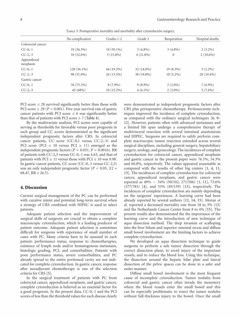
3.2. Postoperative Mortality and Morbidity. A total of 41(6.1%) among 667 patients died postoperatively. Mortalityrate (3.6%, 14/391) after CC-0,-1 resections was significantlylower than that (8.7%, 24/276) after CC-1,-2 resections(Table 3). Causes of deaths were septic shock (N = 14),fistula and peritonitis (N = 12), multiple organ failure (N =4), tumor progression (N = 4), lung embolism (N = 2),cardiac arrhythmia (N = 2), bleeding from duodenal ulcer(N = 1), and massive abdominal bleeding (N = 1). There
was no difference between the complication rates and diseasecategories.
Major complication (grade 3-4 complications) occurredin 126 patients (18.9%). A reoperation was necessary in36 patients (5.4%). The experienced complications wereabdominal abscess (N = 45), bowel fistula (N = 19),anastomosis or stump leakage (N = 18), ileus (N = 9),leakage from urinary bladder (N = 8), perforation of stomach(N = 6), abdominal bleeding (N = 5), bile leak (N = 3),perforation of diaphragm (N = 3), respiratory failure (N =3), renal failure (N = 1), arrhythmia (N = 1), bleeding fromduodenal ulcer (N = 1), and others (N = 4).
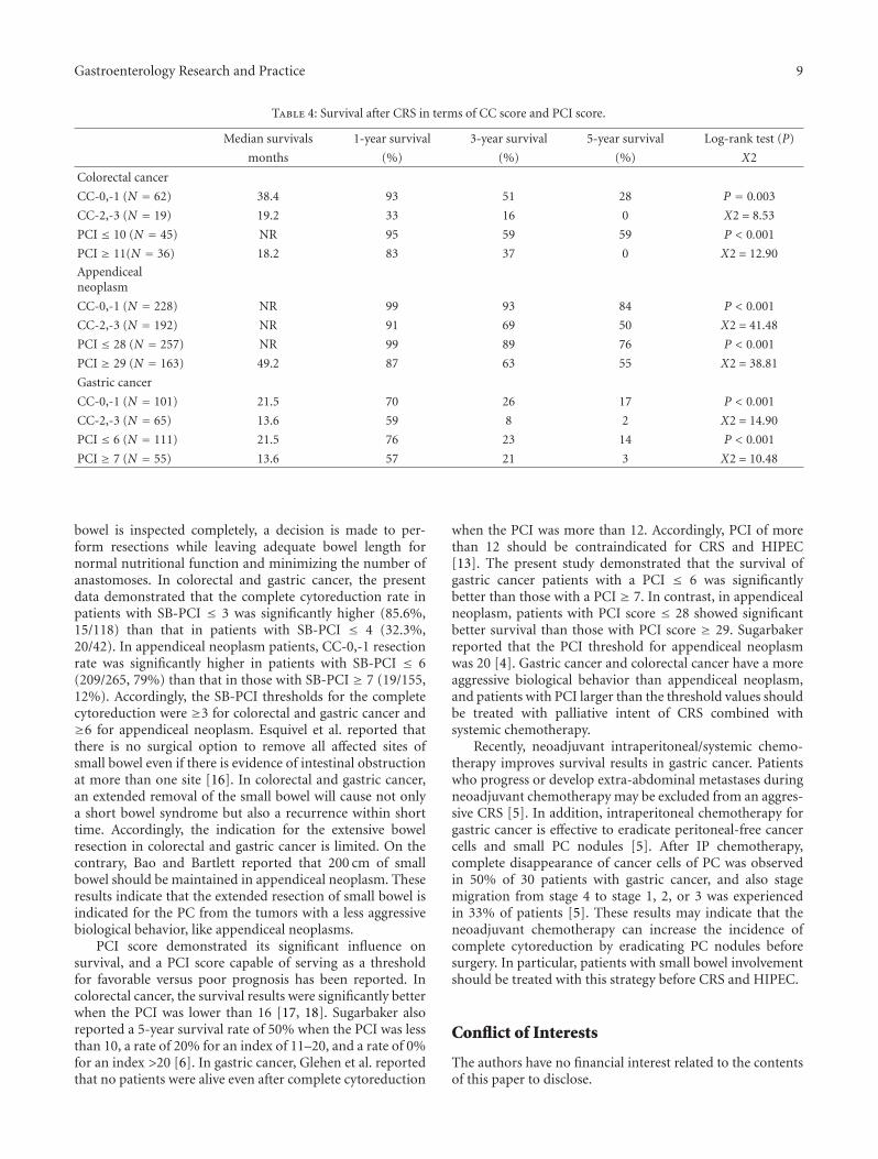
3.3. Survival after CRS. The overall 1-year, 3-year, and 5-yearsurvival rates and median survivals of the three groups areshown in Table 4. Univariate analysis showed that the lymphnode status, tumor differentiation, gender, and performancestatus did not have prognostic impact on survival. The 5-yearsurvival rates after CC-0,-1 resection for colorectal cancer,appendiceal neoplasm, and gastric cancer patients were 28%,84%, and 17%, respectively. In contrast, those in CC-2,-3groups were 0%, 50%, and 2% respectively.
The 5-year survival rate of colorectal cancer patients withPCI score ≤ 10 was significantly better than that with PCIscore ≥ 11 (P < 0.001). In appendiceal cancer, patients with
8 Gastroenterology Research and Practice
Table 3: Postoperative mortality and morbidity after cytoreductive surgery.
No complication Grades 1-2 Grade 3 Reoperation Hospital deaths
Colorectal cancer
CC-0,-1 35 (56.5%) 19 (30.1%) 3 (4.8%) 3 (4.8%) 2 (3.2%)
CC-2,-3 10 (52.6%) 3 (15.8%) 4 (21.0%) 0 2 (10.6%)
Appendicealneoplasm
CC-0,-1 128 (56.1%) 44 (19.3%) 32 (14.0%) 19 (8.3%) 5 (2.2%)
CC-2,-3 98 (51.0%) 26 (13.5%) 38 (19.8%) 10 (5.2%) 20 (10.4%)
Gastric cancer
CC-0,-1 76 (75.2%) 8 (7.9%) 9 (8.9%) 2 (2.0%) 7 (6.9%)
CC-2,-3 45 (68%) 10 (15.2%) 4 (6.1%) 2 (3.0%) 5 (7.6%)
PCI score ≤ 28 survived significantly better than those withPCI score ≥ 29 (P < 0.001). Five-year survival rate of gastriccancer patients with PCI score ≤ 6 was significantly betterthan that of patients with PCI score ≥ 7 (Table 4).
By the multivariate analysis, PCI scores were capable ofserving as thresholds for favorable versus poor prognosis ineach group and CC scores demonstrated as the significantindependent prognostic factors after CRS. In colorectalcancer patients, CC score (CC-0,1 versus CC-2,-3) andPCI score (PCI ≤ 10 versus PCI ≥ 11) emerged as theindependent prognostic factors (P = 0.031, P = 0.0016). RRof patients with CC-2,3 versus CC-0,-1 was 4.63, and that ofpatients with PCI ≥ 11 versus those with PCI ≤ 10 was 9.98.In gastric cancer patients, CC score (CC-0,-1 versus CC-2,3)was an only independent prognostic factor (P < 0.05, X2 =68.47, RR = 26.5).
4. Discussion
Current surgical management of the PC can be performedwith curative intent and potential long-term survival whena strategy of CRS combined with HIPEC is used to selectpatients.
Adequate patient selection and the improvement ofsurgical skills of surgeons are crucial to obtain a completemacroscopic cytoreduction, which is a leading predictor ofpatient outcome. Adequate patient selection is sometimesdifficult for surgeons with experience of small number ofcases with PC. Many criteria have to be assessed in eachpatient: performance status, response to chemotherapies,existence of lymph node and/or hematogenous metastasis,histologic grading, PCI, and comorbidities. Patients withpoor performance status, severe comorbidities, and PCalready spread to the entire peritoneal cavity are not indi-cated for complete cytoreduction. In gastric cancer, responseafter neoadjuvant chemotherapy is one of the selectioncriteria for CRS [5].
In the surgical treatment of patients with PC fromcolorectal cancer, appendiceal neoplasm, and gastric cancer,complete cytoreduction is believed as an essential factor fora good prognosis. In the present data, CC-0,-1 and the PCIscores of less than the threshold values for each disease clearly
were demonstrated as independent prognostic factors afterCRS plus perioperative chemotherapy. Peritonectomy tech-niques improved the incidence of complete cytoreduction,as compared with the ordinary surgical techniques [6, 9–12]. However, patients often with advanced metastases anda limited life span undergo a comprehensive therapy ofmultivisceral resection with several intestinal anastomosisand HIPEC. Surgeons are required to safely perform com-plete macroscopic tumor resection extended across severalsurgical disciplines, including general surgery, hepatobiliarysurgery, urology, and gynecology. The incidences of completecytoreduction for colorectal cancer, appendiceal neoplasmand gastric cancer in the present paper were 76.5%, 54.3%and 60.8%, respectively. The values appeared reasonable ascompared with the results of other big centers [1, 4, 11,13]. The incidences of complete cytoreduction for colorectalcancer, appendiceal neoplasm, and gastric cancer werereported as 49% ∼ 54% (50/102, 271/506) [1, 11], 73.6%(577/783) [4], and 53% (85/159) [13], respectively. Theincidences of complete cytoreduction are mainly dependingon the surgeons’ experiences. A learning curve had beenalready reported by several authors [12, 14, 15]. Moran etal. reported a decreased mortality rate from 18 to 3% [12]and the Netherlands Cancer Center from 8 to 4% [15]. Thepresent results also demonstrated the the importance of thelearning curve and the introduction of new technique ofaqua dissection method. The deep invasion or scallopinginto the liver hilum and superior omental recess and diffusesmall bowel involvement are the limiting factors to achievecomplete cytoreduction.
We developed an aqua dissection technique to guidesurgeons to perform a safe tumor dissection through thecorrect dissection plane, to avoid injury of the importantvessels, and to reduce the blood loss. Using this technique,the dissection around the hepatic hilar plate and lateraldissection of the pelvic spaces can be done in a safer andeasier manner.
Diffuse small bowel involvement is the most frequentcause of incomplete cytoreduction. Tumor nodules fromcolorectal and gastric cancer often invade the mesenterywhere the blood vessels enter the small bowel and thiscan be especially problematic to resect the tumor noduleswithout full-thickness injury to the bowel. Once the small
Gastroenterology Research and Practice 9
Table 4: Survival after CRS in terms of CC score and PCI score.
Median survivals 1-year survival 3-year survival 5-year survival Log-rank test (P)
months (%) (%) (%) X2
Colorectal cancer
CC-0,-1 (N = 62) 38.4 93 51 28 P = 0.003
CC-2,-3 (N = 19) 19.2 33 16 0 X2 = 8.53
PCI ≤ 10 (N = 45) NR 95 59 59 P < 0.001
PCI ≥ 11(N = 36) 18.2 83 37 0 X2 = 12.90
Appendicealneoplasm
CC-0,-1 (N = 228) NR 99 93 84 P < 0.001
CC-2,-3 (N = 192) NR 91 69 50 X2 = 41.48
PCI ≤ 28 (N = 257) NR 99 89 76 P < 0.001
PCI ≥ 29 (N = 163) 49.2 87 63 55 X2 = 38.81
Gastric cancer
CC-0,-1 (N = 101) 21.5 70 26 17 P < 0.001
CC-2,-3 (N = 65) 13.6 59 8 2 X2 = 14.90
PCI ≤ 6 (N = 111) 21.5 76 23 14 P < 0.001
PCI ≥ 7 (N = 55) 13.6 57 21 3 X2 = 10.48
bowel is inspected completely, a decision is made to per-form resections while leaving adequate bowel length fornormal nutritional function and minimizing the number ofanastomoses. In colorectal and gastric cancer, the presentdata demonstrated that the complete cytoreduction rate inpatients with SB-PCI ≤ 3 was significantly higher (85.6%,15/118) than that in patients with SB-PCI ≤ 4 (32.3%,20/42). In appendiceal neoplasm patients, CC-0,-1 resectionrate was significantly higher in patients with SB-PCI ≤ 6(209/265, 79%) than that in those with SB-PCI ≥ 7 (19/155,12%). Accordingly, the SB-PCI thresholds for the completecytoreduction were ≥3 for colorectal and gastric cancer and≥6 for appendiceal neoplasm. Esquivel et al. reported thatthere is no surgical option to remove all affected sites ofsmall bowel even if there is evidence of intestinal obstructionat more than one site [16]. In colorectal and gastric cancer,an extended removal of the small bowel will cause not onlya short bowel syndrome but also a recurrence within shorttime. Accordingly, the indication for the extensive bowelresection in colorectal and gastric cancer is limited. On thecontrary, Bao and Bartlett reported that 200 cm of smallbowel should be maintained in appendiceal neoplasm. Theseresults indicate that the extended resection of small bowel isindicated for the PC from the tumors with a less aggressivebiological behavior, like appendiceal neoplasms.
PCI score demonstrated its significant influence onsurvival, and a PCI score capable of serving as a thresholdfor favorable versus poor prognosis has been reported. Incolorectal cancer, the survival results were significantly betterwhen the PCI was lower than 16 [17, 18]. Sugarbaker alsoreported a 5-year survival rate of 50% when the PCI was lessthan 10, a rate of 20% for an index of 11–20, and a rate of 0%for an index >20 [6]. In gastric cancer, Glehen et al. reportedthat no patients were alive even after complete cytoreduction
when the PCI was more than 12. Accordingly, PCI of morethan 12 should be contraindicated for CRS and HIPEC[13]. The present study demonstrated that the survival ofgastric cancer patients with a PCI ≤ 6 was significantlybetter than those with a PCI ≥ 7. In contrast, in appendicealneoplasm, patients with PCI score ≤ 28 showed significantbetter survival than those with PCI score ≥ 29. Sugarbakerreported that the PCI threshold for appendiceal neoplasmwas 20 [4]. Gastric cancer and colorectal cancer have a moreaggressive biological behavior than appendiceal neoplasm,and patients with PCI larger than the threshold values shouldbe treated with palliative intent of CRS combined withsystemic chemotherapy.
Recently, neoadjuvant intraperitoneal/systemic chemo-therapy improves survival results in gastric cancer. Patientswho progress or develop extra-abdominal metastases duringneoadjuvant chemotherapy may be excluded from an aggres-sive CRS [5]. In addition, intraperitoneal chemotherapy forgastric cancer is effective to eradicate peritoneal-free cancercells and small PC nodules [5]. After IP chemotherapy,complete disappearance of cancer cells of PC was observedin 50% of 30 patients with gastric cancer, and also stagemigration from stage 4 to stage 1, 2, or 3 was experiencedin 33% of patients [5]. These results may indicate that theneoadjuvant chemotherapy can increase the incidence ofcomplete cytoreduction by eradicating PC nodules beforesurgery. In particular, patients with small bowel involvementshould be treated with this strategy before CRS and HIPEC.
Conflict of Interests
The authors have no financial interest related to the contentsof this paper to disclose.
10 Gastroenterology Research and Practice
References
[1] V. J. Verwaal, S. van Ruth, E. de Bree et al., “Random-ized trial of cytoreduction and hyperthermic intraperitonealchemotherapy versus systemic chemotherapy and palliativesurgery in patients with peritoneal carcinomatosis of colorec-tal cancer,” Journal of Clinical Oncology, vol. 21, no. 20, pp.3737–3743, 2003.
[2] V. J. Verwaal, S. Bruin, H. Boot, G. van Slooten, and H. vanTinteren, “8-Year follow-up of randomized trial: cytoreduc-tion and hyperthermic intraperitoneal chemotherapy versussystemic chemotherapy in patients with peritoneal carcino-matosis of colorectal cancer,” Annals of Surgical Oncology, vol.15, no. 9, pp. 2426–2432, 2008.
[3] T. D. Yan and D. L. Morris, “Cytoreductive surgery and peri-operative intraperitoneal chemotherapy for isolated colorectalperitoneal carcinomatosis: experimental therapy or standardof care?” Annals of Surgery, vol. 248, no. 5, pp. 829–835, 2008.
[4] P. H. Sugarbaker, “Epithelial appendiceal neoplasms,” CancerJournal, vol. 15, no. 3, pp. 225–235, 2009.
[5] Y. Yonemura, Y. Endou, M. Shinbo et al., “Safety and efficacyof bidirectional chemotherapy for treatment of patients withperitoneal dissemination from gastric cancer: selection forcytoreductive surgery,” Journal of Surgical Oncology, vol. 100,no. 4, pp. 311–316, 2009.
[6] T. A. Sugarbaker, D. Chang, P. Koslowe, and P. H. Sugarbaker,“Patterns of spread of recurrent intraabdominal sarcoma.,”Cancer Treatment and Research, vol. 82, pp. 65–77, 1996.
[7] P. Piso, P. Slowik, F. Popp, M. H. Dahlke, G. Glockzin, andH. J. Schlitt, “Safety of gastric resections during cytoreductivesurgery and hyperthermic intraperitoneal chemotherapy forperitoneal carcinomatosis,” Annals of Surgical Oncology, vol.16, no. 8, pp. 2188–2194, 2009.
[8] P. H. Sugarbaker, “Peritonectomy procedures,” Annals of Sur-gery, vol. 221, no. 1, pp. 29–42, 1995.
[9] P. Jacquet and P. H. Sugarbaker, “Clinical research method-ologies in diagnosis and staging of patients with peritonealcarcinomatosis,” Cancer Treatment and Research, vol. 82, pp.359–374, 1996.
[10] P. H. Sugarbaker, “Successful management of microscopicresidual disease in large bowel cancer,” Cancer Chemotherapyand Pharmacology, vol. 43, pp. S15–S25, 1999.
[11] O. Glehen, F. Kwiatkowski, P. H. Sugarbaker et al., “Cytore-ductive surgery combined with perioperative intraperitonealchemotherapy for the management of peritoneal carcino-matosis from colorectal cancer: a multi-institutional study,”Journal of Clinical Oncology, vol. 22, no. 16, pp. 3284–3292,2004.
[12] B. J. Moran, “Decision-making and technical factors accountfor the learning curve in complex surgery,” Journal of PublicHealth, vol. 28, no. 4, pp. 375–378, 2006.
[13] O. Glehen, F. N. Gilly, C. Arvieux et al., “Peritoneal car-cinomatosis from gastric cancer: a multi-institutional studyof 159 patients treated by cytoreductive surgery combinedwith perioperative intraperitoneal chemotherapy,” Annals ofSurgical Oncology, vol. 17, no. 9, pp. 2370–2377, 2010.
[14] T. D. Yan, M. Links, S. Fransi et al., “Learning curvefor cytoreductive surgery and perioperative intraperitonealchemotherapy for peritoneal surface malignancy—a journeyto becoming a Nationally Funded Peritonectomy Center,”Annals of Surgical Oncology, vol. 14, no. 8, pp. 2270–2280,2007.
[15] R. M. Smeenk, V. J. Verwaal, and F. A. N. Zoetmulder,“Learning curve of combined modality treatment in peri-toneal surface disease,” British Journal of Surgery, vol. 94, no.11, pp. 1408–1414, 2007.
[16] J. Esquivel, D. Elias, D. Baratti, S. Kusamura, and M. Deraco,“Consensus statement on the loco regional treatment ofcolorectal cancer with peritoneal dissemination,” Journal ofSurgical Oncology, vol. 98, no. 4, pp. 263–267, 2008.
[17] D. Elias, F. Blot, O. A. A. El Otmany et al., “Curative treatmentof peritoneal carcinomatosis arising from colorectal cancerby complete resection and intraperitoneal chemotherapy,”Cancer, vol. 92, no. 1, pp. 71–76, 2001.
[18] J. L. Koh, T. D. Yan, D. Glenn, and D. L. Morris, “Evaluation ofpreoperative computed tomography in estimating peritonealcancer index in colorectal peritoneal carcinomatosis,” Annalsof Surgical Oncology, vol. 16, no. 2, pp. 327–333, 2009.

Submit your manuscripts athttp://www.hindawi.com
Stem CellsInternational
Hindawi Publishing Corporationhttp://www.hindawi.com Volume 2014
Hindawi Publishing Corporationhttp://www.hindawi.com Volume 2014
MEDIATORSINFLAMMATION
of
Hindawi Publishing Corporationhttp://www.hindawi.com Volume 2014
Behavioural Neurology
EndocrinologyInternational Journal of
Hindawi Publishing Corporationhttp://www.hindawi.com Volume 2014
Hindawi Publishing Corporationhttp://www.hindawi.com Volume 2014
Disease Markers
Hindawi Publishing Corporationhttp://www.hindawi.com Volume 2014
BioMed Research International
OncologyJournal of
Hindawi Publishing Corporationhttp://www.hindawi.com Volume 2014
Hindawi Publishing Corporationhttp://www.hindawi.com Volume 2014
Oxidative Medicine and Cellular Longevity
Hindawi Publishing Corporationhttp://www.hindawi.com Volume 2014
PPAR Research
The Scientific World JournalHindawi Publishing Corporation http://www.hindawi.com Volume 2014
Immunology ResearchHindawi Publishing Corporationhttp://www.hindawi.com Volume 2014
Journal of
ObesityJournal of
Hindawi Publishing Corporationhttp://www.hindawi.com Volume 2014
Hindawi Publishing Corporationhttp://www.hindawi.com Volume 2014
Computational and Mathematical Methods in Medicine
OphthalmologyJournal of
Hindawi Publishing Corporationhttp://www.hindawi.com Volume 2014
Diabetes ResearchJournal of
Hindawi Publishing Corporationhttp://www.hindawi.com Volume 2014
Hindawi Publishing Corporationhttp://www.hindawi.com Volume 2014
Research and TreatmentAIDS
Hindawi Publishing Corporationhttp://www.hindawi.com Volume 2014
Gastroenterology Research and Practice
Hindawi Publishing Corporationhttp://www.hindawi.com Volume 2014
Parkinson’s Disease
Evidence-Based Complementary and Alternative Medicine
Volume 2014Hindawi Publishing Corporationhttp://www.hindawi.com
Related Documents


![Clinical Study - hindawi.com fileglucose-based solutions and peritonitis episodes may lead to persistent increase of peritoneal transport rate [11]. ... out, thus, 53 patients finished](https://static.cupdf.com/doc/110x72/5cc011d988c99337188be68d/clinical-study-solutions-and-peritonitis-episodes-may-lead-to-persistent-increase.jpg)