5-(Perylen-3-yl)Ethynyl-arabino-Uridine (aUY11), an Arabino-Based Rigid Amphipathic Fusion Inhibitor, Targets Virion Envelope Lipids To Inhibit Fusion of Influenza Virus, Hepatitis C Virus, and Other Enveloped Viruses Che C. Colpitts, a,b Alexey V. Ustinov, c Raquel F. Epand, d Richard M. Epand, d Vladimir A. Korshun, c Luis M. Schang a,b,e Department of Medical Microbiology and Immunology, a Li Ka Shing Institute of Virology, b Department of Biochemistry, e University of Alberta, Edmonton, Alberta, Canada; Shemyakin-Ovchinnikov Institute of Bioorganic Chemistry, Russian Academy of Sciences, Moscow, Russia c ; Department of Biochemistry and Biomedical Sciences, McMaster University, Hamilton, Ontario, Canada d Entry of enveloped viruses requires fusion of viral and cellular membranes. Fusion requires the formation of an intermediate stalk structure, in which only the outer leaflets are fused. The stalk structure, in turn, requires the lipid bilayer of the envelope to bend into negative curvature. This process is inhibited by enrichment in the outer leaflet of lipids with larger polar headgroups, which favor positive curvature. Accordingly, phospholipids with such shape inhibit viral fusion. We previously identified a com- pound, 5-(perylen-3-yl)ethynyl-2=-deoxy-uridine (dUY11), with overall shape and amphipathicity similar to those of these phos- pholipids. dUY11 inhibited the formation of the negative curvature necessary for stalk formation and the fusion of a model en- veloped virus, vesicular stomatitis virus (VSV). We proposed that dUY11 acted by biophysical mechanisms as a result of its shape and amphipathicity. To test this model, we have now characterized the mechanisms against influenza virus and HCV of 5-(perylen-3-yl)ethynyl-arabino-uridine (aUY11), which has shape and amphipathicity similar to those of dUY11 but contains an arabino-nucleoside. aUY11 interacted with envelope lipids to inhibit the infectivity of influenza virus, hepatitis C virus (HCV), herpes simplex virus 1 and 2 (HSV-1/2), and other enveloped viruses. It specifically inhibited the fusion of influenza vi- rus, HCV, VSV, and even protein-free liposomes to cells. Furthermore, aUY11 inhibited the formation of negative curvature in model lipid bilayers. In summary, the arabino-derived aUY11 and the deoxy-derived dUY11 act by the same antiviral mecha- nisms against several enveloped but otherwise unrelated viruses. Therefore, chemically unrelated compounds of appropriate shape and amphipathicity target virion envelope lipids to inhibit formation of the negative curvature required for fusion, inhib- iting infectivity by biophysical, not biochemical, mechanisms. V iral entry is an attractive antiviral target. Clinical entry inhib- itors target viral glycoproteins to inhibit their attachment to cellular receptors (e.g., maraviroc) (1) or to prevent the confor- mational changes of virion glycoproteins needed to drive fusion between viral and cellular membranes (e.g., enfuvirtide) (2). These approaches, unfortunately, have the same limitations as tar- geting any other viral protein, such as selection for resistance and narrow specificity (3). Experimental compounds have been pro- posed to inhibit the infectivity of unrelated enveloped viruses by disruption of virion envelopes (e.g., PD 404182 [42]), inhibition of fusion by poorly understood mechanisms (e.g., arbidol [48]), or by irreparable damage to the virion lipid envelopes (e.g., LJ-001 [49]). From these and related studies, it is clear that antiviral ap- proaches targeting conserved structural elements of virions, such as the virion envelope, can result in broad-spectrum activity against unrelated enveloped viruses. All enveloped viruses require the fusion of viral and cellular membranes to enter the cell (4). The energy required for this pro- cess is provided by the attachment, binding, and conformational changes of viral fusion proteins, which undergo major structural rearrangements during fusion from the pre- to the postfusion states. The three different classes of viral fusion proteins differ structurally but mediate fusion in an overall similar manner (5). The insertion of the viral fusion peptides disrupts the target mem- branes, resulting in the formation of a fusion intermediate known as the hemifusion stalk, in which only the outer leaflets of the two membranes are fused (6). The inner leaflets subsequently fuse, resulting in a small pore that then enlarges to allow full fusion. Formation of the hemifusion stalk requires the lipid bilayer to form local negative curvature, an energetically demanding rear- rangement. Fusion is consequently promoted by enrichment in the outer leaflet of lipids that induce negative curvature, such as oleic acid (6). Conversely, enrichment in the outer leaflet of lipids that favor positive curvature, specifically those with hydrophilic headgroups larger than their hydrophobic tails (such as lysophos- phatidylcholine [LPC]), impairs fusion (6). Such phospholipids prevent the fusion of enveloped viruses, such as influenza virus (7, 8), rabies virus (9), and Sendai virus (10). However, such phos- pholipids also tend to be disruptive to all membranes (including cellular ones), to have signaling activities, to be toxic, and to be too rapidly metabolized to be useful as drugs. We previously identified a synthetic compound, 5-(perylen-3- Received 17 October 2012 Accepted 24 December 2012 Published ahead of print 2 January 2013 Address correspondence to Luis M. Schang, [email protected]. Supplemental material for this article may be found at http://dx.doi.org/10.1128 /JVI.02882-12. Copyright © 2013, American Society for Microbiology. All Rights Reserved. doi:10.1128/JVI.02882-12 3640 jvi.asm.org Journal of Virology p. 3640 –3654 April 2013 Volume 87 Number 7

Welcome message from author
This document is posted to help you gain knowledge. Please leave a comment to let me know what you think about it! Share it to your friends and learn new things together.
Transcript
5-(Perylen-3-yl)Ethynyl-arabino-Uridine (aUY11), an Arabino-BasedRigid Amphipathic Fusion Inhibitor, Targets Virion Envelope LipidsTo Inhibit Fusion of Influenza Virus, Hepatitis C Virus, and OtherEnveloped Viruses
Che C. Colpitts,a,b Alexey V. Ustinov,c Raquel F. Epand,d Richard M. Epand,d Vladimir A. Korshun,c Luis M. Schanga,b,e
Department of Medical Microbiology and Immunology,a Li Ka Shing Institute of Virology,b Department of Biochemistry,e University of Alberta, Edmonton, Alberta,Canada; Shemyakin-Ovchinnikov Institute of Bioorganic Chemistry, Russian Academy of Sciences, Moscow, Russiac; Department of Biochemistry and Biomedical Sciences,McMaster University, Hamilton, Ontario, Canadad
Entry of enveloped viruses requires fusion of viral and cellular membranes. Fusion requires the formation of an intermediatestalk structure, in which only the outer leaflets are fused. The stalk structure, in turn, requires the lipid bilayer of the envelope tobend into negative curvature. This process is inhibited by enrichment in the outer leaflet of lipids with larger polar headgroups,which favor positive curvature. Accordingly, phospholipids with such shape inhibit viral fusion. We previously identified a com-pound, 5-(perylen-3-yl)ethynyl-2=-deoxy-uridine (dUY11), with overall shape and amphipathicity similar to those of these phos-pholipids. dUY11 inhibited the formation of the negative curvature necessary for stalk formation and the fusion of a model en-veloped virus, vesicular stomatitis virus (VSV). We proposed that dUY11 acted by biophysical mechanisms as a result of itsshape and amphipathicity. To test this model, we have now characterized the mechanisms against influenza virus and HCV of5-(perylen-3-yl)ethynyl-arabino-uridine (aUY11), which has shape and amphipathicity similar to those of dUY11 but containsan arabino-nucleoside. aUY11 interacted with envelope lipids to inhibit the infectivity of influenza virus, hepatitis C virus(HCV), herpes simplex virus 1 and 2 (HSV-1/2), and other enveloped viruses. It specifically inhibited the fusion of influenza vi-rus, HCV, VSV, and even protein-free liposomes to cells. Furthermore, aUY11 inhibited the formation of negative curvature inmodel lipid bilayers. In summary, the arabino-derived aUY11 and the deoxy-derived dUY11 act by the same antiviral mecha-nisms against several enveloped but otherwise unrelated viruses. Therefore, chemically unrelated compounds of appropriateshape and amphipathicity target virion envelope lipids to inhibit formation of the negative curvature required for fusion, inhib-iting infectivity by biophysical, not biochemical, mechanisms.
Viral entry is an attractive antiviral target. Clinical entry inhib-itors target viral glycoproteins to inhibit their attachment to
cellular receptors (e.g., maraviroc) (1) or to prevent the confor-mational changes of virion glycoproteins needed to drive fusionbetween viral and cellular membranes (e.g., enfuvirtide) (2).These approaches, unfortunately, have the same limitations as tar-geting any other viral protein, such as selection for resistance andnarrow specificity (3). Experimental compounds have been pro-posed to inhibit the infectivity of unrelated enveloped viruses bydisruption of virion envelopes (e.g., PD 404182 [42]), inhibitionof fusion by poorly understood mechanisms (e.g., arbidol [48]),or by irreparable damage to the virion lipid envelopes (e.g., LJ-001[49]). From these and related studies, it is clear that antiviral ap-proaches targeting conserved structural elements of virions, suchas the virion envelope, can result in broad-spectrum activityagainst unrelated enveloped viruses.
All enveloped viruses require the fusion of viral and cellularmembranes to enter the cell (4). The energy required for this pro-cess is provided by the attachment, binding, and conformationalchanges of viral fusion proteins, which undergo major structuralrearrangements during fusion from the pre- to the postfusionstates. The three different classes of viral fusion proteins differstructurally but mediate fusion in an overall similar manner (5).The insertion of the viral fusion peptides disrupts the target mem-branes, resulting in the formation of a fusion intermediate knownas the hemifusion stalk, in which only the outer leaflets of the two
membranes are fused (6). The inner leaflets subsequently fuse,resulting in a small pore that then enlarges to allow full fusion.
Formation of the hemifusion stalk requires the lipid bilayer toform local negative curvature, an energetically demanding rear-rangement. Fusion is consequently promoted by enrichment inthe outer leaflet of lipids that induce negative curvature, such asoleic acid (6). Conversely, enrichment in the outer leaflet of lipidsthat favor positive curvature, specifically those with hydrophilicheadgroups larger than their hydrophobic tails (such as lysophos-phatidylcholine [LPC]), impairs fusion (6). Such phospholipidsprevent the fusion of enveloped viruses, such as influenza virus (7,8), rabies virus (9), and Sendai virus (10). However, such phos-pholipids also tend to be disruptive to all membranes (includingcellular ones), to have signaling activities, to be toxic, and to be toorapidly metabolized to be useful as drugs.
We previously identified a synthetic compound, 5-(perylen-3-
Received 17 October 2012 Accepted 24 December 2012
Published ahead of print 2 January 2013
Address correspondence to Luis M. Schang, [email protected].
Supplemental material for this article may be found at http://dx.doi.org/10.1128/JVI.02882-12.
Copyright © 2013, American Society for Microbiology. All Rights Reserved.
doi:10.1128/JVI.02882-12
3640 jvi.asm.org Journal of Virology p. 3640–3654 April 2013 Volume 87 Number 7
yl)ethynyl-2=-deoxy-uridine (dUY11), with the same overall shapeand amphipathicity as such lipids (11). dUY11 inhibited the for-mation of the negative curvature necessary to form the hemifu-sion stalk. Furthermore, dUY11 prevented the fusion of a modelenveloped virus, vesicular stomatitis virus (VSV). Consistently,dUY11 inhibited the infectivity of several enveloped viruses atnanomolar concentrations but did not inhibit the infectivity ofnonenveloped viruses. Although dUY11 is a nucleoside derivative,it does not act by classical nucleosidic mechanisms (11).
We had proposed that dUY11 was the first member of a familyof compounds that inhibit infectivity by biophysical (not bio-chemical) mechanisms, according to their shapes and amphipath-icities, not to chemical interactions. We called these compoundsrigid amphipathic fusion inhibitors (RAFIs). According to ourmodel, chemically distinct molecules of the same overall shapeand amphipathicity should also target virion envelope lipids toprevent fusion of viral and cellular membranes. Furthermore, allof these compounds should act by the same mechanisms againstall enveloped viruses, including important human pathogens.
We had shown that an arabino-configured nucleoside,5-(perylen-3-yl)ethynyl-arabino-uridine (aUY11), also inhibitedthe infectivity of herpes simplex virus 1 (HSV-1) (11). However,the antiviral mechanism of aUY11 remained uncharacterized. Wehave now tested our model using the arabino-derived aUY11 andclinically important viruses, including influenza virus and hepati-tis C virus (HCV). The arabino-derived aUY11 differs from thedeoxy-derived dUY11 in that it has a 2= hydroxyl group in thenucleoside moiety. If the compounds acted by traditional bio-chemical mechanisms, then this modification should result in dif-ferent biological activities. If these compounds acted by biophys-ical mechanisms, however, then the arabino-configured aUY11and the deoxyuridine-configured dUY11 should act by the samemechanisms. Moreover, the antiviral mechanisms should be thesame against several enveloped viruses, regardless of their partic-ular fusion proteins.
Here, we report that aUY11 interacts with the envelope lip-ids of influenza virus, HCV, HSV-1, and HSV-2 and inhibitsthe infectivity of these and other enveloped viruses. aUY11inhibits lipid mixing in fluorescence-dequenching fusion as-says for influenza virus, HCV, and VSV, which represent thethree classes of fusion proteins. Furthermore, aUY11 localizesto the hydrophobic core of the lipid membrane of protein-freeliposomes and inhibits their acid-induced fusion to cells in theabsence of any viral protein. aUY11 did not affect membranefluidity but instead inhibited the transition of model lipid bi-layers from lamellar to hexagonal phases, a transition that re-quires the formation of negative curvature. Such activities areconsistent with the inhibition of the negative curvature neces-sary for virion envelopes to fuse to cell membranes as the mainantiviral mechanism of 5-perylene-ethynyl uracil deoxy- orarabino-derived nucleosides. We conclude that aUY11 anddUY11 act by the same biophysical mechanisms, targeting thevirion envelope lipids to prevent the formation of the negativecurvature necessary for fusion, thereby inhibiting the infectiv-ity of enveloped viruses. Our findings show that RAFIs areindeed a novel family of antiviral molecules that act by bio-physical mechanisms against unrelated enveloped viruses, in-cluding clinically important human pathogens.
MATERIALS AND METHODSCompounds. The synthesis of aUY11 and other RAFIs has been describedpreviously (11–13). The compounds were dissolved in dimethyl sulfoxide(DMSO) as 10 mM stocks, stored at �20°C, and resuspended just beforeuse to the indicated concentrations in warm Dulbecco’s modified Eagle’smedium (DMEM). Equivalent concentrations of DMSO were used in thecontrols.
Cells and viruses. African green monkey Vero fibroblasts, NIH 3T3cells, and Madin-Darby canine kidney (MDCK) cells were cultured inDMEM supplemented with 5% fetal bovine serum (FBS), 50 U/ml peni-cillin, and 50 �g/ml streptomycin at 37°C in 5% CO2. Huh7.5 cells (14)and HEK293 cells were cultured in DMEM supplemented with 10% fetalbovine serum (FBS), 50 U/ml penicillin, and 50 �g/ml streptomycin at37°C in 5% CO2.
To prepare HSV-1 and HSV-2 stocks, Vero cells were infected at amultiplicity of infection (MOI) of 0.05 PFU/cell of HSV-1 strain KOS orHSV-2 strain 186 (from P. Schaffer, Harvard Medical School). Viralstocks were prepared according to standard procedures (15).
To prepare VSV or Sindbis virus stocks, Vero cells were infected withVSV or Sindbis virus (MOI, 0.02 PFU/cell) at 37°C in 5% CO2. The inoc-ulum was removed, and the infected cells were overlaid with DMEM-5%FBS. Culture medium was collected after 48 h and centrifuged at 3,200 �g for 30 min at 4°C to pellet cell debris. The supernatant was then centri-fuged at 10,000 � g for 2 h at 4°C, and the resulting virion pellet wasresuspended in DMEM, aliquoted, and stored at �80°C.
HCV strain JFH-1 (16) was originally obtained from Takaji Wakita(Tokyo Metropolitan Institute for Neuroscience, Tokyo, Japan) throughLorne Tyrrell (University of Alberta, Edmonton, Canada). To preparevirus stock, Huh7.5 cells were infected with 0.2 focus-forming units(FFU)/cell of HCV JFH-1 and passaged as necessary. After 7 days, culturemedium was collected and centrifuged at 800 � g for 5 min at 4°C to pelletcell debris. The supernatant was filtered through a 0.22-�m filter andconcentrated using Amicon 100,000-molecular-weight cutoff filters (Mil-lipore, Billerica, MA). The resulting virus stocks were titrated by focus-forming assays and stored at �80°C.
Influenza virus strains A/Puerto Rico/8/1934 (H1N1), A/USSR/90/77(H1N1), A/Aichi/2/68 (H3N2), and A/Port Chalmers/1/73 (H3N2) wereobtained from Veronika von Messling (INRS-Institut Armand-Frappier,Research Centre, Quebec, Canada). To prepare virus stocks, MDCK cellswere infected with 0.01 PFU/cell of influenza virus. The infected cells werethen incubated in DMEM supplemented with 0.2% bovine serum albu-min (BSA) and 2 �g/ml tosylphenylalanyl chloromethyl ketone (TPCK)trypsin (Sigma-Aldrich, Oakville, ON, Canada) in 5% CO2 at 33°C forapproximately 2 days, until at least 90% of the cells displayed cytopathiceffects (cell rounding with minimal detachment). Virions were harvestedas described for VSV.
Murine cytomegalovirus (mCMV) strain RM427� (17), containing alacZ insertion in the nonessential immediate-early 2 gene, was originallyobtained from Edward Mocarski (Emory University, Atlanta, GA)through Denise Hemmings (University of Alberta, Edmonton, Alberta,Canada). To prepare virus stocks, NIH 3T3 cells were infected with 0.01PFU/cell of mCMV RM427�. The infected cells were incubated at 33°C in5% CO2 for approximately 4 days, until at least 90% of the cells displayedcytopathic effects (cell rounding with minimal detachment). Two daysafter 90% of the cells displayed cytopathic effects, virions were harvestedas described for HSV-1 and HSV-2.
Poliovirus (PV) strain Mahoney was obtained from Jiang Yin andMichael N. James (University of Alberta, Edmonton, Alberta, Canada)and propagated in Vero cells as described for VSV. Adenovirus (AdV)expressing green fluorescent protein (GFP) was obtained from Rene Ja-cobs and Dennis Vance (University of Alberta, Edmonton, Alberta, Can-ada) and was titrated in HEK293 cells maintained with DMEM-10% FBS.Mammalian orthoreovirus type 3 was kindly provided by Maya Shmulev-itz (University of Alberta, Edmonton, Alberta, Canada).
RAFI Mechanism of Action against Influenza Virus and HCV
April 2013 Volume 87 Number 7 jvi.asm.org 3641

Infectivity assays. Vero cells (5 � 105 cells/well in 6-well plates) wereinfected with a 200-�l inoculum containing approximately 200 PFU ofHSV-2, VSV, Sindbis virus, poliovirus, or reovirus preexposed to aUY11,dUY11, or DMSO vehicle for 10 min at 37°C. The inocula were removedafter 1 h, and the monolayers were washed and overlaid with 2% methyl-cellulose or 1% agarose (for reovirus) containing 5% FBS. The infectedcells were incubated in 5% CO2 at 37°C until well-defined plaques devel-oped (typically, 1 to 2 days postinfection). The cells were then fixed andstained with crystal violet in methanol (0.5% [wt/vol] crystal violet, 17%[vol/vol] methanol in H2O).
For influenza virus, MDCK cells (6 � 105 cells/well in 6-well plates)were infected with a 200-�l inoculum containing approximately 200 PFUof influenza virus A/Puerto Rico/8/1934, A/USSR/70/99, A/Aichi/2/68, orA/Port Chalmers/1/73 preexposed to compound or DMSO vehicle for 10min at 37°C. The inocula were removed after 1 h, and the monolayers werewashed and overlaid with 0.8% agarose (containing 0.1 �g/ml TPCK-trypsin). Plaques were visualized using neutral red or crystal violetstaining.
Huh7.5 cells (9 � 104 cells/well in 24-well plates) were infected with150 �l of HCV strain JFH-1 (containing �100 FFU) preexposed toaUY11, dUY11, or DMSO vehicle for 10 min at 37°C. The inocula wereremoved 4 h later, and the monolayers were washed and overlaid withfresh DMEM-10% FBS containing no drug. The infected cells were fixedat 72 h postinfection with cold methanol-acetone (1:1) and processed forimmunocytochemistry for HCV core protein. Cells were incubated withprimary mouse IgG anti-core antibody MA1-080 (Thermo Scientific,Rockford, IL) diluted 1:300 for 2 h at room temperature, followed byaddition of secondary biotinylated horse anti-mouse IgG antibody (Vec-tor Laboratories, Burlingame, CA) for 30 min at room temperature. Thefixed cells were then treated with the avidin-biotin-peroxidase complex(Vectastain ABC kit; Vector Laboratories) for 30 min, followed by addi-tion of ImmPact SG peroxidase substrate (Vector Laboratories). Foci werecounted under the microscope.
For mCMV, NIH 3T3 cells (3 � 105 cells/well in 12-well plates) wereinfected with a 150-�l inoculum containing approximately 200 PFU ofmCMV RM427� preexposed for 10 min at 37°C to compound or DMSOvehicle. The inocula were removed after 1 h, and the monolayers werewashed and overlaid with DMEM-10% FBS. Foci of infected cells weredetected after 24 h using a LacZ cell detection kit (InvivoGen, SanDiego, CA).
For AdV, virions were exposed to aUY11, dUY11, or DMSO for 10min at 37°C prior to infecting HEK293 cell monolayers (5 � 105 cells/wellin 12-well plates). The inocula were removed after 1 h, and the monolay-ers were washed and overlaid with fresh DMEM-10% FBS. Infected cellsexpressing GFP were visualized after 24 h using a fluorescence microscopewith a UV light source (Leica DM IRB, Itzlar, Germany).
Time-of-addition assays. For pretreatment experiments, MDCK,Huh7.5, or Vero cells were treated with aUY11 or dUY11 for 1 h at 37°C.The cells were then washed three times with DMEM warmed to 37°C andinfected with influenza virus A/PR/34/8, HCV JFH-1, or HSV-1, respec-tively, in the absence of any drug. Infectivity was assessed by plaque orfocus formation. To test whether aUY11 or dUY11 was effective whenadded to already-infected cells, MDCK, Huh7.5, or Vero cells were in-fected with 5 or 0.5 PFU/cell of influenza virus A/PR/34/8, HCV JFH-1, orHSV-1 KOS. The inocula were removed after 1 h of adsorption. The in-fected cells were washed and then overlaid with DMEM supplementedwith 10% FBS (HCV) or 5% FBS (influenza virus and HSV-1) and aUY11for 24 (influenza virus and HSV-1) or 48 (HCV) hours. The supernatantsand cell lysates were then harvested as described previously. Influenzavirus and HSV-1 virions were pelleted by centrifugation at 10,000 � g andresuspended in drug-free DMEM. HCV virions were concentrated usingAmicon centrifugal filters with a 100-kDa molecular-mass cutoff. Titra-tions were performed using logarithmic dilutions of the viruses (in drug-free medium) and MDCK, Vero, or Huh7.5 cells. Harvested virions wereexamined for the presence of aUY11 by obtaining its fluorescence spectra.
Liposome preparation. Cholesterol, �-oleoyl-�-palmitoyl-L-�-phos-phatidylcholine (POPC), and 1,2-dioleoyl-L-�-phosphatidylcholine(DOPC) were obtained from Sigma-Aldrich (Oakville, ON, Canada).POPC liposomes were prepared by hydrating 2 �mol dry lipid with 1 ml of180 mM Na2HPO4, 10 mM citric acid (pH 7.4), followed by vortexing.DOPC-cholesterol (1.7:1 molar ratio) liposomes were prepared by thehydration method. DOPC and cholesterol were dissolved and mixed in500 �l chloroform, which was then evaporated; 1 ml of 180 mMNa2HPO4, 10 mM citric acid (pH 7.4) was added to the resulting lipidfilm, and the mixture was vortexed to form large multilamellar liposomes.Octadecyl rhodamine B chloride (R18; Invitrogen, Grand Island, NY) wasadded (to a final concentration of 5 mol%) as required by first mixing R18and lipids as ethanol and chloroform solutions, respectively.
R18 labeling of VSV, HCV, and influenza virus virions. Virions werelabeled with 0.59 �M (VSV and HCV) or 1.8 �M (influenza virus) R18 byincubating VSV, influenza virus (108 PFU), or HCV JFH-1 (106 FFU) with1.97 �l (VSV and HCV) or 5.91 �l (influenza virus) of 300 �M R18 inethanol in 1 ml of 180 mM Na2HPO4, 10 mM citric acid (pH 7.4) for 1 hat room temperature on a rotary shaker in the dark. The labeled virionswere purified through a column containing 4 ml of Sephadex G-100 resin.The column was equilibrated by washing it twice with approximately 12ml of 180 mM Na2HPO4, 10 mM citric acid (pH 7.4) (henceforth referredto as fusion buffer). Labeled virions (1 ml) were then added to the columnand eluted with fusion buffer. Fractions of approximately 500 �l werecollected every minute for 15 min. The viral-protein concentration ineach fraction was determined using a Bradford assay (Bio-Rad). Fractionsof labeled VSV, HCV, or influenza virus virions were titrated on Vero,Huh7.5, or MDCK cells. For HCV and influenza virus, fractions contain-ing infectious virus were then pooled and concentrated using Amicon100,000-molecular-weight cutoff filters (Millipore, Billerica, MA). Thepooled fractions were tested for R18 incorporation by R18 dequenchingafter addition of Triton X-100 to a final concentration of 0.1% (VSV andinfluenza virus) or 0.01% (HCV), using a QuantaMaster 40 scanningspectrofluorometer (Photon Technology International, Birmingham, NJ)equipped with a 75-W xenon lamp. R18 was excited at 560 nm. Fluores-cence was detected at 590 nm using a model 814 switchable photon-counting/analog photomultiplier detection unit with an R1527 photo-multiplier tube. Data were collected using FeliX32 software (PhotonTechnology International, Birmingham, NJ).
Spectra. Emission spectra of aUY11 or dUY11 were collected using theQuantaMaster 40 spectrofluorometer. aUY11 or dUY11 was added to 2.5ml of 180 mM Na2HPO4, 10 mM citric acid (pH 7.4) buffer, or to 2.5 mlof 1-octanol (Sigma), to a final concentration of 48 nM or 0.48 nM, re-spectively, in a polymethacrylate cuvette prewarmed to 37°C. Alternately,aUY11 or dUY11 was added to 107 PFU of VSV, HSV-1 or -2, or influenzavirus; 106 FFU of HCV; or 2 nmol POPC liposomes in 2.5 ml of fusionbuffer at 37°C to a final concentration of 48 nM. For other experiments,approximately 104 influenza virus, HCV, or HSV-1 virions produced bycells treated with vehicle, or equivalent volumes from cells treated withaUY11, were diluted to 2.5 ml in 180 mM Na2HPO4, 10 mM citric acid(pH 7.4). Emission spectra were obtained at the maximum excitationwavelength, 455 nm, and examined from 475 to 575 nm. Spectra werenormalized to the highest fluorescence signal intensity obtained for allconditions.
Confocal microscopy. Near-confluent Vero cells seeded onto 22- by22-mm coverslips (Fisher) placed in 6-well plates were cultured inDMEM-5% FBS, 50 U/ml penicillin, and 50 �g/ml streptomycin in 5%CO2 for �16 h at 37°C. The cells were first incubated with 250 nMPKH26-GL fluorescent dye (Sigma) for 10 min at 37°C and were thenwashed twice with 2 ml/well of room temperature DMEM. The washedcells were exposed to 2 �M aUY11 or dUY11 for 1, 5, 15, 40, or 120 min at37°C. The RAFIs were removed, and the cells were washed twice with 2ml/well of DMEM at room temperature. The cells were then fixed with10% formalin for 30 min at room temperature, washed once with 1� PBSat room temperature, and mounted onto glass slides using Mowiol
Colpitts et al.
3642 jvi.asm.org Journal of Virology

mounting medium (10% Mowiol in 25% glycerol in 0.2 M phosphatebuffer, pH 7.4).
Confocal microscopy was performed using a Leica SP5 laser scanningconfocal microscope with a 100� oil immersion (numerical aperture,1.44) lens. Fluorescent images were obtained using a 5-mW argon laser(458 nm) to excite RAFIs and a 1-mW HeNe laser (543 nm) to excitePKH26, with band-pass filters of 470 to 535 nm (RAFIs) and 560 to 650nm (PKH26). The pinhole aperture was set to 1.0 Airy unit for each chan-nel. The images were collected as 8-bit images using Leica ApplicationSuite (LAS) microscope software and adjusted for contrast and brightnessin Microsoft PowerPoint. Scale bars were added using LAS or Fiji ImageJ(NIH, Bethesda, MD) software.
Differential scanning calorimetry. Mixtures of dielaidoylphosphati-dylethanolamine (DEPE) (Avanti Polar Lipids, Alabaster, AL) and aUY11were made by dissolving the components in chloroform-methanol (2:1).The solvent was then evaporated under nitrogen gas and placed in a vac-uum desiccator for 3 h. The dried films were hydrated with 0.8 ml of 20mM PIPES, 0.14 M NaCl, 1 mM EDTA, pH 7.4, by vortexing; degassed;and placed in the sample cell of a Nano II calorimeter (Calorimeter Sci-ences Corp., UT), and buffer was placed in the reference cell. The lamel-lar-to-inverted-hexagonal phase transition temperature was evaluated ata heating scan rate of 1°C/min. The cell volume was 0.34 ml, and the totallipid concentration was 2.5 mg/ml. The results were plotted in ORIGIN7.0 and analyzed with DA-2 (Microcal, Inc.)
Fusion assays. Vero, MDCK, or Huh7.5 cells were cultured inDMEM-5% FBS (Vero and MDCK) or DMEM-10% FBS (Huh7.5) asdescribed previously. The cells were washed with phosphate-buffered sa-line (PBS) (1 mM KH2PO4, 150 mM NaCl, 3 mM Na2HPO4 [pH 7.4]) andincubated for less than 5 min at 37°C in 3 ml of 1� trypsin in PBS togenerate single-cell suspensions. The single-cell suspensions were thenwashed twice by centrifugation in fusion buffer at 800 � g for 5 min at 4°Cin an A-4-62 rotor in an Eppendorf 5810R centrifuge.
R18-labeled VSV fusion experiments were performed as previouslydescribed (11). R18-labeled influenza virus or HCV (1 � 104 PFU) wasexposed to 0.1% DMSO or RAFI in 100 �l for 10 min at 37°C and thenincubated on ice for 3 min. The virions thus exposed were then diluted3.5-fold by mixing with 1 � 106 prechilled MDCK (influenza virus) orHuh7.5 or Vero (HCV) cells in 250 �l of fusion buffer and incubated onice for 30 min (influenza virus) or 60 min (HCV) to allow binding but notfusion. The virus-cell complexes were then washed with fusion buffer bycentrifugation at 300 � g for 5 min at 4°C. The virus-cell pellets wereresuspended in 2.5 ml of the same buffer prechilled to 4°C. Fusion wastriggered by increasing the temperature to 37°C and lowering the pH to 5(influenza virus) or 5.5 (HCV) by adding 500 mM citric acid. For HCVfusion, we also tested pH 4, 4.5, 5, and 6. An identical sample was kept atpH 7.4 by adding fusion buffer, and an additional nonfusion control wasmaintained at 4°C (pH 7.4). Equivalent aliquots were removed at discretetime points, fixed with 10% formalin, and transferred to polymethacrylatecuvettes (Sigma, Oakville, ON, Canada). Fluorescence was excited at 560nm and detected at 590 nm, using a QuantaMaster 40 scanning spectro-fluorometer and FeliX32 software. Percent fusion was calculated fromchanges in fluorescence according to the following equation: percent fu-sion [(F5.5/Fmax) � (Finitial/Fmax)] � 100, where F5.5 is the fluorescenceat pH 5.5 at each time point, Finitial is the initial fluorescence of the com-plex, and Fmax is the total fluorescence after Triton X-100 lysis to a finalconcentration of 0.01% (HCV) or 0.1% (influenza virus). F7.4, the fluo-rescence at pH 7.4 for each time point, is also shown on the graphs.
R18-labeled DOPC-cholesterol liposomes (2 nmol) were exposed to0.1% DMSO or 2 �M aUY11 in a minimal volume for 10 min at 37°C andthen incubated on ice for 3 min. The liposomes thus exposed were thenmixed with 1 � 106 prechilled Vero cells in fusion buffer and incubated onice for 10 min. The liposome-cell mixtures were then diluted to 2.38 ml infusion buffer and warmed to 37°C prior to transfer to polymethacrylatecuvettes. Fusion was triggered by increasing the temperature to 37°C andlowering the pH to 5.5 by addition of 500 mM citric acid. An identical
sample was kept at pH 7.4 by adding fusion buffer. Fluorescence wasexcited at 560 nm and detected at 590 nm, using a QuantaMaster 40scanning spectrofluorometer and FeliX32 software. Percent fusion ofliposomes was calculated from changes in fluorescence according to thefollowing equation: percent fusion {[(F5.5 � Finitial)/(Fmax � Finitial)] �[(F7.4 � Finitial)/(Fmax � Finitial)]} � 100, where F5.5 is the fluorescence atpH 5.5 at each time point, F7.4 is the fluorescence at pH 7.4 at each timepoint, Finitial is the fluorescence of the complex after incubation for 10 minat pH 7.4 at 37°C, and Fmax is the total fluorescence after lysis with TritonX-100 (at a final concentration of 0.1%).
Membrane fluidity. 1,6-Diphenyl-1,3,5-hexatriene (DPH) (Sigma)(18) was dissolved in tetrahydrofuran and then added to DOPC-choles-terol liposomes (20 nmol) to a final concentration of 2 �M. To allowinsertion of DPH into the hydrophobic core of the lipid membrane, theliposome-DPH mixture was incubated for 10 min at 37°C. The DPH-labeled liposomes were then incubated with aUY11 or cholesterol for 10min at 37°C and transferred to cuvettes prewarmed to 37°C. DPH fluo-rescence was excited at 350 nm, and emission was measured at 450 nmusing a QuantaMaster 40 scanning spectrofluorometer and FeliX32 soft-ware. Fluorescence polarization (P) was calculated according to the fol-lowing equation: P (IVV � IVH)/(IVV � IVH), where IVV and IVH are theintensities obtained with polarizers aligned parallel and perpendicular tothe polarized excitation beam, respectively.
RESULTSaUY11 inhibits the infectivity of otherwise unrelated envelopedviruses, including important human pathogens. We first testedthe effects of the arabino-configured aUY11 (Fig. 1) on the infec-tivity of unrelated DNA or RNA enveloped viruses, including im-portant human pathogens (influenza virus, HCV, HSV-2, andSindbis virus) and model viruses (mCMV and VSV). These vi-ruses are internalized by different mechanisms, fuse to differentcell membranes, and have fusion proteins of all three classes.However, they all require the formation of negative membranecurvature for the fusion of their envelopes with the cellular mem-branes.
Influenza virus, HSV-2, or Sindbis virus virions (�200 PFU)were exposed to aUY11, dUY11, or DMSO vehicle control at 37°Cin DMEM for 10 min prior to infecting Vero (HSV-2 and Sindbisvirus) or MDCK (influenza virus) cell monolayers. Infectivity wasassessed by plaquing efficiency. For HCV infections, JFH-1 virions(�200 FFU) were exposed to aUY11, dUY11, or DMSO vehiclecontrol in serum-free medium for 10 min at 37°C prior to infect-ing Huh7.5 cell monolayers. Foci of infected cells were detected byimmunocytochemistry and counted under the microscope. Infec-tivity was expressed as a percentage of the infectivity of virionstreated with vehicle control.
aUY11 inhibited the infectivity of several strains of influenzavirus (50% inhibitory concentration [IC50], 0.078 to 0.283 �M),HCV JFH-1 (IC50, 0.187 �M), HSV-2 (IC50, 0.031 �M), and Sind-bis virus (IC50, 0.006 �M) (Fig. 2A). Similarly, dUY11 inhibitedthe infectivity of several strains of influenza virus (IC50, 0.097 to0.187 �M), HCV JFH-1 (IC50, 0.107 �M), and HSV-2 (IC50, 0.052�M) (Fig. 2A). The IC50s against each virus were similar foraUY11 and dUY11. We had previously shown that aUY11 anddUY11 both inhibit the infectivity of HSV-1 at similar concentra-tions (IC50, 0.131 �M and 0.048 �M, respectively) (11).
aUY11 also inhibited the infectivity of model DNA and RNAviruses, mCMV and VSV, respectively. mCMV RM427� virions(�200 PFU) were exposed to aUY11, dUY11, or DMSO vehiclecontrol in DMEM for 10 min at 37°C prior to infecting NIH 3T3cell monolayers. Foci of infected cells were identified 24 h later by
RAFI Mechanism of Action against Influenza Virus and HCV
April 2013 Volume 87 Number 7 jvi.asm.org 3643

LacZ expression. Infectivity was expressed as focus-forming effi-ciency relative to that of vehicle-treated virions. VSV virions(�200 PFU) were exposed to aUY11, dUY11, or DMSO vehiclecontrol in DMEM for 10 min at 37°C prior to infecting Vero cellmonolayers. Infected cells were incubated until plaques devel-oped. Infectivity was assessed by plaquing efficiency and is ex-pressed relative to that of vehicle-treated virions. aUY11 anddUY11 inhibited the infectivity of mCMV (IC50, 0.037 �M and0.013 �M, respectively) and VSV (IC50, 0.005 �M and 0.002 �M,respectively) (Fig. 2B).
aUY11 inhibited the infectivity of unrelated enveloped viruses,including important human pathogens, in the nanomolar range(Table 1). In contrast, and consistent with the proposed mecha-nism of action, aUY11 did not inhibit the infectivity of three non-enveloped viruses, poliovirus, adenovirus, and reovirus (Fig. 2C).Taken together, our results suggest that aUY11 likely targets virionenvelopes.
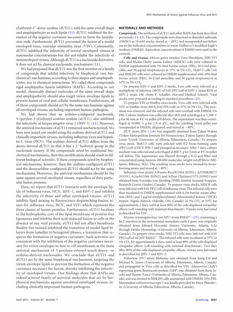
aUY11 localizes to virion envelope lipids. Fluorescence spec-tra depend on the polarity of the fluorochrome environment. Totest the localization of aUY11 in virions, we analyzed its fluores-cence spectra in different environments. aUY11 was added to 2.5ml of aqueous fusion buffer or to 2.5 ml of 1-octanol to a finalconcentration of 48 nM or 0.48 nM, respectively, in a polymeth-acrylate cuvette prewarmed to 37°C. Alternatively, aUY11 wasadded to 107 PFU of VSV, 107 PFU of HSV-1, 106 PFU of influenzavirus, 106 FFU of HCV, or 2 nmol protein-free POPC liposomes inthe same aqueous buffer to a final concentration of 48 nM. We alsoanalyzed the fluorescence spectra of dUY11 in the same environ-ments. Emission spectra were obtained at the maximum excita-tion wavelength, 455 nm, and were normalized to the highest flu-orescence signal intensity obtained for all conditions (set as 1).
As we had observed for dUY11 in the presence of a modelenveloped virus, VSV (11), the fluorescence spectra of aUY11were most similar when in the presence of HSV-1, HSV-2, influ-
enza virus, or HCV virions or protein-free liposomes (Fig. 3A).These viruses have different glycoproteins and different glycopro-tein contents but share a hydrophobic environment in the lipidcores of their envelopes. These spectra were most similar to thespectrum of aUY11 in octanol, which mimics the hydrophobiccore of the membrane, and clearly distinct from its spectrum inaqueous buffer without virions or liposomes. These results areconsistent with aUY11 localizing to a hydrophobic environmentin virions, such as the hydrophobic lipid core of the virion enve-lope, as we had proposed based on our previous studies withdUY11 and a model enveloped virus (11). More interestingly, weshow here that aUY11 localizes to a similar hydrophobic environ-ment in clinically important viruses, such as influenza virus, HCV,and herpesviruses.
We also took advantage of the intrinsic fluorescence of aUY11to evaluate its potential localization to other lipid membranes.Near-confluent Vero cell monolayers seeded onto coverslips weretreated with 250 nM PKH26-GL fluorescent cell dye (a generalmembrane dye) for 10 min at 37°C and then exposed to 2 �MaUY11 or dUY11 for 1 to 120 min, also at 37°C. The cells werewashed, fixed with 10% formalin, and mounted onto glass slides.aUY11 localized to plasma and intracellular membranes (Fig. 3B;see Movies S1A and B in the supplemental material). In summary,aUY11 localizes to cellular lipid membranes.
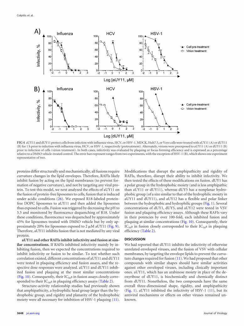
aUY11 protects cells from infection with influenza virus.Since aUY11 localizes to cellular membranes, we next tested theeffects of aUY11 on virion infectivity to cells treated before infec-tion. Cells were exposed to aUY11 for 1 h at 37°C. Following threewashes, the cells were infected with influenza virus, HCV, orHSV-1 and evaluated for plaque or focus formation. Infectivitywas expressed as a percentage of the infectivity in cells pretreatedwith vehicle control. Under these conditions, aUY11 protected thecells from infection by influenza virus and, less efficiently, HCVand HSV-1 (IC50s, 0.55, 3.5, and 4 �M, respectively) (Fig. 4A). For
FIG 1 Chemical and three-dimensional (3D) structures of the RAFIs aUY11, dUY11, aUY12, dUY5, and dUY1. The 3D structures (11) are displayed in threeorthogonal perspectives (gray, carbon; blue, hydrogen; red, oxygen; dark blue, nitrogen).
Colpitts et al.
3644 jvi.asm.org Journal of Virology

influenza virus, aUY11 acted at concentrations similar to thosewhen virions were exposed (the IC50 was only �1.5-fold higherthan when virions were preexposed). Similarly, dUY11 protectedcells from infection by influenza virus and, less efficiently, by HCVand HSV-1 (Fig. 4B). In summary, aUY11 and dUY11 protect cellsfrom infection by influenza virus at concentrations similar tothose at which they inhibit the infectivity of influenza virusvirions.
aUY11 inhibits the infectivity of influenza virus, HCV, orHSV-1 virions produced by treated cells. Viruses acquire theirenvelopes from the membranes of infected cells, and aUY11 local-izes to cell membranes (Fig. 3B). Therefore, we tested the effects ofaUY11 on the infectivity of virions produced by already infectedcells. MDCK, Huh7.5, or Vero cells were first infected with 5 or 0.5PFU/cell of influenza virus, HCV, or HSV-1. After 1 h, the inoculawere removed, and the cells were washed and overlaid with me-dium containing aUY11 for 24 or 48 h (for influenza virus andHSV-1 or HCV, respectively). Supernatants and cell lysates were
harvested, and influenza virus and HSV-1 virions were pelletedand resuspended in medium with no drugs, whereas HCV virionswere concentrated on a 100-kDa molecular-mass cutoff and thendiluted in medium with no drug to be titrated. Almost no infec-tious virus could be recovered from already infected cells incu-bated with 6 �M aUY11 (Fig. 5A). The concentrations at whichaUY11 inhibited the production of infectious virus by cells treatedafter infection were very similar to the concentrations at which itinhibited the infectivity of exposed virions. In cells infected at anMOI of 5, the IC50 for influenza virus, HCV, or HSV-1 was 0.2, 0.4,or 0.35 �M, respectively. In cells infected at an MOI of 0.5, theIC50 for influenza virus, HCV, or HSV-1 was 0.06, 0.14, or 0.28�M, respectively. We had previously shown that the RAFI dUY11does not affect viral replication in previously infected cells and hasno apparent effects on virion assembly or integrity, only minimalones on virion budding, and no cytotoxic effects (11). We havenow shown (Fig. 3B) that aUY11 localizes to the membranes oftreated cells. Therefore, the effects on the virions produced by cells
FIG 2 aUY11 is active against enveloped but otherwise unrelated viruses. (A) Infectivity of HSV-2, HCV, influenza virus, or Sindbis virus exposed to aUY11(open squares) or dUY11 (filled circles), tested by plaquing efficiency or focus-forming efficiency (average standard deviation [SD]; n � 3). (B) Infectivity ofmodel DNA and RNA enveloped viruses exposed to aUY11 or dUY11, tested by plaquing efficiency or focus-forming efficiency (average SD; n � 3). (C)Infectivity of nonenveloped poliovirus (open triangles), adenovirus (filled inverted triangles), or reovirus (filled diamonds) exposed to aUY11 (average range;n 2).
RAFI Mechanism of Action against Influenza Virus and HCV
April 2013 Volume 87 Number 7 jvi.asm.org 3645

treated after infection are likely the result of inhibition of the in-fectivity of the progeny virions, which acquire their envelopes bybudding from the cell membranes to which aUY11 localizes. Inthis model, aUY11 in the cellular membranes would become in-corporated into the virion envelopes during budding. To test themodel, we took advantage of the intrinsic fluorescence of aUY11and examined its fluorescence spectra in 104 influenza virus,HCV, or HSV-1 virions produced by cells treated with vehicle orin equivalent volumes from cells treated with aUY11. The charac-teristic emission spectrum of aUY11 was detected, at decreasingintensities, for samples treated with 6, 2, or 0.6 �M aUY11. Thespectrum was not detected at lower concentrations, most likely asa result of the limiting virions (100- or 1,000-fold fewer HCV andinfluenza virus or HSV-1 virions, respectively, than in Fig. 3A). Asexpected, aUY11 fluorescence was not detected in samples fromthe untreated cells (Fig. 5B). In summary, aUY11 added to alreadyinfected cells associated with the progeny virions produced.
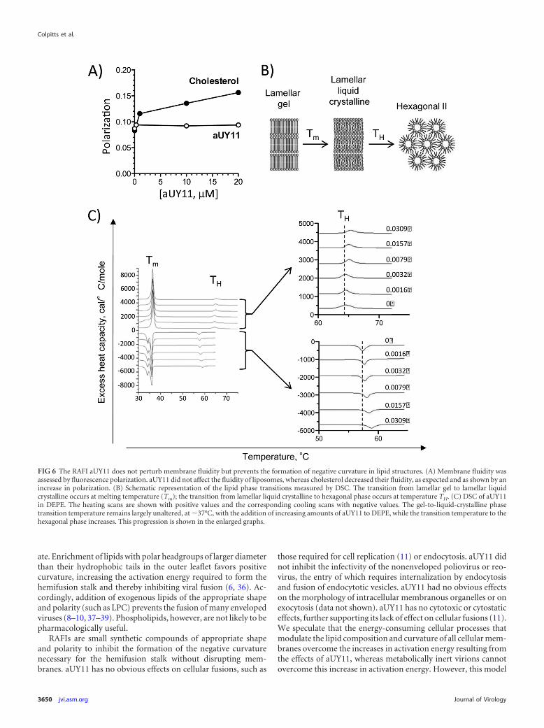
aUY11 does not perturb membrane fluidity. Compoundsthat target membranes can modulate their fluidity, which in turnaffects the infectivity of enveloped virions (20). For example, cho-lesterol increases the ordering of phospholipid acyl chain packing(21). Decreases in membrane fluidity reduce the infectivity of en-veloped virions, such as HIV (22). We therefore tested the effectsof aUY11 on membrane fluidity, using the DPH fluorescencepolarization method (23). As membrane fluidity decreases (suchas by addition of cholesterol), the polarization of DPH fluores-cence increases. The addition of aUY11 up to 20 �M (approxi-mately 100 times the IC50) to DOPC liposomes did not result inany such increase in DPH polarization (Fig. 6A), indicating thataUY11 does not affect membrane fluidity.
aUY11 inhibits the formation of lipid structures with nega-tive curvature. We next used differential scanning calorimetry(DSC) to test whether aUY11 inhibits the formation of lipid struc-tures with negative curvature. DEPE lamellar phases were recon-stituted with increasing concentrations of aUY11. The transitionfrom the flat morphology of the lamellar phase to the negativecurvature of the inverted hexagonal phase was evaluated by DSC(Fig. 6B). Less than 3% aUY11 in DEPE increased the transition
temperature between the lamellar and inverted hexagonal phasesby �1°C (Fig. 6C). These effects on the transition temperaturerequired for the formation of negative monolayer curvature, char-acterized by a regression of 29 7, indicate that aUY11 disfavorsthe formation of negative membrane monolayer curvature.
The cooling scans, which evaluate the reciprocal inverted-hex-agonal-to-lamellar phase transition, exhibit a characteristic hys-teresis caused by kinetic factors. They also show that aUY11 raisesthe hexagonal-to-bilayer transition temperature, characterized bya regression of 49 7 (Fig. 6C). The facts that a characteristictransition due to hysteresis is recovered on each cooling scan andthat the cooling regression is similar to that obtained in the heat-ing scans demonstrate that aUY11 did not affect the integrity ofthe multilamellar membranes. They were not disrupted or lysedby aUY11, as can also be ascertained by the constancy of the maintransition, which appears at 37°C on reheating. The hysteresis ofDSC transitions of phosphatidylethanolamines has been previ-ously noted (24).
Taken together, the DSC shows that aUY11 raises the transi-tion temperature necessary to form inverted hexagonal phaseswithout disrupting the membranes or affecting their fluidity(Fig. 6A). This is consistent with inhibitors of fusion that preventthe formation of negative monolayer curvature (25).
aUY11 inhibits the fusion of viral and cellular membranes.Since aUY11 and dUY11 inhibit the changes in membrane curva-ture required for fusion (Fig. 6C), we next tested whether aUY11inhibited virion-envelope-to-cell-membrane fusion. We used flu-orescence-dequenching fusion assays, which analyze lipid mixingbetween the outer leaflets of virions and target cells.
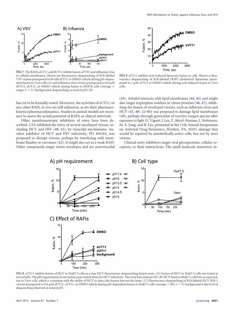
VSV virions labeled with self-quenching concentrations of R18were exposed to aUY11 prior to mixing with Vero cells. Fusionwas triggered by lowering the pH and analyzed by the dequench-ing of R18 fluorescence. Fluorescence was dequenched by �15%when VSV virions exposed to DMSO vehicle were induced to fuseto target cells, but by only 5% when VSV virions exposed to aUY11were induced to fuse under the same conditions (Fig. 7A).
We next tested the effects of aUY11 and dUY11 on fusion of aclinically important virus, influenza virus (A/Puerto Rico/8/
TABLE 1 Inhibition of infectivity of enveloped but otherwise unrelated viruses or nonenveloped viruses
Virus family Virus
IC50 (�M)a
aUY11 dUY11
Orthomyxoviridae Influenza virus A/PR/8/34 (H1N1) 0.283 0.130 0.187 0.051Influenza virus A/USSR/90/77 (H1N1) 0.078 0.019 0.097 0.021Influenza virus A/Aichi/2/68 (H3N2) 0.078 0.007 0.143 0.045Influenza virus A/PC/1/73 (H3N2) 0.090 0.026 0.117 0.032
Flaviviridae HCV JFH-1 0.187 0.060 0.107 0.041Togaviridae Sindbis 0.006 0.001 0.011 0.005b
Rhabdoviridae VSV 0.005 0.001 0.002 0.001Herpesviridae HSV-1 KOS 0.131 0.034b 0.048 0.012b
HSV-2 186 0.031 0.008 0.052 0.003mCMV 0.037 0.016 0.013 0.004
Picornaviridae Polio �200 �200b
Adenoviridae Adenovirus �20 �20b
Reoviridae Reovirus �20 NDa Average SD (n 3). ND, not done.b Data from reference 11.
Colpitts et al.
3646 jvi.asm.org Journal of Virology
34[H1N1]). Influenza virus virions labeled at self-quenching con-centrations of R18 were exposed to aUY11 or dUY11 prior tomixing with MDCK cells. Fusion was triggered by increasing thetemperature and decreasing the pH to 5. Under these conditions,fluorescence was dequenched by approximately 8% for influenzavirus virions treated with DMSO vehicle but by less than 1%(background levels) for influenza virus virions treated with 600nM dUY11 or aUY11, even after 2.5 h (influenza virus fuses in lessthan 10 min). The background dequenching at neutral pH was�2% after 2.5 h (Fig. 7B).
According to our model, RAFIs should inhibit the infectivity ofenveloped (but otherwise unrelated) viruses by the same mecha-nisms. We tested whether aUY11 also inhibited the fusion of an-other clinically relevant virus, HCV. To this end, we first devel-oped a fusion assay using HCV JFH-1 virions and Huh7.5 cells.HCV JFH-1 virions labeled at self-quenching concentrations ofR18 were mixed with Huh7.5 cells. Fusion was triggered by in-
creasing the temperature to 37°C and decreasing the pH (Fig. 8A).As expected, HCV fused only to Huh7.5 cells, and not to Verocells, at an optimum pH of 5.0 to 5.5, consistent with fusion toearlier endosomes (Fig. 8B). In summary, the requirements for pHand cell type in our fusion assays match the requirements for HCVinfectivity.
We then used the HCV fusion assays to test whether aUY11 ordUY11 inhibited the fusion of HCV to Huh7.5 cells. R18-labeledHCV JFH-1 virions were exposed to aUY11 or dUY11 and thenmixed with Huh7.5 cells. Fluorescence was dequenched by ap-proximately 12% for HCV virions treated with DMSO vehicle butby less than 5% for HCV virions treated with 600 nM dUY11 oraUY11 (IC99 in infectivity assays), in the range of the backgrounddequenching at neutral pH in these assays (�3%) (Fig. 8C).
aUY11 inhibits fusion by acting on lipids, not proteins. In-fluenza virus has a class I fusion protein, whereas HCV is thoughtto have a class II and VSV a class III protein. Although these fusion
FIG 3 aUY11 localizes to lipid membranes. (A) The emission spectra of aUY11 and dUY11 in virions or protein-free liposomes are most similar, and closelyresemble their spectra in hydrophobic environments. aUY11 or dUY11 was added to aqueous buffer (gray) or to 1-octanol (black) to a final concentration of 48nM or 0.48 nM, respectively. aUY11 or dUY11 (48 nM final concentration) was also added to 106 PFU of influenza virus A (blue), 106 FFU of HCV (orange), 107
PFU of HSV-1 (green), 107 PFU of HSV-2 (teal), or 107 PFU of VSV (pink) or to 2 nmol liposomes (red) in 2.5 ml of aqueous buffer. Fluorescence was excitedat 455 nm. Emission spectra for dUY11 in VSV virions, liposomes, aqueous buffer, and octanol were determined previously (11). (B) The RAFI aUY11 localizesto cellular lipid membranes. Vero cell monolayers exposed to PKH26 general membrane dye for 10 min at 37°C were washed and then exposed to aUY11 ordUY11 for 1, 5, 15, 40, or 120 min at 37°C. Shown are confocal microscopy images; scale bars, 25 �m.
RAFI Mechanism of Action against Influenza Virus and HCV
April 2013 Volume 87 Number 7 jvi.asm.org 3647
proteins differ structurally and mechanistically, all fusions requirecurvature changes in the lipid envelopes. Therefore, RAFIs likelyinhibit fusion by acting on the lipid membranes (to prevent for-mation of negative curvature), and not by targeting any viral pro-tein. To test this model, we next analyzed the effects of aUY11 onthe fusion of protein-free liposomes to cells, fusion that is inducedunder acidic conditions (26). We exposed R18-labeled protein-free DOPC liposomes to aUY11 and then added the liposomesthus exposed to cells. Fusion was triggered by decreasing the pH to5.5 and monitored by fluorescence dequenching of R18. Underthese conditions, fluorescence was dequenched by approximately45% for liposomes treated with DMSO vehicle but by only ap-proximately 20% for liposomes exposed to 2 �M aUY11 (Fig. 9).Therefore, aUY11 inhibits fusion that is not mediated by any viralprotein.
aUY11 and other RAFIs inhibit infectivity and fusion at sim-ilar concentrations. If RAFIs inhibited infectivity mainly by in-hibiting fusion, then we expected the concentrations required toinhibit infectivity or fusion to be similar. To test whether suchcorrelation existed, different concentrations of aUY11 and dUY11were tested in plaquing efficiency and fusion assays, and the re-spective dose responses were analyzed. aUY11 and dUY11 inhib-ited fusion and plaquing at the most similar concentrations(Fig. 10). Consequently, their IC50s in fusion assays closely corre-sponded to their IC50s in plaquing efficiency assays (Table 2).
Structure-activity relationship studies had previously shownthat amphipathicity, a hydrophilic head group larger than the hy-drophobic group, and rigidity and planarity of the hydrophobicmoiety were all necessary for inhibition of HSV-1 plaquing (11).
Modifications that disrupt the amphipathicity and rigidity ofRAFIs, therefore, disrupt their ability to inhibit infectivity. Wethen tested the effects of these modifications on fusion. dUY1 hasa polar group in the hydrophobic moiety (and is less amphipathicthan aUY11 or dUY11), whereas dUY5 has a nonplanar hydro-phobic group (of a size similar to that of the hydrophilic moiety inaUY11 and dUY11), and aUY12 has a flexible and polar linkerbetween the hydrophobic and hydrophilic groups (Fig. 1). Severalconcentrations of dUY1, dUY5, and aUY12 were tested in VSVfusion and plaquing efficiency assays. Although these RAFIs varyin their potencies by over 100-fold, each inhibited fusion andplaquing at similar concentrations (Fig. 10). Consequently, theirIC50s in fusion closely corresponded to their IC50s in plaquingefficiency (Table 2).
DISCUSSION
We had reported that dUY11 inhibits the infectivity of otherwiseunrelated enveloped viruses, and the fusion of VSV with cellularmembranes, by targeting the envelope lipids to prevent the curva-ture changes required for fusion (11). We had proposed that othercompounds with similar shapes should have similar activitiesagainst other enveloped viruses, including clinically importantones. aUY11, which has an arabinose moiety in place of the de-oxyribose of dUY11, is biochemically and chemically distinctfrom dUY11. Nonetheless, the two compounds have the sameoverall three-dimensional shape, rigidity, and amphipathicity(Fig. 1). aUY11 inhibited the infectivity of HSV-1 (11), but itsantiviral mechanisms or effects on other viruses remained un-known.
FIG 4 aUY11 and dUY11 protect cells from infection with influenza virus, HCV, or HSV-1. MDCK, Huh7.5, or Vero cells were treated with aUY11 (A) or dUY11(B) for 1 h prior to infection with influenza virus, HCV, or HSV-1, respectively (pretreatment). Alternately, virions were preexposed to aUY11 (A) or dUY11 (B)prior to infection of cells (virion treatment). In both cases, infectivity was evaluated by plaquing or focus-forming efficiency and is expressed as a percentagerelative to a DMSO vehicle-treated control. The error bars represent ranges from two experiments, with the exception of HSV-1 (B), which shows one experimentrepresentative of two.
Colpitts et al.
3648 jvi.asm.org Journal of Virology

aUY11 inhibited, with similar potencies, the infectivity of clin-ically important viruses (influenza virus A, HCV, HSV-1, HSV-2,and Sindbis virus) with DNA or RNA genomes, which replicate inthe nucleus or in the cytoplasm and which use different cellularreceptors and enter cells by different fusion mechanisms. In con-trast, it failed to inhibit the infectivity of nonenveloped viruses.The targets of aUY11 are therefore conserved only among envel-oped viruses. We propose that aUY11 most likely targets the con-served lipid core of the envelopes. Consistently, aUY11 interactswith a hydrophobic component of enveloped virions (such as theenvelope lipids) or protein-free liposomes and inhibits the fusionof these viruses and liposomes to target cells.
Influenza virus hemagglutinin (HA), like other class I fusionproteins, is predominantly composed of alpha helices containingan N-terminal hydrophobic fusion peptide (27) and forms trimersbefore and after fusion. The proposed mechanism for HA suggeststhe folding of the uncleaved protein to a metastable state (28),which is then activated by cleavage (27, 29). Fusion is triggered bylow pH, resulting in irreversible conformational changes leadingto a more stable postfusion conformation (30). HCV E2 is pro-posed to be a class II fusion protein, like those in other flaviviridae(31), which consist predominantly of beta sheets with the fusionpeptide in internal loops (32). Class II fusion proteins are dimersparallel to the virion surface in their prefusion state but undergoconformational rearrangements to form postfusion trimers pro-jecting perpendicularly from the virion envelope (32). VSV G pro-tein is a class III fusion protein consisting of a mixed alpha helixand beta sheet structure (33). Class III fusion proteins undergo
reversible conformational changes. Extended exposure to low pHinactivates the virions, but fusion is recovered when the pH israised again (34).
The fusion proteins of influenza virus, HCV, and VSV repre-sent all three different classes, with different structures and fusionmechanisms. However, aUY11 and dUY11 inhibited fusion of allthree viruses at similar concentrations. Their targets are thereforenot likely the fusion proteins but something even more conservedamong enveloped viruses. aUY11 also inhibited acid-induced fu-sion of protein-free liposomes to cells (in the absence of any viralprotein), strongly supporting the model in which RAFIs inhibitinfectivity by acting on lipids. Although liposome fusion was in-hibited only partially, RAFIs are the only small molecules knownto inhibit this process, indicating that they directly target the lip-ids. The lipids in the envelopes of all enveloped viruses (or vesi-cles) must form a hemifusion stalk structure when fusing to targetcell membranes (4). This process requires the formation of nega-tive curvature by the outer leaflet of the envelope. aUY11 inhibitsthe transition from the lamellar phase (flat) to the hexagonal IIphase (with negative curvature), indicating that it inhibits the for-mation of the negative curvature required for fusion. These resultssupport the model in which RAFIs target virion envelope lipids toprevent fusion of viral and cellular membranes by biophysicalmechanisms (i.e., inhibiting the formation of negative curvaturein virion envelopes).
Virion fusion is affected by the lipid composition of the enve-lope (35). The molecular shape of lipids affects the formation ofthe negative curvature of the hemifusion stalk fusion intermedi-
FIG 5 aUY11 inhibits the infectivity of influenza virus, HCV, and HSV-1 virions produced by infected cells. (A) Cells infected with 5 or 0.5 PFU or FFU per cellof influenza virus, HCV, or HSV-1 were incubated in the presence of aUY11 for 23 h (influenza virus and HSV-1) or 44 h (HCV). Supernatants and cell lysateswere harvested and titrated for the presence of infectious virus. (B) Virions harvested in panel A were tested for the presence of aUY11 by examining itsfluorescence spectra. aUY11 fluorescence was excited at 455 nm.
RAFI Mechanism of Action against Influenza Virus and HCV
April 2013 Volume 87 Number 7 jvi.asm.org 3649
ate. Enrichment of lipids with polar headgroups of larger diameterthan their hydrophobic tails in the outer leaflet favors positivecurvature, increasing the activation energy required to form thehemifusion stalk and thereby inhibiting viral fusion (6, 36). Ac-cordingly, addition of exogenous lipids of the appropriate shapeand polarity (such as LPC) prevents the fusion of many envelopedviruses (8–10, 37–39). Phospholipids, however, are not likely to bepharmacologically useful.
RAFIs are small synthetic compounds of appropriate shapeand polarity to inhibit the formation of the negative curvaturenecessary for the hemifusion stalk without disrupting mem-branes. aUY11 has no obvious effects on cellular fusions, such as
those required for cell replication (11) or endocytosis. aUY11 didnot inhibit the infectivity of the nonenveloped poliovirus or reo-virus, the entry of which requires internalization by endocytosisand fusion of endocytotic vesicles. aUY11 had no obvious effectson the morphology of intracellular membranous organelles or onexocytosis (data not shown). aUY11 has no cytotoxic or cytostaticeffects, further supporting its lack of effect on cellular fusions (11).We speculate that the energy-consuming cellular processes thatmodulate the lipid composition and curvature of all cellular mem-branes overcome the increases in activation energy resulting fromthe effects of aUY11, whereas metabolically inert virions cannotovercome this increase in activation energy. However, this model
FIG 6 The RAFI aUY11 does not perturb membrane fluidity but prevents the formation of negative curvature in lipid structures. (A) Membrane fluidity wasassessed by fluorescence polarization. aUY11 did not affect the fluidity of liposomes, whereas cholesterol decreased their fluidity, as expected and as shown by anincrease in polarization. (B) Schematic representation of the lipid phase transitions measured by DSC. The transition from lamellar gel to lamellar liquidcrystalline occurs at melting temperature (Tm); the transition from lamellar liquid crystalline to hexagonal phase occurs at temperature TH. (C) DSC of aUY11in DEPE. The heating scans are shown with positive values and the corresponding cooling scans with negative values. The gel-to-liquid-crystalline phasetransition temperature remains largely unaltered, at �37°C, with the addition of increasing amounts of aUY11 to DEPE, while the transition temperature to thehexagonal phase increases. This progression is shown in the enlarged graphs.
Colpitts et al.
3650 jvi.asm.org Journal of Virology
has yet to be formally tested. Moreover, the activities of aUY11, orany other RAFI, in vivo are still unknown, as are their pharmaco-kinetics/pharmacodynamics. Studies in animal models are neces-sary to assess the actual potential of RAFIs as clinical antivirals.
Other membranotropic inhibitors of entry have been de-scribed. C5A inhibited the entry of several enveloped viruses, in-cluding HCV and HIV (40, 41), by virucidal mechanisms. An-other inhibitor of HCV and HIV infectivity, PD 404182, wasproposed to disrupt virions, perhaps by interfering with mem-brane fluidity or curvature (42). It might also act as a weak RAFI.Other compounds target virion envelopes and are nonvirucidal
(43). Arbidol interacts with lipid membranes (44, 45) and mightalso target tryptophan residues in virion proteins (46, 47), inhib-iting the fusion of enveloped viruses, such as influenza virus andHCV (47, 48). LJ-001 was proposed to damage lipid membranes(49), perhaps through generation of reactive oxygen species afterexposure to light (F. Vigant, J. Lee, Z. Akyol-Ataman, L. Robinson,M. E. Jung, and B. Lee, presented at the 11th Annual Symposiumon Antiviral Drug Resistance, Hershey, PA, 2010), damage thatwould be repaired by metabolically active cells, but not by inertvirions.
Clinical entry inhibitors target viral glycoproteins, cellular re-ceptors, or their interactions. The small molecule maraviroc in-
FIG 7 The RAFIs aUY11 and dUY11 inhibit fusion of VSV and influenza virusto cellular membranes. Shown are fluorescence dequenching of R18-labeledVSV virions preexposed to 60 nM aUY11 or DMSO vehicle during pH-depen-dent fusion to Vero cells (A) and influenza virus virions preexposed to 0.6 �MdUY11, aUY11, or DMSO vehicle during fusion to MDCK cells (average range; n 2) (background, dequenching at neutral pH) (B).
FIG 8 aUY11 inhibits fusion of HCV to Huh7.5 cells in a new HCV fluorescence dequenching fusion assay. (A) Fusion of HCV to Huh7.5 cells was tested atseveral pHs. The pH requirements in our fusion assay match those for HCV infectivity. The error bars indicate SD. (B) HCV fused to Huh7.5 cells but, as expected,not to Vero cells, which is consistent with the ability of HCV to infect the former but not the latter. (C) Fluorescence dequenching of R18-labeled HCV JFH-1virions preexposed to 0.6 �M dUY11, aUY11, or DMSO vehicle during pH-dependent fusion to Huh7.5 cells (average SD; n 3); background is the level ofdequenching observed at neutral pH.
FIG 9 aUY11 inhibits acid-induced liposome fusion to cells. Shown is fluo-rescence dequenching of R18-labeled DOPC-cholesterol liposomes preex-posed to 2 �M aUY11 or DMSO vehicle during acid-induced fusion to Verocells.
RAFI Mechanism of Action against Influenza Virus and HCV
April 2013 Volume 87 Number 7 jvi.asm.org 3651

hibits the interactions between HIV gp120 and CCR5 (1), whereasthe biomimetic peptide enfuvirtide inhibits the structural rear-rangements of HIV gp41 (19, 50–52). Antibodies have also beenexplored as inhibitors of infectivity, mostly against respiratorysyncytial virus (RSV) (53). Unfortunately, peptides and antibod-ies have poor oral bioavailability or stability (54). Furthermore, allthese approaches suffer from the limitations of targeting any viralprotein, such as selection for resistance (3, 55), which is particu-larly important for the viral surface proteins that commonlychange rapidly to avoid immune responses (56). In contrast,RAFIs target viral envelope lipids, which are not encoded in theviral genome and are an essential structural component of all en-veloped viruses. Therefore, they are expected to have a higherbarrier to selection for resistance. Consistent with this expecta-tion, we have not yet been able to select for dUY11-resistantHSV-1 mutants.
Importantly, RAFIs also protect cells from infection. aUY11was particularly effective at preventing infection by influenza vi-rus, which is internalized by endocytosis before fusion is triggeredby the conformational changes in the virion glycoproteins at the
low pH in the late endosomes (57). Pretreatment of cells was lesseffective at inhibiting the infectivity of HSV-1, which fuses to mostcell types at neutral pH at the plasma membrane (58), or HCV,which follows a more complex entry pathway to fuse at a pHtypical of earlier endosomes. aUY11 is internalized to many intra-cellular membranes. It may well accumulate in the late endo-somes, which would result in a high-concentration reservoir towhich the influenza virus, but not HSV-1 or HCV, virions wouldbe exposed before fusion. Alternatively, the lack of inactivation ofHSV-1 or HCV at low pH may allow these virions to make re-peated attempts to fuse until one is successful.
aUY11 is also effective for treating cells already infected withinfluenza virus, HCV, or HSV-1. Although it does not protect thecells from virus-induced cytopathic effects, as expected from itsmechanisms of action, the virions produced from the infectedcells are not infectious. This effect is likely due to the incorpora-tion of aUY11 in progeny virion envelopes, which are acquiredduring budding from the cellular membranes to which aUY11localizes.
In summary, chemically distinct compounds with the sameoverall three-dimensional shape and amphipathicity inhibit viralinfectivity by inhibiting fusion of viral and cellular membranes.aUY11 inhibits viral fusion due to its shape and amphipathicitythrough biophysical, not biochemical, mechanisms. Specifically, itinhibits the formation of the negative curvature necessary for theformation of the hemifusion stalk, a critical step in the fusion ofenveloped viruses. aUY11 acts by the same biophysical mecha-nisms on several enveloped but otherwise unrelated viruses, in-cluding important human pathogens, such as influenza virus,HCV, and HSV-1/2. Here, we defined the mechanisms of RAFIs
FIG 10 RAFIs inhibit VSV plaquing efficiency and fusion to Vero cells at similar concentrations. Shown are comparisons of inhibition of fusion (open squares)or infectivity (filled circles) by RAFIs. Lipid-mixing assays were conducted as described in the legend to Fig. 7. For plaquing assays, approximately 200 VSV virionswere exposed to increasing concentrations of aUY11, dUY11, aUY12, dUY5, or dUY1 for 10 min at 37°C. The virions thus exposed were then used to infect 5 �106 Vero cells seeded in 6-well plates. Plaquing efficiency was calculated as a percentage of plaques formed by virions exposed to DMSO vehicle.
TABLE 2 IC50 for fusion correlates with IC50 for plaquing efficiency
RAFI
IC50 (�M)
Fusion Plaquing
dUY11 0.011 0.009aUY11 0.010 0.010dUY5 3.0 2.5aUY12 5.0 2.5
Colpitts et al.
3652 jvi.asm.org Journal of Virology

against such clinically important viruses. These mechanisms arefully consistent with those we had proposed based on dUY11 anda single model enveloped virus, VSV (11). We conclude thatRAFIs are a novel family of antiviral compounds that act by bio-physical mechanisms to prevent fusion of viral to cellular mem-branes.
ACKNOWLEDGMENTS
This research was supported by Canadian Institute of Health Researchgrants to L.M.S. (MOP 106452) and R.M.E. (MOP 86608) and the Bur-roughs Wellcome Fund (L.M.S.). L.M.S. is a Burroughs Wellcome Inves-tigator in the Pathogenesis of Infectious Disease. V.A.K. was supported bythe Molecular and Cellular Biology Program of the Russian Academy ofSciences; A.V.U. was supported by the Dynasty Foundation and a RussianPresident scholarship for young investigators (project SP-2494.2012.4).C.C.C. thanks the Natural Sciences and Engineering Research Council ofCanada and Alberta Innovates-Health Solutions for graduate scholar-ships.
We are grateful to Takaji Wakita, Charles Rice, Veronika vonMessling, Edward Mocarski, and Maya Shmulevitz for the kind gifts ofHCV JFH-1, Huh7.5 cells, influenza virus strains, mCMV, and reovirusT3, respectively, and to Gary Eitzen for his assistance with the fusionassays.
REFERENCES1. Dorr P, Westby M, Dobbs S, Griffin P, Irvine B, Macartney M, Mori J,
Rickett G, Smith-Burchnell C, Napier C, Webster R, Armour D, PriceD, Stammen B, Wood A, Perros M. 2005. Maraviroc (UK-427,857), apotent, orally bioavailable, and selective small-molecule inhibitor ofchemokine receptor CCR5 with broad-spectrum anti-human immunode-ficiency virus type 1 activity. Antimicrob. Agents Chemother. 49:4721–4732.
2. Wild CT, Shugars DC, Greenwell TK, McDanal CB, Matthews TJ. 1994.Peptides corresponding to a predictive alpha-helical domain of humanimmunodeficiency virus type 1 gp41 are potent inhibitors of virus infec-tion. Proc. Natl. Acad. Sci. U. S. A. 91:9770 –9774.
3. Wei X, Decker JM, Liu H, Zhang Z, Arani RB, Kilby JM, Saag MS, WuX, Shaw GM, Kappes JC. 2002. Emergence of resistant human immuno-deficiency virus type 1 in patients receiving fusion inhibitor (T-20) mono-therapy. Antimicrob. Agents Chemother. 46:1896 –1905.
4. Harrison SC. 2008. Viral membrane fusion. Nat. Struct. Mol. Biol. 15:690 – 698.
5. Earp LJ, Delos SE, Park HE, White JM. 2005. The many mechanisms ofviral membrane fusion proteins. Curr. Top. Microbiol. Immunol. 285:25– 66.
6. Chernomordik LV, Kozlov MM. 2003. Protein-lipid interplay in fusionand fission of biological membranes. Annu. Rev. Biochem. 72:175–207.
7. Chernomordik LV, Leikina E, Frolov VA, Bronk P, Zimmerberg J.1997. An early stage of membrane fusion mediated by the low pH confor-mation of influenza hemagglutinin depends upon membrane lipids. J. CellBiol. 136:81–93.
8. Gunther-Ausborn S, Praetor A, Stegmann T. 1995. Inhibition of influ-enza-induced membrane fusion by lysophosphatidylcholine. J. Biol.Chem. 270:29279 –29285.
9. Gaudin Y. 2000. Rabies virus-induced membrane fusion pathway. J. CellBiol. 150:601– 612.
10. Yeagle PL, Smith FT, Young JE, Flanagan TD. 1994. Inhibition ofmembrane fusion by lysophosphatidylcholine. Biochemistry 33:1820 –1827.
11. St Vincent MR, Colpitts CC, Ustinov AV, Muqadas M, Joyce MA,Barsby NL, Epand RF, Epand RM, Khramyshev SA, Valueva OA,Korshun VA, Tyrrell DL, Schang LM. 2010. Rigid amphipathic fusioninhibitors, small molecule antiviral compounds against enveloped viruses.Proc. Natl. Acad. Sci. U. S. A. 107:17339 –17344.
12. Andronova VL, Skorobogatyi MV, Manasova EV, Berlin I, KorshunVA, Galegov GA. 2003. Antiviral activity of some 5-arylethynyl 2=-deoxyuridine derivatives. Bioorg. Khim. 29:290 –295.
13. Skorobogatyi MV, Ustinov AV, Stepanova IA, Pchelintseva AA,Petrunina AL, Andronova VL, Galegov GA, Malakhov DA, Korshun
VA. 2006. 5-Arylethynyl-2=-deoxyuridines, compounds active againstHSV-1. Org. Biomol. Chem. 4:1091–1096.
14. Blight KJ, McKeating JA, Rice CM. 2002. Highly permissive cell lines forsubgenomic and genomic hepatitis C virus RNA replication. J. Virol. 76:13001–13014.
15. Lacasse JJ, Schang LM. 2010. During lytic infections, herpes simplex virustype 1 DNA is in complexes with the properties of unstable nucleosomes.J. Virol. 84:1920 –1933.
16. Wakita T, Pietschmann T, Kato T, Date T, Miyamoto M, Zhao Z,Murthy K, Habermann A, Krausslich HG, Mizokami M, BartenschlagerR, Liang TJ. 2005. Production of infectious hepatitis C virus in tissueculture from a cloned viral genome. Nat. Med. 11:791–796.
17. Stoddart CA, Cardin RD, Boname JM, Manning WC, Abenes GB,Mocarski ES. 1994. Peripheral blood mononuclear phagocytes mediatedissemination of murine cytomegalovirus. J. Virol. 68:6243– 6253.
18. Lentz BR. 1989. Membrane “fluidity” as detected by diphenylhexatrieneprobes. Chem. Phys. Lipids 50:171–190.
19. Schmidt AG, Yang PL, Harrison SC. 2010. Peptide inhibitors of dengue-virus entry target a late-stage fusion intermediate. PLoS Pathog.6:e1000851. doi:10.1371/journal.ppat.1000851.
20. Harada S. 2005. The broad anti-viral agent glycyrrhizin directly modu-lates the fluidity of plasma membrane and HIV-1 envelope. Biochem. J.392:191–199.
21. Lande MB, Donovan JM, Zeidel ML. 1995. The relationship betweenmembrane fluidity and permeabilities to water, solutes, ammonia, andprotons. J. Gen. Physiol. 106:67– 84.
22. Harada S, Yusa K, Monde K, Akaike T, Maeda Y. 2005. Influence ofmembrane fluidity on human immunodeficiency virus type 1 entry.Biochem. Biophys. Res. Commun. 329:480 – 486.
23. Shinitzky M, Barenholz Y. 1978. Fluidity parameters of lipid regionsdetermined by fluorescence polarization. Biochim. Biophys. Acta 515:367–394.
24. Epand RM, Epand RF. 1988. Kinetic effects in the differential scanningcalorimetry cooling scans of phosphatidylethanolamines. Chem. Phys.Lipids 49:101–104.
25. Epand RM. 1986. Virus replication inhibitory peptide inhibits the con-version of phospholipid bilayers to the hexagonal phase. Biosci. Rep.6:647– 653.
26. Connor J, Yatvin MB, Huang L. 1984. pH-sensitive liposomes: acid-induced liposome fusion. Proc. Natl. Acad. Sci. U. S. A. 81:1715–1718.
27. Wilson IA, Skehel JJ, Wiley DC. 1981. Structure of the haemagglutininmembrane glycoprotein of influenza virus at 3A resolution. Nature 289:366 –373.
28. Chen J, Lee KH, Steinhauer DA, Stevens DJ, Skehel JJ, Wiley DC. 1998.Structure of the hemagglutinin precursor cleavage site, a determinant ofinfluenza pathogenicity and the origin of the labile conformation. Cell95:409 – 417.
29. Wiley DC, Skehel JJ. 1987. The structure and function of the hemagglu-tinin membrane glycoprotein of influenza virus. Annu. Rev. Biochem.56:365–394.
30. Bullough PA, Hughson FM, Skehel JJ, Wiley DC. 1994. Structure ofinfluenza haemagglutinin at the pH of membrane fusion. Nature 371:37– 43.
31. Krey T, d’Alayer J, Kikuti CM, Saulnier A, Damier-Piolle L, Petitpas I,Johansson DX, Tawar RG, Baron B, Robert B, England P, Persson MA,Martin A, Rey FA. 2010. The disulfide bonds in glycoprotein E2 of hep-atitis C virus reveal the tertiary organization of the molecule. PLoS Pathog.6:e1000762. doi:10.1371/journal.ppat.1000762.
32. Kielian M. 2006. Class II virus membrane fusion proteins. Virology 344:38 – 47.
33. Roche S, Rey RA, Gaudin Y, Bressanelli S. 2007. Structure of the prefu-sion form of the vesicular stomatitis virus glycoprotein G. Science 315:843– 848.
34. Gaudin Y, Tuffereau C, Segretain D, Knossow M, Flamand A. 1991.Reversible conformational changes and fusion activity of rabies virus gly-coprotein. J. Virol. 65:4853– 4859.
35. Teissier E, Pecheur EI. 2007. Lipids as modulators of membrane fusionmediated by viral fusion proteins. Eur. Biophys. J. 36:887– 899.
36. Chernomordik L. 1996. Non-bilayer lipids and biological fusion interme-diates. Chem. Phys. Lipids 81:203–213.
37. Chernomordik LV, Leikina E, Kozlov MM, Frolov VA, Zimmerberg J.1999. Structural intermediates in influenza haemagglutinin-mediated fu-sion. Mol. Membr. Biol. 16:33– 42.
RAFI Mechanism of Action against Influenza Virus and HCV
April 2013 Volume 87 Number 7 jvi.asm.org 3653

38. Stiasny K, Heinz FX. 2004. Effect of membrane curvature-modifyinglipids on membrane fusion by tick-borne encephalitis virus. J. Virol. 78:8536 – 8542.
39. Vogel SS, Leikina EA, Chernomordik LV. 1993. Lysophosphatidylcho-line reversibly arrests exocytosis and viral fusion at a stage between trig-gering and membrane merger. J. Biol. Chem. 268:25764 –25768.
40. Bobardt MD, Cheng G, de Witte L, Selvarajah S, Chatterji U, Sanders-Beer BE, Geijtenbeek TB, Chisari FV, Gallay PA. 2008. Hepatitis C virusNS5A anchor peptide disrupts human immunodeficiency virus. Proc.Natl. Acad. Sci. U. S. A. 105:5525–5530.
41. Cheng G, Montero A, Gastaminza P, Whitten-Bauer C, Wieland SF,Isogawa M, Fredericksen B, Selvarajah S, Gallay PA, Ghadiri MR,Chisari FV. 2008. A virocidal amphipathic alpha-helical peptide that in-hibits hepatitis C virus infection in vitro. Proc. Natl. Acad. Sci. U. S. A.105:3088 –3093.
42. Chamoun AM, Chockalingam K, Bobardt M, Simeon R, Chang J,Gallay P, Chen Z. 2012. PD 404,182 is a virocidal small molecule thatdisrupts hepatitis C virus and human immunodeficiency virus. Antimi-crob. Agents Chemother. 56:672– 681.
43. Teissier E, Penin F, Pecheur EI. 2011. Targeting cell entry of envelopedviruses as an antiviral strategy. Molecules 16:221–250.
44. Pecheur EI, Lavillette D, Alcaras F, Molle J, Boriskin YS, Roberts M,Cosset FL, Polyak SJ. 2007. Biochemical mechanism of hepatitis C virusinhibition by the broad-spectrum antiviral arbidol. Biochemistry 46:6050 – 6059.
45. Villalain J. 2010. Membranotropic effects of arbidol, a broad anti-viralmolecule, on phospholipid model membranes. J. Phys. Chem. B 114:8544 – 8554.
46. Leneva IA, Russell RJ, Boriskin YS, Hay AJ. 2009. Characteristics ofarbidol-resistant mutants of influenza virus: implications for the mecha-nism of anti-influenza action of arbidol. Antiviral Res. 81:132–140.
47. Teissier E, Zandomeneghi G, Loquet A, Lavillette D, Lavergne JP,Montserret R, Cosset FL, Bockmann A, Meier BH, Penin F, Pecheur EI.2011. Mechanism of inhibition of enveloped virus membrane fusion bythe antiviral drug arbidol. PLoS One 6:e15874. doi:10.1371/journal.pone.0015874.
48. Boriskin YS, Leneva IA, Pecheur EI, Polyak SJ. 2008. Arbidol: a broad-spectrum antiviral compound that blocks viral fusion. Curr. Med. Chem.15:997–1005.
49. Wolf MC, Freiberg AN, Zhang T, Akyol-Ataman Z, Grock A, Hong PW,Li J, Watson NF, Fang AQ, Aguilar HC, Porotto M, Honko AN,Damoiseaux R, Miller JP, Woodson SE, Chantasirivisal S, Fontanes V,Negrete OA, Krogstad P, Dasgupta A, Moscona A, Hensley LE, WhelanSP, Faull KF, Holbrook MR, Jung ME, Lee B. 2010. A broad-spectrumantiviral targeting entry of enveloped viruses. Proc. Natl. Acad. Sci. U. S. A.107:3157–3162.
50. Dwyer JJ, Wilson KL, Davison DK, Freel SA, Seedorff JE, Wring SA,Tvermoes NA, Matthews TJ, Greenberg ML, Delmedico MK. 2007.Design of helical, oligomeric HIV-1 fusion inhibitor peptides with potentactivity against enfuvirtide-resistant virus. Proc. Natl. Acad. Sci. U. S. A.104:12772–12777.
51. Kilby JM, Hopkins S, Venetta TM, DiMassimo B, Cloud GA, Lee JY,Alldredge L, Hunter E, Lambert D, Bolognesi D, Matthews T, JohnsonMR, Nowak MA, Shaw GM, Saag MS. 1998. Potent suppression of HIV-1replication in humans by T-20, a peptide inhibitor of gp41-mediated virusentry. Nat. Med. 4:1302–1307.
52. Liu R, Tewari M, Kong R, Zhang R, Ingravallo P, Ralston R. 2010. Apeptide derived from hepatitis C virus E2 envelope protein inhibits a post-binding step in HCV entry. Antiviral Res. 86:172–179.
53. Huang K, Incognito L, Cheng X, Ulbrandt ND, Wu H. 2010. Respira-tory syncytial virus-neutralizing monoclonal antibodies motavizumaband palivizumab inhibit fusion. J. Virol. 84:8132– 8140.
54. Vlieghe P, Lisowski V, Martinez J, Khrestchatisky M. 2010. Synthetictherapeutic peptides: science and market. Drug Discov. Today 15:40 –56.
55. Baba M, Miyake H, Wang X, Okamoto M, Takashima K. 2007. Isolationand characterization of human immunodeficiency virus type 1 resistant tothe small-molecule CCR5 antagonist TAK-652. Antimicrob. Agents Che-mother. 51:707–715.
56. Iannello A, Debbeche O, Martin E, Attalah LH, Samarani S, Ahmad A.2006. Viral strategies for evading antiviral cellular immune responses ofthe host. J. Leukoc. Biol. 79:16 –35.
57. Yoshimura A, Kuroda K, Kawasaki K, Yamashina S, Maeda T, OhnishiS. 1982. Infectious cell entry mechanism of influenza virus. J. Virol. 43:284 –293.
58. Wittels M, Spear PG. 1991. Penetration of cells by herpes simplex virusdoes not require a low pH-dependent endocytic pathway. Virus Res. 18:271–290.
Colpitts et al.
3654 jvi.asm.org Journal of Virology
Related Documents