5 Multiple Gestations

Welcome message from author
This document is posted to help you gain knowledge. Please leave a comment to let me know what you think about it! Share it to your friends and learn new things together.
Transcript

5 Multiple Gestations

CLINICAL IMAGAGINGAN ATLAS OF DIFFERENTIAL DAIGNOSIS
EISENBERG
DR. Muhammad Bin Zulfiqar PGR-FCPS III SIMS/SHL

• Fig FUS 5-1 Dichorionic gestation with two separate placentas. An anterior placenta (P1) is associated with one twin and a posterior placenta (P2) is seen with the other twin. The two sacs are separated by a thick membrane (arrow).13

• Fig FUS 5-2 Dichorionic triplet gestation. Two triplets lie in one chorionic cavity with a common placenta (1 and 2) and the third (3) in its own chorionic cavity with its own placenta.14

• Fig FUS 5-3 Thin and thick dividing membranes. (A) Sonogram of a dichorionic, diamniotic twin gestation shows a thick dividing membrane (arrow). (B) Sonogram of a monochorionic, diamniotic twin gestation shows a thin dividing membrane (arrow).15

• Fig FUS 5-4 Conjoined twins. Transverse sonogram shows conjoining of twins across the anterior abdomen (long arrow). The two fetal spines (short arrows) are seen posteriorly in each.14
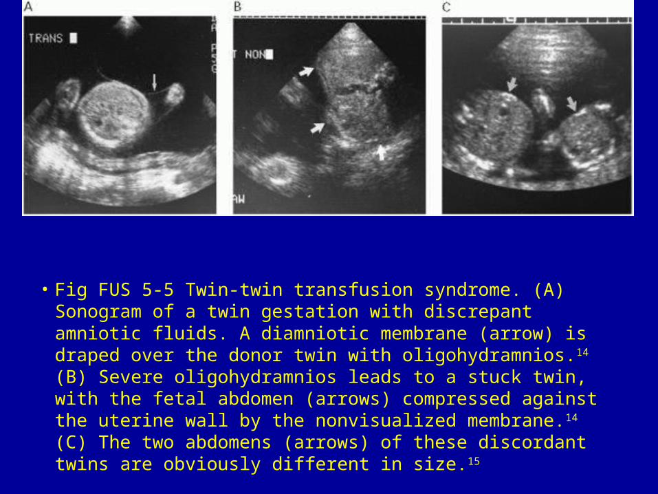
• Fig FUS 5-5 Twin-twin transfusion syndrome. (A) Sonogram of a twin gestation with discrepant amniotic fluids. A diamniotic membrane (arrow) is draped over the donor twin with oligohydramnios.14 (B) Severe oligohydramnios leads to a stuck twin, with the fetal abdomen (arrows) compressed against the uterine wall by the nonvisualized membrane.14 (C) The two abdomens (arrows) of these discordant twins are obviously different in size.15

• Fig FUS 5-6 Acardiac twins. (A) Transverse sonogram through the trunk of the acardiac twin shows massive skin thickening (arrows) around the abdomen (arrowheads). (B) Doppler study of the umbilical artery of the acardiac twin shows flow in the reverse direction, toward the fetus. (C) Doppler study of the umbilical vein shows reversed flow away from the fetus.16

• Fig FUS 5-7 Fetus papyraceus. (A) Sonogram of the paper-thin fetal head (long arrows) of a twin that died several months previously. The fetal remnants are compressed against the uterine wall by the live co-twin (short arrows). (B) Longitudinal scan of the compressed fetus shows the head (long arrow) and spine (short arrows). The thrombosed umbilical cord (curved arrow) extends from the fetus toward the uterine wall.14

Related Documents