49-Iodo-49-Deoxydoxorubicin Disrupts the Fibrillar Structure of Transthyretin Amyloid Joana Almeida Palha,* ² Dario Ballinari, ‡ Nadia Amboldi, ‡ Isabel Cardoso,* § Rui Fernandes,* Vittorio Bellotti, ¶ Giampaolo Merlini, ¶ and Maria Joa ˜ o Saraiva* § From the Amyloid Unit,* Instituto de Biologia Molecular e Celular, Porto e Instituto Superior de Cie ˆncias da Sau ´ de, Paredes, Portugal; Pharmacia and Upjohn, ‡ Discovery Research Oncology, Nerviano, Italy; the Instituto de Cie ˆncia Biome ´dicas Abel Salazar, § Universidade do Porto, Portugal; and the Biotechnology Research Laboratories, ¶ University Hospital IRCCS Policlinico San Matteo and the Department of Biochemistry, University of Pavia, Italy Transthyretin (TTR) is a tetrameric protein synthe- sized mainly by the liver and the choroid plexus, from where it is secreted into the plasma and the cerebrospinal fluid , respectively. Some forms of poly- neuropathy , vitreopathy , and cardiomyopathy are caused by the deposition of normal and/or mutant TTR molecules in the form of amyloid fibrils. Familial amyloidotic polyneuropathy is the most common form of TTR amyloidosis related to the V30M variant. It is still unclear the process by which soluble pro- teins deposit as amyloid. The treatment of amyloid- related disorders might attempt the stabilization of the soluble protein precursor to retard or inhibit its deposition as amyloid; or aim at the resorption of the deposited amyloid. The anthracycline 4*-iodo-4*-de- oxydoxorubicin (I-DOX) has been shown to reduce the amyloid load in immunoglobulin light-chain amy- loidosis. We investigated 1) whether I-DOX has affin- ity for TTR amyloid in tissues, 2) determined the I-DOX binding constants to TTR synthetic fibrils , and 3) determined the nature of the effect of I-DOX on TTR fibrils. We report that 1) I-DOX co-localizes with amy- loid deposits in tissue sections of patients with famil- ial amyloidotic polyneuropathy; 2) I-DOX strongly interacts with TTR amyloid fibrils and presents two binding sites with k d of 1.5 3 10 211 mol/L and 5.6 3 10 210 mol/L , respectively; and 3) I-DOX disrupts the fibrillar structure of TTR amyloid into amorphous material , as assessed by electron microscopy but does not solubilize the fibrils as confirmed by filter assays. These data support the hypothesis that I-DOX and less toxic derivatives can prove efficient in the treatment of TTR-related amyloidosis. (Am J Pathol 2000, 156:1919 –1925) Transthyretin (TTR), a tetrameric protein that binds thy- roxine and retinol, is synthesized mainly by the liver and the choroid plexus, from where it is secreted into the plasma and the cerebrospinal fluid, respectively. 1 Nor- mal and mutated TTR variants are associated with the deposition of amyloid fibrils in several tissues, causing predominantly polyneuropathy and/or cardiomyopathy. The process that leads to the deposition of soluble TTR into an amyloid fibril is still unknown. It is hypothesized that an amyloidogenic intermediate should be initially formed and prompt the protein to deposit. It is also pos- sible that some tissue-specific factor enables a particular protein to deposit and this may vary in the different types of amyloidosis. 2 In familial amyloidotic polyneuropathy (FAP) associated with a V30M mutation, the amyloid fibrils deposit primarily in the peripheral nerve. During the course of the disease, FAP patients may suffer from vitreous opacities caused by the presence of TTR fibrils, which can be easily removed and represent an excellent native fibrillar material for analysis. The only therapy avail- able for FAP is liver transplant, where the amyloidogenic precursor protein is eliminated from the blood. Some patients improve in autonomic function. Serum amyloid P component scintigraphy analysis has shown regression of visceral amyloid after liver transplantation. 3 One pa- tient has also been described where regeneration of the peripheral nerve was observed without a decrease in the amyloid load. 4 Although it is general agreement that liver transplantation arrests the progression of the disease to some extent, it is a drastic therapy and the clinical im- provement is not observed in all instances. In addition, the choroid plexus continues to synthesize the mutated protein, and it is unknown whether the cerebrospinal fluid constitutes a route for TTR access to the nerve. There- fore, potential alternative therapies could aim either at the inhibition of amyloid deposition and/or at the resorption of already formed amyloid. Several compounds are being tested with such purposes in FAP and in other amyloid- oses. 5,6 The anthracycline 49-iodo-49-deoxydoxorubicin (I- DOX) may be such a drug. I-DOX strongly binds in vitro to Supported by grants SAU/1290/95 from Praxis XXI, Portugal, BMH4- CT98 –3689 from the European Community, E.793 from Telethon, Italy and by a grant from the University Hospital IRCCS Policlinico San Matteo, Pavia, Italy. Accepted for publication February 9, 2000. Address reprint requests to Maria Joa ˜ o Saraiva, Amyloid Unit, Instituto de Biologia Molecular e Celular, Rua do Campo Alegre, 823. 4150 Porto, Portugal. E-mail: [email protected]. American Journal of Pathology, Vol. 156, No. 6, June 2000 Copyright © American Society for Investigative Pathology 1919

Welcome message from author
This document is posted to help you gain knowledge. Please leave a comment to let me know what you think about it! Share it to your friends and learn new things together.
Transcript
49-Iodo-49-Deoxydoxorubicin Disrupts the FibrillarStructure of Transthyretin Amyloid
Joana Almeida Palha,*† Dario Ballinari,‡
Nadia Amboldi,‡ Isabel Cardoso,*§
Rui Fernandes,* Vittorio Bellotti,¶
Giampaolo Merlini,¶ and Maria Joao Saraiva*§
From the Amyloid Unit,* Instituto de Biologia Molecular e
Celular, Porto e Instituto Superior de Ciencias da Saude,†
Paredes, Portugal; Pharmacia and Upjohn,‡ Discovery Research
Oncology, Nerviano, Italy; the Instituto de Ciencia Biomedicas
Abel Salazar, § Universidade do Porto, Portugal; and the
Biotechnology Research Laboratories,¶ University Hospital IRCCS
Policlinico San Matteo and the Department of Biochemistry,
University of Pavia, Italy
Transthyretin (TTR) is a tetrameric protein synthe-sized mainly by the liver and the choroid plexus,from where it is secreted into the plasma and thecerebrospinal fluid, respectively. Some forms of poly-neuropathy, vitreopathy, and cardiomyopathy arecaused by the deposition of normal and/or mutantTTR molecules in the form of amyloid fibrils. Familialamyloidotic polyneuropathy is the most commonform of TTR amyloidosis related to the V30M variant.It is still unclear the process by which soluble pro-teins deposit as amyloid. The treatment of amyloid-related disorders might attempt the stabilization ofthe soluble protein precursor to retard or inhibit itsdeposition as amyloid; or aim at the resorption of thedeposited amyloid. The anthracycline 4*-iodo-4*-de-oxydoxorubicin (I-DOX) has been shown to reducethe amyloid load in immunoglobulin light-chain amy-loidosis. We investigated 1) whether I-DOX has affin-ity for TTR amyloid in tissues, 2) determined theI-DOX binding constants to TTR synthetic fibrils, and3) determined the nature of the effect of I-DOX on TTRfibrils. We report that 1) I-DOX co-localizes with amy-loid deposits in tissue sections of patients with famil-ial amyloidotic polyneuropathy; 2) I-DOX stronglyinteracts with TTR amyloid fibrils and presents twobinding sites with kd of 1.5 3 10211 mol/L and 5.6 310210 mol/L, respectively; and 3) I-DOX disrupts thefibrillar structure of TTR amyloid into amorphousmaterial, as assessed by electron microscopy but doesnot solubilize the fibrils as confirmed by filter assays.These data support the hypothesis that I-DOX and lesstoxic derivatives can prove efficient in the treatmentof TTR-related amyloidosis. (Am J Pathol 2000,156:1919–1925)
Transthyretin (TTR), a tetrameric protein that binds thy-roxine and retinol, is synthesized mainly by the liver andthe choroid plexus, from where it is secreted into theplasma and the cerebrospinal fluid, respectively.1 Nor-mal and mutated TTR variants are associated with thedeposition of amyloid fibrils in several tissues, causingpredominantly polyneuropathy and/or cardiomyopathy.The process that leads to the deposition of soluble TTRinto an amyloid fibril is still unknown. It is hypothesizedthat an amyloidogenic intermediate should be initiallyformed and prompt the protein to deposit. It is also pos-sible that some tissue-specific factor enables a particularprotein to deposit and this may vary in the different typesof amyloidosis.2 In familial amyloidotic polyneuropathy(FAP) associated with a V30M mutation, the amyloidfibrils deposit primarily in the peripheral nerve. During thecourse of the disease, FAP patients may suffer fromvitreous opacities caused by the presence of TTR fibrils,which can be easily removed and represent an excellentnative fibrillar material for analysis. The only therapy avail-able for FAP is liver transplant, where the amyloidogenicprecursor protein is eliminated from the blood. Somepatients improve in autonomic function. Serum amyloid Pcomponent scintigraphy analysis has shown regressionof visceral amyloid after liver transplantation.3 One pa-tient has also been described where regeneration of theperipheral nerve was observed without a decrease in theamyloid load.4 Although it is general agreement that livertransplantation arrests the progression of the disease tosome extent, it is a drastic therapy and the clinical im-provement is not observed in all instances. In addition,the choroid plexus continues to synthesize the mutatedprotein, and it is unknown whether the cerebrospinal fluidconstitutes a route for TTR access to the nerve. There-fore, potential alternative therapies could aim either at theinhibition of amyloid deposition and/or at the resorption ofalready formed amyloid. Several compounds are beingtested with such purposes in FAP and in other amyloid-oses.5,6
The anthracycline 49-iodo-49-deoxydoxorubicin (I-DOX) may be such a drug. I-DOX strongly binds in vitro to
Supported by grants SAU/1290/95 from Praxis XXI, Portugal, BMH4-CT98–3689 from the European Community, E.793 from Telethon, Italy andby a grant from the University Hospital IRCCS Policlinico San Matteo,Pavia, Italy.
Accepted for publication February 9, 2000.
Address reprint requests to Maria Joao Saraiva, Amyloid Unit, Institutode Biologia Molecular e Celular, Rua do Campo Alegre, 823. 4150 Porto,Portugal. E-mail: [email protected].
American Journal of Pathology, Vol. 156, No. 6, June 2000
Copyright © American Society for Investigative Pathology
1919
various natural amyloid fibrils composed of immunoglob-ulin light chains, amyloid A, TTR (V30M), b-protein, andb2-microglobulin7,8 and leads to amyloid resorption inpatients with light-chain amyloidosis.9 In addition, I-DOXdelays the appearance of experimental prion disease inhamsters prolonging the survival time.10 The metabolicactive I-DOX metabolite, 13-dihydro-I-DOX (I-DOXOL),can reach the cerebrospinal fluid.9 This feature may berelevant for diseases where cerebrospinal fluid bathesthe site of amyloid deposition.
The application of I-DOX in the treatment of TTR-re-lated amyloidosis needs investigation at the tissue andmolecular levels. We investigated binding of I-DOX in situon FAP tissues and in vitro on native and synthetic TTRfibrils by morphological and biochemical analysis, to de-termine the nature of the interaction and the effect on TTRfibrils on binding.
Materials and Methods
Anthracyclines
I-DOX, I-DOXOL, and DOX (doxorubicin) were kindly pro-vided by Pharmacia and Upjohn, Nerviano, Italy.
TTR Fibrils
Native TTR amyloid vitreous fibrils were obtained froma FAP patient with V30M TTR subjected to vitreoctomy,whereas kidney fibrils were extracted, postmortem,from another FAP patient using water extractions aspreviously described.11 Both the kidney and vitreousfibrils showed the characteristic apple birefringenceunder polarized light after Congo red binding. Recom-binant TTR proteins were isolated from the periplasmicspace of Escherichia coli after osmotic shock, and pu-rified by ion-exchange chromatography and prepara-tive electrophoresis.12 Wild-type or V30M-TTR syn-thetic amyloid fibrils were prepared from acidificationof 2-mg/ml recombinant TTR solutions and assessedby thioflavin T fluorescence assay.13,14 Shortly, excita-tion spectra were recorded on a Jasco FP-770 spec-trofluorometer (Jasco International, Tokyo, Japan) at25°C with 30 mmol/L thioflavin T in 50 mmol/L of gly-cine-NaOH buffer, pH 9.0, in 1-ml assay volume. Prep-arations showing the characteristic novel excitationmaxima at 450 nm on thioflavin T binding were used inthe experiments.
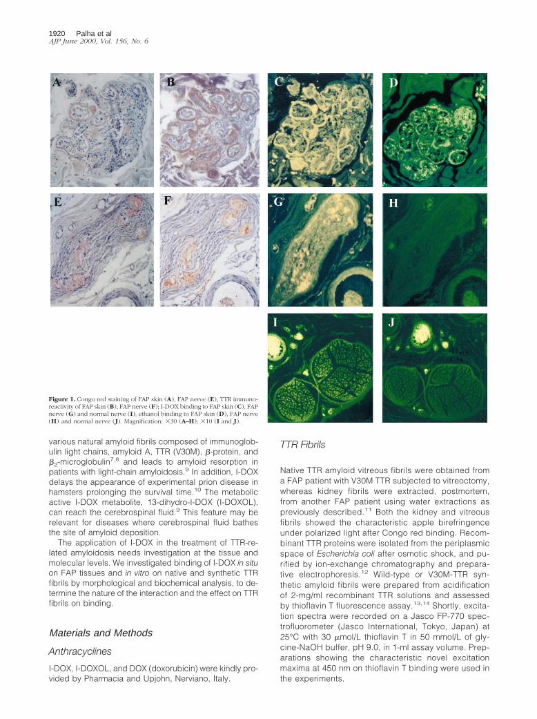
Figure 1. Congo red staining of FAP skin (A), FAP nerve (E); TTR immuno-reactivity of FAP skin (B), FAP nerve (F); I-DOX binding to FAP skin (C), FAPnerve (G) and normal nerve (I); ethanol binding to FAP skin (D), FAP nerve(H) and normal nerve (J). Magnification: 330 (A–H); 310 (I and J).
1920 Palha et alAJP June 2000, Vol. 156, No. 6
Tissue Sections
Deparaffinized tissue sections of nerve and skin biopsiesfrom FAP patients were incubated for 20 minutes with80% ethanol saturated with NaCl followed by 0.5%Congo red in 80% alkaline ethanol saturated with NaCl,and analyzed under polarized light.15,16 Amyloid wasidentified by the characteristic green birefringence. Im-munohistochemistry using polyclonal anti-TTR antibodies(DAKO, Glostrup, Denmark) following standard proce-dures, and developed with 3,39-diaminobenzidine con-firmed the TTR nature of the amyloid deposits.
Anthracycline Binding
Fluorescence and Confocal Microscopy
Deparaffinized tissue sections were incubated for 20minutes with 1025 mol/L I-DOX or I-DOXOL in 80% eth-anol saturated with NaCl. Control samples were incu-bated with 80% ethanol saturated with NaCl. Anthracy-cline fluorescence was determined with a Leica DMLBmicroscope (Wetzlar, Germany; excitation filter 585 nm,emission filter 615 nm). Kidney-extracted TTR fibrils weretreated in the same way but observed under a Bio-RadMRC 600 confocal microscope (Bio-Rad, Hercules, CA)using a 568 nm excitation filter.
Quantitative Methods
For the Scatchard analysis a fixed amount of fibrils(TTR or insulin) was incubated for 2 hours at 20°C withsolutions of I-DOX in water ranging from 1027 mol/L to7.5 3 1025 mol/L. The samples were centrifuged at15,000 3 g for 10 minutes at 4°C and fibril pellets werewashed three times with 1 ml of water. Bound com-pounds were extracted by adding 200 ml of HCl 0.6N/EtOH (1:1) and vigorously shaken for 20 minutes. Thenthe samples were spun at 15,000 3 g and supernatantswere injected into high performance liquid chromatogra-
phy. RP-high performance liquid chromatography analy-sis was performed using the following instrumental con-ditions: Waters (Milford, MA) equipment comprising 717Autosampler, 474 spectrofluorometer, and 510 pump.Column was C8 (Waters WAT 054270 4.6 3 250 mm) withprecolumn C8 WATO 54250 Sentry. Samples were run at
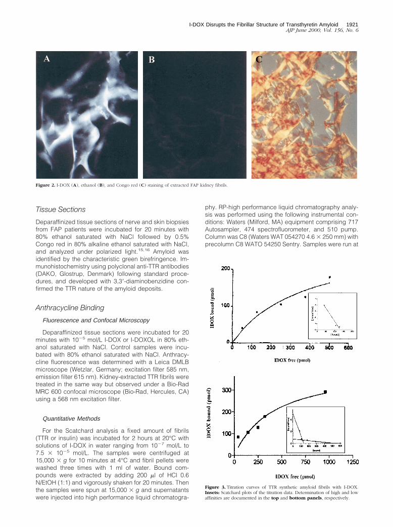
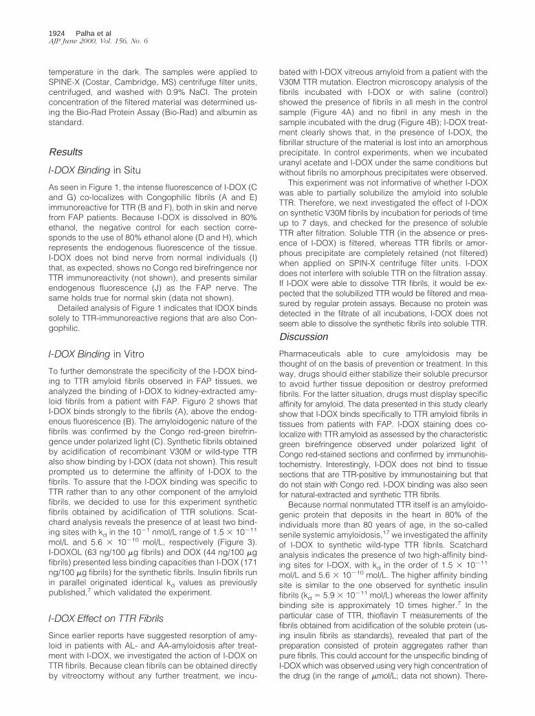
Figure 2. I-DOX (A), ethanol (B), and Congo red (C) staining of extracted FAP kidney fibrils.
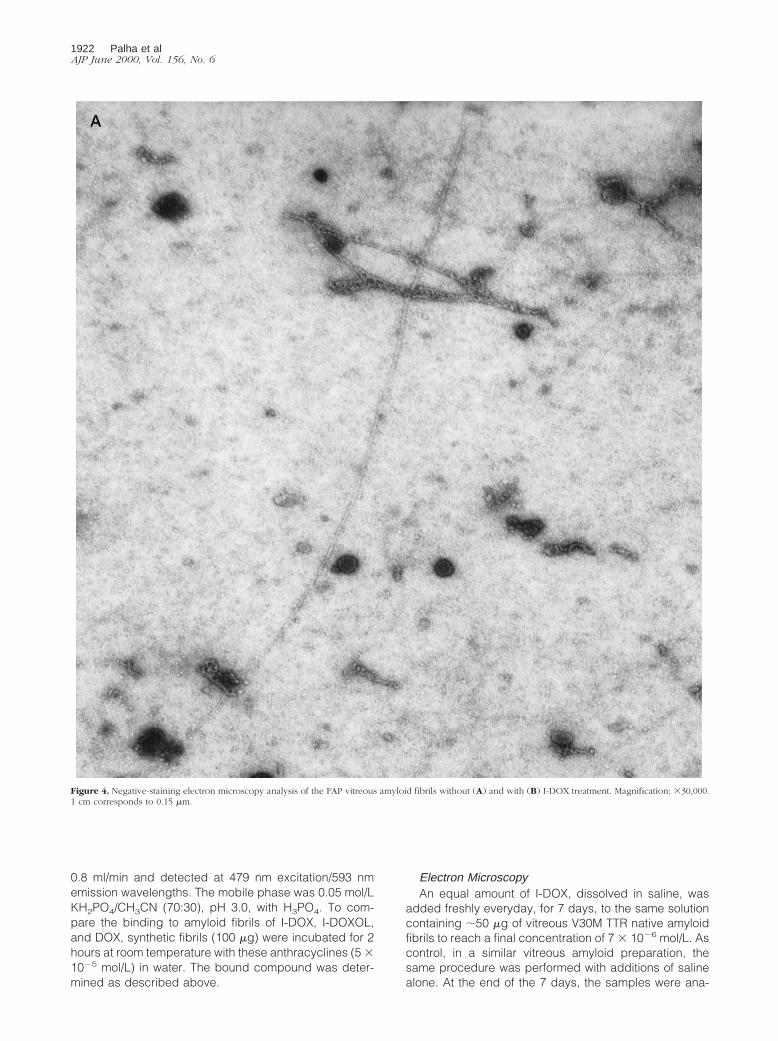
Figure 3. Titration curves of TTR synthetic amyloid fibrils with I-DOX.Insets: Scatchard plots of the titration data. Determination of high and lowaffinities are documented in the top and bottom panels, respectively.
I-DOX Disrupts the Fibrillar Structure of Transthyretin Amyloid 1921AJP June 2000, Vol. 156, No. 6
0.8 ml/min and detected at 479 nm excitation/593 nmemission wavelengths. The mobile phase was 0.05 mol/LKH2PO4/CH3CN (70:30), pH 3.0, with H3PO4. To com-pare the binding to amyloid fibrils of I-DOX, I-DOXOL,and DOX, synthetic fibrils (100 mg) were incubated for 2hours at room temperature with these anthracyclines (5 31025 mol/L) in water. The bound compound was deter-mined as described above.
Electron MicroscopyAn equal amount of I-DOX, dissolved in saline, was
added freshly everyday, for 7 days, to the same solutioncontaining ;50 mg of vitreous V30M TTR native amyloidfibrils to reach a final concentration of 7 3 1026 mol/L. Ascontrol, in a similar vitreous amyloid preparation, thesame procedure was performed with additions of salinealone. At the end of the 7 days, the samples were ana-
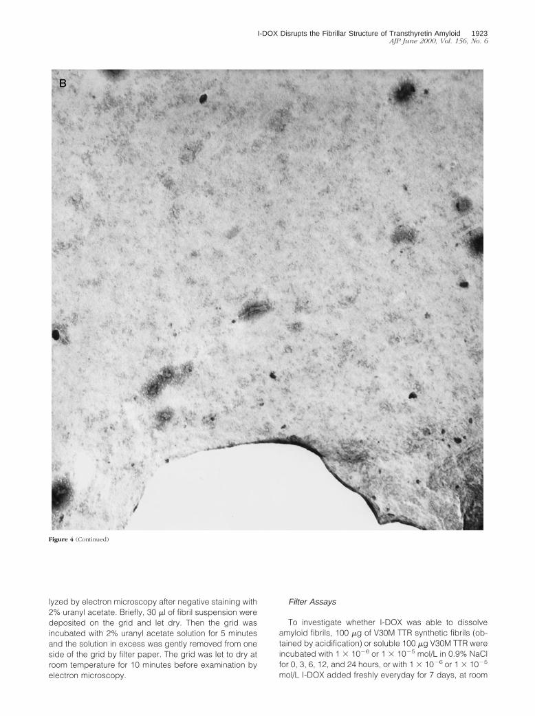
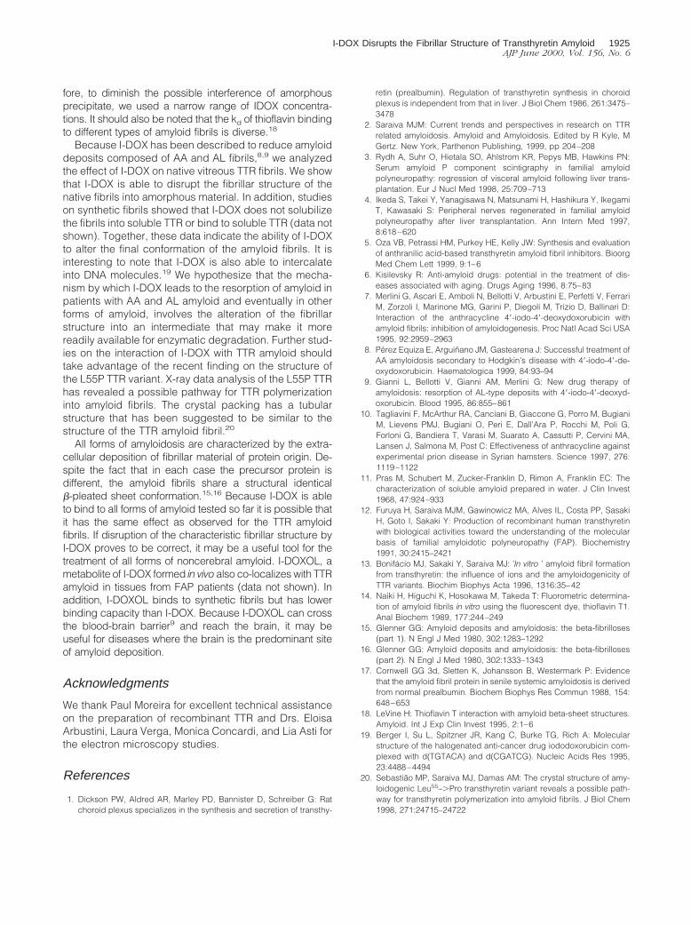
Figure 4. Negative-staining electron microscopy analysis of the FAP vitreous amyloid fibrils without (A) and with (B) I-DOX treatment. Magnification: 330,000.1 cm corresponds to 0.15 mm.
1922 Palha et alAJP June 2000, Vol. 156, No. 6
lyzed by electron microscopy after negative staining with2% uranyl acetate. Briefly, 30 ml of fibril suspension weredeposited on the grid and let dry. Then the grid wasincubated with 2% uranyl acetate solution for 5 minutesand the solution in excess was gently removed from oneside of the grid by filter paper. The grid was let to dry atroom temperature for 10 minutes before examination byelectron microscopy.
Filter Assays
To investigate whether I-DOX was able to dissolveamyloid fibrils, 100 mg of V30M TTR synthetic fibrils (ob-tained by acidification) or soluble 100 mg V30M TTR wereincubated with 1 3 1026 or 1 3 1025 mol/L in 0.9% NaClfor 0, 3, 6, 12, and 24 hours, or with 1 3 1026 or 1 3 1025
mol/L I-DOX added freshly everyday for 7 days, at room
Figure 4 (Continued)
I-DOX Disrupts the Fibrillar Structure of Transthyretin Amyloid 1923AJP June 2000, Vol. 156, No. 6
temperature in the dark. The samples were applied toSPINE-X (Costar, Cambridge, MS) centrifuge filter units,centrifuged, and washed with 0.9% NaCl. The proteinconcentration of the filtered material was determined us-ing the Bio-Rad Protein Assay (Bio-Rad) and albumin asstandard.
Results
I-DOX Binding in Situ
As seen in Figure 1, the intense fluorescence of I-DOX (Cand G) co-localizes with Congophilic fibrils (A and E)immunoreactive for TTR (B and F), both in skin and nervefrom FAP patients. Because I-DOX is dissolved in 80%ethanol, the negative control for each section corre-sponds to the use of 80% ethanol alone (D and H), whichrepresents the endogenous fluorescence of the tissue.I-DOX does not bind nerve from normal individuals (I)that, as expected, shows no Congo red birefringence norTTR immunoreactivity (not shown), and presents similarendogenous fluorescence (J) as the FAP nerve. Thesame holds true for normal skin (data not shown).
Detailed analysis of Figure 1 indicates that IDOX bindssolely to TTR-immunoreactive regions that are also Con-gophilic.
I-DOX Binding in Vitro
To further demonstrate the specificity of the I-DOX bind-ing to TTR amyloid fibrils observed in FAP tissues, weanalyzed the binding of I-DOX to kidney-extracted amy-loid fibrils from a patient with FAP. Figure 2 shows thatI-DOX binds strongly to the fibrils (A), above the endog-enous fluorescence (B). The amyloidogenic nature of thefibrils was confirmed by the Congo red-green birefrin-gence under polarized light (C). Synthetic fibrils obtainedby acidification of recombinant V30M or wild-type TTRalso show binding by I-DOX (data not shown). This resultprompted us to determine the affinity of I-DOX to thefibrils. To assure that the I-DOX binding was specific toTTR rather than to any other component of the amyloidfibrils, we decided to use for this experiment syntheticfibrils obtained by acidification of TTR solutions. Scat-chard analysis reveals the presence of at least two bind-ing sites with kd in the 1021 nmol/L range of 1.5 3 10211
mol/L and 5.6 3 10210 mol/L, respectively (Figure 3).I-DOXOL (63 ng/100 mg fibrils) and DOX (44 ng/100 mgfibrils) presented less binding capacities than I-DOX (171ng/100 mg fibrils) for the synthetic fibrils. Insulin fibrils runin parallel originated identical kd values as previouslypublished,7 which validated the experiment.
I-DOX Effect on TTR Fibrils
Since earlier reports have suggested resorption of amy-loid in patients with AL- and AA-amyloidosis after treat-ment with I-DOX, we investigated the action of I-DOX onTTR fibrils. Because clean fibrils can be obtained directlyby vitreoctomy without any further treatment, we incu-
bated with I-DOX vitreous amyloid from a patient with theV30M TTR mutation. Electron microscopy analysis of thefibrils incubated with I-DOX or with saline (control)showed the presence of fibrils in all mesh in the controlsample (Figure 4A) and no fibril in any mesh in thesample incubated with the drug (Figure 4B); I-DOX treat-ment clearly shows that, in the presence of I-DOX, thefibrillar structure of the material is lost into an amorphousprecipitate. In control experiments, when we incubateduranyl acetate and I-DOX under the same conditions butwithout fibrils no amorphous precipitates were observed.
This experiment was not informative of whether I-DOXwas able to partially solubilize the amyloid into solubleTTR. Therefore, we next investigated the effect of I-DOXon synthetic V30M fibrils by incubation for periods of timeup to 7 days, and checked for the presence of solubleTTR after filtration. Soluble TTR (in the absence or pres-ence of I-DOX) is filtered, whereas TTR fibrils or amor-phous precipitate are completely retained (not filtered)when applied on SPIN-X centrifuge filter units. I-DOXdoes not interfere with soluble TTR on the filtration assay.If I-DOX were able to dissolve TTR fibrils, it would be ex-pected that the solubilized TTR would be filtered and mea-sured by regular protein assays. Because no protein wasdetected in the filtrate of all incubations, I-DOX does notseem able to dissolve the synthetic fibrils into soluble TTR.
Discussion
Pharmaceuticals able to cure amyloidosis may bethought of on the basis of prevention or treatment. In thisway, drugs should either stabilize their soluble precursorto avoid further tissue deposition or destroy preformedfibrils. For the latter situation, drugs must display specificaffinity for amyloid. The data presented in this study clearlyshow that I-DOX binds specifically to TTR amyloid fibrils intissues from patients with FAP. I-DOX staining does co-localize with TTR amyloid as assessed by the characteristicgreen birefringence observed under polarized light ofCongo red-stained sections and confirmed by immunohis-tochemistry. Interestingly, I-DOX does not bind to tissuesections that are TTR-positive by immunostaining but thatdo not stain with Congo red. I-DOX binding was also seenfor natural-extracted and synthetic TTR fibrils.
Because normal nonmutated TTR itself is an amyloido-genic protein that deposits in the heart in 80% of theindividuals more than 80 years of age, in the so-calledsenile systemic amyloidosis,17 we investigated the affinityof I-DOX to synthetic wild-type TTR fibrils. Scatchardanalysis indicates the presence of two high-affinity bind-ing sites for I-DOX, with kd in the order of 1.5 3 10211
mol/L and 5.6 3 10210 mol/L. The higher affinity bindingsite is similar to the one observed for synthetic insulinfibrils (kd 5 5.9 3 10211 mol/L) whereas the lower affinitybinding site is approximately 10 times higher.7 In theparticular case of TTR, thioflavin T measurements of thefibrils obtained from acidification of the soluble protein (us-ing insulin fibrils as standards), revealed that part of thepreparation consisted of protein aggregates rather thanpure fibrils. This could account for the unspecific binding ofI-DOX which was observed using very high concentration ofthe drug (in the range of mmol/L; data not shown). There-
1924 Palha et alAJP June 2000, Vol. 156, No. 6
fore, to diminish the possible interference of amorphousprecipitate, we used a narrow range of IDOX concentra-tions. It should also be noted that the kd of thioflavin bindingto different types of amyloid fibrils is diverse.18
Because I-DOX has been described to reduce amyloiddeposits composed of AA and AL fibrils,8,9 we analyzedthe effect of I-DOX on native vitreous TTR fibrils. We showthat I-DOX is able to disrupt the fibrillar structure of thenative fibrils into amorphous material. In addition, studieson synthetic fibrils showed that I-DOX does not solubilizethe fibrils into soluble TTR or bind to soluble TTR (data notshown). Together, these data indicate the ability of I-DOXto alter the final conformation of the amyloid fibrils. It isinteresting to note that I-DOX is also able to intercalateinto DNA molecules.19 We hypothesize that the mecha-nism by which I-DOX leads to the resorption of amyloid inpatients with AA and AL amyloid and eventually in otherforms of amyloid, involves the alteration of the fibrillarstructure into an intermediate that may make it morereadily available for enzymatic degradation. Further stud-ies on the interaction of I-DOX with TTR amyloid shouldtake advantage of the recent finding on the structure ofthe L55P TTR variant. X-ray data analysis of the L55P TTRhas revealed a possible pathway for TTR polymerizationinto amyloid fibrils. The crystal packing has a tubularstructure that has been suggested to be similar to thestructure of the TTR amyloid fibril.20
All forms of amyloidosis are characterized by the extra-cellular deposition of fibrillar material of protein origin. De-spite the fact that in each case the precursor protein isdifferent, the amyloid fibrils share a structural identicalb-pleated sheet conformation.15,16 Because I-DOX is ableto bind to all forms of amyloid tested so far it is possible thatit has the same effect as observed for the TTR amyloidfibrils. If disruption of the characteristic fibrillar structure byI-DOX proves to be correct, it may be a useful tool for thetreatment of all forms of noncerebral amyloid. I-DOXOL, ametabolite of I-DOX formed in vivo also co-localizes with TTRamyloid in tissues from FAP patients (data not shown). Inaddition, I-DOXOL binds to synthetic fibrils but has lowerbinding capacity than I-DOX. Because I-DOXOL can crossthe blood-brain barrier9 and reach the brain, it may beuseful for diseases where the brain is the predominant siteof amyloid deposition.
Acknowledgments
We thank Paul Moreira for excellent technical assistanceon the preparation of recombinant TTR and Drs. EloisaArbustini, Laura Verga, Monica Concardi, and Lia Asti forthe electron microscopy studies.
References
1. Dickson PW, Aldred AR, Marley PD, Bannister D, Schreiber G: Ratchoroid plexus specializes in the synthesis and secretion of transthy-
retin (prealbumin). Regulation of transthyretin synthesis in choroidplexus is independent from that in liver. J Biol Chem 1986, 261:3475–3478
2. Saraiva MJM: Current trends and perspectives in research on TTRrelated amyloidosis. Amyloid and Amyloidosis. Edited by R Kyle, MGertz. New York, Parthenon Publishing, 1999, pp 204–208
3. Rydh A, Suhr O, Hietala SO, Ahlstrom KR, Pepys MB, Hawkins PN:Serum amyloid P component scintigraphy in familial amyloidpolyneuropathy: regression of visceral amyloid following liver trans-plantation. Eur J Nucl Med 1998, 25:709–713
4. Ikeda S, Takei Y, Yanagisawa N, Matsunami H, Hashikura Y, IkegamiT, Kawasaki S: Peripheral nerves regenerated in familial amyloidpolyneuropathy after liver transplantation. Ann Intern Med 1997,8:618–620
5. Oza VB, Petrassi HM, Purkey HE, Kelly JW: Synthesis and evaluationof anthranilic acid-based transthyretin amyloid fibril inhibitors. BioorgMed Chem Lett 1999, 9:1–6
6. Kisilevsky R: Anti-amyloid drugs: potential in the treatment of dis-eases associated with aging. Drugs Aging 1996, 8:75–83
7. Merlini G, Ascari E, Amboli N, Bellotti V, Arbustini E, Perfetti V, FerrariM, Zorzoli I, Marinone MG, Garini P, Diegoli M, Trizio D, Ballinari D:Interaction of the anthracycline 49-iodo-49-deoxydoxorubicin withamyloid fibrils: inhibition of amyloidogenesis. Proc Natl Acad Sci USA1995, 92:2959–2963
8. Perez Equiza E, Arguinano JM, Gastearena J: Successful treatment ofAA amyloidosis secondary to Hodgkin’s disease with 49-iodo-49-de-oxydoxorubicin. Haematologica 1999, 84:93–94
9. Gianni L, Bellotti V, Gianni AM, Merlini G: New drug therapy ofamyloidosis: resorption of AL-type deposits with 49-iodo-49-deoxyd-oxorubicin. Blood 1995, 86:855–861
10. Tagliavini F, McArthur RA, Canciani B, Giaccone G, Porro M, BugianiM, Lievens PMJ, Bugiani O, Peri E, Dall’Ara P, Rocchi M, Poli G,Forloni G, Bandiera T, Varasi M, Suarato A, Cassutti P, Cervini MA,Lansen J, Salmona M, Post C: Effectiveness of anthracycline againstexperimental prion disease in Syrian hamsters. Science 1997, 276:1119–1122
11. Pras M, Schubert M, Zucker-Franklin D, Rimon A, Franklin EC: Thecharacterization of soluble amyloid prepared in water. J Clin Invest1968, 47:924–933
12. Furuya H, Saraiva MJM, Gawinowicz MA, Alves IL, Costa PP, SasakiH, Goto I, Sakaki Y: Production of recombinant human transthyretinwith biological activities toward the understanding of the molecularbasis of familial amyloidotic polyneuropathy (FAP). Biochemistry1991, 30:2415–2421
13. Bonifacio MJ, Sakaki Y, Saraiva MJ: ’In vitro ’ amyloid fibril formationfrom transthyretin: the influence of ions and the amyloidogenicity ofTTR variants. Biochim Biophys Acta 1996, 1316:35–42
14. Naiki H, Higuchi K, Hosokawa M, Takeda T: Fluorometric determina-tion of amyloid fibrils in vitro using the fluorescent dye, thioflavin T1.Anal Biochem 1989, 177:244–249
15. Glenner GG: Amyloid deposits and amyloidosis: the beta-fibrilloses(part 1). N Engl J Med 1980, 302:1283–1292
16. Glenner GG: Amyloid deposits and amyloidosis: the beta-fibrilloses(part 2). N Engl J Med 1980, 302:1333–1343
17. Cornwell GG 3d, Sletten K, Johansson B, Westermark P: Evidencethat the amyloid fibril protein in senile systemic amyloidosis is derivedfrom normal prealbumin. Biochem Biophys Res Commun 1988, 154:648–653
18. LeVine H: Thioflavin T interaction with amyloid beta-sheet structures.Amyloid. Int J Exp Clin Invest 1995, 2:1–6
19. Berger I, Su L, Spitzner JR, Kang C, Burke TG, Rich A: Molecularstructure of the halogenated anti-cancer drug iododoxorubicin com-plexed with d(TGTACA) and d(CGATCG). Nucleic Acids Res 1995,23:4488–4494
20. Sebastiao MP, Saraiva MJ, Damas AM: The crystal structure of amy-loidogenic Leu55–.Pro transthyretin variant reveals a possible path-way for transthyretin polymerization into amyloid fibrils. J Biol Chem1998, 271:24715–24722
I-DOX Disrupts the Fibrillar Structure of Transthyretin Amyloid 1925AJP June 2000, Vol. 156, No. 6
Related Documents