Copyright © 2000-2016 Mark Brandt, Ph.D. 128 Signal transduction In order to interact properly with their environment, cells need to allow information as well as molecules to cross their cell membranes. Information in many single-celled and all multicellular organisms is transmitted in the form of molecules. However, the molecule itself does not contain the information. The molecule only carries the fact of the information, not the information itself. The way the cell acts on the arrival of the information determines the information content; in effect, the cell itself decides how to respond to the information. Glucose (in most cells) is not a signaling molecule, because the cell merely uses glucose as a metabolite, while insulin is a signaling molecule, because its primary function is to modify cellular responses, rather than to act as a source of energy or biosynthetic intermediates. The term “signal transduction” refers to the conversion of the information carried by signaling molecules into changes in cellular activities. Requirements for signal transduction 1) Signal transduction requires a receptor, a cellular protein that recognizes the signaling molecule. In the absence of the receptor, the ligand has no effect. Cells that lack receptors for a given ligand do not respond to that ligand. (Note: a ligand is a signaling molecule that binds to a particular receptor.) Receptors must bind the signaling molecule with high affinity. Receptors must bind ligand at physiological ligand concentrations. If the K d for the ligand is 10 µM while the physiological concentration of the ligand is 0.1 nM, the protein is not the receptor for that ligand. For example, the estrogen receptor has a K d for estradiol of 0.1 – 0.2 nM; in mature females, the serum concentration of estradiol usually varies between about 0.1 and 1.3 nM depending on the phase of the menstrual cycle. Receptors must exhibit specificity for the ligand. Receptors should bind ligands, but should not bind closely related molecules. This is not an absolute requirement, and in most cases, high enough concentrations of competing ligands may result in receptor binding by competitor. However, most receptors are able to distinguish fairly similar molecules; a receptor typically binds the actual hormone with 10-fold to 10,000-fold higher affinity than it exhibits for chemically related molecules. For example, the estrogen receptor binds testosterone with about 50,000- fold lower affinity than it does estradiol, although the molecules are structurally similar. OH OH O HO Testosterone Estradiol

Welcome message from author
This document is posted to help you gain knowledge. Please leave a comment to let me know what you think about it! Share it to your friends and learn new things together.
Transcript
Copyright © 2000-2016 Mark Brandt, Ph.D.
128
Signal transduction In order to interact properly with their environment, cells need to allow information as well as molecules to cross their cell membranes. Information in many single-celled and all multicellular organisms is transmitted in the form of molecules. However, the molecule itself does not contain the information. The molecule only carries the fact of the information, not the information itself. The way the cell acts on the arrival of the information determines the information content; in effect, the cell itself decides how to respond to the information. Glucose (in most cells) is not a signaling molecule, because the cell merely uses glucose as a metabolite, while insulin is a signaling molecule, because its primary function is to modify cellular responses, rather than to act as a source of energy or biosynthetic intermediates. The term “signal transduction” refers to the conversion of the information carried by signaling molecules into changes in cellular activities. Requirements for signal transduction
1) Signal transduction requires a receptor, a cellular protein that recognizes the signaling molecule.
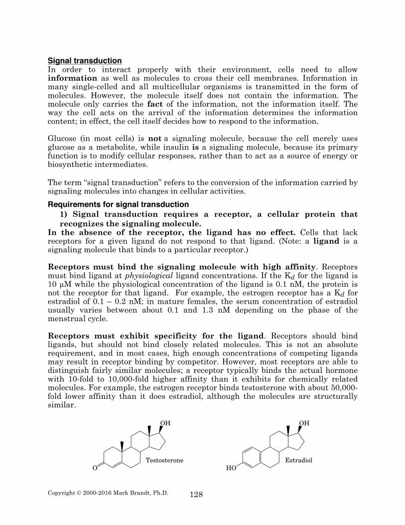
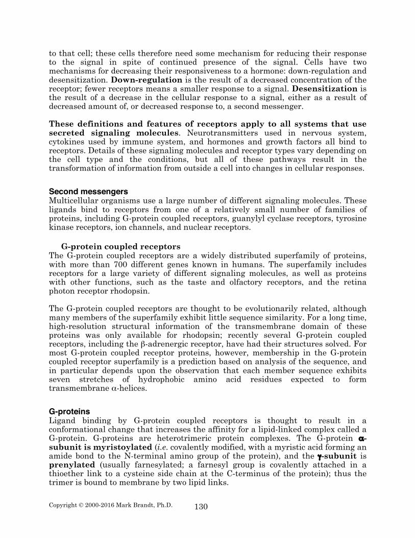
In the absence of the receptor, the ligand has no effect. Cells that lack receptors for a given ligand do not respond to that ligand. (Note: a ligand is a signaling molecule that binds to a particular receptor.) Receptors must bind the signaling molecule with high affinity. Receptors must bind ligand at physiological ligand concentrations. If the Kd for the ligand is 10 µM while the physiological concentration of the ligand is 0.1 nM, the protein is not the receptor for that ligand. For example, the estrogen receptor has a Kd for estradiol of 0.1 – 0.2 nM; in mature females, the serum concentration of estradiol usually varies between about 0.1 and 1.3 nM depending on the phase of the menstrual cycle. Receptors must exhibit specificity for the ligand. Receptors should bind ligands, but should not bind closely related molecules. This is not an absolute requirement, and in most cases, high enough concentrations of competing ligands may result in receptor binding by competitor. However, most receptors are able to distinguish fairly similar molecules; a receptor typically binds the actual hormone with 10-fold to 10,000-fold higher affinity than it exhibits for chemically related molecules. For example, the estrogen receptor binds testosterone with about 50,000-fold lower affinity than it does estradiol, although the molecules are structurally similar.
OH OH
O HOTestosterone Estradiol

Copyright © 2000-2016 Mark Brandt, Ph.D.
129
Receptors must exhibit a finite capacity for the ligand (usually, the binding of one ligand molecule per receptor molecule). The requirement for finite capacity arose from pharmacological studies performed before the isolation of the receptor proteins. Some compounds bind glass tubes and other experimental system components with high affinity and stereochemical specificity; glass tubes typically have vast numbers of binding sites, while cells generally do not. Ligand binding must elicit a direct biological effect. Some non-receptor proteins have high affinity for ligands, but ligand binding to these proteins has no effect on cellular functioning, and therefore these proteins are not receptors.
2) Signal transduction requires a mechanism for linking ligand binding to biological changes within a cell.
The hormone is the “first messenger”. In most cases, the binding of the hormone to the receptor elicits the release of a “second messenger”: a compound that acts like an intracellular hormone. The release of the second messenger is the first step in transmitting the signal from the outside to the inside of the cell. Second messengers allow amplification of a signal; one hormone molecule binding to one receptor can result in the production of many second messenger molecules. Second messengers allow the convergence of multiple pathways, because more than one second messenger pathway can affect specific cellular enzymes. Second messengers also allow more than one hormone to have similar effects in some cell types, because both hormones result in production of the same second messenger. Second messengers also allow modulation of a signal; one hormone can alter the response to a second hormone by affecting the production of the second messenger.
3) Signal transduction results in a variety of types of biological effects. Hormonal effects can be mediated by one of two major processes. The first, and faster process is an alteration in the activity of existing proteins. This can involve the opening or closing of ion channels, binding of a second messenger causing activation or inhibition of an enzyme, or the covalent modification of existing proteins (especially phosphorylation or dephosphorylation of enzymes) resulting in altered enzymatic activity. The second, and slower process is the alteration in the amount of a protein. This is usually the result of activation or inhibition of gene transcription; it may also be due to changes in the rate of degradation of the protein.
4) Signal transduction pathways have at least one mechanism for turning the signal off.
The most obvious method for turning off a signal is the dissociation of the ligand from the receptor. Ligand dissociation can be induced a cellular process, or can occur due to a decrease in the circulating ligand concentration. Most second messengers have short half-lives within the cell, and therefore when the hormone dissociates, the second messenger levels decrease, terminating the signal. However, in some cases, the signaling molecule concentration remains high for prolonged periods. For many cells, chronic response to a signal results in deleterious changes
Copyright © 2000-2016 Mark Brandt, Ph.D.
130
to that cell; these cells therefore need some mechanism for reducing their response to the signal in spite of continued presence of the signal. Cells have two mechanisms for decreasing their responsiveness to a hormone: down-regulation and desensitization. Down-regulation is the result of a decreased concentration of the receptor; fewer receptors means a smaller response to a signal. Desensitization is the result of a decrease in the cellular response to a signal, either as a result of decreased amount of, or decreased response to, a second messenger. These definitions and features of receptors apply to all systems that use secreted signaling molecules. Neurotransmitters used in nervous system, cytokines used by immune system, and hormones and growth factors all bind to receptors. Details of these signaling molecules and receptor types vary depending on the cell type and the conditions, but all of these pathways result in the transformation of information from outside a cell into changes in cellular responses. Second messengers Multicellular organisms use a large number of different signaling molecules. These ligands bind to receptors from one of a relatively small number of families of proteins, including G-protein coupled receptors, guanylyl cyclase receptors, tyrosine kinase receptors, ion channels, and nuclear receptors.
G-protein coupled receptors The G-protein coupled receptors are a widely distributed superfamily of proteins, with more than 700 different genes known in humans. The superfamily includes receptors for a large variety of different signaling molecules, as well as proteins with other functions, such as the taste and olfactory receptors, and the retina photon receptor rhodopsin. The G-protein coupled receptors are thought to be evolutionarily related, although many members of the superfamily exhibit little sequence similarity. For a long time, high-resolution structural information of the transmembrane domain of these proteins was only available for rhodopsin; recently several G-protein coupled receptors, including the β-adrenergic receptor, have had their structures solved. For most G-protein coupled receptor proteins, however, membership in the G-protein coupled receptor superfamily is a prediction based on analysis of the sequence, and in particular depends upon the observation that each member sequence exhibits seven stretches of hydrophobic amino acid residues expected to form transmembrane α-helices. G-proteins Ligand binding by G-protein coupled receptors is thought to result in a conformational change that increases the affinity for a lipid-linked complex called a G-protein. G-proteins are heterotrimeric protein complexes. The G-protein α-subunit is myristoylated (i.e. covalently modified, with a myristic acid forming an amide bond to the N-terminal amino group of the protein), and the γ-subunit is prenylated (usually farnesylated; a farnesyl group is covalently attached in a thioether link to a cysteine side chain at the C-terminus of the protein); thus the trimer is bound to membrane by two lipid links.
Copyright © 2000-2016 Mark Brandt, Ph.D.
131
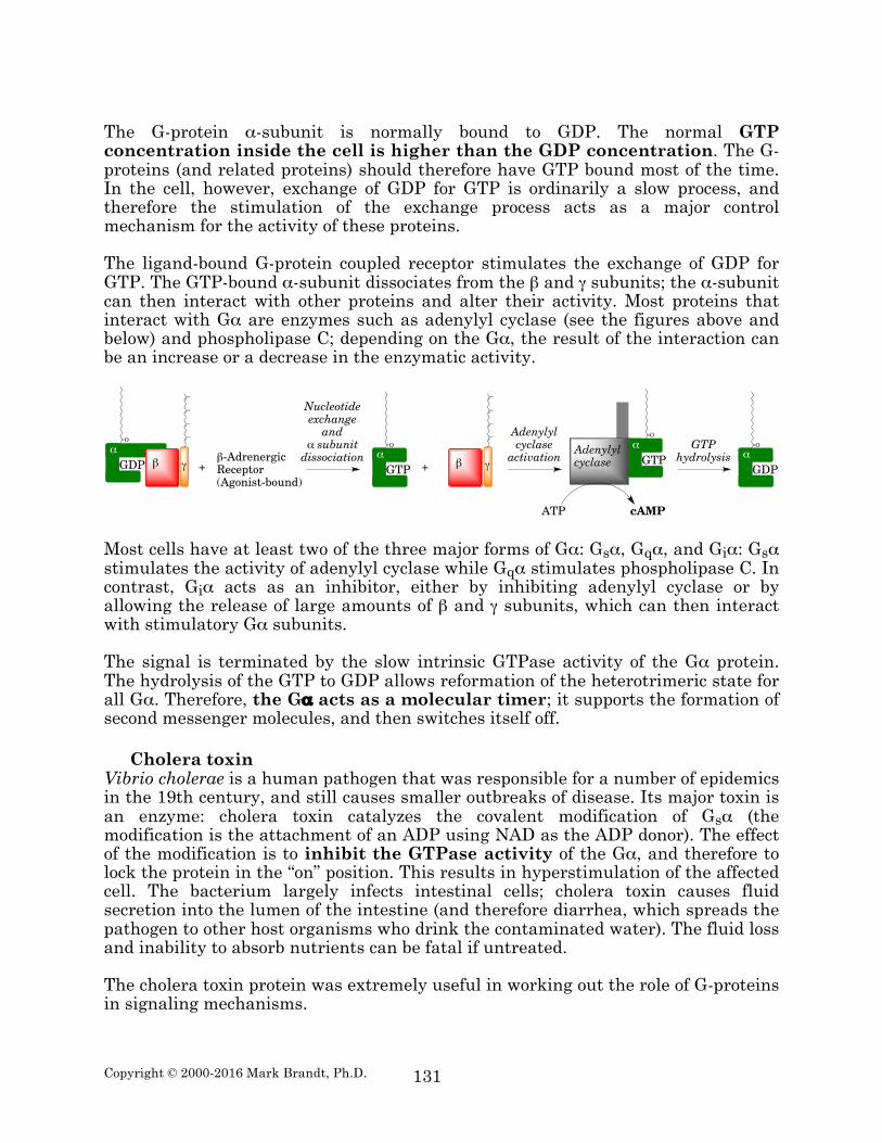
The G-protein α-subunit is normally bound to GDP. The normal GTP concentration inside the cell is higher than the GDP concentration. The G-proteins (and related proteins) should therefore have GTP bound most of the time. In the cell, however, exchange of GDP for GTP is ordinarily a slow process, and therefore the stimulation of the exchange process acts as a major control mechanism for the activity of these proteins. The ligand-bound G-protein coupled receptor stimulates the exchange of GDP for GTP. The GTP-bound α-subunit dissociates from the β and γ subunits; the α-subunit can then interact with other proteins and alter their activity. Most proteins that interact with Gα are enzymes such as adenylyl cyclase (see the figures above and below) and phospholipase C; depending on the Gα, the result of the interaction can be an increase or a decrease in the enzymatic activity.
Most cells have at least two of the three major forms of Gα: Gsα, Gqα, and Giα: Gsα stimulates the activity of adenylyl cyclase while Gqα stimulates phospholipase C. In contrast, Giα acts as an inhibitor, either by inhibiting adenylyl cyclase or by allowing the release of large amounts of β and γ subunits, which can then interact with stimulatory Gα subunits. The signal is terminated by the slow intrinsic GTPase activity of the Gα protein. The hydrolysis of the GTP to GDP allows reformation of the heterotrimeric state for all Gα. Therefore, the Gα acts as a molecular timer; it supports the formation of second messenger molecules, and then switches itself off.
Cholera toxin Vibrio cholerae is a human pathogen that was responsible for a number of epidemics in the 19th century, and still causes smaller outbreaks of disease. Its major toxin is an enzyme: cholera toxin catalyzes the covalent modification of Gsα (the modification is the attachment of an ADP using NAD as the ADP donor). The effect of the modification is to inhibit the GTPase activity of the Gα, and therefore to lock the protein in the “on” position. This results in hyperstimulation of the affected cell. The bacterium largely infects intestinal cells; cholera toxin causes fluid secretion into the lumen of the intestine (and therefore diarrhea, which spreads the pathogen to other host organisms who drink the contaminated water). The fluid loss and inability to absorb nutrients can be fatal if untreated. The cholera toxin protein was extremely useful in working out the role of G-proteins in signaling mechanisms.
β γGDPα
+β-AdrenergicReceptor(Agonist-bound)
β γGTPα
+
Nucleotideexchangeand
α subunitdissociation
O
GTPαAdenylyl
cyclase
ATP cAMP
OO
Adenylylcyclaseactivation
GTPhydrolysis
GDPα
O
Copyright © 2000-2016 Mark Brandt, Ph.D.
132
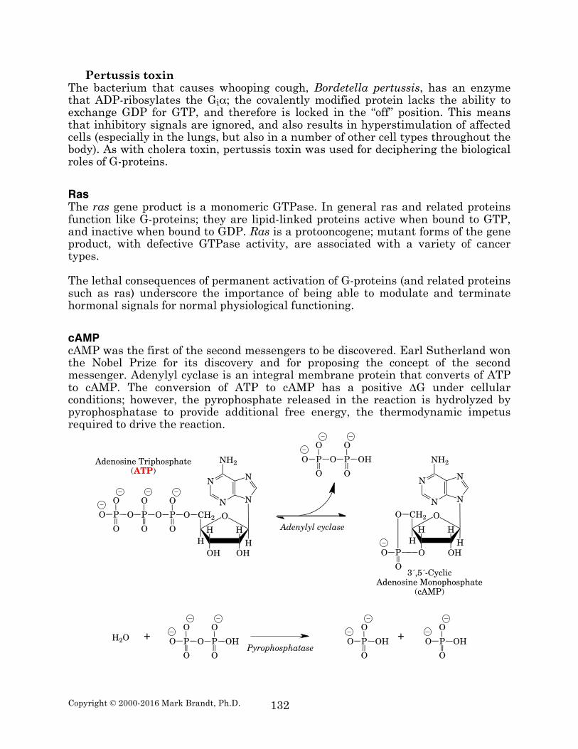
Pertussis toxin The bacterium that causes whooping cough, Bordetella pertussis, has an enzyme that ADP-ribosylates the Giα; the covalently modified protein lacks the ability to exchange GDP for GTP, and therefore is locked in the “off” position. This means that inhibitory signals are ignored, and also results in hyperstimulation of affected cells (especially in the lungs, but also in a number of other cell types throughout the body). As with cholera toxin, pertussis toxin was used for deciphering the biological roles of G-proteins. Ras The ras gene product is a monomeric GTPase. In general ras and related proteins function like G-proteins; they are lipid-linked proteins active when bound to GTP, and inactive when bound to GDP. Ras is a protooncogene; mutant forms of the gene product, with defective GTPase activity, are associated with a variety of cancer types. The lethal consequences of permanent activation of G-proteins (and related proteins such as ras) underscore the importance of being able to modulate and terminate hormonal signals for normal physiological functioning. cAMP cAMP was the first of the second messengers to be discovered. Earl Sutherland won the Nobel Prize for its discovery and for proposing the concept of the second messenger. Adenylyl cyclase is an integral membrane protein that converts of ATP to cAMP. The conversion of ATP to cAMP has a positive ∆G under cellular conditions; however, the pyrophosphate released in the reaction is hydrolyzed by pyrophosphatase to provide additional free energy, the thermodynamic impetus required to drive the reaction.
P
O
O
O O P
O
O
O
HH
OHOHH
CH2
H
O
N
N N
N
NH2
P
O
O
O
Adenosine Triphosphate(ATP)
O
HH
OHOH
CH2
H
O
N
N N
N
NH2P
O
O
O O P
O
O
OH
PO
O3´,5´-Cyclic
Adenosine Monophosphate(cAMP)
Adenylyl cyclase
P
O
O
O O P
O
O
OHPyrophosphatase
P
O
O
O OH P
O
O
O OHH2O ++

Copyright © 2000-2016 Mark Brandt, Ph.D.
133
The general mechanism for cAMP action is as follows. The hormone (epinephrine in the example shown below) interacts with the receptor. The receptor-hormone complex then activates the exchange of GDP for GTP by a G-protein, resulting in dissociation of the activated α-subunit. Note that the receptor-hormone complex can activate a number of G-proteins before the ligand dissociates. The Gsα•GTP complex activates adenylyl cyclase, resulting in cAMP formation; the enzyme produces considerable numbers of cAMP molecules, which also amplifies the signal.
Once released, cAMP primarily acts on cAMP-dependent protein kinase. Binding of cAMP results in dissociation of the regulatory subunit of the inactive kinase complex, releasing the catalytic subunit. The catalytic subunit then alters the activity of substrate proteins by phosphorylating the proteins on serine and threonine residues. The cAMP system was discovered during studies on the regulation of the liver enzyme glycogen phosphorylase. The cAMP-dependent protein kinase phosphorylation of phosphorylase kinase results in activation of phosphorylase kinase. Active phosphorylase kinase phosphorylates (and activates) glycogen phosphorylase;
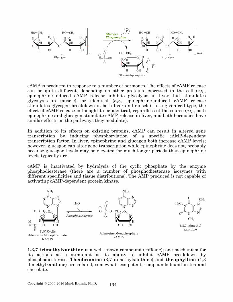
Finally, the active glycogen phosphorylase catalyzes the removal of glucose (in the form of glucose-1-phosphate) from glycogen.
β-AdrenergicReceptorPDB ID 3SN6)
Gsα
Gβ
Gγ
O
OH
OH
H2N
CH3 OH
Epinephrine
Gsα•GTP(PDB ID 1AZT)
O
O
Adenylyl CyclaseGsα•GTP(PDB ID 3C16)
ATP
cAMP
Adenylyl Cyclase(inactive)
cAMP-DependentProtein KinasePhosphorylase
KinasePhosphorylaseKinase
P
ATP ADP
cAMP
GlycogenPhosphorylase
ATP
ADP
GlycogenPhosphorylase
P
Copyright © 2000-2016 Mark Brandt, Ph.D.
134
cAMP is produced in response to a number of hormones. The effects of cAMP release can be quite different, depending on other proteins expressed in the cell (e.g., epinephrine-induced cAMP release inhibits glycolysis in liver, but stimulates glycolysis in muscle), or identical (e.g., epinephrine-induced cAMP release stimulates glycogen breakdown in both liver and muscle). In a given cell type, the effect of cAMP release is thought to be identical, regardless of the source (e.g., both epinephrine and glucagon stimulate cAMP release in liver, and both hormones have similar effects on the pathways they modulate). In addition to its effects on existing proteins, cAMP can result in altered gene transcription by inducing phosphorylation of a specific cAMP-dependent transcription factor. In liver, epinephrine and glucagon both increase cAMP levels; however, glucagon can alter gene transcription while epinephrine does not, probably because glucagon levels may be elevated for much longer periods than epinephrine levels typically are. cAMP is inactivated by hydrolysis of the cyclic phosphate by the enzyme phosphodiesterase (there are a number of phosphodiesterase isozymes with different specificities and tissue distributions). The AMP produced is not capable of activating cAMP-dependent protein kinase.
1,3,7 trimethylxanthine is a well-known compound (caffeine); one mechanism for its actions as a stimulant is its ability to inhibit cAMP breakdown by phosphodiesterase. Theobromine (3,7 dimethylxanthine) and theophylline (1,3 dimethylxanthine) are related, somewhat less potent, compounds found in tea and chocolate.
O H
H
OH
OH
H
H
OH
CH2
H
HO
O
O H
H
OH
OH
H
H
CH2
H
HO
nGlycogen
O H
H
OH
OH
H
H
OH
CH2
H
HO
O
O H
H
OH
OH
H
H
CH2
H
HO
n–1GlycogenP
O
O
O OH O H
H
OH
OH
H
H
OH
CH2
H
HO
P
O
O
O O
Glucose-1-phosphate
GlycogenPhosphorylase
P
N
N N
N
O
H3C
O
CH3
CH3
1,3,7-trimethylxanthine
O
HH
OHOH
CH2
H
O
N
N N
N
NH2
PO
O 3´,5´-CyclicAdenosine Monophosphate
(cAMP)
O
HH
OHOHH
CH2
H
O
N
N N
N
NH2
P
O
O
O
Adenosine Monophosphate(AMP)
Phosphodiesterase
H2O

Copyright © 2000-2016 Mark Brandt, Ph.D.
135
A number of hormones work by inhibiting adenylyl cyclase (by one of several mechanisms, with the most well understood being activation of Giα), and result in reduced cAMP levels. The action of other hormones is mediated by stimulation of phosphodiesterase activity, which also reduces cAMP levels. Finally, some hormones work by stimulating protein phosphatase activity. The phosphatase removes the phosphate added by the protein kinase, and therefore reverses the effect of the kinase. The phosphatases have varying specificity; some are capable of turning off most signals, while others only dephosphorylate specific proteins. cGMP Unlike adenylyl cyclase, guanylyl cyclase-coupled receptors consist of a single protein that contains both the receptor function and the enzymatic activity. Two classes of guanylyl cyclase proteins are known. One class is comprised of cell surface proteins, which act as receptors for some peptide hormones (e.g., atrial natriuretic peptide). The second class is comprised of soluble intracellular proteins. The activator for these receptors seems to be nitric oxide (•N=O). Nitric oxide is a very short acting hormone, with half-life of a few seconds. The nitric oxide system provides useful examples of the effects of modulators of second messenger systems. One major effect of nitric oxide is vasodilation. Heart disease is a consequence of insufficient blood flow to the heart, and vasodilation is one (somewhat temporary) treatment of this problem. Nitroglycerin (1,2,3-propanetriol trinitrate)8 increases nitric oxide levels in cardiac arteries and therefore increases blood flow; it has been used as a drug for more than a century, although its mechanism of action was only discovered recently.
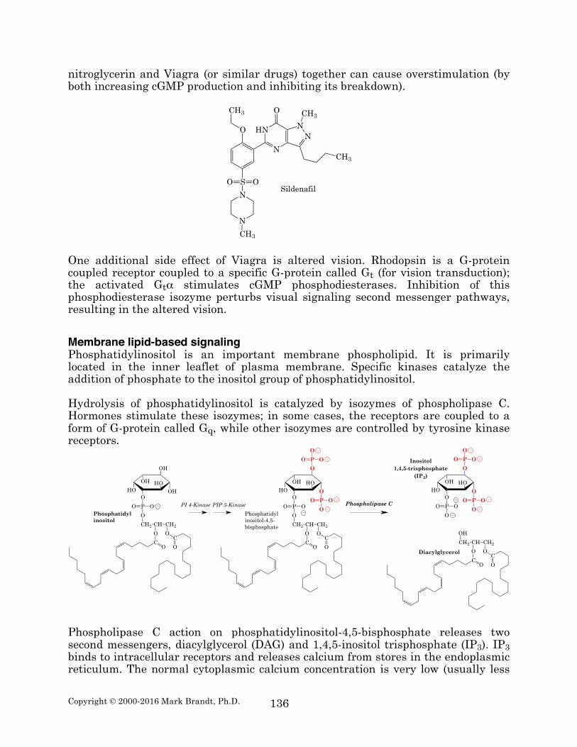
A second potential approach to this problem is to inhibit cGMP breakdown, because nitric oxide uses cGMP as its second messenger. Phosphodiesterase V is a cGMP-specific phosphodiesterase found in certain tissues; its action is responsible for turning off the signal. An inhibitor of this enzyme (sildenafil, better known under the trade name Viagra) prolongs the effect of nitric oxide. This explains why taking
8 Nitroglycerin is a powerful shock-sensitive high explosive. Alfred Nobel invented dynamite as a more user-friendly form of nitroglycerin, and used the proceeds to fund the Nobel Prize trust organizations.
O
O
O
NO2
NO2
O2N 1,2,3-propanetrioltrinitrate
Copyright © 2000-2016 Mark Brandt, Ph.D.
136
nitroglycerin and Viagra (or similar drugs) together can cause overstimulation (by both increasing cGMP production and inhibiting its breakdown).
One additional side effect of Viagra is altered vision. Rhodopsin is a G-protein coupled receptor coupled to a specific G-protein called Gt (for vision transduction); the activated Gtα stimulates cGMP phosphodiesterases. Inhibition of this phosphodiesterase isozyme perturbs visual signaling second messenger pathways, resulting in the altered vision. Membrane lipid-based signaling Phosphatidylinositol is an important membrane phospholipid. It is primarily located in the inner leaflet of plasma membrane. Specific kinases catalyze the addition of phosphate to the inositol group of phosphatidylinositol. Hydrolysis of phosphatidylinositol is catalyzed by isozymes of phospholipase C. Hormones stimulate these isozymes; in some cases, the receptors are coupled to a form of G-protein called Gq, while other isozymes are controlled by tyrosine kinase receptors.
Phospholipase C action on phosphatidylinositol-4,5-bisphosphate releases two second messengers, diacylglycerol (DAG) and 1,4,5-inositol trisphosphate (IP3). IP3 binds to intracellular receptors and releases calcium from stores in the endoplasmic reticulum. The normal cytoplasmic calcium concentration is very low (usually less
HN
N
NN
CH3
CH3
O
O
CH3
SO O
N
N
CH3
Sildenafil
CO
O
O
OO
OH
OH
P OO
O
CH2 CH CH2OC
O
Phosphatidylinositol
Phospholipase CPI 4-Kinase PIP 5-Kinase
CO
O
O
OO
OH
O
P OO
O
CH2 CH CH2OC
O
PO
O
O
Phosphatidylinositol-4,5-bisphosphate
O
OO
OH
O
P OO
O
PO
O
OInositol1,4,5-trisphosphate
(IP3)
CO
O
OH
CH2 CH CH2OC
O
Diacylglycerol
OHHH H
H O
PO O
O
O
PO O
O
HH
Copyright © 2000-2016 Mark Brandt, Ph.D.
137
than 1 µM). IP3 release is one mechanism for increasing intracellular calcium concentration. Diacylglycerol and increased calcium together activate protein kinase C, a serine/threonine-specific protein kinase that alters the activity of a variety of proteins by phosphorylating them. Opening of calcium channels can also increase intracellular calcium levels. In addition to protein kinase C, increased intracellular calcium also interacts with a small, highly conserved, calcium-binding protein called calmodulin. Calcium induces a conformational change in calmodulin; the calcium-calmodulin complex binds and modulates the activity of a number of other proteins in a calcium-dependent manner. Calcium/calmodulin-dependent protein kinase is another major intracellular regulator kinase. Calcium/calmodulin also regulates some of the phosphodiesterases that deactivate cAMP and cGMP. Calcium also acts as a regulator of some protein phosphatases, enzymes that reverse the effects of kinases. Removal of the phosphate added by protein kinases is an important part of the signal also; in some cases, it terminates the signal, while in others, the phosphate-removal is the signal. Calcium is heavily involved in muscle contraction, because calcium/calmodulin also regulates myosin light-chain kinase and calcium directly regulates troponin C (a calcium-binding protein related to calmodulin). Tyrosine kinases A third major type of cell surface receptor contains an intrinsic protein kinase activity that is specific for tyrosine residues. For many of these receptors, ligand binding results in dimerization of the receptor, while for others, the ligand binding probably causes a conformational change in the receptor. Tyrosine kinase receptors include the receptor for insulin and the receptors for a number of growth factors. The activated tyrosine kinase phosphorylates a number of proteins. The result of these phosphorylation events is an activation of a number of second messenger pathways, including ras and some isozymes of phospholipase C. SH2 is the abbreviation for src-homology domain-2; src is a tyrosine kinase oncogene product that contains the first of these domains to be characterized. SH2 domains are phosphotyrosine-binding motifs found in proteins that interact with tyrosine kinases substrates. The SH2 domain requires more than a phosphotyrosine for binding, with different versions having somewhat different sequence specificities. SH2 domains are responsible for tyrosine phosphorylation-dependent protein-protein interactions. Signal propagation by tyrosine kinases depends in part on specific protein-protein interactions. Neurotransmission Neurons frequently use ion channels opening or closing as their primary method for responding to signal molecule binding. Many of these channels contain both receptor and channel functions in the same protein; such receptors comprise a fourth major class of receptor. The signal can be propagated in several ways. One
Copyright © 2000-2016 Mark Brandt, Ph.D.
138
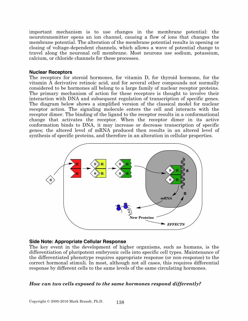
important mechanism is to use changes in the membrane potential: the neurotransmitter opens an ion channel, causing a flow of ions that changes the membrane potential. The alteration of the membrane potential results in opening or closing of voltage-dependent channels, which allows a wave of potential change to travel along the neuronal cell membrane. Most neurons use sodium, potassium, calcium, or chloride channels for these processes. Nuclear Receptors The receptors for steroid hormones, for vitamin D, for thyroid hormone, for the vitamin A derivative retinoic acid, and for several other compounds not normally considered to be hormones all belong to a large family of nuclear receptor proteins. The primary mechanism of action for these receptors is thought to involve their interaction with DNA and subsequent regulation of transcription of specific genes. The diagram below shows a simplified version of the classical model for nuclear receptor action. The signaling molecule enters the cell and interacts with the receptor dimer. The binding of the ligand to the receptor results in a conformational change that activates the receptor. When the receptor dimer in its active conformation binds to DNA, it may increase or decrease transcription of specific genes; the altered level of mRNA produced then results in an altered level of synthesis of specific proteins, and therefore in an alteration in cellular properties.
Side Note: Appropriate Cellular Response The key event in the development of higher organisms, such as humans, is the differentiation of pluripotent embryonic cells into specific cell types. Maintenance of the differentiated phenotype requires appropriate response (or non-response) to the correct hormonal stimuli. In most, although not all cases, this requires differential response by different cells to the same levels of the same circulating hormones. How can two cells exposed to the same hormones respond differently?
!
! "
! "
! "
! "
! "
"
"
!"#$
#$%&'()*$+,-
%&&%'()
"!

Copyright © 2000-2016 Mark Brandt, Ph.D.
139
A complete answer to this question would require an entire textbook. The following is a partial list containing a few general examples that illustrate some of the mechanisms that cells use to allow the correct response to a given stimulus.
1) Presence of the receptor: Most cells do not respond to the steroid hormone progesterone because they lack the progesterone receptor. This is the simplest method of responding “correctly”: for a cell without the receptor for a hormone, that hormone does not exist, regardless of the circulating levels of that hormone. In some cases, the amount of receptor may be important in regulating the response to the hormone also: in general, a cell with a higher level of receptor has a stronger response than a cell with a lower level.
2) Alteration or inactivation of the hormone: Many cells are capable of
metabolizing hormones to inactive molecules. If this occurs before the hormone binds to the receptor, the cell will not respond to the hormonal signal. In addition, some cells are capable of converting inactive molecules to hormones; if this happens, the cell may respond to the hormone before surrounding cells are exposed to the hormone.
3) Regulation of second messengers: Most hormones act through one of a small
number of second messenger molecules. For many of these second messengers, the cell can modulate the amount of second messenger that is present. For example, cells can increase the GTPase activity of Gα, inactivating the Gα more rapidly, which would lower the amount of cAMP produced. Alternatively, the cell may decrease the activity of phosphodiesterase, and therefore increase the cAMP concentration within the cell.
4) Regulation of protein phosphorylation: A large subset of second messenger
systems function by altering phosphorylation states of proteins (e.g., by activation of the cAMP-dependent protein kinase, or by activation of protein kinase C, or by activation of protein phosphatases that remove the added phosphate). Phosphorylation states of cellular proteins can vary dramatically as different pathways increase rates of addition or removal of the phosphates; in addition, some proteins may be phosphorylated at multiple sites, usually by different enzymes, with the effect of the modification at different locations having differing effects. Depending on the modification enzymes present, and the different kinase and phosphatase pathways being activated, the cell can integrate signals of different types to react appropriately to all of the signals to which it can respond at a given time.
The important concept to remember at this point is that the response to a given hormone depends on a number of other factors. The response cannot be considered in isolation; it depends on the cell type, on the presence or absence of other proteins, and on the levels of other hormones to which the cell can respond, or to which the cell has recently responded.
Copyright © 2000-2016 Mark Brandt, Ph.D.
140
Summary Signal transduction is the mechanism that a cell uses to convert an extracellular signal to changes in intracellular processes. It is thus a method for moving information across membranes. In most cases, the binding of a ligand to its receptor results in release of second messenger molecules. Second messenger molecules include cAMP, cGMP, calcium, inositol trisphosphate, and diacylglycerol, among others. One mechanism of second messenger action is the activation of protein kinases (e.g., cAMP-dependent protein kinase, protein kinase C, and calcium/calmodulin dependent protein kinase); the kinases alter the activity of a variety of proteins by phosphorylation. Other mechanisms of second messenger action include stimulation of second messenger-breakdown enzymes and of phosphatases that dephosphorylate proteins. Some receptors contain intrinsic tyrosine kinase activity; these receptors alter cellular functions by directly phosphorylating proteins on tyrosine residues. Some receptors interact directly with DNA and with the transcription machinery to alter rates of gene transcription. Finally, some proteins are ligand operated ion channels; these alter ion gradients across the membrane, and therefore directly alter cellular properties.
Related Documents