Asian Pacific Journal of Cancer Prevention, Vol 13, 2012 2753 DOI:http://dx.doi.org/10.7314/APJCP.2012.13.6.2753 ROS and Induction of Apoptosis by Rubia cordifolia in HEp-2 Cells Asian Pacific J Cancer Prev, 13, 2753-2758 Introduction Cancer of the larynx is the most common type of head and neck cancer. The high incidence of larynx cancer is primarily attributable to the habit of tobacco, betel quid chewing and alcohol consumption. It was estimated by NCI’s SEER Cancer Statistics Review that among Asians, 2.4 per 100,000 men and 0.3 per 100,000 women were diagnosed in 2003-2007. The treatment of these types of oral cancers includes surgery and/or radiotherapy, which are often associated with loss of function, disfigurement and reduced quality of life (Day et al., 2003). Recently, advances in chemotherapeutic agents for the treatment of larynx cancer have been highlighted but the survival rate of patients has not improved significantly. The development of novel therapeutic agents targeting the malignant behavior of these cancers is important to improve the prognosis of treatment (Ghashm et al., 2010). In a scenario where conventional medicine has failed to develop techniques that could reduce the incidence of death due to larynx cancer, complementary and alternative medicine is slowly emerging as an option. There are a number of alternative medicine systems based on traditional theories and philosophy that have originated in specific geographical areas and evolved over the years. The most widely practiced of these include the Unani, Ayurveda & Siddha and the Chinese system of medicine that have 1 Department of Biochemistry, University of Madras, Guindy campus, Chennai -600025, Tamilnadu, 2 School of Chemical and Biotechnology, SASTRA University, Thanjavur-613401, India *For correspondence: [email protected] Abstract Rubia cordifolia Linn, which belongs to the Rubiaceae family, is a well-known herb used in Ayurvedic medicine. In the present study, we investigated the influence of a methanolic extract (RC) on the induction of apoptosis in HEp-2 (human laryngeal carcinoma) cell line, as evidenced by cytotoxicity, morphological changes and modification in the levels of pro-oxidants. Inhibition of cell proliferation and lactate dehydrogenase (LDH) release increased in a time and dose-dependent manner. Further, reduced glutathione (GSH), glutathione transferase (GST) and protein levels decreased and lipid peroxidation increased significantly on RC treatment in a dose dependent manner when compared to controls. Based on the results we determined the optimal dose as 30mg/ ml and the apoptotic effect of RC extract (30 mg/ml) on HEp-2 cells was confirmed by fluorescent microscopy and transmission electron microscopy (TEM) based on morphological and ultrastructural changes. RC extract suppressed the proliferation of HEp-2 oral cancer cells inducing apoptotic cell death in vitro. These results point to potential of RC extract as an agent for the treatment of laryngeal squamous cell carcinoma. Keywords: Antiproliferative activity - cancer cells - medicinal plant - in vitro studies RESEARCH COMMUNICATION Induction of Apoptosis by Methanolic Extract of Rubia Cordifolia Linn in HEp-2 Cell Line is Mediated by Reactive Oxygen Species PN Shilpa 1 *, V Sivaramakrishnan 2 , S Niranjali Devaraj 1 originated from the Arab, India and China respectively. These medicinal systems have identified more than 700 individual herbal extracts as well as several drug preparations which claim to treat and/or prevent several diseases including cancer (Shafi et al., 2009). The process of programmed cell death, or apoptosis, is generally characterized by distinct morphological characteristics and energy-dependent biochemical mechanisms .Apoptosis has emerged as an important mechanism for anticancer effects of many naturally occurring and synthetic agents (Cheon et al., 2006; Ichikawa and Aggarwal, 2006; Kim et al., 2007; Samudio et al., 2005; Shishodia and Aggarwal, 2004; Xiao and Singh, 2007; Xiao and Singh, 2008; Xiao et al., 2006c). During the past decade, however, the evidence is gradually being shown that many cancer chemotherapeutic agents are not totally free from side effects. It is thus considered important to screen apoptotic inducers from plants, either in the form of crude extracts or as active components isolated from them. Rubia cordifolia, Linn (locally known as manjistha), a perennial herbaceous climber belonging to the family Rubiaceae, is very common on Higher ghats, Mahabaleswar, Amboli and Maharastra state in India. It has long cylindrical and typically red colored roots (Adwankar et al., 1980). Rubia cordifolia is an important medicinal plant which is used for treatment of various

Welcome message from author
This document is posted to help you gain knowledge. Please leave a comment to let me know what you think about it! Share it to your friends and learn new things together.
Transcript
Asian Pacific Journal of Cancer Prevention, Vol 13, 2012 2753
DOI:http://dx.doi.org/10.7314/APJCP.2012.13.6.2753 ROS and Induction of Apoptosis by Rubia cordifolia in HEp-2 Cells
Asian Pacific J Cancer Prev, 13, 2753-2758
Introduction
Cancer of the larynx is the most common type of head and neck cancer. The high incidence of larynx cancer is primarily attributable to the habit of tobacco, betel quid chewing and alcohol consumption. It was estimated by NCI’s SEER Cancer Statistics Review that among Asians, 2.4 per 100,000 men and 0.3 per 100,000 women were diagnosed in 2003-2007. The treatment of these types of oral cancers includes surgery and/or radiotherapy, which are often associated with loss of function, disfigurement and reduced quality of life (Day et al., 2003). Recently, advances in chemotherapeutic agents for the treatment of larynx cancer have been highlighted but the survival rate of patients has not improved significantly. The development of novel therapeutic agents targeting the malignant behavior of these cancers is important to improve the prognosis of treatment (Ghashm et al., 2010). In a scenario where conventional medicine has failed to develop techniques that could reduce the incidence of death due to larynx cancer, complementary and alternative medicine is slowly emerging as an option. There are a number of alternative medicine systems based on traditional theories and philosophy that have originated in specific geographical areas and evolved over the years. The most widely practiced of these include the Unani, Ayurveda & Siddha and the Chinese system of medicine that have
1Department of Biochemistry, University of Madras, Guindy campus, Chennai -600025, Tamilnadu, 2School of Chemical and Biotechnology, SASTRA University, Thanjavur-613401, India *For correspondence: [email protected]
Abstract
Rubia cordifolia Linn, which belongs to the Rubiaceae family, is a well-known herb used in Ayurvedic medicine. In the present study, we investigated the influence of a methanolic extract (RC) on the induction of apoptosis in HEp-2 (human laryngeal carcinoma) cell line, as evidenced by cytotoxicity, morphological changes and modification in the levels of pro-oxidants. Inhibition of cell proliferation and lactate dehydrogenase (LDH) release increased in a time and dose-dependent manner. Further, reduced glutathione (GSH), glutathione transferase (GST) and protein levels decreased and lipid peroxidation increased significantly on RC treatment in a dose dependent manner when compared to controls. Based on the results we determined the optimal dose as 30mg/ml and the apoptotic effect of RC extract (30 mg/ml) on HEp-2 cells was confirmed by fluorescent microscopy and transmission electron microscopy (TEM) based on morphological and ultrastructural changes. RC extract suppressed the proliferation of HEp-2 oral cancer cells inducing apoptotic cell death in vitro. These results point to potential of RC extract as an agent for the treatment of laryngeal squamous cell carcinoma. Keywords: Antiproliferative activity - cancer cells - medicinal plant - in vitro studies
RESEARCH COMMUNICATION
Induction of Apoptosis by Methanolic Extract of Rubia Cordifolia Linn in HEp-2 Cell Line is Mediated by Reactive Oxygen SpeciesPN Shilpa1*, V Sivaramakrishnan2, S Niranjali Devaraj1
originated from the Arab, India and China respectively. These medicinal systems have identified more than 700 individual herbal extracts as well as several drug preparations which claim to treat and/or prevent several diseases including cancer (Shafi et al., 2009). The process of programmed cell death, or apoptosis, is generally characterized by distinct morphological characteristics and energy-dependent biochemical mechanisms .Apoptosis has emerged as an important mechanism for anticancer effects of many naturally occurring and synthetic agents (Cheon et al., 2006; Ichikawa and Aggarwal, 2006; Kim et al., 2007; Samudio et al., 2005; Shishodia and Aggarwal, 2004; Xiao and Singh, 2007; Xiao and Singh, 2008; Xiao et al., 2006c). During the past decade, however, the evidence is gradually being shown that many cancer chemotherapeutic agents are not totally free from side effects. It is thus considered important to screen apoptotic inducers from plants, either in the form of crude extracts or as active components isolated from them. Rubia cordifolia, Linn (locally known as manjistha), a perennial herbaceous climber belonging to the family Rubiaceae, is very common on Higher ghats, Mahabaleswar, Amboli and Maharastra state in India. It has long cylindrical and typically red colored roots (Adwankar et al., 1980). Rubia cordifolia is an important medicinal plant which is used for treatment of various
P N Shilpa et al
Asian Pacific Journal of Cancer Prevention, Vol 13, 20122754
ailments in Ayurvedic system of medicine (Adwankar et al., 1980; Sertoli et al., 1994; Sridevi, 2011). The major active component of Rubia cordifolia Linn are anthraquinones glycosides and include 1-hydroxy 2-methoxy anthraquinone, 1, 4-dihydroxy-2-methyl-5-methoxy anthraquinone, 1, 3-dimethoxy 2-carboxy anthraquinone and rubiadin (1,3-dihydroxy-2-methyl anthraquinone) (Dosseh et al., 1981). In the Indian system of medicine this plant is in clinical use for the management of several disorders including cancer (Patel et al., 2011a). The plant Rubia cordifolia has been reported for anti-inflammatory (Kasture et al., 2001), immunomodulatory (Joharapurkar et al., 2003), anticonvulsant, anxiolytic (Kasture et al., 2000) and anti-tumor activities (Patel et al., 2011b). In the ethno-botanical claims, it is mentioned that the roots are used for the treatment of jaundice by the folk tribes of West Bengal and Uttaranchal. But to the best of our knowledge there is no scientific report on the anti-proliferative and apoptosis inducing effect of Rubia cordifolia. Hence, the present study was carried out to elicit the anti-proliferative and apoptosis inducing effect of the extract of extract of Rubia cordifolia in vitro, using HEp-2 cells as the experimental model. Materials and Methods
Chemicals and reagents Dulbecco’s Modified Eagle Medium (DMEM), fetal calf serum, penicillin, streptomycin and trypsin / ethylenediamine tetraacetic acid (EDTA) were purchased from Life Technologies, Inc. (GrandIsland, NY, USA). Dimethyl sulphoxide and 3-(4, 5-dimethyl thizol-2-yl)-2, 5-diphenyl tetrazolium bromide (MTT) were purchased from Sigma Chemical Co. (St. Louis, MO, USA).
Plant material and extraction Roots of Rubia cordifolia (RC) were purchased from market. The plant material was identified and duly authenticated by the Center for Advanced Studies in Botany, Guindy Campus, University of Madras, Chennai. A voucher specimen of the collected sample was deposited in the departmental museum for future reference. Roots were dried in shade and pulverized. The powder was treated with petroleum ether for dewaxing and later subjected to cold percolation using methanol (95%, v/v) as solvent. The extract was concentrated under vacuum and dried in a vacuum desiccator and stored at room temperature. RC extract was dissolved in sterile water and concentration of 5, 10, 15, 20, 25 and 30 mg/ml were used for the various experiments.
Cell culture Human Epithelial carcinoma (HEp-2) cell line was obtained from National Center for Cell Sciences, Pune, India. The cells were grown at 37 °C under a humidified, 5% CO2 atmosphere in DMEM medium supplemented with 10% fetal calf serum (FCS), 2 mM glutamine, 100 units/ml of penicillin and 50 μg/ml of streptomycin.
Trypan blue exclusion test Cell viability was determined by trypan blue exclusion
test after cells were harvested after designated treatments with different concentration of drug, using trypan blue. Equal amount of cell suspension and 0.4% trypan blue were mixed for 1-2 min and 10 microliter of the mixture were taken to the hemocytometer and then the cells were counted in all four field under the light microscope (Freshney, 1994).
Cytotoxicity Assay HEp-2 Cells were seeded in 96-well microtitre plates (1×10⁶ cells per well), and left to adhere to the plastic plates overnight before being exposed to different concentrations of RC. In control well, 100 μl of medium alone was added. Then, cells in each well were incubated at 37°C in 10μl of MTT (5 mg/ml) for 4 hr. After the incubation period, the well contents were removed and the reaction was terminated by addition of 100μl of dimethyl sulphoxide and 12.5μl of glycine buffer (0.1 M glycine, 0.1 M NaCl, pH 10.5) to each well. Absorbance was read at 570 nm using a microplate ELISA reader (Mosmann, 1983). Growth inhibition was expressed as percentage.
Biochemical assays Lactate dehydrogenase (LDH) was estimated by the method of (Nieland, 1955). The activity of Glutathione-S-Transferase (GST) was determined by the method of (Habig et al., 1974). Reduced glutathione (GSH) was estimated by the method of (Moron et al., 1979). Lipid peroxide (LPO) content was determined by thiobarbituric acid reaction as described by (Okhawa et al., 1979).
Morphological studies
Fluorescent microscopy - Dual staining. Morphological changes of the cellular nucleus after treatment was studied by staining with acridine orange/ethidium bromide. Cells were seeded on coverslip in 35 mm sterile petriplates. After the designated treatments, the cells were harvested by trypsinization, washed with PBS and about 25μl of cell suspension was then mixed with 1μl of the dye mixture, containing both 100 μg/ml acridine orange and 100 μg/ml ethidium bromide in Phosphate buffered saline (PBS). After staining, the cells were visualized immediately under a fluorescent microscope.
Transmission electron microscopy (TEM). After trypsinization, suspended cells were centrifuged at 800 g and fixed in 1% gluteraldehyde in 0.1 M phosphate buffer (pH-7.4) overnight at 4°C. Then, post fixed with 1% OSO4 in 0.1 M cacodylate buffer (pH-7.4) for 1 hr at 4°C, the cell cultures were dehydrated in graded ethanol solutions and embedded in epon according to the method of pease ultrathin sections of the cell from atleast two selected blocks per well were counter stained with uranyl acetate and lead citrate and were examined with Philips CM10 TEM (David et al., 1973).
Statistical analysis Results are expressed as Mean ± SD. The results were statistically evaluated using one-way analysis of variance (ANOVA) by SPSS software version 10 for repeated
Asian Pacific Journal of Cancer Prevention, Vol 13, 2012 2755
DOI:http://dx.doi.org/10.7314/APJCP.2012.13.6.2753 ROS and Induction of Apoptosis by Rubia cordifolia in HEp-2 Cells
measurements and Post-hoc Dunnet’s test for multiple comparisons was performed. The mean difference was significant at the 0.001, 0.01, 0.05 levels.
Results
The plant products serve as vital sources of blocking and suppressing agent that interfere with the carcinogenic process. Since mechanism relating to cell death and cancer attenuation by plant products has received limited attention, the present work was carried out to illustrate apoptotic and antiproliferative effect of RC. HEp-2 cells were treated with extracts equivalent to 5, 10, 15, 20, 25 and 30 mg/ml. Cell proliferation was inhibited in a dose-dependent manner after exposure to RC at dosages above 10 mg. The percentage of viable cells was 62.87%, 54.67%, 38.99%, 26.92%, 20.32%, 15% for 5, 10, 15, 20, 25 and 30 mg/ml of RC respectively, whereas in cell control the number of viable cells was
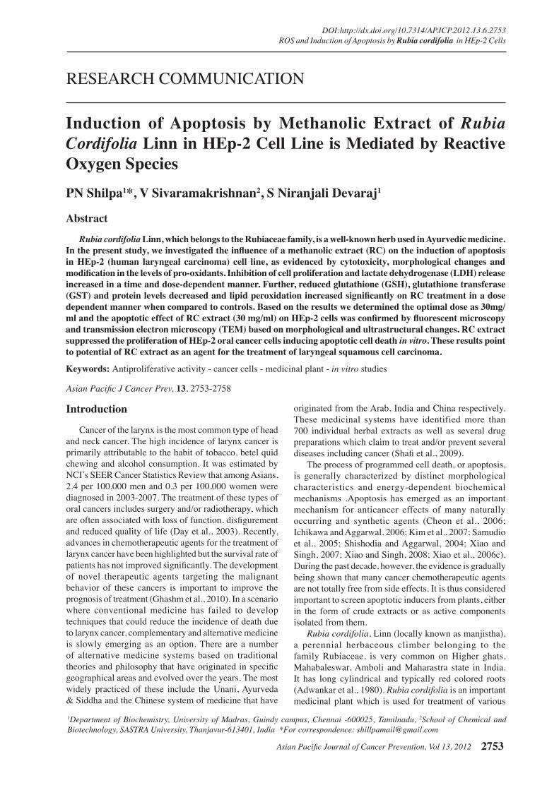
79.06% (Figure 1). The LD50 of RC determined by MTT utilization analysis in HEp-2 was 10 mg/ml. In response to RC, the number of cells in culture also progressively and dose- dependently declined. At 4 mg RC, cell proliferation ceased, whereas at concentration greater than 4 mg, cells died with significant decreases in cell numbers and in a time-dependent manner (24 h, 48 h) (Figure 2). According to MTT assay the reduction of a tetrazolium component
Figure 1. Effect of RC. A) HEp-2 Cell viability, B) RC on LDH release, C) GSH in HEp-2 cells, D) GST in HEp-2 cells, E) TBARS in HEp-2 cells, F) Protein in HEp-2 cells
Figure 2. Cytotoxic Activity of RC on HEp-2 cells
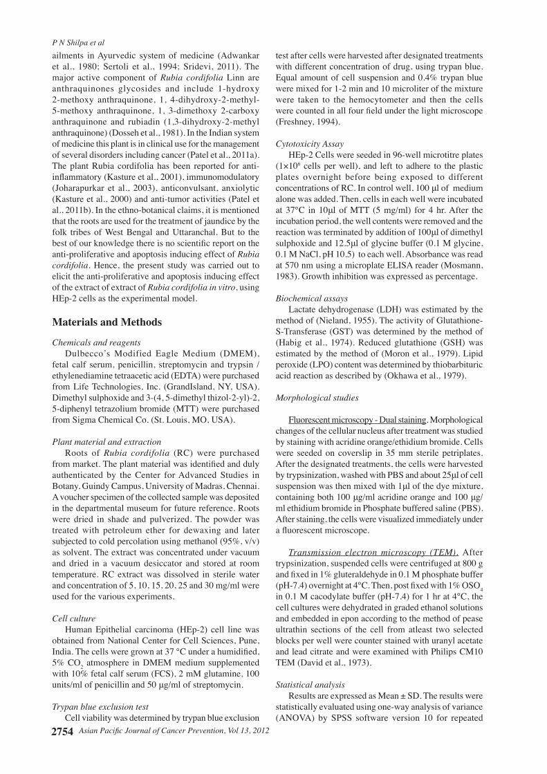
Figure 3. Apoptosis of HEp-2 cells treated with RC (20 mg) Acridine orange/Ethidium bromide. (a)Viable cells with normal nuclei (VN)-Bright green chromatin with organized structure.(b) Viable cells with apoptotic nuclei (VA)-Bright green chromatin highly condensed or fragmented.(c) Non-viable cells with normal nuclei (NVN)-Bright orange chromatin with organized structure.(d) Non-viable cells with apoptotic nuclei (NVA)-Bright orange chromatin, highly condensed or fragmented
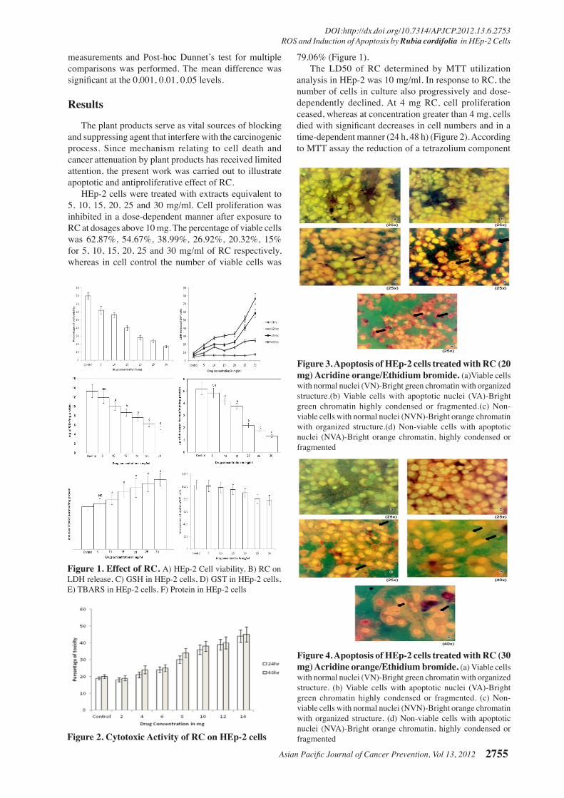
Figure 4. Apoptosis of HEp-2 cells treated with RC (30 mg) Acridine orange/Ethidium bromide. (a) Viable cells with normal nuclei (VN)-Bright green chromatin with organized structure. (b) Viable cells with apoptotic nuclei (VA)-Bright green chromatin highly condensed or fragmented. (c) Non-viable cells with normal nuclei (NVN)-Bright orange chromatin with organized structure. (d) Non-viable cells with apoptotic nuclei (NVA)-Bright orange chromatin, highly condensed or fragmented
P N Shilpa et al
Asian Pacific Journal of Cancer Prevention, Vol 13, 20122756
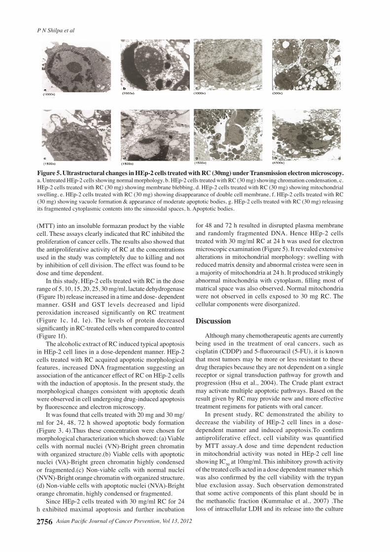
(MTT) into an insoluble formazan product by the viable cell. These assays clearly indicated that RC inhibited the proliferation of cancer cells. The results also showed that the antiproliferative activity of RC at the concentrations used in the study was completely due to killing and not by inhibition of cell division. The effect was found to be dose and time dependent. In this study, HEp-2 cells treated with RC in the dose range of 5, 10, 15, 20, 25, 30 mg/ml, lactate dehydrogenase (Figure 1b) release increased in a time and dose- dependent manner. GSH and GST levels decreased and lipid peroxidation increased significantly on RC treatment (Figure 1c, 1d, 1e). The levels of protein decreased significantly in RC-treated cells when compared to control (Figure 1f). The alcoholic extract of RC induced typical apoptosis in HEp-2 cell lines in a dose-dependent manner. HEp-2 cells treated with RC acquired apoptotic morphological features, increased DNA fragmentation suggesting an association of the anticancer effect of RC on HEp-2 cells with the induction of apoptosis. In the present study, the morphological changes consistent with apoptotic death were observed in cell undergoing drug-induced apoptosis by fluorescence and electron microscopy. It was found that cells treated with 20 mg and 30 mg/ml for 24, 48, 72 h showed apoptotic body formation (Figure 3, 4).Thus these concentration were chosen for morphological characterization which showed: (a) Viable cells with normal nuclei (VN)-Bright green chromatin with organized structure.(b) Viable cells with apoptotic nuclei (VA)-Bright green chromatin highly condensed or fragmented.(c) Non-viable cells with normal nuclei (NVN)-Bright orange chromatin with organized structure.(d) Non-viable cells with apoptotic nuclei (NVA)-Bright orange chromatin, highly condensed or fragmented. Since HEp-2 cells treated with 30 mg/ml RC for 24 h exhibited maximal apoptosis and further incubation
for 48 and 72 h resulted in disrupted plasma membrane and randomly fragmented DNA. Hence HEp-2 cells treated with 30 mg/ml RC at 24 h was used for electron microscopic examination (Figure 5). It revealed extensive alterations in mitochondrial morphology: swelling with reduced matrix density and abnormal cristea were seen in a majority of mitochondria at 24 h. It produced strikingly abnormal mitochondria with cytoplasm, filling most of matrical space was also observed. Normal mitochondria were not observed in cells exposed to 30 mg RC. The cellular components were disorganized.
Discussion
Although many chemotherapeutic agents are currently being used in the treatment of oral cancers, such as cisplatin (CDDP) and 5-fluorouracil (5-FU), it is known that most tumors may be more or less resistant to these drug therapies because they are not dependent on a single receptor or signal transduction pathway for growth and progression (Hsu et al., 2004). The Crude plant extract may activate multiple apoptotic pathways. Based on the result given by RC may provide new and more effective treatment regimens for patients with oral cancer.
In present study, RC demonstrated the ability to decrease the viability of HEp-2 cell lines in a dose-dependent manner and induced apoptosis.To confirm antiproliferative effect, cell viability was quantified by MTT assay.A dose and time dependent reduction in mitochondrial activity was noted in HEp-2 cell line showing IC50 at 10mg/ml. This inhibitory growth activity of the treated cells acted in a dose dependent manner which was also confirmed by the cell viability with the trypan blue exclusion assay. Such observation demonstrated that some active components of this plant should be in the methanolic fraction (Kummalue et al., 2007) .The loss of intracellular LDH and its release into the culture
Figure 5. Ultrastructural changes in HEp-2 cells treated with RC (30mg) under Transmission electron microscopy. a. Untreated HEp-2 cells showing normal morphology, b. HEp-2 cells treated with RC (30 mg) showing chromation condensation, c. HEp-2 cells treated with RC (30 mg) showing membrane blebbing, d. HEp-2 cells treated with RC (30 mg) showing mitochondrial swelling, e. HEp-2 cells treated with RC (30 mg) showing disappearance of double cell membrane, f. HEp-2 cells treated with RC (30 mg) showing vacuole formation & appearance of moderate apoptotic bodies, g. HEp-2 cells treated with RC (30 mg) releasing its fragmented cytoplasmic contents into the sinusoidal spaces, h. Apoptotic bodies.
Asian Pacific Journal of Cancer Prevention, Vol 13, 2012 2757
DOI:http://dx.doi.org/10.7314/APJCP.2012.13.6.2753 ROS and Induction of Apoptosis by Rubia cordifolia in HEp-2 Cells
medium is an indicator of irreversible cell death due to cell membrane damage which increased with time and dose (Fotakis and Timbrell, 2006). Further, cytotoxicity was also confirmed by protein assay.
Images obtained from live cell cultures under proantrocyanin administration demonstrated that many of the cells were clustering, rounding, and blebbing – morphologic changes that are often suggestive of apoptosis (King et al., 2007). Abrupt cell shrinkage, the earliest light microscopic feature in apoptosis occurs almost simultaneous to nuclear changes and is evidenced as cells in culture lose contact with adjacent cells, during which convolution begins to form shrinkage which occurs due to a net outward fluid movement, likely because of the inhibition of the Na+-K+- Cl- co-transporter system(Wilcock and Hickman, 1988). The morphological changes observed after Cranberry and Grape seed extract treatment in oral squamous carcinoma cell lines were similar to the effect caused by RC (Chatelain et al., 2011). Further, chromatin condensation and nuclear fragmentation of treated cells were clearly evident on examination under a fluorescent microscope. Acridine orange is a vital dye that will stain both live and dead cells, whereas ethidium bromide will stain only those cells that have lost their membrane integrity (Raju et al., 2004). Based on the MTT assay, 10 mg, 20 mg, 30 mg/ml and the morphological features of a cell line in apoptosis were dose dependent, i.e., a stronger apoptosis signal was induced with higher concentrations of the respective extract. Cytoplasmic shrinking and hypercondensed chromatin observed by (Stander et al., 2007) are also not exclusive properties of apoptotic processes and can also indicate the presence of autophagic processes (Guimaraes and Linden, 2004).
Cleavage of nuclear DNA is regarded as a biochemical hallmark of apoptosis. It is well known that apoptosis is induced either by depletion of endogenous antioxidants or by generation of free radicals (Wyllie, 1980). We have also analysed the intracellular GSH levels in HEp-2 cells after treatment with the RC. The GSH levels were significantly lower when compared to control cells. Cellular GSH plays an important role in protection against oxidative stress-induced injury. Depletion of GSH levels has been shown to enhance susceptibility to oxidative stress-induced cytotoxicity.This result was consistent with Quercitin (Ramos and Aller, 2008). The depletion of endogenous GSH sensitized the cells to RC treatment and thereby increased the apoptotic cells, thus suggesting inactivation by the formation of GSH-RC conjugate, and GSTs are involved in catalyzing the GSH conjugate formation (Mari et al., 2009). Therefore, it was interesting to measure GST activity in the present study especially when RC elevated the endogenous GSH levels. Recent findings suggest that GSTs play an important role in carcinogenicity and resistance to drugs and their levels are elevated in several tumors (Mari et al., 2009). Exposure of mammalian cells to a variety of chemical agents results in increased levels of xenobiotic metabolizing enzymes. These enzymes function as an intracellular detoxification system and by decreasing the levels of compounds capable of generating ROS; they are part of the enzymatic antioxidant defense
against oxidative stress (Sies, 1991). GSTs are only part of a complex detoxification system because multiple enzymes are involved in the redox cycling of GSH (Spanou et al., 2010). These observations suggest that reactive oxygen species generation plays a key role in the induction of apoptosis in HEp-2 cells.
In conclusion, our result clearly demonstrated that RC inhibits cell proliferation in HEp-2 cell line and induced apoptosis through the elevation of reactive oxygen species generation. This could be attributed to phytochemicals present in the extract. Even though further studies are needed to confirm these properties of each component the present work opens new perspectives for cancer treatment based on ethnopharmacological studies.
References
Adwankar MK, Chitnis MP, Khandalekar DD, Bhadsavale CG (1980). Anticancer acvtivity of the extracts of Rubia cordifolia Linn. Ind J Exp Biol, 18, 102-9
Chatelain K, Phippen S, McCabe J, et al (2011). Cranberry and grape seed extracts inhibit the proliferative phenotype of oral squamous cell carcinomas. Evid Based Complement Altern Med, ??, 1-12
Cheon JH, Kim JS, Kim JM, et al (2006). Plant sterol guggulsterone inhibits nuclear factor-kB signaling in intestinal epithelial cells by blocking IkB kinase and ameliorates acute murine colitis. Inflamm Bowel Dis, 12, 1152-61.
David GFX, Hebert J, Wright CS (1973). The ultrastructure of the pineal ganglion in the ferret. J Anat, 115, 79-97.
Day TA, Davis BK, Gillespie MB, et al (2003). Oral cancer treatment. Curr Treat Options Oncol, 4, 27-41.
Dosseh C, Tessier AM, Delaveau P (1981). New quinones in Rubia cordifolia L. Roots, III. Planta Med, 43, 360-6.
Fotakis G, Timbrell JA (2006). In vitro cytotoxicity assays: Comparison of LDH, neutral red, MTT and protein assay in hepatoma cell lines following exposure to cadmium chloride. Toxicol Lett, 160, 171-7.
Freshney I (1994). Culture of Animal Cells: a manual of basic techniques. New York, John Wiley & Sons. Fourth Edition, 330-1
Ghashm AA, Othman NH, Khattak MN, Ismail NM, Saini R (2010). Antiproliferative effect of Tualang honey on oral squamous cell carcinoma and osteosarcoma cell lines. BMC Complement Altern Med, 10, 49-57.
Guimaraes A, Linden R (2004). Programmed cell deaths. Apoptosis and alternative deathstyles. Eur J Biochem, 271, 1638-50.
Habig WH, Pabst MJ, Jakoby WB (1974). Glutathione-S-transferase, the first enzymatic step in mercapturic acid formation. J Biol Chem, 249, 7130-139.
Hsu S, Singh B, Schuster G (2004). Induction of apoptosis in oral cancer cells: agents and mechanisms for potential therapy and prevention. Oral Oncol, 40, 461-73.
Ichikawa H, Aggarwal BB (2006). Guggulsterone inhibits osteoclastogenesis induced by receptor activator of nuclear factor-β ligand and by tumor cells by suppressing nuclear factor-β activation. Clinc Cancer Res, 12, 662-8.
Joharapurkar AA, Deode NM, Zambad SP, Umathe SN (2003). Immunomodulatory activity of alcoholic extract of Rubia cordifolia Linn. Indian Drugs, 40, 179-81.
Kasture SB, Kasture VS, Chopde CT (2001) Anti-inflammatory activity of Rubia cordifolia roots. J Nat Remed, 1, 111-5.
Kasture VS, Desmukh VK, Chopde CT (2000). Anticonvulsant
P N Shilpa et al
Asian Pacific Journal of Cancer Prevention, Vol 13, 20122758
and behavioral actions of triterpine isolated from Rubia cordifolia Linn. Ind J Exp Biol, 38, 675-80.
Kim YA, Xiao D, Xiao H, et al (2007). Mitochondria-mediated apoptosis by diallyl trisulfide in human prostate cancer cells is associated with generation of reactive oxygen species and regulated by Bax/Bak. Mol Cancer Ther, 6, 1599-609.
King M, Chatelain K, Farris D, et al (2007). Oral squamous cell carcinoma proliferative phenotype is modulated by proanthocyanidins: a potential prevention and treatment alternative for oral cancer. BMC Complement Altern Med, 7, 22-32.
Kummalue T, Charoenrat OO, Jiratchariyakul W, et al (2007). Antiproliferative effect of Erycibe elliptilimba on human breast cancer cell line. J Ethnopharm, 110, 439-43.
Mari M, Morales A, Colell A, Garcı´a-Ruiz C, Fernandez-Checa JC (2009). Mitochondrial glutathione, a key survival antioxidant. Antioxid Redox signal, 11, 2685-700.
Moron MS, De Pierre JW, Mannervik B (1979). Levels of glutathione, glutathione reductase and glutathione-S-transferase in rat lung and liver. Biochim Biophys Acta, 82, 67-70.
Mosmann T (1983). Rapid colorimetric assay for cellular growth and survival: application to proliferation and cytotoxicity assays. J Immunol Methods, 65, 55-63.
Nieland AA (1955). Lactic dehydrogenase of heart muscle. Methods in enzymology, 1, 449-54.
Okhawa H, Ohishi N, Yagi K (1979). Assay of lipid peroxides in animal tissues by thiobarbituric acid reaction. Anal Biochem, 95, 351-8.
Patel PR, Nagari AA, Patel RC, Rathod DK, Patel VR (2011a). Invitro anticancer activity of Rubia cordifolia against Hela and Hep2 cell line. Int J Pharmacy Pharmcol Sci, 3, 70-1.
Patel PR, Raval BP, Karanth HA, Patel VR (2011b). Potent antitumor activity of Rubia cordifolia. Int J Phytomed, 2, 44-6.
Raju J, Patlolla JM, Swamy MV, Rao CV (2004). Diosgenin, a steroid saponin of Trigonella foenum graecum (Fenugreek), inhibits azoxymethane-induced aberrant crypt foci formation in F344 rats and induces apoptosis in HT-29 human colon cancer cells. Cancer Epidem Biomar, 13, 1392-8.
Ramos AM, Aller P (2008). Quercetin decreases intracellular GSH content and potentiates the apoptotic action of the antileukemic drug arsenic trioxide in human leukemia cell lines. Biochem Pharmacol, 75, 1912-23.
Samudio I, Konopleva M, Safe S, McQueen T, Andreeff M (2005). Guggulsterones induce apoptosis and differentiation in acute myeloid leukemia: identification of isomer-specific antileukemic activities of the pregnadienedione structure. Molecular Cancer Ther, 4, 1982-92.
Sertoli A, Francalanci S, Giorgini S (1994) Prevention of allergic contact dermatitis with alternative products. Contact Dermatitis, 31, 322-3.
Shafi G, Munshi A, Hasan TN, et al (2009). Induction of apoptosis in HeLa cells by chloroform fraction of seed extracts of Nigella sativa. Cancer Cell Int, ??, 29-37.
Shishodia S, Aggarwal BB (2004). Guggulsterone inhibits NF-kappaB and IkappaBalpha kinase activation, suppresses expression of anti-apoptotic gene products, and enhances apoptosis. J Biol Chem, 279, 47148 -58.
Sies H (1991). Oxidative stress: from basic research to clinical applications. Am J Med, 91, 31-8.
Spanou C, Stagos D, Aligiannis N, Kouretas D (2010). Influence of potent antioxidant leguminosae family plant extracts on growth and antioxidant defense system of Hep2 cancer cell line. J Med Food, 13, 149-55.
Sridevi Sivakami PL, Nandhini C (2011). Extraction, isolation of medicinal plant pigments for natural food colours. Plant
Arch, 11, 479-84.Stander BA, Marais S, Steynberg TJ, et al (2007). Influence
of Sutherlandia frutescens extracts on cell numbers, morphology and gene expression in MCF-7 cells. J Ethnopharm, 112, 312-8.
Wilcock E, Hickman JA (1988). Characterisation of a Na+-K+-CL- cotransporter in alkylating agents-sensitive L1210 murine leukemia cells. Biochim Biophys Acta, 946, 359-69.
Wyllie AH (1980) Glucocorticoid-induced thymocyte apoptosis is associated with endogenous endonuclease activation. Nature, 284, 555-6.
Xiao D, Singh SV (2007). Phenethyl isothiocyanate inhibits angiogenesis in vitro and ex vivo. Cancer Res, 67, 2239-46.
Xiao D, Singh SV (2008). z-Guggulsterone, a constituent of Indian Ayurvedic medicinal plant Commiphora mukul, inhibits angiogenesis in vitro and in vivo. Mol Cancer Ther, 7, 171-80.
Xiao D, Vogel V, Singh SV (2006c). Benzyl isothiocyanate-induced apoptosis in human breast cancer cells is initiated by reactive oxygen species and regulated by Bax and Bak. Mol Cancer Ther, 5, 2931-45.
Related Documents