1QQ#11 for 10:30 1. Retrograde axonal transport limits the rate of axonal regeneration to 1-2 mm/day. 2. The cell body of an afferent neuron is located in the ventral root. 3. Microglia are a component of the blood- brain barrier. 4. The neural crest consists of cells, some of which will become somatic motoneurons. Revise two of the following misleading statements. Your revision cannot consist of a “not” statement.

1QQ#11 for 10:30 1.Retrograde axonal transport limits the rate of axonal regeneration to 1-2 mm/day. 2.The cell body of an afferent neuron is located in.
Dec 18, 2015
Welcome message from author
This document is posted to help you gain knowledge. Please leave a comment to let me know what you think about it! Share it to your friends and learn new things together.
Transcript

1QQ#11 for 10:30
1. Retrograde axonal transport limits the rate of axonal regeneration to 1-2 mm/day.
2. The cell body of an afferent neuron is located in the ventral root.
3. Microglia are a component of the blood-brain barrier.
4. The neural crest consists of cells, some of which will become somatic motoneurons.
Revise two of the following misleading statements. Your revision cannot consist of a “not” statement.

1QQ#11 for 11:30
1. Anterograde axonal transport limits the rate of axonal regeneration to 200 mm/day.
2. The cell body of an interneuron is located in the dorsal root ganglion.
3. Microglia adjust the concentration of ions and neurotransmitters in the interstitial fluid surrounding neurons of the CNS.
4. The neural crest consists of cells, some of which will become interneurons.
Revise two of the following misleading statements. Your revision cannot consist of a “not” statement.
Topics covered with board drawings
Axonal regeneration
• Peripheral vs central• Role of growth cones• Timecourse of recovery• Wallerian degeneration• Schwann cells and
Schwann tubes• CNS and spreading
necrosis
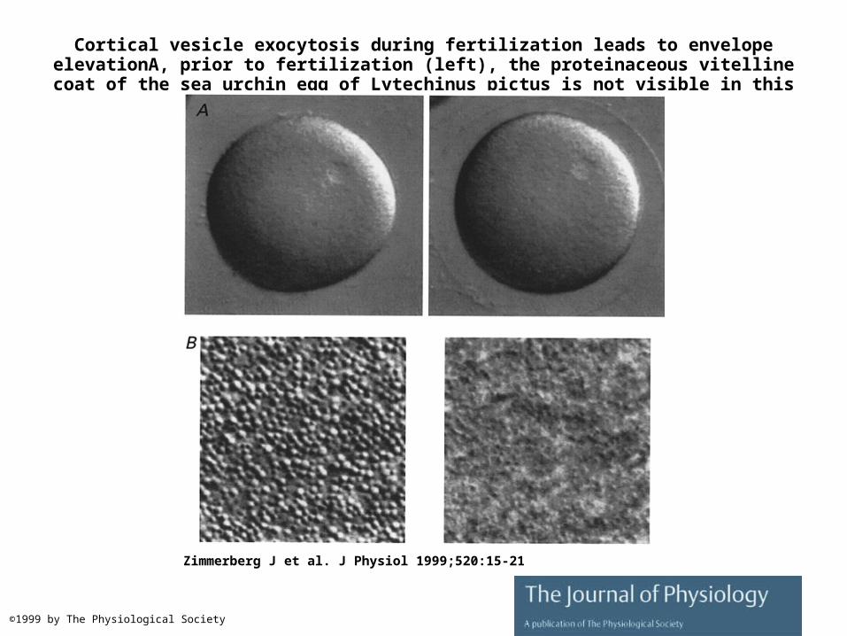
Cortical vesicle exocytosis during fertilization leads to envelope elevationA, prior to fertilization (left), the proteinaceous vitelline coat of the sea urchin egg of Lytechinus pictus
is not visible in this differential interference contrast image.
Zimmerberg J et al. J Physiol 1999;520:15-21
©1999 by The Physiological Society
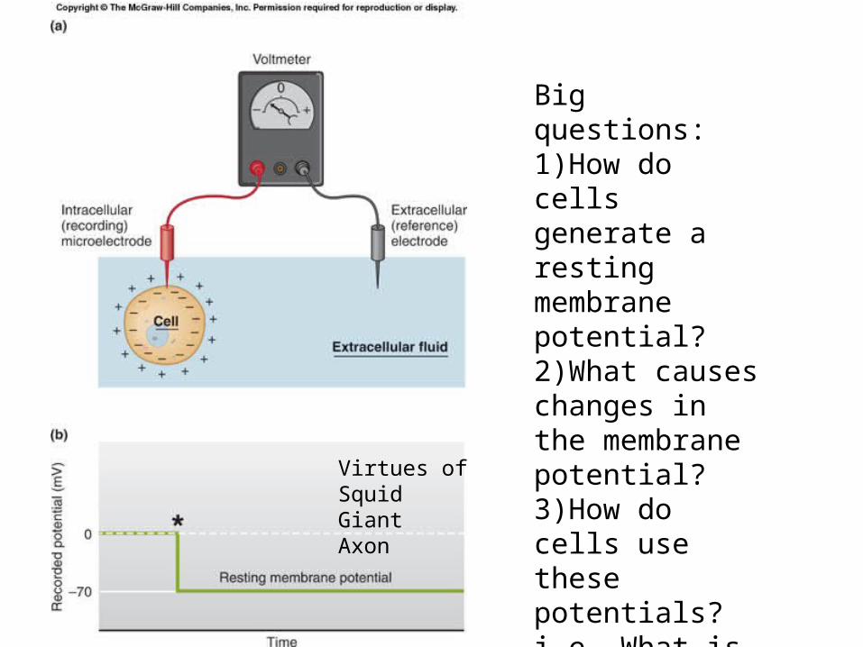
Virtues of SquidGiantAxon
Big questions:1)How do cells generate a resting membrane potential?2)What causes changes in the membrane potential?3)How do cells use these potentials? i.e. What is their purpose?

Fig. 06.09
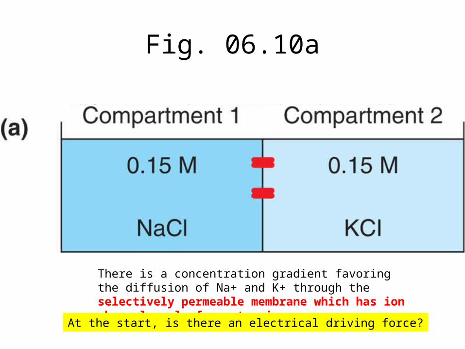
Fig. 06.10a
There is a concentration gradient favoring the diffusion of Na+ and K+ through the selectively permeable membrane which has ion channels only for potassium.
At the start, is there an electrical driving force?

Fig. 06.10b
With K+ channels open, K+ diffuses down its concentraiton gradient, leaving behind CL- ions which are not permeable through the membrane. As more and more K+ move to the left, the compartment they leave becomes more and more negatively charged.
Is there an electrical driving force?
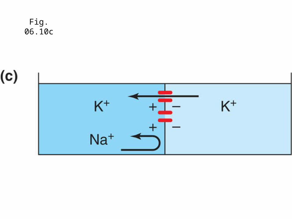
Fig. 06.10c

Fig. 06.10d
Soon, the accumulation of negative charges seriously impeded the diffusion of K+ as the electrostatic force builds up in opposition to the concentration driving force.
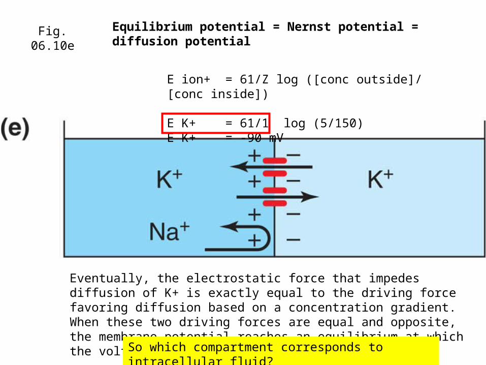
Fig. 06.10eEquilibrium potential = Nernst potential = diffusion potential
Eventually, the electrostatic force that impedes diffusion of K+ is exactly equal to the driving force favoring diffusion based on a concentration gradient. When these two driving forces are equal and opposite, the membrane potential reaches an equilibrium at which the voltage is called
So which compartment corresponds to intracellular fluid?
E ion+ = 61/Z log ([conc outside]/ [conc inside])
E K+ = 61/1 log (5/150)E K+ = -90 mV
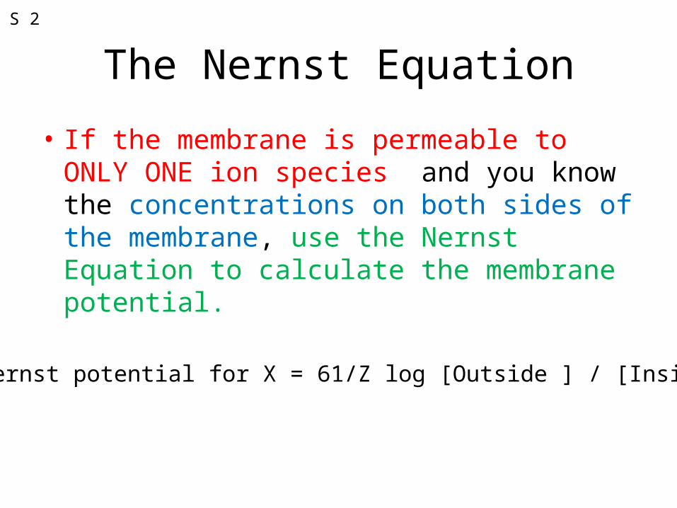
The Nernst Equation
• If the membrane is permeable to ONLY ONE ion species and you know the concentrations on both sides of the membrane, use the Nernst Equation to calculate the membrane potential.
Nernst potential for X = 61/Z log [Outside ] / [Inside]
S 2

Fig. 06.10eEquilibrium potential = Nernst potential = diffusion potential
E ion+ = 61/Z log ([conc outside]/ [conc inside])
E K+ = 61/1 log (5/150)E K+ = -90 mV
150 mM5 mM
K+50 mM
Predict the change in membrane potential if K+ were added to the extracellular fluid.
S 1
What hormone regulates the levels of Na+ and K+ in extracellular fluid?
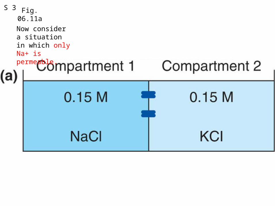
Fig. 06.11aS 3
Now consider a situation in which only Na+ is permeable.

Fig. 06.11bS 4

Fig. 06.11cS 5
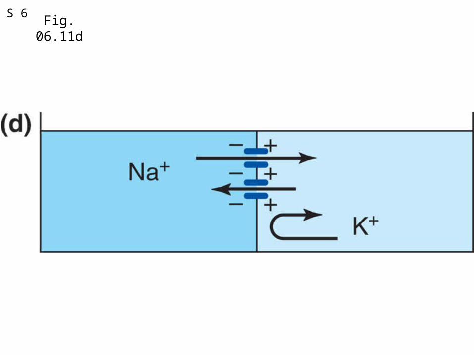
Fig. 06.11dS 6

Fig. 06.11eEquilibrium potential for Na+
E Na+ = 61/1 log (145/15)
E Na + = +60 mV
145 mM 15 mM
Extracellular Intracellular
So, given these concentrations of Na+ and a membrane permeable only to Na+, use Nernst equation to calculate what the membrane potential would be.
At the equilibrium potential, no net movement of Na+ because driving forces (concentration and electrical) are exactly equal and opposite.
S 7

1QQ#12 for 10:30
1. Ligand-gated ion channels are found exclusively in the axonal membrane.
2. Leak channels are found only at the distal tips of sensory neurons.
3. Trigger zones and axons hillocks are the sites which convert action potentials to graded potentials.
4. Graded potentials are conducted non-decrementally and are well suited for long-distance signalling.
Revise two of the following misleading statements. Your revision cannot consist of a “not” statement.

1QQ#12 for 11:30
1. Ligand-gated ion channels are found exclusively in the membrane of dendrites.
2. Leak channels are found only at the distal tips of efferent neurons.
3. Trigger zones and axons hillocks are the sites where action potentials are converted to graded potentials.
4. Action potentials are conducted non-decrementally and are well suited for long-distance signaling.
Revise two of the following misleading statements. Your revision cannot consist of a “not” statement.

Test 1 Monday• Thermoregulation
• Glucose homeostasis
• Negative feedback, positive feedback, feedforward
• Endocrine System and endocrine disorders
• Reflexes. Neurons and glial cells
• Equilibrium potentials and Resting Membrane potential.
Comprehensive list of topics at http://webs.wofford.davisgr/bio342/test1study2012.htm

Electrical and concentration gradient driving forces for Sodium and Potassium
How does the membrane potential change if 1) permeability to sodium increases2) Permeability to potassium increases
Why is resting membrane potential closer to EK than ENa?
What would happen to membrane potential if suddenly PNa
became very great?
Size and Direction of Arrows show driving forces!
The G-H-K Equation!S 8
How can the membrane become suddenly more permeable to Na+?
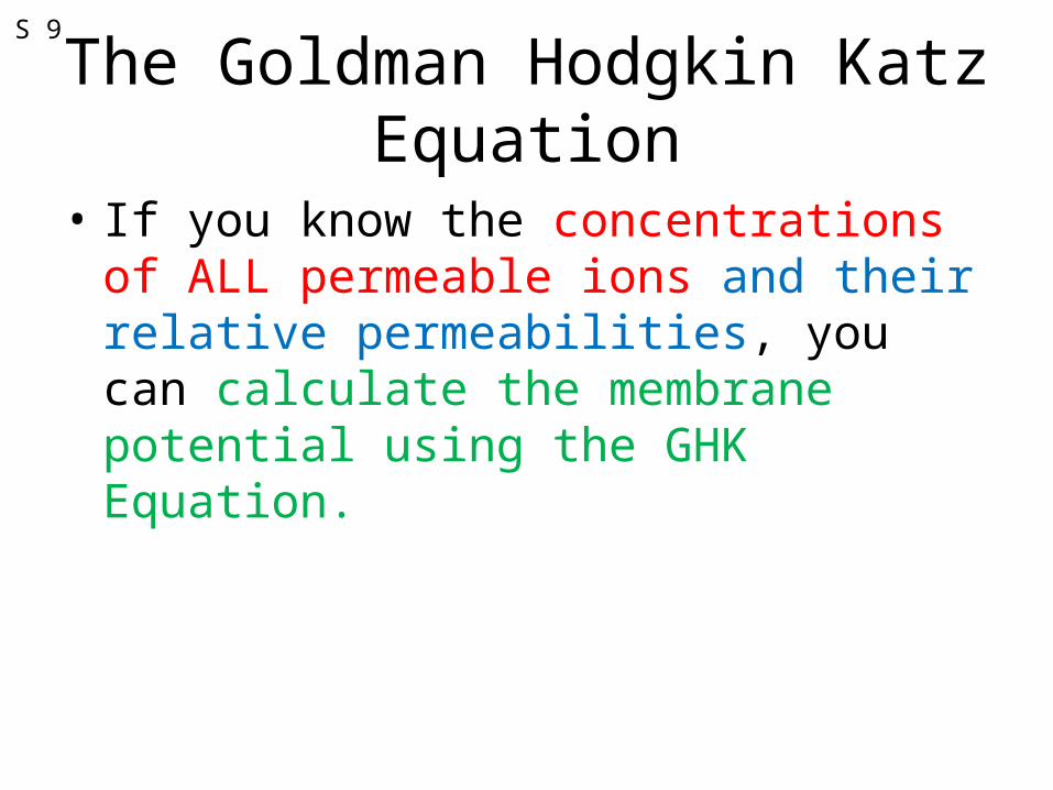
The Goldman Hodgkin Katz Equation
• If you know the concentrations of ALL permeable ions and their relative permeabilities, you can calculate the membrane potential using the GHK Equation.
S 9

At RMP, some Na+ leaks in, some K+ leaks out.
S 10
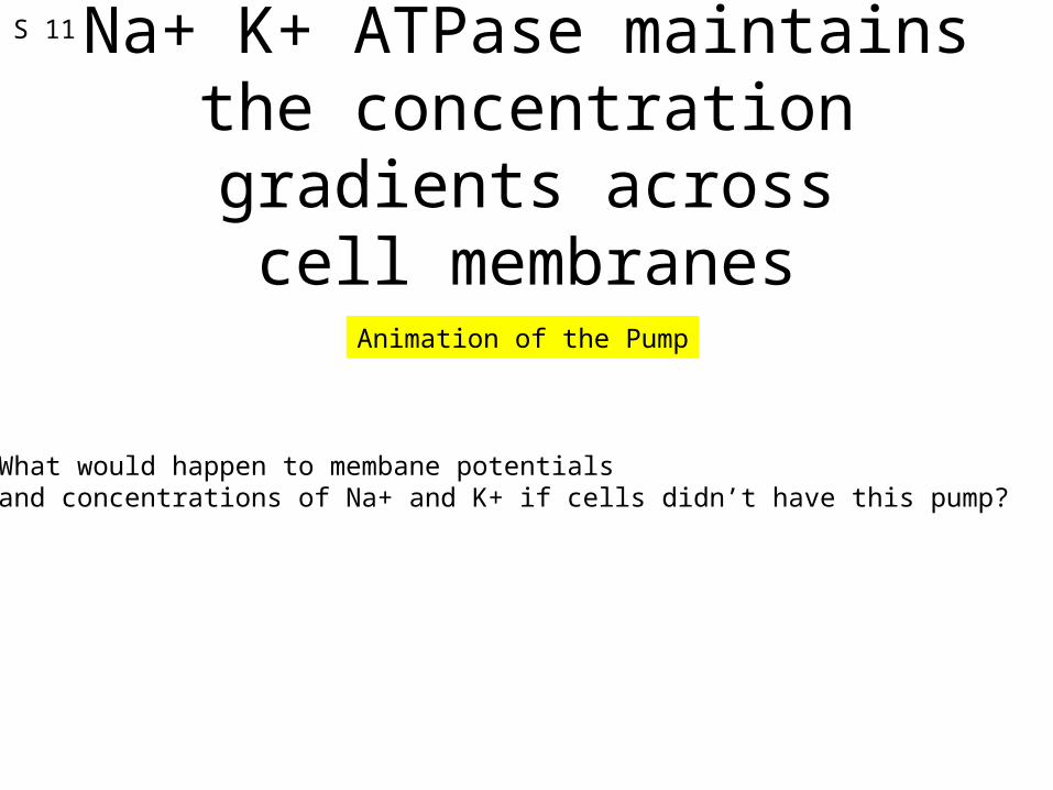
Na+ K+ ATPase maintains the concentration gradients across
cell membranes
Animation of the Pump
What would happen to membane potentials and concentrations of Na+ and K+ if cells didn’t have this pump?
S 11

Animations of the Origin of Resting Membrane Potential
Animation of Resting Membrane Potential (single ion)
YouTube animation of Na-K-ATPase, Sodium Co-transporter, and K Leak channels
Origin of Resting Membrane Potential and intracellular recording
S 12

S 13
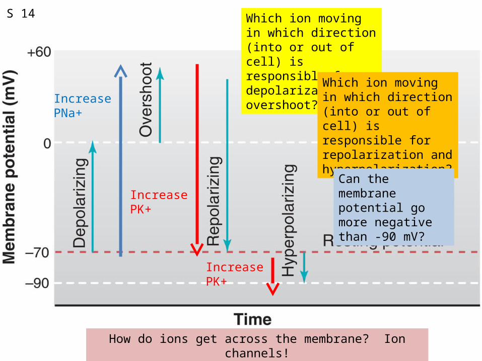
Which ion moving in which direction (into or out of cell) is responsible for depolarization and overshoot? Which ion moving in
which direction (into or out of cell) is responsible for repolarization and hyperpolarization?
Can the membrane potential go more negative than -90 mV?
Increase PK+
Increase PNa+
S 14
Increase PK+
How do ions get across the membrane? Ion channels!
Related Documents