MEETING ABSTRACTS Open Access 18th International Symposium on Intensive Care and Emergency Medicine Brussels, Belgium. 17–20 March 1998 Published: 1 March 1998 These abstracts are available online at http://ccforum.com/supplements/2/S1 MEETING ABSTRACTS P001 Hemodynamics in induced whole body hyperthermia T Kerner 1 , M Deja 1 , O Ahlers 1 , J Löffel 2 , H Riess 2 , P Wust 3 , D Pappert 1 , H Gerlach 1 1 Abteilung für Anaesthesiologic und operative Intensivmedizin, Virchow- Charité der Humboldt-Universität zu Berlin, Deutschland; 2 Abteilung für Hämatologiel Onkologie, Virchow-Charité der Humboldt-Universität zu Berlin, Deutschland; 3 Abteilung für Strahlenklinik und poliklinik, Virchow-Charité der Humboldt-Universität zu Berlin, Deutschland Critical Care 1998, 2(Suppl 1):P001 Background: Whole body hyperthermia induced by radiative systems has been used in therapy of malignant diseases for more than ten years. Von Ardenne and co-workers have developed the ‘ systemiche Krebs-Mehrschritt-Therapic’ (sKMT), a combined regime including whole body hyperthermia of 42°C, induced hyperglycaemia and relative hyperoxaemia with additional application of chemotherapy. This concept has been employed in a phase I/II clinical study for patients with metastatic colorectal carcinoma at the Virchow-Klinikum since January 1997. Methods: The sKMT concept was performed eleven times under intravenous general anaesthesia, avoiding volatile anaesthetics. Core temperatures of up to 42°C were reached stepwise by warming with infrared-A-radiation (IRATHERM 2000®). During the whole procedure blood glucose levels of 380–450 mg/dl were maintained as well as PaO 2 levels above 200 mmHg. Extensive invasive monitoring was performed in all patients including measurements with the REF-Ox-Pulmonary artery catheter with continuous measuring of mixed venous saturation (Baxter Explorer®) and invasive monitoring of arterial blood pressure. Data for calculation of hemodynamic and gas exchange parameters were collected four times, at temperatures of 37°C, 40°C, 41.8–42°C and 39°C, during measurements FiO 2 was 1.0 at all times. Fluids were given in order to keep central-venous and Wedge pressure within normal range during the whole procedure. Statistics were performed using the Wilcoxon Test. Results: Statistically significant differences were found between heart rate, cardiac index and systemic vascular resistance comparing data at 37°C and 42°C. Heart rate and cardiac index increased to a maximum at 42°C (P < 0.0001) whereas systemic vascular resistance had its minimum at 42°C (P < 0.0001). Mean arterial pressure dropped with increasing temperature, differences were not significant. Calculation of stroke volume index and ventricular volumes showed only a slight decrease in endsystolic volumes with increasing temperature, the resulting differences in right ventricular ejection fraction were marginally significant (P = 0.038) comparing 42°C to baseline. Right ventricular stroke work index as well as mean pulmonary arterial pressure increased at 42°C (P = 0.0115 and P = 0.0037), pulmonary vascular resistance only dropped little compared to systemic vascular resistance, left ventricular stroke work index even dropped with increasing temperature, though showing no significant difference. Values for mixed venous oxygen saturation did not vary during therapy, pulmonary right-left shunt showed a temperature associated increase (P = 0.0323) to a maximum at 42°C. Conclusion: Under the procedure of sKMT cardiac function in patients, who do not have any pre-existing cardiac impairment, can be maintained almost unchanged, ie with normal right and left ventricular pressure, despite an increase in right ventricular stroke work Acknowledegment: Supported by Deutsche Krebshilfe. P002 Induced hyperthermia causes significant changes in lymphocytes O Ahlers 1 , T Boehnke 1 , T Kerner 1 , M Deja 1 , D Keh 1 , J Löffel 2 , B Hildebrandt 2 , H Riess 2 , P Wust 3 , D Pappert 1 , H Gerlach 1 1 Abteilung für Anästhesiologie und operative Intensivmedizin (GD: Prof. Dr. KJ Falke),Virchow-Charitè der Humboldt-Universität, 13344 Berlin, Deutschland; 2 Abteilung für Hämatologie/Onkologie (GD: Prof. Dr. D. Huhn), Virchow-Charitè der Humboldt-Universität, 13344 Berlin, Deutschland; 3 Strahlenklinik und -poliklinik (GD: Prof. Dr. h.c. R. Felix), Virchow-Charitè der Humboldt-Universität, 13344 Berlin, Deutschland Critical Care 1998, 2(Suppl 1):P002 Background: Changes in lymphocyte subpopulations are determined under several clinical conditions eg during activation of the immune system. Our aim was to analyze the influence of induced elevated body temperatures on lymphocytes in patients without infections or other physiological stimulators of the immune system. Therefore we examined blood of patients with metastatic colorectal carcinoma during whole body hyperthermia of 42°C caused by infrared-A-radiation. This is used as part of so called ‘systemische Krebs-Mehrschritt-Therapie’ (sKMT), which was started as a phase I/II clinical study at Virchow-Klinikum in 1997. Methods: Lymphocyte subpopulations were investigated by flow- cytometry-analysis. Blood samples were obtained before beginning of therapy at 37°C, at 40°C, at the end of the plateau of 42°C and after therapy at 37°C again. Time between investigations was about 2 h. Subpopulations were natural killer cells, T-Cells, IL2-Receptor on T-Cells, T4-Cells and T8-Cells. Cell counts were compared by using a Wilcoxon rank sum test. Results: The number of lymphocytes per nl decreased significantly from 37°C to 42°C (Fig. 1). This effect was mainly caused by a significant decrease of the absolute T4-Cell count and a slight decrease of the T8- Cell count with a resulting significant decrease of T-Cells. In addition, IL2- Receptor expression on T-Cells, as a marker for activation, decreased significantly. In contrast, the number of natural killer cells per nl Critical Care 1998, Volume 2 Suppl 1 http://ccforum.com/supplements/2/S1

Welcome message from author
This document is posted to help you gain knowledge. Please leave a comment to let me know what you think about it! Share it to your friends and learn new things together.
Transcript
MEETING ABSTRACTS Open Access
18th International Symposium on Intensive Careand Emergency MedicineBrussels, Belgium. 17–20 March 1998
Published: 1 March 1998
These abstracts are available online at http://ccforum.com/supplements/2/S1
MEETING ABSTRACTSP001Hemodynamics in induced whole body hyperthermiaT Kerner1, M Deja1, O Ahlers1, J Löffel2, H Riess2, P Wust3, D Pappert1,H Gerlach11Abteilung für Anaesthesiologic und operative Intensivmedizin, Virchow-Charité der Humboldt-Universität zu Berlin, Deutschland; 2Abteilung fürHämatologiel Onkologie, Virchow-Charité der Humboldt-Universität zu Berlin,Deutschland; 3Abteilung für Strahlenklinik und poliklinik, Virchow-Charité derHumboldt-Universität zu Berlin, DeutschlandCritical Care 1998, 2(Suppl 1):P001
Background: Whole body hyperthermia induced by radiative systemshas been used in therapy of malignant diseases for more than tenyears. Von Ardenne and co-workers have developed the ‘systemicheKrebs-Mehrschritt-Therapic’ (sKMT), a combined regime including wholebody hyperthermia of 42°C, induced hyperglycaemia and relativehyperoxaemia with additional application of chemotherapy. Thisconcept has been employed in a phase I/II clinical study for patientswith metastatic colorectal carcinoma at the Virchow-Klinikum sinceJanuary 1997.Methods: The sKMT concept was performed eleven times underintravenous general anaesthesia, avoiding volatile anaesthetics. Coretemperatures of up to 42°C were reached stepwise by warming withinfrared-A-radiation (IRATHERM 2000®). During the whole procedureblood glucose levels of 380–450 mg/dl were maintained as well as PaO2
levels above 200 mmHg. Extensive invasive monitoring was performedin all patients including measurements with the REF-Ox-Pulmonaryartery catheter with continuous measuring of mixed venous saturation(Baxter Explorer®) and invasive monitoring of arterial blood pressure.Data for calculation of hemodynamic and gas exchange parameterswere collected four times, at temperatures of 37°C, 40°C, 41.8–42°C and39°C, during measurements FiO2 was 1.0 at all times. Fluids were givenin order to keep central-venous and Wedge pressure within normalrange during the whole procedure. Statistics were performed using theWilcoxon Test.Results: Statistically significant differences were found between heartrate, cardiac index and systemic vascular resistance comparing data at37°C and 42°C. Heart rate and cardiac index increased to a maximum at42°C (P < 0.0001) whereas systemic vascular resistance had its minimumat 42°C (P < 0.0001). Mean arterial pressure dropped with increasingtemperature, differences were not significant. Calculation of strokevolume index and ventricular volumes showed only a slight decrease inendsystolic volumes with increasing temperature, the resulting differencesin right ventricular ejection fraction were marginally significant (P = 0.038)comparing 42°C to baseline. Right ventricular stroke work index as well as
mean pulmonary arterial pressure increased at 42°C (P = 0.0115 and P =0.0037), pulmonary vascular resistance only dropped little compared tosystemic vascular resistance, left ventricular stroke work index evendropped with increasing temperature, though showing no significantdifference. Values for mixed venous oxygen saturation did not varyduring therapy, pulmonary right-left shunt showed a temperatureassociated increase (P = 0.0323) to a maximum at 42°C.Conclusion: Under the procedure of sKMT cardiac function in patients,who do not have any pre-existing cardiac impairment, can be maintainedalmost unchanged, ie with normal right and left ventricular pressure,despite an increase in right ventricular stroke workAcknowledegment: Supported by Deutsche Krebshilfe.
P002Induced hyperthermia causes significant changes in lymphocytesO Ahlers1, T Boehnke1, T Kerner1, M Deja1, D Keh1, J Löffel2, B Hildebrandt2,H Riess2, P Wust3, D Pappert1, H Gerlach11Abteilung für Anästhesiologie und operative Intensivmedizin (GD: Prof. Dr.KJ Falke),Virchow-Charitè der Humboldt-Universität, 13344 Berlin,Deutschland; 2Abteilung für Hämatologie/Onkologie (GD: Prof. Dr. D. Huhn),Virchow-Charitè der Humboldt-Universität, 13344 Berlin, Deutschland;3Strahlenklinik und -poliklinik (GD: Prof. Dr. h.c. R. Felix), Virchow-Charitè derHumboldt-Universität, 13344 Berlin, DeutschlandCritical Care 1998, 2(Suppl 1):P002
Background: Changes in lymphocyte subpopulations are determinedunder several clinical conditions eg during activation of the immunesystem. Our aim was to analyze the influence of induced elevated bodytemperatures on lymphocytes in patients without infections or otherphysiological stimulators of the immune system. Therefore we examinedblood of patients with metastatic colorectal carcinoma during wholebody hyperthermia of 42°C caused by infrared-A-radiation. This is used aspart of so called ‘systemische Krebs-Mehrschritt-Therapie’ (sKMT), whichwas started as a phase I/II clinical study at Virchow-Klinikum in 1997.Methods: Lymphocyte subpopulations were investigated by flow-cytometry-analysis. Blood samples were obtained before beginning oftherapy at 37°C, at 40°C, at the end of the plateau of 42°C and after therapyat 37°C again. Time between investigations was about 2 h. Subpopulationswere natural killer cells, T-Cells, IL2-Receptor on T-Cells, T4-Cells and T8-Cells.Cell counts were compared by using a Wilcoxon rank sum test.Results: The number of lymphocytes per nl decreased significantly from37°C to 42°C (Fig. 1). This effect was mainly caused by a significantdecrease of the absolute T4-Cell count and a slight decrease of the T8-Cell count with a resulting significant decrease of T-Cells. In addition, IL2-Receptor expression on T-Cells, as a marker for activation, decreasedsignificantly. In contrast, the number of natural killer cells per nl
Critical Care 1998, Volume 2 Suppl 1http://ccforum.com/supplements/2/S1
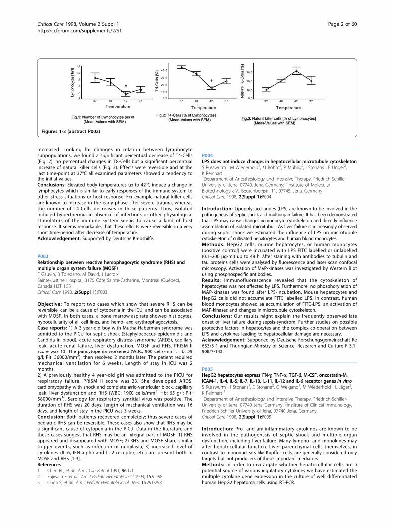
increased. Looking for changes in relation between lymphocytesubpopulations, we found a significant percentual decrease of T4-Cells(Fig. 2), no percentual changes in T8-Cells but a significant percentualincrease of natural killer cells (Fig. 3). Effects were reversible and at thelast time-point at 37°C all examined parameters showed a tendency tothe initial values.Conclusions: Elevated body temperatures up to 42°C induce a change inlymphocytes which is similar to early responses of the immune system toother stress situations or host response. For example natural killer cellsare known to increase in the early phase after severe trauma, whereasthe number of T4-Cells decreases in these patients. Thus, isolatedinduced hyperthermia in absence of infections or other physiologicalstimulators of the immune system seems to cause a kind of hostresponse. It seems remarkable, that these effects were reversible in a veryshort time-period after decrease of temperature.Acknowledgement: Supported by Deutsche Krebshilfe.
P003Relationship between reactive hemophagocytic syndrome (RHS) andmultiple organ system failure (MOSF)F Gauvin, B Toledano, M David, J LacroixSainte-Justine Hospital, 3175 Côte Sainte-Catherine, Montréal (Québec),Canada H3T 1C5Critical Care 1998, 2(Suppl 1):P003
Objective: To report two cases which show that severe RHS can bereversible, can be a cause of cytopenia in the ICU, and can be associatedwith MOSF. In both cases, a bone marrow aspirate showed histiocytes,hypocellularity of all cell lines, and hemo- and erythrophagocytosis.Case reports: 1) A 3 year-old boy with Mucha-Haberman syndrome wasadmitted to the PICU for septic shock (Staphylococcus epidermidis andCandida in blood), acute respiratory distress syndrome (ARDS), capillaryleak, acute renal failure, liver dysfunction, MOSF and RHS. PRISM IIscore was 13. The pancytopenia worsened (WBC: 900 cells/mm3; Hb: 59g/l; Plt: 36000/mm3), then resolved 2 months later. The patient requiredmechanical ventilation for 6 weeks. Length of stay in ICU was 2months.2) A previously healthy 4 year-old girl was admitted to the PICU forrespiratory failure. PRISM II score was 23. She developed ARDS,cardiomyopathy with shock and complete atrio-ventricular block, capillaryleak, liver dysfunction and RHS (WBC: 1900 cells/mm3; Hb: 65 g/l; Plt:58000/mm3). Serology for respiratory syncitial virus was positive. Theduration of RHS was 20 days; length of mechanical ventilation was 16days, and length of stay in the PICU was 3 weeks.Conclusion: Both patients recovered completely; thus severe cases ofpediatric RHS can be reversible. These cases also show that RHS may bea significant cause of cytopenia in the PICU. Data in the literature andthese cases suggest that RHS may be an integral part of MOSF: 1) RHSappeared and disappeared with MOSF; 2) RHS and MOSF share similartrigger events, such as infection or neoplasia; 3) increased level ofcytokines (IL-6, IFN-alpha and IL-2 receptor, etc.) are present both inMOSF and RHS [1-3].References1. Chen RL, et al: Am J Clin Pathol 1991, 96:171.2. Fujiwara F, et al: Am J Pediatr Hematol/Oncol 1993, 15:92-98.3. Ohga S, et al: Am J Pediatr Hematol/Oncol 1993, 15:291-298.
P004LPS does not induce changes in hepatocellular microtubule cytoskeletonS Russwurm1, M Wiederhold1, KJ Böhm2, P Mühlig2, I Stonans1, E Unger2,K Reinhart11Department of Anesthesiology and Intensive Therapy, Friedrich-Schiller-University of Jena, 07740, Jena, Germany; 2Institute of MolecularBiotechnology e.V., Beutenbergstr, 11, 07745, Jena, GermanyCritical Care 1998, 2(Suppl 1):P004
Introduction: Lipopolysaccharides (LPS) are known to be involved in thepathogenesis of septic shock and multiorgan failure. It has been demonstratedthat LPS may cause changes in monocyte cytoskeleton and directly influenceassemblation of isolated microtubuli. As liver failure is increasingly observedduring septic shock we estimated the influence of LPS on microtubulecytoskeleton of cultivated hepatocytes and human blood monocytes.Methods: HepG2 cells, murine hepatocytes, or human monocytes(positive control) were incubated with LPS FITC labelled or unlabelled(0.1–200 μg/ml) up to 48 h. After staining with antibodies to tubulin andtau proteins cells were analysed by fluorescence and laser scan confocalmicroscopy. Activation of MAP-kinases was investigated by Western Blotusing phosphospecific antibodies.Results: Immunofluorescence revealed that the cytoskeleton ofhepatocytes was not affected by LPS. Furthermore, no phosphorylation ofMAP-kinases was found after LPS-incubation. Mouse hepatocytes andHepG2 cells did not accumulate FITC labelled LPS. In contrast, humanblood monocytes showed an accumulation of FITC-LPS, an activation ofMAP-kinases and changes in microtubule cytoskeleton.Conclusions: Our results might explain the frequently observed lateonset of liver failure during sepsis-syndrom. Further studies on possibleprotective factors in hepatocytes and the complex co-operation betweenLPS and cytokines leading to hepatocellular damage are necessary.Acknowledgement: Supported by Deutsche Forschungsgemeinschaft Re653/5-1 and Thuringian Ministry of Science, Research and Culture F 3.1-908/7-143.
P005HepG2 hepatocytes express IFN-g, TNF-a, TGF-b, M-CSF, oncostatin-M,ICAM-1, IL-4, IL-5, IL-7, IL-10, IL-11, IL-12 and IL-6 receptor genes in vitroS Russwurm1, I Stonans1, E Stonane2, G Weigand1, M Wiederhold1, L Jäger2,K Reinhart 1
1Department of Anesthesiology and Intensive Therapy, Friedrich-Schiller-University of Jena, 07740 Jena, Germany; 2Institute of Clinical Immunology,Friedrich-Schiller-University of Jena, 07740 Jena, GermanyCritical Care 1998, 2(Suppl 1):P005
Introduction: Pro- and antiinflammatory cytokines are known to beinvolved in the pathogenesis of septic shock and multiple organdysfunction, including liver failure. Many lympho- and monokines mayalter hepatocellular function. Liver parenchymal cells themselves, incontrast to mononuclears like Kupffer cells, are generally considered onlytargets but not producers of these important mediators.Methods: In order to investigate whether hepatocellular cells are apotential source of various regulatory cytokines we have estimated themultiple cytokine gene expression in the culture of well differentiatedhuman HepG2 hepatoma cells using RT-PCR.
Figures 1-3 (abstract P002)
Critical Care 1998, Volume 2 Suppl 1http://ccforum.com/supplements/2/S1
Page 2 of 60
Results: Our findings demonstrate that HepG2 cells express mRNAs forIFN-g, TNF-a, TGF-b, M-CSF, oncostatin-M, ICAM-1, IL-4, IL-5, IL-7, IL-10, IL-11, IL-12 and IL-6R. At the same time the expression of IL-1, IL-2, IL-3, IL-6, CD40 ligand and IL-2R genes was not detected.Conclusions: Hepatocytes are potential producers of a variety ofcytokines, some of them being able to regulate hepatocellular functionsdirectly, others are important regulators of leukocyte functions. Thus, onthe one hand, hepatocytes may express autoregulatory cytokines and, onthe other hand, influence the functions of other liver cells like Kupffer, Itoor endothelial cells. Due to their large amount, liver parenchymal cellscould be an important source of systemically acting pro- and anti-inflammatory and other regulatory cytokines.Acknowledgement: Supported in part by grants from the DeutscheForschungsgemeinschaft (Re 653/5-1) and the Thuringian Ministry ofScience, Research and Culture (B301-95026).
P006Autoantibodies against oxidated low density lipoproteins (oLAb) andprocalcitonin (PCT) as prognostic markers for patients suffering fromsepsis and systemic inflammatory response syndrome (SIRS)J Reiger, F Tatzber, G Ziervogel, U Köller, G Grimm2nd Medical Department and Institute of Laboratory Medicine, GeneralHospital, St Veiterstrab e 47, 9020 Klagenfurt, AustriaCritical Care 1998, 2(Suppl 1):P006
Objective: To investigate the role of lipidperoxidation and infectionduring acute sepsis we measured antoantibodies against oxidated LDL(oLAb) and procalcitonin (PCT) comparing the neopterin as a marker ofmacrophages activation and CRP as marker of inflammation.Design: A prospective, descriptive cohort study.Patients: 23 patients admitted to the ICU with verified sepsis (n=12, s=6)or SIRS (n=11, s = 6).Measurements and results: The clinical severity of the disease wasasessed using the APACHE II score over a period of 24 h after admission.Determination of serum levels of all parameters under study wasperformed on daily drawn serum samples. Surviving septic patientsproduced significantly increasing oLAbs (P < 0.001) as significantlydecreasing PCT levels (P < 0.001). In contrast, in non-survivors oLAbswere decreasing (P < 0.05) and PCT levels were increasing (P < 0.05).The identical effect was found for the SIRS group with the exception, thatthe significance of PCT in survivors was slightly lower (P < 0.05).Conclusion: Despite both patient groups were rather small, we considerthat the measurement of oLAb as well as PCT to be a useful prognosticmarker concerning the outcome of sepsis as well as of SIRS patients.
P007Endotoxin-induced adhesion of human red blood cells to vascularendothelium does not depend on the presence of leukocytes but ismodified by different flow patternO Eichelbrönner, A Sielenkämper, G Cepinskas, I Chin-Yee, W SibbaldUniversity of Western Ontario, A.C. Burton Vascular Biology Laboratory,London Health Sciences Centre, 375 South Street, N6A 4G5, London,Ontario, CanadaCritical Care 1998, 2(Suppl 1):P007
Introduction: Endotoxin-induced (ETX) adhesion of leucocytes to vascularendothelial cells is a well investigated phenomenon. However, little isknown about the effects of endotoxin on erythrocyte-endothelial cellinteractions . The objective of this study was to investigate the effects ofETX on adhesion of human red blood cells (RBC) to human vascularendothelial cells (HUVEC) in the presence or absence of leukocytes andunder different conditions of flow.Methods: Endothelial cells were obtained from human umbilical veins andcultured as first and second passage monolayers and then grown tocomplete confluency on cover slips. RBC were harvested from fresh blooddonated by healthy volunteers, washed with isotonic NaCl andresuspended to a hematocrit of 30% in medium (M199, GIBCO, Canada).Group A (n = 7) served as a control, whereas in group B, C and D (n = 7 ineach) both RBC and HUVECs were incubated with ETX (75 μg/ml) at 37°Cfor 2 h. In group E whole blood was incubated with ETX and thereafter
RBC were isolated as described above. The HUVEC coated cover slips wereplaced in a flowing chamber and perfused with the RBC suspensions at aflow rate of 0.65 ml/min in group A, B and E and 1.3 ml/min in group C. Ingroup D stop and go flow patterns found in the microcirculation of septicanimals were mimicked by applying a flow rate of 0.65 ml/min with fourstops (4–6 s) per minute. The flow chambers were arranged on amicroscope and connected to a video recording system. In eachexperiment 15–20 sites of a defined area were recorded and analysed.Results: The control group showed an adhesion of 71 ± 8 cells/mm2.Incubation of HUVECs and RBC with ETX increased RBC adhesion ingroup B to 172 ± 25 cells/mm2 (P < 0.05). Incubation of whole bloodincluding leucocytes with ETX did not exhibit a different degree ofadhesion compared to group B. When flow rate was elevated to 1.3 ml/min, the number of adherent RBC decreased to 89 ± 20/mm2 comparableto the control group with a flow rate of 0.65 ml/min. In group D withintermittent stops of flow, RBC adhesion increased to 274 ± 35 cells/mm2
(P < 0.05) compared to control, group B and group C.Conclusion: Incubation of RBC and HUVECs with endotoxin promotederythrocyte adhesion to human vascular endothelium. The presence ofleukocytes during the endotoxin exposure did not affect the degree ofadhesion. Elevating flow rate, however, reduced erythrocyte adhesion,while stop and go flow pattern favoured erythrocyte adhesion. Thesefindings suggest that altered RBC may contribute to the microcirculatoryinjury observed in sepsis.
P008Adhesion molecule, soluble adhesion molecule, and cytokine levels inpatients with severe burnsS Endo1, H Nakae2, Y Yamada1, K Inada3, S Taniguchi11Critical Care and Emergency Center, Iwate Medical University, 19-1Uchimaru, Morioka 020, Japan; 2Department of Critical Care Medicine, AkitaUniversity Medical School, 1-1-1 Hondo, Akita 010, Japan; 3Department ofBacteriology, Iwate Medical University Medical School, 19-1 Uchimaru,Morioka 020, JapanCritical Care 1998, 2(Suppl 1):P008
Object: We measured endotoxins, inflammatory cytokines and solubleadhesion molecules in the blood of 17 severe burn patients to determinethe involvement of these factors in the pathophysiology in burn patients.Design: Prospective study.Patients: Seventeen patients with burns with a total burn surface area of20% or more and a burn index of 15% or more.Measurement and main results: Endotoxin was measured by anendotoxin-specific assay. Tumor necrosis factor-a, interleukin 6 andinterleukin 8 were measured by an enzyme-linked immunosorbent assay.Soluble adhesion molecules were also measured by an enzyme-linkedimmunosorbent assay. CD11a, CD11b, and CD18 were measured by a flowcytometry. Their levels were high in the non-surviving group, the septicshock group, and the multiple organ dysfunction syndrome group,suggesting the possibility of a close connection between them and theevolution of the pathophysiology in patients with burns complicated byinfection (Table).Conclusion: Soluble adhesion molecules were found to indirectly reflectthe level of endothelial cell adhesion molecules, suggesting thatinflammatory cytokines may also be involved as factors in their production.
Table (abstract P008). Comparisons of the factors in thesepsis group and the sepsis-free group
Sepsis (-) (n=4) Sepsis (+) (n=13) P value
Endotoxin (pg/ml) 8.1 ± 10.1 12.5 ± 8.8 0.1630
TNF-a (pg/ml) 74.5 ± 58.7 638.1 ± 792.7 0.0151
IL-6 (pg/ml) 65.3 ± 112.4 754.9 ± 862.1 0.0346
IL-8 (pg/ml) 83.9 ± 11.7 280.9 ± 114.8 0.0168
slCAM-1 (ng/ml) 682.1 ± 291.3 924.6 ± 542.1 0.0362
sELAM-1 (ng/ml) 261.7 ± 23.0 342.1 ± 53.4 0.0101
sVCAM-1 (ng/ml) 1245.1 ± 422.6 3028.9 ± 1861.2 0.0438
Critical Care 1998, Volume 2 Suppl 1http://ccforum.com/supplements/2/S1
Page 3 of 60

P009Circulating markers of endothelial activation in acetaminophen inducedacute liver failureW Bernal, P Langley, J WendonInstitute of Liver Studies, Kings College Hospital, London SE5 9RS, UKCritical Care 1998, 2(Suppl 1):P009
Background: Once defined clinical criteria are fulfilled, survival followingacetaminophen overdose is very poor without emergency livertransplantation. The early identification of patients developing multipleorgan failure (MOF) is important for prompt listing for transplantation.MOF is associated with early elevations of the circulating markers ofendothelial activation E-Selectin, VCAM-1 and ICAM-1 and Von WillebrandFactor (VWF). We determined their levels and those of Interleukin-6 (IL-6)on admission in acetaminophen overdose patients, to evaluate their usein the identification of those who would require transplantation.Patients: Nine healthy controls and 20 patients were studied. Elevenpatients became encephalopathic and seven patients either died orrequired emergency liver transplantation.Methods: E-selectin, VCAM, ICAM, IL-6 and VWF were determined onadmission using commercial ELISA. Statistical testing used Mann-WhitneyU tests and Spearmans Rank correlation. Results were corrected formultiple testing.Results: All molecules assayed were significantly higher in patients thancontrols (Pc < 10-4). IL-6 alone was higher in non survivors than survivors[158 pg/ml (range 22–440) vs 32 pg/ml (3.5–177), Pc < 0.05]. Adhesionmolecule levels were consistent with multiple organ failure of a non-septic aetiology. VWF was elevated in those patients who becameencephalopathic : 371 U/dl (149–658) vs 178 U/dl (65–340), P < 0.04 andcorrelated strongly with mean arterial pressure (r = 0.664, P < 0.005).Conclusions: ICAM, VCAM and E-selectin are poorly predictive ofoutcome in ALF, but suggest a non-septic aetiology of MOF. Thepathogenesis of encephalopathy may involve endothelial activation andcardiovascular dysfunction. IL-6 appears the most useful early marker of apoor outcome in ALF.
P010Circulating endothelial adhesion molecules in critically ill septicpatientsC Bouza Alvarez1, M Sancho1, A Velasco1, N Longo2, P Sanchez-Mateos21Intensive Care Unit, General Universitario Gregorio Marañón, Madrid,Spain;2Immunology service, General Universitario Gregorio Marañón, Madrid,SpainCritical Care 1998, 2(Suppl 1):P010
Introduction: Recently it has been demonstrated the importance of theendothelium in the physiopathology of organ failure and death inexperimental sepsis and soluble forms of adhesion molecules have beenshown to be excellent markers of endothelial activation or even damagein this setting [1] But their definite role in clinical sepsis has not beenclearly defined [2]. The objective of this study is to assess whethercirculating concentrations of endothelial adhesion molecules [endothelialleukocyte adhesion molecule-1 (SLLAM-1) and intercellular adhesionmolecule-1 (sICAM-1)] can be used to define, confirm or predict theoutcome in critically ill septic patients.Methods: Prospective, longitudinal, descriptive cohort study with notherapeutic interventions in which participated 63 patients admitted tothe ICU with the clinical diagnosis of severe sepsis (21) or septic shock(42) [3] and 10 healthy adults that served as controls. Blood samples fromand indwelling arterial catheter were collected from patients on days 1.3and 7 after their admission to the ICU for measuring plasmaconcentrations of sELAM-1 and sICAM-1 (ELISA, R&D Systems).Additionally biological data (age, sex, APACHE II, blood leukocyte count,platelet count, coagulation, haemodynamic and blood gas variables,serum lactate levels, need of vassopressor support, culture results, SOFAscore, individual organ dysfunction) and mortality were obtained.Statistical analysis was performed using Student t-test, Mann-WhitneyU test. P < 0.05 was considered significant.Results: Plasma levels of sELAM-1 and sICAM-1 on day 1 and 3 weresignificantly higher (P < 0.001) in septic patients than in controls. Patientswith bacteremia had higher levels of ICAM-1 on day 1 (P = 0.024) and 3
(P = 0.008). Soluble levels of the molecules measured did not correlatewith any other clinical or laboratory parameter at admission. With regardto outcome sELAM-1 on day 1 was significantly elevated in patientsthat developed coagulopathy and ICAM-1 on day 3 in those patientswho developed shock (P = 0.007), acute renal failure (P = 0.02) orcoagulopathy (P = 0.05). Both levels of sELAM-1 and ICAM-1 were similarin surviving and nonsurviving patients.Conclusions: Plasma endothelial adhesion molecules concentrations areelevated in critically ill septic patients but their levels show a high degree ofindividual variation and have a poor global correlation with biological dataand final outcome. sICAM-1 on day 3 might be useful for identifying thesubgroup of patients who will develop multiorgan failure and thereforecould benefit from a more agressive preventive or therapeutic approach.References1. Gearing AHJ, Newman W: Circulating adhesion molecules in disease.
Immunol Today 1993, 14:506.2. Boldt J, Muller M, Khun D, et al: Circulating adhesion molecules in the
critically ill: a comparison between trauma and sepsis patients. IntensCarc Med 1996, 22:122-128.
3. American College of Chest Physician / Society of Critical Care MedicineConsensus Conference: Definitions of sepsis and organ failure andguidelines for the use of innovative therapies in sepsis. Crit Care Med1992, 20:864-874.
P011Antibodies to ICAM-1 and PECAM-1 ameliorate pulmonary injury afterintestinal ischemia-reperfusion in the ratA Börjesson, XD Wang, R AnderssonDepartment of Surgery, Lund University Hospital, 221 85 Lund, SwedenCritical Care 1998, 2(Suppl 1):P011
Full text: Intestinal ischemia-reperfusion (I/R) gives rise to acute lunginjury characterized by neutrophil sequestration and microvascular injury.Important mediators of I/R-associated injury include neutrophils andplatelet-activating factor (PAF). During conditions of inflammationneutrophils and endothelial cells show an increased expression ofadhesion molecules. ICAM-1 on endothelial cells is needed for highaffinity bonds between neutrophils and endothelial cells, necessary forthe further transmigration of neutrophils, where PECAM-1 is involved.In the present study, a significant increase in albumin leakage overthe pulmonary capillaries, as well as increased pulmonary MPO(myeloperoxidase)-content was found in rats subjected to 30 minintestinal ischemia (by clamping the superior mesenteric artery) followedby 12 h reperfusion. Treatment with anti-ICAM-1 or anti-PECAM-1monoclonal antibodies significantly reduced the otherwise occuringincrease in both albumin leakage and pulmonary MPO-content inpancreatitis animals. There was also an increase in serum IL-1b levelsafter intestinal I/R, which could be prevented by use of antibodies toICAM-1 and PECAM-1.In conclusion we found that the acute lung injury seen after intestinal I/Rin the rat to a large extent could be prevented by blocking the adhesion/transmigration process of pulmonary leukocytes.
P012Lipid peroxidation parameters and antioxidant status of critically illintensive care unit patientsKH Smolle1, G Khoschsorur1, W Wonisch2, F Tatzber21Departments of Internal Medicine and Surgery, Karl Franzens University,Graz, Austria; 2Institute of Biochemistry, Karl Franzens University, Graz, AustriaCritical Care 1998, 2(Suppl 1):P012
Full text: Lipid peroxidation (LPO) is believed to play a crucial role in severaldisorders involving free radical action. Especially intensive care unit (ICU)patients suffer from situations, which lead to formation of free radicals andsubsequent LPO, eg septicemia, multi organ failure or ischemic situations.Methods for the measurement of LPO products are usually restricted tospecialists due to relatively complicated methodology. On the other hand,there is demand for routine methods to monitor LPO and/or antioxidativecapacity of ICU patients. The scope of this study was to evaluate andcompare some methods for the measurement of LPO products and
Critical Care 1998, Volume 2 Suppl 1http://ccforum.com/supplements/2/S1
Page 4 of 60
antioxidant capacity with special respect to therapeutic intervention andclinical outcome of the patients.Ten consecutive ICU patients (2 female, 8 male) received daily antioxidantinfusions including glutamine. Blood samples were drawn daily at 8:00and 11:00 a.m. before onset of infusion therapy. After the end of infusiontherapy, one sample was taken each day at 15:00 p.m. Each patient wasmonitored for 6 to 8 consecutive days. Plasma was obtained bycentrifugation and was stored at -80°C until use. Malonic dialdehyde(MDA) was determined by HPLC and used as a reference method. Totalantioxidant status (TAS, Randox, U.K.) was determined photometrically at560 nm. Human antibodies against oxidised LDL (oLAb) were measuredby ELISA (EliTec, Austria) in addition to our routine diagnostic program.During the observation period, one patient (male, 39 years) died, while allothers recovered. None of the three methods evaluated was clearlyindicative for the fatal outcome, although trends could be observed. Outof more than 200 single determinations, less than 10% were within thenormal range for MDA (<0.7 μmol/l)and TAS (1.3–1.77 mmol/l). Everythird sample exceeded the normal range of MDA twofold (>1.4 μmol/l),and half of the samples gave antioxidant capacities of less than half ofthe normal range of TAS (<0.7 mmol/l). In samples taken after antioxidanttherapy, there was a clear trend to higher TAS levels, but not highenough to strike the normal range. Concerning oLAb titres, comparedwith normal healthy subjects, we observed a significant trend towardslower titres, especially in septicemic patients.These data convincingly support the hypothesis, that LPO is one veryimportant factor in a great variety of disorders of ICU patients. Althoughnone of the parameters evaluated was indicative for the fatal outcome ofone patient, results obtained by these methods clearly showed the criticalsituation of these ICU patients and were to some extent indicative fortherapeutical success. Due to their relative simplicity, TAS and oLAb canbe adapted to routine laboratories with clinical chemistry and/or ELISAequipment. MDA measurement still requires a HPLC unit. From theseresults it is tempting to speculate upon importance of antioxidanttherapies for the outcome of critically ill ICU patients, but further researchis necessary to find clear and convincing solutions for that aspect.Dedication: The authors dedicate this study to Prof. H. Esterbauer, whodied in early 1997.
P013CD64 upregulation on peripheral granulocytes is not a marker of sepsisand does not correlate with serum concentrations of granulocytecolony-stimulating factor (G-CSF) in postoperative/posttraumaticpatients with severe sepsisM Weiss1, C Selig1, M Ruoff1, H Feist1, C Karcher1, A Koch1, A Reuter 1,EM Schneider21Department of Anesthesiology, Universität, Ulm, Germany; 2Department ofExperimental Anesthesiology, Universität, Ulm, GermanyCritical Care 1998, 2(Suppl 1):P013
Purpose: To study whether the modulation of the expression of CD64 onthe surface of neutrophils correlates with the inflammatory response andchanges in serum concentrations of G-CSF in postoperative/posttraumaticpatients with severe sepsis and septic shock.Methods: Sixteen of these patients were studied upon admission to theintensive care unit (ICU) staying for more than 5 days. In these patients, alongitudinal analysis on the kinetics of leukocyte counts, the expressionof CD64 and G-CSF serum concentrations was performed on a daily basisuntil discharge from the ICU. Surface expression was tested by flowcytometry using a Coulter Epics XL-MCL (Coulter Electronis, Krefeld,Germany). Results are expressed as a ratio between the mean channelvalue of the CD64-positive granulocyte fraction and the isotype controlIgG1, ie CD64/IgG1.Results: In all patients, CD64 was homogeneously expressed on allgranulocytes. Six out of the 16 patients responded with an increase inCD64/IgG1 > 2.5 following manifestation of an infectious focus. In theremaining 10 patients CD64/IgG1 remained or declined below 2.5 andeven below 1.5 despite bacterial infection, severe sepsis and septic shock.High expression of CD64-density (ratio > 2.5) occured incidentally withlow serum concentrations of G-CSF (< 170 pg/ml) in individual patientsand vice versa, i. e., low CD64 ratio < 1.5 and high G-CSF (up to 65,000pg/ml). In a single patient with shock not due to infection, CD64/IgG1
remained below 1.7, despite serum concentrations of G-CSF up to 2300pg/ml. Serum concentrations of G-CSF did not correlate with theexpression of CD64 (r = 0.02–0.61 for individual patients).Conclusions: G-CSF has been proven a relevant hematopoietic factor tocope with acute inflammation and sepsis in vivo. CD64 expression hasbeen suggested to indicate G-CSF serum activity and activation ofneutrophils in vivo, and to serve as a marker of sepsis. The non-responsiveness of CD64 to G-CSF indicates that other factors must beinvolved and that active counterregulatory effects occur in patients withsevere sepsis and septic shock. Thus, CD64 expression cannot serve as alongterm marker of sepsis.
P014Selenium substitution in patients with severe systemic inflammatoryresponse syndrome (SIRS)MWA Angstwurm, J Schottdorf, J Schopohl, R GärtnerMedizinische Klinik der Universität München, Ziemssenstr, 1, 80336 München,GermanyCritical Care 1998, 2(Suppl 1):P014
Objective: To determine the effect of selenium substitution on morbidityand mortality in patients with systemic inflammatory response syndrome(SIRS).Design: Controlled randomized prospective pilotstudy comparingpatients with or without selenium substitution in an intensive care unit ofan university hospital for internal medicine. 42 patients were includedwith SIRS due to infection and a minimal APACHE-II score of 15 points onthe day of admission. The selenium substitution group (Se+) receivedsodiumselenite during 9 days (535 μg for 3 days, 285 μg for 3 days and155 μg for 3 days) and thereafter 35 μg per day i.v. (Se+, n = 2), thecontrol group (Se-) received 35 μg sodiumselenite throughout treatmentperiod (Se-, n = 21).Interventions: Morbidity and clinical outcome was monitored by scoringusing the APACHE-III score, occurrence of acute renal failure, need andlength of mechanical ventilation and hospital mortality. Blood samples onday 0, 3, 7 and 14 were analysed for serum selenium concentration andglutathion peroxidase activity.Main results: The median APACHE-III score on admission, age, sex,underlying diseases, serum selenium levels and glutathion peroxidaseactivities on admission were identical in both groups. In Se+ patientsserum selenium levels and glutathion peroxidase activity normalisedwithin 3 days, whereas in controls both parameters remained low (P <0.0001). APACHE-III score improved on days 7 and 14 in the Se+ group(P = 0.018). Hemofiltration of acute renal failure was necessary in 9 Se-compared to 3 Se+ patients (P = 0.035). Overall mortality in Se- groupwas 55 % versus 33.5 % in the Se+ group.Conclusion: Selenium substitution in patients with SIRS improves clinicaloutcome and reduces the incidence of acute renal failure.
P015Granulocyte colony-stimulating factor (G-CSF) enhances superoxideproduction in acute liver failure (ALF): an in vivo effectM Clapperton, J Wendon, N RolandoInstitute of Liver Studies, King’s College School of Medicine and Dentistry,London, UKCritical Care 1998, 2(Suppl 1):P015
Methods: The in vitro ability of G-CSF to improve neutrophil superoxideproduction has been previously demonstrated in ALF patients [1].However, the in vivo effect on neutrophil function is unknown. ThereforeG-CSF was given to three groups of ALF patients (n = 6) as a dailyinfusion at four different dosages (25, 50, 100 and 150 µg/m2).Superoxide production was measured after fMLP stimulation, before andat 24 and 96 h.Results: See table.G-CSF significantly enhanced superoxide production at 96 h (P < 0.05).Furthermore, this effect was observed at doses below the standardtherapeutic dose of 150 µg/m2. Further studies are needed to determinethe therapeutic value of G-CSF in the prevention and treatment ofinfection in ALF patients.
Critical Care 1998, Volume 2 Suppl 1http://ccforum.com/supplements/2/S1
Page 5 of 60
Reference1. Rolando N, Clapperton M, et al: Effect of granulocyte colony stimulating
factor (G-CSF) on neutrophil function in patients with acute liver failure.J Hepatol 1996, 25:100.
P016Pulmonary injury after intestinal ischemia and reperfusion injury:effects of treatment with lexipafant, a PAF antagonistA Börjesson, XD Wang, R AnderssonDepartment of Surgery, Lund University hospital, 221 85 Lund, SwedenCritical Care 1998, 2(Suppl 1):P016
Full text: Intestinal ischemia and reperfusion (I/R) can lead to thedevelopment of pulmonary injury characterized by increased leakage ofmacromolecules over pulmonary capillaries, and leukocyte sequestration.Important mediators of I/R-associated injury include polymorphonucleargranulocytes (PMNs) and platelet-activating factor (PAF). PMNs exposed toPAF demonstrate an increased expression of adhesion molecules, leading toincreased PMN-endothelial cell interactions. Once adherent, PAF can furtheractivate PMNs to increased respiratory burst activity and degranulation,leading to damage of the pulmonary endothelium. In the present study, wefound an increased leakage of radiolabeled human serum albumin in thelungs of rats after 30 min intestinal ischemia (by clamping of the superiormesenteric artery) followed by 3 and 12 h reperfusion. The increasedleakage could at least in part be prevented by lexipafant, a potent PAF-antagonist, administered intraperitoneally, after the onset of reperfusion.Pulmonary myeloperoxidase (MPO) content also increased after intestinal I/R, indicating PMN sequestration through the pulmonary endothelium. Inconclusion, pulmonary injury in the rat, characterized by increased leakageof radiolabeled albumin over the endothelial barrier, correlated well withincreased pulmonary MPO-content, implying the involvement of PMNs inthe pathogenesis of remote organ injury after experimental intestinal I/R.Treatment with a PAF antagonist, lexipafant, decreased the severityof pulmonary damage. The potential clinical use of PAF-antagonists inI/R-associated pulmonary injury remains to be evaluated.
P017Pentoxifylline in severe sepsis: a double-blind, randomizedplacebo-controlled studyKH Staubach, J Schröder, P Zabel, F StüberDept. of Surgery, Medical University of Lübeck and Kiel, ForschungszentrumBorstel (Parkallee 35, D-23845 Borstel)Critical Care 1998, 2(Suppl 1):P017
Full text: Pentoxifylline (POF) inhibits macrophage production of tumornecrosis factor alpha (TNF) as a central mediator in sepsis. To evaluate thetherapeutic effect of POF in patients with sepsis a prospective, double-blind,placebo-controlled study in two centers (Lübeck, Kiel) was performed. 51patients were included and randomized to receive POF continuously (1 mg/kg bw/h i.v.) or saline solution as placebo over 28 days maximally or untilpatients were discharged from ICU or died. Bioactivity of TNF and interleukin(IL)-6, MOF-score according to Goris as well as organ function weredetermined at diagnosis, daily from day 1 to 7, on day 10, 14, 17, 21, 24 and28 after diagnosis of sepsis. There were no differences in patientscharacteristics at diagnosis concerning APACHE II score [17 ± 4 (mean ± SD)]for POF and 18 ± 5 points for placebo), MOF-score (10.5 vs 10.7) or organfunction. At study entrance 23 of 27 patients in the POF-group and 21/24 inthe placebo-group had septic shock. No adverse effects of POF-treatmentwere observed. The 28 day mortality rate was 30% (8/27) in POF treated
patients and 33% (8/24) in the control group. Hospital mortality was 41%(11/27) and 54% (13/24). Serum concentrations of TNF and IL-6 were notsignificantly different throughout the evaluation. MOF-score was lower inPOF treated patients after day 4 compared to placebo treated patientswhich reached significant differences on day 14 and 21 (P < 0.05, unpairedt-test). PaO2/FioO2-ratio was significantly improved in POF treated patientsfrom day 10 (266 ± 132) to day 21 (346 ± 142) compared to the placebogroup (201 ± 161 vs 221 ± 112, P < 0.05 and P < 0.01 on day 14 and 17).Pressure-adjusted heart rate (HR×CVP/MAP) was significantly improved fromday 6 to day 10 (P < 0.05) in patients treated with POF compared to thecontrol group. A multi-center trial is needed to evaluate the efficacy inimproving organ function and outcome in severe sepsis.
P018Treatment of severe sepsis in patients with highly elevated IL-6 levelswith anti-TNF monoclonal antibody MAK 195F: The Ramses studyK ReinhartDepartment of Anesthesiology, Friedrich-Schiller-University, Jena, GermanyCritical Care 1998, 2(Suppl 1):P018
Full text: This prospective placebo-controlled double-blind clinical studyinvestigated the efficacy of the monoclonal anti-TNF antibody MAK 195F(Afelimomab) compared to placebo in patients suffering from severe sepsisand septic shock with highly elevated IL-6 levels. Patients were stratified atentry into the trial by a semi-quantitative rapid stick test into those withIL-6 serum levels and below 1000 pg/ml serum. High IL-6 patients wererandomised to receive either MAK 195F as 9 single doses for 1 mg/kg in8 h intervals or placebo as additive treatment and followed for 28 days.Low IL-6 patients were not randomised and only followed for survival.Altogether 944 patients were enrolled into the study before the trial wasstopped prematurely. This early termination was based on the results of aplanned interim analysis which releaved that it was very unlikely that thestudy would finally end up with a positive result when completed as planned.From those 944 patients 446 were stratified to be randomised, 224received MAK 195F and 222 received placebo, 498 patients were notrandomised. Mortality rates in the randomised patients was 57.7% in theplacebo-treated group and 54.0% in the MAK-treated group. Themortality rate in the non-radomised patients was 39.6%. Whereas themortality difference between the two randomised patient groups did notreach statistical significance, the difference between the randomised andnon-randomised patients was highly statistically significant (P < 0.001).In conclusion, this study demonstrated that the innovative IL-6 rapid stickused to stratify the study patients was a powerful tool to identify patientswith a high mortality risk in sepsis. However, the study failed to proof theefficacy of MAK 195F to reduce mortality in septic patients with highlyelevated IL-6 levels. A similar study is currently still ongoing in North-America.Acknowledgement: This study was supported by Knoll AG, Ludwigshafen,Germany
P019C1-Inhibitor substitution as ultima ratio therapy in septic shock:experience with 15 patientsB Vangerow, G Marx, M Leuwer, H RueckoldtDept. of Anaesthesiology 1, Hannover Medical School, 30625 Hannover,GermanyCritical Care 1998, 2(Suppl 1):P019
Introduction: Plasma C1-Inhibitor (C1-INH) is the major inhibitor of boththe classical pathway of complement system and contact activation. Arelative deficiency of C1-INH has been proposed as an importantcontributor to the development of shock and organ failure [1,2]. Amongothers, our working group has published two cases of successful C1-INHsubstitution in septic patients recently [3].Methods: In a retrospective study we investigated data of 15 patientswith septic shock who received C1-INH concentrate as ultima ratiotherapy. The mean substitutional dosage of C1-INH concentrate(Berinert HS ’, Centeon) was 10.300 units. Mean values for C1-INHactivity, antigenic C1-INH level, fluid balance, hemodynamic andrespiratory parameters were compared 24 h prior to 96 h after C1-INHadministration.
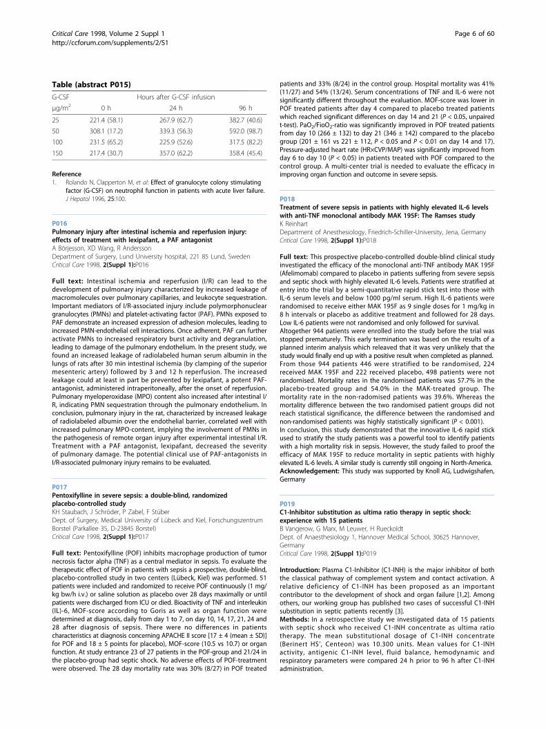
Table (abstract P015)
G-CSF Hours after G-CSF infusion
µg/m2 0 h 24 h 96 h
25 221.4 (58.1) 267.9 (62.7) 382.7 (40.6)
50 308.1 (17.2) 339.3 (56.3) 592.0 (98.7)
100 231.5 (65.2) 225.9 (52.6) 317.5 (82.2)
150 217.4 (30.7) 357.0 (62.2) 358.4 (45.4)
Critical Care 1998, Volume 2 Suppl 1http://ccforum.com/supplements/2/S1
Page 6 of 60
Results: After substitutional therapy the antigenic C1-INH level increasedsignificantly (from 159 to 228%) while the functional C1-INH activity didnot change statistically. The daily fluid balance decreased from +2695 to-186 ml/24 h (P = 0.05), hemodynamic parameters were not affected.There was a good tendency towards higher arterial-to-inspired oxygenratios (P = 0.55) after C1-INH administration.Conclusion: Our data show significantly higher antigenic C1-INH levelsafter substitutional therapy but no difference in C1-INH functional activity.This may be due to complex formation of C1-INH with its target proteases.Prior to C1-INH administration the patients had positive fluid balances andimpaired arterial-to-inspired oxygen ratios. If This is looked upon as anequivalent to capillary leakage these results encourage to perform arandomised controlled study on C1-INH substitution in septic patients.References1. Nuijens JH, et al: Proteolytic inactivation of plasma C1 inhibitor in sepsis.
J Clin Invest 1989, 84:443.2. Hack CE, et al: C1 -inhibitor substitution therapy in septic shock and in
vascular leak syndrome induced by high doses of interleukin-2. IntensiveCare Med 1993, 19:19-28.
3. Vangerow B, et al: Clinical improvement after administration of C1-Inhibitor in two patients with SIRS and capillary leak syndrome. IntensiveCare Med 1996, 22 (suppl 3):367.
P020Increased monocyte activation and tissue factor expression in patientswith sepsisB Dohrn1, S Russwurm1, J Vickers2, A Meier-Hellmann1, P Spangenberg2,W Lösche 3, K Reinhart11Klinik für Anästhesiologie und Intensivmedizin der Friedrich-Schiller-Universität Jena, 07740 Jena, Gemany; 2Fachbereich MedizintechnikderFachhochschule Jena, Germany; 3Zentrum für Vaskuläre Biologie undMedizin (Erfurt) der Friedrich-Schiller-Universität Jena, GermanyCritical Care 1998, 2(Suppl 1):P020
Background: Tissue factor (TF) is believed to play an important role in theinitiation of intravascular coagulation. Accumulating evidence has beenprovided that TF expression in monocytes may determine disturbances inintravascular coagulation and outcome in sepsis. A direct proof of anincreased monocyte TF activity and/or antigen in septic patients is still lacking.Patients and methods: 47 patients treated in the Intensive Care Unit ofan university hospital were included in the study. Sepsis and the degreeof organ dysfunction were assessed according to the criteria of the ACCP/SCCM Consensus Conference and the SOFA score. Blood samples from anarterial catheter were anticoagulated with sodium citrate. Binding tomonocytes of FITC-labelled monoclonal antibodies against TF and CD11bwas determined by flowcytometry.Results: Compared to non-septic patients (n = 20) monocytes of septicpatients (n = 27) bound significantly higher amounts of anti- TF antibody(mean fluorescence intensity - MFI: 14.3 ± 3.3 vs. 18.9 ± 6.4; P < 0.01).The same was true for binding of the antibody against the activation-dependent antigen CD11b. MFI in non-septic patients amounted to 266 ±72 and in septic patients 335 ± 86 (P < 0.005). Using a non-parametricanalysis we found significant correlation between anti-TF and anti-CD11bbinding (P < 0.03) as well as between SOFA Score and anti-CD11b oranti-TF binding (P < 0.03 and 0.02, respectively).Conclusion: The results indicate that monocytes in septic patients are on ahigher activation level and have higher amounts of TF antigen whencompared to non-septic ICU patients. Thus, the study confirmed the proposedrole of monocyte TF expression for sepsis-associated activation of coagulation.
P021Significance of the changes in blood fibrinogen levels as an acutephase reactant in septic DICY Otomo, H Henmi, M Honma, H Kato, J Inoue, T AraiNational Hospital Tokyo Disaster Medical Center, Dept. of Critical Care andTraumatology, 3256 Midori-cho, Tachikawa, Tokyo 190, JapanCritical Care 1998, 2(Suppl 1):P021
Background: Although treatment of DIC is said to be more effectivewhen it is started at early stage, early diagnosis of DIC occurring in
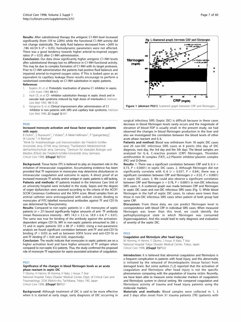
surgical infectious SIRS (Septic DIC) is difficult because in these casesdecrease in blood fibrinogen levels rarely occurs and the magnitude ofelevation of blood FDP is usually small. In the present study, we hadobserved the changes in blood fibrinogen production in the liver andalso we investigated the correlation between the blood levels of otheracute phase reactant and IL-6.Patients and method: Blood was withdrawn from 18 septic DIC casesand 20 non-DIC infectious SIRS cases at 4 points (the day of DICdiagnosis, next day, the 3rd day and the 5th day). The blood samples areanalyzed for IL-6, C-reactive protein (CRP) fibrinogen, Thrombinantithrombin III complex (TAT), a2-Plasmin inhibitor-plasmin complex(PIC) and D-Dimer.Results: 1. There was a significant correlation between CRP and IL-6 (r =0.73, P < 0.0001) in septic DIC cases. 2. Although fibrinogen did notsignificantly correlate with IL-6 (r = 0.027, P < 0.84), there was asignificant correlation between CRP and fibrinogen (r = 0.55, P < 0.0001)in septic DIC cases. 3. We could also observe a significant correlationbetween CRP and fibrinogen (r = 0.78, P < 0.0001) in non-DIC infectiousSIRS cases. 4. A scattered graph was made between CRP and fibrinogenin septic DIC cases and non-DIC infectious SIRS cases (Fig. 1). While bloodfibrinogen in the half of septic DIC cases, its values were lower thanthose in non-DIC infectious SIRS cases when patient of both group hadsame CRP.Discussion: From these data, we can predict fibrinogen level incorrespondence with blood CRP in individual SIRS cases. When measuredfibrinogen was lower than this level, we could identify thepathophysiological state in which fibrinogen was consumed(hypercoagulation). And this would lead to early diagnosis and evaluationof severity of the septic DIC.
P022Coagulation and fibrinolysis after head injuryM Homma, H Henmi, Y Otomo, J Inoue, H Kato, T AraiNational Hospital Tokyo Disaster Medical Center, Tokyo, JapanCritical Care 1998, 2(Suppl 1):P022
Introduction: It is believed that abnormal coagulation and fibrinolysis isa frequent complication in patients with head injury, and this abnormalityis initiated by the released of thromboplastin (tissue factor) fromdamaged brain. But some authors [1,2] reported that the activation ofcoagulation and fibrinolysis after head injury is not the specificphenomenon comparing with the population of trauma victim. Recently,we have been able to measure some molecular markers of coagulationand fibrinolysis system in clinical setting. We compared coagulation andfibrinolysis activity of trauma and head injury patients using themolecular markers.Materials and methods: Blood samples were collected in 1, 3and 5 days after onset from 31 trauma patients (TR) (patients with
Figure 1 (abstract P021) Scattered graph between CRP and fibrinogen.
Critical Care 1998, Volume 2 Suppl 1http://ccforum.com/supplements/2/S1
Page 7 of 60

head-Abbreviated Injury Score (AIS) = 3 were excluded), 12 head injury(HI) patients (patients with other system AIS = 3 were excluded) and 27cerebrovascular disease patients (CVD) (patients with intracranialhemorrhage or subarachnoid hemorrhage), and thrombin-antithrombin IIIcomplex (TAT), a 2 plasmin inhibitor plasmin complex (PIC), D-dimer,plasminogen activator inhibitor (PAI-1), tissue plasminogen activator(tPA)-PAI-I complex (tPAI-C), thrombomodulin (TM) and protein C activity(PCA) were measured.Results: PCA and PAI- I on 1 day had good correlation with injuryseverity score (ISS). TAT, D-dimer and PAI-1 of TR were significantly (P <0.05) higher than those of CVD, and PCA of TR were significantly lowerthan those of CVD, but there were no difference between TR and HI.Conclusions: HI and TR had significantly activation of coagulation andfibrinolysis compared with CVD, but there were no evidence that HI hadsignificantly activation of coagulation and fibrinolysis compared with TR.References1. Gando S, Tedo I, Kubota M: Posttrauma coagulation and fibrinolysis. Crit
Care Med 1992, 20:594-600.2. Sørensen J, Jensen H, Rahr H: Haemostatic activation in patients with
head injury with and without simultaneous multiple trauma. Scand J CinLab Invest 1993, 53:659-665.
P023Close relationship of PAI-1 (plasminogen activator inhibitor-1) withmultiple organ dysfunction syndrome and abnormal glucosemetabolism investigated by means of artificial pancreasM Hoshino1, Y Haraguchi2, M Sakai1, H Saegusa1, K Hayashi1, N Horita1,H Ohsawa11Department of Intensive and Critical Care Medicine, Tokyo Police Hospital;2National Hospital Tokyo Disaster Medical Center, Fujimi 2-10-41 Chiyoda-ku,Tokyo, Japan 102Critical Care 1998, 2(Suppl 1):P023
Background: There are many reports in recent years that the PAI-I isrelated to cardiovascular diseases and glucose intolerance especially indiabetic patients. But the role of role of PAI-1 to organ dysfunction andglucose in acutely ill patients is not clearly analysed. We invesigated thecontribution of coagulopathy including abnormal PAI-1 level to organdysfunction and to abnormal glucose metabolism in serious patients bymeans of the bedside type artificial pancreas (AP).Materials and methods: Thirty-five serious patients with glucoseintolerance, consisting of 25 males and 10 females aged from 23 years to 75years, were investigated. Primary diseases were as follows: 12 patients withpneumonia, seven with abdominal diseases treated by surgery, six withacute pancreatitis, three with hepatobiliary disorders, two with diabetic foot,and five with other diseases. Analysed items were, 1) regarding multipleorgan failure (MOF), MOFscore (calculated from the MOF criteria of JapaneseAssociation for Critical Care Medicine), 2) regarding glucose metabolism, (a)M value (mg/kg per min: measured by the euglycemic hyperinsulinemicglucose clamp method with AP. The clamped blood glucose level was 80mg/dl with the insulin infusion rate of 1.12 mU/kg per min) as an indicatorof peripheral glucose tolerance, (b) MCRI (ml/kg per min: metabolicclearance rate of insulin: measured by the glucose clamp method) as an
indicator of insulin metabolic rate, 3) regarding coagulation and fibrinolysis,(a) DIC (disseminated intravascular coagulation) score calculated from theDIC criteria of the Ministry of Health and Wealth of Japan, (b) PAI-I, (c) tPA-PAI (tissue plasminogen activator-PAI-1 complex), (d) FDP, (e) anti-thrombin III, (f) D-dimer, (g) PIC, (h) TAT, (i) plasminogen, (j) protein-C, 4) TM(thrombomodulin) as a parameter of endothelial cell injury, and 5) serum fat(free fatty acid (FFA), triglyceride, cholsterol). AP used was STG-22, made byNIKKISOH Corp. in Japan.Results: There were correlations between the following parameters. 1)Between MOF and other parameters; positive correlation between DICscoreand MOFscore (y = 0.533x + 2.70, n = 26, r2 = 0.705), MOFscore and PAI-I(y = 0.00996x + 0.588, n = 14, r2 = 0.282), MOFscore and tPA-PAI (n = 15, r2
= 0.239), 2) between PAI-I or tPA-PAI and other parameters; (a) negativecorrelation between tPA-PAI and M value (y = –17.4x + 98.9, n = 13, r2 =0.665), b) positive correlation between PAI-I and MCRI (y = 16.4x-108, n = 8,r2 = 0.532), tPA-PAI and MCRI (n = 8, r2 = 0354), TM and PAI-1 (y = 0.0155x +2.55, n = 13, r2 = 0.635), TM and tPA-PAI (n = 14, r2 = 0.660), FFA and PAI-1(y = 0.000490x + 0.144, n = 10, r2 = 0.477), FFA and tPA-PAI (n = 11, r2 =0.429). Other parameters but PAI-I and tPA-PAl related to coagulopathyshowed no definite relationships with MOFscore, M value, MCRI, TM and FFAConclusion: Multiple organ dysfunction syndrome, glucose intolerance,and increased insulin metabolism were revealed to be closely related toincreased PAI-I or tPA-PAI, which reflect decreased fibrinolysis. Injury ofendothelium and increased serum FFA level were thought to be relatedto increased PAI-I and tPA-PAI, which may explain the progression ofMOF partly. In addition, PAI-1 and tPA-PAI seemed to be sensitive markerof, and might be one of the risk factors of multiple organ dysfunctionsyndrome and nutritional metabolic disorder.
P024Use of antithrombin III in cancer patients with sepsis complicated withdisseminated intravascular coagulopathyM Polansky, J Varon, WK HootsThe University of Texas M.D. Andersen Cancer Center, Division ofAnesthesiology and Critical Care, 1515 Holcombe Box 42, Houston, TX, USA77030Critical Care 1998, 2(Suppl 1):P024
Introduction: Mortality rate from sepsis is estimated to be 20% to 60% withdisseminated intravascular coagulation (DIC) often accompanying sepsis.Replacement therapy with antithrombin III (ATIII) concentrate has beenhypothesized to be a means for attenuating DIC, since ATIII is rapidlyconsumed during DIC. A reliable index of poor prognosis is an initial decreaseof serum ATIII level. The purpose of this pilot study was to evaluate the effectsof the administration of ATIII in patients sepsis complicated by DIC.Materials: All adult patients admitted to the surgical intensive care unit atthe University of Texas M.D. Anderson Cancer Center with clinical evidence ofsepsis and DIC were eligible for inclusion. Exclusion criteria consisted ofleukemia and treatment with heparin. Patients received 100 units/kg of idealbody weight of antithrombin III (Thrombate III®) at time zero, repeated 12 hlater, and daily for three days. Serum ATIII levels were drawn prior toenrollment, prior to and after each dose, and for 2 days after the last dose.Levels were analyzed at the conclusion of the study. No other interventionwas provided by the investigators. Patients were otherwise managed by theirprimary care providers and consultants as their clinical situation warranted.Results: The study population included 4 women and 1 man with anaverage age of 59.6 years (33–84). Severity of illness was measured by amean APACHE II of 21.8 (19–24) and a mean TISS of 55.6 (45–70) at thetime of study enrollment. The ICU and hospital mortality rate were 80%,with one patient not surviving to complete the study regimen. SerumATIII levels at the time of enrollment ranged from 22 to 78 MUI/ml,averaging 45.4 (normal 80–120). The one surviving patient had thehighest ATIII level (78 MUI/ml) prior to study enrollment. A rise in ATIIIlevels after treatment was found in each patient, with all but one patienthaving levels normal or supranormal after the first dose of treatment.One patient, with the lowest pretreatment level (22 MUI/ml), had levelsthat remained below normal during most of treatment and wasdecreased (51 MUI/ml) at day 2 post treatment.Conclusions: We have found a variable response in ATIII levels in patientswith sepsis after ATIII treatment. Future studies involving ATIII replacementshould include the use of ATIII levels to guide dosing regimens.
Table (abstract P022)
TR HI CVD
numbers of pt. 31 12 27
day 2.7 ± 0.3 2.5 ± 0.6 3.3 ± 0.4
TAT 84.1 ± 13.8* 77.7 ± 31.2 28.7 ± 7.4*
PIC 3 ± 0.4 4.2 ± 2.1 2.7 ± 0.5
D dimer 20.8 ± 2.7* 19.1 ± 5.3 6.9 ± 1.1*
PAI-1 30.6 ± 3.1* 30.7 ± 5.4 17.3 ± 1.4*
tPAI-C 19.7 ± 1.8 20.3 ± 3.8 19.2 ± 1.8
cTM 29.9 ± 2.8 25.9 ± 2.4 28.2 ± 1.4
PCA 84.2 ± 3.0* 83.6 ± 5.5 99.5 ± 3.2*
(mean ± SE; *P < 0.05, TR vs CVD)
Critical Care 1998, Volume 2 Suppl 1http://ccforum.com/supplements/2/S1
Page 8 of 60

P025Effect of antithrombin III on inflammatory immune response in patientswith severe sepsisD Inthorn1, JN Hoffmann1, WH Hartl1, D Mühlbayer1, M Jochum2
1Dept. of Surgery Klinikum Grob hadern and Ludwig-Maximilians-University,Marchioninistr. 15, 81377 München, Germany; 2Division of ClinicalBiochemistry in the Dept. of Surgery Klinikum Innenstadt, Ludwig-Maximilians-University, Marchioninistr. 15, 81377 München, GermanyCritical Care 1998, 2(Suppl 1):P025
Full text: Antithrombin III (AT III) is an important inhibitor of thrombinactivity, as well as of many other proteases of the coagulation system. AT IIIadministration showed beneficial effects on septic multiple organdysfunction in clinical and experimental studies. It was the aim of this studyto determine whether continuous long-term AT III supplementation altersthe systemic inflammatory response in patients with severe sepsis. In aprospective study 29 surgical patients with severe sepsis were randomlyassigned to receive either conventional intensive care treatment (n = 15,control group) or additional AT III supplementation with a plasma AT IIIactivity >120% during a fourteen day study period (n = 14, AT III group).Plasma Interleukin (IL)-6 and IL-8, and the circulating adhesion moleculesICAM-1 and soluble E-selectin, as well as PMN elastase were determined dailyby ELISA. Total leukocyte count and C-reactive protein (CRP) were measureddaily and body temperature was registered. Compared to control patients, adownregulation of plasma IL-6, and, to a lesser degree, also of plasma IL-8was observed in the AT III group (P < 0.05). AT III supplementation preventedthe continuous increase in ICAM-1 plasma concentration observed in controlpatients and lead to a significant fall in serum soluble E-selectin and in CRPconcentration (P < 0.05). This fall corresponded to a downregulation of bodytemperature over time (P < 0.05). There was no AT III effect on PMN elastaseconcentration or total leukocyte count. Our results show that long-termantithrombin III supplementation attenuates the systemic inflammatoryresponse in patients with severe sepsis.
P026Release of endothelin in an experimental model of shock and ARDS insheepU Friess, M Quintel, H Roth, F Esen, C Beyer, HJ BenderDept. of Anesthesia and Intensive Care, Klinikum Mannheim, Faculty ofClinical Medicine, University of Heidelberg, GermanyCritical Care 1998, 2(Suppl 1):P026
Objectives: The Endothelins (ETs) are a class of 21 amino-acid peptides,which were first described in 1988 as vasoactive peptides produced inthe endothelial cells of the porcine aorta. Meanwhile three differentsubtypes have been identified. ETs act as potential vasocontrictors in thecardiovascular system. In the lungs, ETs induce bronchospasm and viavasoconstriction pulmonary hypertension followed by severe edemaformation. It is well known, that ETs are synthesized and present in thelung. Additionally a high number of ET-receptors has been identifed inthe lung. Recent studies show elevated ET-plasma concentration invarious diseases like cardiogenic shock, subarachnoidal bleeding, COPD,sepsis and trauma. This study was performed to evaluate the ET-plasmaconcentration after induced hypovolemic shock states and ARDS.Methods: The experiment was performed in 14 anesthesized, andmechanically ventilated sheep (21–28 kg BW) during two different consecutiveshock models (A: 3.5% BW ultrafiltrate withdrawal by hemofiltration; B: Bloodwithdrawal up to 3.5% of BW) and after lung lavage induced ARDS. A 4 Fcatheter was placed in the A. carotis to obtain blood pressure. Cardiac outputwas determinated by a 7.5 F fiberoptic thermodilution catheter placed into themain pulmonary artery. Blood samples from the A. carotis (CAR) and the A.pulmonalis (PUL) and urine samples were taken at baseline, 15 min afterhemofiltration, 15 min after blood withdrawal and 5, 20, 60 min after lunglavage. ET-concentrations were measured by RIA (Nichols, Bad Homburg, FRG).Transpulmonary flux was calculated by the equation: (ET - concentration [A.pulmonalis] - [A. carotis])/cardiac output. Statistical analysis was performedusing Wilcoxon sign rank test.Results: A significant increase of plasma-ET concentrations from baselinelevels in the A. carotis and A. pulmonalis (CAR 7.89 ± 3.9 μg/ml; PUL 8.22 ±4.1 μg/ml) was observed during hypovolemia with ultrafiltration (CAR
14.9 ± 7.8 μg/ml, P < 0.01; PUL 15.4 ± 7.1 μg/ml, P < 0.01), and duringhypovolemia with blood-withdrawal (CAR 14.4 ± 6.6 μg/ml, P < 0.01; PUL13.6 ± 6.4 μg/ml, P < 0.01), and 60 min after ARDS (CAR 10.6 ± 5.0 μg/ml,P < 0.05). No significant difference was found between plasma-ETconcentration in CAR and PUL at any state. Urine-ET concentration showeda slight, but not significant increase after ARDS, but no alterations inhypovolemic states. Flux calculation did not indicate significant changes.Conclusion: The presented data show a significant increase in plasma-ETconcentration after induced hypovolemic shock. It can be speculated thatthe potential vasoconstrictory effects of ETs act as a paracrine regulatorof vascular tone. In contrast to other studies we could not find asignificant change in the early state of induced ARDS. In a saline lavagemodel desquamation of bronchial and bronchialar epithelium, hyalinemembranes and accumulations of pyknotic cells in peterminal airspacescan be observed. These pathological changes may explain the lack of anearly response due to partial destruction of endothelium cells.
P027Effect of interleukin-6 and interleukin-10 on nitric oxide production byendothelial cellsV Brix-Christensen, Y Liu, E Tonnesen, G StefanoState University of New York at Old Westbury, NY and University Hospital ofAarhus, DenmarkCritical Care 1998, 2(Suppl 1):P027
Introduction: Nitric oxide (NO) is synthesised from conversion of L-arginineto citrulline by NO-synthase (NOS). NO mediates vascular dilation, musclerelaxation and platelet aggregation. Constitutive NOS (cNOS) exists inendothelial cells but not in vascular smooth muscle. Among others thecytokines IL-6 and IL-10 can be measured in high concentrations duringsepsis. IL-10 has an inhibitory effect on the NO synthesis in LPS stimulatedmacrophages by inhibiting NOS. Recently, IL-10 have been reported to havea stimulatory effect on the NO production by endothelial cells in humansaphenous veins in a concentration dependent way.Aim: To investigate the in vitro effect of IL-10 and IL-6 on NO productionby endothelial cells in human saphenous veins.Methods: Human saphenous veins from patients undergoing CPB were used.The vessels were suspended in phosphate buffered saline (PBS) with theendothelial side up. The tip of the amperometic probe was positioned 10 μmabove the cell surface. Concentrations of the NO gas in the PBS solution weremeasured in real time with a DUO 18 computer data acquisition.Results: The vessels were exposed to IL-10 (10-8 M), IL-6 (10-6 M) anti-IL-6(l0-6 M) alone or in combination. IL-10 increased the NO production bythe endothelial cells. IL-6 on its own did not affect the NO production.When IL-6 was added minutes following the IL-10 administration, itdiminished the IL-10 induced NO release. Anti-IL-6 did not have anyinfluence on the NO production, but the IL-6 inhibition of IL-10stimulated NO release, was blocked when anti IL-6 was added.Conclusion: These results show that IL-10 stimulate NO release fromendothelial cells. Addition of IL-6 can attenuate the effect of IL-10 onendothelial cells.
P028Effects of nitric oxide (NO) on platelets in neonatesD Keh1, I Kürer1, M Gerlach1, W Dudenhausen2, W Woltmann2, K Falke 1,H Gerlach 1
1From the Clinic of Anesthesiology and Intensive Care Medicine, VirchowClinic, Humboldt University, Berlin, Germany; 2Department of Obstetrics,Virchow Clinic, Humboldt University, Berlin, GermanyCritical Care 1998, 2(Suppl 1):P028
Full text: Nitric oxide (NO) is known to play a crucial role in primaryhemostasis due to its platelet-inhibitory properties. To investigate the effectsof NO on neonatal platelets, umbilical cord blood from 10 healthy termneonates was obtained immediately after birth. Blood samples from boththe mothers and non-pregnant female blood donors (n = 10) were analyzedin parallel as controls. Citrated platelet rich plasma was incubated with theNO-donor SIN-1 (10 μM), an active metabolite of molsidomine, prior toactivation with 10 μM adenosine-diphosphate (ADP) or 0.05 U/ml humana-thrombin (both ED50), or with agonists or buffer only. Platelet activation
Critical Care 1998, Volume 2 Suppl 1http://ccforum.com/supplements/2/S1
Page 9 of 60

was analyzed in a two-color flow cytometry assay using phycoerythrine (PE)or fluoresceine-isothiocyanate (FITC) conjugated monoclonal antibodiesdirected against glycoprotein-(GP)-Ib (CD42b) and activated fibrinogenreceptor GPIIb-IIIa (PAC-1), respectively. No significant differences for PAC-1binding to resting platelets (baseline) was observed between neonate,mother or control, indicating absence of pregnancy- or delivery-inducedactivation of circulating platelets (see figure). No significant differences ofplatelet response to ADP or thrombin were found between controls andmothers, indicating normal platelet response of the mother. Compared toboth the mother and control, neonatal GPIIb-IIIa activation was significantly(P < 0.05) depressed, ie newborn platelets revealed hyporeactivity to ADPand thrombin. NO significantly (P = 0.001) inhibited GPIIb-IIIa activation in allgroups, however, percentual downregulation of GPIIb-IIIa-exposure was notsignificantly different in newborns, mothers, or controls (ca. 70 ± 5%). Thisdemonstrates depressed response of neonatal platelets to agonists, butnormal reaction to platelet inhibitory compounds such as NO. Sincenewborn platelets are starting from a lower level of activation, NO nearlydiminished platelet activation. This might be of clinical relevance, sincehemorrhagic complications remain a serious problem in critically ill termneonates, or even more in premature newborns. Several studies showedreduced platelet function in both healthy volunteers and patients with adultrespiratory distress syndrome (ARDS) during inhalation of gaseous NO. NO-inhalation is well established in the pediatric ICU for treatment of infantrespiratory distress syndrome (IRDS) or persistent pulmonary hypertension.The presented data indicate that inhalation of NO might be an additionalrisk-factor for bleeding complications in critically ill neonates.Acknowledgement: Supported by Deutsche ForschungsgemeinschaftDFG Fa 139/4-2
P029Nitric oxide (NO) metabolite levels are not increased duringhypotensive periods in human sepsisDH Jenkins1,3, HL Frankel1,3, AK May1, H Nguyen2, K Simo1, CW Schwab1,S Bina41Department of Surgery, University of Pennsylvania Medical Center, 3400Spruce Street, Philadelphia, PA 19104, USA; 2National Naval Medical Center,Uniformed Services University of the Health Sciences; 3Departments ofSurgery, Uniformed Services University of the Health Sciences;4Anesthesiology of the Uniformed Services University of the Health SciencesCritical Care 1998, 2(Suppl 1):P029
Purpose: Excess NO production has been proposed to cause thehemodynamic derangements of septic shock. This study was undertaken
to determine whether serum NO metabolite levels correlate withhemodynamic changes in human sepsis.Methods: A 12-month prospective study of surgical ICU patients withSIRS, sepsis, severe sepsis or septic shock was undertaken. Serum NO2/NO3 levels were determined by chemiluminescence from blood drawnduring blood culture acquisition or hypotension (SBP <90 mmHg), thendaily for 7 days or until ICU discharge or death. The following werecollected: vital signs, pulmonary artery catheter data, white blood cellcount (WBC) and arterial base deficit (BD). T-test analysis compared NO2/NO3 levels in hypotensive and normotensive patients. Pearson correlationcoefficients were determined for NO2/NO3 and the variables above. Dataare mean ± SEM; P < 0.05 defined significance.Results: NO2/NO3 levels were 12.2 ± 3.8 pmol/μl in 13 hypotensive vs 12.7 ±2.0 in 9 normotensive patients (NS). Correlation coefficients (r2) are depicted:Conclusions: NO metabolite levels are not different in hypotensive andnormotensive septic patients. There is poor correlation between NOmetabolite levels and physiologic changes in human sepsis. Further studyregarding the role of NO as the principal vasodilator in sepsis iswarranted.
P030Serum TNF alpha and IL-8 levels in difficult weaning patientsV Cerny, P Zivny, P Dostal, R ParizkovaDepartment of Anesthesiology and Intensive Care, Charles University, Facultyof Medicine, 50005 Hradec Králové, Czech RepublicCritical Care 1998, 2(Suppl 1):P030
Introduction: Metabolic status and severity of illness play important rolein developing ventilatory dependency during long term ventilatorysupport in critically ill patients. The aim of the study was to evaluateTNFa and IL-8 serum levels during weaning from long term ventilatorysupport.Methods: After institutional approval 26 critically ill patients wereprospectively studied during ventilatory support and weaning. All patientswere weaned according to standard weaning protocol. Blood sampleswere drawn daily and collected until analysis. Apache II score, ventilatorydays and ‘weaning’ days were recorded. After successful weaning patientswere divided in two groups according to the length of weaning (W):group S (W ≤ 3 days, n = 6), group L (W ≤ 3 days, n = 20). TNFa and IL-8serum levels were selected and measured at the time of admission (T1),on the last day of full ventilatory support (T2), on the day when weaningwas started (T3) and on the first day after 24 h of spontaneousventilation (T4). Values are expressed as a mean ± SD, t-test or MannWhitney Rank Sum test where appropriate were used for statisticalanalysis (Sigma Stat Statistical Software, Jandel Co., USA), P < 0.05 wasconsidered statistically significant.Results: Total ventilatory and weaning days were 24.1 ± 11, resp. 7.5 ± 3in group L and 10.7 ± 4.6 resp. 2.6 ± 1.7 in group S. Selected results ofTNFa (in pg/ml) and IL-8 (in pg/ml) are presented in the table.
Figure (abstract P028) Effects of SIN-1 on GPIIb-IIIa activation in platelets from cord blood (neonate), mother and female control, activated with ADP orthrombin.
Table (abstract P029)
SBP P CI SVO2 SVR WBC BD
NO2/NO3 0.04 0.01 0.10 0.16 0.05 0.09 0.04
Critical Care 1998, Volume 2 Suppl 1http://ccforum.com/supplements/2/S1
Page 10 of 60
Discussion: Serum TNFa and IL-8 levels were persistent in patients withprolonged ventilatory support. This suggests that these mediators mayalso be involved in ventilatory failure leading to difficult weaning afterlong term mechanical ventilation.Acknowledgement: Supported by grant IGA MZ CR 3674-3.
P031Matrix metalloproteinase-1, soluble Fas ligand, and soluble Fas antigenlevels in patients with multiple organ dysfunction syndromeS Endo1, T Kasai1, T Takakuwa1, H Nakae2, Y Yamada1, K Inada3, S Taniguchi11Critical Care and Emergency Center, Iwate Medical University, 19-1 Uchimaru,Morioka 020, Japan; 2Department of Critical Care Medicine, Akita UniversityMedical School, 1-1-1 Hondo, Akita 010, Japan; 3Department of Bacteriology,Iwate Medical University School, 19-1 Uchimaru, Morioka 020, JapanCritical Care 1998, 2(Suppl 1):P031
Full text: Fas L induces apoptosis by binding to Fas. It is reported thatthe extracellular matrix degrading enzyme, metalloproteinase, is involvedin conversion from membrane Fas L to soluble Fas (sFas).In this study, the involvement of matrix metalloproteinase-1 (MMP-1),soluble Fas ligand (sFasL), and soluble Fas antigen (sFas) in apoptosis atthe onset of multiple organ dysfunction syndrome (MODS) was examined.The subjects were 52 patients. Blood sampling was continued from thetime before or at the onset of MODS up to its recovery or death. MMP-1,the tissue inhibitor of metalloproteinase-1 (TIMP-1), MMP-1/TIMP-1complex, sFas L, sFas, tumor necrosis factor-a (TNF-a), TNF receptor I(TNFR I), TNFR II, nitrite/nitrate (NOx), transforming growth factor b (TGF-b), and endotoxin were measured.With the increase in dysfunctional organs, sFas, TNF-a, and NOxincreased, and showed significantly higher levels in those that died thanin the survivors. Their levels were significantly correlated with the MMP-1level, and the MMP-1 level was negatively correlated with the TGF-b level.At the onset of MODS, the sFas, TNF-a, and NOx levels were high, suggestingthat apoptosis occurred. It was also suggested that MMP-1 was involved insFas and TNF-a production, and TGF-b suppressed MMP-1 production.
P032Endothelium activation during prolonged extra-corporeal circulationB Adamik, B Pliskowska, PS Berezowicz, J Kübler, B Golêbiowska, A KüblerDepartment of Anaesthesiology and Intensive Therapy, Wroclaw Universityof Medicine, Chalubinskiego 1a, 50-368 Wroclaw, PolandCritical Care 1998, 2(Suppl 1):P032
Objectives: The aim of this study was to monitor the plasma levels ofsoluble adhesion molecules in patients undergoing cardiac surgery inextracorporeal circulation (EC). We investigated the relationships betweenthe levels of adhesins released during the prolonged extracorporealcirculation and postoperative morbidity.Study design: We assessed 10 patients (NYHA class II or III) undergoingelective cardiac surgery with flat-sheet membrane oxygenation. Theprocedures were: coronary artery bypass grafting, cardiac valve implantationor both. The duration of CPB was 135–246 min, core temperature 28–31°C.The patient statuses were assessed daily with SOFA and Marshall MODSscores. The soluble vascular cell adhesion molecule (sVCAM-1), solubleintercellular adhesion molecule (sICAM-1), soluble endothelial leukocyteadhesion molecule (sELAM-1), lactate levels and WBC were measured beforeinduction of anaesthesia, during EC, one and two days after operation.Materials and methods: Arterial blood samples were collected on heparin.Plasma was immediately obtained by centrifugation and then frozen at -70°C
until the assay was performed. Activity of sVCAM-1, sICAM-1 and sELAM-1was measured using enzyme- linked immunoabsorbent assays (ELISA).Results: The plasma concentrations of sVCAM-1 were significantly higherduring the entire study (mean 954.38, 818.60, 1291.09, 1264.09 ng/ml). Infour patients who developed postoperative multiple organ dysfunctionfailure the sVCAM-1 were significantly higher than in those with uneventfulrecovery. sICAM-1 levels after operation were above the normal range(mean 272.88, 236.97, 294.64, 410.52 ng/ml). There was no significantdifferences in plasma sELAM-1 levels during the whole time of observation(mean 43.07, 29.45, 48.57, 49.18 ng/ml before anaesthesia, during surgery,on day 2 and 3, respectively). sICAM-1 levels after operation were abovethe normal range (mean 272.88, 236.97, 294.64, 410.52 ng/ml).Conclusions: Extracorporeal circulation with membrane oxygenation isfollowed by a significant endothelium response and release of solubleadhesion molecules into the bloodstream. There is the correlationbetween the markedly increasing level of sVCAM-1 and sICAM-1 and theappearance of postoperative complications.
P033Systemic inflammatory response syndrome (SIRS) without systemicinflammationA Takala, I Jousela, O Takkunen, KT Olkkola, S-E Jansson, M Leirisalo-Repo,H RepoDepartment of Anaesthesia, Helsinki University, Haartmaninkatu 4, FIN-00029Helsinki, FinlandCritical Care 1998, 2(Suppl 1):P033
Background: Criteria of SIRS, developed to detect early systemicinflammation (SI), are sensitive enough to include also patients (pts)without SI. To address this problem, we developed composite systemicinflammation score (CSIS) based on phagocyte activation markers toevaluate degree of SI in acutely ill pts with SIRs.Patients and methods: 100 pts at medical emergency were included.CD11b expression levels on neutrophils (PMNs) and monocytes (MO)were determined by flow cytometry, serum levels of IL-6, IL-1b, andTNF-a by ELISA, and of C-reactive protein (CRP) by immunoturbidimetry.To calculate CSIS, IL-6, IL-1b, TNF-a, CRP and CD1 expression levels wereeach scored 0 to 4 points; maximum CSIS is 20 points.Results: 61 pts had SIRS2, 14 SIRS3, 2SIRS4, 20 sepsis, 2 severe sepsis,and 1 septic shock. Drug intoxication (17 pts) and acute myocardialinfarction (15 pts) were most common diagnoses. CD11b expressionincreased in order : SIRS2 = SIRS3 < Sepsis; CRP in order: SIRS2 < SIRS3 <Sepsis, and IL-6 and IL-1b both in order: SIRS2 < SIRS3 < Sepsis. 13.8%of pts had CSIS 0 indicating virtually absence of SI. CSIS of SIRS2 pts(median 1.5; range 0–8) was lower than that of SIRS3 pts (3.5; 0–9, P =0.013) and that of Sepsis pts (5; 3–10, P < 0.001); also SIRS3 differed fromSepsis (P = 0.0018). Among 81 pts with CSIS = 1, proportions of pts withincreased IL-6, CRP, CD11b, IL-1b, and TNF-a were 79.0%, 72.8%, 61.7%,25.9% and 2.5%, respectively.Conclusions: Quantifying phagocyte CD11b expression and circulatingIL-6 and CRP concurrently provides a means to identify patient whomeets SIRS criteria but lacks SI.
P034Measurement of circulating ICAM-1 in patients with acute lung injuryon ventilators in intensive care unitC Hoong Thye, R Rajamanon mani, G Ong Siok Yan, R QvistDepartment of Anesthesiology, Faculty of Medicine, University Hospital,University of Malaya, 50603 Kuala Lumpur, MalaysiaCritical Care 1998, 2(Suppl 1):P034
Full text: Serum levels of circulating inter cellular adhesion molecule-1(ICAM-1) were measured by usign a sandwich-type Enzyme LinkedImmunosorbent Assay (ELISA) kit (R&D System Inc). cICAM-1 was detectedin the serum of fifteen normal healthy volunteers with an age range of20 to 56 years, who had an average level of 172.4 ng/ml and a range of92.59 ng/ml to 305.32 ng/ml. A significant elevation of cICAM-1 levelswas observed (P < 0.0006) in patients with acute lung injury as comparedwith the normal. The trends of the patients’ cICAM-1 were determined bysequential measurements during their stay in ICU. Seven patients
Table (abstract P030)
T1 T2 T3 T4
Group S TNFa 10.8 ± 5.59 10.6 ± 9.52 6.42 ± 7.11 0*
Group L TNFa 11.7 ± 7.67 12.6 ± 5.6 7.8 ± 5.9 7.15 ± 6.04*
Group S IL-8 55.2 ± 33.3 65.8 ± 37.2 47.3 ± 42.0 6.32 ± 10.8*
Group L IL-8 205.2 ± 356.8 89.9 ± 101.6 73.8 ± 100.4 38.7 ± 27.6*
*P < 0.05
Critical Care 1998, Volume 2 Suppl 1http://ccforum.com/supplements/2/S1
Page 11 of 60

demonstrated a rising trend of serum cICAM-1. The remaining patientshowed a falling trend, with a marked elevation (>1300 ng/ml) of cICAM-1 level on the day of admission into ICU. The cICAM-1 levels of the eightpatients were correlated with two clinical parameters: ratio of arterialoxygenation to inspired oxygen fraction (PF ratio), and pressure adjustedheart rate. Despite a poor correlation of cICAM-1 levels with the PF ratio,cICAM-1 levels were elevated to at least 400 ± 20 ng/ml, when the PFratio was low (<300). The infection pattern in these patients were alsoanalyzed by bacteriological investigations done on clinical samples. Themeasurements of cICAM-1 levels may be important in understanding thepathophysiology of critically ill patients with acute lung injury. Furtherstudies on the immunological mechanism and modulation of theinjurious inflammatory response may benefit critically ill patients with thedevelopment of effective immunotherapy.
P035Human peripheral blood mononuclear cells express mRNA forprocalcitonin; modulation by lipopolysaccharides and sepsis relatedcytokinesM Oberhoffer1, S Russwurm1, I Stonans1, E Stonane2, H Vogelsang2, U Junker2,L Jäger2, K Reinhart11Department of Anesthesiology and Intensive Therapy, Friedrich-Sehiller-University of Jena, 07740 Jena, Germany; 2Institute of Clinical Immunology,Friedrich-Sehiller-University of Jena, 07740 Jena, GermanyCritical Care 1998, 2(Suppl 1):P035
Introduction: Procalcitonin (PCT), the precursor of calcitonin, is producedunder physiological conditions in the thyroid. It was recently identified asa marker of severe especially bacterial infection. Elevated PCT levels havebeen demonstrated in septic patients also after thyroidektomy. Thereforeother sites of PCT expression have to be considered.Methods: Possible PCT mRNA expression in peripheral bloodmononuclear cells (MNC) from healthy humans was assessed by reversetranscriptase polymerase chain reaction (RT-PCR) using a novel primer set(B.R.A.H.M.S, Berlin). Levels of PCT mRNA expression were estimated incontrol and cultures of MNC stimulated by lipopolysaccharides (LPS) andsepsis related cytokines. Restriction mapping was performed to verify thespecificity of PCR primers for human PCT by digestion of PCR amplifiedthyroid medullary carcinoma cDNA.Results: RT-PCR analysis demonstrated that MNC express mRNA for PCTand that LPS as well as various cytokines may modulate this expression.In the cultures stimulated with LPS from E. coli B4 and Salmonellaabortus equi the final RT-PCR product for PCT mRNA was elevated up to200% to 2300% in comparison with 100% in control culture. Pronouncedeffects were also observed for IL-lb, 100–1800%; IL-6, 200-3500%; TNF-a,200-9000%; IL-2, 250–1500%. IL-10 had no effect on the expression ofmRNA for PCT.Conclusions: We demonstrate for the first time that PCT is expressed inMNC. This expression is modulated by bacterial LPS and sepsis relatedcytokines. Therefore MNC may be one of the sources for elevated plasmaPCT levels in septic patients.
P036Procalcitonin, cytokines and C-reactive protein in systemicinflammatory responseP Tesinsky, Z Rusavy, I Novak, V SramekDepartment of Internal Medicine I, Charles University Hospital, Plzeñ, CzechRepublicCritical Care 1998, 2(Suppl 1):P036
Background: Procalcitonin (PCT) is a biochemical marker with ahigh specificity for bacterial and mycotic etiology of inflam-mation. Its production is selectively induced by endotoxin-cytokinesmechanism.Aim of study: To verify differential diagnostic value of PCT ininflammation of different etiology and intensity and to test practical useof PCT estimation.Methods: In a prospective pilot study, 14 patients with various maindiagnoses admitted to a medical ward entered the study. Serum levels ofPCT, interleukin 6 (IL-6), C-reactive protein (CRP), body temperature and
APACHE-II score were estimated in 12 h intervals within 6 days. Resultswere statistically evaluated by linear correlation analysis.Results: 1. Patients with bacterial sepsis showed highest levels of PCT.Elevated levels of PCT were also present in patients with systemicautoimmune diseases. Patients with viral etiology of inflammatory processand those with no inflammatory process had normal levels of PCT.2. Significant correlation was found between PCT and IL-6 (r2 = 0.6568),and PCT and CRP (r2 = 0.3714) in patients with bacterial sepsis.Correlation of CRP and IL-6 levels was less significant (r2 = 0.1968).3. No correlation between PCT and number of leukocytes, bodytemperature, lactate, and sedimentation of erythrocytes was observedneither in septic nor in non-septic individuals.Discussion: Estimation of serum procalcitonin level in septic patientsseems to be a reliable and easy-to-do test which corresponds with IL-6.As laboratory estimation of cytokines is relatively difficult and expensive,procalcitonin can be a good variant.
P037Procalcitonin in paediatric sepsisM Hatherill, SM Tibby, A Mclntyre, IA MurdochPaediatric Intensive Care Unit, Guy’s Hospital, London SE1 9RT, UKCritical Care 1998, 2(Suppl 1):P037
Objective: To document the relationships between procalcitonin (PCT),C-reactive protein (CRP) and leucocyte count (WCC) in children withsepsis, and to compare their diagnostic value in septic shock.Methods: PCT, CRP and WCC were measured on admission to the PICUin 146 children, median age 18 months (range 0.1-202), mortality 22/146(15%). PCT was measured by immunoluminometric assay (BRAHMSDiagnostika, Berlin, Germany). Patients were categorized a prioriaccording to clinical and laboratory data as having either septic shock(n = 51) [1], localized bacterial infection (n = 33), viral infection (n = 14)or as non-infected controls (n = 48). Data were analysed by Kruskal-WallisANOVA and Dunn’s test for multiple comparisons.Results: PCT was significantly higher in septic patients compared tolocalized infection, viral infection and controls respectively (all P < 0.001).Receiver operating curve analysis indicates PCT > 20 ng/ml as the bestdiscriminator of septic shock with positive and negative predictive valuesof 87% and 89%. In contrast, CRP > 35 mg/l had positive and negativepredictive values of 65% and 83%.Conclusion: Procalcitonin is a better discriminator of systemic fromlocalized bacterial infection than either CRP or leucocyte count.Reference1. Saez-Llorens X, McCracken GH: Sepsis syndrome and septic shock in
pediatrics: current concepts of terminology, pathophysiology, andmanagement. J Pediatr 1993, 123:497-508.
P038Urinary procalcitonin associated with a microbiologically diagnosedpneumonitis: preliminary resultsF Chiappini1, MC Matita1, P De Sole2, R Fresu2, L Frigieri1, L Fuso1, G Pagliari11Departments of Respiratory Physiology; 2Clinical Chemistry, CatholicUniversity Rome, ItalyCritical Care 1998, 2(Suppl 1):P038
Full text: Procalcitonin (PCT), the prohormone of calcitonin, is a 116amino-acid protein stimulated by bacterial inflammation and producedby neuroendocrine cells of different organs, included the lung.Elevated serum levels of PCT have been found in several diseases suchas sepsis syndromes, inhalation burn injury, infectious pneumonitis.PCT has been measured in body fluids other than serum in only fewpapers and with conflicting results. The aim of the present study wasto verify whether PCT levels measured in the serum, urine, andbronchoalveolar lavage (BAL) fluid could be a marker of pulmonaryinfection, diagnosed by a positive cultural bacterial exam in thebronchial washing (BW).Until now, 10 patients with pulmonary diseases were studied. Four ofthem had a radiologically diagnosed pneumonitis, 3 had a non-small celllung cancer (NSCLC), 3 had other non-infective lung diseases. All thepatients underwent a flexible fiberoptic bronchoscopy (FOB) with BAL
Critical Care 1998, Volume 2 Suppl 1http://ccforum.com/supplements/2/S1
Page 12 of 60
and BW. The quantitative determination of PCT was performed in theserum, urine and BAL fluid by the immunoluminometric assay (ILMA)using the LUMItest PCT kit (BRAHMS Diagnostica, Berlin, Germany).PCT was not detectable in BAL fluids of all the patients. A positivedetermination of PCT was obtained in a total of 4 patients : in 2 ofthem in urine, in 1 patient in both urine and serum, and in 1 patientin serum. A trend towards a significant association between smokinghabit and urinary PCT was found (PCT = 0 in non-smokers, 0.73 ± 1.27ng/ml in ex-smokers, 1.77 ± 2.06 ng/ml in smokers). Urinary PCT waslower in patients with a radiologically diagnosed pneumonitis than inpatients with NSCLC (0.95 ± 1.90 vs 1.83 ± 1.68 ng/ml, respectively).However a positive coltural exam for bacteria in BW was found in only1 out of 4 patients with a radiologically diagnosed pneumonitis and in2 out of 3 patients with NSCLC. Condidering the 3 patients with apositive coltural exam for bacteria in BW, a trend towards a significanthigher urinary PCT values was found in these patients in comparisonwith the 7 patients with a negative coltural exam (1.83 ± 1.68 vs 0.54± 1.43 ng/ml, respectively). The 4 patients with a positivedetermination of PCT in urine and/or serum had a significant highernumber of total cells in the BAL fluid in comparison with theremaining 6 patients (218 000 ± 155 000 vs 60 000 ± 30 000respectively, P = 0.01).In conclusion, these preliminary results seem to indicate that an increasedlevel of urinary PCT is associated with the presence of pulmonaryinfection also complicating other lung diseases such as NSCLC anddemonstrated by a positive coltural exam for bacteria in BW. Urinary PCTreflects the severity of alveolitis which usually derives from pulmonaryinfection. Taking into account the smoking habit of the patient, urinaryPCT could be a simple and non-invasive marker useful in the follow-up ofpatients with a microbiologically diagnosed pneumonitis.
P039Procalcitonin (PCT) for monitoring ICU patients with severe polytraumaand early SIRS: results of a prospective studyRJ Stiletto1, G Kirchner2, E Ziering11Department of Trauma Surgery ICU, Philipps-University Marburg, BaldingerStrasse, 35033 Marburg/Lahn, Germany; 2Department of Clinical Chemistry,Philipps-University Marburg, Baldinger Strasse, 35033 Marburg/Lahn,GermanyCritical Care 1998, 2(Suppl 1):P039
Introduction: PCT has been described by a number of authors for its use inmonitoring severely ill patients. An increase of serum PCT concentration is
usually found in a systemic response of the organism eg in severe sepsis orseptic shock. Yet the exact patho-physiological mechanisms or possiblefunction of PCT remain unclear. In this open prospective study we used PCTfor the monitoring of intensive care unit (ICU) patients with severe polytraumaand early systemic inflammatory response syndrome (SIRS) in clinical routine.Methods: PCT was measured using a commercial immunoluminometricassay (LUMItest ProCT by BRAHMS Diagnostica GmbH, Berlin, Germany).The assay was adapted to a Liamat system (Byk Sangtec). All 26 patientshad a mean ISS (Injury Severity Score) of >50 and fulfilled the criteria forSIRS (systemic inflammatory response syndrome). There were 4 patientswith septic episodes and 4 patients with manifestation of an ARDS. Inaddition, IL6 and CRP levels were measured. Blood samples for laboratoryanalysis were taken daily.Results: If not indicated otherwise numbers are given as median (25th–75th percentile). We did 145 PCT measurements in 26 patients which weobserved for an average of 14 days after admission. Overall we found aPCT of 0.5 μg/l (0.2–2.6 μg/l). Evaluating the data of patient withoutsepsis, we found the highest PCT values in patients with severe SIRS andat least 2 major organ systems involved, especially when severe pelvictrauma was sustained. A Kruskal Wallis analysis revealed significantdifferences in the median PCT concentrations in these 3 groups. SIRSonly: 0.2 μg/l (0.1–0.55 μg/l), severe SIRS: 5.8 μg/l (1.4–34.2 μg/l), Sepsis:0.5 μg/l (0.3–3.5 μg/l).Conclusions: Aim of our study was to investigate the value of PCT inmonitoring of critically ill trauma patients in an evidence basedprocedure. We confirmed the data of other authors regarding the PCTlevels in SIRS. However, we found that patients with major traumaespecially the combination of pelvic and chest trauma developed PCTlevels that in previous studies were found in severe bacterial sepsisonly.
P040Postoperative plasma concentrations of procalcitonin after differenttypes of surgeryM Meisner1, K Tschaikowsky1, A Hutzler1, C Schick2, J Schmidt11University of Erlangen-Nuremberg, Dept. of Anesthesiology, Krankehausstr,12, 91054 Erlangen, Germany; 2Dept. of Surgery, Krankehausstr, 12, 91054Erlangen, GermanyCritical Care 1998, 2(Suppl 1):P040
Introduction: Procalcitonin (PCT) is an inflammatory induced proteinindicating severe bacterial infection or systemic inflammation in
Table 1 (abstract P040) Maximal postoperative PCT plasma concentrations (μg/l) of patients with a regularpostoperative course during a 5 day observation period
Type of surgery median 75% perc. 90% perc. maximal concentr.
Non-abdominal surgery* (n=37) 0.38 0.55 1.06 2.5
Cholecystectomy (n=11) 0.49 0.60 0.62 0.62
Resection of intestine** (n=20) 1.50 2.49 3.00 5.13
Major abdom. and retroperitoneal s.*** (n=12) 0.54 1.57 5.53 5.76
Cardiac and thoracic surgery (n=37) 0.61 1.24 1.96 4.96*hip-replacement, peripheral vascular surgery, thyroidectomy, hernia surgery; **resection of colon, sigma, rectum, gastrectomy; ***esophagectomy, Whipple’soperation, aortic aneurysm; perc., percentile
Table (abstract P037)
Septic P Local P Viral P Control
PCT 91.6 (22-323) <0.001 7.2 (2-17) NS 0.4 (0-0.8) NS 0 (0-1.4)
CRP 86 (29-113) NS 27 (10-94) NS 10 (7-29) NS 9 (7-16)
WCC 12.9 (5.7-18) NS 9.7 (6.7-17.4) NS 6.9 (4.5-10) NS 13.5 (7.8-16.4)
PCT (ng/ml), CRP (mg/l) and WCC (× 109/l), median (interquartile range)
Critical Care 1998, Volume 2 Suppl 1http://ccforum.com/supplements/2/S1
Page 13 of 60
critically ill patients. To use PCT for diagnosis of infection or systemicinflammation also in the postoperative period, PCT should notsubstantially be induced by the surgical trauma in patients with aregular postoperative course. We thus measured PCT and CRP plasmaconcentrations after different types of surgery in patients with a regularpostoperative course.Methods: Postoperative plasma concentrations of PCT and CRP wereprospectively measured preoperative and 5 days postoperative in 117patients with a regular postoperative course and no signs of infection orinflammation after different types of surgery (Table 1).Results: Postoperative induction of PCT largely depends on the type ofsurgery. Intestinal surgery and major abdominal operations more oftenincrease PCT, whereas after primarily aseptic surgery PCT is normal in themajority of patients (Table 1). CRP concentrations were increased after alltypes of surgery (data not shown).Conclusions: Postoperative moderately increased PCT plasmaconcentrations may be expected also without infection orinflammation in some patients after certain types of surgery. SincePCT concentrations are significantly more elevated during sepsis andsevere bacterial infections, PCT might indicate infection orinflammation also postoperative. The significance of PCT for diagnosisof postoperative infections however should be evaluated by furtherinvestigations.
P041Enhanced preoperative C-reactive protein plasma levels predictpostoperative infections in patients undergoing cardiac surgeryEJ Fransen1, JG Maessen1, TWO Elenbaas1, EEHL van Aamhem1,E Stobberingh2, H Visschers2, MP van Diejen-Visser31Department of Cardiopulmonary, AZM, PO BOX 5800, 6202 AZ Maastricht,The Netherlands; 2Department of Microbiology, AZM, PO BOX 5800, 6202 AZMaastricht, The Netherlands; 3Department of Clinical Chemistry, AZM, POBOX 5800, 6202 AZ Maastricht, The NetherlandsCritical Care 1998, 2(Suppl 1):P041
Introduction: Some patients undergoing cardiac surgery show enhancedlevels of C-reactive protein (CRP) before surgery. Recently, postoperativelyenhanced CRP levels have been related to postoperative infections. Thequestion therefore arises whether preoperatively enhanced CRP levels area risk factor for postoperative infections in patients undergoing cardiacsurgery.Measurements and results: CRP was measured in 863 patientsundergoing cardiac surgery with cardiopulmonary bypass. CPR levelswere determined on daily intervals from the day before surgery tillday 6 after surgery. Furthermore, we documented infectious diseasesrelated data. Patients developing an infection during thepostoperative course had significantly higher CRP levels at the daybefore surgery, 17.7 ± 4 mg/l vs. 7.8 ± 0.7 mg/l. Furthermore, CRPlevels in patients developing an infection were significantly higher atday 1, 4 and still at day 6 after surgery. The incidence ofpostoperative infections was significantly higher in patients withenhanced preoperative CRP levels than in those with normalpreoperative CRP levels (upper quartile vs. lower three quartiles),25.0% vs. 11.2% respectively. Furthermore, the length of postoperativehospital stay was significantly longer in patients with enhancedpreoperative CRP levels than in those with normal preoperative CRPlevels, 9.6 ± 0.8 vs 7.6 ± 0.3 days. Multivariate analysis including thevariables enhanced preoperative CRP, and CPB duration, age, gender,and diabetes mellitus demonstrated that preoperatively enhanced CRPwast the most important independent variable predictingpostoperative infection (OR, 2.7; 95% CI, 1.6 to 4.3).Conclusion: This study shows for the first time that preoperativemeasurement of CRP may offer a useful, predictive marker in riskstratification for postoperative infections in patients scheduled for cardiacsurgery.
P042In vitro production by whole blood of pro- and anti-inflammatorycytokines in neonates with transposition of the great arteries havingundergone arterial switch operationMC Seghaye1, K Schumacher1, RG Grabitz1, S Busse1, S Däbritz2, BJ Messmer2,G von Bernuth 1, J Duchateau31Department of Paediatric Cardiology, Aachen University of Technology,Aachen, Germany; 2Department of Thoracic- and Cardiovascular Surgery,Aachen University of Technology, Aachen, Germany; 3Department ofImmunology, Hôpital Brugmann, Bruxelles, BelgiumCritical Care 1998, 2(Suppl 1):P042
Background: Cardiac operations induce a systemic inflammatory reactionwhich could influence reactivity of immune competent cells in the daysor weeks following the operative procedure and thus postoperativeinfectious morbidity.Objectives: To test the hypothesis of whether pro- and anti-inflammatorycytokine synthesis is modified in neonates having undergone cardiacoperation.Methods: 15 neonates with transposition of the great arteries wereinvestigated before as well as 5 and 14 days after arterial switch operation.Mean age at operation was 6 days. Whole blood (1 ml) was stimulated invitro with bacterial endotoxin (LPS) (1 ng/ml) and the concentrations of thepro-inflammatory cytokines TNFa and IL8 and of the natural anti-inflammatory cytokine IL10 were measured in the cell culture supernatant.Results: As compared with the control culture, there was a significantproduction of TNFa, IL8 and IL10 before as well as 5 and 14 days afterthe operation. In comparison with the preoperative results, TNFa and IL8-production decreased significantly 5 days after the operation (P < 0.02and < 0.05, respectively) whereas IL10-synthesis remained unchanged.There was no significant difference between cytokine production beforeand 14 days after the operation.Conclusions: Our results demonstrate that neonates having undergonecardiac operation display transient postoperative inhibition of thereactivity of immune competent cells with decreased production of pro-inflammatory cytokines. In contrast, the production of the natural anti-inflammatory cytokine IL10 remains postoperatively unaltered and couldbe in turn, at least in part, involved in the postoperative inhibition ofTNFa and IL8 synthesis.
P043Postoperative morbidity following cardiopulmonary bypass may beattributed to endotoxemiaPM Walker, DM Foster, AD Romaschin, D Harris, TE David, JC MarshallThe Toronto Hospital, 200 Elizabeth Street, Rm 218-2-EN, Toronto, ON,Canada M5G 2C4Critical Care 1998, 2(Suppl 1):P043
Introduction: Increased concentrations of endotoxin (ET) have beenmeasured in the plasma of patients undergoing cardiopulmonary bypass(CPB), however the significance of the increase is uncertain. Using a newrapid, reliable, whole blood method for ET detection, we sought todetermine the prevalence of endotoxemia in a group of patientsundergoing CPB, and describe the relationship between endotoxemia andpostoperative morbidity.Design: Prospective, observational trial in a Canadian tertiary medicalcentre.Subjects: Twenty-seven patients undergoing coronary artery bypass graftsurgery (CABG) or valvular heart surgery.Methods: Whole blood samples (1 ml) were drawn at arterial lineinsertion, immediately postoperative, and at 24 h. Known pre-operativerisk factors were assessed and subjects were assigned a mortality riskscore [1]. MOD [2] scores and APACHE II scores were calculated at 24 h orat discharge from CVICU.
Critical Care 1998, Volume 2 Suppl 1http://ccforum.com/supplements/2/S1
Page 14 of 60
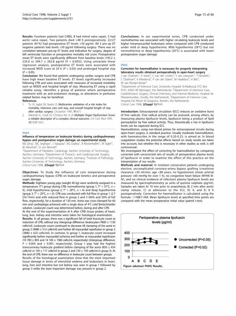
Results: Fourteen patients had CABG, 8 had mitral valve repair, 5 hadaortic valve repair. Two patients died >48 h postoperatively. 22/27patients (81%) had preoperative ET levels >50 pg/ml. All 5 pre-op ETnegative patients had levels >50 pg/ml following surgery. There was nocorrelation between pre-op ET levels and indication for surgery, degree ofleft ventricular function or preoperative mortality risk score. Postoperativemean ET levels were significantly different from baseline levels (195.2 ±224.0 vs 399.7 ± 282.8 pg/ml [P = 0.003]). Using univariate linearregression analysis, postoperative ET levels were associated withincreased MOD score at 24 h (P < 0.05) and prolonged hospital stay(P < 0.05).Conclusion: We found that patients undergoing cardiac surgery and CPBhave high mean baseline ET levels. ET levels significantly increasedfollowing CPB and were associated with measures of increased morbiditysuch as MODS and hospital length of stay. Measuring ET using a rapidreliable assay, identifies a group of patients where perioperativetreatment with an anti-endotoxin strategy, or alterations in perfusionrelated factors may be beneficial.References1. Tu JV, Jaglal SB, Naylor D: Multicentre validation of a risk index for
mortality, intensive care unit stay, and overall hospital length of stayafter cardiac surgery. Circulation 1995, 91:677.
2. Marshall JC, Cook DJ, Christou NV, et al: Multiple Organ Dysfunction Score:a reliable descriptor of a complex clinical outcome. Crit Care Med 1995,23:1638-1652.
P044Influence of temperature on leukocyte kinetics during cardiopulmonarybypass and postoperative organ damage: an experimental studyMa Qing1, MC Seghaye1, J Vazquez2, RG Grabitz1, B Klosterhalfen1, M Sigler3,BJ Messmer2, G von Bernuth11Department of Paediatric Cardiology, Aachen University of Technology,Aachen, Germany; 2Department of Thoracic- and Cardiovascular Surgery,Aachen University of Technology, Aachen, Germany; 3Institute of Pathology,Aachen University of Technology, Aachen, GermanyCritical Care 1998, 2(Suppl 1):P044
Objectives: To Study the influence of core temperature duringcardiopulmonary bypass (CPB) on leukocyte kinetics and perioperativeorgan damage.Methods: Eighteen young pigs (weight 40 kg) were randomly assigned to atemperature (T°) group during CPB: normothermia (group 1, T° = 37°C, n =6), mild hypothermia (group 2, T° = 28°C, n = 6) and deep hypothermia(group 3, T° = 20°C, n = 6). CPB was conducted with full flow in group 1 (2.7l/m2/min) and with reduced flow in group 2 and 3 (60% and 50% of fullflow, respectively), for a duration of 120 min. Aorta was cross-clamped for 60min and cardioplegia achieved with a single dose of 4°C cold Bretschneidersolution. Leukocyte count was determined before, during and after CPB.At the end of the experimentation (6 h after CPB) tissue probes of heart,lung, liver, kidney and intestine were taken for histological examination.Results: In all groups, there was a significant fall of total leucocyte count atinduction of CPB, without any intergroup difference (leukocytes=7800 ± 1150cells/ml). Leukocyte count continued to decrease till clamping of the aorta ingroup 2 (4980 ± 513 cells/ml) and further till myocardial reperfusion in group 3(3880 ± 625 cells/ml). In contrast, in group 1, leukocyte count increasedsignificantly before myocardial ischemia and further at myocardial reperfusion(10 700 ± 803 and 14 100 ± 1960 cells/ml, respectively) (intergroup difference:P < 0.005 and < 0.001, respectively). Group 1 pigs had the highesttranscoronary leukocyte gradient before clamping of the aorta (850 ± 454cells/ml vs 150 ± 117 cells/ml in group 2 and 150 ± 159 cells/ml in group 3). Atthe end of CPB, there was no difference in leukocyte count between groups.Results of the histological examination show that the most importanttissue damage in terms of interstitial oedema and leukostasis in heart,lung, liver and intestine but not kidney was seen in group 1 followed bygroup 3 while the least important damage was present in group 2.
Conclusions: In our experimental series, CPB conducted undernormothermia was associated with higher circulating leukocyte levels andhigher intramyocardial leukostasis when compared with CPB conductedunder mild or deep hypothermia. Mild hypothermia (28°C) but notnormothermia or deep hypothermia (20°C) is associated with lowerpostoperative organ damage.
P045Correction for haemodilution is necessary for properly interpretinglaboratory results obtained perioperatively in open-heart surgeryJ van Zwienen1, A Voets3, C van der Linden3, S van Leeuwen1, T Hendriks1,C Doelman2, K Miederna2, P van der Starre2, M Haalebos2, H Bilo2,W van Rooijen-Butijn21Department of Intensive Care, University Hospital St-Radboud, P.O. Box9101, 6500 HB Nijmegen, the Netherlands; 2Department of Intensive Care,Cardiothoracic Surgery, Clinical Chemistry, and Internal Medicine, Hospital deWeezenlunden, Zwolle, the Netherlands; 3Department of Intensive Care,Hospital De Wever & Gregorius, Ileerlen, the NetherlandsCritical Care 1998, 2(Suppl 1):P045
Introduction: Extracorporeal circulation (ECC) induces an oxidative burstof free radicals. Free radical activity can be assessed, among others, bymeasuring plasma lipofuscin levels, lipofuscin being a product of lipidperoxydation by free radical activity. Thus, theoretically a rise in lipofuscinlevels can be expected during ECC.Haemodilution, using non-blood primes for extracorporeal circuits duringopen-heart surgery, is standard practice. Usually moderate haemodilution,with haematocrites in the range of 0.20-0.25, is aimed at. In bloodcoagulation studies the potential effects hereof on study results are takeninto account, but whether this is necessary in other studies as well, is stillcontroversial.We investigated the effect of correcting for haemodilution by comparingcorrected with uncorrected sets of results of perioperative plasma levelsof lipofuscin in order to examine the effect of this practice on theinterpretation of our results.Methods and material: In nineteen consecutive patients undergoingelective, uncomplicated coronary-artery bypass grafting (creatinineclearance >50 ml/min, age <80 years, no hypotension (mean arterialpressure <60 mmHg for over 1 h), no congestive heart failure (NYHA III-IV), and no clinical evidence of infection) plasma lipofuscin levels aremeasured by spectrophotometry as units of quinine sulphate (pg/ml).Samples are taken A) 10 min prior to anaesthesia, B) 2 min after aorticclamp release, C) at admission to the ICU, D) 4, and E) 8 hpostoperatively. Correction for haemodilution is calculated using theformula : 1-HtB/1-HtA. Mean lipofuscin levels at specified time points arecompared with the mean preoperative initial value (paired t-test).
Figure (abstract P045) Results.
Critical Care 1998, Volume 2 Suppl 1http://ccforum.com/supplements/2/S1
Page 15 of 60
Discussion: Uncorrected plasma lipofuscin levels show no rise duringopen-heart surgery, whereas corrected values, which are all statisticallysignificantly higher in comparison to the initial preoperative value, do.This is in agreement with the theoretically expected, and thus offers theopportunity for correct interpretation.The above underscores the importance of correcting for haemodilution. Itis essential for a proper interpretation of perioperative laboratory resultsand consequent understanding of the pathophysiology of ECC in open-heart surgery.
P046Myocardial cell damage related to arterial switch operation in neonatesand the role of pro-inflammatory, cytokinesMC Seghaye1, RG Grabitz1, HH Hövels-Gürich1, J Duchateau3, S Däbritz2,BJ Messmer2, G von Bernuth11Departments of Paediatric Cardiology, Aachen University of Technology,Aachen, Germany; 2Thoracic- and Cardiovascular Surgery, Aachen Universityof Technology, Aachen, Germany; 3Department of Immunology, HôpitalBrugmann, Brussels, BelgiumCritical Care 1998, 2(Suppl 1):P046
Objectives: To investigate the role of myocardial depressant pro-inflammatory cytokines on myocardial cell damage related to arterialswitch operation (ASO) in neonates.Methods: 35 neonates (mean age 7 days) with transposition of the greatarteries undergoing ASO were prospectively studied. ASO was performedusing combined low-flow cardiopulmonary bypass (CPB) andcardiocirculatory arrest (CCA) under deep hypothermia. Patients withmyocardial hypocontractility at the end of the procedure or 4 and 24 hpostoperatively (po), as observed at echocardiography, were defined ashaving myocardial dysfunction (MD). Troponin-T (Tropo-T) and thecreatine kinase isoform CKMB served as markers for myocardial celldamage during and after ASO. Interleukine (IL)-6 (a myocardialdepressant cytokine) and IL-8 (involved in ischemia-reperfusion damages)served as marker for the systemic inflammatory reaction related to ASO.Results: Five patients showed MD after ASO, all patients survived. Age atASO, duration of CPB, CCA or aortic clamping were not different in patientswith and without MD. In all patients, plasma levels of Tropo-T increasedsignificantly during CPB (P < 0.0001). At the end of ASO, patients with MDhad significantly higher values of Tropo-T than patients without MD (P <0.002). In contrast to Tropo-T, levels of CKMB did not increase during CPBand were not different in patients with and without MD. IL6 and IL8increased significantly in all patients during CPB (P < 0.001, respectively).Patients with MD had significantly higher IL6 and IL8 values 4 h po thanpatients without MD (P < 0.001, respectively). Tropo-T- and IL6 valuesmeasured 4 h after ASO were significantly correlated to each other (P < 0.05).Conclusions: Our results demonstrate myocardial cell damage (as shownby Tropo-T liberation) and systemic inflammatory reaction (as shown byIL6- and IL8-production) related to ASO performed with combined low-flow CPB and CCA under deep hypothermia in neonates. Patients withMD have more important myocardial cell damage and higher magnitudeof systemic inflammatory reaction than have patients without thiscomplication. IL6 and IL8 both are possibly directly involved in thedamage to the myocardial cell in this setting.
P047A new model for evaluation of thrombosis and ischaemia/reperfusioninjuryHB Ravn, LB Ilkjaer, U Moeldrup, L Jensen, M Chew, S Johnsen,L Birk-Soerensen, E Tønnesen, VE HjortdalAarhus University Hospital, DK-8200 Aarhus N, DenmarkCritical Care 1998, 2(Suppl 1):P047
Introduction: Experimental studies on ischaemia/reperfusion injury havetraditionally focused on the isolated effect following simple occlusion ofthe nutrient artery. In myocardial infarction the reperfusion injury isfurther complicated by the presence of a thrombogenic area in thecoronary artery that may cause repetitive episodes of re-occlusion andembolization.
Methods: In eight pigs a thrombogenic area was created in the leftanterior descending artery (LAD) by exposure of adventitia into thelumen. The LAD was occluded proximal to the lesion for 50 min, followedby 4 h of reperfusion. Troponin-T (TNT), creatine kinase (CK), andcollagen-induced platelet aggregation (CPA) were measured 4 timesduring the experiment Indium-labeled platelets were given 30 min priorto harvesting of the hearts.Results: The infarct size/area at risk was 40 (35–63)% in the presentstudy. TNT and CK increased significantly to 1.7 (0.6–3.5) μg/l (P < 0.001)and 1480 (1105–2249) U/l (P < 0.02), respectively. Infarct size correlatedsignificantly with TNT-3h (r=0.85, P < 0.002), but not with CK-3h. Plateletaggregation decreased by 34% (P < 0.05) 15 min of reperfusion, butreturned to base-line. Platelet accumulation in the left ventricle wassignificantly higher in area at risk [194 (157–206)%] compared to area notat risk (100%), and to the right ventricle [137 (120–142)%]; (P < 0.05).Conclusion: In the present study Troponin-T showed a better correlationwith infarct size than CK. A decreased reactivity of circulating plateletswas observed after reperfusion and significantly more platelets werefound in the area at risk. These results indicate that activated plateletsbecome entrapped in the myocardium and may aggravate thereperfusion injury.
P048Treatment of hypercoagulation state in acute myocardial infarctionVM Kreines, SI Sharovarnikov, AI Kravchenko, VM Vlasov, VH Vaizov,IM UstyantsevaState Scientific and Clinical Center of Coal Miner’s Health Protection, 652509,Kemerovo region, Leninsk-Kuznesky, RussiaCritical Care 1998, 2(Suppl 1):P048
Full text: It is known that thrombolysis does not always prevent thedevelopment of early repeated thrombosis of coronary vessels. We havedeveloped a method of extracorporeal thermal plasmamodification —thermoplasmasorbtion (TPS), which allows to reach significant decrease ofits coagulation state (complete removal of fibrinogen, reduction of activityof the external and internal coagulation factors) and decrease of bloodviscosity. Is shown, that reinfusion of modificated plasma reduces plateletand red blood cells aggregation. 40 patients with myocardial infarction inacute period underwent the analysis. The TPS was applied to 20 patients(group 1) and 20 underwent conservative treatment (group 2). Duringclinical supervision more expressive positive dynamics changes were notedin all the patients of group 1: stenocardic cupping already in the 1st day oftreatment, hemodynamic stabilization, absence of thrombotic and othercomplications. The improvement of clinical state of the patients of thisgroup was proved by system and central hemodynamic rate: increase ofend diastolic volume, decrease of systole frequencies, increase of ejectionfraction with stable head ejection. In our opinion the given changes testifyto the improvement of heart work with inclusion of a rate thermalplasmosorbtion in complex treatment of acute myocardial infarction.
P049Artificial blood as a way to treat acute myocardial infarctionTA Pertseva, LV Usenko, OG CherkasovaDniepropetrovsk State Medical Academy, Dzerzynsky str. 9, Dniepropetrovsk320044, UkraineCritical Care 1998, 2(Suppl 1):P049
Introduction: Artificial blood–perfluorochemical emulsion Perftoran is a wayto improve oxygenation of the heart during periods of hypoxia and ischemia.This study was performed to determine the efficacy of an intravenousinfusion of Perftoran for patients with acute myocardial infarction.Methods: Forty patients, males not older then 60 with first acutemyocardial infarction (AMI) were observed, 20 to receive Perftoran in thefirst 6 h of AMI (1st group) and 20 to receive no Perftoran (2nd group),along with 20 mg of intravenous nitroglycerin given over 8 h. Minimaleffective dosage of Perftoran 4–5 ml/kg intravenous was used, infusion ratewas 20–40 drops per minute. We investigated composite clinical outcomes,ECG changes, infarct size, creatine kinase activity, hemodynamic parameters,acid-base status, blood gases, oxygen supply and consumption.
Critical Care 1998, Volume 2 Suppl 1http://ccforum.com/supplements/2/S1
Page 16 of 60

Results: On the first and following days of AMI the repeated episodes ofchest pain were observed in 81% cases in the second group and in 57%cases in the first group (P < 0.001). The patients of the first group also hadpositive ECG changes on the second day of the AMI: Development ofterminal T-wave inversion, ST-segment normalization and 2 patients had Q-wave reduction. In Perftoran-treated patients non-damaged volume of leftventricle (in % from the first damage) was 25.06 ± 5.40 (P < 0.05) comparedwith traditional-treated parients, 6.60 ± 2.31%. By the 3rd-5th days of AMI inthe first group there was observed an increase of arterial-venous O2 contentdifference, O2 delivery and 2 and more times than in the 2nd group.Conclusions: The use of Perftoran in patients with AMI performs apositive effect on clinical characteristics of AMI, leads to reduce infarctsize and decrease of ischemia in affected area.
P050Morbidity in the SICU after transmyocardial laser revascularizationC De La Cruz, J Cruz, A Castillo, E Corbacho, M Damas, J SarmientoSurgical Intensive Care Unit, ‘Virgen de la Victoria’ University Hospital,Campus Universitario Teatinos, Apartado 3091, 29010 Malaga, SpainCritical Care 1998, 2(Suppl 1):P050
Background: Transmyocardial laser revascularization (TMLR) is a moderntechnique of myocardial revascularization in patients with symptomaticcoronary artery disease that is refractory to maximal medical therapy andwho are unsuitable candidates for conventional methods of myocardialrevascularization [1-3]. There are no studies showing the clinical events ofthe early postoperative period.Objectives: To report the morbidity appeared in the surgical intensivecare unit (SICU) after TMLR.Methods: Description of the postoperative evolution in the SICU ofpatients who underwent TMLR between March and October 1997.Postoperative complications, clinical trendmarks and length of stay wererecorded. Data are presented as mean values (range).Results: TMLR was performed in eight patients, four men and four women,age 62 years (51-74), angina class 4. Cardiac events were: low cardiacoutput, 5; acute myocardial infarction, 1. Symptomatic angina, 1 and silentischemia, 1. One patient received mechanical ventilation during 17 daysbecause severe acute lung injury with a lung injury score (LIS) of 2.6;extubation was performed in the remaining 9.5 h (4-24) after admittancein the SICU, with a maximal LIS of 1.08 (0.5-2). Postoperative chest tubesdrainage ranged between 280 and 1890 ml. Thrombocytopenia was aconstant finding. No other complications were found and no patient died.The patient with prolonged mechanical ventilation stayed in the SICU 27days; length of stay was 3.7 days (1-6) in the remaining.Conclusions: Morbidity in the SICU after TMLP seems to be focused inthe cardiovascular, respiratory and hematological systems. Thesepreliminary findings need to be confirmed with other similar studies.References1. J Thorac Cardiovasc Surg 1996, 111:1047-1053.2. Circulation 1995, 92 (suppl II):1158-1165.3. J Am Coll Cardiol 1995, 25:258-263.
P051Intra-operative events do not predict perioperative myocardialinfarctionSK Appavu, R Haley, SR Patel, A KhorasaniDepartments of Surgery and Anesthesia, Cook County Hospital and theDepartment of Surgery, University of Illinois College of Medicine, Chicago,Illinois, USACritical Care 1998, 2(Suppl 1):P051
Full text: Perioperative myocardial infarction (PMI) is infrequent, but has highmortality rate. Therefore, a diagnostic work up to rule out PMI is usuallyperformed when cardiac or hemodynamic events occur during majoroperations. We performed this prospective study to determine whether or notthese intra operative events accurately predicted the development of PMI. Wehypothesized that they did. We studied patients who underwent majorsurgical procedures and a subsequent diagnostic work up to rule out PMIwhile they were in the recovery room. PMI work up included physical
assessment, countinuous ECG monitoring, and three 12 lead ECG and cardiacenzymes performed every 8 h. Data collection included patient demographics,diagnoses, pre-existing cardiac risk factors, type and duration of anesthesiaand surgery, intra operative cardiac and hemodynamic events, results of PMIwork up, incidence of PMI and patient outcome. Fifty-eight patients enteredthe study, 28 males and 30 females. Their mean age was 61.8 years.Preexisting cardiac conditions included angina in 5 patients, previous MI inanother 5, coronary artery bypass in 5, diabetes in 31, atherosclerotic arterialdisease in 26, smoking in 35 and hypertension in 42. The mean anesthesiaand operative times were 332.8 and 237.9 min, respectively. Intra operativeevents included hypotension in 20 patients, tachycardia in 15, hemorrhage in6, ECG changes in 3, hypertension in 8 and physician concern for PMI risk in 6.Only one of 58 patients (1.7%) developed PMI.Conclusion: Intra operative cardiac events alone are not very specificpredictors of perioperative myocardial infarction.
P052Painless acute myocardial infarction and diabetes mellitusŠ GrmecDepartment of Emergency Medicine, ZD Ptuj, Potrceva 19a, Ptuj 2250,SloveniaCritical Care 1998, 2(Suppl 1):P052
Full text: With diabetes mellitus, painless acute myocardial infarctus (PAMI) is awell-known clinical phenomen. Possible explanations include microangiopathy,diabetic neuropathy and abnormalities in myocardial metabolism in humans(hyperinsulinemia, hyperglycemia and insulin resistance).Various studies show that with diabetes mellitus approximately 25–50%of AMI presents with only slight pain or without pain. For emergencyprehospital services this feature plays an extremely important differentialdiagnostic role.This is a presentation of our clinical experiences from January 1995 toSeptember 1997. During this period, we dealt with 69 (28 female and 41male) cases of AMI with patients suffering from diabetes.All the patients had previously been registered at the regional diabetescenter and had undergone treatment: 22 (31.8%) with insulin therapy and47 (68.2%) with oral antidiabetes medicine. The average age of thepatients was 51.3 ± 5.8 years.All the patients underwent the ECG and enzyme analysis of AMI andpreinterventional blood glucose levels. Thirty-two patients had no cardiacpain, only non-specific symptomatology (nausea, vomiting, dyspnea,perspiration, tiredness, palpitations, non-specific precordial sensations) ora dominating clinical picture of hyperglycemia (ketoacidosis) orhypoglycemia. For 4 patients we were not able to obtain relevantanamnestic/heteroanamnestic data.Hyperglycemia (15.3–31.5 mmol/l; 20.61 ± 4.08) was verified in 43patients (62.3%), of them 5 cases with coma (ketoacidosis).Hypoglycemia (0.8–2.0 mmol/l; 1.47 ± 0.43) was registered in 11 patients(15.1%), of them 2 cases with coma.Euglycemia (3.52–6.37 mmol/l; 5.28 ± 0.82) was registered in 15 patients(22.6%).Conclusion: PAMI must to be excluded in the treatment of an emergencypatient for whom there is an anamnestic or heteroanamnestic record ofdiabetes. Treatment of ketoacidosis should be carried out promptly, becauseof the high mortality rate with already developed ketoacidosis-coma andAMI. It is vital that the differential diagnosis is carried out promptly andaccurately to ensure accurate and prompt therapeutic treatment
P053A new system for quantitative determination of troponin T andmyoglobin in the emergency room and in the intensive care unitU Waldenhofer, MM Hirsch, AN Laggner, HA Katus, M Müller-Bardorff,C Sylvén, G Rasmanis, PO Collinson, W Gerhardt, G Hafner, R Zerback,R LeinbergerBoehringer Mannheim GmbH, Sandhoferstr, 131, 68305 Mannheim, GermanyCritical Care 1998, 2(Suppl 1):P053
Full text: We present here the results of a multicentre evaluation ofa small system (Cardiac reader) for the rapid and quantitative
Critical Care 1998, Volume 2 Suppl 1http://ccforum.com/supplements/2/S1
Page 17 of 60

determination of troponin T (CARDIAC T Quantitative test) and myoglobin(CARDIAC M test) in heparinized venous whole blood (150 μl).The measuring unit of the Cardiac reader contains a CCDcamera that optically reads the signal and the control line of therespective immunochemical test strips. The tests have a reaction time of10 or 14 min. The quantitative measuring range for the troponin Tdetermination on the Cardiac reader is 0.1 to 3 μg/l and 30 to 700 μg/lfor CARDIAC M.The within-series imprecision was very acceptable for CARDIAC TQuantitative (CV 10 to 15%) and good for CARDIAC M (CV 5 to 10%).Compared with the Enzymun-Test Troponin T, the results obtained withCARDIAC T Quantitative show a high correlation (n = 40; r = 0.967;y = -0.006 + 1.093x). The method comparison between CARDIAC M andTinaquant® Myoglobin shows that both assays agree well (n = 82;r = -0.959; y = 3.6 + 0.976x).To assess the clinical efficiency, CARDIAC T Quantitative results of allfive study centres (481 samples of patients with suspected acutecoronary syndromes) were evaluated in a nine-field comparison tableversus Enzymun-Test Troponin T. More than 90% clinical concordancewere achieved with only 4 results that were clinically grossly discordant(0.8%); the corresponding Enzymun-Test Troponin T results weremoreover directly at the cut-off point (0.10 mg/l). Sensitivity andspecificity relative to Enzymun-Test Troponin T were 97 and 96%,respectively.With the Cardiac reader reliable quantitative results can be easilyobtained for both cardiac markers and entered into the hospital datasystem. Consequently, the Cardiac reader is especially suitable for use inemergency rooms and coronary care units.
P054Intravenous magnesium reduces infarct size following anischaemia/reperfusion injury combined with a thrombogenic lesionin the LADHB Ravn, LB Ilkjaer, U Moeldrup, L Jensen, M Chew, S Johnsen,L Birk-Soerensen, E Tønnesen, VE HjortdalAarhus University Hospital, DK-8200 Aarhus N, DenmarkCritical Care 1998, 2(Suppl 1):P054
Introduction: Experimental studies have demonstrated that magnesium(Mg) therapy can protect the ischemic myocardium and modulatereperfusion injury. In myocardial infarction the reperfusion injury isfurther complicated by the presence of a thrombogenic area in theaffected coronary artery that may lead to repetitive reocclusion andembolisation.Methods: We investigated the effect of Mg on infarct size in arandomised, blinded study in pigs inflicted with a thrombogenic lesion inthe left anterior descending artery (LAD) and mechanical occlusion of thevessel proximal to the lesion. Coronary occlusion was maintained for 50min followed by 4 h reperfusion. Magnesium sulphate (6 mmol/30 minfollowed by 3 mmol/h) or saline was given at 20 min of coronaryocclusion and continued for 4 h. 111Indium-labeled platelets were given30 min prior to harvesting of the hearts. Troponin-T (TNT) was measuredat base-line and 3 h into reperfusion.Results: The infarct size/area at risk (IS/AR) ratio in the placebo groupwas 40 (35–63)% (n = 8) compared with 16 (9–40)% (n = 6) in the Mgtreated animals (P < 0.05). Platelet accumulation in AR was reduced with15% in the Mg treated animals [placebo-group: 194 (157–205)% vs Mg-group: 166(131–213)%; NS]. Cellular leakage of TNT increased significantlyin both groups and was at 3 h highest in the placebo group: 1.7 (0.6–3.5μg/l compared to 0.3 (0.1–1.5) μg/l in the Mg-treated animals (NS).Conclusion: The present study demonstrates that iv Mg infusion is ableto reduce infarct size with more than 50% in this model where anischaemia/reperfusion injury was combined with endothelial damage inthe nutrient artery. The reduction in infarct size may be due tocomplementary actions on platelets and a cellular protection of themyocardium.
P055Remifentanil in cardiac anesthesia: influence on postoperative ICUcourseF Guarracino, A Bossi, M Tedesco, D De Cosmo, R De StefaniDepartment of Cardiothoracic Intensive Care, Umberto 1 Hospital, ViaCirconvallazione 50, 30171 Mestre (Ve), ItalyCritical Care 1998, 2(Suppl 1):P055
Introduction: Remifentanil (R) a new short-acting opioid, has beenintroduced in cardiac total intravenous anesthesia to solve the problemsconnected with the adminstration of long-acting opioids. Aim of ourstudy was to evaluate the efficacy of R in optimizing mechanicalventilation and ICU stay after cardiac surgery.Methods: We studied 30 consecutive pts, EF >40% scheduled for electivecoronary surgery. Pts were randomized in two different groups: Group R (16pts) receiving remifentanil, and group F treated with moderate dosefentanyl (14 pts). Induction of anesthesia was performed in both groupswith fentanyl 15 μg/kg per min, vecuronium 0.15 mg/kg and thiopentone0.5–1 mg/kg. In group R anesthesia was then maintained with continuousinfusion of propofol 1–3 mg/kg per h and remifentanil 0.5–1 μg/kg per minrunning throughout the surgical procedure. In group F the maintenance wasobtained with propofol 1–3 mg/kg per h and fenatyl 5–7 μg/kg per hrunning throughout the surgical procedure too. Propofol was continued atdose 0.5–1 mg/kg per h in the ICU until 30 min previous extubation in allpts. In adjunction pts of group R received remifentanil 0.1–0.2 μg/kg per minuntil extubation. In both groups we evaluated the duration of mechanicalventilation and of ICU stay. All data were analysed with t-test (P < 0.05).Results: Mean time of mechanical ventilation was 4 ± 1.3 h in group F vs9 ± 15 h in group R (P < 0.05). In group R mean ICU stay was shorterthan group F: 18 h vs 27 h (P < 0.05). Global ICU cost charge/pt was $930in group R vs $1390 in group F, according to DRG coefficients.Discussion: Our experience suggests that remifentanil, combined withpropofol, can improve early extubation and reduce ICU stay in uncomplicatedCABG pts with good EF, so leading to a trend in cost reduction.
P056Continuous infusion of propofol for sedation of pediatric patientsfollowing open-heart surgeryA Darwish, S Mousa, B Afrane, M El-Barbary, Z HaleesKing Faisal Specialist Hospital and Research Center, PO BOX 3354, Riyadh11211, Saudi ArabiaCritical Care 1998, 2(Suppl 1):P056
Introduction: The aim of this study was to prospectively evaluate safetyand efficacy of propofol infusion as an alternative sedative agent inchildren with open-heart surgery.Methods: Twenty-nine pediatric patients with complex congenital lesions,aged between 4 and 48 months, who underwent open-heart surgery,received propofol infusion as sedation after midazolam infusion wasineffective. The effective sedative dose of propofol was between 1.5 and7.0 mg/kg per h (median 3 mg/kg per h). Measurement of heart rate (HR)electrocardiogram (ECG), blood pressure (BP), arterial blood gas (ABG)including pH, bicarbonate (HCO3), partial carbon dioxide pressure (PCO2),and blood oxygen saturation (PO2); and blood chemistry including liverfunction tests (LFTs), triglycerides (TG), and total bilirubin (TB), were alldone prior to and during propofol therapy.
Table (abstract P056)
Parameter Change in median values P values
HR (beats/min) -10 0.009
BP (mmHg) -2 0.001
Triglyceride (mmol/l) 0.2 0.001
Critical Care 1998, Volume 2 Suppl 1http://ccforum.com/supplements/2/S1
Page 18 of 60
Results: After 10–288 h of propofol infusion (median 74 h), no statisticallysignificant differences were found in the parameters measured except adecrease in median HR with attendant decrease in BP, and an increase inmedian TG.Conclusion: Continuous sedation with propofol is a safe and effective,alternative agent for sedation of infants and children after surgical repairof compex congenital heart disease.
P057Gamma-hydroxybutyrate: an ‘old’ and very interesting drug: newperspectives?DR PichlmeierInstitut für Anesthesiologie, Technical University Munich, GermanyCritical Care 1998, 2(Suppl 1):P057
Background and goal of study: Due to the unique property of lackingcardiovasculary and respiratory depressive effects, GHB is a veryinteresting alternative for sedation of critically ill patients.Materials and methods: An epileptogenic effect described in someanimals after high doses of GHB has not been seen in 20 patientsanalysed with on-line EEG under bolus-application and 20 minafterwards. We also show that the long duration of the effect of asingle or repetitive dose of GHB can in fact be reversed by theapplication of Physostigmine.Results and discussions: No signs of cerebral hyperexcitability werefound in 20 patients receiving an anesthetic dosage of GHB. The problemof sodium-overload should not be an argument any longer as soon asthe newly developed, sodium-free drug is available. The third argumentagainst this drug, meaning the long and individually not calculable timeof effect, does also not seem to be a good argument against it’s use,since anaesthesia from GHB can be antagonized quickly by theapplication of physostigmine.Conclusions: GHB is a very interesting drug for anaesthesia andsedation on ICU due to its missing depressive cardiovasculary andrespiratory effects and many more aspects. So people should not allowthemselves not to use that drug on their patients for the three mostusually uttered reasons being sodium overload, cerebral convulsionsand long and uncalculable time of action. All these arguments seemuntrue.References1. Maitre M: Progr Neurobiol 1997, 51:337.2. Piehlmeier R, Schneck HJ: Intensiv- und Notfallbehandlung 1991, 16:106-112.
P058Rapid and safe intubation with rocuronium bromide in critically illpatientsMA Estecha1, JM Molina1, CA De la Cruz2, L Ruiz Del Fresno1, D Daga1,A García-Alcántara1, JL Carpintero11Intensive Care Unit, ‘Virgen de la Victoria’ University Hospital, CampusUniversitario Teatinos, Apartado 3091, 29010 Malaga, Spain; 2SurgicalIntensive Care Unit, ‘Virgen de la Victoria’ University Hospital, CampusUniversitario Teatinos, Apartado 3091, 29010 Malaga, SpainCritical Care 1998, 2(Suppl 1):P058
Background: Critically ill patients need sometimes orotraqueal intubationand mechanical ventilation. A muscle relaxant with rapid onset time,producing good intubation conditions is required. Succinylcholineprovides this goal, but it has several undesirable side effects. Rocuroniumbromide is a new muscle relaxant with an onset time shorter than otherbecause its low potency, allowing orotracheal intubation at 60–90 s,being free of cardiovascular and other side effects.Objectives: The aim of this study is to assess in a prospective manneronset time, and intubation conditions at 60 s, and side effects after adose of rocuronium bromide.
Methods: Patients aged 18–70 years admitted to the ICU who requiredmechanical ventilation, were included in this study. Neuromuscular diseasewas an exclusion criterian. After fentanyl 1 μg/kg and etomidate 0.2 mg/kg,patients received a bolus dose of 0.6 mg/kg of rocuronium bromide. Heartrate (HR), systolic arterial pressure (SAP), diastolic arterial pressure (DAP) andmean arterial pressure (MAP) were monitored before and afteradministration of bolus dose. Neuromuscular response was monitored witha peripheral nerve stimulator with two electrodes on the wrist. Train of four(TOF) was the mode chosen for stimulation and the number of responses atthe thumb was recorded by tactile evaluation. Intubation conditions wereassessed following previous published scores, in excellent, good, poor andimpossible. Data are presented as mean values; statistical analysis was madeby Wilcoxon test, considering P ≤ 0.05 as significance level.Results: Eight patients, mean age 54.5 years, three man and five women,were included in the study, all of them requiring mechanical ventilationbecause respiratory failure. Onset time of bolus dose of rocuroniumbromide was 3.2 min; maximal degree of blockade was 80% in onepatient and 100% in the remaining; and clinical duration was 24.2 min.Intubation conditions were excellent in six patients and good two ones.There were not significant changes in HR, SAP, DAP and MAP afterrocuronium regarding control values and no other types of side effectsappeared.Conclusions: Rocuronium bromide allows rapid and safe intubation undersuitable conditions in critically ill patients, without haemodynamic changes.
P059The use of continuous epidural anaesthesia in intensive care units inPolandA Szukiewicz1, F Mayzner-Zawadzka1, D Kosson 1, D Szukiewicz21Department of Anaesthesiology and Intensive Therapy, Warsaw UniversitySchool of Medicine, Poland; 2Chair of General and Experimental Pathology,Warsaw University School of Medicine, PolandCritical Care 1998, 2(Suppl 1):P059
Introduction: The aim of the study was to estimate the range of use ofepidural anaesthesia in Intensive Care Units. Our study is a part of a largequestionnaire surveillance study aimed at estimation of the widespreadof regional anaesthesia in Poland.Methods: The questionnaire was sent to 376 hospitals for adults, of apatients bed quantity of 250 and more, or specialised in orthopaedics,thoracic surgery, heart surgery or gynaecology and obstetrics. Head ofDepartment of Anaesthesiology was asked to answer over 75 questionsregarding the use of regional anaesthesia in his hospital. In regard ofepidural anaesthesia in Intensive Care questions included : the presenceof epidural anaesthesia, duration of catheter persist, use of cathetertunnelization technique, and the most frequent indications for epiduralanaesthesia application in Intensive Care Unit. General questionsincluded: the percentage of regional anaesthesia in all procedures, andfor how long time they perform regional anaesthesia.Results: We have obtained 118 responses (31.36%), 13 from universityhospitals, 70 from regional hospitals, 17 from district hospitals, and 18from specialised centres. Regional anaesthesia made from 3.6% to 83.3%(median value 21.4%) of all anaesthetic procedures. Application ofepidural anaesthesia in ICU was confirmed in 64 hospitals (54.24%respondents), in 10 hospitals the epidural anaesthesia was not performedat all, in 28 there wasn’t any Intensive Care Unit. The main indicationsreported were: acute pancreatitis, postoperative pain treatment, chesttrauma and severe trauma. Occasionally epidural anaesthesia was used inmyocardial infarction, asthma, vascular changes in lower extremities andin cancer patients. The mean time of use of catheter was 7 days.Tunnelization of the catheter was widely used in ICU practice.Conclusions: Epidural anaesthesia has a place in Intensive Care practicein Poland. The use of this technique is restricted by hospital habits andcosts. It is used mainly in acute pancreatitis, postoperative pain treatmentand trauma.
Critical Care 1998, Volume 2 Suppl 1http://ccforum.com/supplements/2/S1
Page 19 of 60
P060Electrocardiographic abnormalities in patients with cerebrovasculardiseaseMB Passavanti1, MA Tedesco2, F Massimo1, G Ratti2, G Di Salvo2, F Argenzio1,G Limongelli2, C Aurilio1, M Chiefari11Department of Anesthesia and Intensive Care, 2nd University of Naples,Piazza Miraglia 80138 Naples, Italy; 2Medical Surgical Institute Cardiology, 2ndUniversity of Naples, Piazza Miraglia 80138 Naples, ItalyCritical Care 1998, 2(Suppl 1):P060
Full text: Cerebral damage is frequently accompanied by ECG changesthat consist mostly in repolarization disturbance in patients withoutcoronary artery disease. Repolarization changes consist of sometimesdramatic ST segment elevations and inverted T waves in patients withnormal hearts. These alterations resemble those of ischemic heart diseaseand may cause diagnostic mistakes.We selected and studied 23 patients, 14 men and 9 women, mean age 63± 4 years, with recent stroke. Each patient underwent standard ECG,cardiac enzymes, M-mode and 2-D echocardiography and Doppler.On ECG, all patients presented inverted T waves and no alterations of STsegment. Cardiac enzymes were in the normal range and echocardiogramshowed no wall motion abnormalities. Only 13 patients underwentcoronary arteriography and all had normal coronary arteriograms.Our findings confirm previous reports that associate acute cerebralaccidents and repolarization abnormalities.There is little information regarding a relationship between specific strokelocation and disturbances in repolarization in humans, but there is substantialevidence that ‘catecholamine storm’, characterized by copious release ofnorepinephrine from cardiac b-1 receptor sites, during acute cerebral accidentsis responsible for myocardial damage reflected in sub-endocardial injury.
P061Sepsis: cause of late deterioration of intracranial pressure in patientswith severe traumatic brain injury?P Dostal, V Cerny, R ParizkovaDepartment of Anaesthesia and Intensive Care, Charles University, Faculty ofMedicine, 50005, Hradec Králové, Czech RepublicCritical Care 1998, 2(Suppl 1):P061
Introduction: Increased intracranial pressure (ICP) in patients with severetraumatic brain injury without findings of CT scan deterioration isfrequently observed late during the course of treatment. The aim of thisstudy was to evaluate the possible relationship between the late increaseof ICP in head injured patients and the development of sepsis duringtheir course on ICU.Methods: 22 consecutive patients (median age 18) admitted to the ICUwith severe traumatic brain injury (GCS < 8) and maintained according toa standard treatment protocol were retrospectively studied and dividedinto three groups. Group I (n = 6) were patients who developed nointracranial hypertension during their ICU stay, group II (n = 9) werepatients with early intracranial hypertension and group III (n = 7) werepatients who developed ICP deterioration on the 5th day or later afteradmission. Age, sex, initial GCS, incidence of secondary insults during first24 hours after the injury, CT finding on admission, incidence of sepsisduring the ICU stay, coincidence of sepsis with ICP deterioration,modified SOFA score (without neurological component) on the 2nd and7th day of ICU stay, day of ICP deterioration and mortality in the groupswere recorded. Fisher’s exact test, t-test, and one way ANOVA test wereused for statistical analysis (SigmaStat Statistical Software, Jandel Co.,USA), P < 0.05 was considered statistically significant.Results: No differences in age, sex, initial GCS, modified SOFA score onthe 2nd day and incidence of secondary insults during first 24 h after theinjury among groups were observed The overall incidence of sepsis was33% (7/22 patients) and mortality rate was 22.2% (5/22 patients). The lateintracranial hypertension was more frequently associated with thedevelopment of sepsis in group III (coincidence of sepsis and ICPdeterioration in 1/9 patients in group II and 5/7 patients in group III, P =0.035). Subsequent analysis in group III showed tendency to ICPdeterioration in patients with sepsis compared to patients without sepsis
(5/6 septic patients developed ICP deterioration compared to 2/5nonseptic patients with ICP deterioration) but the difference did notreach statistical significance (P = 0.165).Discussion: Frequent coincidence of late ICP deterioration and sepsis inpatients with severe traumatic brain injury was observed. Late ICPdeterioration could be in some cases the result of sepsis inducedchanges in vascular reactivity due to overproduction of NO and othervasodilatatory mediators.Acknowledgement: Partially supported by grant IGA MZ CR 3672-3.
P062Determinants of natriuretic peptides in early septic shockR Witthaut1, Ch Busch1, P Fraunberger2, A Walli2, D Seidel2, G Pilz2, K Werdan11Chair of Cardiac Intensive Care, Kröllwitz Clinic, Martin-Luther UniversityHalle-Wittenberg, Ernst-Grube-Str. 40, 06097 Halle, Germany; 2Institute ofClinical Chemistry, Ludwig-Maximilians University Munich, Marchioninistr.15,81377 Munich, GermanyCritical Care 1998, 2(Suppl 1):P062
Background and objectives: Circulating natriuretic peptides (atrialnatriuretic peptide, brain natriurelic peptide) are sensitive indicators ofthe severity of heart failure. Comparable high plasma levels of natriureticpeptides has been found in severe heart failure (NYHA III-IV) and patientswith septic shock (Am Heart J 1993, 126:466–468). Impaired heartfunction, changes of cardiopulmonary hemodynamics andproinflammatory cytokines, ie interleukin-6 (IL-6) or tumor-necrosis-factor-a (TNF-a) might contribute to the modulation of natriuretic peptiderelease (Am Heart J 1997, 79:1128–1131). Therefore, we studied theinterrelation between IL-6, soluble TNF-receptors (TNF-R-p55, TNF-R-p75),atrial natriuretic peptide (ANP) and brain natriuretic peptide (BNP) as wellas heart function, cardiopulmonary and systemic hemodynamics inpatients at the first two days following the diagnosis of septic shock.Methods: Patients without acute renal failure (ARF) fullfilling criteriaof septic shock (APACHE 28.4 ± 16.9; ELEBUTE 19.2 ± 4.2; n = 17).Determination of IL-6, TNF-R-p55, TNF-R-p75, ANP and BNP in plasma as wellas cardiac index (CI), mean arterial pressure (MAP), right atrial pressure (RA),heart rate (HR), systemic vascular resistance (SVR) and mean pulmonaryarterial pressure (PAP). Mean ± SD; r2 = Pearsons correlation coefficient.Results: IL-6 was significantly correlated to ANP (P < 0.01). During thefirst 2 days following diagnosis, IL-6 and ANP concordingly decreased(4033 ± 5657 vs 1978 ± 4751 pg/ml and 28.3 ± 16.6 vs 23.3 ± 15.1 pg/ml, respectively). BNP remained unchanged (12.4 ± 15.7 vs 12.8 ± 17.2pg/ml), on both days inversely correlated to CI (P < 0.05). Accordingly, nosignificant differences of CI (4.5 ± 0.9 vs. 4.5 ± 1.0 l/min/m2) or SVR (533.6± 136.9 vs 595.7 ± 210.2 dyn × s × cm-5) could be determined. The fall ofANP from day 1 to 2 was independent of changes in RA (11.2 ± 4.2 vs.11.8 ± 3.5 mmHg), HR (136.8 ± 27.2 vs. 142.4 vs. 62.6 bpm) or PAP (27.0± 6.6 vs 28.3 ± 4.6 mmHg). There was no significant change from day 1to 2 of TNF-R-p55 (7.6 ± 3.5 vs 8.3 ± 6.0 pg/ml) and TNF-R-75 (11.0 ± 6.2vs 12.9 ± 8.6 pg/ml). On day 2, ANP and BNP significantly correlated to CI(r2 = -0.682 and r2 = -0.574, respectively; P < 0.05), while CI inverselydepend on SVR (r2 = -0.801; P < 0.05). No significant negative correlationcould be calculated between ANP, BNP and SVR or MAP.Conclusions: Patients in early septic shock without ARF showed a directlycorrelated, concordant decrease of IL-6 and ANP independent of changes inRA, HR, PAP or CI. Vasodilative BNP release was inversely correlated to CI.Thus, interleukin-6 and left ventricular heart function might play a differentrole in the regulation of ANP- and BNP release in early septic shock.
P063Prolonged hypernatremia safely controls elevated intracranial pressurein pediatric head injury patientsS Khanna, B Fisher, B PetersonChildren’s Hospital, Division of Pediatric Critical Care Medicine, Suite 115MOB, 3030 Children’s Way, San Diego, CA 92123, USACritical Care 1998, 2(Suppl 1):P063
Introduction: The standard treatment of elevated intracranial pressure (ICP)includes the administration of hyperosmotic solutions. Osmolar diuretics
Critical Care 1998, Volume 2 Suppl 1http://ccforum.com/supplements/2/S1
Page 20 of 60

may lead to intravascular dehydration, hypotension, pre-renal azotemia andreduction of cerebral blood flow. Hypertonic saline solutions effectivelyreduce raised intracranial pressure in animals and humans withoutproducing intravascular dehydration. We evaluated the degree ofintracranial pressure control induced by the administration of 3% hypertonicsaline (508 mOsms/I NaCl) in pediatric patients after traumatic brain injury.Methods: We retrospectively reviewed the charts of 68 children whosuffered traumatic brain injury with intracranial hypertension (ICP >20mmHg). These patients received the conventional modalities for thetreatment of intracranial hypertension including elevation of head (15°),pain control, hyperventilation, mannitol and sodium thiopentothaladministration. In addition, they received continuous 3% saline infusionsto reduce intracranial pressure below 20 mmHg. Hypertonic salineinfusions were continuous and ranged between 2–7 days dependingupon the patient ’s requirement for control of raised intracranialpressure.Results: The mean dose of 3% saline was 17.23 ± 5 ml/kg/day. Thehighest, lowest and average serum sodium was 182, 140 and 158 ± 28mEq/l respectively. The mean daily dose of mannitol was 2 ± 0.65 gm/kg/day. The highest, lowest and average serum osmolarity was 380, 276and 316 ± 39 mOsm/l respectively. The mean serum creatinine was 0.7± 0.3 mg/dl. The intracranial pressure averaged 20 mmHg 92% of thetime, 20–30 mmHg 7% and greater than 30 mmHg 1% of the timeduring the first seven days post injury. Ten (15%) of 68 patientsexpired. However only two (3%) children died of uncontrolledintracranial hypertension.Conclusions: We conclude that administration of 3% hypertonic saline topediatric brain injured patients with elevated intracranial pressure inconjunction with standard treatment modalities results in effectivecontrol of intracranial pressure. Hypernatremia and hyperosmolarity aresafely tolerated in brain injured pediatric patients.
P064Disorders of the effective osmolality in central nervous system injuryM Balík1, A Kazda21Department of Anesthesiology and Resuscitation, University Hospital, Unemocnice 2, 12808 Prague 2, Czech Republic; 2Department of ClinicalBiochemistry, Med. Faculty-Policlinics, Karlovo nám. 32, 121 II Prague 2,Czech RepublicCritical Care 1998, 2(Suppl 1):P064
Full text: Disorders of the effective osmolality regulation are frequentin the cerebral affections that originate from trauma, vascular disease,imflamation or tumors. Hypo-osmolality and hyponatremia arepresented in two different states: Inappropriate Vaseopressin SecretionSyndrome (IAHDS) and Cerebral Salt Wasting Syndrome (CSWS). Quickdifferential diagnose is important because the treatment of bothsyndromes is essentially different. Typical cause of hypernatremia iscentral diabetes insipidus (DI). The group of available calculated renalfunction parameters is applied in the differential diagnosis of thesesyndromes. They are creatinine clearance, excretion fraction of waterand sodium, electrolyte clearance and electrolyte free water clearance.Investigation of ADH and natriuretic pepride could be evenmisleading. Pathophysiologic consequence of the state given byinappropriate elevation of one hormone can be the elevation of thesecond one.The mentioned topic is documented with the four selected patients fromthe ICU. The syndromes were diagnosed using the computer programevaluating renal functions by means of 13 routinely monitored valuesand 12 output parameters.Authors present brief clinical courses, biochemical results and calculatedrenal function parameters by one patient with the IADHS, by one with DI,by one with CSWS passing over to DI and by one with DI passing toIADHS. The importance of complex access to evaluation of renalfunctional parameters is stressed. These are influenced by actual therapy,eg by diuretics or osmotherapy.Conclusions: Mentioned four cases are the example of the effective useof the currently investigated values in the diagnosis and monitoring ofthe effective osmolality disorders in cerebral affections.
P065Perfusion pressure changes after ANP administration in preeclampsia:in vitro studyD Szukiewicz1, A Szukiewicz2, D Maslinska 1, M Markowski31Dept. of General & Experimental Pathology, 2nd Faculty of Medicine,University School of Medicine, Warsaw, Poland; 2Dept. of Anaesthesia &Intensive Therapy, 2nd Faculty of Medicine, University School of Medicine,Warsaw, Poland; 3Dept. of Obstetrics & Gynaecology, 2nd Faculty ofMedicine, University School of Medicine, Warsaw, PolandCritical Care 1998, 2(Suppl 1):P065
Introduction: Regulation of the blood flow in human placental tissue bychanges of the vascular resistance is very particular, because of lack ofsympathetic innervation in placental vessels [1]. The number ofguanylate-coupled receptors in placental (issues, corresponding to ANP-receptors, has been found to be greater in preeclampsia, than in normalpregnancy [2]. The aim of the study was to investigate the influence ofvarious doses of atrial natriuretic peptide (ANP) on perfusion pressure inpreeclampsia.Methods: Heparinized placentas (n = 14) obtained after preeclamp tic(n = 7; 1st group) and normal (n = 7; 2nd group, control) pregnanciesdelivered at term by cesarean sections were perfused in vitro usingmodified Cedard manner. Perfusion pressure, measured continuously, wasthe main parameter of the vascular status changes. The placental vesselswere submaximally preconstricted by continuous infusion of the nitric-oxide synthase inhibitor N-?)-nitro-L-arginine (NOLA; 100 µmol/l, 10 min).Over 120 min depending on the period of experiment (30, 45, 60, 75min) human synthetic aANP were administered (25, 50, 100, 200 nmol/l, 2min increments).Results: Mean value of perfusion pressure increase after NOLA infusionwas 5.63 kPa (± SD 0,36), the differences between groups I and II did notreach statistical significance.Conclusion: The responsiveness of the fetoplacental vasculature to theANP given in higher doses (100, 200 nmol/l, 2 min increments) isincreased in preeclampsia (P < 0.05).References1. Am J Obstet Gynecol 1993, 168:1616.2. Am J Obstet Gynecol 1994, 170:142-147.
P066Management of subarachnoid hemorrhage patients: is early surgerybetter than delayed?V ŠvigeljDept. of Neurology, University Medical Centre, SI-1525 Ljubljana, SloveniaCritical Care 1998, 2(Suppl 1):P066
Full text: There is no general consensus with regard to management ofsubarachnoid hemorrhage (SAH) patients. Early surgery has beenadvocated in order to minimize the rebleed risk, however it is morehazardous than delayed surgery, because the brain is tight and swollen,and the risk of vasospasm and infarction is increased.The purpose of this retrospective study is to present our experiences inmanagement of SAH patients. Between 1990–1995, 196 (59.4%) femaleand 134 (40.6%) male patients were admitted to our unit due to suddenonset of a headache. Subarachnoid hemorrhage was proven by CT scanand/or lumbar puncture and in 161 (48.8%) patients, three-vesselsangiography showed one or more aneurysms. The conservatively treatedgroup of SAH patients was statistically older than surgically treated group(P = 0.002). There was no statistically significant difference regarding agebetween male and female patients in both treated groups. Average
Table (abstract P065) Perfusion pressure decrease (kPa)after a ANP administration (mean values ± SD)
Group 30-32 min 45-47 min 60-62 min 75-77 min
I Preeclampsia 1.01 ± 0.17 1.49 ± 0.22 1.99 ± 0.23 2.33 ± 0.39
II Normal pregnancy 0.94 ± 0.2 1.22 ± 0.19 1.43 ± 0.26 1.79 ± 0.24
Critical Care 1998, Volume 2 Suppl 1http://ccforum.com/supplements/2/S1
Page 21 of 60

timing of aneurysm surgery was 73.9 ± 75.5 h after the onset of bleeding(range: 4–550 h).All but 20 patients (all conservatively treated) received nimodipine toprevent or treat vasospasm. In 14 (4.2%) patients vasospasm was seenon angiograms and 34 (21.1%) patients developed clinically significantvasospasm postoperatively between days 3–5. Mortality rate inconservatively treated group was 23.1%, and in surgically treatedgroup 5%.We conclude that early surgery (between days 1–3) is better, however itis more hazardous due to higher risk of vasospasm. Rebleed was themajor problem in conservatively treated group of SAH patients, but overall outcome was better.
P067Low albuminemia on vasospasm following subarachinoid hemorrhage(SAH)K Ikeda1, T Ikeda1, M Usuda1, N Ishii1, S Nakajima2, T Fukuda2, M Hasue21Department of Anesthesiology, Hachioji Medical Center of Tokyo MedicalCollege, Japan; 2Department of Neurosurgery, Hachioji Medical Center ofTokyo Medical College, JapanCritical Care 1998, 2(Suppl 1):P067
Introduction: The aim of this study was to determine the mechanism oflow albuminemia on vasospasm following subarachnoid hemorrhage(SAH).Patients and methods: We evaluated the changes in serum and urinaryNAG, and renin-angiotensin substances (RAS) [plasma renin activity (PRA),angiotensin I and II] on day 1, 3, 7 and 14 following SAH. The subjectswere 43 patients with SAH, who were classified as Group 1 [vasospasm (+)]and Group 2 [vasospasm (-)] according to the angiography findingsbetween days 5 to 7 after the clipping operation. We performedhypervolemic and hypertensive therapy from day 3 to prevent vasospasm.Results: We observed a significant decrease in serum albumin, and anincrease in urine albumin and NAG in both groups from day 3, and asignificantly higher amount of urinary albumin and NAG in Group 1 incomparison with data in Group 2. We also observed a significant increaseof PRA and angiotensin I in both groups from day 3, and a significantlyhigher amount was observed in Group 1 than in Group 2. A significantincrease in angiotensin II was observed in both groups on day 3, but wedid not observe any significant difference between the two groups. Allthe RAS data was within the normal range.Conclusions: An increase in renal concentrations of NAG is recognized asrenal tubular dysfunction in vasospasm Group 1. We concluded thathigher concentrations of urinary albumin related to renal tubulardysfunction is one of the causes of low albuminemia on vasospasm.
P068Monitoring of hypervolemic hemodilution and hypertensive (HHH)therapy in subarachnoid hemorrhage (SAH) patients with pulmonaryartery catheter (PAC)JL Nates2, M Jauss1, S Singh2, D Krieger21Dept. of Psychiatry, University of Heidelberg, Heidelberg, Germany;2Departments of Anesthesiology-CCM and Neurological Sciences, 6431Fannin Street, MSB 5.020, University of Texas Health Sciences Center-Houston, TX, USA 77030Critical Care 1998, 2(Suppl 1):P068
Background: Cerebral arterial vasospasm continues to be a majorcomplication of aneurysmal SAH. Prophylactic HHH therapy may bebeneficial reducing delayed ischemia after early aneurysm clipping.Minimal standards of monitoring are not well defined.The aim of our study was to evaluate the contribution of PAC andcranscranial Doppler (TCD) monitoring during HHH-therapy in patientswith SAH after early operative intervention.Methods: We observed 37 ICU patients with SAH during HHH therapy. Allpatients received hypervolemic hemodilution therapy aiming for ahematocrit of 33–38%, 10–12 mmHg central venous pressure (or 15–18mmHg pulmonary wedge pressure), and 160–200 mmHg systolic arterial
pressure during the risk period for vasospasm. We evaluated clinicalfindings, PAC values and mean flow velocity (MFV) on both middlecerebral arteries (MCA).Results: Outcome correlated with clinical state on admission (Fisher-exact test, P < 0.05). A constantly elevated systemic vascular resistance(SVR) was associated with a good outcome while declining SVR duringclinical course with a poor outcome (analysis of variance for repeatedmeasurements, P < 0.05). A successful HHH therapy according to theabove hemodynamic criteria did not reduce occurrence of vasospasm(Fisher-exact test). In patients without vasospasm (MFV ≤ 140 cm/s, n =16), we were able to develop a multiple regression model thatexplained 44% of variance of MFV by consideration of SVRI andhematocrit.Conclusion: SVRI monitoring and manipulation using PAC during HHHtherapy may affect outcome positively. An association of MFV with SVRIand hematocrit could be demonstrated.
P069Diagnostics and treatment algorythms of acute brain injury periodVV Agadganjan, SA Kravtsov, VM Kreines, AV NovokshonovState Scientific and Clinical Center of Coal Miners’ Health Protection,Leninsk-Kuznetsky, Russia, 652509Critical Care 1998, 2(Suppl 1):P069
Full text: On the basis experience in 137 patients treatment weelaborated working classification of acute brain injury period which madeit possible to compose diagnostic and intensive care algorithms. Theclassification is based upon the determination of main pathophysiologicalprocesses characterizing the duration of the acute injury period andtakes into account stress reaction, primary reperfusion, cerebraledema, recurring reperfusion, adaptation as well as the display of theprocesses upon the evaluation of patient state severity (compensated,subcompensated, decompensated, terminal).Patient state of severity was evaluated on the basis of remainingcapacities of the vitally important patient organs and systems (centralnervous system according to Glasgo coma scale, vegetative nervoussystem state — cardiointervalography, cardiovascular system, respiratorysystem, homeostasis systems — by Appache II scale). The classificationswe present takes into account the dynamic complex pathophysiologicalprocesses taking place in patient organism, and gives an opportunity offast change in main accents of intensive care. Diagnostics and treatmentalgorithms on the basis of the present classification with the othermodern methods of surgical treatment allowed to improve theposttreatment outcomes, to reduce early (till 10 days) lethality in theacute brain injury period by 8%.
P070Cranial computed tomography in the emergency evaluation of adultpatients without a recent history of head trauma: a prospectiveanalysisAJ Sproesser, SG Rothrock, J FalkDepartment of Emergency Medicine, Orlando Regional Medical Center, 86W. Underwood, Ste. 201. Orlando, Florida 32806, USACritical Care 1998, 2(Suppl 1):P070
Objectives: To examine the pattern of nontrauma cranial CT use in anurban ED, to identify the rate of significant CT abnormalities in headache,and to develop criteria for restricting the ordering of CT scans.Methods: A prospective, observational study of a case series of adultswho underwent cranial CT scanning for non-traumatic headache wasperformed at the ED of an urban teaching hospital with an annual censusof 55 000. Clinically significant CT scans were defined as: 1) acute stroke,2) CNS malignancy, 3) acute hydrocephalus, 4) intracranial bleeding or 5)intracranial infection. X2 recursive partioning was used to derive adecision rule to restrict ordering of CT scans.Results: Only 12 (4%) of 291 CT scans revealed clinically significantabnormalities. The presence of headache with vomiting was 100%sensitive (95%CI: 74–100%) and 49% specific (95%CI: 43–56%) in
Critical Care 1998, Volume 2 Suppl 1http://ccforum.com/supplements/2/S1
Page 22 of 60
detecting clinically significant CT scans. This set of features had positiveand negative values of 8%(95%CI: 4–13%) and 100% (95% CI: 97–100%),respectively.Conclusion: Clinically significant CT abnormalities were uncommon in theheadache patients population, suggesting that current criteria forordering nontrauma cranial CT scans may be too liberal. In this study, aset of clinical criteria was derived that may be useful at separatingpatients into high- and low-risk categories for clinically significant cranialCT abnormalities. Before these results are applied clinically, these criteriashould be validated in larger, prospective studies.
P071Cerebral blood flow velocities in hypoxemic patientsF Chiappini1, L Fuso1, G Pagliari1, M Visocchi2, B Cioni2, M Meglio21Department of Respiratory Physiology, Catholic University Rome, Italy;2Department of Neurosurgery, Catholic University Rome, ItalyCritical Care 1998, 2(Suppl 1):P071
Full text: According to the literature, in normocapnic hypoxia cerebralblood flow (CBF) increases in order to maintain a steady oxygen (O2)delivery as well as to prevent a significant drop in cerebral venous O2
tension. The aim of the present study is to investigate transcranialDoppler (TCD) velocitometric patterns in hypoxemic patients. CBFvelocity was evaluated before and after inhalation of an O2 enrichedmixture in order to detect differences in O2 delivery when chronichypoxia was corrected. Eight patients (mean age 71.4 ± 2.2 yrs) withrespiratory failure (RF) (hypoxemia without hypercapnia), were studiedbefore starting a long-term oxygen therapy. None had a history ofcardiovascular and/or hematologic diseases. A group of 8 healthysubjects, matched for age and gender, were also studied as referencegroup. Both patients with RF and normal subjects underwent a CBFvelocity measurement, in supine position, by using TCD (Quantascope,Toshiba T2100, Vital Science, Amsterdam, The Netherlands) both inbasal conditions (while breathing room air) and during the inhalation ofan O2 enriched mixture through a Venturi mask for at least 30 min.Heart rate was recorded and arterial blood gases were measured with aemogasanalyzer ABL-330 (Radiometer, Copenhagen) before theassessment of CBF velocity both in basal conditions and during theinhalation of the O2 enriched mixture. A significant increase of PaO2
and SaO2 during the inhalation of the O2 enriched mixture incomparison with basal conditions was found both in patients with RF(PaO2: 54.1 vs 84.9 mmHg, P = 0.004; SaO2: 86.6 vs 95.8%, P = 0.003)and in normal subjects (PaO2: 82.9 vs 112.2 mmHg, P = 0.0004; SaO2:95.6 vs 97.8%, P = 0.003). The CBF velocities decreased in both groups(32.2 vs 27.9 cm/s, P = 0.011 in RF patients; 29.8 vs 19.9 cm/s, P =0.0003, in normal subjects). However, the reduction of CBF velocitieswas lower in RF patients in comparison with the normal subjects (-4.3 ±3.5 vs -10 ± 4.4 cm/s, respectively, P = 0.012).In conclusion, the lower decrease of the CBF velocities in RF patientsduring the inhalation of an O2 enriched mixture seems to indicate thatchronic hypoxia induces a quite maximal vasodilatation to protect CBFand, thus, to maintain an adequate O2 delivery to the brain.
P072Use of single photon emission computed tomography (SPECT) in thefollow-up of carbon monoxide poisoned patients treated with HBOtherapy: a case reportM Maione, L Socciarelli, S Falini, G Scalmani, G Poggi, M BrauzziHyperbaric Unit, P.O. Misericordia, Via Senese, 58100 Grosseto, ItalyCritical Care 1998, 2(Suppl 1):P072
Full text: It is widely known that carbon monoxide (CO) has the toxiceffects of tissue hypoxia and produces acute neurologic deficits and asevere neurologic reaction may be delayed for days or week after anoxicexposure. The prediction of outcome during the acute stages or thelatest period is difficult in most cases because of variations in age,previous state of health, duration and severity of exposure, individualsusceptibility and manner of treatment.
The initial laboratory findings do not provide any prognostic clues andattempts at further predicting the clinical outcome of acute CO poisoningby means of brain CT scan or other tests have remained unsuccessful.SPECT provides tomographic images of cerebral perfusion and it has beenused in clinical practice.We experienced two cases of CO poisoning in which SPECT was performed1 month later from acute poisoning; they had different outcome and theclinical situation was directly related to SPECT imaging results.The patients were a mother and her son, age 55 and 25 years, who wereaccidentally poisoned by a domestic bath heater. They were foundunconscious at home and their GCS (Glasgow Coma Score) upon theirarrival in the hospital was 3. Both patients were referred to our facilityand improved greatly after HBO treatment (90 min at 2.8 ATA and 30min at 1.9 ATA); the younger was almost totally awake and the oldertook a little bit longer period for regaining consciousness. The MRI inboth cases showed edema and necrosis in both pallidi globi. Clinically,the younger recovered fully and the older had mild neurologic sequelaeas ataxic walking and light dizziness, the last one disappeared in fewdays. They were treated for a total of 20 sessions of HBO at 2 ATA for 60min.The SPECT brain imaging was totally negative for the younger andshowed a bilateral decrease of blood flow in the subcortical motor nucleiin the older patient.We can conclude that SPECT imaging is more sensitive and more directlyrelated with the clinical picture of the patient than MRI and we arestrongly convinced to use this diagnostic test in all cases of severecarbon monoxide poisoning.
P073The effects of diaspirin crosslinked hemoglobin (DCLHb) onoxygenation, perfusion and resuscitation: preclinical experienceK Burhop1, C Ince2, A Gulati3, D Malcolm4
1Baxter Healthcare Corporation, 25212 West State Route 120 WG2-3S, RoundLate, IL 60073-9799, USA; 2Amsterdam Medical Center, Amsterdam, theNetherlands; 3University of Illinois-Chicago, Chicago, Illinois, USA; 4USUHS,Bethesda, Maryland, USACritical Care 1998, 2(Suppl 1):P073
Full text: Preclinical studies of DCLHb, a highly solution of stabilizedhuman hemoglobin tetramers, in animals with normovolemichemorrhagic shock have demonstrated its ability to quickly restore meanarterial blood pressure, base deficit, and subcutaneous and mucosal pO2
to baseline levels or higher. In addition, DCLHb has been shown to becapable of preserving normal gut architecture, decreasing bacterialtranslocation, increasing blood flow to key tissues and organs, anddecreasing mortality in some models.Administration of DCLHb to hemorrhaged animals may serve to create asmall shift in blood flow from the muscle, resulting in a large increase inflow to vital organs. This shift may have a significant beneficial effect incertain indications, such as shock, and result in maintenance of vitalorgan perfusion. A further study of DCLHb as a low-volume resuscitationagent in severe hypovolemic, hemorrhagic shock in pigs demonstratedthe agent’s ability to improve gut microvascular oxygenation. Inthis study, palladium porphyrine phosphorescence was used as a markerof microvascular pO2, the results indicated that DCLHb restoresmicrovascular oxygenation to pre-hemorrhagic levels.The studies outlined above and others have shown the ability of DCLHbto maintain vital organ perfusion consistently. This property ofDCLHb may be explained in part by examining its pharmacologicalcharacteristics. For example, DCLHb has a pressor effect, involving anumber of different autocrine systems in the body, including interactionswith nitric oxide, endothelin, and the a-adrenergic system. DCLHbappears to work through multiple endogenous systems and mechanisms,each of which probably interact.In summary, studies in animal models of hemorrhagic shock indicate theDCLHb has oxygen-carrying capabilities similar to fresh blood and has thepotential to treat blood loss and ischemic situations, such as hemorrhagicshock, in humans. Trials are underway to investigate the safety andefficacy of DCLHb in such patient populations.
Critical Care 1998, Volume 2 Suppl 1http://ccforum.com/supplements/2/S1
Page 23 of 60
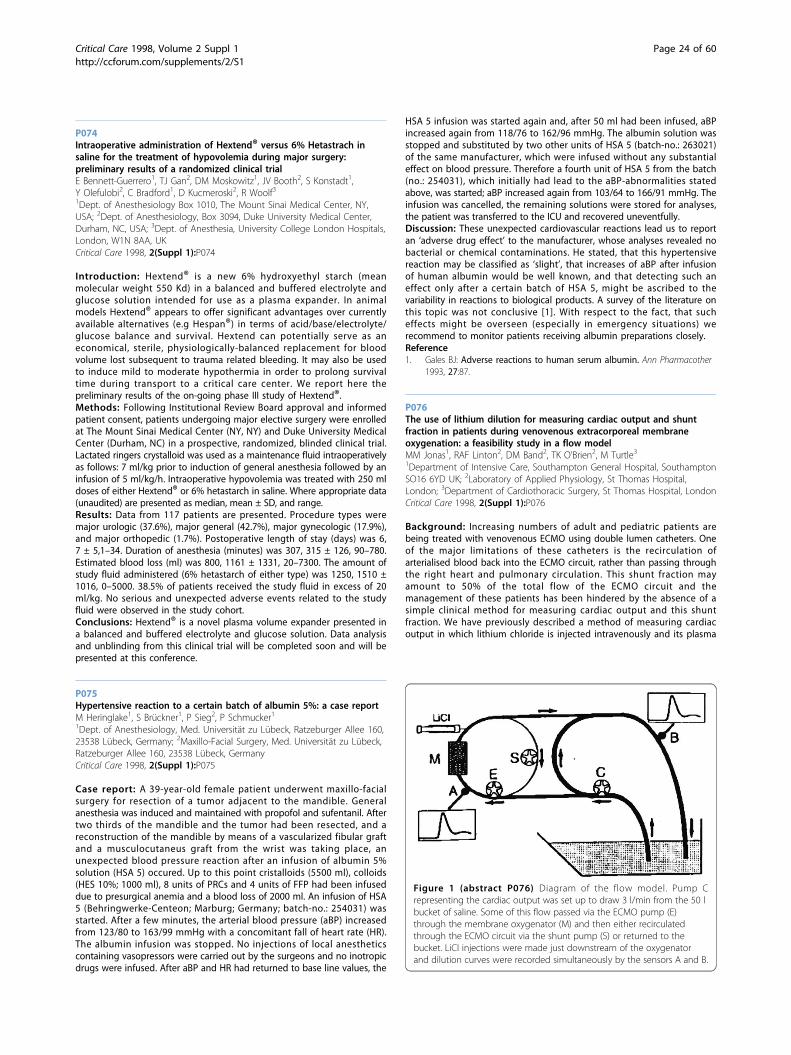
P074Intraoperative administration of Hextend® versus 6% Hetastrach insaline for the treatment of hypovolemia during major surgery:preliminary results of a randomized clinical trialE Bennett-Guerrero1, TJ Gan2, DM Moskowitz1, JV Booth2, S Konstadt1,Y Olefulobi2, C Bradford1, D Kucmeroski2, R Woolf31Dept. of Anesthesiology Box 1010, The Mount Sinai Medical Center, NY,USA; 2Dept. of Anesthesiology, Box 3094, Duke University Medical Center,Durham, NC, USA; 3Dept. of Anesthesia, University College London Hospitals,London, W1N 8AA, UKCritical Care 1998, 2(Suppl 1):P074
Introduction: Hextend® is a new 6% hydroxyethyl starch (meanmolecular weight 550 Kd) in a balanced and buffered electrolyte andglucose solution intended for use as a plasma expander. In animalmodels Hextend® appears to offer significant advantages over currentlyavailable alternatives (e.g Hespan®) in terms of acid/base/electrolyte/glucose balance and survival. Hextend can potentially serve as aneconomical, sterile, physiologically-balanced replacement for bloodvolume lost subsequent to trauma related bleeding. It may also be usedto induce mild to moderate hypothermia in order to prolong survivaltime during transport to a critical care center. We report here thepreliminary results of the on-going phase III study of Hextend®.Methods: Following Institutional Review Board approval and informedpatient consent, patients undergoing major elective surgery were enrolledat The Mount Sinai Medical Center (NY, NY) and Duke University MedicalCenter (Durham, NC) in a prospective, randomized, blinded clinical trial.Lactated ringers crystalloid was used as a maintenance fluid intraoperativelyas follows: 7 ml/kg prior to induction of general anesthesia followed by aninfusion of 5 ml/kg/h. Intraoperative hypovolemia was treated with 250 mldoses of either Hextend® or 6% hetastarch in saline. Where appropriate data(unaudited) are presented as median, mean ± SD, and range.Results: Data from 117 patients are presented. Procedure types weremajor urologic (37.6%), major general (42.7%), major gynecologic (17.9%),and major orthopedic (1.7%). Postoperative length of stay (days) was 6,7 ± 5,1–34. Duration of anesthesia (minutes) was 307, 315 ± 126, 90–780.Estimated blood loss (ml) was 800, 1161 ± 1331, 20–7300. The amount ofstudy fluid administered (6% hetastarch of either type) was 1250, 1510 ±1016, 0–5000. 38.5% of patients received the study fluid in excess of 20ml/kg. No serious and unexpected adverse events related to the studyfluid were observed in the study cohort.Conclusions: Hextend® is a novel plasma volume expander presented ina balanced and buffered electrolyte and glucose solution. Data analysisand unblinding from this clinical trial will be completed soon and will bepresented at this conference.
P075Hypertensive reaction to a certain batch of albumin 5%: a case reportM Heringlake1, S Brückner1, P Sieg2, P Schmucker11Dept. of Anesthesiology, Med. Universität zu Lübeck, Ratzeburger Allee 160,23538 Lübeck, Germany; 2Maxillo-Facial Surgery, Med. Universität zu Lübeck,Ratzeburger Allee 160, 23538 Lübeck, GermanyCritical Care 1998, 2(Suppl 1):P075
Case report: A 39-year-old female patient underwent maxillo-facialsurgery for resection of a tumor adjacent to the mandible. Generalanesthesia was induced and maintained with propofol and sufentanil. Aftertwo thirds of the mandible and the tumor had been resected, and areconstruction of the mandible by means of a vascularized fibular graftand a musculocutaneus graft from the wrist was taking place, anunexpected blood pressure reaction after an infusion of albumin 5%solution (HSA 5) occured. Up to this point cristalloids (5500 ml), colloids(HES 10%; 1000 ml), 8 units of PRCs and 4 units of FFP had been infuseddue to presurgical anemia and a blood loss of 2000 ml. An infusion of HSA5 (Behringwerke-Centeon; Marburg; Germany; batch-no.: 254031) wasstarted. After a few minutes, the arterial blood pressure (aBP) increasedfrom 123/80 to 163/99 mmHg with a concomitant fall of heart rate (HR).The albumin infusion was stopped. No injections of local anestheticscontaining vasopressors were carried out by the surgeons and no inotropicdrugs were infused. After aBP and HR had returned to base line values, the
HSA 5 infusion was started again and, after 50 ml had been infused, aBPincreased again from 118/76 to 162/96 mmHg. The albumin solution wasstopped and substituted by two other units of HSA 5 (batch-no.: 263021)of the same manufacturer, which were infused without any substantialeffect on blood pressure. Therefore a fourth unit of HSA 5 from the batch(no.: 254031), which initially had lead to the aBP-abnormalities statedabove, was started; aBP increased again from 103/64 to 166/91 mmHg. Theinfusion was cancelled, the remaining solutions were stored for analyses,the patient was transferred to the ICU and recovered uneventfully.Discussion: These unexpected cardiovascular reactions lead us to reportan ‘adverse drug effect’ to the manufacturer, whose analyses revealed nobacterial or chemical contaminations. He stated, that this hypertensivereaction may be classified as ‘slight’, that increases of aBP after infusionof human albumin would be well known, and that detecting such aneffect only after a certain batch of HSA 5, might be ascribed to thevariability in reactions to biological products. A survey of the literature onthis topic was not conclusive [1]. With respect to the fact, that sucheffects might be overseen (especially in emergency situations) werecommend to monitor patients receiving albumin preparations closely.Reference1. Gales BJ: Adverse reactions to human serum albumin. Ann Pharmacother
1993, 27:87.
P076The use of lithium dilution for measuring cardiac output and shuntfraction in patients during venovenous extracorporeal membraneoxygenation: a feasibility study in a flow modelMM Jonas1, RAF Linton2, DM Band2, TK O’Brien2, M Turtle31Department of Intensive Care, Southampton General Hospital, SouthamptonSO16 6YD UK; 2Laboratory of Applied Physiology, St Thomas Hospital,London; 3Department of Cardiothoracic Surgery, St Thomas Hospital, LondonCritical Care 1998, 2(Suppl 1):P076
Background: Increasing numbers of adult and pediatric patients arebeing treated with venovenous ECMO using double lumen catheters. Oneof the major limitations of these catheters is the recirculation ofarterialised blood back into the ECMO circuit, rather than passing throughthe right heart and pulmonary circulation. This shunt fraction mayamount to 50% of the total flow of the ECMO circuit and themanagement of these patients has been hindered by the absence of asimple clinical method for measuring cardiac output and this shuntfraction. We have previously described a method of measuring cardiacoutput in which lithium chloride is injected intravenously and its plasma
Figure 1 (abstract P076) Diagram of the flow model. Pump Crepresenting the cardiac output was set up to draw 3 l/min from the 50 lbucket of saline. Some of this flow passed via the ECMO pump (E)through the membrane oxygenator (M) and then either recirculatedthrough the ECMO circuit via the shunt pump (S) or returned to thebucket. LiCI injections were made just downstream of the oxygenatorand dilution curves were recorded simultaneously by the sensors A and B.
Critical Care 1998, Volume 2 Suppl 1http://ccforum.com/supplements/2/S1
Page 24 of 60
concentration-time curve measured in arterial blood using a lithium-selective electrode [1].Methods: The patient circulation and ECMO circuit were represented by aflow model (Fig. 1). Pump C was set at 3 1/min throughout. Pump E (ECMO)and S (shunt) were varied to provide 3 shunt fractions, (approximately 12,25, 50%) at each of 3 ECMO flow rates (approximately 0.5, 1.0 and 2.0 l/min).Following a bolus injection of lithium chloride (0.15 mmol), its dilutioncurves were recorded simultaneously by sensors A and B (see Fig. 1). Theflows through pump S and pump C were calculated from the 9 pairs oflithium dilution curves and compared to the actual flows delivered.Results: This method would be suitable for adult and pediatric patients(in whom a smaller dose of lithium would be given). Although cardiacoutput could be measured during ECMO if the lithium was injected intothe pulmonary artery, the proposed method avoids pulmonary arterycatheterisation and allows shunt flows to be calculated.Reference1. Linton, et al: Crit Care Med 1997, 25:1796-1800.
P077Cardiac output estimation with transesophageal DopplerA Donati1, R Coltrinari1, D Achilli1, E Adrario1, L Mariani1, P Bechi1, P Pelaia1,P Pietropaoli21Institute of Medical and Surgical Emergency, University of Ancona, Italy;2Institute of Anesthesiology and Intensive Caff, Uuiversity ‘La Sapienza’,Rome, ItalyCritical Care 1998, 2(Suppl 1):P077
Background: Transesophageal Doppler monitoring provides continuous,non invasive monitoring of cardiac output by measurement of aortic flowvelocities. The first transesophageal monitors developed for clinical useperformed inconsistently in anesthetized patients and thus failed to gainwide clinical acceptance [1,2]. A second generation of transesophagealDoppler improved performance compared to the first generation, even if itwas still unsuitable to accurately measure absolute cardiac output values [3].Material and methods: A new developed transesophageal Dopplerdevice for cardiac output estimation (COdopp) monitoring (ODM II,Abbott, USA), which displays aortic blood flow velocity in real time, wasprospectively evaluated in 7 critically ill patients. One patient was a COPDwith an acute heart failure, the second was abdominal aortic aneurysmpost-operative with multiple organ failure, the third a politrauma, and thelast a comatose patient affected by an intracerebral hemorrhage. The aimof the study was to assess the reliability and accuracy of this Dopplerdevice, in comparison with the thermodilution method. A 7.5-Frpulmonary catheter (Abbott, USA) was inserted via left succlavian vein inthe 4 patients, and, after hemodynamic stabilization, a Doppler probe
was positioned in esophagus, following Doppler signals of discendingaorta. To estimate cardiac output, diameter of aorta was automaticallycalculated from a nomogram based on patient characteristic, includingsex, age, height and weight. As reference standard, thermodiluition COmeasurements (COtd) were obtained using iced temperature injectateand a dedicated, calibrated computer (Horizon 2000, Mennen). During a5-min period where the mean arterial pressure variation was less than ±5 mmHg, repeated simultaneous COtd and COdopp measurements wereobtained and defined an epoch. An epoch was accepted for analysiswhen three CO values with less than 15% variation, largest to smallest,were obtained for each method of CO determination.Results: 51 couplets of data were obtained. Hearth rate range was 56-124.Cardiac output range measured by thermodiluition was 3.4-11.5 while COrange measured by Doppler was 3.6-11.2. Linear regression was good(COdopp = COtd × 0.9155 ± 0.7334; r = 0.9745 and P < 0.0001; r2 = 0.9496).Bland and Aitman test [4] showed an acceptable difference of the twostandard deviation range of 1.56 l/min (bias = 0.141176 ± 0.389449 l/min [SD]).Conclusion: Compared with previous results these data improved thereliability of the new transesophageal Doppler to measure absolute value ofcardiac output. Further studies are needed to confirm the accuracy of thisnew transesophageal Doppler device in hemodynamically instable patient.References1. Spahn DR, Schmid ER, Jenni R, von Segesser L, Turina M, Tornic M,
Baetsher A: Non invasive versus invasive measurement of cardiac outputafter cardiac surgery:clinical validation. J Cardiothorac Anesth 1990,4:46-59.
2. Siegal LC, Shafer SL, Martinez GM, Ream AK, Scott JC: Comparison ofsimultaneous intraoperative measurements of cardiac output bythermodiluition, esophageal Doppler an electrical impedance inanesthetized patients Anesthesiology 1987, 67:A181.
3. Schmid ER, Spahn DR, Tornic M: Reliability of a new generationtransesophageal Doppler device for cardiac output monitoring. AnestAnalg 1993, 77:971-979.
4. Bland JH, Altman DG: Statistical methods for assessing agreementbetween two methods of clinical measurement. Lancet 1986, i:307-310.
P078Femoral artery catheterisation for cardiac output measurement usingthe femoral artery thermodilution technique does not compromise limbperfusionNH Boyle, B Ng, H Berkenstadt, PC Roberts, AC Barber, RC Mason, A McLuckie,RJ BealeDepartments of Surgery and Intensive Care, Guy’s Hospital, St Thomas’Street, London SE1 9RT, UKCritical Care 1998, 2(Suppl 1):P078
Background: Measurement of cardiac output and extra-vascular lungwater in patients with Acute Respiratory Distress Syndrome receivingvasopressors involves femoral artery catheterisation through a 4.5-Frsheath, with potential risk of vascular compromise to the limb. Scanninglaser Doppler flowmetry is a new non-invasive technique for assessingtissue blood flow, making up to 250 point measurements of perfusionper second as a laser is scanned over a surface, creating a grey scalephotographic and a colour perfusion. We have used this technique toassess pedal skin perfusion following Femoral artery catheterisation inaddition to standard clinical evaluation.Method: 10 intubated, ventilated and sedated patients without clinicalevidence of peripheral arterial disease were randomised to right or left leg
Table (abstract P078)
Perfusion Units (PUs) Pre mobilisation Immediately post-insertion 24 h post-insertion
Insertion 230.5 205.7 217.1
legs (21.5-592) (19.3-621.7) (29.6-467.9)
Non-insertion 218.1 188.8 219.9
legs (19.3-586) (19-601.8) (29.7-410.5)
Insertion minus 12.46 16.8 -2.9
non-insertion legs (-67.2 to 17.5) (-41 to 142) (-22.7 to 198)
Table (abstract P076). Results for the 9 injections (l/min)
Pump C 3.0 3.0 3.0 3.0 3.0 3.0 3.0 3.0 3.0
Calculated 3.0 3.1 3.2 2.8 3.2 3.1 3.0 2.9 2.6
‘cardiac output’
Pump E 0.52 0.52 0.52 1.01 1.01 1.01 2.0 2.0 2.0
Pump S 0.06 0.12 0.25 0.12 0.25 0.5 0.25 0.5 1.0
Calculated 0.07 0.14 0.26 0.15 0.25 0.53 0.32 0.57 1.1
shunt flow
Critical Care 1998, Volume 2 Suppl 1http://ccforum.com/supplements/2/S1
Page 25 of 60

catheterisation (Pulsiocath 2024L, Pulsion, Munich, Germany). 9 werereceiving vasopressors (Noradrenaline 0.03–0.48 μg/kg/min, Adrenaline0.09–0.8 μg/kg per min). Room temperature was constant and both legswere uncovered for 15 min equilibration. Measurements were made beforeand after insertion, and at 24 h. Laser Doppler scans (Moor LDI, MoorInstruments Ltd, Axminster, Devon, UK) of the plantar aspect of the feet,and systemic mean arterial and maximum calf occlusion pressures wererecorded. The sole of the foot was outlined from the photographic image,allowing calculation of mean perfusion units (PUs) from the correspondingperfusion image (>17 000 individual perfusion measurements).Results: Mean systemic and occlusion pressures, and skin perfusion wereunchanged between legs, and between measurement time points, followinginsertion of the catheters. Mean biases prior to, immediately after and 24 hfollowing catheterisation were 12.46, 16.35 and 2,85 PUs (95% confidenceintervals -36.2 to 61.1, -17.9 to 50.6 and -48.7 to 54.4 PUs) respectively. Thelimits of agreement were -132.5 to 157.4, -85.3 to 119 and 156.7 to 151 PUs.Conclusions: Femoral artery catheterisation for double-indicator dilutionmeasurements does not reduce calf occlusion pressures or foot skinperfusion in patients receiving vasopressor drugs. Scanning laser Dopplerflowmetry is easily used to assess changes in foot perfusion and theeffect of interventions that may reduce blood flow to the skin of the foot.
P079Measurement of cardiac output in infants less than 10 kg: accuracy offemoral artery thermodilution as compared to direct FickSM Tibby, M Hatherill, G Jones, IA MurdochDept. of Paediatric Intensive Care, Guy’s Hospital, London SE1 9RT, UKCritical Care 1998, 2(Suppl 1):P079
Introduction: Femoral artery thermodilution (FATD) has proven a usefultechnique for the measurement of cardiac output (CO) in children as itavoids the problems associated with pulmonary artery catheterisarion byplacing the thermistor in the femoral artery. We wished to evaluate theaccuracy of FATD using a commercially available device (COLD Z-021,Pulsion, Munich) amongst infants less than 10 kg.Methods: 20 ventilated infants were studied following cardiac surgery,median weight 4.7 kg (range 2.5–10 kg). Absence of anatomical shuntwas confirmed with Doppler Echocardiography. When haemodynamicallystable, a 1.3 Fr thermistor was placed into the femoral artery via apercutaneously placed 22 gauge cannula. Five consecutive FATDmeasurements were made using iced 5% Dextrose solution (1.5 ml + 0.15ml/kg body weight) given via a central venous cannula, then averaged.Over the same time period CO was also measured utilising the Fickprinciple, with O2 consumption measured by a metabolic monitor(Deltatrac, Datex, Helsinki). Cuffed endotracheal tubes were used if airleaks of >5% of inspiratory tidal volume were present. Arterial and mixedvenous O2 contents were calculated using co-oximetry.Results: There were no line related complications. Mean Fick CO was 0.77l/min (range 0.32 to 2.21) mean FATD CO was 0.76 l/min (range 0.28 to2.05). The mean bias was 0.009 l/min (95% confidence interval -0.03 to0.05) with limits of agreement (mean bias ± 1.96 sd) of -0.15 to 0.17 l/min. The mean FATD coefficient of variation was 5.6%.Conclusions: FATD is a safe, reproducible, clinically acceptable techniquefor bedside CO measurement in ventilated infants.
P080Time course evolution of ventilatory response to inspiratory unloadingin patientsG Annat, P Mahul, S Duperret, B Delafosse, D Weismann, J-P VialeFaculté de médecine Lyon Grange-Blanche, laboratoire de physiologic, 8,avenue Rockefeller, 69373 Lyon Cedex 08, FranceCritical Care 1998, 2(Suppl 1):P080
Introduction: Inspiratory unloading decreases ventilatory drive and workof breathing in patients undergoing mechanical ventilation. We examinedthe time course of this effect in patients receiving permanent ventilatorysupport, provided by pressure support ventilation (PSV), or intermittentventilatory support, provided by biphasic positive airway pressure (BIPAP).Methods: Two sets of measurements were taken from 8 COPD patientsdurign the period of weaning from mechanical ventilation: First, during
spontaneous breathing, then during the first 15 respiratory cyclesfollowing the onset of PSV; second, during BIPAP set in such a mannerthat one spontaneous breath took place between two pressure-assistedbreaths. The following variables were measured: VT, PO.1, meantransdiaphragmatic pressure (Pdi), inspiratory work (Wi), anddiaphragmatic electrical activity (EMG-di).Results: The first breath following the onset of PSV was associated withan increase in VT and a drop in Pdi and Wi performed per liter, withunchanged values of PO.l, Wi performed per breath, and EMG-di. Thesame phenomena were observed for the assisted breath of BIPAP ascompared to the preceding spontaneous breath. During the subsequentbreaths of PSV; PO.l Wi, and EMG-di decreased progressively up to thesixth to eighth breath, and VT returned to pre-PSV values.Conclusion: The decrease in ventilatory drive associated with PSV takesplace from the first breath onwards, but requires 6 to 8 breaths to befully achieved. This transient period could explain the characteristics ofthe pressure-assisted breaths intermixed with spontaneous breaths duringBIPAP ventilation: an increased VT for an unchanged work per breath,suggesting an enhanced inspiratory efficiency.
P081Interactive ventilation: first experience with patient controlled weaningby using a Siemens-SV 300 Automode® ventilatorH Roth, M Quintel, R Tillmans, G Lansche, U Friess, P Herrmann, HJ BenderDepartment of Anesthesiology and Intensive Care, Klinikum Mannheim,Faculty of Clinical Medicine, University of Heidelberg, GermanyCritical Care 1998, 2(Suppl 1):P081
Introduction: The new Siemens SV-300 A ventilator switchesautomatically between control and support mode of ventilation. At thefirst sign of respiratory effort, the ventilator allows the patient to breathespontaneously with a preset pressure or volume support. In case ofapnea, the ventilator automatically changes to a preset volume orpressure control mode. In our study the combination of pressure controland pressure support mode was used. 20 patients (pts) who underwentbrain tumor surgery were randomized either to the Automode®-Group (n= 10, age 24–76 years, mean 49.7) or to the conventional weaningprocedure with manually regulated SIMV-mode (Manual Mode, n = 10,age 22–70, mean 47.9) immediately after entering the ICU. Time fromentering the study to extubation (weaning time) was measured. PCO2-Levels during the weaning period were measured for each patient infixed time intervals. The number of manipulations on the respiratorduring the whole weaning period was counted for each patient.Aim of the study: Identification of malfunctions, cases of apnea andcomparison of average weaning time, pCO2 levels and number ofmanipulations necessary during the weaning period.
Figure 1 (abstract P081) Average weaning time (minutes, 10 pts eachgroup with STD).
Critical Care 1998, Volume 2 Suppl 1http://ccforum.com/supplements/2/S1
Page 26 of 60

Results: In the Automode®-Group no malfunctions or cases of apneawere observed. Average weaning time was shorter in the Automode®-Group (Fig. 1), pCO2-levels seemed to be more stable (Fig. 2). Thenumber of manipulations on the respirator were significantly lower in theAutomode®-Group (Fig. 3).Conclusions: With the new Automode®-option weaning seems to bemore comfortable for the patient and the staff taking care. Automode® isan approach to adapt the machine to the patient and not the patient tothe machine. Spontaneous ventilation is possible at an early point and atevery time during the whole weaning process. PCO2 levels seem to bemore stable as a sign of a better adaptation to the patients ventilatoryneeds. Shortening of the total weaning time seems to be possible.Significant reduction of alarm beeps and consecutively less manipulationson the respirator (no apnea alarm, no struggling with SIMV-mode) reducestress for the nursing staff.
P082Mechanical ventilation effects on distal organs: preliminary reportsF Valenza1, S Sibilla1, G Porro1, G Nicolini2, G Tredici2, L Gattinoni11Istituto di Anestesia e Rianimazione, Ospedale Policlinico di Milano-IRCCS,via F Sforza 35, 20100 Milano, Italy; 2Istituto di Anatomia Umana, Universitadi Milano, Milano, ItalyCritical Care 1998, 2(Suppl 1):P082
Full text: In order to test systemic effects of mechanical ventilation, westudied organs ‘distal’ to the lung by means of an in-vivo animal model.
A total of 22 Sprague Dawley rats were anesthetized, curarized andmechanically ventilated (Harvard Rodent, mod. 683). Once total lungcapacity was estimated (8 rats), a total of 7+7 rats were randomized toreceive tidal volume of 25% and 75% of inspiratory capacity (CI),respectively. Ventilation strategies were: a) 25%CI (9.8 ± 0.7 ml/kg),respiratory rate (RR) 57.8 ± 5.9 bpm, positive end expiratory pressure(PEEP) 4.29 ± 0.64 cmH2O, mean airway pressure (Pawm) 6.96 ± 0.61cmH2O, peak inspiratory airway pressure (Pawp) 13.4 ± 2.12; b) 75%CI(31.3 ± 3 ml/kg), RR 18.3 ± 3.3, PEEP zero, Pawm 6.13 ± 0.53, Pawp 28.7 ±2.43. Arterial pressure (invasively monitored), paO2 pHa, and paCO2 werenot statistically different between groups throughout the experiment.After 1 h of ventilation animals were sacrificed, liver and kidney isolatedand fixed in 4% formalin, cut and H&H stained for optic microscopy.Organs from rats ventilated with 75%CI were consistently different fromthose ventilated with edema, and liver more homogeneously edematous.From a morphometric assay conducted on 5+5 rats, 75%CI liver resultedrepresented by more empty zone (index of edema) as compared to 25%CI liver (18.17 ± 5.02 vs 11.89 ± 2.69, respectively; P < 0.001). Weconclude that rats ventilated for an hour with a tidal volume equal to75%CI are characterized by different liver and kidney morphology fromthose of rats ventilated with 25%CI, at the same (Pawm), arterial pressure,acid-base status and oxygenation.
P083Aspiration of airway dead space (ASPIDS) in mechanically ventilatedpatientsE De Robertis1, G Servillo1, F Rossano1, B Jonson2, R Tufano11Department of Anaesthesia and Intensive Care, University of Naples‘Federico II’, Via S. Pansini 5, 80131, Napoli, Italy; 2Department of ClinicalPhysiology, Lund University Hospital S-221 85, Lund, SwedenCritical Care 1998, 2(Suppl 1):P083
Introduction: Alveolar ventilation can be improved and CO2 clearanceenhanced by reducing dead space. We studied a system for theaspiration of gas from the airway dead space (Vdaw) denoted ASPIDS.During ASPIDS, in the late expiration, CO2 rich gas is aspirated from thedistal end of the tracheal tube; simultaneously fresh gas is injected in theinspiratory line. Our hypothesis was that CO2-laden gas in the Vdaw couldbe eliminated and, keeping alveolar ventilation constant, a reduction inminute ventilation (MV) and airway pressure (Paw) achieved.Materials and Methods: Six patients (4m/2f, mean age 56 ± 23 years)mechanically ventilated for cerebral pathologies (Crs: 78 ± 20 ml/cmH2O)with a computer controlled Servo Ventilator 900C (basal ventilation: MV:7.7 l/min; RR: 12.7; Ti: 33%; Tpaus: 5%; PEEP: 5 cmH2O), were studied. Thecomputer controls 2 solenoid valves for aspiration and injection. The aimwas to keep PaCO2 constant during ASPIDS while decreasing MV. ASPIDSwas expected to clear from CO2 the volume of connecting tubings (ie150 ml). Consequently, MV was decreased, during ASPIDS, of 150 × RR (ie1900 ml). Data collected after 20 min of ASPIDS were compared withbaseline ventilation using a T test. The following parameters wererecorded: HR, mABP, MV, Pawpeak, VCO2, blood gases.
Figure 2 (abstract P081) Mean pCO2-levels during weaning period (mmHg with STD for each patient).
Figure 3 (abstract P081) Average number of manipulations.
Critical Care 1998, Volume 2 Suppl 1http://ccforum.com/supplements/2/S1
Page 27 of 60
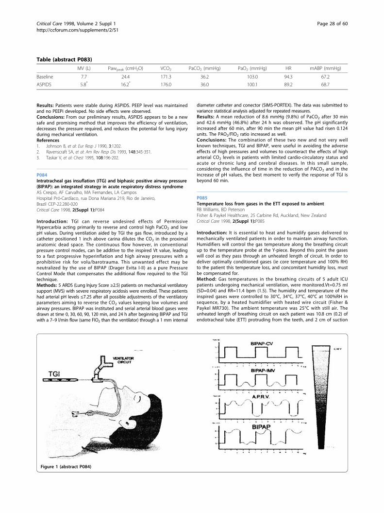
Results: Patients were stable during ASPIDS. PEEP level was maintainedand no PEEPi developed. No side effects were observed.Conclusions: From our preliminary results, ASPIDS appears to be a newsafe and promising method that improves the efficiency of ventilation,decreases the pressure required, and reduces the potential for lung injuryduring mechanical ventilation.References1. Johnson B, et al: Eur Resp J 1990, 3:1202.2. Ravenscraft SA, et al: Am Rev Resp Dis 1993, 148:345-351.3. Taskar V, et al: Chest 1995, 108:196-202.
P084Intratracheal gas insuflation (ITG) and biphasic positive airway pressure(BIPAP): an integrated strategy in acute respiratory distress syndromeAS Crespo, AF Carvalho, MA Fernandes, LA CamposHospital Pró-Cardíaco, rua Dona Mariana 219, Rio de Janeiro,Brazil CEP-22.280-020Critical Care 1998, 2(Suppl 1):P084
Introduction: TGI can reverse undesired effects of PermissiveHypercarbia acting primarily to reverse and control high PaCO2 and lowpH values. During ventilation aided by TGI the gas flow, introduced by acatheter positioned 1 inch above carina dilutes the CO2 in the proximalanatomic dead space. The continuous flow however, in conventionalpressure control modes, can be additive to the inspired Vt value, leadingto a fast progressive hyperinflation and high airway pressures with aprohibitive risk for volu/barotrauma. This unwanted effect may beneutralized by the use of BIPAP (Drager Evita I-II) as a pure PressureControl Mode that compensates the additional flow required to the TGItechnique.Methods: 5 ARDS (Lung Injury Score ≥2.5) patients on mechanical ventilatorysupport (MVS) with severe respiratory acidosis were enrolled. These patientshad arterial pH levels ≤7.25 after all possible adjustments of the ventilatoryparameters aiming to reverse the CO2 values keeping low volumes andairway pressures. BIPAP was instituted and serial arterial blood gases weredrawn at time 0, 30, 60, 90, 120 min, and 24 h after beginning BIPAP and TGIwith a 7–9 l/min flow (same FIO2 than the ventilator) through a 1 mm internal
diameter catheter and conector (SIMS-PORTEX). The data was submitted tovariance statistical analysis adjusted for repeated measures.Results: A mean reduction of 8.6 mmHg (9.8%) of PaCO2 after 30 minand 42.6 mmHg (46.8%) after 24 h was observed. The pH significantlyincreased after 60 min, after 90 min the mean pH value had risen 0.124units. The PAO2/FlO2 ratio increased as well.Conclusions: The combination of these two new and not very wellknown techniques, TGI and BIPAP, were useful in avoiding the adverseeffects of high pressures and volumes to counteract the effects of higharterial CO2 levels in patients with limited cardio-circulatory status andacute or chronic lung and cerebral diseases. In this small sample,considering the influence of time in the reduction of PACO2 and in theincrease of pH values, the best moment to verify the response of TGI isbeyond 60 min.
P085Temperature loss from gases in the ETT exposed to ambientRB Williams, BD PetersonFisher & Paykel Healthcare, 25 Carbine Rd, Auckland, New ZealandCritical Care 1998, 2(Suppl 1):P085
Introduction: It is essential to heat and humidify gases delivered tomechanically ventilated patients in order to maintain airway function.Humidifiers will control the gas temperature along the breathing circuitup to the temperature probe at the Y-piece. Beyond this point the gaseswill cool as they pass through an unheated length of circuit. In order todeliver optimally conditioned gases (ie core temperature and 100% RH)to the patient this temperature loss, and concomitant humidity loss, mustbe compensated for.Method: Gas temperatures in the breathing circuits of 5 adult ICUpatients undergoing mechanical ventilation, were monitored.Vt=0.75 ml(SD=0.04) and RR=11.4 bpm (1.5). The humidity and temperature of theinspired gases were controlled to 30°C, 34°C, 37°C, 40°C at 100%RH insequence, by a heated humidifier with heated wire circuit (Fisher &Paykel MR730). The ambient temperature was 25°C with still air. Theunheated length of breathing circuit on each patient was 10.8 cm (0.2) ofendotracheal tube (ETT) protruding from the teeth, and 2 cm of suction
Table (abstract P083)
MV (L) Pawpeak (cmH2O) VCO2 PaCO2 (mmHg) PaO2 (mmHg) HR mABP (mmHg)
Baseline 7.7 24.4 171.3 36.2 103.0 94.3 67.2
ASPIDS 5.8* 16.2* 176.0 36.0 100.1 89.2 68.7
Figure 1 (abstract P084)
Critical Care 1998, Volume 2 Suppl 1http://ccforum.com/supplements/2/S1
Page 28 of 60
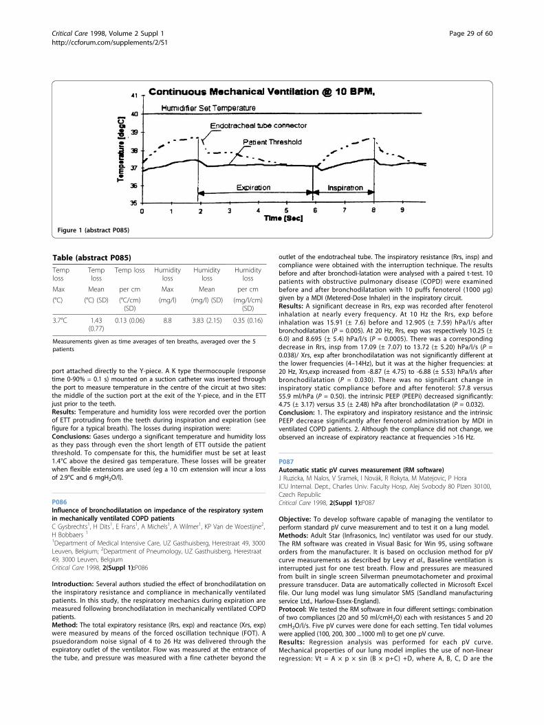
port attached directly to the Y-piece. A K type thermocouple (responsetime 0-90% = 0.1 s) mounted on a suction catheter was inserted throughthe port to measure temperature in the centre of the circuit at two sites:the middle of the suction port at the exit of the Y-piece, and in the ETTjust prior to the teeth.Results: Temperature and humidity loss were recorded over the portionof ETT protruding from the teeth during inspiration and expiration (seefigure for a typical breath). The losses during inspiration were:Conclusions: Gases undergo a significant temperature and humidity lossas they pass through even the short length of ETT outside the patientthreshold. To compensate for this, the humidifier must be set at least1.4°C above the desired gas temperature. These losses will be greaterwhen flexible extensions are used (eg a 10 cm extension will incur a lossof 2.9°C and 6 mgH2O/l).
P086Influence of bronchodilatation on impedance of the respiratory systemin mechanically ventilated COPD patientsC Gysbrechts1, H Dits1, E Frans1, A Michels1, A Wilmer1, KP Van de Woestijne2,H Bobbaers 1
1Department of Medical Intensive Care, UZ Gasthuisberg, Herestraat 49, 3000Leuven, Belgium; 2Department of Pneumology, UZ Gasthuisberg, Herestraat49, 3000 Leuven, BelgiumCritical Care 1998, 2(Suppl 1):P086
Introduction: Several authors studied the effect of bronchodilatation onthe inspiratory resistance and compliance in mechanically ventilatedpatients. In this study, the respiratory mechanics during expiration aremeasured following bronchodilatation in mechanically ventilated COPDpatients.Method: The total expiratory resistance (Rrs, exp) and reactance (Xrs, exp)were measured by means of the forced oscillation technique (FOT). Apsuedorandom noise signal of 4 to 26 Hz was delivered through theexpiratory outlet of the ventilator. Flow was measured at the entrance ofthe tube, and pressure was measured with a fine catheter beyond the
outlet of the endotracheal tube. The inspiratory resistance (Rrs, insp) andcompliance were obtained with the interruption technique. The resultsbefore and after bronchodi-latation were analysed with a paired t-test. 10patients with obstructive pulmonary disease (COPD) were examinedbefore and after bronchodilatation with 10 puffs fenoterol (1000 μg)given by a MDI (Metered-Dose Inhaler) in the inspiratory circuit.Results: A significant decrease in Rrs, exp was recorded after fenoterolinhalation at nearly every frequency. At 10 Hz the Rrs, exp beforeinhalation was 15.91 (± 7.6) before and 12.905 (± 7.59) hPa/l/s afterbronchodilatation (P = 0.005). At 20 Hz, Rrs, exp was respectively 10.25 (±6.0) and 8.695 (± 5.4) hPa/l/s (P = 0.0005). There was a correspondingdecrease in Rrs, insp from 17.09 (± 7.07) to 13.72 (± 5.20) hPa/l/s (P =0.038)/ Xrs, exp after bronchodilatation was not significantly different atthe lower frequencies (4–14Hz), but it was at the higher frequencies: at20 Hz, Xrs,exp increased from -8.87 (± 4.75) to -6.88 (± 5.53) hPa/l/s afterbronchodilatation (P = 0.030). There was no significant change ininspiratory static compliance before and after fenoterol: 57.8 versus55.9 ml/hPa (P = 0.50). the intrinsic PEEP (PEEPi) decreased significantly:4.75 (± 3.17) versus 3.5 (± 2.48) hPa after bronchodilatation (P = 0.032).Conclusion: 1. The expiratory and inspiratory resistance and the intrinsicPEEP decrease significantly after fenoterol administration by MDI inventilated COPD patients. 2. Although the compliance did not change, weobserved an increase of expiratory reactance at frequencies >16 Hz.
P087Automatic static pV curves measurement (RM software)J Ruzicka, M Nalos, V Sramek, I Novák, R Rokyta, M Matejovic, P HoraICU Internal. Dept., Charles Univ. Faculty Hosp, Alej Svobody 80 PIzen 30100,Czech RepublicCritical Care 1998, 2(Suppl 1):P087
Objective: To develop software capable of managing the ventilator toperform standard pV curve measurement and to test it on a lung model.Methods: Adult Star (Infrasonics, Inc) ventilator was used for our study.The RM software was created in Visual Basic for Win 95, using softwareorders from the manufacturer. It is based on occlusion method for pVcurve measurements as described by Levy et al., Baseline ventilation isinterrupted just for one test breath. Flow and pressures are measuredfrom built in single screen Silverman pneumotachometer and proximalpressure transducer. Data are automatically collected in Microsoft Excelfile. Our lung model was lung simulator SMS (Sandland manufacturingservice Ltd., Harlow-Essex-England).Protocol: We tested the RM software in four different settings: combinationof two compliances (20 and 50 ml/cmH2O) each with resistances 5 and 20cmH2O/l/s. Five pV curves were done for each setting. Ten tidal volumeswere applied (100, 200, 300 ...1000 ml) to get one pV curve.Results: Regression analysis was performed for each pV curve.Mechanical properties of our lung model implies the use of non-linearregression: Vt = A × p × sin (B × p+C) +D, where A, B, C, D are the
Table (abstract P085)
Temploss
Temploss
Temp loss Humidityloss
Humidityloss
Humidityloss
Max Mean per cm Max Mean per cm
(°C) (°C) (SD) (°C/cm)(SD)
(mg/l) (mg/l) (SD) (mg/l/cm)(SD)
3.7°C 1.43(0.77)
0.13 (0.06) 8.8 3.83 (2.15) 0.35 (0.16)
Measurements given as time averages of ten breaths, averaged over the 5patients
Figure 1 (abstract P085)
Critical Care 1998, Volume 2 Suppl 1http://ccforum.com/supplements/2/S1
Page 29 of 60

regression parameters, however for mathematical simplicity we usedlinear regression Vt = A × p +B. The correlation coefficient and A, Bparameters were pooled from each settings measurement and mean ±SD were calculated:Our results showed little variability for each setting. Resistance didn’taffect the measurement of compliance as was expected.Conclusion: RM software for PC is reliable on repetitive measurementsand precise enough to measure static pV curves in a lung model withtidal volumes exceeding 150 ml. Further validation with independentpneumotachometer and pressure transducer is necessary. Thereafter RMsoftware probably can be used in clinical settings.
P088Time required for PaO2 equilibration during mechanical ventilation withchanges in FiO2
N Cakar, M Tugrul, A Demirarslan, T Erdem, Ö Akinci, F Esen, L TelciDepartment of Anesthesiology and Intensive Care Medicine, Istanbul MedicalFaculty of University of Istanbul, Çapa Klinikleri 34390 Istanbul, TurkeyCritical Care 1998, 2(Suppl 1):P088
Introduction: Although it’s a general practice to obtain blood gas sampleafter 30 min when FiO2 was changed during mechanical ventilation, exacttiming for blood gas analysis has not been very well documented. In thisstudy we aimed to investigate the time required for PaO2 equilibrationduring pressure controlled ventilation (PCV) with changes in FiO2.Patients and methods: 25 adult patients with stable blood gas values (5female, 20 male) on PCV (Servo 900C Siemens, Elema, Sweden) withFiO2:0.35, frequency: 12 breaths/min, tidal volume: 10 ml/kg and PEEP:5 cmH2O were studied.Following a control blood gas measurement at FiO2:0.35 and determinationof PaO2 (PaO235), FiO2 was increased to 0.55 for 60 min and again returnedto 0.35 without any change in ventilatory parameters. Blood samples wereobtained and immediate blood gas measurements were performed on 3, 5,7, 9, 11, 15, 20, 25, 30 min in both periods and at the 60th minute inFiO2:0.55 period. The PaO2 value measured at the 60th minute of FiO2:0.55(PaO255) was accepted as representative of equilibration.The timing of PaO2 values reaching either the PaO255 and PaO235 levelswere recorded for each patient and average (Mean ± SD) of these valueswere calculated.Results: The average (Mean ± SD) time period necessary to reach PaO255and PaO235 were 9 ± 7.33 min and 7.08 ± 6.02 min respectively. Thenumbers and the percentages of the patients reaching their own PaO255and PaO235 values for each sampling time were listed in Table 1.Conclusion: We concluded that after FiO2 changes during PCV, 10 minwill be adequate for obtaining the blood gas sample representative forequilibration of PaO2.
P089Pre-hospital tracheal intubation assisted by fibroscopy in patients withsuspicion of cervical spine lesion: a pilot studyP-H Bassene1, A d’Hollander2, J-P Faller1, A Kara1, J-B Braun11C.H.G., 14 rue de Mulhouse, Belfort, F-9000, France; 2Dept. Anesthesia-Ressuscitation, C.H.G.CUB Erasme, 14 rue de Mulhouse, Belfort, F-9000,France, 808 Rte de Lennick, Bruxelles, B-1070, BelgiumCritical Care 1998, 2(Suppl 1):P089
Introduction: There is still some controversy about the best manner tomanage, in the pre-hospital phase, the patients requesting trachealintubation and suspected to suffer from a cervical spine lesion. Thepresent observation reports the use of a new, entirely autonomous,equipment for in the field tracheal fibroscopy.Methodology: An anaesthetist, trained to tracheal fibroscopy, has at hisdisposal a medical vehicle and the equipment for emergency pre-hospitaltracheal intubation.Results: Five patients were intubated with the ‘in the field’ trachealfibroscope (9, 16, 21, 22 and 62 years of age). The intubation was carriedout without any problem on 4 victims despite their spine wasimmobilised with a cervical collar and that blood was present in thepharynx. One intubation proved itself to be more difficult on a patientwho had undergone several unsuccessful and traumatising attempts bythe first physician on the scene.Discussion: Some practical details seem to be at importance for correctmanagement of a fibroscopic guided tracheal intubation in the pre-hospital environment. First, the quality of the images transmitted by thefiberoptic equipment; second, the oxygenation of the patient during thetracheal intubation process; third, the aseptic aspects of this manoeuvre;fourth, the ‘ready to use’ aspect of the associated material (gloves,suction); fifth, the training level of the physician in charge of the trachealintubation process; sixth, the level of accompanying analgesia oranaesthesia.Conclusion: As tracheal intubation through fibroscopy could be a ‘gentle’technique, the use of ‘in the field’ fibroscopic equipment would beencouraged in a pre-hospital phase to reduce the risk of hypoxaemiaassociated with many difficult tracheal intubations and the severity ofpharyngeal and laryngeal trauma produced by the ‘classical’ direct visionlaryngoscope.
P091Bedside percutaneous tracheostomy: experience with 40 critically illpatientsSF Kahveci, S Gören, O Kutlay, B Özcan, G KorfahDepartment of Anesthesiology and Reanimation, School ofMedicineUniversity of Uludag, Bursa, TurkeyCritical Care 1998, 2(Suppl 1):P091
Text: In intensive care unit patients requiring prolonged mechanicalventilation, tracheostomy is necessary. As an alternative to the standardsurgical method, percutaneous techniques are available.Since November 1996, 40 patients (34M, 6F) have been electively selectedfor percutaneous tracheostomy (PCT) at a University Hospital with a 9-bedcombined medical-surgical ICU. PCT was performed at bedside with thePortex Percutaneous Tracheostomy Kit (Portex Ltd, Kent, England). Theprocedure time and early complications were recorded.Patients were between 18 and 86 years (46.4 ± 3.1). The procedure wassuccessful in all patients and the average duration of placement was9.05 ± 0.7 min (3–20 min). An 8 or 8.5 mm cannula was inserted ineach case, introduced between the 1st and 2nd or 2nd and 3rdtracheal cartilages.
Table (abstract P087)
A B r2
compl. 50 ml/cmH2O, 36.7 ± 1.3 101.6 ± 21.5 0.975
rezist. 5 cmH2O/l/s
compl. 50 ml/cmH2O, 35.5 ± 1.1 92.1 ± 19.2 0.97
rezist. 20 cmH2O/l/s
compl. 20 ml/cmH2O, 21.6 ± 0.4 114.6 ± 17.0 0.98
rezist. 5 cmH2O/l/s
compl. 20 ml/cmH2O, 20.8 ± 0.6 121.4 ± 24.6 0.98
rezist. 20 cmH2O/l/s
Table 1 (abstract P088) The numbers and the percentages of the patients reaching their own PaO255 and PaO235values for each sampling time
Minute 3 5 7 9 11 15 20 25 30
FiO2:0.55 Period 8 (32%) 4 (16%) 2 (8%) 4 (16) 1 (4%) 3 (12%) 1 (4%) 1 (4%) 1 (4%) n = 25
FiO2:0.35 Period 8 (32%) 7 (28%) 5 (20%) 2 (8%) - 1 (4%) 1 (4%) - 1 (4%) n = 25
Critical Care 1998, Volume 2 Suppl 1http://ccforum.com/supplements/2/S1
Page 30 of 60
The average duration of artificial ventilation before PCT was 9.8 ± 0.8days (2-28 days), after PCT was 12.1 ± 1.2 days (1–32 days). Meanduration of PCT was 20.9 ± 3.6 days (1–140 days). There were no PCT-related deaths. The only procedure related complication was bleeding atthe stoma site which resolved within applied pressure. 20 patients weredecannulated. Early clinical examination revealed hoarseness in onepatient. Stomas were closed within a few days, leaving an approximately1 cm length scar.Our study suggests that PCT is a simple and safe method and can beperformed rapidly at the bedside in the ICU.
P092Esophageal acid and bile reflux in mechanically ventilated patientsH Dits1, A De Weerdt1, A Wilmer1, J Tack2, E Frans1, H Bobbaers11UZ Gasthuisberg University of Leuven, Herestraat 49, Leuven, 3000, Belgium;2Dept. of Gastroenterology, UZ Gasthuisberg University of Leuven, Herestraat49, Leuven, 3000, BelgiumCritical Care 1998, 2(Suppl 1):P092
Introduction: Previous studies using scintigraphy have documented ahigh incidence (61%) of gastroesophageal reflux in mechanicallyventilated (MV) patients. The aim of this study was to assess, over aprolonged period of time, the incidence and type (bile or acid) of reflux,the effect of body position on reflux, and the potential relationshipbetween pathological reflux and esopahagitis in mechanically ventilatedpatients.Methods: 24 critically ill, MV patients (mean APACHE II score: 21, meanage 64 ± 14 yrs, 15 men), hospitalized in a medical intensive care unit,were prospectively included for 24-h esophageal pH and duodeno-gastroesophageal reflux (DGER) studies (Digitrapper III pH-meter andfiber-optic sensor for the presence of bilirubin, Bilitec 2000, Synectics,Sweden) with single sensors placed 5 cm proximal to the loweresophageal sphincter (LES) and instrumented with a nasogastric tube.Exclusion criteria were: MV <3 or >8 days, previous initiation of enteralfeeding, history of reflux esophagitis or gastric surgery, acutegastrointestinal bleeding or exophageal varices, or medication withprokinetic agents. All patients received stress ulcer prophylaxis withranitidine 50 mg iv, tid. On the day before the study patientsunderwent esophageal endoscopy to determine the presence ofesophagitis. Patients were placed in supine semi-recumbent positionand turned on either left or right lateral sides at intervals as deemedappropriate by the attending nurse. The following variables wereanalyzed depending on body position (total time, time in supine, right,or left position): % time pH <4 (normal <3.4 %) or % time bilirubinabsorption >0.14 (normal: <3 %), total duration of reflux, and numberreflux episodes. Data are medians with interquartile ranges (IQR),significance was tested with the Mann-Whitney U test. Fisher’s exacttest was used to analyze relationships between the presence orabsence of pathological reflux and esophagitis.Results: After a median of 5 days of MV (IQR 3.8) 12 of 24 patients (50%)patients had pathological DGER (median % reflux time over the wholerecording time independent of body position: 8%, IQR 0.62%). In contrast,only one patient had significant acid reflux (4.8% of the recording time)and the median pH in the lower esophagus for all 24 patients was 6.7(IQR 6.7). The median duration where bile salts were present in the loweresophagus was 112 min (IQR 0.865 min), in 8 of 24 patients this timeperiod exceeded 5 h. The median number of DGER episodes was 12 (IQR0.37). The relative time of pathological reflux was significantly higher inleft lateral and supine position as compared to right lateral position (7.1%IQR 0.64% and 8%, IQR 0.65% vs. 4.7% IQR 0.70%, P < 0.01. 12 of 24patients (50%) had esophagitis. There was a significant positiverelationship between the presence of pathological DGER and thepresence of esophagitis (P = 0.04).Conclusions: Under standard stress ulcer prophylaxis with ranitidine,critically ill patients with MV have a high incidence (50%) of DGER butnot of acidic esophageal reflux. Reflux is highest in the left lateral and inthe semirecumbent position. The presence of bile salts in the esophagusfor prolonged periods of time suggest that: 1) the barrier function of theLES and the clearance function of the esophagus are deranged, 2)esophagitis in these patients may not only be a result of mechanicalirritation due to the nasogastric tube, but may also be a chemical
esophagitis, 3) intestinal fluids refluxing into the esophagus may beconducive to ventilator-associated pneumonia.
P093Adrenocortical function and outcome in critically ill patientsE Bermejo, T Lajo, JC Sotillo, MJ Fernández, E Palencia, C BouzaIntensive Care Unit, Hospital General Universitario ‘Gregorio Moranon’,Madrid, SpainCritical Care 1998, 2(Suppl 1):P093
Objective: To assess the value of measuring adrenocortical functionparameters in predicting outcome and mortality in critically ill patients.Methods: Prospective clinical investigation with no therapeuticinterventions in which were involved 63 consecutive patients admitted tothe medical intensive care unit. Adrenocortical function was assessed byplasma cortisol and dehydroepiandrosterone sulfate (DHEA-S) levels ondays 1, 3 and 7 and by the short synthetic ACTH stimulation testperformed within 24 h of admission to the ICU. Individual variables werecompared with severity scores, biological data and outcome. Statisticalanalysis: Student test, Mann-Whitney U test P < 0.05 was consideredsignificant.Results: Mean age 57 ± 19, APACHE II score at admission 20 ± 7, therewere 37 (54.4%) septic patients. In hospital mortality rate 43.1%. Busalcortisol concentrations were increased with a mean value of 32.22 ± 19.3.Of overall cortisol determinations 94% were above 15 μg/dl. Nocorrelations were found between these levels and factors such asAPACHE II score, MODS score, haemodynamic measurements, amount ofvassopresor support neither mortality. Cortisol levels on day 3 correlatedwith the use of dopamine (p:.03) and noradrenaline (p:.02) and were ableto discriminate between survivors and non-survivors (median 13.65 vs30.3, P < 0.005). The response to ACTII test had a mean value of 45.4 ±18 with a mean difference with basal cortisol levels of 13.85 ± 10. No oneof these measurements did show any relationship with clinical variablesof outcome. Global basal concentrations of DHEA-S were lower (median65, range: 1–447) than normal values (r: 120–240) and showed a markedtendency to decrease over the time (median 30 on days 3 and 7). Levelsof DHEA-S on day 1 showed a weak correlation with APACHE II score(r. 0.33; P = 0.039) DHE-S on day 3 was much higher in survivors than innon-survivors patients (median 41.1 vs 20.7; P = 0.002).Two patterns of adrenocortical function were identified. In survivingpatients plasma cortisol levels were normal or increased and graduallydecreased whereas levels of DHEA-S showed an opposite pattern. In theother hand, in non-survivors cortisol levels persisted elevated and DHEA-Sdecreased. The ratio DHEA-S/cortisol on day 3 was a good indicator offinal outcome with a median value of 1.92 in survivors and 0.79 in non-survivors (P = 0.009).Conclusion: In critically ill patients the basal adrenocortical function ischaracterized by high plasma cortisol and low DHEA-S levels. The DHEA-S/cortisol plasma concentrations on day 3 might be used as a prognosticmarker for identifying final outcome in this population.
P094Treatment of postoperative lung injury in patients with SIRS bymethylprednisoloneM Sumida1, T Fukada1, Y Tsukazaki1, N Kobayashi1, Y Mukubou1, T Sumida2,K Taira1, M Kawamata11Department of Anesthesiology, Tokyo Women’s Medical College, 2-1-10Nishiogu, Arakawa-ku, 116, Japan; 2Department of Anesthesiology, TokyoWomen’s Medical College Institute of Medical ScienceSt Marianna UniversitySchool of Medicine, 2-1-10 Nishiogu, Arakawa-ku, 116, JapanCritical Care 1998, 2(Suppl 1):P094
Introduction: Intraperitoneal hyperthermic perfusion (IPHP) is performedclinically as one treatment for cancer patients with advanced gastrointestinalcancer. All patients are fallen the systemic inflammatory response syndrome(SIRS) by IPHP. In our previous work, we clarified the elevated TNF-a in theserum of IPHP patients [1]. Glucocorticoids are well known inhibitors ofTNF-a [2]. We examined serum TNF-a levels before and after administrationof methylprednisolone (MPS) and compaired the lung injury score inpostoperative days between the group of MPS and control.
Critical Care 1998, Volume 2 Suppl 1http://ccforum.com/supplements/2/S1
Page 31 of 60

Methods: Seventeen patients with gastrointestinal cancer were undergonesurgery combined with IPHP. Group 1 (10 patients): MPS was notadminstrated. Group 2 (7 patients): MPS was administered before andduring IPHP. We measured serum TNF-a levels and evaluate themetabolism, hematorogy, hemodynamics and the post-operative lung injury.Results: In group 1, serum TNF-a levels was increased to 46.0 ±25.7 pg/ml (mean ± SD) at the end of IPHP and patients were SIRS withthe postoperative lung injury. In group 2, TNF-a was not detected butpostoperative lung injury were improved.Conclusion: MPS inhibited the production of TNF-a which was able toimprove postoperative lung injury. Pre-emptive MPS might be one ofstrategy for treatment of lung injury during SIRS.
P095A placebo-controlled study on the effects of corticosteroid inhalationtherapy in ammonia induced lung injury in rabbitsE Sjöblom, J Höjer, PEJ Kulling, K Stauffer, A Suneson, U LudwigsDept. of Medicine, Medical Intensive Care Unit, Södersjukhuset, Stockholm,118 83, SwedenCritical Care 1998, 2(Suppl 1):P095
Background: The use of inhalation corticosteroids in toxic pulmonaryoedema caused by exposure to irritating gases is controversial. Theireffects have not been demonstrated in controlled studies.Study design: A randomised blind placebo-controlled study of theeffects of budesonide inhalation in a rabbit model of toxic lung injuryinduced by ammonia inhalation.Animals: Sixteen New Zealand White rabbits.Intervention: Lung injury was induced by inhalation of a defined amountof aerosolised ammonia. Thirty and 150 min later the animals wererandomised to receive either inhalation therapy with 0.5 mg budesonideor placebo.Measurements and results: Airway pressures, haemodynamics and gasexchange were measured at baseline, 5 and 15 min after the ammoniaadministration, and every 30 min during a 6-h period after the first blindinhalation therapy with corticosteroids or placebo. The ammoniainhalation resulted in an acute severe lung injury detected after 15 minas a decrease in PaO2 from 23.3 (± 3.6) to 11.0 (±3.6) kPa (P < 0.005) andan increase in peak airway pressure from 13 (±2) to 17 (±2) cmH2O (P <0.005). During the 6-h observation period, the blood gas parametersimproved in all rabbits. In comparison to placebo, budesonide did notresult in improved gas exchange or reduced airway pressure levels duringthe observation period.Conclusion: In this animal model, corticosteroid inhalation therapy hadno acute effects on ammonia induced lung injury.
P096Late steroid therapy improves gas exchange and reduces organdysfunction in acute lung injury caused by pneumococcal pneumoniaT Varpula1, V Pettilä1, E Rintala21Department of Surgery, Helsinki University Central Hospital, Helsinki, Finland;2Department of Surgery Department of Internal Medicine, Helsinki UniversityCentral Hospital, Helsinki, FinlandCritical Care 1998, 2(Suppl 1):P096
Introduction: Prophylactic or early glucocorticoid therapy have notproved to be useful in the treatment of acute lung injury (ALI). However,there is evidence of the beneficial effects of steroids in thefibroproliferative stage of ALI (late steroid therapy).Materials and methods: We analyzed the clinical data of all patientswho suffered from a severe pneumococcal pneumonia during 1993–97and were treated in the ICU at our institution. Patients who requiredmechanical ventilation more than 10 days (n = 18) were selected for thestudy. Based on empirical data, late steroid therapy was started for 11patients because of persistent impairment of gas exchange and/orinflammatory process of the lungs, and the remaining 7 patients servedas controls. Methylprednisolone was intravenously administered with adaily dose of 80 mg and 40 mg. The dosage was gradually decreased.General treatment of the patient groups was similar including lungprotective ventilatory strategies, prone positioning and invasive
hemodynamic monitoring The steroid therapy was started within 9.3 days(range, 7–13 days) after hospitalization. Accordingly, the values of thecontrol group were monitored on the day 10 after hospitalization.Mortality was assessed on the day 30.Results: Age, sex, APACHE II-score and lung injury score, Multi-organDysfunction Score (MODS), CRP-level and PaO2/FiO2-ratio werecomparable at the start of steroid therapy or day 10. The changes in CRP,MODS and PaO2/FiO2 between the day steroid treatment started (day 0)and 3 days thereafter differed significantly between the two groups.Conclusions: Although the sample size of this retrospective study wassmall, our results support the increasing evidence of the advantages oflate steroid therapy in acute lung injury.
P097Corticoid treatment increases the risk of enterobacter aerogenesinfection in intensive care unitM Piagnerelli, P Lejeune, Y Glupczynski, E CarlierDepartment of Intensive Care Unit, André Vésale Hospital, Montigny-le-Tilleul,BelgiumCritical Care 1998, 2(Suppl 1):P097
Text: The emergence of multiresistant Enterobacter aerogenes (E.a.) hasserious implications in the management of intensive care patients becauseof increasing risk of nosocomial infection acquisition attributed not only tothe severity of the underlying disease, but also to invasive procedures andcontaminated life-support equipment. Acquisition of E.a. usually occurseither by colonization with an endogenous strain selected de novo from thepatient’s own flora (eg broad-spectrum antibiotic follow-up therapy) or bypatient-to-patient transmission of strains by the health personnel. E.a. is adifficult-to-treat organism since it is intrinsically resistant to beta-lactamantibiotics and it readily develops multiple drug resistance following theclinical usage of broad-spectrum cephalosporins or carbapenems.A prospective study comparing risk-factors for E.a. colonization/infectionhas been conducted in a 12-bed ICU patients (median age, length of stay,mortality rate, mechanical ventilation, antibiotherapy prior to ICUadmission, urinary catheter, surgical drain and corticoid therapy). FromFebruary 25, 1995 up to May 12, 1995, prescription order of perinealswabs has been undertaken to all patients prior to admission, dischargeand on a weekly basis, to rule out the unit specific incidence of E.a.acquisition. 146 patients were included in this study. Out of which,14 (9.6%) had one or more perineal swabs positive for E.a. and 7 of themhad a positive swab the day of the ICU admission.The E.a. colonization/infection was not significantly associated withpatient’s median age, mortality rate, antiobiotherapy prior to ICUadmission, mechanical ventilation, urinary catheter and surgical drain.A significant association had been noted with corticoid therapy and ICUlength of stay (respectively 80 days for E.a. + and 3.0 days for E.a.-), alsohad been noted that the median SAPS 2 score for colonized/infectedpatients is superior to the median SAPS 2 score of the ICU patients(43 versus32). Nine patients with E.a. colonization/infection developpedinfection. Despite appropriate antimicrobial therapy, E.a. colonization wasassociated with a crude mortality rate of 43%.These data suggest that corticoid therapy is a risk factor in E.a.colonization/infection in ICU patients and perhaps, indeed, increases thepatients mortality rate. Further prospective study are needed to optimizethese data and precise the other risk factors. The potential effects of riskfactors emphasize the importance of specific measures for infectioncontrol in critically ill patients.
P098Helicobacter pylori infection in intensive care: increased prevalence anda new nosocomial infectionMS Robertson, JF Cade, RL ClancyDepartment of Pathology, The Royal Melbourne Hospital University ofNewcastle, Parkville, VIC 3050, Newcastle, NSW 2300, AustraliaCritical Care 1998, 2(Suppl 1):P098
Objective: The pathogenesis of acute gastric stress ulceration in theseriously ill is uncertain and any role of Helicobacter pylori infection isunknown. We aimed to assess the relationship between H. pylori
Critical Care 1998, Volume 2 Suppl 1http://ccforum.com/supplements/2/S1
Page 32 of 60

serological status and stress ulceration in seriously ill patients, as well asH. pylori serological status in Intensive Care nurses as a marker fornosocomial infection.Design: Prospective epidemiological survey.Setting: Adult Intensive Care Unit in a University teaching hospital.Patients: 100 patients, 100 nurses and 500 blood donors as communitycontrols.Interventions: H. pylori serological status was measured in patients, staffand controls using a rapid whole blood test. Upper gastrointestinalbleeding and risk factors for acute stress ulceration were recorded.Measurements and main results: In seriously ill patients, H. pyloriseropositivity (67%) was significantly higher than in the control group(39%) (P < 0.001). In patients, seropositivity was not related to age,country of birth, diagnostic category, severity of illness or risk score forstress ulceration. There was a trend towards increased macroscopicgastric bleeding in seropositive patients. In Intensive Care nurses,H. pylori seropositivity (40%) was significantly higher than in age’matchedcontrols (19%) (P < 0.001). Only duration of Intensive Care nursing wassignificantly associated with seropositivity (P = 0.02).Conclusions: The unexpectedly high H. pylori seropositivity rate in thisseriously ill cohort raises the possibility that under Intensive Careconditions H. pylori infection may modulate responses to illness andinjury, with consequent clinical implications. Furthermore, the elevatedseropositivity rate in Intensive Care nurses suggests that H. pylori can benosocomially transmitted.
P099Tetanus not forgottenHY Zolkepli, G PhutaneICU, General Hospital, 15586 Kota Bharu, Kelantan, MalaysiaCritical Care 1998, 2(Suppl 1):P099
Introduction: Tetanus still lingers with us although we have fairly goodimmunization programme in the country. There are no cases of neonataltetanus in our hospital for the past 10 years.Materials and methods: Retrospective study was done on 12 cases ofgeneralised adult tetanus diagnosed clinically and admitted to ourhospital from Jan 1995 to Nov 1997.Management: Three cases were treated conservatively in the medicalward. Nine cases were admitted directly to ICU from the A&E Dept. Ofthe cases admitted to ICU, three were on conservative treatment:isolation in semi dark room, invasive monitoring, anti-tetanus toxoid,human hyperimmune antite-tanus globulin, titrated diazepam infusion, i.v.crystalline penicilline and metronidazole, and wound desloughing. Sixneeded ventilatory support: two underwent tracheostomy, and only oneneeded labetalol infusion. Proper nutrition was taken care of in all thepatients.Observation: Conservative management in the medical ward wassatisfactory and all patients were discharged well. Patients admitted toICU: 88.88% are above the age of 50 years, males more than females,average incubation period was 10 days, all patients had outdoor injuries,average stay was 14 days, 55.55% had autonomic dysfunction, 66.66%had nasocomial infection, there were two deaths (22.22%): one due tosevere uncontrolled autonomic dysfunction with bradycardia, another dueto septicaemia.Conclusion: Tetanus is still a problem in elderly age group in our humidclimate. Autonomic dysfunction is among the major complications.Titrated diazepam infusion without ventilation had satisfactory results.
P100Nosocomial infection surveillance in Belgian ICUs: aim andmethodology of the feedbackG Hanique1, C Suetens2, H Carsauw2, B Jans2, O Ronveaux2, R Mertens21University St Luc Hospital UCL, Hippocrate Avenue, Brussel, Belgium;2Scientific Institut for Public Health Luois Pasteur, Rue J. Wytsman, Brussles,BelgiumCritical Care 1998, 2(Suppl 1):P100
Introduction: The risk for nosocomial infecions (NI) in ICU’s is five to tentimes higher than in the other hospital care units, with increasing
mortality, morbidity, length of stay and costs. Previous studiesdemonstrated a significant decrease in NI after the set up of effectivesurveillance programs. If the surveillance is performed in a multicentricsetting with representative sample, the results can be dysplayed as awhole but also aggregated for each individual ICU (IICU). The productionof meaningful feedback reports may help IICU to evaluate its ownsituation as compared to that of the whole studied group (WS).Objectives: The aim of the study was to provide IICU with its ownincidence of Nosocomial Pneumonia (NPN) and Blood Stream Infection(NBSI), and their mortality rate after controlling for the risk factors. Therisk factors (RF) are the case mix at admission (severity of illness = SAPSIIscore and the pathologies), the exposition to devices at risk (mechanicalventilation and central venous catheters) and the length of ICU stay.These 3 last RF are counfounding variables depending on the intensistyof ICU resources’ use.Material and methods: At the end of 1995, all Belgian ICUs were invitedto join a voluntary prospective NI surveillance network which was basedon the criteria defined in the HELICS project. Participation was for one aretwo 3-months periods, starting in January 1996. During the first twotrimesters of 1996, 64 different ICU’s, from 28% of the Belgian acute carehospitals, have joined the study. A total of 8475 patients were observed.Data included: (i) patient characteristics at admission: administrative data,type of admission, SAPSII score, prior surgery or antibiotics, impairedimmunity and infection at entry; (ii) during ICU stay, daily exposure tomechanical ventilation, central venous catheters and treatments; (iii) if aNPN and/or a NBSI occured during ICU stay, diagnostic criteria andmicrobiological data; (iv) discharge data: vital status and date.Results: Two kinds of feedbacks were produced for IICU with resultsgrouped by trimester: descriptive and analytical. The descriptivefeedbacks reported firstly, the observed results of the WS and IICUconcerning the case mix at admission, the intensity of devices use, lengthof ICU stay, mortality and NI rates and secondly, the distribution of thosevariables throughout the WS as well as the localisation of IICU, expressedin percentile, within each distribution. The analytical feedbacks producedspecific standardised rates (SR) for the intensity of devices use and lengthof stay weighted for the case mix, and the SR for mortality and NI ratesweighted for the ICU resources use and the case mix. Moreover, those SRmay vary widely as a consequence of hazard, specially for small ICUs, sothat confidence interval (CI) must be calculated for IICU specific SR.
P101Epidemiological impact of the antimicrobial therapy on Acinetobacterspp. isolated from ITU patientsT Nikodemski, S Giedrys-Kalemba, R Bohatyrewicz, M Zukowski, I Bilska,C PakulskiDept. of Anaesthesiology and Intensive Care Medicine, Dept. of Microbiologyand Immunology, Pomeranian School of Medicine, Panstwowy SzpitalKliniczny Nr 1, ul. Uni. Lubelskiej 1, 71-344 Szczecin, PolandCritical Care 1998, 2(Suppl 1):P101
Background: Acinetobacter spp. are Gram-negative bacteria which maybe isolated as comensals from human skin, throat and intestine, but arealso responsible for hospital infection. Today Acinetobacter is consideredto be a significant nosocomial pathogen in outbreaks of nosocomialinfections predominantly in intensive therapy units (ITU).Purpose: To analyse epidemiology and factors influencing Acinetobacterspp. outbreaks.Setting: A six-bedded surgical ITU in a 700-bed teaching hospital. Theresearch was conducted from January 1995 till June 1996.Methods: Relationships between amount of isolated Acinetobacter spp.strains (in total 190) and antibiotics consumption, expressed in defineddaily dose (DDD), were analysed using linear correlation. To find out
Table (abstract P101)
amikacin aminoglycosides cefotaxim
P < 0.05 P < 0.05 P = 0.01
y=-0.02x+15.99 y=-0.009x+16.0 y=-0.05x+13.08
r =-0.58 r =-0.54 r =-0.62
Critical Care 1998, Volume 2 Suppl 1http://ccforum.com/supplements/2/S1
Page 33 of 60

whether there is any relation between Acinetobacter out-breaks andoverpopulation on ITU, all data were categorised for the frequency table.Paerson Chi-square test was used to identify relationship between thecrosstabulated variables.Results: Strong correlation between resistant Acinetobacter isolates andamikacin, aminoglycosides and cefotaxim consumption is shown in table.Crosstabulated variables analyses proved influence of overpopulation onITU on the Acinetobacter outbreak’s (P < 0.05).Conclusion: This study illustrates the influence of antimicrobial therapyon outbreaks of resistant Acinetobacter strains.
P102Control of nosocomial infections in ICU patients in the PomeranianSchool of Medicine in PolandT Nikodemski, S Giedrys-Kalemba, R Bohatyrewicz, M Zukowski,E Marzec-LewensteinDept. of Anaesthesiology and Intensive Care Medicine, Dept. of Microbiologyand Immunology, Pomeranian School of Medicine, Panstwowy SzpitalKliniczny Nr 1, ul. Unii Lubelskiej 1, 71-344 Szczecin, PolandCritical Care 1998, 2(Suppl 1):P102
Background: Antibiotics represent the most commonly prescribedmedical therapies for hospitalised patients. The practice of ‘spirallingempiricism’ has increasingly led to the unnecessary administration ofantibiotics, resulting in the emergence of infections with antibiotic-resistant bacteria.Purpose: To define the influence of antibiotic use on the etiology ofnosocomial infection.Setting: A six-bedded surgical intensive therapy unit (ITU) in a 700-bedteaching hospital. The research was conducted from January 1995 tillJune 1996. In August 1995 we have changed antibiotic guidelines on ourITU (third generation cephalosporins, fluoroquinolones and vancomycinwere used only as the last option and never in prevention)Methods: We have compared three consecutive periods of 6 months(I’95, II’95, I’96). 1276 samples for microbiological culture, mainly frombronchial tree, wounds, blood and urine, were obtained in routinemanner. From 60% positive cultures 1216 strains were isolated.Antibiotics consumption was expressed in defined daily dose (DDD).Results: Over 18-months there was no statistically significant deference inmortality rate on our ITU. There were statistically lower monthlyconsumptions of ceftriaxone form 60 to 0 DDD (P < 0.05) and quinolonesfrom 282 to 110 DDD (P < 0.05) in I’95 and II’95 respectively. Vancomycinuse decreased from 133 DDD in I’95 to 34 DDD in I’96 (P < 0.05). Therewas no statistically significant increase in amount of isolated pathogenstrains in analysed periods. We have observed improvement in activitiesof third generation cephalosporins and fluoroquinolones.Conclusion: This study illustrates the influence of antimicrobal therapyon the species and the resistance of strains isolated in nosocomialinfection. Restrictive antibiotics policy do not affect ITU outcome. Betterstrategies for antibiotic administration in the ITU setting may improvetheir efficacy and reduce costs. Therefore, antibiotic policy would bemandatory in each hospital and department.
P103ICU acquired late pneumonia: epidemiological, clinical, bacteriologicaland histological aspects of a 3 years studyG Tulli1, A Cellerini2, A Farese3, V Mangani1, R Oggioni1, F Zolfanelli41Intensive Care Unit; 2Department of Radiology; 3Department of infectiousdiseases; 4Department of Pathology and Histology, Nuovo Ospedak SanGivanni di Dio, Via di Torregalli 3, 50143 Firenze, ItalyCritical Care 1998, 2(Suppl 1):P103
Objective: To evaluate the incidence and the characteristics of ICUacquired late pneumonia and to correlate the clinical diagnosis ofpneumonia to histological findings; to identify and emphasize the riskfactors of its onset.Subject and methods: ICU acquired late pneumonia was defined by thepresence of new and persistent infiltrates to the chest X-ray (appeared atleast 72 h after admission) in addition to at least one of the following: a)purulent sputum; b) T° >38°C or <36°C; c) white blood cells count >12 000
or <4 000/mm3. Tracheal aspirate, BAL or PSB were performed to obtainmicrobiological samples. We evaluated 573 patients, 380 males (66%) and193 females (34%), consecutively admitted in a medical and surgical ICUfrom 1994 to 1996; the median age was 64 years. Patients ventilated at theadmission were 395 (69%); 229 patients (40%) were admitted in ICU after asurgical procedure. The Apache II and the SAPS I scores at the admissionwere respectively >16 in 46% of patients and >9 in 76% of patients. Theoverall ICU mortality was 33%. We observed 112 episodes of pneumonia(crude incidence 19%): 52% of patients had undergone a previous surgicalprocedure; Apache II score was >16 in 63% of patients while Saps I scorewas >9 in 93%. Pneumonia lethality was 49%. All the dead patients wereventilated. In 47% postmortem examination was performed. Chi square testwith Yates correction, T Student’s test and Fisher’s exact test wereperformed for statistical analysis.Results: The incidence of pneumonia was higher in males than infemales (22% vs 13%; P < 0.05); its frequency was higher in surgicalthan in medical patients (25% vs 15%; P < 0.01) and in ventilatedpatients than in not ventilated (26% vs 4%; P < 0.000001). Incidencein patients with Apache II >16 was 27% (P < 0.0001 vs Apache II <16); inpatients with SAPS I >9 frequency was 24% (P < 0.00001 vs SAPS <9).The mean length of ICU stay (LOS) of patients with pneumonia was 36days, while LOS of patients without pneumonia was 13 days (P <0.0000000001). In 94 patients (84%) we obtained positive microbiologicalsamples; gram positive germs were observed in 44% of isolates, gramnegative in 38%, fungi in 17%. Post-mortem examination confirmed thediagnosis of pneumonia in 50% of cases; acute pulmonary oedema, ARDSand pulmonary infarction were the most common causes of misdiagnosis.Conclusions: Pneumonia is one of the most common nosocomialinfections, particularly in ventilated critically ill patients, and it isburdened by high lethality. Our data evidence that male sex, mechanicalventilation, high severity disease indexes scores, previous surgery andlength of ICU stay are important risk factors for the onset of latepneumonia. Relationship between ARDS and pneumonia is strict, as ARDSmay be both the result or the cause of pneumonia. How best todiagnose ICU acquired late pneumonia is still under debate, consideringthe frequent discrepancy between clinical and histological diagnosis.
P104The contribution of viral presence to severe exacerbation of asthmaT Stefanovski, Z Arsovski, G BalabanClinic of Pulmology and Allergy, Medical Faculty Skopje, MacedoniaCritical Care 1998, 2(Suppl 1):P104
Purpose of the study was to evaluate the contribution of viral presence asa possible trigger of the severe asthma. Allergens, physical effort, cold-air,gastroesophageal reflux and other possible triggers were not taken intoconsideration in this study.84 patients with severe asthma hospitalized at the Clinic of Pulmologyduring the winter period of 1996, were clinically assessed by randomchoice. We have examined blood samples for viral serology the first dayof their hospitalization and after two weeks. The following results wereobtained expressed in percentage. In 31 (36.9%) patients we foundpositive isolates for viral presence: Influenzae A in 12 patients (38.7%),Parainfluenzae in 6 patients (19.3%) RSV in 5 patients (16.1%),Mycoplasma pneumoniae in 3 (9.6%) and RV in 5 (16.1%). Positive viralpresence after 2 weeks showed 6.4 % (2 patients).Although this is a small group of patients to be taken for definitiveconclusions about the viral infection as a major trigger for exacerbationof severe asthma, their presence especially in winter period plays animportant role in worsening of the disease.
P105Function of different ICU ventilators under hyperbaric conditionsW Stahl, E Calzia, P RadermacherSektion Anästhesiologische Pathophysiologie und Verfahrensentwicklung,Department of Anesthesiology, University of Ulm, GermanyCritical Care 1998, 2(Suppl 1):P105
Introduction: ICU-ventilators are designed for use at normobaricconditions. Under certain circumstances patients need mechanical
Critical Care 1998, Volume 2 Suppl 1http://ccforum.com/supplements/2/S1
Page 34 of 60

ventilation during hyperbaric oxygen therapy. The aim of theinvestigation was to analyse the function of four different ICU-ventilators[EVITA-4, Microvent, Oxylog 2000 HBO (Drägerwerk AG, Germany) andServo ventilator 900 C (Siemens-Elema, Sweden] under 5 (normobaric, 1.3,1.6, 1.9 and 2.8 ATA, EVITA-4 and Oxylog 2000 HBO) or 6 (additionally6 ATA, Servo ventilator 900 C and Microvent) different atmosphericpressure conditions, regarding the difference of present and effectivelyapplied volume and pressure from those set at the control panel.Methods: We tested the following ventilator modes: volume (VCV) andpressure controlled ventilation (PCV, EVITA-4 and Servo ventilator 900 Conly), the electromechanical lung simulator LS 1500 (Drägerwerk AG,Germany) consisting of a motor driven bellow.During VCV tidal volume was set at (VT) = 750 ml, respiratory rate (f) =15/min, I:E ratio = 1:2, positive endexpiratory pressure (PEEP) = 0 cmH2O.During PCV (f) = 10/min inspiratory pressure (Pinsp) was set in order toachieve a Vt of 750 ml keeping all others ventilator parameters constant. TheVT applied by the ventilator was measured as the linear displacement of thebellows at each ambient pressure. (Paw) was measured inside of the bellows.Results: During VCV applied Vt of the ventilator decreases significantlywith increasing ambient pressure while PCV with constant Pinsp and PEEPVt was stable at each depth.Conclusions: Under hyperbaric conditions PCV should be preferred dueto the stability of VT applied. If PCV is not available, VT needs to beadjusted during VCV depending on the actual ambient pressure.Acknowledgement: We thank Drägerwerk AG, Germany for kindlyproviding us the lung simulator LS 1500.
P106Work of breathing imposed by different ventilators under hyperbaricconditionsE Calzia, W Stahl, P RadermacherSektion Anästhesiologische Pathophysiologie und Verfahrensentssuklung,Department of Anesthesiology. University of Ulm, GermanyCritical Care 1998, 2(Suppl 1):P106
Introduction: The continuous increase in gas density caused bycompression is likely to affect the work of spontaneous breathing (WOB)under hyperbaric conditions. In intubated patients breathingspontaneously through demand flow ventilator systems WOB isadditionally influenced by the WOB component imposed by theventilator (WOBimp). Since the gas density dependent components of the
ventilators available (eg inspiratory and expiratory valves) have beendesigned for use under normobaric conditions, WOBimp may differsignificantly with increasing ambient pressure. Our aim was to analyzeWOB as well as WBimp with four different ventilators under hyperbaricconditions in a lung model study.Methods: spontaneous breathing was simulated by the electromechanicallung model LS 1500 (Drägerwerk AG, Germany) consisting of a motor drivenbellows. Lung model settings were: spontaneous tidal volume (VT) = 500 ml,respiratory rate (f) = 20/min, compliance of the respiratory system (CRS) =50 ml/cm H2O and resistance (R) = 5 cm H2O×s/l. The ventilators EVITA-4,Microvent, Oxylog 2000 HBO (Drägerwerk AG, Germany) and Servoventilator 900C (Siemens-Elema, Sweden) were tested in the modes CPAP(0 cm H2O) and Pressure Support Ventilation (PSV, 5 and 10 cm H2O overCPAP except of the Oxylog 2000 HBO that do not provide a PSV-mode)under 5 (1, 1.3, 1.6, 1.9 and 2.8 ATA, EVITA-4 and Oxylog 2000 HBO) or 6(additionally 6 ATA, Servo ventilator 900 C and Microvent) differentatmospheric pressure conditions respectively. The motor driving electricalsignal modulated by the linear displacement of the bellows was used aspleural pressure (Ppl), the displacement of the bellows as flow (V) signal.Airway pressure (Paw) was measured inside the bellows. WOB and WOBimp
were calculated by integration of the inspiratory Ppl and Paw drop over Vrespectively.Conclusion: WOB and WOBimp, increased significantly with atmosphericpressure. Due to the marked differences between the ventilators the propertiesof each one needs to be known in order to assess the impact on WOB.Acknowledgement: We thank Drägerwerk AG, Germany for kindlyproviding us the lung simulator LS 1500.
P107Respiratory mechanics during nitrous oxide and xenon anesthesia withand without metacholine-induced bronchoconstrictionE Calzia, T Handschuh, W Stahl, T Marx, P Radermacher, M GeorgieffSektion Anästhesiologische Pathophysiologie und Verfahrensentwicklung,Department of Anesthesiology, University of Ulm, GermanyCritical Care 1998, 2(Suppl 1):P107
Introduction: The use of xenon (Xe) as inhalative anesthetic agent mightimpair respiratory mechanics because of the relatively high density of thisgas (5.897 g/l) when compared to nitrogen (N2, 1.250 g/l) or nitrousoxide (N2O, 1.964 g/l). The aim of this study was to compare inspiratoryairway resistance (Raw) as well as mean and peak airway pressure (Paw)
Figure (abstract P106) Results.
Critical Care 1998, Volume 2 Suppl 1http://ccforum.com/supplements/2/S1
Page 35 of 60

in pigs under normal conditions and with metacholine-inducedbronchoconstriction during Xe versus N2O anesthesia.Methods: Two groups of anesthetized and paralysed pigs (N2O n = 5, Xen = 6) were ventilated with constant inspiratory flow provided by a semi-closed anesthesia circuit (CICERO, Drägerwerk AG, Germany) with a freshgas supply correspondoing to half-minute ventilation. Ventilator settingswere: tidal volume (VT) = 12 ml/kg, respirtory rate (f) = 12/min, durationof inspiration (TI) = 1.5 s, inspiratory breath hold (iBH) = 1 s and PEEP = 0cmH2O. Paw was measured through a port proximal to the endotrachealtube with a differential pressure transducer (140PC, Honeywell Inc.,Plymouth, MN) and airflow (V) by a heated pneumotachograph (FleischNo. 2, Fleisch, Switzerland) connected with a differential pressuretransducer. Animals were ventilated for three sequential periods of 45min with a (control) mixture of 70% N2 and 30% O2 (period 1 and 3) anda testgas mixture of 70% Xe or N2O and 30% O2 (period 2). At the end ofeach period we performed three series of end-inspiratory airwayocclusions in order to calculate Raw and determined peak Paw as well asmean Paw (for a period of 2 min). For the second part of the studywe induced a bronchoconstriction by a continuous metacholine infusion(16-32 μg/kg per min) during ventilation with the control gas mixtureand repeated the same study protocol once again.
Results: X = P < 0.05. Friedman ranks signed ANOVA, followed, whensignificant, by the Student-Newman-Keuls method.Conclusions: In contrast to the marked increase in Raw, changes in Pawduring Xe anesthesia were less impressive but should be individuallyconsidered whenever using this gas as inhalative anesthetic agent.Acknowledgement: We thank Drägerwerk AG for kindly providing theCICERO EM.
P108Arterial oxygen partial pressures during nitrous oxide and xenoneliminationW Stahl, T Handschuh, E Calzia, T Marx, P Radermacher, M GeorgieffSektion Anästhesiologische Pathophysiologie und Verfahrensentwicklung,Department of Anesthesiology, University of Ulm, GermanyCritical Care 1998, 2(Suppl 1):P108
Introduction: A fall in arterial oxygen partial pressure (PaO2) duringnitrous oxide (N2O) elimination is a well known phenomenondenominated as diffusive hypoxia that is caused by the relatively fast
Figure (abstract P107) Results. X =P < 0.05. Friedman ranks signed ANOVA, followed, when significant, by the Student-Newman-Keuls method.
Figure (abstract P108) Results. Data are mean ± SD, *P < 0.05 RS Mann Whitney.
Critical Care 1998, Volume 2 Suppl 1http://ccforum.com/supplements/2/S1
Page 36 of 60

N2O-elimination compared with the slower simultaneous N2-uptake. Onthe one hand, the occurence of diffusive hypoxia during Xenon (Xe)elimination could be supposed because of the rapid recovery from aninhalational (Xe)-anesthesia that can be explained by the low blood/gaspartition coefficient (lXe = 0.l4, lN20 = 0.47). On the other hand, sincediffusion of gas molecules is highly dependent on molecular weight(MWN2 = 28, MWN2O = 44, MWXe = 133), the velocity of Xe-eliminationshould not exceed N2-uptake very much. Therefore we compared PaO2
during the course of Xe- and N2O elimination.Methods: After approval by the institutional animal ethics committee 11pigs (Xe group=6, N2O=5) were anestethized and paralyzed. The animalswere ventilated using a standard semiclosed anesthesia circuit (CiceroEM, Drägerwerk AG, Germany) with a fresh gas flow (70% Xe + 30% O2
or 70% N2O + 30% O2) corresponding to the minute ventilation.Ventilatory parameters were: tidal volume (Vt) = 12 ml/kg BW, respiratorytime (f) = 12/min, inspiration rate (Ti) = 1.5 s, inspiratory breath hold =1.0 s. After equilibration (45 min) fresh gas composition was switchedfrom Xe or N2O respectively to nitrogen (N2) keeping FiO2 as well as theventilatory parameters constant. PaO2 was measured continuouslythroughout the equilibration period and up to 15 min after switching toN2 by the continuous blood gas monitoring device Paratrend 7(Biomedical Sensors, New York, NY).Results: Data are mean ± SD, *P < 0.05 RS Mann Whitney.Conclusions: Since PaO2 decreased to a much lesser degree during Xe-elimination compared with N2O. Diffusive hypoxia during recovery fromXe-anesthesia seems to be unlikely to occur.Acknowledgement: We thank Drägerwerk AG, Germany for kindlyproviding us the Cicero-Xenon EM.
P109The effect of positive end-expiratory pressure during partial liquidventilation in acute respiratory failureG Zobel1, S Rödi1, B Urlesberger2, A Trantina3, D Dacar31Depts of Pediatrics; 2Neonatology; 3Cardiac Surgery, University of Graz,AustriaCritical Care 1998, 2(Suppl 1):P109
Objectives: To investigate the effects of PEEP application during partialliquid ventilation (PLV) on gas exchange and lung mechanics in ananimal model of ARF.Design: Prospective, randomized studySetting: University research laboratorySubjects: Six piglets weighing 7 to 12 kgInterventions: After induction of anesthesia, tracheostomy and controlledmechanical ventilation animals were instrumented with two central venouscatheters, a pulmonary artery and two arterial catheters, and an ultrasonicflow probe around the pulmonary artery. Acute respiratory failure wasinduced by the infusion of oleic acid (0.08 ml/kg) and repeated lunglavages with 0.9% Nacl (20 ml/kg), the protocol consisted of 4 differentPEEP levels (PEEP 0, 5, 10 and 15 cmH2O) randomly applied during PLV.The oxygenated and warmed PFC liquid (30 ml/kg) was instilled into thetrachea over 5 min without changing the ventilator settings.
Measurements and results: Airway presures, tidal volumes, respiratorycompliance, airway resistance and arterial blood gases were measured.Data were obtained at baseline and after lung injury and at each PEEPlevel during PLV. Data analysis: values are given as mean ± SEM.Comparisons were made by ANOVA for repeated measures. A P value of<0.05 was considered significant.The infusion of oleic acid combined with 2–5 lung lavage induced asignificant reduction of PaO2/FiO2 from 485 ± 28 torr to 68 ± 3.2 torr (P <0.01) and of Cstat from 1.3 ± 0.06 to 0.67 ± 0.04 ml/cmH2O/kg (P < 0.01).Conclusions: Partial liquid ventilation is a useful technique to improveoxygenation in severe ARDS. The application of PEEP during PLV furtherimproves oxygenation and lung mechanics.Acknowledgement: The work was supported by grants of the AustrianNationalbank Nr:6496
P110A new management of humidification under superimposedhigh-frequency jet-ventilation in combination with a newprototype of a jet-ventilatorP Kraincuk, C Schabernig, G Ihra, A Kepka, A AloyDept. of Anesthesiology and General Intensive Care, University of Vienna,AKH-Wien, Waehringer Guertel 18-20, 1090 Vienna, AustriaCritical Care 1998, 2(Suppl 1):P110
Background and goal of study: This study introduces a new prototypeof an eletronic jet ventilator which is capable of simultaneously deliveringtwo separate jet streams with different frequencies to the patient. Thisventilation technique is called superimposed high-frequency jet-ventilation (SHFJV). We want to show, in a prospective, randomized,observational study, that the problems of humidification, under SHFJVcan be prevented, using this new jet-ventilator (Alexander 1, Fa. Fesco,Vienna, Austria).Material and methods: After institutional approval forty patients, 16female, 24 male with a median age of 67 years (range 55 to 79 years) wererandomly allocated to one of four groups (A, B, C, D) ten in each group,receiving either SHFJV by the Alexander 1 (group A and group B), or highfrequency oscillation (HFO) by a VDR 4 (group C), or conventional mechanicalventilation (CMV) by an Evita (Group D). All patients were ventilated for morethan 100 h because of respiratory insufficiency [1]. The relative humidity (RH)of the inspiration gas (FM-C1, E u E. Elektronic, Unterwiesen, Austria) and itstemperature (temperature sensor line 21076 A, Hewlett Packard, Palo Alto,CA) were measured twice a day. Bronchoscopy (Olympus BF Type 20D,Olympus Optical Co., LTD, Shirikawa, Japan) was performed twice a day too.Humidification and warning of the ventilation gas was done using threedifferent humidification systems. In group A, we used a combined form of ahumidification and warming-system. Whereas the entrained gas (bias flow)was humidified by a hot water humidifier (Aquapor, Type 8406640 Fa.Dräger, Corp., Lübeck, Germany), humidification of the jet gas was achievedby a continuous infusion of 0.9% saline via a special jet adapter into the highpressure jet line. The instilled saline infusion was warmed by a fluid warmer(HL-90 INT, Level 1 Technologies Corp., Rockland, MA, USA) up to 37°C beforeit reached the jet gas. In group B, we used the same system without the fluid
Table (abstract P109). Blood gases and respiratory variables during PLV with different PEEP levels
BL ALI PEEP-0 PEEP-5 PEEP-10 PEEP-15
PaO2/FiO2 (torr) 485 ± 28** 68 ± 3 269 ± 49** 274 ± 53** 320 ± 50** 401 ± 46**
Qs/Qt(%) 20.5 ± 5** 48 ± 3 33 ± 3** 33 ± 3** 30 ± 2** 24 ± 2**
pH 7.39 ± 0.01 7.28 ± 0.02 7.26 ± 0.03 7.26 ± 0.03 7.27 ± 0.04 7.29 ± 0.1
PaCO2 (torr) 42 ± 2 44 ± 4 47 ± 5 48 ± 5 47 ± 6 45 ± 5
PIP (cmH2O) 26.1 ± 1** 38.5 ± 2.4 52.6 ± 1.6* 43 ± 2.4* 35 ± 0.8 39.5 ± 0.9
Pplat(crnH2O) 17.1 ± 1** 31.6 ± 2.3 25.6 ± 1.7* 25.6 ± 1.4* 27.3 ± 1.1* 32.6 ± 1.2
Paw (cmH2O) 9.3 ± 0.2** 14.1 ± 0.47 12.8 ± 0.3 14 ± 0.2** 17 ± 0.2* 21.5 ± 0.2*
Cstat† 1.3 ± 0.1** 0.67 ± 0.04 0.71 ± 0.04 0.82 ± 0.1* 0.95 ± 0.1* 0.94 ± 0.1*
Rawm†† 20 ± 3.9* 25.5 ± 4.4 30.6 ± 4.6* 26.5 ± 3.1 21.1 ± 3.4 21 ± 4.1*P < 0.05 vs ALI, **P < 0.01 vs ALI, BL: baseline; ALI: Acute Lung Injury; †ml/cmH2O/kg,
††cmH2O/l/s
Critical Care 1998, Volume 2 Suppl 1http://ccforum.com/supplements/2/S1
Page 37 of 60

warmer. In group C and D, we used a hot water humidifier. In all four groups,several patients were exposed to intermittent prone position in case ofextremely severe ARDS. Data were statistically analyzed using t-test andFischer-test (rectification according to Yates).Results and discussion: To evaluate the amount of tracheobronchialmucosal injury, a scoring system was used, taking into account the size ofthe area and the extent of macroscopic epithelial damage. Group A, Cand D had a mucosal injury score of zero, no significant pathologicevidence could be seen. The temperature values (group A: 31.4°C; groupC: 32.1°C; group D: 34.2°C:) were insignificantly different. In group B, thetemperature was significantly lower (27°C) than in all other groups.Inflammation of the tracheal mucosa was found and the mucosal injuryscore was significantly higher.Conclusion: Using the ALEXANDER 1 for SHFJV with high humidity,almost all the problems, which are associated to this ventilationtechnique, like tracheal epithelial inflammation, necrotizingtracheobronchits (NT), etc can be prevented.Reference1. Schuster DP, et al: Crit Care Med 1997, 5:230-235.
P111A new method of quantification of atelectatic areas: a pilot studyP Kraincuk, G Ihra, F Vorbeck, A Kepka, C Schabernig, A AloyDept. of Anesthesiology and General Intensive Care, University of Vienna,AKH-Wien, Waehringer Gurtel 18-20, 1090 Vienna, AustriaCritical Care 1998, 2(Suppl 1):P111
Background and goal of study: This study reports of a new way toverify the recruitment of atelectatic areas with a mobile CT, in patientswho are served superimposed high frequency jet ventilation (SHFJV) [1].Material and methods: After institutional approval four patients, threemale and one female with a mean age of 52 years (ranging 21 to 83 years)received SHFJV for four hours by a prototype of a new electronic jetventilator named ALEXANDER 1. Blood gas analyses were taken every halfhour during the whole study period. A thorax CT (mobile CT, PhilipsTomoscan M, Netherlands), was taken of every patient before and after theperiod of ventilation with the possibility of 3D reconstruction. The ventilatedareas of each thoracic CT-picture were digitally remastered and volumetriclymeasured by a serial scan of the lungs (matrix 512 to 512, zoom factor 1.4;slicethickness 1.0 cm, pitch 1.0 cm, trash hold of -400 to -1000 HE). A highresolution CT (HR-CT) was performed of every patient’s lungs (slicethickness0.2 cm, pitch 1.5 cm, same trash hold and same matrix as in the serial scan)before and after jet-ventilation, to detect any alteration of the parenchymaor changing of (he diameters of the bronchi. Data export was performed viaDICOM III on a Philips Easy Vision 2,1.2. Sun Sparc Station 5.Results and discussion: The situation of every lung improved. Theaverage amount of the improvement of the ventilated areas were 329cm, which represented an average increase of 11.7%. In analogy of thesevalues, we saw that the PaO2 measurements of the arterial blood gasanalyses were improving too. The average increase of the PaO2 valueamounted to 40.4% within the period of SHFJV, meaning that theimprovement of the ventilation (recruitment of the atelectatic areas) leadsto a better oxygenation. Regarding the HR CT, we saw no dilatation ofthe bronchi, but a decrease of the atelctatic areas.Conclusions: The improvement of the ventilation and oxygenation canbe umpirely objectified with this new method. This possibility improvesthe critically regarded situation of the SHFJV.Reference1. Keszler H, et al: Crit Care Med 1982, 10:514.
P112Improved oxygenation with superimposed high frequency jetventilation at resonant frequencyA Kepka, G Ihra, C Schabernig, P Kraincuk, A AloyDepartment of Anesthesiolog and Intensive Care Medicine, University ofVienna, AKH, 1090 Vienna, AustriaCritical Care 1998, 2(Suppl 1):P112
Introduction: Superimposed high-frequency jet-ventilation (SHFJV) is anew form of a combined high frequency (HF) jet ventilation technique
in treatment of ARDS. Since the HF jet-stream improves oxygenationpartially due to enhancement of the intra-pulmonary gas mixture [1]the question arises which frequency would be the most effective.Animal models and in vitro experiments demonstrate that the bestresults may be achieved with frequencies close to the resonantfrequency (RF) of the lungs [1,2]. In this study we tried to find RF usingSHFJV.Materials and methods: SHFJV is a combination of a pressure controlledlow-frequent and a pressure controlled HF jet-ventilation. The RF wasstudied in two patients groups with SHFJV. Group 1 included pulmonaryhealthy patients (n = 11) and group 2 included patients with ARDS(n = 13). The individual RF was determined in each patient analysing theventilation-pressure waves which were recorded at 0.5 Hz intervalsstarting with 5 Hz up to 16.5 Hz. The amplitudes of the recorded pressurewaves were analyzed and the average calculated at each examinedfrequency. The frequency with the maximum pressure amplituderepresented the RF [3]. After RF was determined the patients wereventilated for 30 min using SHFJV at a frequency different than the RFfollowed by 30 min of SHFJV at RF. FiO2 levels and airway pressure didnot change in the study period. Blood gas analysis was performed inintervals of 5 min throughout the study.Results: In contrast to non-resonant frequency in group 1 PaO2increasedfrom 113 mmHg to 122 mmHg with SHFJV at RF. In group 2 PaO2
increased from 89 mmHg to 101 mmHg with SHFJV at RF.Discussion: Our results demonstrate an improvement of PaO2 whenapplying HF ventilation at the individual RF compared to non-resonantfrequencies. Although the effects of SHFJV, as with other HF jetventilation techniques, are not fully understood, studying RF with itsimprovement of oxygenation may be another step towards explainingthe beneficial effects of HF ventilation techniques observed.References1. Smith BE: Acta Anaesthesiol Scand 1989, 33 (suppl):65.2. Schultze A: Eur J Pediatr 1991, 150:671.3. Vito Brusasco: J AppI Physiol 1986, 60:885.
P113Nitric oxide application during superimposed high-frequencyjet-ventilation in patients with ARDSA Kepka, C Schabernig, G Ihra, P Kraincuk, A AloyDepartment of Anesthesiology and Intensive Care Medicine, University ofVienna, AKH, 1090 Vienna, AustriaCritical Care 1998, 2(Suppl 1):P113
Introduction: Inhaled nitric oxide (NO) reduces pulmonary hypertensionin severe ARDS and improves arterial oxygenation by redistributing bloodflow to areas with a normal ventilation/perfusion ratio [1]. As high peakinspiratory pressure (PIP) may contribute to pulmonary damageventilatory strategies like high-frequency (HF) ventilation search tominimize PIP. This study was designed to evaluate the combined benefitsof superimposed high-frequency jet-ventilation (SHFJV), a new HF jet-ventilation technique, with addition of inhalational NO.Methods: 10 patients with severe ARDS were evaluated. All patientswhere ventilated with a conventional respirator (Evita, Dräger,Germany). The indication for SHFJV was a Horrowitz Index below 100.An electronic prototype jet-ventilator (Alexander 1, Reiner, Austria)used for SHFJV provided simultaneous low-(14-20/min) and high-frequency (400-600/min) jet-streams, which were applied with aspecial jet-adapter. For application of NO we used the Pulmonox mini(Messer Griesheim, Austria) including a specially developed computersoftware for precise addition of NO (10 ppm) to the HF jet-streamduring inspiration [2]. After 3 h of jet-ventilation we additionallyapplied NO.
Table 1 (abstract P113)
CPPV SHFJV SHFJV and NO
Oxygenation 19.7 16.8 13.9
Horrowitz 96 118 149
PIP (cmH2O) 34 30 30
Critical Care 1998, Volume 2 Suppl 1http://ccforum.com/supplements/2/S1
Page 38 of 60
Results: PaO2/FiO2 and Horowitz index increased during SHFJV and NOapplication, while PIP could be reduced maintaining the same levels ofCO2 (Table 1).Conclusion: The beneficial effect of SHFJV on oxygenation can becombined with the selective effects of inhaled NO. Thereby, impairedpulmonary function can be further improved.References1. Rossaint R: Eur J Anaesthesiol 1994, 11:43.2. SchragI E: Anaesthetist 1995, 44:843.
P114Nitric oxide improves pulmonary vascular resistance but notoxygenation in ARDS, heart transplant, and pulmonary hypertensionMC DaSilva, JS Smith, RN Cooney, TL Bass, SA BlosserFrom the Department of Trauma/Critical Care Surgery, Penn State College ofMedicine, The Milton S. Hershey Medical Center, Hershey, PA 17033, USACritical Care 1998, 2(Suppl 1):P114
Introduction: We study the effects of inhaled NO on oxygenation (P/Fratio), and pulmonary vascular resistance (PVR) in 15 patients with ARDS,cardiac transplant and pulmonary hypertension for a minimum of 48 h.Methods: Data were collected prior to NO therapy, 24 and 48 h after NOwas initiated. For each subject PVR and P/F ratio for the three timeperiods were plotted against the corresponding dose of inhaled NO(ppm). Using these plots linear regression analysis was performed. Theslopes of the regression lines (b) were averaged for each of the threegroups and a student’s t-test was performed on each group’s data toidentify significant correlation.Results: The PVR was found to be decreased (b = -5.38), (P < 0.01) in all threegroups. NO increased the P/F ratio (b = 1.69), but this was not statisticallysignificant. When considering the groups individually, the ARDS group (n = 3)had on average a decrease in PVR (b = -7.02), and improvement of P/F ratio(b = 4.71). This group received an average concentration of NO of 22.7 ppmfor an average of 10 days. The cardiac transplant group (n = 6) showedimprovement in the PVR (b = 5.59) with almost no effect on the P/F ratio (b =0.345). This group received an average of NO of 34.8 ppm for an average of6 days. The pulmonary hypertension group (n = 6) revealed a significantreduction in the PVR (b = -3.81) with a P < 0.025, and there was increase inthe P/F ratio (b = 1.528). This group received an average concentration of NOof 41.4 ppm for an average of 5.5 days.Conclusion: Inhaled NO had a greater effect on PVR than on P/F ratio. Inthe cardiac transplant group, NO decreased the PVR but had little effecton oxygenation (P/F). In the pulmonary hypertension group, NOdecreased the PVR but not improvement in P/F ratio. In this study, theonly significant effect of NO was on the PVR in patients considered as awhole and in the pulmonary hypertension subgroup. Patients with ARDShad the least benefit of NO therapy. We attribute this lack of response inoxygenation to severe underlying pulmonary condition as well as othercomorbid illnesses.
P115Inhaled nitric oxide: how to deliver it?E Troncy, M Francœur, G BlaiseLaboratory of Anœsthesia, Department of Anesthesia, Centre Hospitalier deI’Université de Montreal - Campus Notre-Dame; 1560 Sherbrooke St East,Montréal, Québec, H2L 4M1, CanadaCritical Care 1998, 2(Suppl 1):P115
Introduction: Clinical applications of inhaled nitric oxide (inhNO) therapygave rise to the development of delivery systems applicable to routineclinical care. This study reviewed the various components necessary foran adequate clinical use of inhNO, covering the NO gas mixture cylinders,inhNO delivery techniques and specifications.Methods: Computerized search (CURRENT CONTENTS, MEDLINE) ofpublished original research and review articles (~260), conferenceabstracts and compendiums up to December 1997 (~60), personal fileswith clinical and experimental use, and contact with expert informantswere selected in technical, experimental and clinical reports from therecent English, French, German, and Spanish literature if pertaining to theadministration of inhNO.
Results: The production of NO gas mixture cylinders must be certifiedwith respect to gas purity, stability, and concentration (limits between100 and 1000 parts-per-million), guaranteed calibration, and specific color.In order to maintain a safe and constant concentration of NO in inspiredgas, NO would need to be injected proportionally to the flow in the maincircuit and followed by adequate mixing of NO and N2/O2. This would beexpected to occur with: (1) mixing a constant proportion of N2, O2 andNO pre-ventilator (or with a gas mixing chamber): with this system, flowin the inspiratory limb could be continuous or phasic, constant orvariable, and not be expected to effect NO concentration; but drawbacksare inability to precisely adjust preventilator mixing, increased timeavailability for creation of toxic NO2 gas, and putative deterioration of theventilator internal components by the oxidative gas; (2) NO is injected inthe inspiratory limb with some limitations: at a continuous and constantrate of NO and gas flow for infant ventilators; sequentially (duringinspiration only) of an adult phasic flow ventilator where main circuitflow is at a constant rate (volume ventilation, square wave flow, andpatient heavily sedated or sedated/paralyzed); (3) with variable flow rate(decelerating, ramp, and sine) for volume-, pressure-controlled ventilationor variation in patient ventilatory effort, NO injection in the inspiratorylimb is titrated using an ultrafast response system that consists of aninspiratory limb pneumotach (or thermoanemometer) generating anelectrical signal to a mass flow meter that adjusts the NO injection on amillisecond basis as flow varies within or between breaths. Thesynchronized delivery allows a precise and constant administration of NOwith a minimal production of NO2 and oxidation danger.Discussion: The great expectancies generated by inhNO action have ledresearchers to design personal inhNO delivery system but only withmitigated results. At present, biomedical companies are finding a financialinterest in designing a delivery system which will suit the needs ofclinicians considering the effect of the ventilatory mode, ventilatorysettings, NO delivery device being used, and injection site.
P116Evolution of thorax X-rays in ARDS patients with or without inhalednitric oxideE Troncy1, L Van Tulder2, S Carignan4, J Prénovault4, J-P Collet5, S Shapiro5,J-G Guimond3, L Blair3, T Ducruet5, M Francoeur1, M Charbonneau3,G Cousineau4, G Blaise11Laboratory of Anesthesia, Dept. of Anesthesia, CHUM - Campus Notre-Dame, 1560 Sherbrooke St East, Montréal, Québec, H2L 4M1, Canada; 2Dept.of Radiology, CHUM - Campus Notre-Dame, 1560 Sherbrooke St East,Montréal, Québec, H2L 4M1, Canada; 3Dept. of Medicine, CHUM - CampusNotre-Dame, 1560 Sherbrooke St East, Montréal, Québec, H2L 4M1, Canada;4Dept. of Anesthesia, CHU Liege, Liege, Belgium; 5Centre for ClinicalEpidemiology and Community Studies, Jewish General Hospital, McGillUniversityCritical Care 1998, 2(Suppl 1):P116
Introduction: In a pilot randomized placebo-controlled clinical trial ofacute respiratory distress syndrome (ARDS) patients treated with inhalednitric oxide (inhNO), we have measured a chest X-ray score during10 days post-randomization for control (15 patients receiving usual care)and experimental (15 patients with usual care plus inhNO) and compareits evolution in time and between groups with lung function andoutcome.Methods: This study was approved by the Human Research and EthicCommittee of Centre Hospitalier de I’Université de Montréal. Alltherapeutic interventions were standardized. Lung function was assessedby hypoxia score (HS = PaO2/FiO2), PEEP level, dead space ventilation,lung compliance, and venous admixture. The optimal dose of inhNO wasdetermined daily between 0.5 and 40 parts-per-million. Nonresponders toinhNO were defined as patients presenting a 20% increase in HS afterinitial optimal dose of inhNO determination. Daily morning radiographswere obtained on portable chest X-ray equipment. After completing thestudy, 255 radiographs of the 30 ARDS patients were analyzed in blindedand serial fashion by three independent readers. Based on previousstudies [1-3], radiographic criteria were selected in order to reflect thepattern of air-space consolidation (parenchymal opacification, atelectasia)associated with vascular pattern (indistinct vessels) and possible presenceof interstitial pattern (septal lines, peribronchial cuffing, pleural effusion)
Critical Care 1998, Volume 2 Suppl 1http://ccforum.com/supplements/2/S1
Page 39 of 60

particularly considering the evolution with time (until day 10 afterrandomization). The reliability of the technique was determined bylooking at intra- (45 radiographs blindly evaluated twice) and inter-observer variabilities.Results and discussion: ARDS resulted mainly from sepsis (25/30).Observed baseline characteristics were similar between groups. Duringthe first day, HS increased greatly in patients treated with inhNO: +70.4mmHg (+59%) vs. +14.2 mmHg (+9.3%) for control group (P = 0.02),venous admixture decreased from 25.7 to 15.2% in the inhNO group,and only from 19.4 to 14.9 % in the control group (P = 0.05). The statusof the lung correlated well with the chest X-ray score. Intra- and inter-observer variability were reasonable. Five out of 15 patients werenonresponders. After the first day of therapy, no further beneficialeffect of inhNO could be detected, whereas studied parameters werenever affected by usual care in the control group. Forty percent ofpatients treated with inhNO were alive and weaned from mechanicalventilation 30 days after randomization compared to 33.3% in thecontrol group (P = 0.83). The 30-day mortality rate was similar in thetwo groups: 60% in patients treated with inhNO vs 53.3% withoutinhNO (P = 0.71); most deaths (11/17) were due to multiple organdysfunction syndrome (MODS). On the 5 direct lung injury-inducedARDS, only 1/3 died in the control, 0/2 in the inhNO group. The 30-daymortality rate of nonresponders, and responders to inhNO was 80%,and 50%, respectively. No correlation was found between the evolutionof the chest X-ray score and the outcome, the ARDS origin or thetreatment.References1. Int Crit Care Dig 1988, 7:2-4.2. Chest 1992, 102:565S-570S.3. Am J Respir Crit Care Med 1995, 152:329-335.
P117Adenosine as pulmonary vasodilator in ARDSJM Molina1, MA Estecha1, JL Carpintero1, C De La Cruz2, AJ Molina31Intensive Care Unit; 2Surgical Intensive Care Unit, ‘Virgen de la Victoria’University Hospital, Campus Universitario Teatinos, Apartado 3091, 29010-Málaga, Spain; 3Electronic Technology Department, Sevilla UniversityCritical Care 1998, 2(Suppl 1):P117
Background: Pulmonary hypertension plays a major role in thephysiopathology of ARDS. Because some studies report a predictive valueof increased pulmonary vascular resistances on mortality in patients withARDS, treatment of this pulmonary hypertension seems to be justified.However, traditional intravenous vasodilators agents have bothpulmonary and sistemics effects. Adenosine, a new agent, has beenshown to have potent selective pulmonary vasodilator actions in patientswith primary pulmonary hypertension when is infused directly inpulmonary artery.Objectives: To assess the vasodilator pulmonary effects of adenosine andits consecuences on cardiac performance and gas exchange, in patientswith ARDS-induced pulmonary hypertension.Methods: Patients with ARDS admitted to the ICU were included inthis prospective study. All of them received mechanical ventilation(PCV-IRV). Haemodynamic monitoring was made by Swan-Ganzcatheter. Arterial pressure was monitored in an invasive mode and
mixed venous saturation was recorded too. Cardiac output wasmeasured with thermodilution method. Haemodynamic stability in thelast 12 h regardless the use of inotropic drugs, a pO2 ≥ 100 mmHgand a pH≥ 7.2, were goals to achieve before entering in the study.Variables not measured directly, were calculated with standarformulae. Transthoracic echocardiography was used to assess systolicand diastolic function of both ventricles, by calculating left ventricularejection fraction (LVEF) and right ventricular shortening fraction (RVSF)in the assessment of systolic function and measuring maximal flowvelocities of E and A waves in the case of diastolic function.Continuous intravenous infusion of adenosine was performed by acentral venous line with increasing doses of 0.001, 0.01, 0.03 and 0.05mg/kg per min. Haemodynamic and oxymetric parameters andechocardiographic measurements were recorded before the start ofadenosine infusion and after each increase in the dose. Statisticalanalysis was made with ANOVA and Student ’s T tests, consideringP ≤ 0.05 as significance level.Conclusions: Adenosine decreases pulmonary hypertension in ARDS onlyin high doses, but producing systemic effects too, decreasing cardiacoutput and mean arterial pressure. Gas exchange is impaired becauseinhibiton of hypoxic pulmonary vasoconstriction. Diastolic function isimproved but systolic one is impaired in both ventricles after the use ofadenosine.
P118Use of prostacyclin in pulmonary hypertensionR Vicente, LM López, F Ramos, JM Borro, P Morales, R MonteroDepartment of Anesthesiolog/Reanimation, Hospital Universitario La Fe. AvdaCampanar 21, 46009 Valencia, SpainCritical Care 1998, 2(Suppl 1):P118
Introduction: The use of drugs such as epoprostenol which dilate thepulmonary vessels is indicated in patients with primary pulmonaryhypertension in order to improve their clinical status before beingtransplanted.We describe our experience in six patients with primary pulmonaryhypertension who received prostacyclin and underwent lungtransplantation sometime later.Material and methods: Six patients aged between 19 and 50 years old (2female, 4 male). Base diagnosis was primary pulmonary hypertension. In allof them a haemodynamic study with Swan-Ganz catheter was performed.Patients were treated initially with acenocumarol and diltiazem. If theypresented auricular fibrilation or low right ventricular function, digoxin wasassociated. If the end telediastolic right ventricular pressure was increased,dicumarinic was associated. If the right ventricular function became worse,prostacyclin was added to the treatment.Results: Dose of prostacyclin: 4–6 ng/kg per minTime of waiting before lung transplantation: 25 days to 6 monthsClinical improvement of right ventricular function: 5 patientsDysnea after moderate efforts: 3 patientsImprovement of pulmonary artery pressure: 6 patientsConclusions: Prostacyclin is an usefull treatment of pulmonaryhypertension to improve the clinical status and the haemodynamicparameters in these patients and it helps until lung transplantation canbe performed.
Table (abstract P117)
Eyecc. PVRI PVRI Ratio Ratio speed
MAP MpaP cF CI Vol LVSWI RVSWI SVRI totale arteriolar PVRI/SRVI PaO2 Qs/Qt Flow RV A/E
Basal 82 28 112 3.8 57 37 12 1496 581 354 0.39 109 29 0.33/0.5
First infusion 71* 25* 99* 3.1* 54* 30* 10* 1523* 634* 407* 0.42* 92* 37* 0.54/0.45*
Second infusion 70* 27* 97* 3.2* 56.7 32* 12* 1476* 664* 393* 0.45* 90* 36* 0.63/0.35*
Third infusion 63* 21* 95* 3.4* 61.6* 31* 10* 1228* 486* 231* 0.39 83* 42* 0.55/0.41*
Fourth infusion 60* 18* 90* 3.5* 62.1* 30* 9* 1170* 429* 167* 0.37* 80* 41* 0.4 7/0.43*
*Denote statisitical significance to respect basal values
Critical Care 1998, Volume 2 Suppl 1http://ccforum.com/supplements/2/S1
Page 40 of 60
P119Use of a clinical protocol to assess the respective indications of proneposition and nitric oxide in patients with ARDSO Raguin, T Rusterholtz, C Berton, S Gayol, MN Liegeon, C Tournoud,A Jaeger, P SauderMedical Intensive Care Unit, CHRU et Université Louis Pasteur, 67091Strasbourg Cedex, FranceCritical Care 1998, 2(Suppl 1):P119
Inhalation of nitric oxide (NO) and prone position ventilation (PPV) havebeen showed to improve the PaO2/FiO2 ratio in patients with acuterespiratory distress syndrome (ARDS). The aim of this study was toidentify which of the following treatments (PPV, NO or PPV + NO)improved the PaO2/FiO2 ratio most in a patient. Ten consecutive patientspresenting with severe acute respiratory failure (PaO2/FiO2 <150 mmHg)unrelated to congestive heart failure were studied.The time course of the treatment was as follow: supine position (SPV) (H0control value), PPV (H0 to H2), SPV (H2 to H3), PPV + NO (H3 to H5) andSPV + NO (H5 to H6). Oxymetric parameters were measured at the end ofeach cycle of treatment. During the protocol, ventilator settings wereunchanged. A patient was considered as responder to a treatment whenthe PaO2/FiO2 ratio increased by a value of 20 mmHg compared to thecontrol value. After the protocol time course, each patient received thetreatment according to the best oxymetric results.Two patients were no responders to PPV and 8 patients responded toPPV with an increase of PaO2/FiO2 of 122.8 ± 104.5 mmHg (mean ± sd).Five patients were responders to NO with an increase of PaO2/FiO2 of51.8 ± 20.2 mmHg. Only one patient was not responsive to NO and toPPV, but responded to PPV + NO combination (+ 65 mmHg).According to the results, the treatments selected were: 1) PPV + NO in 6patients: increase of PaO2/FiO2 by a mean of 149.7 ± 89.6 mmHg; 2) PPVin 3 patients: increase of PaO2/FiO2 by a mean of 131.3 ± 124 mmHg; 3)NO alone in one patient: increase of PaO2/FiO2 by 75 mmHg.Although the aim of the study was not to compare the 2 methods, itseems nevertheless that PPV was more effective than NO. An additiveeffect of PPV and NO was found in 6 out of 10 patients and especially inone patient not responding to PPV or NO alone.
P120The role of positioning in the prevention and therapy of ALI and ARDSin polytrauma patients: results of a prospective study with 100 casesRJ Stiletto, E Brück, E Ziering, L GotzenDepartment of Trauma Surgery/ICU, Philipps-University Marburg, BaldingerStrasse, 35033 Marburg/Lahn, GermanyCritical Care 1998, 2(Suppl 1):P120
Introduction: Since its introduction into intensive care medicine in theearly 80s, the positive effects of kinetic therapy have been proven indifferent patient groups. However, polytrauma patients with severe chestinjuries have not yet been studied in large numbers. According to theprinciples of evidence based medicine, only a small body of literatureexists. With this in mind, we planned to create a greater data base in thissubgroup of trauma patients to work out a baseline for an evidencebased level I study.Methods: From 1994 to 1997, in a consecutive series 100 patients weretreated in an open prospective trial with a standardized protocolincluding therapy guidelines for the application of either kinetic therapyor prone positioning. There were 73 male and 27 female patients with amean age of 35 years. In 80% of the cases, a traffic accident was thecause of injury. The severity of trauma and illness in this group wascharacterized by an ISS (injury severity score) of 35 (15–75) and anAPACHE II of 11 (6–28) on the first day. Outcome predictors wereventilation time, stay on the ICU and in hospital, as well as total hospitalmortality. A short-term outcome parameter was an increase of PaO2/FiO2
ratio in the first 72 h.Results: 100 patients were treated with this concept in a kinetic bed withpositioning. Whenever possible, extreme positioning of 60° was used, in
10 patients with severe atelectasis prone positioning was performed inaddition for a mean time of 2 days. The mean treatment time in thekinetic bed was 5 days (2–21), the time on ventilator therapy was 12 (3–62), length of stay on the ICU 18 (5–67) days and in hospital 28.5 (8–220)days. Positive PaO2/FiO2 ratio changes in the first 72 h of the therapywere observed in 70% of patients. Mortality in the treatment group was10%. Cause of death in all patients was the development of multipleorgan failure.Conclusions: Analysing our results, we found a much better outcomethan predicted by the initial scoring in this severly injured patient group.The rate of secondary lung damage was very low. According to ourfindings, standardized kinetic therapy is one approach to improve theoutcome in polytrauma patients by reducing severe pulmonarycomplications.
P121Low cost prone positioning of critically ill ARDS patients with the MPS(modular prone positioning system)R Stiletto, E Brück, G BittnerDepartment of Trauma Surgery/ICU, Philipps-University Marburg, BaldingerStrasse, 35460 Marburg/Lahn, GermanyCritical Care 1998, 2(Suppl 1):P121
Introduction: Despite the many benefits of prone positioning in criticallyill patients with respiratory failure and acute respiratory distress syndrome(ARDS) on the intensive care unit its technical aspects are not yetsufficiently defined. Different approaches exist with special beds forprone positioning, but these are difficult to handle, often not availableand involve high costs. With this in mind, we developed an easyhandling prone positioning system (MPS) that requires no special bedsand run at low cost.Methods: From 01.01.1996 to 31.07.97, 10 patients with severe atelectasisand ARDS were treated in the authors’ institution by the MPS. The causefor prone positioning was in all cases bad gas exchange (PO2/FiO2 <200mmHg) and severe atelectasis of conventional X-ray and CT scan. Thepositioning was performed over consecutive cycles of 12 h supported byphysiotherapeutical measures like percussion and bronchioalveolarlavage. During the prone positioning the positive end-expiratory pressure(PEEP) was raised to 10–12 mmHg in the initial phase.Results: The intermittent prone positioning was performed for a mean of3 days (1–4). In all cases the PO2/FiO2 ratio rose to values over 300mmHg after 24 h. According to the treating nurses and intensivists thehandling of the MPS is easy and it provides better monitoring of thepatient than previously used methods. The rental costs of the system,$600 for the prototype, were significantly lower than the costs for specialprone positioning beds.Conclusion: The MPS is a cost effective device yielding many benefits forprone positioning in critically ill patients with severe atelectasis and ARDS.
P122Pulmonary gas exchange in pigs improves in the prone position withabdominal distensionM Mure, RW Glenny, KB Domino, MP HlastalaDepartments of Anesthesiology, Medicine, Physiology, and Biophysics,University of Washington, Seattle WA 98-195, USACritical Care 1998, 2(Suppl 1):P122
Partial pressure of oxygen (PaO2) in arterial blood tend to be higher inthe prone compared to supine position. Whether similar improvementsin gas exchange occur in the presence of abdominal distension is notclear. We studied the effect of position on gas exchange in eightketamine anesthetized, mechanically ventilated pigs with abdominaldistension. A intraabdominal rubber balloon, was used to increaseintraabdominal pressure. Gas exchange was measured in the supineand prone positions, with and without abdominal distension, in randomorder using the multiple inert gas elimination technique. In presence ofnormal abdomen, only PaO2 increased (P < 0.05). In contrast, in the
Critical Care 1998, Volume 2 Suppl 1http://ccforum.com/supplements/2/S1
Page 41 of 60
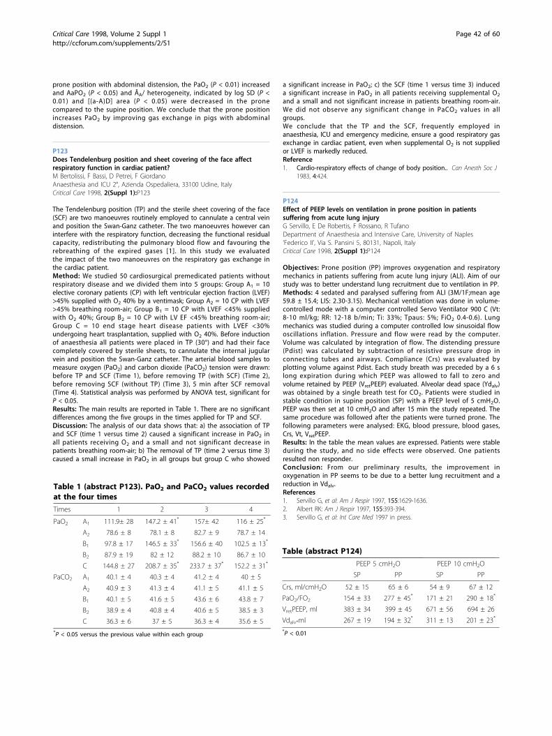
prone position with abdominal distension, the PaO2 (P < 0.01) increasedand AaPO2 (P < 0.05) and ÃA/ heterogeneity, indicated by log SD (P <0.01) and [(a-A)D] area (P < 0.05) were decreased in the pronecompared to the supine position. We conclude that the prone positionincreases PaO2 by improving gas exchange in pigs with abdominaldistension.
P123Does Tendelenburg position and sheet covering of the face affectrespiratory function in cardiac patient?M Bertolissi, F Bassi, D Petrei, F GiordanoAnaesthesia and ICU 2°, Azienda Ospedaliera, 33100 Udine, ItalyCritical Care 1998, 2(Suppl 1):P123
The Tendelenburg position (TP) and the sterile sheet covering of the face(SCF) are two manoeuvres routinely employed to cannulate a central veinand position the Swan-Ganz catheter. The two manoeuvres however caninterfere with the respiratory function, decreasing the functional residualcapacity, redistributing the pulmonary blood flow and favouring therebreathing of the expired gases [1]. In this study we evaluatedthe impact of the two manoeuvres on the respiratory gas exchange inthe cardiac patient.Method: We studied 50 cardiosurgical premedicated patients withoutrespiratory disease and we divided them into 5 groups: Group A1 = 10elective coronary patients (CP) with left ventricular ejection fraction (LVEF)>45% supplied with O2 40% by a ventimask; Group A2 = 10 CP with LVEF>45% breathing room-air; Group B1 = 10 CP with LVEF <45% suppliedwith O2 40%; Group B2 = 10 CP with LV EF <45% breathing room-air;Group C = 10 end stage heart disease patients with LVEF <30%undergoing heart trasplantation, supplied with O2 40%. Before inductionof anaesthesia all patients were placed in TP (30°) and had their facecompletely covered by sterile sheets, to cannulate the internal jugularvein and position the Swan-Ganz catheter. The arterial blood samples tomeasure oxygen (PaO2) and carbon dioxide (PaCO2) tension were drawn:before TP and SCF (Time 1), before removing TP (with SCF) (Time 2),before removing SCF (without TP) (Time 3), 5 min after SCF removal(Time 4). Statistical analysis was performed by ANOVA test, significant forP < 0.05.Results: The main results are reported in Table 1. There are no significantdifferences among the five groups in the times applied for TP and SCF.Discussion: The analysis of our data shows that: a) the association of TPand SCF (time 1 versus time 2) caused a significant increase in PaO2 inall patients receiving O2 and a small and not significant decrease inpatients breathing room-air; b) The removal of TP (time 2 versus time 3)caused a small increase in PaO2 in all groups but group C who showed
a significant increase in PaO2; c) the SCF (time 1 versus time 3) induceda significant increase in PaO2 in all patients receiving supplemental O2
and a small and not significant increase in patients breathing room-air.We did not observe any significant change in PaCO2 values in allgroups.We conclude that the TP and the SCF, frequently employed inanaesthesia, ICU and emergency medicine, ensure a good respiratory gasexchange in cardiac patient, even when supplemental O2 is not suppliedor LVEF is markedly reduced.Reference1. Cardio-respiratory effects of change of body position.. Can Anesth Soc J
1983, 4:424.
P124Effect of PEEP levels on ventilation in prone position in patientssuffering from acute lung injuryG Servillo, E De Robertis, F Rossano, R TufanoDepartment of Anaesthesia and Intensive Care, University of Naples‘Federico II’, Via S. Pansini 5, 80131, Napoli, ItalyCritical Care 1998, 2(Suppl 1):P124
Objectives: Prone position (PP) improves oxygenation and respiratorymechanics in patients suffering from acute lung injury (ALI). Aim of ourstudy was to better understand lung recruitment due to ventilation in PP.Methods: 4 sedated and paralysed suffering from ALI (3M/1F;mean age59.8 ± 15.4; LIS: 2.30-3.15). Mechanical ventilation was done in volume-controlled mode with a computer controlled Servo Ventilator 900 C (Vt:8-10 ml/kg; RR: 12-18 b/min; Ti: 33%; Tpaus: 5%; FiO2 0.4-0.6). Lungmechanics was studied during a computer controlled low sinusoidal flowoscillations inflation. Pressure and flow were read by the computer.Volume was calculated by integration of flow. The distending pressure(Pdist) was calculated by subtraction of resistive pressure drop inconnecting tubes and airways. Compliance (Crs) was evaluated byplotting volume against Pdist. Each study breath was preceded by a 6 slong expiration during which PEEP was allowed to fall to zero andvolume retained by PEEP (VretPEEP) evaluated. Alveolar dead space (Ydalv)was obtained by a single breath test for CO2. Patients were studied instable condition in supine position (SP) with a PEEP level of 5 cmH2O.PEEP was then set at 10 cmH2O and after 15 min the study repeated. Thesame procedure was followed after the patients were turned prone. Thefollowing parameters were analysed: EKG, blood pressure, blood gases,Crs, Vt, VretPEEP.Results: In the table the mean values are expressed. Patients were stableduring the study, and no side effects were observed. One patientsresulted non responder.Conclusion: From our preliminary results, the improvement inoxygenation in PP seems to be due to a better lung recruitment and areduction in Vdalv.References1. Servillo G, et al: Am J Respir 1997, 155:1629-1636.2. Albert RK: Am J Respir 1997, 155:393-394.3. Servillo G, et al: Int Care Med 1997 in press.
Table 1 (abstract P123). PaO2 and PaCO2 values recordedat the four times
Times 1 2 3 4
PaO2 A1 111.9± 28 147.2 ± 41* 157± 42 116 ± 25*
A2 78.6 ± 8 78.1 ± 8 82.7 ± 9 78.7 ± 14
B1 97.8 ± 17 146.5 ± 33* 156.6 ± 40 102.5 ± 13*
B2 87.9 ± 19 82 ± 12 88.2 ± 10 86.7 ± 10
C 144.8 ± 27 208.7 ± 35* 233.7 ± 37* 152.2 ± 31*
PaCO2 A1 40.1 ± 4 40.3 ± 4 41.2 ± 4 40 ± 5
A2 40.9 ± 3 41.3 ± 4 41.1 ± 5 41.1 ± 5
B1 40.1 ± 5 41.6 ± 5 43.6 ± 6 43.8 ± 7
B2 38.9 ± 4 40.8 ± 4 40.6 ± 5 38.5 ± 3
C 36.3 ± 6 37 ± 5 36.3 ± 4 35.6 ± 5*P < 0.05 versus the previous value within each group
Table (abstract P124)
PEEP 5 cmH2O PEEP 10 cmH2O
SP PP SP PP
Crs, ml/cmH2O 52 ± 15 65 ± 6 54 ± 9 67 ± 12
PaO2/FO2 154 ± 33 277 ± 45* 171 ± 21 290 ± 18*
VretPEEP, ml 383 ± 34 399 ± 45 671 ± 56 694 ± 26
Vdalv*ml 267 ± 19 194 ± 32* 311 ± 13 201 ± 23*
*P < 0.01
Critical Care 1998, Volume 2 Suppl 1http://ccforum.com/supplements/2/S1
Page 42 of 60

P125Cytokine removal from plasma of patients with SIRS by theBiologic-DTPF™H Levy1, SR Ash2,3, J Steczko3, DJ Carr31Department of Internal Medicine, University of New Mexico, 2211 LomasBlvd NE, Albuquerque, New Mexico 87131, USA; 2Purdue University and StElizabeth Medical Center, West Lafayette, Indiana, USA; 3HemoCleanse, Inc.,West Lafayette, Indiana, USACritical Care 1998, 2(Suppl 1):P125
The BioLogic-DTPF System™ uses a single lumen 10F venous catheterand combines a hemodiabsorption system using 2 l of sorbentsuspension, flowing through the dialysate side of a parallel platecellulosic dialyzer with whole blood perfusion on the blood side (theBioLogic-DT™ System), in series with a push-pull pheresis system(BioLogic-PF System™). Bidirectional plasma flow (80–100 ml/min) acrossthe 0.5 micron PF membranes provide direct contact between plasmaprotein and powdered sorbent suspension of either charcoal orcharcoal and silica. In vitro tests using whole blood have demonstratedthat the BioLogic-DTPF clears cytokines (TNF-a, IL-lb, and IL-6) at a rateof 15–25 ml/min, without evidence of saturation of sorbent during90 min run times. Systemic Inflammatory Response Syndrome (SIRS) isthe most common manifestation of cyrokine mediated disease in ICUpatients. A Food and Drug Administration-approved InvestiganonalDevice Exemption permits a phase I preliminary safety and efficacystudy of a single 6 h BioLogic-DTPF treatment of 8 patients with SIRSand organ failure due to sepsis.Results: In treatment of 4 patients with charcoal, no adverse eventsoccurred, hemodynamics were stabilized and less pressor agent wasrequired. Cytokine levels decreased during treatment and remainedsignificantly lower the next day.The broad and nonspecific cytokine binding by the BioLogic-DTPF mayquench both the proinflammatory and anti-inflammatory responses ofSIRS and prove beneficial in treatment of patients.
P126Immunological effects of CWHD in critically ill patientsD Kehler, E Tonnesen, P ToftDepartment of Anaesthesia and Intensive Care, Aarhus University Hospital,8000 Aarhus C, DenmarkCritical Care 1998, 2(Suppl 1):P126
Acute renal failure as part of the multi-organ dysfunction syndrome(MODS) is a severe complication in critically ill patients. Hemodynamicinstability is common in these patients and exacerbated by hemodialysis(HD). Therefore, continuous veno-venous hemodiafiltration (CVVHD) isused as an alternative to conventional HD. The aim of the study was totest the hypothesis that initiation of CVVHD elicits an acute activation ofthe immune system followed by immunosuppression.Patient and methods: Fifteen consecutive, septic patients with acuterenal failure were treated with CVVHD in the ICU. The studywas approved by the Regional Ethical Committee on Human Researchand informed consent was obtained from the patients or a close relative.The cellular response was measured as the distribution of the CD3+, CD4+
and CD8+ lymphocytes (flow cytometry), and the leukocyte adhesionmolecules CD1la, CD11b, CD16, CD18, CD44 and CD42 (flow cytometry).The humoral response was estimated as plasma cortisol (RIA), theproinflammatory cytokines TNFa and IL-8 and the anti-inflammatorycytokine IL-10 (ELISA), and the complement split products C3d andC4d (RIE).Results: Ten men and five women were included. Eleven patientsdeveloped MODS after abdominal surgery, 4 patients suffered frommedical diseases. Mean age was 59 years (range 25–75 years). MeanAPACHE II score before CVVHD was 19 (range 8–27). Nine patients (60%)died in septic shock. Mean duration of CVVHD treatment was 9 days(1–21 days). TNFa and IL-8 were detectable in all patients, while IL-10was detectable only in a few patients. Low cytokine concentrations weremeasured in the ultra-filtrate. Big intra- and interindividual variations weredemonstrated for all the immunological parameters. In summary, no
Figure (abstract P125) Results.
Critical Care 1998, Volume 2 Suppl 1http://ccforum.com/supplements/2/S1
Page 43 of 60

significant changes were demonstrated in any of the immunologicalvariables.Conclusions: The hypothesis that CVVHD induces an acute activation ofthe immune system followed by immunosupression was not confirmed inthe present study. The heterogenic patient material with differentunderlying diseases and various duration of illness before start of CVVHDmay contribute to the large variation in the measured immunologicalmarkers.
P127Reversal of intractable circulatory failure complicating septic shock withshort time high volume haemofiltration (ST-HV-CVVH) after failure ofconventional therapy: a prospective evaluationPM Honore1, J Jamez2, M Wauthier2, T Dugernier11Intensive Care Department, St Pierre Hospital, 1340 Ottiguies, Belgium;2Nephrology Department, St Pierre Hospital, 1340 Ottignies, BelgiumCritical Care 1998, 2(Suppl 1):P127
Objectives: According to our previous retrospective study, shorttime highvolume haemofiltration may have beneficial effects on thehaemodynamic course and outcome of patients with refractory septicshock. Therefore, a prospective study was performed to confirm (or not)these preliminary results.Design: Prospective open study over 20 consecutive patients.Setting: Fifteen bedded adult polyvalent intensive care unit in a generalhospital with more than 45% of the admissions coming through theemergency department.Methods: Twenty patients with septic shock were included according toentry criteria including haemodynamic status, acid-base balance, septicstatus, respiratory support and renal status.Technique: It consisted in an exchange of 35 l in a 4 h period of time,achieving a neutral balance. A Gambro device was used with
polyacrylonitrile membranes and bicarbonate was used as buffer. After, thepatient was put on low volume haemofiltration exchanging 24 l a day.Results: 1) Influence on the haemodynamic course: data were analysedusing non parametric statistical methods. A responder was defined using 4criterias as shown by the following table (criteria: C). 2) Influence on theoutcome : 9 patients out of 11 responders survived at day 28. Both groups(survivors N = 9 and non survivors - N = 11) were similar on admission interms of APACHE II score (31.1 vs 32.3 P > 0.05), SAPS II score (69 vs 68.5P > 0.05) but were significantly different regarding the time of intervention(6.1 h vs 14.2 h P < 0.01). The global expected mortality was 79.1% and theobserved mortality was 55%. Using the angular transformation as statisticaltest, the difference was significant with a P value below 0.05.Conclusions: Our prospective open study is confirming our preliminaryresults by suggesting that ST-HV-CVVH is able to reverse intractablecirculatory failure complicating septic shock and to improve survival. Earlyintervention is related with a better outcome. Outcome is well predictedby the early haemodynamic response and not by conventional scoringsystems. In our institution, at the present time, this therapy is restrictedto cases unresponsive to conventional therapy.
P128Successful treatment of refractory toxic streptococcal syndromeassociated with severe lactic acidosis using a combined haemofiltrationtechnique with a bicarbonate based replacement fluid: report of 4consecutive casesPM Honore1, J Jamez2, M Wauthier2, JP Pelgrim1
1Intensive Care Department, St Pierre Hospital, 1340 Ottignies, Belgium;2Nephrology Department, St Pierre Hospital, 1340 Ottignies, BelgiumCritical Care 1998, 2(Suppl 1):P128
Objectives: Devastating toxic strep syndrome has still high mortality race(about 30%) despite all (he therapeutical interventions that have been
Table 1 (abstract P127). Inclusion criteria
1) Haemodynamic status Mean arterial pressure : <55 mmHg
Inotropic support: after failure of maximal dosages of dopamine as last step, epinephrine was used up to
(1 st step) (15 μg/kg/min), dobutamine-epinephrine (2d step) 0.5 μg/kg/min for more than 2 h without
(15 μ/kg/min and 1 μg/kg/min respectively). results.
Cardiac index <2 l/min/m2
Wedge pressure >14<18 mmHg
2) Acid-base balance Arterial pH <7.15
Serum lactate >5 mmol/l
3) Septic status SIRS criteria 3 out of 4
Objective source of sepsis Always present
4) Respiratory support Mechanical ventilation All the patients
paO2/FiO2 ratio <100
5) Renal status No incidence on the inclusion criteria
Table 2 (abstract P127)
Time after onset
of the procedure Parameters Responder group n = 11 Non responder group n = 9 P value
T 0: Time 0 pH 7.14 7.12 >0.05
Mean arterial pressure 50.9 46.1 >0.05
Cardiac index 1.77 1.89 >0.05
Inotropic support E = 38.2 μg/min E = 35.9 μg/min >0.05
T2 = 2 hours Cardiac index (1stC) Increase >50% no S. increase <0.01
after the start SvO2 (2nd C) Increase >25% no S. increase <0.01
T4 = 4 hours pH (3rd C) >7.3 <7.15 <0.01
after the start Inotropic support (4th C) 50% reduction in E/NE no s. reduction <0.01
E, epinephrine; NE, norepinephrine; S, significant.
Critical Care 1998, Volume 2 Suppl 1http://ccforum.com/supplements/2/S1
Page 44 of 60
developped in the last decade. The mortality of the refractory cases liesabove 80%. Several descriptions of favorable outcome have beenpresented using plasmapheresis and intravenous immunoglobin therapy.Amongst the available tools, short lime high volume haemofiltration (ST-HV-SVVH) could be used as therapeutic rescue. We report here aretrospective study of 4 consecutive cases of refractory toxic strepsyndrome in terms of haemodynamic course and outcome.Design: Retrospective study.Setting: Fifteen bedded, adult polyvalent intensive care unit in a generalhospital.Methods: The four cases were in agreement with the consensusdefinition of toxic strep syndrome. They were treated with conventionaltherapy first including high doses of penicillin, surgical debridment whenneeded and adequate critical care therapy.Short time high volume haemofiltration was only used after failure ofconventional therapy.Technique: The technique consisted in the use at first of ST-HV-CVVHexchanging 35 l in a 4 h period of time with achieving a neutral balance. AGAMBRO device was used with polyacrylonitrile membrane (1.6 m2 of activesurface). Bicarbonate was used as buffer. The vascular access was obtainedusing a 14 french double lumen coaxial catheter allowing blood flow of 450ml/min. After, the patient was put on low volume haemofiltration (24 l a day).Results: 1) Influence on the haemodynamic and metabolic courseDespite the dramatic improvement, no ‘P’ value was calculated in view ofthe small number of cases.2) Influence on the outcome: Global expected mortality according to theseverity scoring (APACHE II and SAPS II) was on admission about 82.5%.Observed mortality at day 28: 25% (no P value was calculated). Oneresponder died at day 18 from nosocomial pneumonia.Conclusions: Short time high volume haemofiltration using bicarbonatebased replacement fluid seems to be a valuable non conventional tool intoxic strep syndrom with severe lactic acidosis after failure of classicaltreatment. Response to the therapy is associated with higher likelihood ofimprovement. We need more cases to become statistically significant. Wecan speculate that the M-Protein and exotoxin A wich play a crucial rolein the severity of the disease can be eliminated by the techniqueregarding their molecular weight.
P129Continuous renal replacement therapy in critically ill neonatesS Rödi1, G Zobel1, B Urlesberger2, E Ring1, M Kuttnig-Haim2
1From the Departments of Pediatrics; 2Neonatology, University of Graz,AustriaCritical Care 1998, 2(Suppl 1):P129
Objective: To describe our experience with continuous renal replacementtherapy (CRRT) in critically ill neonates.Design: Prospective case report series.Setting: A 12 bed multidisciplinary pediatric ICU and a 14 bed neonatalICU in a University hospital.Patients: From June 1985 to June 1997 36 critically ill oliguric oranuric neonates underwent continuous arterio-venous (n = 17) orveno-venous (n = 15) renal support. Four neonates were treated withcontinuous ultrafiltration (CUF) during extracorporeal membraneoxygenation (ECMO). Their mean age was 9.8 ± 1.5 days, their meanbody weight 3.01 ± 0.1 kg. All patients were mechanically ventilatedand 88% needed vasopressor support. Indications for CRRT were: lowcardiac output (n = 10), multiple organ system failure (n = 18), severediuretic resistant hypervolemia (n = 3), and severe metabolic crisis(n = 5).Methods: The membrane surface area of the hemofilters ranged from0.015 to 0.2 m2 and the priming volume from 3.7 to 15 ml. For pump-driven hemofiltration a roller pump with pressure alarms, an air trap, anair bubble detector, and small blood lines was used. Fluid balance wascontrolled by a microprocessor controlled unit. The ultrafiltratesubstitution fluid was based on bicarbonate and was partially or totallyreplaced according to the clinical situation.Results: Mean duration of renal support was 97 ± 20 h, ranging from 14to 720 h Operational data and survival rates during arteriovenous andveno-venous hemofiltration and continuous ultrafiltration during ECMOare given in Table 1.Conclusion: Continuous hemofiltration either driven in the arteriovenousor veno-venous mode is a very effective method of renal support forcritically ill neonates to control fluid balance and metabolic derangement.
Table (abstract P128)
Time after onset of the procedure TO T2 (2 hours) T4 (4 hours)
Mean pH 7.03 7.17 7.39
Serum lactate 12.3 mmol/l 11.4 mmol/l 7.1 mmol/l
Cardiac index 1.95 l/min/m2 3.82 l/min/m2 4.34 l/min/m2
Inotropic support A = 0.61 μg/kg/min A = 0.35 μg/kg/min A = 0.19 μg/kg/min
NA = 1.1 μg/kg/min NA = 0.79 μg/kg/min NA = 0.45 μg/kg/min
Table (abstract P128). Status on admission
Haemodynamic status Mean arterial pressure: <52 mmHg
Wedge pressure between 16 and 18 mmHg
Cardiac index 1.95 l/min/m2
Inotropic support
Maximal dosages of dopamine and dobutamine
{norepinephrine (mean) 1.1 μg/kg/min
{epinephrine (mean) 0.61 μg/kg/min
Metabolic Arterial pH (mean) 7.03
Serum lactate (mean) 12.3 mmol/l.
Expected mortality APACHE II: 34.1 (mean) 84%
SAPS II: 71.5 (mean) 81.5%
Critical Care 1998, Volume 2 Suppl 1http://ccforum.com/supplements/2/S1
Page 45 of 60

CUF can be easily performed during ECMO and should be started early inthe presence of severe hypervolemia.
P130Pharmacokinetics of meropenem in intensive care patients receivingcontinuous renal replacement therapyLJ Giles, AC Barber, G Creed, B Ng, RJ Beale, A McLuckieDepartments of Intensive Care and Pharmacy, Guy’s Hospital, St. Thomas’ St.,London SE1 9RT, UKCritical Care 1998, 2(Suppl 1):P130
Introduction: This study was conducted to determine the appropriatedose of meropenem in critically ill patients with acute renal failuretreated by continuous veno-venous haemofiltration (CVVH) orhaemodiafiltration (CVVHDF) who require this antibiotic therapy.Methods: Ten critically ill patients (7m, 3f, mean age 65 years (range:51–77 years), mean weight 80 kg (range: 51–119 kg), mean APACHE II27.7 (range: 21-37) were included. All patients were receiving CVVH(n = 5) (haemofiltration rate 1–2 l/h) or CVVHDF (n = 5)(haemofiltration rate 1–1.5 l/h: dialysis rate 1–1.5 l/h) using apolyacrylonitrile (AN69) hollow fibre 0.9 m2 filter (Multiflow 100,Hospal). All patients received a meropenem dose of 1 g 12 hourly,intravenously over 5 min. Venous serum samples were taken pre-doseand 5, 15, 30, 60, 90, 120, 240, 360, 480 min post-dose on day 3 toapproximate steady state conditions. Haemofiltrate was collected fordetermination of a sieving coefficient. Samples were analysed by highperformance liquid chromatography.Results: Sieving coefficient for meropenem with the AN69 filter was0.93 ± 0.06 (n = 9) indicating free flow across the membrane. Mean(± SD) serum concentrations at pre-dose, 5, 15, 30, 60, 90, 120, 240,360, 480 mins post-dose were: 7.6 (5.1), 90.9 (23.9), 66.0 (13.0), 53.9(15.7), 40.0 (10.3), 39.0 (9.2), 31.0 (10.4), 21.1 (6.9), 17.2 (6.2), 13.8(6.4) mg/l respectively. Actual pre-dose (Ctrough), 5 min (Cpeak) and480 min levels are given below. Serum concentrations remainedabove the MIC90 for Ps. aeruginosa (4 mg/l) in all patients for two-thirds of the dosage interval which is the target recommended for b-lactam antibiotics. A lower dose may not have been sufficient for allthe patients.Conclusion: A meropenem dose of 1 g 12 hourly is adequate in patientstreated with CVVH or CVVHDF using an AN69 HF 0.9 m2 filter.
P131Measuring dobutamine (Db) clearance during continuoushemofiltration (CHF)T Van der Linden, V Chieux, P Cabaret, G Forzy, B Lepoutre, JL DhondtRéanimation, CH St Philibert, BP 249, 59462 Lomme cedex, FranceCritical Care 1998, 2(Suppl 1):P131
Extrarenal purification modifies the pharmacocinetics of many molecules,including the cardiotonics, which are frequently administered inconjunction with these techniques. The objective of this study was tomeasure Db clearance during CHF in ICU patients.Patients and methods: 15 measurements were taken in 5 consecutive ICUpatients (age: 75.4 ± 7 years, SAPS: 17.6 ± 6.6) under Db (posology (D): 16.5 ±5 (μg/kg/min) and CHF (pump: Gambro AK10, hemofilter AN69S Hospal).Samples were taken from plasma afferent to the filter (Ca), the efferentplasma (Ce) and the ultrafiltrate (Cuf). Db clearance was assessed using HPLCwith electrochemical detection in 18% acetonitrile. Clearances werecalculated: instantaneous Cl (iCl = mass of Db extract/Ca) and total body Cl(tbCl = D/Ca) as well as the transmittance coefficient-spectrum (TCS = Cuf/Ca).Results: 1) There was no correlation between the Db concentrations andthe administered dose. 2) Average Db purification was 30% for CHF. 3)The iCl was 78 ± 52 ml, non correlated to the Cuf. 4) Average tbCl was150 ± 160 ml/min, and 93.3% of the tbCl were over 40 ml/min. Therewas a negative polynomial correlation between tbCl and Ca (P < 0.01). 5)The TCS was 0.45 ± 0.28, irrespective of the posology, not influenced bycoprescription of dopamine.Conclusion: During CHF, Db purification is significant and transmembranefiltration is not the only involved mechanism. A non-linear elimination ofthe Db, implicating various factors, remains to be proved.
P132Beneficial use of predilution in reducing the amount of anticoagulationand the occurrence of bleeding during CRRT in critically ill patients: aprospective randomized studyPM Honoré, X Wittebole, M Lemaire, B PirenneDepartment of Intensive Care, St Pierre Hospital, 1340 Ottignies, BelgiumCritical Care 1998, 2(Suppl 1):P132
Objectives: Anticoagulation during CRRT is one of the major issues. Wehave to take into account three parameters: bleeding occurrence, filterlife and filter efficiency. The purpose of the study was to assessprospectively the efficacy of the predilution technique in reducing theamount of anticoagulation during CRRT allowing to use it in critically illpatients at risk for bleeding.Design: Prospective randomized study.Setting: Fifteen bedded, adult polyvalent intensive care unit in a generalhospital.Methods: Twenty consecutive critically ill patients were randomized tohave either a predilution set or a post-dilution one during CRRT using theprisma Hospal pump.Full circuit standard heparinisation was used to achieve an APTT twicebaseline. The vascath site was restricted to the right internal jugular vein.The pump speed was kept at 100 ml/min. Filter efficiency was assessed bythe haemopermeability index (HPI). The HPI represents the ultrafiltrate flowdivided by the transmembrane pressure. The filter efficiency is lost when theHPI has dropped by 70%. Filter life was measured in hours until the circuitclotted off. Data were analysed using non parametric statistical methods.
Table 1 (abstract P129)
CAVH CVVH ECMO+CUF
(n = 15) (n = 17) (n = 4)
Qb (ml/min) 7.0 ± 1.2 23.1 ± 2.4* 45.1 ± 2.8*†
Qf (ml/min/m2) 3.3 ± 0.4 9.5 ± 1.9* 2.3 ± 0.4†
Duration (h) 103 ± 39 103 ± 20 49 ± 13*†
HF-exchange (h) 26.8 ± 6.0 54.4 ± 11.1* 48.4 ± 13.5*
Survival rate(%) 65 67 75
Qb, blood flow rate; Qf, ultrafiltration rates; EF, hemofilter; CAVH, continuousartenovenous hemofiltration; CVVH, venovenous hemofiltration; CUF,continuous ultrafiltration; ECMO, extracorporeal membrane oxygenation;*P < 0.01 CAVH vs CWK, CUF; †P < 0.01 CVVH vs CUF.
Table (abstract P130)
Patient 1 2 3 4 5 6 7 8 9 10
Replacement CVVH CVVHDF
Rates (1/h) 1 1.5 2 2 2 1:1 1:1 1:1 1.5:1.5 1.5:1.5
Ctrough (mg/1) 13.3 4.2 3.4 7.2 14.6 13.3 2.5 6.3 0.5 10.8
Cpeak(mg/l) 90.1 127.7 85.2 73.2 81.7 116.3 101.2 115.0 63.1 55.3
C480mins (mg/l) 19.4 10.5 14.3 11.6 23.3 20.5 6.2 18.7 5.7 7.5
Critical Care 1998, Volume 2 Suppl 1http://ccforum.com/supplements/2/S1
Page 46 of 60
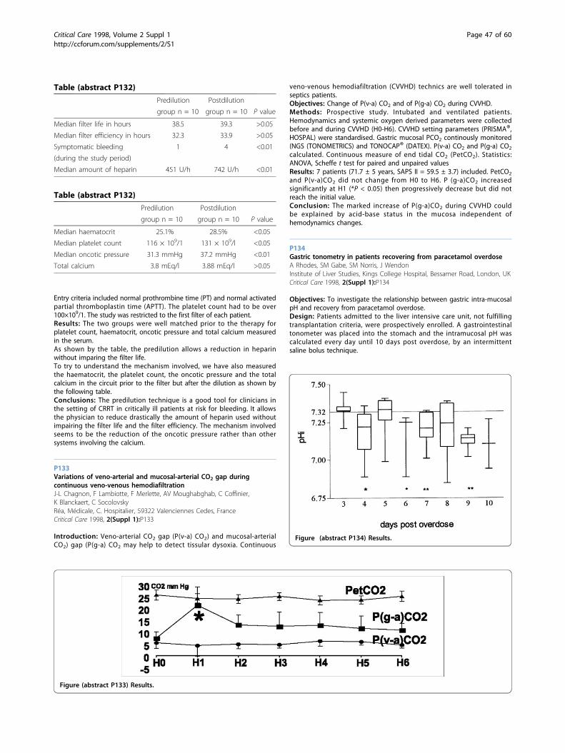
Entry criteria included normal prothrombine time (PT) and normal activatedpartial thromboplastin time (APTT). The platelet count had to be over100×109/1. The study was restricted to the first filter of each patient.Results: The two groups were well matched prior to the therapy forplatelet count, haematocrit, oncotic pressure and total calcium measuredin the serum.As shown by the table, the predilution allows a reduction in heparinwithout imparing the filter life.To try to understand the mechanism involved, we have also measuredthe haematocrit, the platelet count, the oncotic pressure and the totalcalcium in the circuit prior to the filter but after the dilution as shown bythe following table.Conclusions: The predilution technique is a good tool for clinicians inthe setting of CRRT in critically ill patients at risk for bleeding. It allowsthe physician to reduce drastically the amount of heparin used withoutimpairing the filter life and the filter efficiency. The mechanism involvedseems to be the reduction of the oncotic pressure rather than othersystems involving the calcium.
P133Variations of veno-arterial and mucosal-arterial CO2 gap duringcontinuous veno-venous hemodiafiltrationJ-L Chagnon, F Lambiotte, F Merlette, AV Moughabghab, C Coffinier,K Blanckaert, C SocolovskyRéa, Médicale, C. Hospitalier, 59322 Valenciennes Cedes, FranceCritical Care 1998, 2(Suppl 1):P133
Introduction: Veno-arterial CO2 gap (P(v-a) CO2) and mucosal-arterialCO2) gap (P(g-a) CO2 may help to detect tissular dysoxia. Continuous
veno-venous hemodiafiltration (CVVHD) technics are well tolerated inseptics patients.Objectives: Change of P(v-a) CO2 and of P(g-a) CO2 during CVVHD.Methods: Prospective study. Intubated and ventilated patients.Hemodynamics and systemic oxygen derived parameters were collectedbefore and during CVVHD (H0-H6). CVVHD setting parameters (PRISMA®,HOSPAL) were standardised. Gastric mucosal PCO2 continously monitored(NGS (TONOMETRICS) and TONOCAP® (DATEX). P(v-a) CO2 and P(g-a) CO2
calculated. Continuous measure of end tidal CO2 (PetCO2). Statistics:ANOVA, Scheffe t test for paired and unpaired valuesResults: 7 patients (71.7 ± 5 years, SAPS II = 59.5 ± 3.7) included. PetCO2
and P(v-a)CO2 did not change from H0 to H6. P (g-a)CO2 increasedsignificantly at H1 (*P < 0.05) then progressively decrease but did notreach the initial value.Conclusion: The marked increase of P(g-a)CO2 during CVVHD couldbe explained by acid-base status in the mucosa independent ofhemodynamics changes.
P134Gastric tonometry in patients recovering from paracetamol overdoseA Rhodes, SM Gabe, SM Norris, J WendonInstitute of Liver Studies, Kings College Hospital, Bessamer Road, London, UKCritical Care 1998, 2(Suppl 1):P134
Objectives: To investigate the relationship between gastric intra-mucosalpH and recovery from paracetamol overdose.Design: Patients admitted to the liver intensive care unit, not fulfillingtransplantation criteria, were prospectively enrolled. A gastrointestinaltonometer was placed into the stomach and the intramucosal pH wascalculated every day until 10 days post overdose, by an intermittentsaline bolus technique.
Table (abstract P132)
Predilution Postdilution
group n = 10 group n = 10 P value
Median filter life in hours 38.5 39.3 >0.05
Median filter efficiency in hours 32.3 33.9 >0.05
Symptomatic bleeding 1 4 <0.01
(during the study period)
Median amount of heparin 451 U/h 742 U/h <0.01
Table (abstract P132)
Predilution Postdilution
group n = 10 group n = 10 P value
Median haematocrit 25.1% 28.5% <0.05
Median platelet count 116 × 109/1 131 × 109/l <0.05
Median oncotic pressure 31.3 mmHg 37.2 mmHg <0.01
Total calcium 3.8 mEq/l 3.88 mEq/l >0.05
Figure (abstract P133) Results.
Figure (abstract P134) Results.
Critical Care 1998, Volume 2 Suppl 1http://ccforum.com/supplements/2/S1
Page 47 of 60
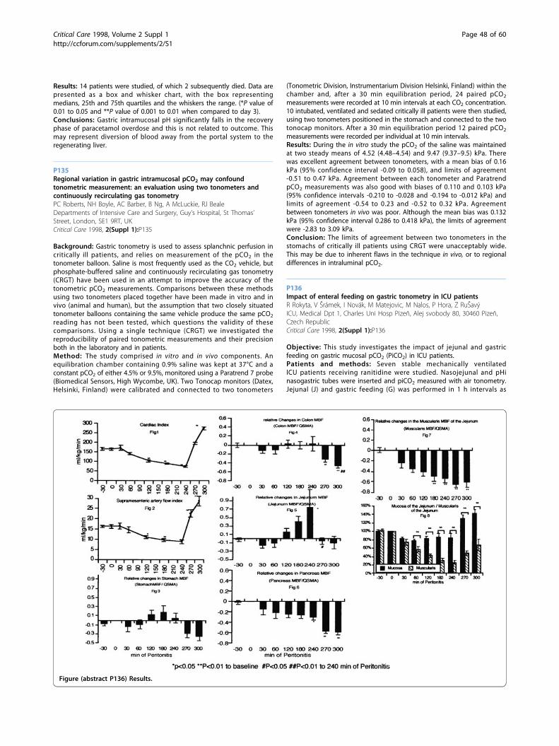
Results: 14 patients were studied, of which 2 subsequently died. Data arepresented as a box and whisker chart, with the box representingmedians, 25th and 75th quartiles and the whiskers the range. (*P value of0.01 to 0.05 and **P value of 0.001 to 0.01 when compared to day 3).Conclusions: Gastric intramucosal pH significantly falls in the recoveryphase of paracetamol overdose and this is not related to outcome. Thismay represent diversion of blood away from the portal system to theregenerating liver.
P135Regional variation in gastric intramucosal pCO2 may confoundtonometric measurement: an evaluation using two tonometers andcontinuously recirculating gas tonometryPC Roberts, NH Boyle, AC Barber, B Ng, A McLuckie, RJ BealeDepartments of Intensive Care and Surgery, Guy’s Hospital, St Thomas’Street, London, SE1 9RT, UKCritical Care 1998, 2(Suppl 1):P135
Background: Gastric tonometry is used to assess splanchnic perfusion incritically ill patients, and relies on measurement of the pCO2 in thetonometer balloon. Saline is most frequently used as the CO2 vehicle, butphosphate-buffered saline and continuously recirculating gas tonometry(CRGT) have been used in an attempt to improve the accuracy of thetonometric pCO2 measurements. Comparisons between these methodsusing two tonometers placed together have been made in vitro and invivo (animal and human), but the assumption that two closely situatedtonometer balloons containing the same vehicle produce the same pCO2
reading has not been tested, which questions the validity of thesecomparisons. Using a single technique (CRGT) we investigated thereproducibility of paired tonometric measurements and their precisionboth in the laboratory and in patients.Method: The study comprised in vitro and in vivo components. Anequilibration chamber containing 0.9% saline was kept at 37°C and aconstant pCO2 of either 4.5% or 9.5%, monitored using a Paratrend 7 probe(Biomedical Sensors, High Wycombe, UK). Two Tonocap monitors (Datex,Helsinki, Finland) were calibrated and connected to two tonometers
(Tonometric Division, Instrumentarium Division Helsinki, Finland) within thechamber and, after a 30 min equilibration period, 24 paired pCO2
measurements were recorded at 10 min intervals at each CO2 concentration.10 intubated, ventilated and sedated critically ill patients were then studied,using two tonometers positioned in the stomach and connected to the twotonocap monitors. After a 30 min equilibration period 12 paired pCO2
measurements were recorded per individual at 10 min intervals.Results: During the in vitro study the pCO2 of the saline was maintainedat two steady means of 4.52 (4.48–4.54) and 9.47 (9.37–9.5) kPa. Therewas excellent agreement between tonometers, with a mean bias of 0.16kPa (95% confidence interval -0.09 to 0.058), and limits of agreement-0.51 to 0.47 kPa. Agreement between each tonometer and ParatrendpCO2 measurements was also good with biases of 0.110 and 0.103 kPa(95% confidence intervals -0.210 to -0.028 and -0.194 to -0.012 kPa) andlimits of agreement -0.54 to 0.23 and -0.52 to 0.32 kPa. Agreementbetween tonometers in vivo was poor. Although the mean bias was 0.132kPa (95% confidence interval 0.286 to 0.418 kPa), the limits of agreementwere -2.83 to 3.09 kPa.Conclusion: The limits of agreement between two tonometers in thestomachs of critically ill patients using CRGT were unacceptably wide.This may be due to inherent flaws in the technique in vivo, or to regionaldifferences in intraluminal pCO2.
P136Impact of enteral feeding on gastric tonometry in ICU patientsR Rokyta, V Šrámek, I Novák, M Matejovic, M Nalos, P Hora, Z RuŠavýICU, Medical Dpt 1, Charles Uni Hosp Pizeñ, Alej svobody 80, 30460 Pizeñ,Czech RepublicCritical Care 1998, 2(Suppl 1):P136
Objective: This study investigates the impact of jejunal and gastricfeeding on gastric mucosal pCO2 (PiCO2) in ICU patients.Patients and methods: Seven stable mechanically ventilatedICU patients receiving ranitidine were studied. Nasojejunal and pHinasogastric tubes were inserted and piCO2 measured with air tonometry.Jejunal (J) and gastric feeding (G) was performed in 1 h intervals as
Figure (abstract P136) Results.
Critical Care 1998, Volume 2 Suppl 1http://ccforum.com/supplements/2/S1
Page 48 of 60

follows (period 1–9): 1 = baseline; 2 = J 40 ml/h; 3 = J 100 ml/h; 4 = nofeeding; 5 = G 40 ml/h; 6 = G 100 ml/h; 7 = G bolus 200 ml; 8 = gastricemptying; 9 = no feeding.Values are provided as means ± SD. ANOVA for repeated measures andWilcoxon test were used for statistical analysis; P < 0.05 was consideredsignificant.Results: Patients remained stable during the study. Baseline pCO2 gap(piCO2-paCO2) was 1.91 ± 0.66 kPa and did not change during the study(ANOVA P = 0.44; Fig. 1)Marked interindividual variability was observed during gastric feedingmainly.Conclusion: In ICU patients jejunal feeding does not influence piCO2. Theeffect of gastric feeding is much less predictable and needs to be testedin each patient.
P137Microcirculatory blood flow in the gastrointestinal tract during earlyseptic shockL Hiltebrand1, A Banic2, GH Sigurdsson11Department of anaesthesia and Intensive Care Medicine; 2Department ofPlastic & Reconstruktive Surgery, University Hospital, Berne, SwitzerlandCritical Care 1998, 2(Suppl 1):P137
The autoregulation of microcirculatory blood flow (MBF) in the gut isimpaired during sepsis. However, there is little data available on thedistribution of blood flow in the different parts of the gastrointestinaltract and on the relationship between mucosal perfusion andsupramesenteric artery flow (QSMA) during sepsis. The aim of this studywas to elucidate the relationship between regional blood flow (QSMA)and the local flow in different tissues and parts of the gut and in thepancreas during early septic shock.Materials and methods: Nine pigs (20–24 kg) were anesthetised endo-tracheally intubated and ventilated. Cardiac index (CI) was measuredwith thermodilution technique and mesenteric artery blood flow (QSMA)with an ultrasound flowprobe. Microcirculatory blood flow (MBF) wascontinuously monitored in multiple organs (pancreas, gastric-, jejunal-,colonic mucosa and muscularis of the jejenum) with a multichannel laser
Doppler system. After baseline measurements a generalised faecalperitonitis was induced by instillation of 20 g of faeces in the abdominalcavity. After 240 min, i.v. fluids were administered which altered thehypodynamic shock to hyperdynamic septic shock.Results and discussion: During the first 240 min (hypodynamic shock) CIand QSMA decreased by 50% (P < 0.01; Figs 1 and 2). Microcirculatory bloodflow in the mucosa of the stomach and colon decreased similarly (Figs3 and 4) while the mucosa of the jejunum was maintained close to baseline(Fig. 5). On the other hand, MBF in the pancreas and the jejunal muscularisdecreased significantly more than the QSMA (Figs 6 and 7). Administrationof i.v. fluids at 240 min was followed by a significant increase in CI andQSMA (Figs 1 and 2). Although this was followed by some increase in MBFin the mucosa of the stomach and colon as well as in the pancreas andjejunal muscularis, the relative amount of flow in these organs significantlydecreased as compared with QSMA (Figs 3,4,5). MBF in the mucosa of thejejunum increased essentially parallel with the QSMA (Fig. 6).Conclusion: a) In septic shock jejenunal mucosal blood flow ismaintained despite decreased mesenteric flow (QSMA), probably throughredistribution of flow from muscularis to the mucosa (Fig. 8).b) Microcirculatory flow in the mucosa of the stomach and colondecreases parallell with QSMA during hypodynamic septic shock, whileduring hyperdynamic sepsis it decreases significantly compared withQSMA (Figs 3 and 4).c) Microcirculatory flow in the pancreas decreases significantly more thanQSMA both during hypodynamic and hyperdynamic septic shock (Fig. 6).
P138The effects of dopexamine on renal function in surgical renal arteryreconstruction: a pilot studyED Cheetham, PDF Dodd, L Gurnani, JM Eddleston, MG WalkerDepartment of Vascular Surgery, Manchester Royal Infirmary, Oxford Road,Manchester, UKCritical Care 1998, 2(Suppl 1):P138
Aims: To evaluate the effects of dopexamine on haemodynamics andrenal function in patients undergoing surgical renal artery reconstruction(RARS).
Figure 1-8 (abstract P137) Onset of Peritonitis at 0 min and Pentastrach administration at 240 min. Data presented as mean ± SEM. Paired ANOVA forrepeated measurements was used for comparison. P < 0.05 was considered statistically siginficant
Critical Care 1998, Volume 2 Suppl 1http://ccforum.com/supplements/2/S1
Page 49 of 60

Methods: Ten patients (aged 54 to 75; 7 males), scheduled for RARS wererandomised, following cardiovascular optimisation, to receive eitherdopexamine (1 μg/kg/min), introduced 2 h before surgery and continuedfor 24 h after surgery (DPX, n = 5), or placebo (PLA, n = 5). Anaesthesiawas similar in both groups. Pre-operatively, all patients were assessed byrenal angiography. Renal function was assessed by plasma creatinine,creatinine clearance and iohexol clearance [1]. Haemodynamics andsplanchnic perfusion were assessed by invasive cardiovascular monitoringand gastric tonometry respectively. Data are expressed as mean ± SD.Statistics: Student’s t test was used.Results: The 2 groups were comparable with reference to demographicdata and surgical procedures (POSSUM predicted risks:DPX mortality21%, morbidity 72%; PLA mortality 22%, morbidity 75%). There was 1death in each group in the first 8 weeks after surgery. Data wasincomplete for iohexol clearance due to technical difficulties (numbersin subscript).There was no significant difference between the groups iniohexol or creatinine clearance (Iox, CreaCl, ml/min), plasma creatinine(Great, μmol/l), urinary volumes (UrVol, l), pHi, oxygen delivery (DO2I)and consumption (VO2I, ml/min/m2) and oxygen extraction ratio (%).Results are shown in the table overleaf (*P < 0.1, significance =P < 0.05):Conclusion: In this small group, dopexamine at this dose did not appearto offer a significant benefit to patients undergoing RARS.Reference1. Brown SC, O’Reilly PH: Iohexol clearance for the determination of
glomerular filtration rate in clinical practice: evidence for a new goldstandard. J Urol 1991, 146:675-679.
P139Comparing the effects of dopexamine and dobutamine on splanchnicparameters in patients with severe sepsisF Esen2, U Friess1, M Quintel 1, K van Ackern11Dept. of Anesthesia logy, University of Heidelberg, Klinitum Mannheim,Germany; 2Dept. of Anesthesiology and Intensive Care, University of Istanbul,Istanbul, TurkeyCritical Care 1998, 2(Suppl 1):P139
Introduction: This prospective randomized study was designed to assessthe effect of low dose dopexamine Hcl (dp) and dobutamine Hcl (db) onsplanchnic blood flow as measured by gastric intramucosal pH (pHi),hepatic metabolism of lidocain to monoethylglicinexylidide (MEGX), andplasma disappearance rate of indocyanine green (PDR/ICG).Methods: 16 critically ill patients, meeting the criteria for the diagnosis ofsevere sepsis, were assigned either to receive dopexamine (1 μg/kg/min)or dobutamine (5 μg/kg/min). Baseline measurements of pHi, MEGX, PDR/ICG as well as hemodynamic, respiratory parameters, oxygen derivedvariables and arterial lactate were measured. At the end of a 2 h infusionof an appropriate drug, a repeated set of the measurements were taken.Results: Both drugs produced significant increases in cardiac index,apparently due to a significant increase in heart rate. SVI (stroke volumeindex) remained unchanged with dopexamine, however some increasewas observed with dobutamine. DO2I (oxygen supply) increased similarly
in both groups (16% dp, 19% db) with VO2I (oxygen consumption)increasing more with db compared to dp. Lactate values after each drugadministration appeared to be significantly lower than baseline values(dp 2.08 ± 0.53, db 2.50 ± 1.99 ± 0.99).No significant improvement in the splanchnic parameters was observedwith dopexamine. PHi increased significantly with db (7.26 ± 0.04 to 7.33± 0.07 P < 0.05). PDR/IICG and MEGX values showed significant increaseswith db (PDR/ICG from 15.5 ± 9.1 to 21.3 ± 10.3%; MEGX from 19.8 ±11.2 to 28.8 ± 18.4 P < 0.05), and stayed constant with dp, however bothparameters measured atbaseline were higher in dp group than in dbgroup.Conclusion: This study demonstrated the expected effects of each drugas mentioned in the previous studies. Our data showed that in severelyseptic patients, dobutamine leads to greater effectiveness in improvingthese accepted splanchnic parameters than dopexamine. However, howwell and accurate these parameters reflect the abnormalities ofsplanchnic organ blood supply and their metabolism needs to beinvestigated by further studies.
P140Hepatic O2 transport and energy balance in hyperdynamic porcineendotoxin shock: NG-monornethyl-L-arginine (L-NMMA) versusnoradrenalineKM Rieger, K Träger, A Vlatten, M Matejovic, T Iber, M Georgieff,P RadermacherSektion APV, Universitätsklinik füur Anästhesiologie, D-89070 Ulm, GermanyCritical Care 1998, 2(Suppl 1):P140
Introduction: Nitric oxide synthase (NOS) inhibition is currentlyinvestigated as a treatment of arterial hypotension associated with septicshock, but controversial data are available with respect to hepatic O2
balance. Therefore we studied the effect of NOS inhibition withL-NMMA on liver O2 transport as well as energy balance, in comparisonto a standard treatment with noradrenaline (NOR) in a hyperdynamicendotoxin (ETX) shock model.Methods: Thirty anesthetized pigs were randomly assigned to 3 groups12 h after induction of endotoxic shock with continuous LPS infusion: ETXgroup: no drug therapy (n = 8); NOR group (n = 11) and L-NMMA group(n = 11): vasopressor therapy with NOR and L-NMMA was titrated tomaintain MAP at preshock levels. Hepatic blood flow (Transonic® Dopplerultrasound), O2 kinetics as well as mean and frequency distribution ofintracapillary HbO2 saturation (remission spectrophotometry, EMPHO)were measured.Results: Data are median and interquartile range. *P < 0.05 vs preshock(RM ANOVA on ranks). **P < 0.05 between groups (ANOVA).Conclusions: Although, in contrast to NOR, L-NMMA did notfurtherincrease macrocirculatory liver O2 availability, there was no differencebetween the two treatments with regard to microcircula-tory O2 supplyand O2 uptake. Hence, neither of the two treatments improved liverenergy balance.Acknowledgement: L-NMMA (546C88) was kindly provided byGlaxoWellcome, UK.
Table (abstract P138)
Pre Drug Post Ind’n DPX End Surg. 24 h post 2–4 months re Drug post Ind’n PLA End Surg. 4 h post 2–4 month
lox 74.3± 31.23 62 ± 51.84 86 ± 29.94 53.6 ± 21.25
CreaCl 56.7 ± 38.3 51.3 ± 21.6
Creat 138 ± 63.5 184.2 ± 92.7 109.3 ± 23* 109.4 ± 25.7 174 ± 50.7 132.7 ± 9.8
% ΔCreat 30.3 ± 31.4 66.6 ± 61.5
UrVol(24) 2.1 ± 1.03 3.2 ± 1.05
pHi 7.39 ± 0.05 7.35 ± 0.09 7.28 ± 0.14 7.39 ± 0.1 7.37 ± 0.04 7.29 ± 0.06
DO21 458 ± 87 520 ± 93* 699 ± 163* 643 ± 224 512 ± 78 412 ± 57 516 ± 81 552 ± 70
VO21 112 ± 11 85 ± 40 101 ± 40 162 ± 71 130 ± 23 87 ± 7 83 ± 21 151 ± 26
O2ER 24 ± 3 16 ± 6 14 ± 5 26 ± 7 26 ± 7 21 ± 3 16 ± 6 27 ± 5
Critical Care 1998, Volume 2 Suppl 1http://ccforum.com/supplements/2/S1
Page 50 of 60

P141Intestinal O2 transport and energy balance during hyperdynamicendotoxic shock in the pig: comparison of noradrenaline andNG-monomethyl-L-arginine (L-NMMA)K Träger, KM Rieger, A Vlatten, M Matejovic, T Iber, M Georgieff,P RadermacherSektion APV, Universitätsklinik fur Anästhesiologie, D-89070 Ulm, GermanyCritical Care 1998, 2(Suppl 1):P141
Introduction: Vasopressor therapy is current practice for treatment ofarterial hypotension associated with septic shock, but may result in gutdysfunction due to intestinal vasoconstriction. Therefore we compared theeffects of vasopressor treatment with the nitric oxide synthase inhibitorL-NMMA with those of noradrenaline (NOR) on intestinal O2 transport aswell as energy balance in a porcine hyperdynamic endotoxic shock model.Methods: Thirty anesthetized pigs were studied; 12 h after induction ofendotoxic shock with continuous LPS infusion animals were randomlyassigned to receive either no drug therapy (ETX, n = 8), or vasopressorsupport with noradrenaline (NOR, n = 11) or L-NMMA (L-NMMA, n = 11),respectively, in order to maintain MAP at preshock levels. Portal venousblood flow (Transonic flow probes), arterial-portal venous O2 extraction,
ileal mucosal intracapillary HbO2 saturation (remission spectrophotometryEMPHO) and arterial-ileal mucosal PCO2 gaps were measured.Results: Data are median and interquartile range. *P < 0.05 vs preshock(RM ANOVA on ranks). (See table overleaf.)Conclusions: Despite well-preserved O2 availability neither of the twotreatments could reverse the endotoxin induced derangement ofintestinal energy balance.Acknowledgement: L-NMMA (546C88) was kindly provided byGlaxoWellcome, UK.
P142Endogenous glucose release in hyperdynamic porcine endotoxin shock:NG-monomethyl-L-arginine (L-NMMA) versus noradrenalineA Vlatten, K Träger, KM Rieger, T Iber, M Matejovic, M Steudle, M Georgieff,P RadermacherSektion APV, Universitätsklinik für Anästhesiologie, D-89070 Ulm, GermanyCritical Care 1998, 2(Suppl 1):P142
Introduction: Inhibition of nitric oxide (NO) synthesis is currentlyinvestigated for the treatment of hypotension associated with septic shock.Since both endotoxin (ETX) and NO have been reported to influence
Table (abstract P140)
Preshock 12 h 18 h 24 h
Liver DO2 ETX 2.3 [1.9;2.8] 2.2 [1.7;2.7] 2.7 [2.0;3.0] 2.0 [1.4;2.6]
ml/min/kg NOR* 2.3 [1.8;2.8] 2.0 [1.5;2.4] 3.1 [2.8;3.6]** 2.6 [1.9;4.1]
L-NMMA 2.5 [2.2;2.9] 2.5 [2.1;2.61 2.3 [1.7;2.8] 2.0 [1.1;2.6]
Hepatic ETX 54 ± 5 51 ± 7 51 ± 7 47 ± 10
intracap. HbO2 NOR 59 ± 6 54 ± 8 61 ± 7 58 ± 12
Mean ± SD L-NMMA 59 ± 8 56 ± 10 55 ± 10 58 ± 7
Liver VO2 ETX 0.7 [0.5;1.1] 0.6 [0.4;0.8] 0.6 [0.4;0.8] 0.6 [0.3;0.7]
ml/min/kg NOR 0.7 [0.5;1.1] 0.6 [0.4;0.8] 0.6 [0.4;0.8] 0.6 [0.3;0.7]
L-NMMA 0.8 [0.5;0.8] 0.8 [0.5;1.0] 0.6 [0.4;0.8] 0.5 [0.3;0.8]
Hepatic ETX* 7.48 [17.46;7.50] 7.38 [7.34;7.42] 7.36 [7.34;7.381 7.30 [7.25;7.34]
venous pH NOR* 7.49 [7.46;7.51] 7.33 [7.25;7.34] 7.32 [7.2817.371 7.22 [7.17;7.37]
L-NMMA* 7.48 [7.44,7.49] 7.32 [7.31 ;7.341 7.29 [7.26;7.321 7.24 [7.16;7.30]
Hepatic ETX* 24 [15;32] 26 [18;30] 34 [28;51] 61 [31;123]
venous Lac/Pyr NOR* 31 [21;37] 38 [25;66] 95 [49;1 01] 104 [41;204]
ratio L-NMMA* 21 [18;29] 28 [28;50] 60 [40;95] 148 [67;187]
Table (abstract P141)
Preshock 12 h 18 h 24 h
Portal venous ETX 23 (19;28) 25 (29;28) 24 (20;29) 28 (19;31)
blood flow NOR* 23 (20;26) 25 (18;28) 36 (26;37) 38 (28;42)
(ml/min/kg) L-NMMA 24 (22;28) 32 (24;40) 26 (23;36) 30 (19;34)
O2 extraction ETX 34 (27;35) 29 (20;33) 26 (23;32) 26 (25;35)
(%) NOR* 32 (26;35) 31 (27;41) 20 (18;24) 22 (17;32)
L-NMMA 31 (29;34) 31 (27;35) 31 (27;31) 31 (24;54)
ileal mucosal ETX 43 ± 13 39 ± 21 37 ± 10 30 ± 10
intracap. HbO2 NOR 46 ± 15 44 ± 12 44 ± 8 45 ± 14
Mean ± SD L-NMMA 48 ± 12 43 ± 11 42 ± 11 42 ± 12
Portal venous ETX* 25.1 [22.0;29.1] 25.3 [22.5;28.5] 31.8 [22.8;34.7] 42.7 [40.0;54.5]
Lac/Pyr ratio NOR* 28.1 [26.1;31.3] 31.3 [24.8;37.6] 46.4 [39.0;53.3] 44.7 [36.6;65.9]
L-NMMA* 20.8 [18.6;29.5] 26.5 [25.3;40.4] 39.9 [28.4;50.5] 48.0 [44.3;53.2]
ileal mucosal- ETX* 12 (9;13) 13 (11;20) 17 (14;25) 20 (19;28)
arterial PCO2 gap NOR* 13 (4;15) 19 (16;29) 19 (13;26) 15 (11;53)
(mmHg) L-NMMA* 13 (11;16) 23 (17;29) 37 (13;53) 44 (28;45)
Critical Care 1998, Volume 2 Suppl 1http://ccforum.com/supplements/2/S1
Page 51 of 60

gluconeogenesis we compared the effects of standard vasopressor therapywith noradrenaline (NOR) to L-NMMA on endogenous glucose release duringlong-term ETX induced shock.Methods: Pigs were studied in a normotensive, hyperdynamic shockmodel with volume resuscitation (HES 6%) to keep intrathoracic bloodvolume constant. Animals were randomly assigned to three groups 12 hafter start of ETX infusion. ETX group: no drug treatment (n = 8); NORgroup: treatment with NOR (n = 11); L-NMMA group: treatment with L-NMMA (n = 11), both titrated to maintain MAP at preshock levels. Hepaticuptake of glucose precursors as well as endogenous glucose formation(stable isotope approach) were measured.Results: Data are median and interquartile range. *P < 0.05 vs preshock(RM ANOVA on ranks).**P < 0.05 between groups (ANOVA).Conclusion: Despite their similar effects on liver hemodynamics and O2
kinetics neither NOR nor L-NMMA influenced the ETX-induceddissociation between liver uptake rates of glucose precursors and total denovo glucose formation rate.Acknowledgement: L-NMMA (546C88) was kindly provided byGlaxoWellcome, UK.
P143Factors influencing serum lactate concentration in ICU newbornsI Maruniak-Chudek, J ŒwietliñskiDepartment of Anesthesiology and Intensive Care of Children, 11 Clinic ofPediatric, Silesian University School of Medicine, Katowice 40-852, ul.Poœpiecha 14, PolandCritical Care 1998, 2(Suppl 1):P143
Serum lactate concentration provides clinically important information onmetabolic status of critically ill neonates.Aim: The purpose of the study was to assess some factors influencinglactate levels during first 48 h of intensive treatment.Setting: A university hospital pediatric intensive care unit (PICU).Methods: 173 neonates (gestational age 35 hbd /median/, mean birthweight 2045 ± 808 g, SNAP score -9 /median/) subsequently admitted toPICU in first two days of life were studied. Blood lactate levels, blood gases,electrolytes and glucose levels were determined at the admission and after24 and 48 h of PICU treatment. SNAP score and data concerning oxygentransport into the tissues were collected simultaneously.Results: Blood lactate levels increased with low mean arterial pressure (P <0.001), low Apgar score (at 5 min), P < 0.01 at admission, P < 0.05 in 24 h,ns in 48 h, low temperature at admission, P < 0.001, low hematocrite P <0.05 and high SNAP value, P < 0.001. No significant correlations betweenlactates and other demographic and clinical data were observed.Conclusions: Based on our results we conclude that hypoperfusion andbad clinical condition (SNAP score) has the strongest impact on bloodlactates. Of interest we found persistent effect of asphyxia evaluated asApgar score and hypothermia at the admission on high lactate incidence.
P144Abdominal sepsis in the surgical intensive care unit: a description andsearch for prognostic factorsP Haraldsen, K Isaksson, Z Zdanowski, R AnderssonDepartment of Surgery, Lund University Hospital, 221 85 Lund, SwedenCritical Care 1998, 2(Suppl 1):P144
Introduction: Patients with AS treated in the surgical ICU suffersubstantial morbidity and mortality, despite efforts of modern intensive
care and economical means being invested. The present study wasperformed in order to describe this cathegory of patients and if possibleto identify factors of potential prognostic importance.Methods: All consecutive patients treated due to AS in the surgical ICUat Lund University Hospital during the period January 1983 to December1995 were analysed retrospectively. Information on demography, scoringaccording to APACHE II, SSS and MODS scores, complications andmortality was assessed.Results: Totally 210 patients, 75 females and 135 males, with a mean ageof 65 years were included in the study. Median time of hospitalizationwas 26 days including 6 days at the ICU accounting for a median cost of48.112 USD/ hospital stay. The main cause of AS was visceral perforation(42%). Abdominal bacterial cultures were dominated by enteric bacteria(80%), while blood cultures showed equal frequences of enteric and skinbacteria (21%). Most patients were subjected to surgical intervention(88%).Complications were frequent, dominated by organ failure, seen in 83%.Pulmonary, cardiac and renal failure were most frequent. Multiple organfailure, defined as failure of >2 organ systems, developed in 52%. Thetotal mortality rate was 28%, the most common cause of death beingMODS (69%). APACHE II and SSS scores did not significantly differbetween patients with or without concommitant MODS nor betweensurvivors and non-survivors. A trend towards correlation between highscores and poorer outcome could be seen. A MODS score of >4predicted development of multiple organ failure (P < 0.001), but did notpredict mortality.The presence of previous deseases did not significantly influence onmorbidity and mortality, nor did age, sex, underlying diagnosis norbacterial culture findings. The occurrence of organ failure correlated withpoor prognosis and fatal outcome, the more organ systems that failed,the stronger the correlation. Pulmonary (P < 0.05), cardiac (P < 0.001) andrenal (P < 0.001) failure all increased the risk of lethal outcome, as didmultiple organ failure (P < 0.001).Conclusions: Abdominal sepsis causes substantial morbidity andmortality, mainly due to the frequent development of multiple organdysfunction syndrome. The condition consumes substantial economicalresources and health care facilities. Factors of prognostic importance areall associated with the development of multiple organ failure syndromeand not to individual factors like age, sex, previous diseases or diagnosis.
P145O’Gilvie’s Syndrome (colonic pseudo-obstruction) in the intensive careunitX Castella, F Torner, L Ribera, J RobustéIntensive Care Medicine Department, Hospital General de Manresa, La CullaS/N, O8240 Manresa (Barcelona), SpainCritical Care 1998, 2(Suppl 1):P145
Background: O’Gilvie’s syndrome (colonic pseudo-obstruction) is agastrointestinal motility disorder affecting the whole bowel but involvingmainly the colon. It produces an acute massive dilation of the largebowel without organic obstruction. The cause of the disease is notknown but it seems to involve some autonomic neurologic disorderaffecting the myenteric plexus. For unknown reasons it affects selectivelyelderly, COPD patients with critical diseases (sepsis, polytrauma, vasculardisease, etc.). The mainstay of the differential diagnosis is to exclude anycause of mechanical ileus. The treatment of the syndrome is mainlysupportive including administration of fluids and electrolytes,total parenteral nutrition, and cisapride. If after these measures
Table (abstract P142)
Preshock 12 h 18 h 24 h
Liver lactate ETX* 13 [11;17] 5 [-1;8] 2 [-3;5] -6 [-16;5]
uptake NOR* 9 [6;16] -2 [-7;4] -4 [-11;3] -5 [-10;6]
(μmol/min/kg) L-NMMA* 9 [7;14] 3 [0;4] -8 [-10;-4] -17 [-24;9]
Endogenous glucose ETX* 25 [20;28] 32 [29;38] 32 [22;39] 36 [29;65]
production NOR* 23 [20;24] 34 [31 ;37] 43 [36;50]** 35 [28;49]
(μmol/min/kg) L-NMMA* 23 [18;26] 30 [27;35] 26 [25;43] 29 [23;33]
Critical Care 1998, Volume 2 Suppl 1http://ccforum.com/supplements/2/S1
Page 52 of 60
pseudo-obstruction still persists, colonoscopic decompression is required.An endoscopic placement of a transanal large bore nasogastric tube inthe transversus colon is suggested. Continuous lavage of the colonthrough the tube could be useful. If after these measures the colon is stilldilated, surgery with cecostomy is indicated.Results: In the next table we report our experience in six cases ofO’Gilvie syndrome patients during the years 1996–1997.Conclusion: In our experience O’Gilvie’s syndrome is much morecommon than reported. Medical treatment including cisapride is valuablebut in severe cases colonoscopic decompression with transanally NG tubeplacement with continous lavage is required. In some cases a surgicaldecompressive cecostomy may be necessary.
P146Gastric emptying as assessed by paracetamol absorption is normal 32 hafter elective abdominal aortic aneurysm surgery (AAA)J Cohen1, R Avrahami2, P Singer 1, A Zelikovski21Depts General ICU; 2Vascular Surgery, Rabin Medical Center, CampusBeilinson, Petah Tikva, Israel, 49100Critical Care 1998, 2(Suppl 1):P146
Major potential complications following AAA include 1) MODS, possiblyrelated to GI barrier dysfunction [1-] and 2) stress ulcers, seen ongastroscopic examination in as many as 80% of patients []. Enteralnutrition may prevent both these complications, especially if given early.However patients are often denied this form of treatment because ofconcerns of delayed gastric emptying. Instead they are subjected tonasogastric (N/G) decompression, shown to be unnecessary and evenharmful [3]. We used the paracetamol absorption test to document thepresence of intact or abnormal gastric emptying in the immediatepostoperative period.Methods: We studied 13 consecutive patients admitted to the ICU forroutine postoperative care following elective AAA. Gastric emptying usingthe paracetamol absorption test was assessed 6, 18 and 32 h followingsurgery. The test was considered normal if the area under theconcentration curve from 0–60 min (AUC60) after giving 1 mg ofparacetamol through the N/G tube was >600 mg/min/1. Other factorsnoted included i) demographic data;and ii) time to extubation, removalN/G tube and start of enteral feeding.Results: Patients mean age was 69 ± 12 years; M:F ratio 12:11) Results gastric emptying (mean ± SD):
2) Mean time to end-points:Conclusions: This study showed that 69% of patients hava normal gastricemptying at 18 h and 92% at 32 h following elective AAA. We suggestthat unnecessary N/G tubes may be removed and enteral nutritionstarted to the benefit of the patient 32 h post AAA.References1. Huber S, et al: J Vasc Surg 1995, 22:287.2. Martin IF: Am Surg 1994, 60:169-174.3. Schwartz C, et al: South Med J 1995, 88:825-830.
P147Glycaemia influences glucose metabolism in sepsis duringhyperinsulinemic clampZ Rušavý, V Šrámek, S Lacigová, P TeŠínský, I Novák, R Rokyta, H KordováMedical Dpt I, Charles Uni Hosp Plseñ, Alej svobody, 30460 Plzeñ, CzechRepublicCritical Care 1998, 2(Suppl 1):P147
Objectives: The optimal glycaemia in critically ill is not known. Weinvestigated glucose metabolism in septic patients during hyperglycaemicclamp.Patients and methods: In 10 non-diabetic stable septic patients onmechanical ventilation with baseline glycaemia >6mmol/1 andcontinuous insulin infusion, two hyperinsulinaemic clamps (targetinsulinaemia 250 U/l) were performed after 8 h of no caloric intake.Target glycaemia was 5 mmol/1 (step 1) and 10 mmol/1 (step 2)respectively. Glucose uptake was calculated as the amount of glucose pertime needed to maintain target glycaemia. Glucose oxidation wascalculated from indirect calorimetry and urinary nitrogen losses. Valuesare provided as means ± SD, paired T test was used for statistical analysisand P < 0.05 was considered significant.Results: Glucose uptake was at step 1 significantly lower than at step 2(3.9 ± 2.5 mg/kg/min and 6.9 ± 1.93 mg/kg/min, respectively; P < 0.001).Glucose oxidation was also significantly lower at step 1 (2.4 ± 1.38 mg/kg/min and 4.46 ± 1.65 mg/kg/min, respectively; P < 0.01). Energyexpenditure did not change (2280 ± 418 kcal/ 24 h and 2235 ± 216 kcal/24 h, respectively).Conclusion: During hyperinsulinaemic clamp in sepsis glucose uptakeand oxidation depend on glycaemia. Higher glycaemia may be beneficialin terms of higher glucose uptake and oxidation which is notaccompanied by higher energy expenditure suggesting the suppressionof catabolism.
P148Role of a short parenteral nutrition coupled with early enteral nutritionin the critically ill: a double-blind randomized study versus placeboPh Bauer, C Charpentier, C Bouchet, F Raffy, N Gaconnet, A LarcanRéanimation médicate - Hôpital Central - F54035 Nancy, FranceCritical Care 1998, 2(Suppl 1):P148
The critically ill, stressed, sometimes undernourished may develop anintestinal dysfunction which leads to multiple organ failure. Early enteralnutrition (EN) is usually recommended but may fail to provide an adequateamount of substrate. Parenteral nutrition (PN) is used in case of failure orcontraindications of EN. However, the role of early EN coupled with PN hasnot been evaluated. We conducted a double-blind, randomized, placebo-controlled study to assess the influence of adjuvant PN (Vitrimix KV,Pharmacia Upjohn) and early EN on the improvement of nutritionalparameters, morbidity and mortality in the critically ill, excepted afterelective surgery. Statistical analysis, in intention to treat, used parametrictests (chi-2, ANOVA). Two groups of 60 patients (EN + placebo versus EN +PN) have been enrolled: 82 males, 38 females, admitted for medical (51%)or 33 non elective surgical emergencies (49%), SAPS2 (42 ± 14), who wereeither normonourished (59%), moderate (32%) or severe undernourished(9%). On follow-up from D0 to D4, we observed a significant improvementin nutritional proteins (transthyretine, RBP, transferrine) in the treatedgroup, but not from D0 to D7. Mortality on D90 was identical in bothgroups (17 vs 18) as were the number of days of ventilatory support (11 vs10), the cumulative number of nosocomial infections (29 vs 30), the lengthof stay in intensive care (16.9 vs 17.3), the OSF score measured on D0, D4,
Table (abstract P145)
Age Sex Diagnostic Treatment Outcome
77 Male COPD/Pneumonia Cecostomy Alive
76 Male COPD/Pyelonephritis Colonic NG Tube Alive
72 Male COPD Cecostomy Alive
68 Male COPD/Pneumonia Colonic NG Tube Alive
67 Male COPD Colonic NG tube+lavage Alive
65 Male COPD Colonic NG tube+lavage Alive
Table (abstract P146)
6 h 18 h 32 h
AUC60 200.6 ± 168.7 681.2 ± 283.2 642.75 ± 397
No. patients
>600 mg/min/l 1 9 12
Table (abstract P146)
Extubation AUC60>600 Removal N/G Enteral
(h) (days) (days) feeding (days)
4.1 1.3 3.2 3.9
Critical Care 1998, Volume 2 Suppl 1http://ccforum.com/supplements/2/S1
Page 53 of 60
D7, D14 and D21 and the OMEGA score (263 vs 244). In contrast, weobserved a significant reduction in the number of days of inotropicsupport (3.8 vs 4.4, P = 0.0001) and the length of hospital stay (31.2 vs33.7, P = 0.0022). These results suggest that a short PN coupled with earlyEN is safe, synergistic and cost-effective. By immediately achieving aminimum energetic uptake, it may provide the time necessary for EN torestore intestinal function.
P149Nutritional protocols improve energy supply and reduce nitrogen lossin critical illnessV Birner, K Ratheiser, A Kranz, Ch Zauner, L Kramer, M Schiefermeier, A GanglFourth Dept. of Medicine, University Hospital, Währinger Guürtel 18-20, 1090ViennaCritical Care 1998, 2(Suppl 1):P149
Efficient artificial nutrition (AN) in critically ill patients may be associatedwith reduced catabolism as well as reduced mortality from sepsis. Toevaluate the present practice of AN at our medical ICU we documentednutritional indicators during time interval A (January–April 1997). At thattime we introduced nutritional protocols during ward rounds to improvethe efficiency of AN during B (May–July 1997) and C (August–October1997). In 103 patients who required AN for at least 4 days we analysedestimated energy requirement (Ere), prescribed energy supply (Ep), realenergy supply (Er), percent of real enteral energy supply (Eer), theprevalence of hyperglycemia (HG) as well as nitrogen loss/24 h (NL) andICU mortality rate (MR).Results: (not including period B):We observed that (a) total and percentage of enteral calory intake couldbe improved, while (b) N2-loss was reduced as was (c) prevalence ofhyperglycemia. (d) Real energy supply remains lower than doctors’prescriptions.Conclusions: Nutritonal protocols (I) improve total and enteral energysupply and thereby (II) reduce catabolism in critically ill patients. (Ill) The‘gap’ between prescriptions and performance proves to be consistent andremains a challenge for continuous quality improvement. A potentialeffect on ICU survival needs to be verified by further optimization of ANin larger patient groups.
P150Acute liver failure (ALF) in a specialist intensive care unit: a 7 yearexperienceAJ Ellis1, A Rhodes1, N Jackson1, JG O’Grady1, PM Harrison1, M Rela1, P Gibb1,N Heaton1, R Williams2, JA Wendon11Inst. of Liver Studies, Kings College Hospital; 2Inst. of Hepatology, UniversityCollege London, London, UKCritical Care 1998, 2(Suppl 1):P150
Objective: To define the workload of a liver intensive care unit with respectto aetiology and outcome, including transplantation in patients with ALF.
Design: A retrospective analysis of 989 patients admitted consecutivelywith severe hepatic dysfunction, over a 7 year period.Results: Aetiology of acute liver failure presented as patient episodes.The spectrum of disease presenting as ALF has remained largelyunchanged except for the recent identification of patients withhaemophagocytic lymphohistiocytosis (11 cases since 1993). Overallsurvival has improved from 50/86 (58%) in 1991 to 101/149 (68%) in1997. Over this time period the number of patients undergoing livertransplantation has increased from 11 in 1991 to 18 in 1997 (toNovember) and ITU survival in this group has remained stable at 82%and 78% respectively.Conclusion: Improvements in the medical management of ALF and theidentification of suitable candidates for liver transplantation have resultedin an increased survival. The prompt recognition and referral of patientswith severe hepatic necrosis to centres offering transplantation mayresult in further improvements in survival.
P151Conservative therapy for blunt abdominal and thoracic traumasV Stylianos, K loannis, D Georgios, A loannis, K Antonios, K Kyriakos,A EmmanouilDepartment of Surgery, General Hospital of Chania, Crete, GreeceCritical Care 1998, 2(Suppl 1):P151
Blunt thoracic and abdominal traumas in adult, are responsible for aremarkable number of admissions in our Clinic. The aim of this study isto establish the criteria for the nonsurgical treatment in such traumas.During the period January 1993–September 1997, 7000 patients withblunt abdominal or thoracic traumas were delivered to the emergencydepartment of our Hospital. 1200 of them were hospitalized. Thisnumber of admissions represent the 15% of all admissions in ourHospital during the same period. The mean age was 36 ± 5 years(range: 22–64 years) whereas the mean hospitalization period was 7 ± 2days (range: 4–10 days). The most common causes were the car andmotor vehicle accidents, the falls and the fights. 75 patients (6.25%)underwent surgical treatment. In 198 cases (16.6%) the traumasconcerned the spleen, in 167 (13.9%) the liver, in 172 (14.3%) the injuryhad to do with the spleen and the liver, in 183 cases (15.25%) thetrauma concerned the genitourinary system and in 215 cases (17.9%)the chest was injured. The rest 265 cases were minor trauma of theabdomen or the chest. All patients had a very closed clinicalobservation with careful physical examination, adequate radiographicstudies (X-ray of the chest, ultrasonography, computed tomography)and blood analysis for baseline chemistries, complete blood count, PT,APTT and amylase. Peritoneal lavage was performed in all cases ofabdominal tenderness. Splenectomy was performed in 31 cases,haemostasis of the liver in 35 cases, nephrectomy in 14 cases andplacement of a ureteral double-J stent in 18 cases. Only 7% (84 cases)of the thoracic blunt traumas required surgical treatment.It seems that the conservative therapy of the blunt abdominal andthoracic traumas by the careful clinical surveillance, the use ofultrasonography and computed tomography, is a safe alternative methodof approaching and treating such type of injuries.
Table (abstract P149)
Day #4 noprotocol
withprotocol
Day #4 noprotocol
withprotocol
A (n = 33) C (n = 25) A (n = 33) C (n = 25)
Ere (kcal/day)
2072 ±463
1852 ±307
HG (%) 24 16*
Ep (kcal/day)
1480 ±663
1759 ±246*
APACHEIII
73 ± 29 76 ± 27
Er (kcal/day)
1146 ±671†
1334 ±504†*
NL 9.4 ± 6.2 6.6 ± 3.5*
Eer (%) 46 71* MR (%) 30 24
P < 0.05 vs period A, †P < 0.02 Ep vs Er, means ± SD (Wilcoxon test)
Table (abstract P150)
1991 1992 1993 1994 1995 1996 1997
Paracetamol overdose 66 86 101 102 140 106 99
Viral 4 9 10 3 16 0 8
NANB hepatitis 4 12 8 7 17 14 12
Drug 4 2 5 15 11 6 4
Other 8 12 20 17 25 20 26
Total 86 121 144 144 199 146 149
Critical Care 1998, Volume 2 Suppl 1http://ccforum.com/supplements/2/S1
Page 54 of 60

P152Score systems and cardiovascular function in a series of consecutivepatients with acute severe acute pancreatitisMV de la Torre, A García-Alcántara, A Poullet, R Toro, MA Estecha,A Hernández, JL CarpinteroServicio de Medicina Intensica, Hospital Universitario V. de la Victoria, 29010-Málaga, SpainCritical Care 1998, 2(Suppl 1):P152
Introduction: The score systems in Severe Acute Pancreatitis (SAP) arebased on the clinical and/or analytical variables and are useful in theEmergency Area Triage (EAT).Objective: To compare general prediction model, disease-specific scoresystems and cardiovascular function situation through the pressure-adjusted heart rate (PAR) with mortality.Method: Prospective study of clinical and laboratory testing in 93 SAPpatients treated in the Intensive Care Unit over five years, from 1991–95.On apply APACHE II and Ranson Score at 24 and 48 h. Cardiovascularfunction at 24, 48 h and first week through PAR, calculated as theproduct of the heart rate (HR) multiplied by the ratio of the right atrial(central venous) pressure (RAP) to the mean arterial pressure (MAP);PAR=HR×RAP/mean BP [1].Results: Average age of the 93 cases was 63.9± 13.9 years (26–88), 45.2%being women. Etiology was biliary in 53.5%, alcoholic in 12.8%,pharmacological in 2.8% and idiopathic in 31.4%.Conclusions: In our SAP patients series 24 h APACHE II score has moreprognostic value than Ranson score in mortality. Cardiovascular functionat 48 h, pressure-adjusted heart rate (PAR), predict clinical outcomes; it isimportant to prevent the ocurrence of potentially-life threatening eventsif there is hemodynamically unstable with a PAR >8. Patients died when aPAR was >14.8 during first week.Reference1. Marshall JC, et al: Multiple Organ Dysfunction Score: a reliable
description of a complex clinical outcome. Crit Care Med 1995, 23:1638.
P153Difficulties in substance abuse rehabilitation in a rural trauma centerSR Eachempati, TJ Peterson, DR Eash, D Nayduch, RL ReedPO Box 3501, Duke University Medical Center, Durham, North Carolina27710, USACritical Care 1998, 2(Suppl 1):P153
Objective: Drug and alcohol abuse is prevalent among trauma patients.The trauma incident however may provide an opportunity to confrontthe patient with a potential abuse problem and facilitate rehabilitation.We sought to evaluate the effectiveness of a program designed to assisttrauma patients with their substance abuse problems in our rural Level Itrauma center.
Methods: Starting in April, 1994, our Trauma service identified andreferred all trauma patients judged to have a potential substance abuseproblem to our Drug, Alcoholism and Addictions Program (DAAP). Thesepatients received pertinent in-hospital counselling and education andwere informed of available follow-up. Telephone interviews wereconducted to determine the long-term effectiveness of the program inregards to a substance-free lifestyle and a potential for further morbidity.Results: From 4/8/94 to 6/26/96, a total of 3397 trauma patients wereentered into the Trauma Registry. Of these, 762 (22.4%) tested positive foralcohol and 473 (13.9%) tested positive for other substances. Of these, 100patients or 10.3% of the net 966 patients who tested positive for one ormore substances were identified and confronted regarding their substanceabuse problem(s). Reasons cited for other patients not being included in theprogram were abbreviated hospital stays, physician apathy, and a failure ofthe subspecialty services to seek this consultation. Only 16 patients could becontacted for follow-up. All but 2 of the 16 patients who were contactedhad stopped or curtailed their alcohol or drug use. Notably, one of the non-rehabilitated patients did subsequently have a traffic citation driving underthe influence of an intoxicating sustance.Conclusions: Despite a programmatic approach, only a fraction ofsubstance-abusing trauma patients are successfully approached intoaltering their lifestyles. In patients where counselling is provided, lifestylechanges are potentially attainable. The poor long-term follow-up of thesepatients may underscore the difficulty of rehabilitation of the traumapopulation.
P154The encephalopathy of acetaminophen induced acute liver failure isassociated with cerebral endothelial activationW Bernal, J WendonInstitute of Liver Studies, Kings College Hospital, London SE5 9RS, UKCritical Care 1998, 2(Suppl 1):P154
Background: The development of acute hepatic encephalopathy in acuteliver failure (ALF) is poorly understood but may be precipitated by sepsisand is associated with sustained elevations of tumor necrosis factor alpha[1]. We have investigated whether the cerebral endothelium of patientswith severe encephalopathy has evidence of cytokine induced activation.Methods: Using immunohistochemistry we have examined the expressionof the markers of endothelial activation ICAM-1, VCAM-1 and E-selectin incerebral vasculature of the brains of 4 acetaminophen induced ALFpatients who died of severe encephalopathy and 2 control patients whodied sudden cardiac deaths. CD31 was used as a positive control.Results: CD31 was detected in vessels of all sections studied. No E-selectin expression was detected. Significant ICAM-1 expression and lesserlevels of VCAM-1 were detected in the cerebral endotheliumofencephalopathic patients but not in controls.Conclusions: Acute hepatic encephalopathy is associated with cerebralendothelial activation. The effects of such activation may include changesin blood brain-barrier permeability [2] to the numerous neurologicallyactive substances circulating in ALF and have a direct role inpathogenesis.References1. Rolando N, et al: Semin Liver Dis 1996, 389.2. Vries H, et al: J Neuroimmunol 1996, 64:37-43.
P155Acute pulmonary edema and transient leukopenia in haloperidol-induced neuroleptic malignant syndromeS-T Hsu1, C-H Hsu 1, C-J Chang21Department of Intemal Medicine; 2Department of Psychiatry, TaiwanProvincial Taipei Hospital, 127, Sze-yuan Road, Hsin-Chuang City, TaipeiCounty, TaiwanCritical Care 1998, 2(Suppl 1):P155
Haloperidol is a very commonly prescribed neuroleptic. To ourknowledge, only one case of haloperidol-induced acute pulmonary
Table (abstract P152)
Mortality
No Yes
Variable n Media SD n Media SD P*
24 h APACHE 66 8.9 3.9 26 13.1 6.8 0.000
48 h APACHE 60 9.2 4.8 20 14.9 8.4 0.000
24 h RANSON 66 2.7 1 26 2.8 1.1 ns
48 h Ranson 66 1.8 1.2 23 2.7 1.4 0.004
24 h PAR 53 5.6 3.3 11 6.1 4.9 ns
48 h PAR 52 7.6 4.8 17 20.7 10.5 0.005
1 week PAR 8 7.3 3.5 6 14.8 7.5 ns*t Test.
Critical Care 1998, Volume 2 Suppl 1http://ccforum.com/supplements/2/S1
Page 55 of 60

edema has been reported. We reported a 33-year-old male adult withschizophrenia who ingested haloperidol around 40 mg, flunitrazepam(Rohypnol®) around 100 mg, and trihexyphenidyl (Artane®) around 30mg. Acute pulmonary edema soon developed and was diagnosed withchest radiograms and arterial blood gas. It resolved 4 days later aftertreatment with endotracheal ventilation and diuretics. The neurolepticmalignant syndrome and transient leukopenia was also found in thiscase. Although the etiology of acute pulmonary edema remainedunknown, we thought it was related to neurogenic origin secondary toneuroleptic malignant syndrome resulting from over-dosage of haloperidol.The neuroleptic malignant syndrome was self-improved 3 weeks later.However, the transient shift from leukopenia to leukocytosis has never beendescribed in the English literature of neuroleptic malignant syndrome.
P156A ten-year-analysis of cardiopulmonary resuscitations in Celjeemergency medical serviceS Kajba1, S Djordjevic21Department of Lung Diseases and Tuberculosis, Topolsica Hospital,Topolsica, Slovenia; 2EMS Celje, Health Center Celje, Celje, Slovenia do Kajbadr. Stanislau, Bolnisnica Topolsica, 3326 Topolsica, SloveniaCritical Care 1998, 2(Suppl 1):P156
Aims: This study was performed to determine the survival rate amongout-of-hospital cardiac arrests in Celje EMS and to identify predictors ofsurvival.Methods: Prehospital cardiopulmonary resuscitations between 1 January1987 and 31 December 1996 in Celje EMS were retrospectively analyzed.Results: 380 cardiac arrests were reported by the ambulance service inthe same period. The patients’ average age was 60.8 ± 17.4 years. 271were males and 109 females. 86 (22.6%) resuscitations were successful. 40(10.5%) patients were then discharged from hospital. The heart diseasewas the main cause for resuscitation in 73.4% of the patients. Theaverage response time was 7.8 ± 5.6 min.Survival was significantly greater with bystander-initiated CPR, initial rhythmof ventricular fibrillation or ventricular tachycardia and shorter response times.There were no differences in age and etiology (cardiac versus non-cardiac) ofcardiac arrest between successfully and unsuccessfully resuscitated patients.Conclusions: The overall survival rate from out-of-hospital cardiac arrestin our EMS is comparable with data published in other studies. Our datarelieved improved survival rates when bystanders CPR was initiated andin an EMS system with short response time.
P157HBO in replantation of extremity segments and autotransplantation oftissue complexesLM Afanasyev, YM Ignatyuk652509, Kemerovo region, Leninsk-Kuznetsky, the 7th district State Sdentific-and-Clinical Center of Coal Miner’s Health ProtectionCritical Care 1998, 2(Suppl 1):P157
The main existing indications for hyperbaric oxygenation (HBO) therapyare well known. But there are some works where the effect of HBO was
studied in such illnesses which are not considered as indications for thismethod. So, the problem of the usage of the HBO under the threatof rejection of cutaneous flaps or grafts is under the study now (Wattel Fet al, 1990).Proceeding from the peculiarities of the therapeutic effect of HBO (TabrahFL et al, 1994), with the help of HBO we have optimised the treatment ofpatients (after the replantation of extremity segments andautotransplantation of tissue complexes) who where operated usingmicrosurgical equipment.24 patients with the given pathology were treated in HBO unitduring 1994–97 years. HBO sessions began in a day after theoperation and where administrated once a day. The highest pressureof isopression was 1.7–2.0 ATA; the duration, 40 min. The averagenumber of sessions for each patients was 8–10. The completeaccommodation of replanted segments and transplants was noted inall the patients. Nevertheless the major accommodation factor ofamputated extremity segments is a careful fulfilment of vascularanastomosis. It guarantees the reconstruction of blood flow inarteries and veins.So we consider it justified use HBO in patients with critical ischemia termof abjuncted extremity segments and prolonged ischemia of complexflaps with their autotransplantation.
P158Management of cardiopulmonary emergencies in hyperbaricmedicineTS Mutzbauer 1, PHJ Mueller21Department of Anesthesiology and Critical Care, Federal Armed ForcesMedical Center, 89070 Ulm; 2Hyperbaric Medicine Institute, County Hospital,83278 Traunstein/GermanyCritical Care 1998, 2(Suppl 1):P158
Introduction: Published data concerning cardiopulmonary emergenciesduring hyperbaric oxygen therapy (HBOT) are available, however,incidence is unknown [1]. Due to a tremendous increase in the numberof HBO treatments throughout Germany since the recent installation ofover 60 new hyperbaric institutions (mainly not attached to hospitals) ahigher dimension of incidents is presumed. In multiplace hyperbaricchambers up to 12 patients are under video supervision from outside bychamber operators. Some problems arising from emergencies duringHBOT are not evident in all situations.Methods: Video recordings of megacode training sessions with anAMBU® resuscitation mannequin in a multiplace/multilock hyperbaricchamber (HAUX Starmed® 2200/5,5) were analyzed. Thereby theparticularity of emergency management under hyperbaric conditions wasevaluated.Results: Without attending personnel in the hyperbaric chamberemergency situations will not always be recognized instantly. A furtherdelay of 30-120 s in attending the emergency originates from theneed of pressure equalization in the personnel lock. During thismaneuver guidance of the patients inside the chamber from outsideby the chamber operator is impossible due to increasing noise.Simultaneous surfacing of all chamber occupants is not advisable, butcannot be avoided in some cases to achieve conditions for efficienttherapy because of limited space within the chamber. Evacuation ofthe emergency patient under pressurised conditions into thepersonnel lock can be facilitated by the use of a dividable stretcherwhich is stored under the opposite rows of seats in the maincompartment.Conclusion: Algorithms for the management of emergencies underhyperbaric conditions are essential as well as appropriate chamberarchitecture. Advanced treatment is accomplished after transfer of theemergency patient into the personnel lock, because panic reactions ofother patients within the main chamber have to be anticipated. Routinemonitoring of ECG, NIBP and tcPO2 in all patients is mandatory if nopersonnel is attending inside the chamber during HBOT.Reference1. Kindwall EP: Management of complications in hyperbaric treatment. In:
Hyperbaric Medicine Practice. EP Kindwall (ed.): Best Publishing Co 1995.
Table (abstract P156)
Successful CPR Unsuccessful CPR P
Number (n) 86 294
Age (years) 61.4 ± 19.4 60.6 ± 16.8 NS
Response time (min) 6.1 ± 3.9 8.3 ± 6.0 0.002
Bystander CPR (%) 30.2 12.6 <0.001
Initial rhythm (%) VF/VT=77.9 VF/VT=25.2
PEA =16.3 PEA =19.0 <0.001
asystole = 5.8 asystole = 55.8
Etiology (%) cardiac = 74.4 cardiac = 73.1 NS
non-cardiac = 25.6 non-cardiac = 26.9
Critical Care 1998, Volume 2 Suppl 1http://ccforum.com/supplements/2/S1
Page 56 of 60

P159Comparison of multiorgan dysfunction (MOD) scores in predictionof 1 -year mortality of ICU patientsV Pettilä, M Pettilä, P Voutilainen, O TakkunenIntensive Care Unit, Department of Surgery, Helsinki University CentralHospital, Helsinki, FinlandCritical Care 1998, 2(Suppl 1):P159
Introduction: MOD and secondary infections are main causes of death inintensive care unit. The developed scores and models for prediction ofmortality, as APACHE II, III and MPM II predict outcome quite accurately ina large population but are rather inaccurate and should be applied withcaution for a single ICU patient. Although the basic idea in development ofMOD scores like MODS, LOD and SOFA was not to predict mortality, but todescribe the number, severity and progression of organ failures, thesescores seem to correlate with mortality. The objective of this study was tocompare these new scores in prediction of 1-year mortality of ICU patients.Methods: In year 1995 our mixed 10-bed ICU had 592 admissions. 333patients were randomly chosen for this study. Data of all admissions wascollected partly prospectively, but the MOD scores were calculatedretrospectively because of availability only since 1996. All deaths were verifiedfrom the Finnish National Registry at June 1997. 1-year mortality rate forvarious values of APACHE III, MODS, LOD and SOFA scores were evaluatedand compared by calculating area under receiver operating curves (AUCs).Results: Areas under ROC (AUC) were 0.7817 for APACHE III, 0.7570 forLOD (day 1), 0.7226 for MODS (day 1) and 0.7215 for SOFA (day 1) inprediction of 1-year mortality.Discussion: There is no clear consensus of method to be used inevaluation of MOD. Several scoring systems, as MODS, LOD and SOFA,have been presented, but they have not been properly compared indifferent patient populations. All of the scores clearly correlated with1-year mortality in this study. The LOD score (at ICU day 1) had thehighest predictive power with the AUC nearly equal to that of APACHE III.Conclusions: In addition to usefulness of MODS, LOD and SOFA scores inassessment of multiorgan dysfunction, these scores may be used inprediction of mortality. Further studies are needed to evaluate thedifferences of these scores in this respect, as well as the possibleadvantage of combining a multiorgan dysfunction score at differenttimepoints (for example ICU days 3, 5 and 7), or its change, with baselineAPACHE III score in prediction of mortality.
P160Comparison of three severity of illness scoring systems for intensivecare unit (ICU) patientsJ Livianu, JMC Orlando, FMB Maciel, JO ProençaHospital Municipal do Jabaquara, São Paulo, BrazilCritical Care 1998, 2(Suppl 1):P160
Background: Scoring systems have been proposed to assist in assessingprognosis, to compare ICU performance and to stratify patients forclinical trials. Three different models of severity of illness scoring systems(APACHE II, SAPS II and MPM 24) have been widely used to evaluatecritically ill patients but which one is better to measure severity of illnessand to predict hospital outcome?Study objective: To compare the performance of these three scoringsystems in the same cohort of patients.Methods: Data was prospectivelly collected for each ICU admission. Inorder to strictly follow the models rules, patients who stayed less than 24h at the ICU or were younger than 18 years or were burn, coronary careor cardiac surgery patients were excluded. The outcome measure wasvital status at hospital discharge. The discrimination was evaluated usingROC curve area and for the calibration was used the Hosmer-Lemeshowgoodness-of-fit testResults: Out of 283 consecutives ICU admissions, there were 172 patientswho were eligible by the criteria and had full sets of data. There were69.2% male and 30.8% female patients; age was 45 ± 18.5 (61% had lessthan 50 years old) and postoperative care took up 99 (57.6%) cases, ofwhich 84 (85%) were emergency surgery. Trauma was the admission causefor 65 (37.8%) patients. APACHE II was 17.6 ± 8.3 and SAPS II was 33.2 ±16.1. ICU mortality rate was 34.3% and hospital mortality rate was 43.6%.
Conclusions: The truest assessment of adequacy of a predictive model isthrough goodness-of-fit test that compares expected with observedfrequencies. It is possible for a method to have a high ROC curve but to notfit an observed set of data well. At this study, all three models showed gooddiscrimination power, that is, they were able to distinguish patients wholived from patients who died. Nevertheless, the calibration was very poor,that is, the predictions did not correlate with the actual outcome across theentire range of risk. This finding may be due to meaningful differencesbetween this study casemix and the original development populations (toomany emergency surgery and trauma patients in this study). Furthermore,resource utilization, type of treatment and quality of care should bereviewed and considered when evaluating hospital mortality.
P161Effectiveness of the PRISM III score for predicting mortality in pediatricintensive care neurologic patientsJS Park1, SH Kim1, SS Sheen2, SM Jeong3, YJ Lee31Department of Pediatrics; 2Department of Preventive Medicine and PublicHealth; 3Department of Anesthesiology, Ajou University, School of Medicine,Slauon, KoreaCritical Care 1998, 2(Suppl 1):P161
Purpose: The Pediatric Risk of Mortality (PRISM) III score was developedfrom the Physiologic Stability Index (PSI) to assess pediatric ICU mortalityand provide an objective data as a severity index. Although the PRISMscore has been applied to many comparisions and analyses in previousstudies, there are few reports applied to pediatric intensive care patientsin Korea. To evaluate the effectiveness of the PRISM III score as a severityindex for expecting mortality and find important variables influencingmortality, we applied this scoring scale to pediatric neurologic patientsadmitted to the ICU and analyzed the data statistically.Methods: Data collection was done by careful review of medical recordsand scored each clinical variable. The outcome at discharge wasdetermined as non-survival, survival, and hopeless discharge.Determination of mortality in the hopeless discharge group was donewithin 48 h after discharge by telephone interview. The study populationswere classified into four groups : CNS infection (26 patients), acuteencephalopathy (31 patients), status epilepticus (35 patients) andcerebrovascular disorder (4 patients). The difference of the PRISM III scorebetween the survival group and non-survival group was compared byusing the nonparametric Mann-Whitney test in the entire study populationand for each diagnostic group. To confirm the degree of fitness betweenthe actual mortality and predicted mortality, the Hosmer-Lerneshowgoodness-of-fit test, a multiple logistic regression model was used. Allclinical variables used for scoring were compared for survivals and non-survivals by the Chi-square test. P values <0.05 were considered significant.Results: The PRISM III score was significantly higher in the non-survivalgroup than in the survival group. Predicted mortality from the PRISM IIIscore has fitted to actual mortality. According to the results of analyses ineach diagnostic groups, the PRISM III score was higher in non-survivals ofthe acute encephalopathy and CNS infection groups, but statisticallyinsignificant in the cerebrovascular disorders and status epilepticus groups.
Table (abstract P160)
APACHE II risk SAPS II risk MPM 24 risk P
mean ± standarddeviation
26.6 ± 23.9 22.1 ± 22.4 21.8 ± 21.3 -
sensitivity (%) 37.33 34.47 29.33 NS
specificity (%) 94.85 97.94 97.94 NS
predictive valuepositive (%)
84.86 92.86 91.67 NS
predictive valuenegative (%)
66.19 65.97 64.19 NS
area under ROC curve 0.8267 0.8573 0.8362 NS
goodness-of-fit test (C)* 38.5398 78.4671 72.060 -
NS, non significant; *df=8 P < 0.00001
Critical Care 1998, Volume 2 Suppl 1http://ccforum.com/supplements/2/S1
Page 57 of 60
The important variables of the PRISM III score associated with mortalitywere mental state, pupil reflex, systolic blood pressure, acidosis, bloodsodium level, blood creatinine level, blood glucose level, and PT/PTT.Conclusions: The PRISM III score is helpful in predicting mortality inpediatric intensive care neurologic patients, especially those in the acuteencephalopathy or the CNS infection groups. However, this score was notuseful in the status epilepticus group, and insignificant in cerebrovasculargroup. Due to the smallness of the study group, more massive andcomprehensive studies are needed as a follow up to this study.
P162Predictive value of APACHE II score is improved by combination withbioelectrical impedance analysis in multiple trauma patientsPHJ Mueller1, C Wachsmuth 1, KF Waschke21Hyperbaric Medicine Institute, County Hospital, 83278 Traunstein/Germany;2Department of Anesthesiology and Critical Care, Heidelberg UniversityHospital, 68135 Mannheim, GermanyCritical Care 1998, 2(Suppl 1):P162
Background: Indication to extensive critical care could be ameliorated bycombining APACHE II (AP II) scores [1] with the Phase-Angle (PA, �), aglobal parameter of nutritional status derived from BioelectricalImpedance Analysis (BIA) [2].Methods: 40 (30 male/10 female) multiple trauma patents (age: 16-81years) with a stay of >5 days on the Intensive Care Unit (ICU) were studiedretrospectively. Routinely obtained daily measurements of nutritional status(BIA 101 Impedance Analyser, RJL-Systems) included calculations of PA.Results: 10/40 patients (25%) died during ICU stay (Table). In all patientswith AP II scores ≥20 (APACHE II-Class ≥5) determination of outcomewould have been correct by Phase-Angle: all deceased patients had aPA <3, while surviving patients had a PA >4 (P < 0.05). Discriminanceanalysis of this data reveals a probability of 100% for prediction ofsurvival and 94% for lethal outcome respectively.Conclusion: Even in this small sample of multiple trauma patients thereis a clear cutoff level of PA >4 for prediction of survival. We suggest theroutine use of BIA for observation of ICU patents, but further studies areneeded to establish the prognostic relevance of this method.References1. Knaus WA, et al: Crit Care Med 1985, 13:818.2. Baumgartner RN, et al: Am J Clin Nutr 1988, 48:16.
P163Evaluation of an integrated intensive care service in a department ofgeriatricsC Köppel1,2, G Fahron2, B Andrich-Heumann11Dept. of Geriatrics, Max Bürger Center; 2Dept. of Nephrology and MedicalIntensive Care, Virchow-Klinikum, Humboldt University Berlin, Sophie-Charlotten-Str. 115, D-14059 Berlin, GermanyCritical Care 1998, 2(Suppl 1):P163
Objective: Transferral of multimorbid elderly patients from a geriatricward to an intensive care unit for deterioration for vital function may
be associated with some serious problems: (1) environmentalchanges may lead to acute disorientation in geriatric patients, (2) theinvasiveness of an extended ICU therapy may be felt to beinappropriate by the patient and his relatives in relation tomultimorbidity and prognosis. However, abstaining from transferral toan ICU and witholding extended therapy may be inappropriate as well.Therefore, extended therapy including ICU service which is adjusted tothe individual prognosis and needs of the elderly patients is required ina geriatric clinic. For economic reasons, this implemented extendedtherapy cannot include the more invasive and costly services of an ICU(eg respirator treatment, continuous hemofiltration). We report ourexperience with the implementation of extended therapy in adepartment of geriatrics.Patients and methods: The nurses of the Department of Geriatrics(University Teaching Hospital) received special training in intensive care.Forty geriatric patients with multimorbidity and acute deterioration oftheir health state (eg impairment of renal function, severe pneumonia,pulmonary embolism, sepsis, cardiac arrhythmia with circulatoryinstability, unstable angina pectoris and contraindication forinterventions) were included in the study. Therapy extension includedcontinuous monitoring of ECG, RR, O2-saturation and fluid balance, maskCPAP ventilation, intensified bronchial suctioning, continuous drugs (egdopamine, norepi-nephrine, epinephrine, furosemide, theophylline,insulin, heparin). Evaluation included outcome, necessity of transferral toan external ICU, geriatric assessment (Barthel index, up-and-go test,Tinetti test as well as APACHE III score on admission and discharge) andacceptance of intensive care treatment implemented in the geriatric clinicby the patient and/or his relatives.Results: In all 40 cases, the suggestion of extended therapy within theDepartment of Geriatrics was accepted by the patients themselves or theirrelatives. Eight patients had to be transferred to an ICU for extended ICUtreatment (intubation and ventilation, continuous hemofiltration, surgicalinterventions). Eighteen patients did not survive. This treatment approachwas highly appreciated by the patients and their relatives.Conclusion: Extended therapy including services normally provided byan ICU which are implemented in a geriatric clinic may be anappropriate alternative or adjunct to transferral to an external ICU inmultimorbid geriatric patients with a limited prognosis of survival.However, all decision-making in a deteriorating geriatric patient hasprimarily to depend on the decision of the patient himself and hisrelatives. Transferral of geriatric patients to an external ICU may beprevented by an implemented extended therapy in a substantialnumber of patients.
P164Comparative study of intensive care unit (ICU) perfomanceJ Livianu, JMC Orlando, FMB Maciel, MS Anção, SB Wey, N Akamine, E KnobelHospital Municipal do Jabaquara, Hospital Albert Einstein, São Paulo, BrazilCritical Care 1998, 2(Suppl 1):P164
Background and objective: It is possible to evaluate ICU performanceusing of severity of illness score, but is this methodology objectiveenough to draw comparison between different units?Methods: Data were prospectivelly collected at two hospitals during 42months. To provide homogeneity, all data were collected following thesame protocol, verified by one author and calculated with the samesoftware, developed at Health Informatics Center-Universidade Federal deSão Paulo-UNIFESP.At hospital B patients were younger (55.8% were 50 years old or less) thanthose of hospital A, 27.8% were admitted due to acute trauma. At hospitalA, 59.4% of patients were 60 years old or older, only 6.7% were admitteddue to trauma, 37.2 % were admitted after elective surgery. The area underROC curve showed good degree of calibration but discrimination was notadequate for both hospitals: hospital B had higher number of observedthan expected deaths at all ranges of risk and hospital A had less observedthan expected deaths only at low-risk admissions.Discussion and conclusion: The comparisons between institutions must becorrected for several factors: variations in casemix, patients co-morbidites,status of previous disease, delays in referral, social factors and access to
Table (abstract P162)
AP II class AP II score n Died � Survivors* Non-survivors*
1 0–4 0 - - -
2 5–9 4 0 5.7 -
3 10–14 8 0 4.3 -
4 15–19 11 0 4.1 -
5 20–24 9 3 4.4 2.9*
6 25–29 2 1 4.9 2.1*
7 0–34 6 6 - 2.2
8 >34 0 - - -*� survivors vs non-survivors, P < 0.05.
Critical Care 1998, Volume 2 Suppl 1http://ccforum.com/supplements/2/S1
Page 58 of 60

current technologies. Furthermore it is questionable the appropriateness of anAmerican index to Brazilian hospitals. Therefore we suggest that hospitalsshould build up their own databases and adjust theses scores accordingly, sothat they can make more relevant comparisons.
P165The recovery room as an intensive care unitA Ziser, M Alkobi, R Markovits, B RozenbergRambam Medical Center, PO BOX 9602, Haifa 31096, IsraelCritical Care 1998, 2(Suppl 1):P165
Introduction: Beds in intensive care units (ICU) are expensive and theirnumber is limited. Unstable mechanically ventilated and postsurgical,patients, sometimes have to be treated outside an ICU, due to lack of anavailable open bed. In our hospital these patients are treated at therecovery room (RR) until a bed is available in the ICU. We prospectivelystudied the admission of acute care patients to the RR.Methods: Patients who where admitted to the RR between March andJune 1997 were studied. Patients were included if they were assigned toan ICU either before or during surgery, but could not be admitted due tolack of space. Primary medical and nursing care was provided by theanesthesiologists and the RR nursing personnel.Results: Forty-three patients were included in the study. Mean (± SD) agewas 51 ± 25 years. Thirty-seven patients (86%) were emergency roomadmissions, 6 of whom did not require surgery.Duration of stay in the RR was 18 ± 17.6 h (median 12, range 2.5–97). Allpatients were intubated and mechanically ventilated, and had a centralvenous and an arterial lines on admission to the RR. Thrity-nine patients(90.7%) were still intubated on transport to the ICU. Three (7.0%) patientswere children, ages 6, 7, 15. One patient died in the RR. Eight patients(18.7%) were assigned the medical/surgical ICU, 25 (58.1%) to theneurosurgical ICU, three (7.0%) to the pediatric ICU and seven (16.2%) toother locations.Discussion: The RR with its monitoring equipment, nursing andanesthesia personnel is an attractive location to treat these patients. Thefollowing limitations were noted: 1. No additional nursing staff wasprovided. Therefore less attention could be paid to the ‘routine’postoperative patients. 2. The space in our RR is limited and at timesimmediate postoperative patients had to be delayed in the operatingroom (OR). This way the yield of OR use was decreased. 3. The primarysurgical services tended to lessen postoperative rounds at their RRpatients, and communication with them was less than optimal.Conclusion: While acute care patients can safely be admitted to RR, theduration of their stay should be as short as possible.
P166Quality of life of long term ICU patients at 6 months after hospitaldischargeI Novák, V Šrámek, E Bokrová, M Bílek, M Matejovic, R Rokyta, P HoraICU, Medical Dpt I, Charles Uni Hospital Plzeñ, Alej suobody 80, 304 60 Plzeñ,Czech RepublicCritical Care 1998, 2(Suppl 1):P166
Objectives: to assess the quality of life (QOL) of hospital survivors whostayed at the medical ICU >3 days at 6 months after hospital discharge.Patients and methods: Questionnaire of American Association forRespiratory Care (QAARC: 7 items, scale: min 1, max 7 points) was mailedto patients admitted to the ICU in 1995 who fulfilled the inclusioncriteria. QAARC of the patients was compared to QAARC of localpopulation. Statistics: means ± SD, Student’s t-test; P < 0.05 consideredsignificant.Results: Out of 386 ICU admissions in 1995, 91 patients fulfilled theinclusion criteria. Response rate to QAARC was 70% (64 questionnaires).Fifty-one patients (mean APACHE II on admission 20.4 ± 4.8, age 51 ± 14years, 35 males and 16 females) were alive at 6 months after hospitaldischarge and were further analyzed. Patients had acceptable butsignificantly lower physical fitness than controls (4.3 ± 1.8 and 5.3 ± 1.4points, respectively P < 0.01) but had better emotivity within the last2 weeks (5.6 ± 1.4 and 5.0 ± 1.3 points, respectively, P < 0.05). QOL wasindependent on admission APACHEII score and diagnosis group.
Table (abstract P164). Results
ICU A B P
ICU beds (adults) 23 10 -
Occupancy rate (%) 69.4 92.4 <0.0001
ICU length of stay (days) 3.4 ± 6.1 9.4 ± 12.1 <0.0001
Total admissions 3928 1194 -
Age (years) 59.5 ± 19 46.3 ± 18.8 <0.0001
% elect.surg-emerg.surg-medical 35.5-17.5-47 5.0-34.5-60.5 <0.0001
APACHE II 14.3 ± 8.3 17.6 ± 9.2 <0.0001
% admissions with APACHE 11 < 20 22.2 39.4 <0.0001
Hospital mortality rate (%) 16.2 43.8 <0.0001
Standard mortality rate (S.M.R.) 0.81 1.61 <0.0001
% admissions with risk death <10 49.5 33.7 <0.0001
area under ROC curve 0.8820 0.8594 NS
goodness of fit test (10 df) 75.1 (P < 0.001) 241.2 (P < 0.001)
Plus-minus values indicate standard deviation; NS, non-significant
Figure (abstract P166) Results.
Critical Care 1998, Volume 2 Suppl 1http://ccforum.com/supplements/2/S1
Page 59 of 60
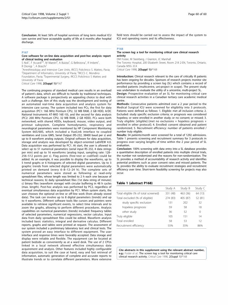
Conclusion: At least 56% of hospital survivors of long term medical ICUcare survive and have acceptable quality of life at 6 months after hospitaldischarge.
P167Core software for on-line data acquisition and post-hoc analysis: reportof clinical testing and evaluationS Noli1, F Accardi2,3, M Maestri4, A Buizza2, G Bellinzona1, R Imberti1,R Dionigi 1, A Braschi11Anesthesiolojay and Intensive Care Units, IRCCS Policlinico S. Matteo, Pavia;2Department of Informatics, University of Pavia; 3IRCCS C. MondinoFoundation, Pavia; 4Experimental Surgery, IRCCS Policlinico S Matteo andUniversity of PaviaCritical Care 1998, 2(Suppl 1):P167
The continuing progress of standard medical care results in an overloadof patient’s data, which are difficult to handle by traditional techniques.A software package is perspectively an appealing choice to deal withsuch a challenge. Aim of this study was the development and testing ofan automated real-time data acquisition and analysis system forintensive care survey. The system included two PCs, the first for dataacquisition (PC1: 166 MHz Pentium CPU, 32 MB RAM, 2 GB HDD, A/DCNational Instruments AT-MIO 16E-10), and the second for data analysis(PC2: 200 MHz Pentium CPU, 32 MB RAM, 2 GB HDD). PCs were bothnetworked, with shared HDDs, keyboard, mouse, video output, andprintout subsystem. Complete hemodynamic, respiratory andneurological parameters were monitored by HP Component MonitoringSystem 66S/68S, which included a VueLink interface to coupledventilators and Licox GMS, Serial Output (RS-232, 38400 baud per s) andup to 8 waveform analog outputs. Original software for data acquisition,storage and analysis was developed by object-oriented LabVIEW 4.0.Data acquisition was performed by PC1. At start, the user is allowed toselect up to 51 numerical parameters (serial input RS 232, 4 data stringsper min) and up to 16 waveforms (analog input, sampling rate: 128–256–512 points per s). Flag points (free-text or codified) could beadded. As an example, it was possible to display the waveforms, up to5 trend graphs or 6 histograms of selected digital parameters. Up to 3graphic trends from selected digital parameters were automaticallyprinted on demand (every 6–8–12–24 h). The complete sets ofnumerical parameters were stored as following: a) read-onlyspreadsheet files, whose length was limited to 2 h each one because oftechnical reasons; b) daily spreadsheet files (1st data string of minute);c) binary files (waveform storage) with circular buffering in 48 h cycles(max. length). Post-hoc analysis was performed by PC2, regardless ofeventual simultaneous data acquisition by PC1. When system starts, theuser chooses the optional on-line or off-line work (from already storeddata). The task can involve up to 8 digital parameters (trends) and upto 4 waveforms. Different software tools like cursors and pointers wereavailable to retrieve significant events, to select time intervals and tozoom the graphs, allowing to perform different procedures. Analysiscapabilities on numerical parameters (trends) included: frequency tablesof selected parameters, numerical regressions, vector calculus. Inputdata from daily spreadsheet files could be edited. Waveform analysisincluded basic statistics, integral and derivative calculus. Differentreports, graphs and tables were printed at request. The assessment ofour system included a preliminary laboratory test and clinical tests. Thesystem proved an easy interface to different equipment. The userinterface and response times were favorably accepted. Data storage anddisplay were reliable and flexible. The equipment can be located atpatient bedside as conveniently as at a ward desk. The use of 2 CPUslinked in a local network allowed effective simultaneous dataacquirement and analysis. Other features included highly configurabledata acquisition, to suit the case at hand, easy and fast retrieval ofinformation, automatic generation of complete and accurate reports toillustrate trends or to correlate different parameters. More extensive
field tests should be carried out to assess the impact of the system toICU and operating rooms and its effectiveness.
P168The screen log: a tool for monitoring critical care clinical researchactivityDM Foster, M Steinberg, J Granton, JC MarshallThe Toronto Hospital, 200 Elizabeth Street, Room 218 2-EN, Toronto, Ontario,Canada M5G 2C4Critical Care 1998, 2(Suppl 1):P168
Introduction: Clinical research relevant to the care of critically ill patientshas been ongoing for decades. Sponsors of research projects monitor siteperformance by providing a screen log (SL) which contains a record ofenrolled patients (multicentre, uni-project in scope). The present studywas undertaken to evaluate the utility of a unicentre, multi-project SL.Design: Prospective evaluation of an SL for monitoring critical careclinical research activities in a Canadian tertiary care academic medicalcentre.Methods: Consecutive patients admitted over a 2 year period to theMedical Surgical ICU were screened for eligibility into 3 protocols.Patients were defined as follows: 1. Eligible: met all inclusion criteria; 2.Excluded: study specific exclusion criteria; or prognosis was consideredhopeless; or were enrolled in another study; or no consent; or missed; 3.Truly eligible: [eligible]-[met no exclusions + hopeless prognosis +enrolled in other protocol]; 4. Enrolled: consent obtained and patientrandomized; 5. Recruitment efficiency: number of patients enrolled /number truly eligible.Results: 54 patients/month were screened for a total of 1292 admissions.Table 1 presents screening and recruitment summary for 3 protocols inprogress over varying lengths of time within the 2 year period of SLevaluation.Conclusion: 100% screening with data entry into a SL database providesa quantitative description of enrolled patients into multiple clinical trials,the number not randomized and the reasons for non-randomization. TheSL provides a method of accountability of research activity and identifiespotential problems such as poor consent rates and missed patients. TheSL provides the ability to evaluate recruitment strategies and recruitmentefficiency over time. Short-term feasibility screening for projects may alsooccur.
Cite abstracts in this supplement using the relevant abstract number,e.g.: Foster et al.: The screen log: a tool for monitoring critical careclinical research activity. Critical Care 1998, 2(Suppl 1):P168
Table 1 (abstract P168)
Study A Study B Study C
Total eligible (% of total screened) 331 (48) 463 (36) 64 (13)
Total excluded (% of eligible) 274 (83) 405 (87) 52 (81)
study specific exclusion 131 202 32
hopeless prognosis 59 58 18
other study 36 52 0
Truly eligible 105 151 14
Total enrolled 58 53 12
Recruitment efficiency 55% 35% 86%
Critical Care 1998, Volume 2 Suppl 1http://ccforum.com/supplements/2/S1
Page 60 of 60
Related Documents