MEETING ABSTRACTS Open Access 9th International Symposium on Intensive Care and Emergency Medicine for Latin America São Paulo, Brasil. June 14-17, 2017 Published: 13 June 2017 Cardiology P01 Complications associated with the use of temporary pacemakers in hospitalized patients awaiting definitive implant procedure in a public hospital in Sao Paulo Andres Eduardo Larrovere Vasquez, Aymée Lustosa Nogueira Torres, Paulo Vinicius Prates Silva, Ana Helena Vicente Andrade, Luciana Gioli-Pereira HMBM - Hospital Municipal Dr Moyses Deutsch, São Paulo, SP, Brasil Critical Care 2017, 21(Suppl 2):P01 Introduction: Indications for temporary and permanent pacemaker im- plantation are well established and atrioventricular blocks are the most common causes. Complications could occur, mainly with temporary pacemakers and they can be related to various conditions as the implant- ation position of the lead, infection and thrombo-embolic events [1,2]. The number of pacemaker implantations in Brazil by the public health system (SUS) is inferior compared to other countries. Besides, the de- mand is growing and the majority of public hospitals do not realize the procedure [3]. This scenario gets worse with the time to wait for a pace- maker and enhance the complications for the hospitalized patients. Objective: We aim to evaluate clinical complications associated with prolonged hospitalization of patients who are waiting for a permanent pacemaker at a public hospital and the comparison of definitive pace- maker implant costs versus hospitalization required for the procedure. Methods: This is an observational retrospective study that was carried out with medical records of the patients admitted at Dr Moysés Deutsch Municipal Hospital from January 2014 to December 2015 with atrioventricular blocks that requiring a temporary pacemaker. The inclu- sion criteria were patients aged at least 18 years old and a diagnosis related to atrioventricular blocks. The clinical data were collected in electronic medical records and the outcomes analyzed were all-cause mortality and clinical-surgical complications during hospitalization. Results: Twenty seven patients that implanted a temporary pacemaker were included. The mean length of hospital stay was approximately 20 days. Eighteen (66.6%) patients presented some intercurrence dur- ing the hospitalization whose main causes were: worsening renal func- tion (22.2%), decompensated heart failure (18.5%), urinary tract infection (18.5%) and pneumonia (11.1%). There were two (7.4%) deaths likely coronary acute syndrome and one (3.4%) due to sepsis. Conclusions: Preliminary results suggest that there is a relation be- tween hospitalization time and number of complications suffered by the patient. Besides, the complications were more frequent in long stay. The costs involved in the definitive pacemaker implantation procedure and an admission to SUS (public hospitals) will still be analyzed and presented later. References 1. The Task Force on cardiac pacing and resynchronization therapy of the European Society of Cardiology (ESC). Developed in collaboration with the European Heart Rhythm Association (EHRA). 2013 ESC Guidelines on cardiac pacing and cardiac resynchronization therapy. European Heart Journal. 2013; 34: 2281–2329. 2. Pereira WL, Cardoso CC, Fumagalli AR, Carbone Filho F. Complicação rara do marcapasso transvenoso temporário: a formação de loopings e nós. Relampa. 2010; 23(3):134-137. 3. Pachón-Mateos JC et.al. RBM – Registro Brasileiro de Marcapassos, Ressincronizadores e Desfibriladores. Relampa. 2013; 26(1):39-49. P02 Omentopexy as a mechanism of stem cell implantation and revascularization in the ischemic myocardium Luiz Fernando Kubrusly 1,2 , Yorgos Graça Salles 1,3 , Camila Moraes Marques 4,1 , Fernando Bermudez Kubrusly 1,2 , Angeline Garcez Massignan 3,1 , Carolina De Marchi Capeletto 3,1 , Luiza Milanesi Abeling 1,3 , Caroline Aragão 5,1 , Andressa de Souza Bertoldi 3,1 , Larissa Maria Vosgerau 3,1 , Gabriel Antonio Coltro 3,1 1 IDC - Instituto Denton Cooley, Curitiba, PR, Brasil; 2 IVEP - Instituto Vita de Ensino e Pesquisa, Curitiba, PR, Brasil; 3 FEPAR - Faculdade Evangélica do Paraná, Curitiba, PR, Brasil; 4 UFPR - Universidade Federal do Paraná, Curitiba, PR, Brasil; 5 HC- UFPR - Hospital de Clínicas/ UFPR,Curitiba, PR, Brasil Critical Care 2017, 21(Suppl 2):P02 Introduction: Despite improved techniques of myocardial revascu- larization in the treatment of ischemic diseases, there are patients who can not be benefited by their diffuse involvement of arteries with diameters incompatible with the techniques [1]. The implant- ation of stem cell at the ischemic myocardial has proved to be able to regenerate myocardium. The omentum is known for applications as highly vascularized graft, full of angiogenic and chemostats factors [2]. Objective: This research intends to investigate the efficacy of omen- topexy as a indirectly method of revascularization and deliverer of stem cells in the ischemic myocardial. Methods: Myocardial infarction was created in 4 pigs by direct ligation of the 1st and 2nd obtuse marginal branches of the circum- flex artery. Lidocaine was administered 0.2% to avoid occurrence of arrhythmias. After 90 minutes of hemodynamic stabilization in 3 ani- mals (Group A), followed by mobilization of the omentum into the mediastinum, the omentum was sutured in the infarcted area. In Group B (1 animal) nothing was done after the infarct. After 30 days of ligation, in both groups, the animals were euthanized. All hearts were removed for histologic evaluation. Nine transversal cuts from the base to the Apex, colored by Hematoxilin-Eosin. It was used CD 34 for expression of stem cells proliferation. Results: Group A (with omentopexy) had progressive fibrosis and thinning of the ventricular wall since the area of the artery liga- ture until the area treated with the omentum. After this point there was progressive atenuation of ischemic changes up to the Apex where almost normal tissue was found. The CD 34 showed presence of stem cell in the myocardium. In Group B (without omentopexy) there were degrees of adherence, but without de- velopment of myocardial vessels. There was evident thinning of the infarcted myocardium. Critical Care 2017, 21(Suppl 2):139 DOI 10.1186/s13054-017-1706-1 © The Author(s). 2017 Open Access This article is distributed under the terms of the Creative Commons Attribution 4.0 International License (http://creativecommons.org/licenses/by/4.0/), which permits unrestricted use, distribution, and reproduction in any medium, provided you give appropriate credit to the original author(s) and the source, provide a link to the Creative Commons license, and indicate if changes were made. The Creative Commons Public Domain Dedication waiver (http://creativecommons.org/publicdomain/zero/1.0/) applies to the data made available in this article, unless otherwise stated.

Welcome message from author
This document is posted to help you gain knowledge. Please leave a comment to let me know what you think about it! Share it to your friends and learn new things together.
Transcript
Critical Care 2017, 21(Suppl 2):139DOI 10.1186/s13054-017-1706-1
MEETING ABSTRACTS Open Access
9th International Symposium on Intensive Careand Emergency Medicine for Latin America
São Paulo, Brasil. June 14-17, 2017Published: 13 June 2017
CardiologyP01Complications associated with the use of temporary pacemakers inhospitalized patients awaiting definitive implant procedure in apublic hospital in Sao PauloAndres Eduardo Larrovere Vasquez, Aymée Lustosa Nogueira Torres,Paulo Vinicius Prates Silva, Ana Helena Vicente Andrade, Luciana Gioli-PereiraHMBM - Hospital Municipal Dr Moyses Deutsch, São Paulo, SP, BrasilCritical Care 2017, 21(Suppl 2):P01
Introduction: Indications for temporary and permanent pacemaker im-plantation are well established and atrioventricular blocks are the mostcommon causes. Complications could occur, mainly with temporarypacemakers and they can be related to various conditions as the implant-ation position of the lead, infection and thrombo-embolic events [1,2].The number of pacemaker implantations in Brazil by the public healthsystem (SUS) is inferior compared to other countries. Besides, the de-mand is growing and the majority of public hospitals do not realize theprocedure [3]. This scenario gets worse with the time to wait for a pace-maker and enhance the complications for the hospitalized patients.Objective: We aim to evaluate clinical complications associated withprolonged hospitalization of patients who are waiting for a permanentpacemaker at a public hospital and the comparison of definitive pace-maker implant costs versus hospitalization required for the procedure.Methods: This is an observational retrospective study that was carriedout with medical records of the patients admitted at Dr MoysésDeutsch Municipal Hospital from January 2014 to December 2015 withatrioventricular blocks that requiring a temporary pacemaker. The inclu-sion criteria were patients aged at least 18 years old and a diagnosisrelated to atrioventricular blocks. The clinical data were collected inelectronic medical records and the outcomes analyzed were all-causemortality and clinical-surgical complications during hospitalization.Results: Twenty seven patients that implanted a temporary pacemakerwere included. The mean length of hospital stay was approximately20 days. Eighteen (66.6%) patients presented some intercurrence dur-ing the hospitalization whose main causes were: worsening renal func-tion (22.2%), decompensated heart failure (18.5%), urinary tractinfection (18.5%) and pneumonia (11.1%). There were two (7.4%)deaths likely coronary acute syndrome and one (3.4%) due to sepsis.Conclusions: Preliminary results suggest that there is a relation be-tween hospitalization time and number of complications suffered bythe patient. Besides, the complications were more frequent in longstay. The costs involved in the definitive pacemaker implantationprocedure and an admission to SUS (public hospitals) will still beanalyzed and presented later.
References1. The Task Force on cardiac pacing and resynchronization therapy of the
European Society of Cardiology (ESC). Developed in collaboration withthe European Heart Rhythm Association (EHRA). 2013 ESC Guidelines oncardiac pacing and cardiac resynchronization therapy. European HeartJournal. 2013; 34: 2281–2329.
© The Author(s). 2017 Open Access This articInternational License (http://creativecommonsreproduction in any medium, provided you gthe Creative Commons license, and indicate if(http://creativecommons.org/publicdomain/ze
2. Pereira WL, Cardoso CC, Fumagalli AR, Carbone Filho F. Complicação rarado marcapasso transvenoso temporário: a formação de loopings e nós.Relampa. 2010; 23(3):134-137.
3. Pachón-Mateos JC et.al. RBM – Registro Brasileiro de Marcapassos,Ressincronizadores e Desfibriladores. Relampa. 2013; 26(1):39-49.
P02Omentopexy as a mechanism of stem cell implantation andrevascularization in the ischemic myocardiumLuiz Fernando Kubrusly1,2, Yorgos Graça Salles1,3, Camila MoraesMarques4,1, Fernando Bermudez Kubrusly1,2, Angeline GarcezMassignan3,1, Carolina De Marchi Capeletto3,1, Luiza Milanesi Abeling1,3,Caroline Aragão5,1, Andressa de Souza Bertoldi3,1, Larissa MariaVosgerau3,1, Gabriel Antonio Coltro3,11IDC - Instituto Denton Cooley, Curitiba, PR, Brasil; 2IVEP - InstitutoVita de Ensino e Pesquisa, Curitiba, PR, Brasil; 3FEPAR - FaculdadeEvangélica do Paraná, Curitiba, PR, Brasil; 4UFPR - UniversidadeFederal do Paraná, Curitiba, PR, Brasil; 5HC- UFPR - Hospital deClínicas/ UFPR,Curitiba, PR, BrasilCritical Care 2017, 21(Suppl 2):P02
Introduction: Despite improved techniques of myocardial revascu-larization in the treatment of ischemic diseases, there are patientswho can not be benefited by their diffuse involvement of arterieswith diameters incompatible with the techniques [1]. The implant-ation of stem cell at the ischemic myocardial has proved to be ableto regenerate myocardium. The omentum is known for applicationsas highly vascularized graft, full of angiogenic and chemostatsfactors [2].Objective: This research intends to investigate the efficacy of omen-topexy as a indirectly method of revascularization and deliverer ofstem cells in the ischemic myocardial.Methods: Myocardial infarction was created in 4 pigs by directligation of the 1st and 2nd obtuse marginal branches of the circum-flex artery. Lidocaine was administered 0.2% to avoid occurrence ofarrhythmias. After 90 minutes of hemodynamic stabilization in 3 ani-mals (Group A), followed by mobilization of the omentum into themediastinum, the omentum was sutured in the infarcted area. InGroup B (1 animal) nothing was done after the infarct. After 30 daysof ligation, in both groups, the animals were euthanized. All heartswere removed for histologic evaluation. Nine transversal cuts fromthe base to the Apex, colored by Hematoxilin-Eosin. It was used CD34 for expression of stem cells proliferation.Results: Group A (with omentopexy) had progressive fibrosis andthinning of the ventricular wall since the area of the artery liga-ture until the area treated with the omentum. After this pointthere was progressive atenuation of ischemic changes up to theApex where almost normal tissue was found. The CD 34 showedpresence of stem cell in the myocardium. In Group B (withoutomentopexy) there were degrees of adherence, but without de-velopment of myocardial vessels. There was evident thinning ofthe infarcted myocardium.
le is distributed under the terms of the Creative Commons Attribution 4.0.org/licenses/by/4.0/), which permits unrestricted use, distribution, andive appropriate credit to the original author(s) and the source, provide a link tochanges were made. The Creative Commons Public Domain Dedication waiverro/1.0/) applies to the data made available in this article, unless otherwise stated.
Critical Care 2017, 21(Suppl 2):139 Page 2 of 36
Conclusions: Omentopexy was able to develop neovascularization inthe ischemic myocardial, preserved its thickness and allowed stemcells to implant at the site of omentopexy.
References1. Cantero MA, et al. Análise dos resultados imediatos da cirurgia de
revascularização do miocárdio com e sem circulação extracorpórea.Rev Bras Cir Cardiovasc.2012;27(1):38-44.
2. Wystrychowski W, et al. Multipotency and cardiomyogenic potential ofhuman adipose-derived stem cells from epicardium, pericardium, andomentum. Stem Cell Research & Therapy.2016;7:84.
P03Anxiety and depressive symptoms in adults and elderly submittedto cardiac surgeryDébora D`Agostini Jorge Lisboa1, Eliane Lucia Colussi1, Marlene Doring1,Gabriela Decol Mendonça1, Gabriela Colussi1, Luana Battistella2, PatriciaDe Carli Tonial Ghisolfi1, Julia Mognon2, Isadora Sisto Rebolho2, IndaiaraMedeiros da Silva2, Renato Ravizon Lisboa31UPF - Universidade de Passo Fundo (Passo Fundo, RS, Brasil); 2HCPF -Hospital da Cidade Passo Fundo (Passo Fundo, RS, Brasil); 3PM -Prefeitura de Marau (Marau, RS, Brasil)Critical Care 2017, 21(Suppl 2):P03
Introduction: The patient undergoing cardiac surgery often experi-ence strong feelings of distress and anxiety befor surgery because itis a highly invasive procedure [1,2]. Anxiety and depression symp-toms before surgery can lead to complications after surgery [3].Objective: Identify the presence of symptoms suggestive of depres-sion and anxiety in patients undergoing cardiac surgeryMethods: It is an uncontrolled prospective cohort study in two hospi-tals in Passo Fundo. Data collection occurred in different times, pre-operative, postoperative during hospitalization and after three monthsof surgery, through the Clinical Sociodemographic Questionnaire, Rat-ing Scale Anxiety and Depression level. Data were analyzed using de-scriptive inferential analysis of the data, at the 0.05 significance level.Results: Observed frequency of symptoms of anxiety in stage I was38.6% at 18.6% phase II and phase III of 8.6% in patients undergoingcardiac surgery with symptoms suggestive of depression observed afrequency in phase I of 12 stage II and 10.0% in stage III of 7.1%.Conclusions: After analyzing the data, patients have a higher degreeof anxiety and depression in the preoperative (phase I), with signifi-cant reduction of these symptoms follow up three months after theprocedure (phase III) (Table 1).
References1. Aikawa, P. et al. Reabilitação cardíaca em pacientes submetidos à cirurgia de
revascularização do miocárdio. Rev Bras Med Esporte, v. 20, n. 1, p. 55–58, 2014.2. Bahramnezhad, F. et al. Quality of Life in Patients Undergoing Percutaneous
Transluminal Coronary Angioplasty (PTCA). Global journal of health science,v. 7, n. 5, p 50, jan. 2015.
3. Choinière, M. et al. Prevalence of and risk factors for persistentpostoperative nonangial pain after c surgery & #8239;: a 2-yearprospective multicentre study. CMAJ, v. 186, n. 7, p. 213–23, 2014.HAD: Escala hospitalar de ansiedade e depressão. Valores expressamfrequência absoluta e relativa
Table 1 (Abstract P03). Prevalence of anxiety and depression inpatients submitted at the study (n = 70), Passo Fundo, 2015
Preoperative Postoerative duringhospitazation
Postoperative threemonths after surgery
Variables n(%) n(%) n(%)
Anxiety (HAD) - yes 27 (38,6) 13 (18,6) 6 (8,6)
Depression (HAD) - yes 9 (12,9) 7 (10,0) 5 (7,1)
HAD Escala hospitalar de ansiedade e depressãoValores expressam frequiência absoluta e relativa
EpidemiologyP04The effects of weekend admission on the mortality of patientsadmitted to intensive care units: the role of organizational factorsFernando Zampieri1, Thiago Lisboa2, Fernando Bozza3, Jorge Salluh3,Marcio Soares31IP-HCor - Instituto de Pesquisa, HCor-Hospital do Coração, São Paulo, SP,Brasil; 2SC-Porto Alegre - Santa Casa de Misericórdia, Porto Alegre, RS, Brasil;3D´Or IR - D’Or Institute for Research and Education. Rio de Janeiro, RJ, BrasilCritical Care 2017, 21(Suppl 2):P04
Introduction: Weekend admission is considered a risk factor for worseoutcomes in critically ill patients, but more robust information on theunderlying mechanisms related to intensive care unit (ICU) organizationis still lacking.Objective: To assess whether ICU organizational and staffing patternscould explain the association between weekend admission and out-comes in critically ill patients.Methods: Retrospective cohort study using the ORCHESTRA study data-base [1] comprising 59,614 patients admitted to the participant ICUsduring 2013. The association between weekend admission and hospitalmortality was assessed using mixed logistic regression adjusting forclinically relevant patient-level (age, severity of organ dysfunctions, co-morbidities, performance status; admission type and length of hospitalstay before ICU admission) and ICU-level (use of checklists on weekend;24/7 full-time intensivists; nurse/bed ratio; unit type and number ofprotocols) characteristics.Results: 41,894 patients (70.3%) were admitted on weekdays and17,720 patients (29.7%) were admitted on weekends. In univariateanalysis, weekend admitted patients had higher ICU (10.9% vs.9.0%, p < 0.001) and hospital (16.5% vs. 13.5%, p < 0.001) mortal-ity rates. After regression, weekend admission was not associatedwith higher hospital mortality (OR 1.05, 95% CI 0.99-1.12, p =0.095). However, a “weekend effect” was still observed in sched-uled surgical patients, as well as in ICUs not holding checklistsand with a decrease in nurse/bed ratio during the weekends. Inaddition, units with a lower number of implemented protocolshad also higher mortality for patients admitted during the week-ends. For unscheduled admissions, no “weekend effect” was ob-served regardless of ICU´s characteristics.Conclusions: Weekend admissions were associated with higher mortalityin certain situations related to potentially modifiable patients’ and cen-ters’ features, representing an opportunity to improve ICU performanceand patients’ outcomes.
Reference1. Soares M, Bozza FA, Angus DC, et al. Organizational characteristics,
outcomes, and resource use in 78 Brazilian intensive care units: theORCHESTRA study. Intensive Care Med. 2015 Dec;41(12):2149-60.
P05Outcome of oncological patients admitted to ICU for full treatmentwho were discharged on exclusive palliative care with consensusto no readmission to ICUAna Paula Agnolon Praça, Pedro Caruso, Antônio Paulo Nassar JuniorAC Camargo - AC Camargo Cancer Center, São Paulo, SP, BrasilCritical Care 2017, 21(Suppl 2):P05
Introduction: The number of oncological patients admitted to intensivecare unit (ICU) is increasing worldwide [1]. However, there have alwaysbeen discussions about the benefit of ICU for certain groups of patients[2] and concerns with end of life care, because it is usually in the ICU thatthe transition from full treatment to exclusive palliative care occurs. Thedecision making process involves patients, relatives, the attending teamand is usually a shared and consensual decision [3]. Our hypotheses arethat the patients discharged from the ICU on exclusive palliate carewould have a high hospital and long-term mortality, would have a smallbut not neglecting ICU readmission rate despite the consensus to no ICUreadmission, and finally some would resume their cancer treatment.
Critical Care 2017, 21(Suppl 2):139 Page 3 of 36
Objective: The objectives were to evaluate on patients dischargedfrom ICU on exclusive palliative care: 1. the hospital and long-termmortality; 2. the ICU readmission rate and 3. the resuming of cancertreatment. We believe that our results will help patients, the attend-ing team and relatives to make a more reasoned decision.Methods: This is a retrospective, descriptive study, performed in amedical-surgical ICU of a cancer center, from April 2012 to April 2016including all patients with solid and hematological tumors, admittedto ICU on full treatment and who were discharged from ICU on ex-clusive palliative care.Results: 13,928 patients were admitted during the study period and351 were admitted to ICU on full code treatment and were dis-charged on exclusive palliative care with consensus of no readmis-sion to ICU. The ICU readmission rate was 9.6%. Fourteen percent ofthe patients resumed their cancer treatment. The hospital mortalitywas 79.5%, the six-month mortality was 96.3% and the one-year mor-tality was 99.1%. From the 20.5% of patients who were dischargedalive, most were on home nursing care (12%). Acute renal failure (OR= 2.42; CI 95%, 1.22-4.79) and delirium (OR = 1.92; CI 95%, 1,02-3.58)were identified as independent risk factors for hospital mortality.Conclusions: Despite the high mortality rates, a significant proportion ofpatients (20%) was discharged from hospital, mainly with home nursingcare. The ICU readmission and resume of cancer treatment rate for exclu-sive palliative patients reflect the need to improve the decision makingprocess of end of life care. The knowledge about risk factors for mortalityalso provides consistent data for the decision-making process.
References1. Taccone FS, Artigas AA, Sprung CL, Moreno R, Sakr Y, Vincent JL.
Characteristics and outcomes of cancer patients in European ICUs.Crit Care 2009;13(1):R15.
2. Kostakou E, Rovina N, Kyriakopoulou M, Koulouris NG, Koutsoukou A.Critically ill cancer patient in intensive care unit: issues that arise. J CritCare. 2014;29(5):817-22.
3. Guidelines for intensive care unit admission, discharge, and triage. TaskForce of the American College of Critical Care Medicine, Society ofCritical Care Medicine. Crit Care Med. 1999;27(3):633-8.
P06Characteristics and outcomes of critical palliative patients in aprivate intensive care unitPaulo Sergio Santos Oliveira, Eduardo da Rosa Borges, Djalma BarbosaJunior, Flavia BarbozaHFCP - Hospital Fornecedores de Cana de Piracicaba, Piracicaba, SP, BrasilCritical Care 2017, 21(Suppl 2):P06
Introduction: In intensive care unit (ICU), the palliative care (PC) playsan important role in critical illness. Ideally, PC delivery should be lon-gitudinal, beginning at the time of a potentially life-limiting diagnosisand continuing throughout the course of the disease [1]. However,many patients only receive the initial palliative approach when theyhave already been hospitalized in the ICU.Objective: The goal of the study is to describe the characteristics ofpatients in palliative care (PC) in ICU compared to the group not under-going palliative care (nonPC). In addition, we made a subgroup analysisin the PC group, comparing oncologic (OC) and non oncologic (Non-OC) patients regarding demographic characteristics and outcomes.Methods: A cross-sectional study was conducted based on a continu-ous register database. The ICU admissions, from September 2015 untilNovember 2016, were included. Demographic information collectedincluded age, gender, type admission (medical or surgical), comorbi-dities (actual neoplasms, chronic obstructive pulmonary disease,hypertension, diabetes, stroke, chronic kidney failure and performancestatus before hospital admission). The markers SAPS3 and SOFA scoreday 1 were collected. In addition, the use of life-enhancing measures(mechanical ventilation, vasoactive drugs and renal replacement ther-apy), the days of hospitalization prior to ICU, length of stay in the ICUand hospital were considered. Finally we evaluated the outcomes inthe ICU and the hospital. For the quantitative variables was used theMann–Whitney test. For the categorical variables, the chi-square test
was used for comparison between groups. After univariate analysis allthose with p < 0.1 were included in the multivariate analysis. Variableswith p < 0.05 of the multivariate analysis were considered significant.We used R (v. 3.3.0) for all analysis.Results: A total of 578 admission in ICU were evaluated, with the PCgroup including 46 (7,96%) e nonPC group 532 (92,04%) patients.After the multivariate analysis, factors such as age (odd: 1,04, CI: 1-1,08),oncological patients (5,67; 1,92-17,76), bedridden previous status (5,81;1,74-20,37), clinical admission (8,72; 1,97-52,88), days of hospitalizationin ICU (1,07; 1,01-1,12) and hospital mortality (8,39; 1,62-39,09) hadsignificant association in PC. In the PC, only age (0,93; 0,88-0,99) wasassociated with the OC.Conclusions: Patients in PC were older, with a significant functionallimitation. The hospitalization was generally for clinical reasons,remaining more days hospitalized and with a higher hospital mor-tality in relation to the nonPC.
Reference1. Levetown, Marcia MD, FAAHPM. Increasing Access to the Benefits of
Palliative Care in the PICU. Pediatric Critical Care Medicine: August 2016 -Volume 17 - Issue 8 - p 804–805.
P07Risk factors for intensive care acquired weakness: a systematicreview and meta-analysisRaquel Annoni1, Jennifer Jones2, Diogenes Seraphim Ferreira3, Susan Berney2,Linda Denehy11Unimelb - The University of Melbourne, Melbourne, Victoria, Australia;2Austin - Austin Health, Melbourne, Victoria, Australia; 3Monash - MonashUniversity, Melbourne, Victoria, AustraliaCritical Care 2017, 21(Suppl 2):P07
Introduction: Previous studies have shown discordant results on the riskfactors and outcomes for intensive care acquired weakness (ICUAW).Objective: We aimed to identify and synthetize the evidence about theprevalence, risk factors and outcomes of ICUAW in critically ill patients.Methods: The systematic review was previously registered on Inter-national Prospective Register of Systematic Reviews (PROSPERO:CRD42014014521). Six electronic databases (PUBMED, MEDLINE,CINAHL, EMBASE, PEDro, SciELO) were searched from 2007 to 2017.Experimental and observational studies were eligible for inclusion if: 1)enrolled adult critically ill patients, 2) ICUAW was evaluated using clin-ical (MRC: Medical Research Council score), electrophysiological tests ora combination of both and, 3) reported at least one comparison be-tween participants with and without ICUAW. A data collection formwas developed and used to extract data from the included studies byone reviewer and cross-checked by a second reviewer. Studies usingMRC score at awakening were included in a meta-analysis.Results: Thirty-seven articles on 29 patient groups (n = 4011 patients)were included. Eighteen studies were conducted in Europe, 6 in theUSA and 1 in each of the following countries: Australia, Brazil, Egypt,India and Vietnam. Twenty-two studies were cohorts, 6 randomizedcontrolled trials (RCTs) and 1 cross-sectional study. Nine observationalstudies (9/23) and 2 RCTs (2/6) had low risk of bias. Fifteen studies usedthe MRC score to diagnose ICUAW and 14 used electrophysiologicaltests. Twenty-seven studies excluded patients with a previous history ofneuromuscular disease. The pooled ICUAW prevalence (95% CI) was40.1% (32.5, 47.7%) regardless of the diagnostic test used and time offirst assessment. Eight studies evaluating 1488 patients were includedin the meta-analysis. Risk factors for ICUAW measured with MRC scoreat awakening included older age, female gender, high SOFA score, sep-sis on ICU admission and any use of corticosteroids during ICU stay(Table 1). In addition, ICUAW was associated with poor outcomes in-cluding longer ICU and hospital length of stay (Table 1).Conclusions: Intensive care acquired weakness occurs in approxi-mately 40% of general critically ill patients. The current meta-analysisprovides evidence of risk factors for MRC diagnosed ICUAW. Furtherresearch should consider including these risk factors when buildingmultivariable models to investigate the contributors to the develop-ment of ICUAW.
Table 1 (Abstract P07). ICUAW risk factors and outcomes
N Effect estimate (95% CI) I2 (P value)
Age (years) 5 MD: 3.46 (0.94, 5.98) 18% (p = 0.30)
Female gender 4 OR: 1.62 (1.22, 2.14) 0% (P = 0.46)
SOFA score 4 MD: 1.96 (1.41, 2.50) 0% (P = 0.77)
Sepsis on admission 3 OR: 1.48 (1.09, 2.00) 0% (P = 0.62)
Use of corticosteroids 3 OR: 2.17 (1.21, 3.91) 45% (P = 0.16)
ICU LOS (days) 3 MD: 8.67 (7.05, 10.28) 0% (P = 0.85)
Hospital LOS (days) 2 MD: 15.31 (11.02, 19.61) 0% (P = 0.67)
MD mean difference, OR odds ratio, LOS length of stay
Critical Care 2017, 21(Suppl 2):139 Page 4 of 36
P08Antibiotics use in intensive care units of a public hospital in theState of Ceará, BrazilAntonio Pergentino Barreira Neto, Alana de Alcântara Brito, Milena deAzevedo Teles, Rafael Cabral Teixeira, Stephanie Wilkes da Silva, IaraSerra Azul Machado Bezerra, Marina Parente Albuquerque, CarlosAugusto Ramos Feijó, Francisco Albano de MenesesHGF – Hospital Geral de Fortaleza, Fortaleza, CE, BrasilCritical Care 2017, 21(Suppl 2):P08
Introduction: The use of antibiotics (ATB) are becoming quite commonin intensive care units (ICUs) throughout the world, being present inprescriptions of the vast majority of patients.Objective: To seek for substrates capable to contribute in futureoptimization of use of antibiotics, due to the emergence of resistantmicroorganisms.Methods: Retrospective study performed through database analysisof patients admitted to three ICUs of the Hospital Geral de Fortaleza,from October 2016 to January 2017.Results: Our sample included 134 patients, mostly male (54.5%), ageaverage of 53.5 ± 19 years (median: 55.5 years), average SOFA at admis-sion was 5.6 ± 4.6 points (median: 5 points), length of stay average of17.8 ± 17.5 days (median: 12 days), ATB use average, per patient, was 4.4± 3.8 (median: 4) and mortality reached 26.1%. In this group of patients,119 (88.8%) used ATB at any moment during hospitalization time and 15(11.2%) did not use it at all. Comparing the two groups, we noticed ahigher SOFA score average among those ar used ATB (6.2 x 1.1, p < 0.05),and higher mortality rates (29.4% x 0%, p < 0,05). The most commonlyused ATBs were meropenem (55.4%), piperacillin-tazobactam (50.4%)and polymyxin B (44.5%). SOFA score average and mortality were particu-larly higher considering patients in whom polymyxin B was used, than innon-users. Still regarding polymyxin B usage, the drug was prescribed to6 (4.5%) patients immediately upon ICU admission.Conclusions: The use of ATB remains very prevalent among patientsadmitted to ICUs. In addition, we were surprised by the amount ofpolymyxin B usage, reaching almost half of the studied population, andeven prescribed to some patients right on ICU arrival. This leads us toimagine that an expressive part of our ICU patients are infected by re-sistant microorganisms, increasing the chance of treatment failure.
P09Women's participation in authorship of original articles inintensive careCarlos Augusto Ramos Feijó, Marina Parente Albuquerque, FranciscoAlbano de MenesesHGF - Hospital Geral de Fortaleza, Fortaleza, CE, BrasilCritical Care 2017, 21(Suppl 2):P09
Introduction: Throughout the world, research shows that the participa-tion of women as authors of original articles in the medical field is stillnot very representative.Objective: To identify the participation of women as authors of originalarticles in the field of Intensive Care in Brazil.Methods: Observational study of women, either as an author in generalor as the first author, of original articles published in the Revista Brasileira
de Terapia Intensiva (RBTI), from 2006 to 2015. The RBTI is an indexedquarterly publication of the Associação de Medicina Intensiva Brasileiraand the Sociedade Portuguesa de Cuidados Intensivos. Review articles,case reports, comments, letters to editors and guidelines were excludedfrom the sample. It was not possible to discriminate the professional cat-egories of the authors.Results: We identified 356 original articles, with 2,049 authors, of which48.5% were women. The mean number of authors was 5.8 (men: 3,women: 2.8). The regions with the highest prevalence of female authorswere Southeast [SE] (52.9%), South [S] (26.1%) and Northeast (17.2%).More than half of the authors came from two states: São Paulo [SP](37.3%) and Rio Grande do Sul [RS] (13.9%). Five of the 27 units of thefederation (UF) had no authors in the period. From the total number ofarticles, 51 (14.3%) were written exclusively by women and 40 (11.2%)only by men. The regions that contributed with the largest portion of thearticles published only by women were SE (63%) and S (20.4%), with 52%of them from SP and 13% from RS. Of the 356 main authors, 198 (55.6%)were women, predominantly from SE (55.1%). Only 14 of the 27 UF hadwomen as the main authors, with SP (41.9%) and RS (14.1%) being themost representative. We also observed the participation of 40 womenfrom other countries in the authorship of the articles, being 8 main au-thors. During the analyzed period, there was a predominance in author-ship of women between 2007 and 2009 and between 2011 and 2014.Conclusions: In Intensive Care publishing environment, the participa-tion of Brazilian women in generating medical research, unlike thatperceived in other countries, accompanies pari passu the male pro-duction. It should be stressed that they take the lead when oneconsiders the position of first author.
References1. Filardo G, da Graca B, Sass DM, Pollock BD, Smith EB, Ashley-Marie Martinez M.
Trends and comparison of female first authorship in high impact medicaljournals: observational study (1994-2014). BMJ. 2016; 352:i847.
2. Erren TC, Groß JV, Shaw DM, Selle B. Representation of women as authors,reviewers, editors in chief, and editorial board members at 6 generalmedical journals in 2010 and 2011. JAMA Intern Med. 2014; 174(4):633-635.
3. Jagsi R, Guancial EA, Worobey CC, Henault LE, Chang Y, Starr R, et al. The“gender gap” in authorship of academic medical literature – a 35-yearperspective. N Engl J Med. 2006; 355:281-87.
P10Medical and surgical admission in an oncology ICU in thenortheast of BrazilAna Paula Pierre Moraes, Gustavo Teixeira Alves, José Ricardo SantosLima, Karina ViegasHTLF - Hospital de Câncer do Maranhão Tarquinio Lopes Filho, São Luis,MA, BrasilCritical Care 2017, 21(Suppl 2):P10
Introduction: Advances in oncology have lead to reduction in mortalityrates nowadays. The cancer patients are usually admitted to the ICUdue to postoperative high-risk surgeries, clinical complications due toacute process, especially infection/sepsis and complications due tochemotherapy [1,2].Objective: To evaluate the hospital mortality and morbidities outcomesin medical and surgical cancer patients requiring ICU admissionMethods: Retrospective study conducted in 11-bed ICU of a public can-cer hospital in São Luis-Maranhão, northeast of Brazil. All patients with adefinitive cancer diagnosis requiring ICU from January to December 2016were classified based on the reason of ICU´s medical and surgical admis-sions. We evaluate demographic and clinical variables at ICU admission,ICU support and outcomes: ICU and hospital length of stay (LOS), ICU re-admission, nosocomial ICU infection, and ICU and hospital mortality, re-spectively. The statistical difference was tested using Pearson´s chi-square or Mann-Whitney tests. The significance level adopted was 0,05.Results: 495 patients fulfilled the study criteria, 239 (48%) were admit-ted due to medical reasons and 256 (52%) due to surgical reasons. Themain reasons for intensive care admission were postoperative care afterelective surgery (47%), infection/sepsis (15%) and respiratory failure(10%). Medical and surgical admission did not differ in sex, age,
Critical Care 2017, 21(Suppl 2):139 Page 5 of 36
nosocomial ICU infection and hospital LOS. Medical admission of can-cer patients had higher SAPS 3 and SOFA scores, higher need for mech-anical ventilation and vasopressors, higher length in ICU (p < 0,001 forall). The overall ICU and hospital mortality were 32% and 50% respect-ively; 55% and 80% for medical ICU admission and 10% and 25% forsurgical ICU admission (p < 0,001) (Table 2).Conclusions: Cancer patients that required ICU admission due to medicalreasons were sicker at ICU admission and had worse outcomes comparedto those admitted due to surgical reasons. The results corroborate the im-portance of early diagnosis, access to medical attendance and promptreference. The possibility to early ICU admission may offer opportunitiesto prevent and better manage life-threatening complication.
References1. Kostakou E, et al. J Crtit Care 2014 Oct; 29(5):817-22.2. Soares M ,et al. Crit Care Med 2010 Jan; 38(1):9-15.
Table 2 (Abstract P10). Characteristics
Characteristics Alln = 495
Surgicaln = 256(51,7%)
Medicaln = 239(48,3%)
p
Demographic variables
Age 60,0 (48-72) 60,5 (51-72) 60,0 (46-71) 0,30
Gender Male 251 (51,7%) 123 (48,0%) 128 (53,6%) 0,22
Anatomic Tumor Site
Gastric and Esofageal 92 (18,6%) 63 (24,6%) 29 (12,1%) <0,001
Colorectal 35 (7,1%) 28 (10,9%) 7 (2,9%) <0,01
Uterine cervix 56 (11,3%) 24 (9,4%) 32 (13,4%) 0,16
Ovarian 42 (8,5%) 31 (12,1%) 11 (4,6%) <0,01
Prostate 42 (8,5%) 19 (7,4%) 23 (9,6%) 0,38
Lung 34 (6,9%) 13 (5,1%) 21 (8,8%) 0,10
Hematology 59 (11,9%) 3 (1,2%) 56 (23,4%) <0,001
Clinical variables
Length hospital stay prior ICU 2,0 (1-11) 2,2 (1-10) 3,0 (0-12) 0,03
Charlson comorbityIndex (points)
2,0 (2-4) 2,0 (2-3) 2,0 (2-6) <0,001
SAPS 3 (points) 48,0 (35-65) 35,5 (29-42) 64,0 (56-77) <0,001
SOFA score on D1 (points) 4,0 (2-7) 2,0 (1-5) 6,0 (3-8) <0,001
ICU support
Mechanical ventilantion on 1 h 175 (35,4%) 81 (31,6%) 94 (39,3%) <0,001
Vasoactive drusg on 1 h 61 (12,3%) 11 (4,3%) 50 (20,9%) <0,001
Mechanical ventilation 209 (42,2%) 83 (32,4%) 126 (52,7%) <0,001
Vasoactive drugs 79 (16,0%) 17 (6,6%) 62 (25,9%) <0,001
Renal replacement therapy 25 (5,1%) 5 (2,0%) 20 (9,2%) 0,001
Outcomes
Readmission 46 (9,3%) 22 (8,6%) 24 (10,0%) 0,57
Nosocomial ICU infection 19 (3,8%) 7 (2,7%) 12 (5,0%) 0,19
ICU Los 3 (1-6) 2,0 (1-4) 5,0 (2-9) 0,001
Hospital Los 16 (8-28) 16,0 (8-29) 17,0 (7-28) 0,84
ICU mortality 157 (31,7%) 25 (9,8%) 132 (55,2%) <0,001
Hospital mortality 248 (50,1%) 62 (24,7%) 187 (80,3%) <0,001
Results for continuous variables are reported as median (interquartile range)
P11Acute Kidney Injury (AKI) related to pregnancy, mortalityand survival of patients treated at the Maternal IntensiveCare Unit of Hospital Materno Infantil de Brasília(ICU/HMIB)Dilson Palhares Ferreira2, Aline Mizusaki Imoto2, Fábio Ferreira Amorim1,Amanda Jacomeli Matsuura1, Jaqueline Lima de Souza3, AdriellRamalho Santana11FEPECS - Fundação de Ensino e Pesquisa em Ciências daSaúde, Brasília/Distrito Federal, Brasil; 2FEPECS - Programa de PósGraduação em Ciências da Saúde, Brasília/Distrito Federal, Brasil;3ESCS - Escola Superior de Ciências da Saúde, Brasília/DistritoFederal, BrasilCritical Care 2017, 21(Suppl 2):P11
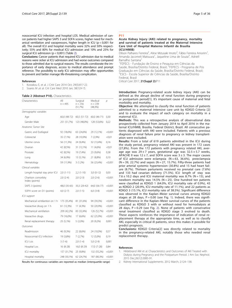
Introduction: Pregnancy-related acute kidney injury (AKI) can bedefined as the abrupt decline of renal function during pregnancyor postpartum period[1]. It’s important cause of maternal and fetalmorbidity and mortality.Objective: We attempted to classify the renal function of patientsadmitted to a maternal intensive care unit by KDIGO Criteria [2]and to evaluate the impact of each category on mortality in amaternal ICU.Methods: This was a retrospective analysis of observational dataprospectively collected from January 2014 to April 2016 in the ma-ternal ICU/HMIB, Brasilia, Federal District, Brazil. All consecutive pa-tients diagnosed with AKI were included. Patients with a previousdiagnosis of renal failure prior to pregnancy or kidney transplant-ation were excluded.Results: From a total of 619 patients admitted in the ICU duringthe study period, pregnancy related AKI was present in 172 cases(27,8%). From the 172 patients with pregnancy related AKI, aver-age age was 29 ± 7 years, gestational age was 32.5 ± 5.7 weeks,APACHE II was 12 ± 7, and SOFA score was 3 ± 3. The major causesof ICU admission were eclampsia (N = 63, 36.6%), preeclampsia(N = 39, 22.7%) and sepsis (N = 27, 15.7%). Fifty-three patients hadprior arterial systemic hypertension (30.8%) and 15 had heart fail-ure (8.7%). Thirteen parturients received no prenatal care (7.6%)and 133 had cesarian delivery (71.5%). ICU length of stay was7.6 ± 10.2 days and ICU maternal mortality was 8.7% (N = 15), andnewborn mortality was 14.5% (N = 25). One hundred ten patientswere classified as KDIGO 1 (64.0%, ICU mortality rate of 0.9%), 43as KDIGO 2 (20.9%, ICU mortality rate of 11.1%), and 22 patients asKDIGO 3 (15.1%, ICU mortality rate of 38.5%). Significant differencewas observed in the Kaplan–Meier survival curves among KDIGOstages at 28 days, P = 0.00 (see Fig. 1). Indeed, there was signifi-cant difference in the Kaplan–Meier survival curves of the patientsclassified as KDIGO 3 with or without need for hemodialysis at28 days, P = 0.29 (see Fig. 2). None of patients with conservativerenal treatment classified as KDIGO stage 3 evolved to death.These aspects reinforces the importance of indication of renal re-placement therapy at the appropriate time, as well as to classifyAKI, especially in critical ill patients, since this makes it possible forpredict prognosis.Conclusions: KDIGO Criteria[2] was directly related to mortalityin the pregnancy-related AKI, notably those who needed renalreplacement therapy.
References1. Hildebrand AM et al. Characteristics and Outcomes of AKI Treated with
Dialysis during Pregnancy and the Postpartum Period. J Am Soc Nephrol.2015 Dec;26(12):3085-91.
2. Kidney International Supplements. 2012 March; 2:124–138.
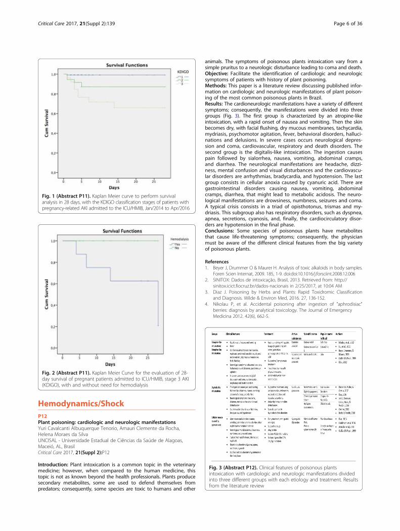
Fig. 1 (Abstract P11). Kaplan Meier curve to perform survivalanalysis in 28 days, with the KDIGO classification stages of patients withpregnancy-related AKI admitted to the ICU/HMIB, Jan/2014 to Apr/2016
Fig. 2 (Abstract P11). Kaplan Meier Curve for the evaluation of 28-day survival of pregnant patients admitted to ICU/HMIB, stage 3 AKI(KDIGO), with and without need for hemodialysis
Fig. 3 (Abstract P12). Clinical features of poisonous plantsintoxication with cardiologic and neurologic manifestations dividedinto three different groups with each etiology and treatment. Resultsfrom the literature review
Critical Care 2017, 21(Suppl 2):139 Page 6 of 36
Hemodynamics/ShockP12Plant poisoning: cardiologic and neurologic manifestationsYuri Cavalcanti Albuquerque Tenorio, Amauri Clemente da Rocha,Helena Moraes da SilvaUNCISAL - Universidade Estadual de Ciências da Saúde de Alagoas,Maceió, AL, BrasilCritical Care 2017, 21(Suppl 2):P12
Introduction: Plant intoxication is a common topic in the veterinarymedicine; however, when compared to the human medicine, thistopic is not as known beyond the health professionals. Plants producesecondary metabolites, some are used to defend themselves frompredators; consequently, some species are toxic to humans and other
animals. The symptoms of poisonous plants intoxication vary from asimple pruritus to a neurologic disturbance leading to coma and death.Objective: Facilitate the identification of cardiologic and neurologicsymptoms of patients with history of plant poisoning.Methods: This paper is a literature review discussing published infor-mation on cardiologic and neurologic manifestations of plant poison-ing of the most common poisonous plants in Brazil.Results: The cardioneurologic manifestations have a variety of differentsymptoms; consequently, the manifestations were divided into threegroups (Fig. 3). The first group is characterized by an atropine-likeintoxication, with a rapid onset of nausea and vomiting. Then the skinbecomes dry, with facial flushing, dry mucous membranes, tachycardia,mydriasis, psychomotor agitation, fever, behavioral disorders, halluci-nations and delusions. In severe cases occurs neurological depres-sion and coma, cardiovascular, respiratory and death disorders. Thesecond group is the digitalis-like intoxication. The ingestion causespain followed by sialorrhea, nausea, vomiting, abdominal cramps,and diarrhea. The neurological manifestations are headache, dizzi-ness, mental confusion and visual disturbances and the cardiovascu-lar disorders are arrhythmias, bradycardia, and hypotension. The lastgroup consists in cellular anoxia caused by cyanuric acid. There aregastrointestinal disorders causing nausea, vomiting, abdominalcramps, diarrhea, that might lead to metabolic acidosis. The neuro-logical manifestations are drowsiness, numbness, seizures and coma.A typical crisis consists in a triad of opisthotonus, trismas and my-driasis. This subgroup also has respiratory disorders, such as dyspnea,apnea, secretions, cyanosis, and, finally, the cardiocirculatory disor-ders are hypotension in the final phase.Conclusions: Some species of poisonous plants have metabolitesthat cause life-threatening symptoms; consequently, the physicianmust be aware of the different clinical features from the big varietyof poisonous plants.
References1. Beyer J, Drummer O & Maurer H. Analysis of toxic alkaloids in body samples.
Foren Scien Internat, 2009. 185, 1-9. doi:doi:10.1016/j.forsciint.2008.12.0062. SINITOX: Dados de intoxicação, Brasil, 2013. Retrieved from: http://
sinitox.icict.fiocruz.br/dados-nacionais in 2/25/2017, at 10:04 AM3. Diaz J. Poisoning by Herbs and Plants: Rapid Toxidromic Classification
and Diagnosis. Wilde & Environ Med, 2016. 27, 136-152.4. Nikolau P, et al. Accidental poisoning after ingestion of "aphrodisiac"
berries: diagnosis by analytical toxicology. The Journal of EmergencyMedicina 2012. 42(6), 662-5.
Critical Care 2017, 21(Suppl 2):139 Page 7 of 36
P13Microvascular reactivity in patients with and without circulatoryshock: an exploratory analysisRenato Carneiro de Freitas Chaves, Roberto Rabello Filho, Ary SerpaNeto, Murillo Santucci Cesar de Assunção, Adriano José Pereira, FlaviaFernandes Manfredi de Freitas, Maria Laura Romagnoli, ThiagoDomingos CorrêaHIAE - Hospital Israelita Albert Einstein, São Paulo, SP, BrazilCritical Care 2017, 21(Suppl 2):P13
Introduction: Abnormalities in microvascular reactivity accessed withthenar near-infrared spectroscopy (NIRS) with a vascular occlusiontest (VOT) have been described in critically ill patients [1].Objective: Our objective was to confirm such findings in termsof static and dynamic NIRS derived parameters in health volun-teers and in critically ill patients with and without circulatoryshock.Methods: This prospective single-center study was approvedby the ethics committee of Hospital Israelita Albert Einstein.Written informed consent was obtained from each participant.Twenty adult healthy volunteers [29 (27-34) years, median(IQR)] and 40 critically ill patients with and without shock (n =20, each) admitted to the ICU within 24 h were included inthis study. Tissue O2 saturation (StO2) was measured at thethenar eminence using an InSpectra StO2 Tissue OxygenationMonitor (model 650; Hutchinson Technology, Hutchinson, MN,USA) using a 15 mm probe. Vascular occlusion test was per-formed by inflating a sphygmomanometer in the upper arm30 mmHg above the systolic arterial pressure, which wasquickly deflated after 3 min of ischemia (1). A research soft-ware (Hutchinson Technology Inc., Hutchinson, MN) was usedfor data collection and analysis.Results: Shock patients (80% septic shock; 20% cardiogenic shock)were older than non-shock patients [66 (56-73) vs. 50 (44-60)years, median (IQR); p = 0.024] and more frequently female [11(55%) vs. 6 (30%); p = 0.017). Shock patients had a higher SAPS IIIscore [53 (45-65) vs. 30 (22-46); p < 0.001], higher SOFA score [8(6-10) vs. 4 (1-5); p < 0.001] and higher 28-day mortality [5 (25.0%)vs. 0 (0.0%); p = 0.047). Shock patients showed a lower maximumStO2 after VOT than patients without shock (Fig. 4). Recovery timeand hyperemia area differed between health volunteers and crit-ically ill patients, but did not differ between patients with andwithout shock.Conclusions: In our studied population, NIRS static and dynamicparameters poorly discriminate shock and non-shock patients.The role of thenar near-infrared spectroscopy in the care ofcritically ill patients needs to be further addressed in largeclinical trials.
Reference1. Lima A et al. The relation of near-infrared spectroscopy with changes
in peripheral circulation in critically ill patients. Crit Care Med. 2011;39(7):1649-54
Fig. 4 (Abstract P13). NIRS derived variables
P14Effect of extracorporeal membrane oxygenation on microcirculationand tissue oxygen saturation in ARDS: a case reportRenato Carneiro de Freitas Chaves, Murillo Santucci Cesar de Assunção, ArySerpa Neto, Roberto Rabello Filho, Bruno de Arruda Bravim, Philipe Francodo Amaral Tafner, Carmen Silva Valente Barbas, Thiago Domingos CorrêaHIAE - Hospital Israelita Albert Einstein, São Paulo, SP, BrazilCritical Care 2017, 21(Suppl 2):P14
Introduction: The impact of ECMO on microcirculation and tissueoxygenation (StO2) in patients with acute respiratory distress syndrome(ARDS) is poorly understood.Objective: Our objective was to evaluate the effect of veno-venousECMO (VV-ECMO) on microcirculation and tissue oxygen saturationduring the first 24 h on ECMO support.Methods: A written informed consent was obtained from a next of kin ofthe patient. We report a case of a 27-year-old Brazilian woman with diag-nosis of Granulomatosis with Poliangiitis. The patient was referred to ourintensive care unit (ICU) due to a severe ARDS requiring venous-venousECMO support. Sublingual microcirculation [Cytocam-IDF imaging® (Brae-dius Medical BV, Huizen, Netherlands)] and thenar StO2 [InSpectra StO2Tissue Oxygenation Monitor (model 650; Hutchinson Technology, Hutch-inson, MN, USA)] were measured immediately before ECMO (Baseline),4 h (T4h) and 24 h (T24h) after the beginning of ECMO [1,2]. Reported pa-rameters from Cytocam-IDF were total vessel density (TVD; mm/mm2),proportion of perfused vessels (PPV; %), perfused vessel density (PVD;mm/mm2) and microvascular flow index (MFI). Vascular occlusion test(VOT) was performed by inflating a sphygmomanometer in the upperarm 30 mmHg above the systolic arterial pressure, which was quicklydeflated after 3 min of ischemia[2]. A research software (HutchinsonTechnology Inc., Hutchinson, MN) was used for NIRS data collection.Results: After 24 h of VV-ECMO, clinical parameters improved, butthe microcirculatory parameters did not (Fig. 5). After 24 h of ECMO,PPV increased, TVD and PVD decreased and MFI remained constant(Fig. 5). While Basal StO2 remained stable after 4 h on ECMO, StO2min and max after VOT improved and ascending slope worsened.The patient died after 19 days in the ICU.Conclusions: Microcirculation abnormalities and microvascular reactivityin ARDS patients on ECMO and their relationship with outcomes in thispopulation of critically ill patients remain poorly understood and need tobe evaluated in future studies.
References1. Aykut G, et al. Cytocam-IDF (incident dark field illumination) imaging for
bedside monitoring of the microcirculation. Intensive Care Med Exp. 2015;3:40.2. Lima A, et al. The relation of near-infrared spectroscopy with changes in
peripheral circulation in critically ill patients. Crit Care Med. 2011; 39(7):1649-54.
Fig. 5 (Abstract P14). Microcirculatory parameters
Fig. 6 (Abstract P16). Performance of available methods to assessfluid responsiveness in spontaneously breathing patients
Critical Care 2017, 21(Suppl 2):139 Page 8 of 36
P15Accuracy of arterial pressure measurement in critically ill patients:The impact of the central to radial pressure gradientRogerio Passos, Adelmo Oliveira, Michel Ribeiro, Joao Ramos, MauricioTeixeira, Andre Gobatto, Marcel Miranda, Paulo BatistaHSR - Hospital São Rafael, Salvador, Bahia, BrasilCritical Care 2017, 21(Suppl 2):P15
Introduction: Invasive arterial pressure monitoring is essential in man-aging critically ill patients. Therefore, the method of measurement andsubsequent accuracy of blood pressure values is important. The radialsite is most commonly used but, in patients requiring vasoactive drugs,gradients in mean arterial pressure (MAP) may develop from the centralto the peripheral arterial tree.Objective: The aim of this study is to evaluate the presence anddeterminants of femoral–radial gradients in MAP in a critically illpopulation.Methods: This was a prospective observational study. Twenty-ninecritically ill patients with clinical indication of invasive arterial pressuremonitoring were included in the study. Simultaneous measurementswere registered in central (femoral) and peripheral (radial) arteries in amedical-surgical intensive care unit. Bias and precision between simul-taneous measurements of MAP via the femoral and radial arteries weredetermined by Bland–Altman analysis; hemodynamic and demographicfactors associated with a MAP gradients were assessed by multiplelinear regressionResults: 215 observations were made in 29 patients. Mean age ofpatients was 65 (SD +/- 14) years, and mean APACHE II score was 24(S+/- 8). Overall mean bias between radial and femoral MAP mea-surements was 6,7 mmHg (limits of agreement, −2.2 to 9.5 mmHg).Multivariate analysis demonstrated that fluid responsive patientswith systolic volume variation (SVV) higher than 15%, norepineph-rine dose higher than 0,6 mcg/kg/min and higher BMI were associ-ated with MAP gradient.Conclusions: Our study demonstrated a systematic difference in MAPmeasured at the radial and femoral sites. The femoral artery may bethe preferred site of measurement in some group of patients.
P16Assessment of fluid responsiveness in spontaneously breathingpatients: a systematic review of literatureRenato Carneiro de Freitas Chaves, Thiago Domingos Corrêa, Ary SerpaNeto, Bruno de Arruda Bravim, Ricardo Luiz Cordioli, Fabio TanzilloMoreira, Murillo Santucci Cesar de AssunçãoHIAE - Hospital Israelita Albert Einstein, São Paulo, SP, BrazilCritical Care 2017, 21(Suppl 2):P16
Introduction: Intravascular volume expansion is a common interven-tion in critically ill patients. Assessment of fluid responsiveness priorto volume expansion is critical to avoid fluid overload, which hasbeen associated with poor outcomes. Maneuvers to assess fluid re-sponsiveness are well established in mechanically ventilated patients[1]; however, few studies evaluated maneuvers to predict fluid respon-siveness in spontaneously breathing patients.Objective: Our objective was to perform a systematic review of litera-ture addressing the available methods to assess fluid responsivenessin spontaneously breathing patients.Methods: Studies were identified through electronic literature searchof PUBMED from 01/08/2009 to 01/08/2016 by two independentauthors. Original articles were selected for inclusion if one of the fol-lowing definitions of fluid responsiveness was adopted: increase instroke volume ≥10%, cardiac output ≥10%, cardiac index ≥10% oraortic velocity-time integral (VTI) ≥10% after a fluid challenge. No re-strictions on language or clinical scenario were adopted. Intensivecare unit (ICU), emergency department (ED) and operating room (OR)patients were included. Fluid challenge was deemed adequate if atleast 5 ml/kg over 30 minutes were intravenously infused. Qualityof included studies was evaluated with Quality Assessment of Diag-nostic Accuracy Studies tool. Primary endpoint was to summarizemethods of assess fluid responsiveness assessment in spontaneously
breathing patients. Secondary end point was to construct a receiveroperating characteristics curve (ROC) for the methods found in litera-ture. Review Manager (RevMan) [Computer program], Version 5.3.Copenhagen, 2014 was used to create the ROC curves.Results: Our search strategy identified 6,156 studies, and three studieswere added through manual search. Of these, seven studies (5 ICUpatients, 1 OR and 1 ED patients) were retrieved and included in this ana-lysis. In total, 329 spontaneously breathing patients were assessed for fluidresponsiveness. Of these, 171 (52%) were deemed fluid responsive. Eight-een maneuvers to assess fluid responsiveness in spontaneous breathingin patients were found (Fig. 6). Deep inspiration maneuver-inducedchange in pulse pressure and deep inspiration maneuver-induced changein velocity peak of femoral artery flow showed the highest accuracy topredict fluid responsiveness in this population of patients (Fig. 7).Conclusions: Our systematic review indicates that spontaneous breathingis not a limitation to accurately assess fluid responsiveness in critically illpatients. Further well-designed studies, with adequate simple size andpower, are necessary to confirm the real accuracy of the different methodsused to assess fluid responsiveness in this population of patients.
Reference1. Marik PE, et. al. Dynamic changes in arterial waveform derived variables
and fluid responsiveness in mechanically ventilated patients: a systematicreview of the literature. Critical care medicine. 2009;37(9):2642-7
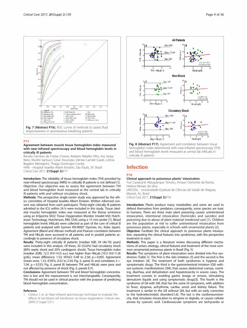
Fig. 7 (Abstract P16). ROC curves of methods to assess fluidresponsiveness in spontaneous breathing patients
Fig. 8 (Abstract P17). Agreement and correlation between tissuehemoglobin index determined with near-infrared spectroscopy (THI)and blood hemoglobin levels measured at central lab (HbLab) incritically ill patients
Critical Care 2017, 21(Suppl 2):139 Page 9 of 36
P17Agreement between muscle tissue hemoglobin index measuredwith near-infrared spectroscopy and blood hemoglobin levels incritically ill patientsRenato Carneiro de Freitas Chaves, Roberto Rabello Filho, Ary SerpaNeto, Murillo Santucci Cesar Assunção, Denise Carnieli Cazati, LetíciaBagatini Meneghini, Thiago Domingos CorrêaHIAE - Hospital Israelita Albert Einstein, São Paulo, SP, BrazilCritical Care 2017, 21(Suppl 2):P17
Introduction: The reliability of tissue hemoglobin index (THI) provided bynear-infrared spectroscopy (NIRS) in critically ill patients is not defined [1].Objective: Our objective was to assess the agreement between THIand blood hemoglobin level measured at the central lab in criticallyill patients with and without circulatory shock.Methods: This prospective single-center study was approved by the eth-ics committee of Hospital Israelita Albert Einstein. Written informed con-sent was obtained from each participant. Thirty-eight critically ill patientsadmitted to the ICU within 24 h were included in this study. Tissue (skel-etal muscle) hemoglobin index was measured at the thenar eminenceusing an InSpectra StO2 Tissue Oxygenation Monitor (model 650; Hutch-inson Technology, Hutchinson, MN, USA) using a 15 mm probe [1]. Bloodhemoglobin levels (HbLab) were collected as part of the care of critical illpatients and analyzed with Sysmex XN-9000® (Sysmex, Inc. Kobe Japan).Agreement (Bland and Altman method) and Pearson correlation betweenTHI and HbLab were accessed in all patients and in pooled patients ac-cordingly to presence of circulatory shock.Results: Thirty-eight critically ill patients [median IQR; 59 (46-70) years]were included in this analysis. Of these, 20 (52.6%) had circulatory shock(80% septic shock and 20% cardiogenic shock). Tissue hemoglobin index[median (IQR); 12.3 (9.9-14.3) a.u.] was higher than HbLab [10.3 (9.0-11.8)g/dL], mean difference: 1.53; 95%CI: 0.48 to 2.58; p = 0.005. Agreement(mean error, 1.53, IC95%, 0.52 to 2.54; Fig. 8, panel A) and correlation (r =1.04, p = 0.533; Fig. 8, panel B) between THI and HbLab were poor andnot affected by presence of shock (Fig. 8, panels E-F).Conclusions: Agreement between THI and blood hemoglobin concentra-tion is low and the measurement is not interchangeable. Consequently,THI should not be used in clinical practice with the purpose of predictingblood hemoglobin concentration.
Reference1. Creteur J, et. al. Near-infrared spectroscopy technique to evaluate the
effects of red blood cell transfusion on tissue oxygenation. Critical care.2009;13 Suppl 5:S11.
InfectionP18Clinical approach to poisonous plants’ intoxicationYuri Cavalcanti Albuquerque Tenorio, Amauri Clemente da Rocha,Helena Moraes da SilvaUNCISAL - Universidade Estadual de Ciências da Saúde de Alagoas,Maceió, AL, BrasilCritical Care 2017, 21(Suppl 2):P18
Introduction: Plants produce many metabolites and some are used todefend themselves from predators; consequently, some species are toxicto humans. There are three main plant poisoning causes: unintentionalintoxication, intentional intoxication (homicides and suicides) andpoisoning due to abuse of plant material (medicinal use) [1]. Childrenare the population at risk to suffer unintentional intoxication frompoisonous plants, especially in schools with ornamental plants [2].Objective: Facilitate the clinical approach to poisonous plants intoxica-tion, separating the clinical features into syndromes, with the perspectivetreatment to each.Methods: This paper is a literature review discussing different mecha-nisms of action, etiology, clinical features and treatment of the most com-mon ornamental poisonous plants in Brazil (Fig. 9).Results: The symptoms of plant intoxication were divided into five syn-dromes (Table 3). The first is the skin irritation [3] and the second is theeye irritation [4]. The treatment of both syndromes is hygiene andsymptomatic drugs. The third is the gastrointestinal irritation (GII) with-out systemic manifestations (SM), that causes abdominal cramps, vomit-ing, diarrhea, and dehydration and hepatotoxicity in severe cases. Thetreatment consists in avoiding gastric lavage or emesis, stimulatingdemulcent liquids and using symptomatic drugs[3]. The fourth is thesyndrome of GII with SM, that has the same GI symptoms, with additionto fever, dyspnea, arrhythmia, cardiac arrest and kidney failure. Thetreatment is similar to the GII without SM, but with an early correctionto the hydroelectrolytic disorders[1,6]. The last is the cardioneurotoxi-city, that simulates intoxication to atropine or digitalis, or causes cellularanoxia by cyanuric acid. Cardiovascular symptoms are tachycardia or
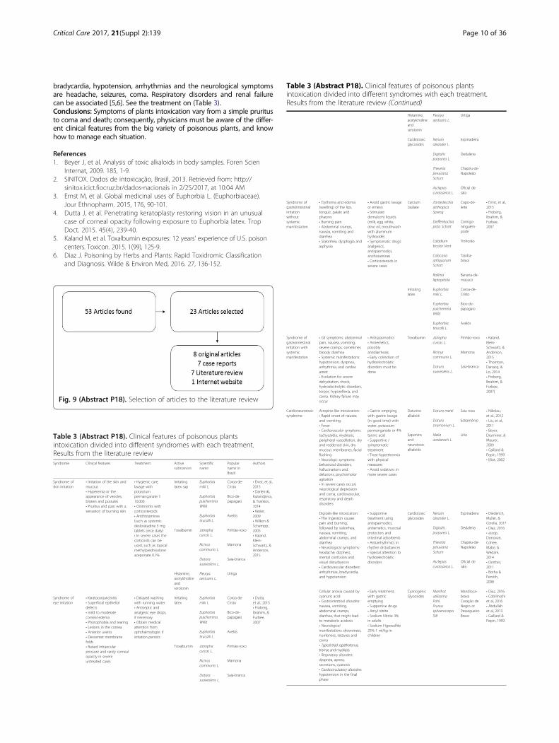
Table 3 (Abstract P18). Clinical features of poisonous plantsintoxication divided into different syndromes with each treatment.Results from the literature review (Continued)
Histamine,acetylcholineandserotonin
Fleuryaaestuans L.
Urtiga
Cardiotoxicglycosides
Neriumoleander L.
Espirradeira
Digitalispurpurea L.
Dedaleira
ThevetiaperuvianaSchum
Chapéu-de-Napoleão
Asclepiascurassavica L.
Oficial desala
Syndrome ofgastrointestinalirritationwithoutsystemicmanifestation
• Erythema and edema(swelling) of the lips,tongue, palate andpharynx• Burning pain• Abdominal cramps,nausea, vomiting anddiarrhea• Sialorrhea, dysphagia andasphyxia
• Avoid gastric lavageor emesis• Stimulatedemulcent liquids(milk, egg white,olive oil, mouthwashwith aluminumhydroxide)• Symptomatic drugs:analgesics,antispasmodics,antihistamines• Corticosteroids insevere cases
Calciumoxalate
ZantedeschiaaethiopicaSpreng
Copo-de-leite
• Ernst, et al.,2015• Froberg,Ibrahim, &Furbee,2007
Dieffenbachiapicta Schott
Comigo-ninguém-pode
Caladiumbicolor Vent
Tinhorão
ColocasiaantiquorumSchott
Taioba-brava
Critical Care 2017, 21(Suppl 2):139 Page 10 of 36
bradycardia, hypotension, arrhythmias and the neurological symptomsare headache, seizures, coma. Respiratory disorders and renal failurecan be associated [5,6]. See the treatment on (Table 3).Conclusions: Symptoms of plants intoxication vary from a simple pruritusto coma and death; consequently, physicians must be aware of the differ-ent clinical features from the big variety of poisonous plants, and knowhow to manage each situation.
References1. Beyer J, et al. Analysis of toxic alkaloids in body samples. Foren Scien
Internat, 2009. 185, 1-9.2. SINITOX. Dados de intoxicação, Brasil, 2013. Retrieved from: http://
sinitox.icict.fiocruz.br/dados-nacionais in 2/25/2017, at 10:04 AM3. Ernst M, et al. Global medicinal uses of Euphorbia L. (Euphorbiaceae).
Jour Ethnopharm. 2015, 176, 90-101.4. Dutta J, et al. Penetrating keratoplasty restoring vision in an unusual
case of corneal opacity following exposure to Euphorbia latex. TropDoct. 2015. 45(4), 239-40.
5. Kaland M, et al. Toxalbumin exposures: 12 years' experience of U.S. poisoncenters. Toxicon. 2015. 1(99), 125-9.
6. Diaz J. Poisoning by Herbs and Plants: Rapid Toxidromic Classificationand Diagnosis. Wilde & Environ Med, 2016. 27, 136-152.
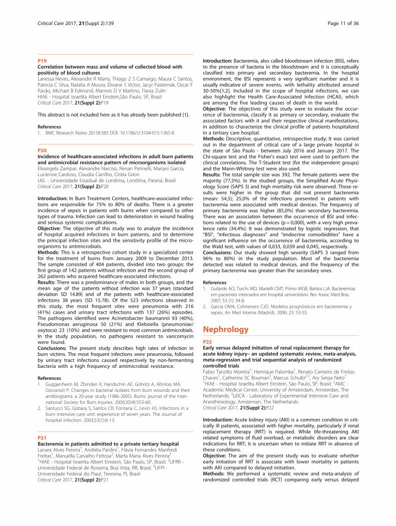
Fig. 9 (Abstract P18). Selection of articles to the literature review
Table 3 (Abstract P18). Clinical features of poisonous plantsintoxication divided into different syndromes with each treatment.Results from the literature reviewSyndrome Clinical features Treatment Active
substancesScientificname
Popularname inBrazil
Authors
Syndrome ofskin irritation
• Irritation of the skin andmucous• Hyperemia or theappearance of vesicles,blisters and pustules• Pruritus and pain with asensation of burning skin
• Hygienic care,lavage withpotassiumpermanganate 1:10,000• Ointments withcorticosteroids• Antihistamines(such as systemicdesloratadine 5 mgtablets once daily)• In severe cases thecorticoids can beused, such as topicalmethylprednisoloneaceponate 0.1%
Irritatinglatex sap
Euphorbiamilii L.
Coroa-de-Cristo
• Ernst, et al.,2015• Darlenski,Kazandjieva,& Tsankov,2014• Kesler,2009• Wilken &Schempp,2005• Kaland,Klein-Schwartz, &Anderson,2015
EuphorbiapulcherrimaWilld
Bico-de-papagaio
Euphorbiatirucalli L.
Avelós
Toxalbumin Jatrophacurcas L.
Pinhão-roxo
Ricinuscommunis L.
Mamona
Daturasuaveolens L.
Saia-branca
Histamine,acetylcholineandserotonin
Fleuryaaestuans L.
Urtiga
Syndrome ofeye irritation
• Keratoconjunctivitis• Superficial epithelialdefects• mild to moderatecorneal edema• Photophobia and tearing• Lesions in the cornea• Anterior uveitis• Descemet membranefolds• Raised intraocularpressure and rarely cornealopacity in severeuntreated cases
• Delayed washingwith running water• Antiseptic andanalgesic eye drops,if necessary• Obtain medicalattention fromophthalmologist ifirritation persists
Irritatinglatex
Euphorbiamilii L.
Coroa-de-Cristo
• Dutta,et al., 2015• Froberg,Ibrahim, &Furbee,2007
EuphorbiapulcherrimaWilld
Bico-de-papagaio
Euphorbiatirucalli L.
Avelós
Toxalbumin Jatrophacurcas L.
Pinhão-roxo
Ricinuscommunis L.
Mamona
Daturasuaveolens L.
Saia-branca
Rollinialeptopetala
Banana-de-macaco
Irritatinglatex
Euphorbiamilii L.
Coroa-de-Cristo
EuphorbiapulcherrimaWilld
Bico-de-papagaio
Euphorbiatirucalli L.
Avelós
Syndrome ofgastrointestinalirritation withsystemicmanifestation
• GII symptoms: abdominalpain, nausea, vomiting,severe cramps, sometimesbloody diarrhea• Systemic manifestations:hypotension, dyspnea,arrhythmia, and cardiacarrest• Evolution for severedehydration, shock,hydroelectrolytic disorders,torpor, hyporeflexia, andcoma. Kidney failure mayoccur
• Antispasmodics• Antiemetics,possiblyantidiarrheals.• Early correction ofhydroelectrolyticdisorders must bedone
Toxalbumin Jatrophacurcas L.
Pinhão-roxo • Kaland,Klein-Schwartz, &Anderson,2015• Thornton,Darracq, &Lo, 2014• Froberg,Ibrahim, &Furbee,2007)
Ricinuscommunis L.
Mamona
Daturasuaveolens L.
Saia-branca
Cardioneurotoxicsyndrome
Atropine-like intoxication:• Rapid onset of nauseaand vomiting• Fever• Cardiovascular symptoms:tachycardia, mydriasis,peripheral vasodilation, dryand reddened skin, drymucous membranes, facialflushing• Neurologic symptoms:behavioral disorders,hallucinations anddelusions, psychomotoragitation• In severe cases occursneurological depressionand coma, cardiovascular,respiratory and deathdisorders
• Gastric emptyingwith gastric lavage(in good time) withwater, potassiumpermanganate or 4%tannic acid• Supportive /symptomatictreatment• Treat hyperthermiawith physicalmeasures• Avoid sedatives inmore severe cases
Daturinealkaloid
Datura metel Saia roxa • Nikolau,et al., 2012• Liu, et al.,2011• Beyer,Drummer, &Maurer,2009• Gaillard &Pepin, 1999• Eliot, 2002
Daturastramonium L.
Estramônio
Saponinsandneurotoxicalkaloids
Meliaazedarach L.
Lírio
Digitalis-like intoxication:• The ingestion causespain and burning,followed by sialorrhea,nausea, vomiting,abdominal cramps, anddiarrhea• Neurological symptoms:headache, dizziness,mental confusion andvisual disturbances• Cardiovascular disorders:arrhythmias, bradycardia,and hypotension
• Supportivetreatment usingantispasmodics,antiemetics, mucosalprotectors andintestinal adsorbents• Antiarrhythmics inrhythm disturbances• Special attention tohydroelectrolyticdisorders
Cardiotoxicglycosides
Neriumoleander L.
Espirradeira • Diederich,Muller, &Cerella, 2017• Diaz, 2016• assop,Donovan,Cohee,Mabe, &Wedam,2014• Oerther,2011• Botha &Penrith,2008
Digitalispurpurea L.
Dedaleira
ThevetiaperuvianaSchum
Chapéu-de-Napoleão
Asclepiascurassavica L.
Oficial desala
Cellular anoxia caused bycyanuric acid:• Gastrointestinal disorders:nausea, vomiting,abdominal cramps,diarrhea, that might leadto metabolic acidosis• Neurologicalmanifestations: drowsiness,numbness, seizures andcoma• Typical triad: opisthotonus,trismas and mydriasis• Respiratory disorders:dyspnea, apnea,secretions, cyanosis• Cardiocirculatory disorders:hypotension in the finalphase
• Early treatment,with gastricemptying• Supportive drugs• Amyl nitrite• Sodium Nitrite 3%in adults• Sodium Hyposulfite25% 1 ml/kg inchildren
CyanogenicGlycosides
ManihotutilissimaPohl.PrunussphaerocarpaSW
Mandioca-bravaCoração deNegro orPessegueiroBravo
• Diaz, 2016• Cobilinschiet al, 2016• Abdullahet al, 2013• Gaillard &Pepin, 1999
Critical Care 2017, 21(Suppl 2):139 Page 11 of 36
P19Correlation between mass and volume of collected blood withpositivity of blood culturesLariessa Neves, Alexandre R Marra, Thiago Z S Camargo, Maura C Santos,Patricia C Silva, Natalia A Moura, Elivane S Victor, Jacyr Pasternak, Oscar FPavão, Michael B Edmond, Marines D V Martino, Flavia ZulinHIAE - Hospital Israelita Albert Einstein,São Paulo, SP, BrasilCritical Care 2017, 21(Suppl 2):P19
This abstract is not included here as it has already been published [1].
References1. BMC Research Notes 20158:383 DOI: 10.1186/s13104-015-1365-8
P20Incidence of healthcare-associated infections in adult burn patientsand antimicrobial resistance pattern of microorganisms isolatedElisangela Zampar, Alexandre Narciso, Renan Petinelli, Mariani Garcia,Lucienne Cardoso, Claudia Carrilho, Cintia GrionUEL - Universidade Estadual de Londrina, Londrina, Paraná, BrasilCritical Care 2017, 21(Suppl 2):P20
Introduction: In Burn Treatment Centers, healthcare-associated infec-tions are responsible for 75% to 80% of deaths. There is a greaterincidence of sepsis in patients with burns when compared to othertypes of trauma. Infection can lead to deterioration in wound healingand serious systemic complications.Objective: The objective of this study was to analyze the incidenceof hospital acquired infections in burn patients, and to determinethe principal infection sites and the sensitivity profile of the micro-organisms to antimicrobials.Methods: This is a retrospective cohort study in a specialized centerfor the treatment of burns from January 2009 to December 2013.The sample consisted of 404 patients, divided into two groups: thefirst group of 142 patients without infection and the second group of262 patients who acquired healthcare-associated infections.Results: There was a predominance of males in both groups, and themean age of the patients without infection was 37 years (standarddeviation SD 14.89) and of the patients with healthcare-associatedinfections 38 years (SD 15.78). Of the 523 infections observed inthis study, the most frequent sites were pneumonia with 216(41%) cases and urinary tract infections with 137 (26%) episodes.The pathogens identified were Acinetobacter baumannii 93 (40%),Pseudomonas aeruginosa 50 (21%) and Klebsiella (pneumoniae/oxytoca) 23 (10%) and were resistant to most common antimicrobials.In the study population, no pathogens resistant to vancomycinwere found.Conclusions: The present study describes high rates of infection inburn victims. The most frequent infections were pneumonia, followedby urinary tract infections caused respectively by non-fermentingbacteria with a high frequency of antimicrobial resistance.
References1. Guggenheim M, Zbinden R, Handschin AE, Gohritz A, Altintas MA,
Giovanoli P. Changes in bacterial isolates from burn wounds and theirantibiograms: a 20-year study (1986-2005). Burns: journal of the Inter-national Society for Burn Injuries. 2009;35(4):553-60.
2. Santucci SG, Gobara S, Santos CR, Fontana C, Levin AS. Infections in aburn intensive care unit: experience of seven years. The Journal ofhospital infection. 2003;53(1):6-13.
P21Bacteremia in patients admitted to a private tertiary hospitalLanara Alves Pereira1, Andréia Pardini1, Flávia Fernandes ManfrediFreitas1, Manuella Carvalho Feitosa2, Marta Maria Alves Pereira31HIAE - Hospital Israelita Albert Einstein, São Paulo, SP, Brasil; 2UFRR -Universidade Federal de Roraima, Boa Vista, RR, Brasil; 3UFPI -Universidade Federal do Piauí, Teresina, PI, BrasilCritical Care 2017, 21(Suppl 2):P21
Introduction: Bacteremia, also called bloodstream infection (BSI), refersto the presence of bacteria in the bloodstream and it is conceptuallyclassified into primary and secondary bacteremia. In the hospitalenvironment, the BSI represents a very significant number and it isusually indicative of severe events, with lethality attributed around30-50%[1,2]. Included in the scope of hospital infections, we canalso highlight the Health Care-Associated Infection (HCAI), whichare among the five leading causes of death in the world.Objective: The objectives of this study were to evaluate the occur-rence of bacteremia, classify it as primary or secondary, evaluate theassociated factors with it and their respective clinical manifestations,in addition to characterize the clinical profile of patients hospitalizedin a tertiary care hospital.Methods: Descriptive, quantitative, retrospective study; It was carriedout in the department of critical care of a large private hospital inthe state of São Paulo - between July 2016 and January 2017. TheChi-square test and the Fisher's exact test were used to perform theclinical correlations. The T-Student test (for the independent groups)and the Mann-Whitney test were also used.Results: The total sample size was 392. The female patients were themajority (77,5%). In the studied groups, the Simplified Acute Physi-ology Score (SAPS 3) and high mortality risk were observed. These re-sults were higher in the group that did not present bacteremia(mean: 54,5); 25,0% of the infections presented in patients withbacteremia were associated with medical devices. The frequency ofprimary bacteremia was higher (85,0%) than secondary bacteremia.There was an association between the occurrence of BSI and infec-tions related to the use of devices (p = 0,000), with a very high preva-lence ratio (34,4%). It was demonstrated by logistic regression, that“BSI”, “infectious diagnoses” and “endocrine comorbidities” have asignificant influence on the occurrence of bacteremia, according tothe Wald test, with values of 0,033, 0,039 and 0,045, respectively.Conclusions: Our study showed high severity (SAPS 3 ranged from96% to 80%) in the study population. Most of the bacteremiadetected was related to medical devices, and the frequency of theprimary bacteremia was greater than the secondary ones.
References1. Guilarde AO, Turchi MD, Martelli CMT, Primo MGB, Batista LJA. Bacteremias
em pacientes internados em hospital universitário. Rev Assoc Med Bras.2007; 53 (1): 34-8.
2. Garcia OMA, Colmenero CJD. Modelos prognósticos em bacteriemia ysepsis. An Med Interna (Madrid). 2006; 23: 53-55.
NephrologyP22Early versus delayed initiation of renal replacement therapy foracute kidney injury– an updated systematic review, meta-analysis,meta-regression and trial sequential analysis of randomizedcontrolled trialsFabio Tanzillo Moreira1, Henrique Palomba1, Renato Carneiro de FreitasChaves1, Catherine SC Bouman2, Marcus Schultz2,3, Ary Serpa Neto11HIAE - Hospital Israelita Albert Einstein, São Paulo, SP, Brasil; 2AMC -Academic Medical Center; University of Amsterdam, Amsterdan, TheNetherlands; 3LEICA - Laboratory of Experimental Intensive Care andAnesthesiology, Amsternan, The NetherlandsCritical Care 2017, 21(Suppl 2):P22
Introduction: Acute kidney injury (AKI) is a common condition in crit-ically ill patients, associated with higher mortality, particularly if renalreplacement therapy (RRT) is required. While life-threatening AKIrelated symptoms of fluid overload, or metabolic disorders are clearindications for RRT, it is uncertain when to initiate RRT in absence ofthese conditions.Objective: The aim of the present study was to evaluate whetherearly initiation of RRT is associate with lower mortality in patientswith AKI compared to delayed initiation.Methods: We performed a systematic review and meta-analysis ofrandomized controlled trials (RCT) comparing early versus delayed
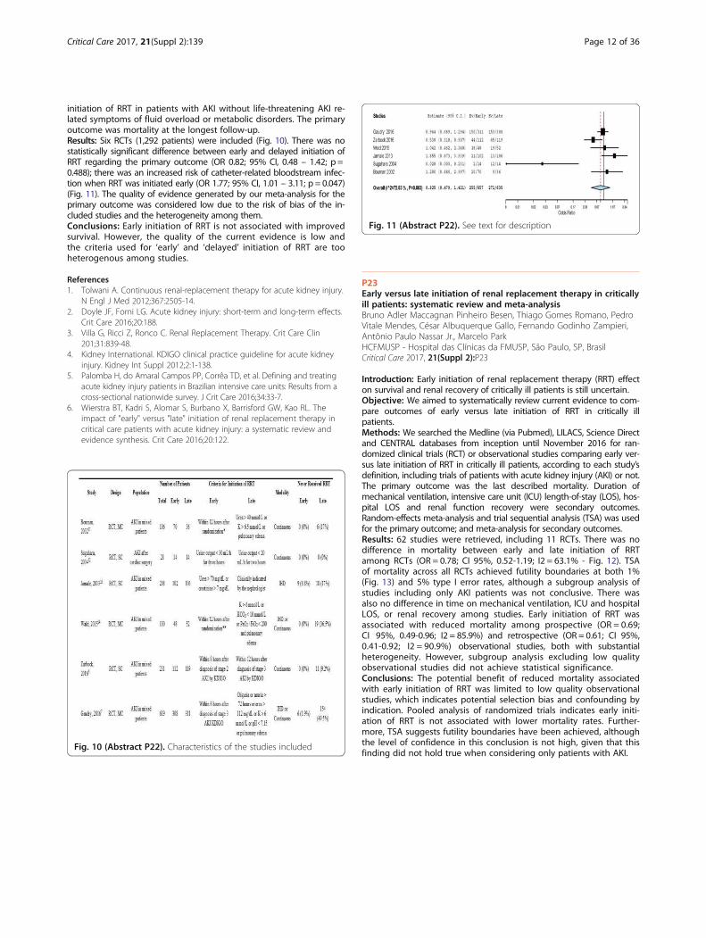
Fig. 11 (Abstract P22). See text for description
Critical Care 2017, 21(Suppl 2):139 Page 12 of 36
initiation of RRT in patients with AKI without life-threatening AKI re-lated symptoms of fluid overload or metabolic disorders. The primaryoutcome was mortality at the longest follow-up.Results: Six RCTs (1,292 patients) were included (Fig. 10). There was nostatistically significant difference between early and delayed initiation ofRRT regarding the primary outcome (OR 0.82; 95% CI, 0.48 – 1.42; p =0.488); there was an increased risk of catheter-related bloodstream infec-tion when RRT was initiated early (OR 1.77; 95% CI, 1.01 – 3.11; p = 0.047)(Fig. 11). The quality of evidence generated by our meta-analysis for theprimary outcome was considered low due to the risk of bias of the in-cluded studies and the heterogeneity among them.Conclusions: Early initiation of RRT is not associated with improvedsurvival. However, the quality of the current evidence is low andthe criteria used for ‘early’ and ‘delayed’ initiation of RRT are tooheterogenous among studies.
References1. Tolwani A. Continuous renal-replacement therapy for acute kidney injury.
N Engl J Med 2012;367:2505-14.2. Doyle JF, Forni LG. Acute kidney injury: short-term and long-term effects.
Crit Care 2016;20:188.3. Villa G, Ricci Z, Ronco C. Renal Replacement Therapy. Crit Care Clin
201;31:839-48.4. Kidney International. KDIGO clinical practice guideline for acute kidney
injury. Kidney Int Suppl 2012;2:1-138.5. Palomba H, do Amaral Campos PP, Corrêa TD, et al. Defining and treating
acute kidney injury patients in Brazilian intensive care units: Results from across-sectional nationwide survey. J Crit Care 2016;34:33-7.
6. Wierstra BT, Kadri S, Alomar S, Burbano X, Barrisford GW, Kao RL. Theimpact of "early" versus "late" initiation of renal replacement therapy incritical care patients with acute kidney injury: a systematic review andevidence synthesis. Crit Care 2016;20:122.
Fig. 10 (Abstract P22). Characteristics of the studies included
P23Early versus late initiation of renal replacement therapy in criticallyill patients: systematic review and meta-analysisBruno Adler Maccagnan Pinheiro Besen, Thiago Gomes Romano, PedroVitale Mendes, César Albuquerque Gallo, Fernando Godinho Zampieri,Antônio Paulo Nassar Jr., Marcelo ParkHCFMUSP - Hospital das Clínicas da FMUSP, São Paulo, SP, BrasilCritical Care 2017, 21(Suppl 2):P23
Introduction: Early initiation of renal replacement therapy (RRT) effecton survival and renal recovery of critically ill patients is still uncertain.Objective: We aimed to systematically review current evidence to com-pare outcomes of early versus late initiation of RRT in critically illpatients.Methods: We searched the Medline (via Pubmed), LILACS, Science Directand CENTRAL databases from inception until November 2016 for ran-domized clinical trials (RCT) or observational studies comparing early ver-sus late initiation of RRT in critically ill patients, according to each study’sdefinition, including trials of patients with acute kidney injury (AKI) or not.The primary outcome was the last described mortality. Duration ofmechanical ventilation, intensive care unit (ICU) length-of-stay (LOS), hos-pital LOS and renal function recovery were secondary outcomes.Random-effects meta-analysis and trial sequential analysis (TSA) was usedfor the primary outcome; and meta-analysis for secondary outcomes.Results: 62 studies were retrieved, including 11 RCTs. There was nodifference in mortality between early and late initiation of RRTamong RCTs (OR = 0.78; CI 95%, 0.52-1.19; I2 = 63.1% - Fig. 12). TSAof mortality across all RCTs achieved futility boundaries at both 1%(Fig. 13) and 5% type I error rates, although a subgroup analysis ofstudies including only AKI patients was not conclusive. There wasalso no difference in time on mechanical ventilation, ICU and hospitalLOS, or renal recovery among studies. Early initiation of RRT wasassociated with reduced mortality among prospective (OR = 0.69;CI 95%, 0.49-0.96; I2 = 85.9%) and retrospective (OR = 0.61; CI 95%,0.41-0.92; I2 = 90.9%) observational studies, both with substantialheterogeneity. However, subgroup analysis excluding low qualityobservational studies did not achieve statistical significance.Conclusions: The potential benefit of reduced mortality associatedwith early initiation of RRT was limited to low quality observationalstudies, which indicates potential selection bias and confounding byindication. Pooled analysis of randomized trials indicates early initi-ation of RRT is not associated with lower mortality rates. Further-more, TSA suggests futility boundaries have been achieved, althoughthe level of confidence in this conclusion is not high, given that thisfinding did not hold true when considering only patients with AKI.
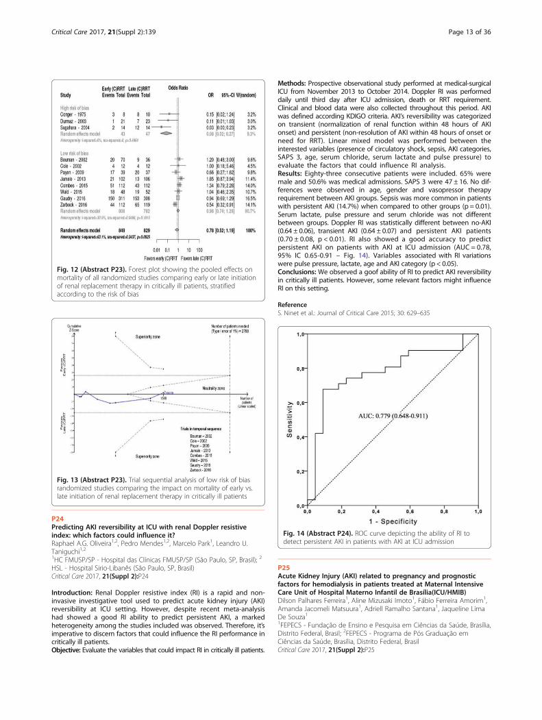
Fig. 14 (Abstract P24). ROC curve depicting the ability of RI todetect persistent AKI in patients with AKI at ICU admission
Fig. 13 (Abstract P23). Trial sequential analysis of low risk of biasrandomized studies comparing the impact on mortality of early vs.late initiation of renal replacement therapy in critically ill patients
Fig. 12 (Abstract P23). Forest plot showing the pooled effects onmortality of all randomized studies comparing early or late initiationof renal replacement therapy in critically ill patients, stratifiedaccording to the risk of bias
Critical Care 2017, 21(Suppl 2):139 Page 13 of 36
P24Predicting AKI reversibility at ICU with renal Doppler resistiveindex: which factors could influence it?Raphael A.G. Oliveira1,2, Pedro Mendes1,2, Marcelo Park1, Leandro U.Taniguchi1,21HC FMUSP/SP - Hospital das Clínicas FMUSP/SP (São Paulo, SP, Brasil); 2
HSL - Hospital Sirio-Libanês (São Paulo, SP, Brasil)Critical Care 2017, 21(Suppl 2):P24
Introduction: Renal Doppler resistive index (RI) is a rapid and non-invasive investigative tool used to predict acute kidney injury (AKI)reversibility at ICU setting. However, despite recent meta-analysishad showed a good RI ability to predict persistent AKI, a markedheterogeneity among the studies included was observed. Therefore, it’simperative to discern factors that could influence the RI performance incritically ill patients.Objective: Evaluate the variables that could impact RI in critically ill patients.
Methods: Prospective observational study performed at medical-surgicalICU from November 2013 to October 2014. Doppler RI was performeddaily until third day after ICU admission, death or RRT requirement.Clinical and blood data were also collected throughout this period. AKIwas defined according KDIGO criteria. AKI’s reversibility was categorizedon transient (normalization of renal function within 48 hours of AKIonset) and persistent (non-resolution of AKI within 48 hours of onset orneed for RRT). Linear mixed model was performed between theinterested variables (presence of circulatory shock, sepsis, AKI categories,SAPS 3, age, serum chloride, serum lactate and pulse pressure) toevaluate the factors that could influence RI analysis.Results: Eighty-three consecutive patients were included. 65% weremale and 50.6% was medical admissions. SAPS 3 were 47 ± 16. No dif-ferences were observed in age, gender and vasopressor therapyrequirement between AKI groups. Sepsis was more common in patientswith persistent AKI (14.7%) when compared to other groups (p = 0.01).Serum lactate, pulse pressure and serum chloride was not differentbetween groups. Doppler RI was statistically different between no-AKI(0.64 ± 0.06), transient AKI (0.64 ± 0.07) and persistent AKI patients(0.70 ± 0.08, p < 0.01). RI also showed a good accuracy to predictpersistent AKI on patients with AKI at ICU admission (AUC = 0.78,95% IC 0.65-0.91 – Fig. 14). Variables associated with RI variationswere pulse pressure, lactate, age and AKI category (p < 0.05).Conclusions:We observed a goof ability of RI to predict AKI reversibilityin critically ill patients. However, some relevant factors might influenceRI on this setting.
ReferenceS. Ninet et al.: Journal of Critical Care 2015; 30: 629–635
P25Acute Kidney Injury (AKI) related to pregnancy and prognosticfactors for hemodialysis in patients treated at Maternal IntensiveCare Unit of Hospital Materno Infantil de Brasília(ICU/HMIB)Dilson Palhares Ferreira1, Aline Mizusaki Imoto1, Fábio Ferreira Amorim1,Amanda Jacomeli Matsuura1, Adriell Ramalho Santana1, Jaqueline LimaDe Souza11FEPECS - Fundação de Ensino e Pesquisa em Ciências da Saúde, Brasília,Distrito Federal, Brasil; 2FEPECS - Programa de Pós Graduação emCiências da Saúde, Brasília, Distrito Federal, BrasilCritical Care 2017, 21(Suppl 2):P25
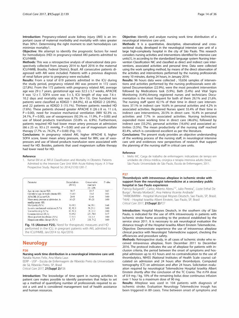
Critical Care 2017, 21(Suppl 2):139 Page 14 of 36
Introduction: Pregnancy-related acute kidney injury (AKI) is an im-portant cause of maternal morbidity and mortality with rates greaterthan 30%1. Determining the right moment to start hemodialysis canminimize mortality1.Objective: We attempt to identify the prognostic factors for needfor hemodialysis (HD) in pregnancy-related AKI patients admitted toICU/HMIB.Methods: This was a retrospective analysis of observational data pro-spectively collected from January 2014 to April 2016 in the maternalICU/HMIB, Brasilia, Federal District, Brazil. All consecutive patients di-agnosed with AKI were included. Patients with a previous diagnosisof renal failure prior to pregnancy were excluded.Results: From a total of 619 patients admitted in the ICU duringthe study period, pregnancy related AKI was present in 172 cases(27,8%). From the 172 patients with pregnancy related AKI, averageage was 29 ± 7 years, gestational age was 32.5 ± 5.7 weeks, APACHEII was 12 ± 7, SOFA score was 3 ± 3, ICU length of stay was 7.6 ±10.2 days and mortality rate was 8.7% (N = 15). One hundred tenpatients were classified as KDIGO 1 (64.0%), 43 as KDIGO 2 (20.9%),and 22 patients as KDIGO 3 (15.1%). Thirteen patients needed HD(7.6%). These patients showed greater APACHE II (26 ± 8 vs. 11 ± 6,P = 0.00), need for invasive mechanical ventilation (IMV 92.3% vs.24.1%, P = 0.00), use of vasopressors (92.3% vs. 11.9%, P = 0.00) anduse of blood products transfusion (53.8% vs. 6.9%). Furthermore,patients required HD had lower mean artery pressure at admission(66 ± 25 vs. 93 ± 21 mmHg, P = 0.00) and use of magnesium sulfatetherapy (7.7% vs. 74.2%, P = 0.00) (Fig. 15).Conclusions: In pregnancy related AKI, higher APACHE II, higherSOFA score, lower mean artery pressure, need for IMV, use of vaso-pressors and use of blood products transfusion were associated withneed for HD. Besides, patients that used magnesium sulfate therapyhad lower need for HD.
Reference1. Kamal EM et al. RIFLE Classification and Mortality in Obstetric Patients
Admitted to the Intensive Care Unit With Acute Kidney Injury: A 3-YearProspective Study. Reprod Sci 2014;21(10):1281-7.
Fig. 15 (Abstract P25). Need for therapeutic measures and HDperformed in the ICU, in pregnant patients with AKI, admitted tothe ICU/HMIB, Jan/2014 to Apr/2016
NeurologyP26Nursing work time distribution in a neurological intensive care unitNatalia Nunes Felix, Ana Maria LausEERP - USP - Escola de Enfermagem de Ribeirão Preto da Universidadede Sã, Ribeirão Preto, SP, BrasilCritical Care 2017, 21(Suppl 2):P26
Introduction: The knowledge of time spent in nursing activities inpatient care makes possible to identify parameters that helps to setup a method of quantifying number of professionals required to as-sist a unit and is considered management tool of health assistanceand human resources.
Objective: Identify and analyze nursing work time distribution of aneurological intensive care unit.Methods: It is a quantitative, descriptive, observational and cross-sectional study, developed in the neurological intensive care unit of alarge high-complexity hospital in the city of São Paulo. This researchconsiders nursing activities and interventions identified for intensive careunits{1}, in according to the standardized language system Nursing Inter-vention Classification NIC and classified as direct and indirect care inter-ventions, associated activities and personal time. Data were collectedusing the work sampling method, by means of the direct observation ofthe activities and interventions performed by the nursing professionalsevery 10 minutes, during 24 hours, in January 2014.Results: 96 hours data were collected , 10,656 samples of interven-tions and activities performed by the nursing professionals were ob-tained Documentation (22.9%), were the most prevalent interventionfollowed by Medications task (5.9%), Bath (5.4%) and Vital SignsMonitoring (4.4%).Among registered nurses and technicians Docu-mentation is the most frequent for both of them 29,1% and 21,6%.The nursing staff spent 42.1% of their time in direct care interven-tions 37.1% in indirect care 16.6% in personal activities and 4.2% inassociated activities. Registered Nurses spent 55.4% of their time inindirect care interventions, 20.5% in direct care. 16.4% in personalactivities and 7.7% in associated activities. Nursing techniciansexpended more working time in direct care (46.6%), followed byindirect care (33.2%), personal activities (16.6%) and associated ac-tivities (3.5%). The mean productivity of the nursing staff reached83.4%, which is considered excellent as per the literature.Conclusions: The present study provides an objective understandingof the working process of the nursing staff, in neurological intensivecare unit and evidences new perspectives of research that supportthe planning of the nursing staff in critical care units.
Reference1. Mello MC. Carga de trabalho de enfermagem: indicadores de tempo em
unidades de clínica médica, cirúrgica e terapia intensiva adulto [tese].São Paulo: Universidade de São Paulo, Escola de Enfermagem; 2011.
P27Thrombolysis with intravenous alteplase in ischemic stroke withsupport from the neurologist telemedicine at a secondary publichospital in Sao Paulo experiencePatricia Bulgarelli1, Carlos Alberto Abreu1,2, Julio Pereira1, Lizzie Erthal DeBurgo1, Renata Morbeck2, Ana Helena Vicente Andrade11HMBOIMIRIM - Hospital Municipal Moyses Deutsch, Sao Paulo, SP, Brasil;2HIAE - Hospital Israelita Albert Einstein, Sao Paulo, SP, BrasilCritical Care 2017, 21(Suppl 2):P27
Introduction: Hospital Moyses Deutsch, in the southern city of SãoPaulo, is indicated for the use of rtPA intravenously in patients withischemic stroke frame according to the protocol established by theservice since 2011. It is necessary to ask neurological evaluation thedistance trough of the Hospital Israelita Albert Einstein Telemedicine.Objective: Demonstrate experience the use of intravenous alteplaseclinical practice with Neurologist Telemedicine support, checking theefficiencies and procedure safety.Methods: Retrospective study, in all cases of ischemic stroke who re-ceived intravenous alteplase, from December 2011 to December2016. The protocol indicates the use of alteplase for patients with in-clusion criteria, the period between the onset of symptoms and hos-pital admission up to 4.5 hours and no contraindication to the use ofthrombolytics, NIHSS (National Institutes of Health Scale course) cal-culated on admission and 24 hours after thrombolysis. Computedtomography (CT) on admission and after 24 hours. Solicitation evalu-ation required by neurological Telemedicine Hospital Israelita AlbertEinstein shortly after the conclusion of the TC Cranio. The rt-PA doseof 0.9 mg / kg, 10% of the remaining bolus dose continuous infusionover 1 hour to a maximum dose of 90 mg.Results: Alteplase was used in 104 patients with diagnosis ofischemic stroke. Evaluation Neurology Telemedicine trough hasbeen triggered in 89 cases. Time between onset of symptoms and
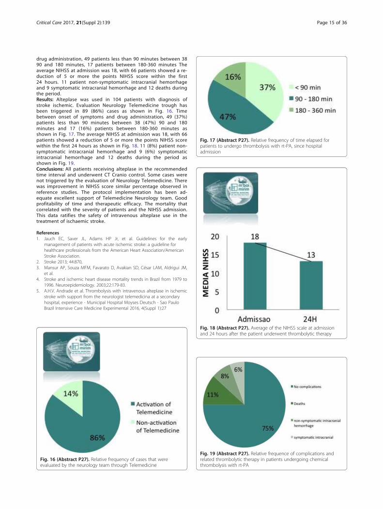
Fig. 17 (Abstract P27). Relative frequency of time elapsed forpatients to undergo thrombolysis with rt-PA, since hospitaladmission
Critical Care 2017, 21(Suppl 2):139 Page 15 of 36
drug administration, 49 patients less than 90 minutes between 3890 and 180 minutes, 17 patients between 180-360 minutes Theaverage NIHSS at admission was 18, with 66 patients showed a re-duction of 5 or more the points NIHSS score within the first24 hours. 11 patient non-symptomatic intracranial hemorrhageand 9 symptomatic intracranial hemorrhage and 12 deaths duringthe period.Results: Alteplase was used in 104 patients with diagnosis ofstroke ischemic. Evaluation Neurology Telemedicine trough hasbeen triggered in 89 (86%) cases as shown in Fig. 16. Timebetween onset of symptoms and drug administration, 49 (37%)patients less than 90 minutes between 38 (47%) 90 and 180minutes and 17 (16%) patients between 180-360 minutes asshown in Fig. 17. The average NIHSS at admission was 18, with 66patients showed a reduction of 5 or more the points NIHSS scorewithin the first 24 hours as shown in Fig. 18. 11 (8%) patient non-symptomatic intracranial hemorrhage and 9 (6%) symptomaticintracranial hemorrhage and 12 deaths during the period asshown in Fig. 19.Conclusions: All patients receiving alteplase in the recommendedtime interval and underwent CT Cranio control. Some cases werenot triggered by the evaluation of Neurology Telemedicine. Therewas improvement in NIHSS score similar percentage observed inreference studies. The protocol implementation has been ad-equate excellent support of Telemedicine Neurology team. Goodprofitability of time and therapeutic efficacy. The mortality thatcorrelated with the severity of patients and the NIHSS admission.This data ratifies the safety of intravenous alteplase use in thetreatment of ischaemic stroke.
References1. Jauch EC, Saver JL, Adams HP Jr, et al. Guidelines for the early
management of patients with acute ischemic stroke: a guideline forhealthcare professionals from the American Heart Association/AmericanStroke Association.
2. Stroke 2013; 44:870,3. Mansur AP, Souza MFM, Favarato D, Avakian SD, César LAM, Aldrigui JM,
et al.4. Stroke and ischemic heart disease mortality trends in Brazil from 1979 to
1996. Neuroepidemiology. 2003;22:179-83.5. A.H.V. Andrade et al. Thrombolysis with intravenous alteplase in ischemic
stroke with support from the neurologist telemedicina at a secondaryhospital, experience - Municipal Hospital Moyses Deutsch - Sao PauloBrazil Intensive Care Medicine Experimental 2016, 4(Suppl 1):27
Fig. 16 (Abstract P27). Relative frequency of cases that wereevaluated by the neurology team through Telemedicine
Fig. 18 (Abstract P27). Average of the NIHSS scale at admissionand 24 hours after the patient underwent thrombolytic therapy
Fig. 19 (Abstract P27). Relative frequence of complications andrelated thrombolytic therapy in patients undergoing chemicalthrombolysis with rt-PA
Critical Care 2017, 21(Suppl 2):139 Page 16 of 36
P28Dysphagia in patients affected by Ischemic StrokeGabriela Decol Mendonça, Débora D`Agostini Jorge Lisboa, Eliane LuciaColussi, Gabriela Colussi, Bianca Knapp Ten Caten, Patricia Jost, AngélicaSavoldi, Patrícia De Carli Tonial GhisolfiUPF - Universidade de Passo Fundo, Passo Fundo, RS, BrasilCritical Care 2017, 21(Suppl 2):P28
Introduction: The presence of the dysphagia symptom is relevant inpatients who had a stroke episode. This symptom can cause numer-ous changes in the patient's clinical condition, so there is a need toobtain data that allow the profile of the patients affected by thisswallowing disorder to be pointed out [1]Objective: Check the rate and degree of dysphagia in patients afterstroke-I evaluated by the speech therapy team.Methods: It is a longitudinal quantitative qualitative study of retro-spective methodology of records from the Hospital da Cidade dePasso Fundo – Rio Grande do Sul. The 152 patient records were ana-lyzed, 89 men and 63 women, in the years 2014 and 2015 withstroke-I. It was included in the study patients with stroke-I diagnosis,of both genders, without age restriction. Did not enter in the searchstroke-I patients who developed to stroke-H and other diseases suchas Parkinson's, Alzheimer's and cancer patients. The evaluation founddemographic and clinical data of the previous patients' history med-ical records.Results: It was found a higher incidence of dysphagia in older adultswith a mean of 70 years old for males. Prevailed mild dysphagia inboth genders and oral dietary. From 152 patients, 6 died. In patientspost-stroke with dysphagia, there are symptoms such as: absence ordelay of the swallowing reflex, increasing the risk of aspiration;Pharyngeal transit levied by absence of intra-oral pressure, alterationof vocal behavior after oral diet suggesting laryngeal penetration, de-crease in larynx elevation; Alteration of the labial sphincter, making itdifficult to start the swallowing process, reducing tongue control dur-ing the oral phase dynamics[1]. Individuals with neurological oropha-ryngeal dysphagia had a higher frequency of severe grade (46%),followed by mild and moderate degrees (27%, respectively) [2]. Thedata are in contrast to the study which indicated that, in patientswho had neurological oropharyngeal dysphagia, the most presentdegree was mild, followed by severe and moderate.Conclusions: It was identified a high incidence of patients experien-cing dysphagia symptom. Strengthening thus the importance of clin-ical assessment in patients after stroke-I episode to detect possibleswallowing disorder.
References1. Abdulmassih EM, Macedo Filho ED, Santos RS, Jurkiewicz AL. Evolução de
pacientes com disfagia orofaríngea em ambiente hospitalar. Art IntOtorrinolaringol. 2009;13(1):55-62. 2. Lacerda NC, Santos SSC. Avaliaçãonutricional de idosos: um estudo bibliográfico. Rev Rene. 2007; 8(1):60-9.
3. Moraes MAS, Coelho WJP, Castro G, Nemr K. Incidência de disfagia emunidade de terapia intensiva de adultos. Rev. CEFAC. 2006;8(2):171-7.
Nutrition /MetabolismP29Protein intake guided by the quadriceps muscle ultrasound in apatient with GBS: case reportDyaiane Santos, Branca Freitas, Debora Carneiro, Silvia Piovacari, EvandroFigueiredo, Diogo ToledoHIAE - Hospital Israelita Albert Einstein, São Paulo, SP, BrasilCritical Care 2017, 21(Suppl 2):P29
Introduction: Guillan Barré Syndrome is caracterized by muscularweakness. The intensity may vary from mild weakness to completetetraplegia with indication to mechanical ventilation which requireshospitalization in an Intensive Care Unit (ICU). Quadriceps muscleultrasonography represents one of the strategies to evaluate the lin-ear development of muscular mass in acute patients, as it is a non –invasive procedure and highly applicable to bedridden patient1.
Nitrogen balance is a non-invasive and accessible technic to evaluatemetabolic stress which consists of comparing the difference betweeningestion and excretion of nitrogen. The higher the losses the higherthe catabolism degree.Objective: Our objective was to evaluate the efficacy of quadricepsmuscle ultrasound serial in the patient with GBS in the ICU, tooptimize to protein intake and minimize muscle mass loss.Methods: This study obtained written informed consent from partici-pant. Male adult patient, 39 years of age, with GBS, in the ICU from8/18/2016 to 11/22/2016. Quadriceps muscle ultrasound, nitrogenbalance and weight check were performed weekly to adjust proteinintake during nutritional therapy. The thickness of the quadriceps mus-culature was quantified with a portable B-mode ultrasound device.With the patient lying supine, knees extended and relaxed, 2 landmarkson each quadriceps were identified and marked. The muscle thicknesswas quantified by the use of onscreen calipers and taken as the dis-tance between the upper margin of the femoral bone and the lowerboundary of the deep fascia of the rectus femoris. Each landmark wasimaged twice and averaged across each leg and then between legs.Results: In the first month at ICU the average protein intake wasbetween 1,6 g to 1,8 g/Kg/day, having an average weight loss of6,2 kg and nitrogen balance of -14. In the second month the averageprotein intake was between 2,0 g to 2,5 g/Kg/day, having an averageweight loss of 10,3 kg, a decrease of 0,36 mm of quadriceps muscleand nitrogen balance of -7,3. In the third month the average proteinintake was 3,5 g/Kg/day, having a stabilized weight, increase of0,16 mm of quadriceps muscle and nitrogen balance of +3,0. Duringdata collection the motor rehabilitation protocol was kept.Conclusions: Quadriceps Ultrasonography serial measures were effi-cient and useful to optimize protein intake. This case showed a cor-relation between negative nitrogen balance and muscle mass lossand between positive nitrogen balance and muscle mass gain assess-ment by US.
ReferenceSimpson F, Doig GS. Bedside nutrition evaluation and physical assessment
techniques in critical illness. Curr Opin Crit Care 2016, 22:303–307.
P30Bedside Ultrasound Is a Practical Measurement Tool for AssessingQuadriceps Muscle: a pilot studyDiogo Toledo, Branca Freitas, Dyaiane Santos, Debora Carneiro, RogérioDib, Silvia Piovacari, Ricardo Cordioli, João Manoel Silva JrHIAE - Hospital Israelita Albert Einstein, São Paulo, SP, BrasilCritical Care 2017, 21(Suppl 2):P30
Introduction: Bedside ultrasound measurement has previously beenused to quantify muscle layer thickness at the quadriceps muscleand some studies about nutritional assessment have showed excel-lent inter-rater and intra-rater reliability of the technique.Objective: The objectives of this study were to evaluate the intra,inter-reliability and facility of measuring quadriceps muscle layerthickness (QMLT) using bedside ultrasound.Methods: The study was approved by the ethics committee of Hos-pital Israelita Albert Einstein. A prospective study was carried out formeasuring QMLT in two healthy volunteers. The thickness of thequadriceps musculature was quantified with a portable B-mode ultra-sound device in volunteers the lying supine, knees extended and re-laxed, beside 2 landmarks on each quadriceps were identified. QMLTwas calculated by measuring at the border between the lower thirdand upper two-thirds between the anterior superior iliac spine (ASIS)and the upper pole of the patella, as well as the measurement of themidpoint between the ASIS and the upper pole of the patella. Themuscle thickness was quantified using onscreen calipers and takenas the distance between the upper margin of the femoral bone andthe lower boundary of the deep fascia of the rectus femoris (Fig. 20).To standardize measurements, an accompanying training with prac-tical lessons and 6 hours’ load time were held for team training.Trainer was 1 physician with advanced training in bedside ultrasound(expert). Trainees were comprised of 3 dietitians, 2 physicians, 1
Critical Care 2017, 21(Suppl 2):139 Page 17 of 36
physiotherapist with no prior ultrasound experience. To validate theimage collection by ultrasound measurements were performed com-paring between expert and all of trainees in different gendervolunteers.Results: Overall, 112 images were examined by expert and comparedto trainees. The correlation of Person found excellent relation be-tween expert and all of trainees R2 > 0.90 (Fig. 21). The best associ-ation was between the expert and dietitians R2 0.99, P < 0.001 andthe worst between expert and physicians R2 0.92, P < 0.001. Regard-ing Bland Altman comparison, the highest percentage of error foundbetween expert and trainees was 5.12% (CI 95% 3.64-12.37) and thelowest was 1.01% (CI 95% 0.72-2.58), in addition the highest bias ofthe values described was -0,12 ± 0,19 and lowest was -0,01 ± 0,04.Conclusions: The data analyzed showed an excellent correlation ofthe measures between expert and trainees. Therefore, this exam as anew nutritional assessment tool is feasible and easily applicable forall health professionals.
Reference1. Tillquist et al Bedside Ultrasound Is a Practical and Reliable Measurement
Tool for Assessing Quadriceps Muscle Layer Thickness. JPEN J ParenterEnteral Nutr.2014; 38:886-890).
Fig. 20 (Abstract P30). See text for description
Fig. 21 (Abstract P30). See text for description
Fig. 22 (Abstract P31). See text for description
P31Skeletal muscle mass assessment in critically ill patients bycomputed tomographyDiogo Toledo1, Amanda Maria Rosa de Oliveira2, Jerusa Toloi2, AmandaCarvalho2, Carla Prado3, João Manoel Silva Jr11HIAE - Hospital Israelita Albert Einstein, São Paulo, SP, Brasil; 2HCB -Hospital do Cancer de Barretos, Barretos, SP, Brasil; 3UAlberta - Universityof Alberta, Canada, Edmonton, AB, CanadaCritical Care 2017, 21(Suppl 2):P31
Introduction: Typically, in ICU, body composition is assessed throughanthropometrics parameters but cannot specifically quantify skeletalmuscle (SM). Recent findings shown that computed tomography (CT)have been used to identify critical ill patients with low SM associatedwith poor clinical outcomes (1).Objective: We have evaluated low muscle mass by CT in comparisonto anthropometric data and outcomes in critical ill patients.Methods: The study was approved by the ethics committee ofHospital do Cancer de Barretos. At the period of 1 year, weincluded patients aged > 18 years, with ICU length of stay morethan 72 hours, who performed abdominal CT around 72 hours ofadmission in the ICU. Demographic data parameters, anthropo-metric data (weight loss and body mass index-BMI), prognosticindex (SAPS 3), complications, ICU length of stay (LOS), hospitalmortality and CT abdomen image were collected for analysis.Skeletal muscle and adipose tissue cross-sectional areas werequantified using single-slice CT scans at the third lumbar vertebra(L3). A ROC for hospital mortality was applied to defined thegroups of patients with sarcopenia and non-sarcopenia by CTanalysis vales. A Cox regression was applied to find independentassociation between sarcopenia and mortality and a 30 days’survival curve adjusted by SAPS 3 and age was used to check thejoint relationship of these features.Results: In the first 72 hours of ICU length of stay, 99 patientswere underwent to abdominal CT. The mean age was 61.64 years,56% male, BMI 24,19 ± 4,49 kg/m2 and hospital mortality was26%. According to weight loss criteria 43.1% of the patients wereclassified as malnourished and 19.4% according to BMI. However,BMI values showed no correlation with abdominal CT values forsarcopenia R2 = 0.39, P < 0.001. The cutoff point for determinationof sarcopenia by CT was 41.2 cm2 /m2 (sensitivity 70%, specificity69.5%, AUC 70.3), this was considered as a reference to classifysarcopenic patients (Fig. 22). The diagnosis of sarcopenia by CTwith the parameters of nutritional evaluation were correlated with64.2% by weight loss criteria and only 35.5% by BMI. Otherwise,sarcopenic patients when compared to nonsarcopenic had aworse 30 days survival (Fig. 23) curve adjusted by age and SAPS3, HR 2.73 CI95% 1.02-7.35 (P = 0.04), higher hospital mortality(41.9% vs 14.6%, P = 0.006) and ICU complication (76.7% vs 52.1%,P = 0.016).Conclusions: The sarcopenia assessed by abdominal CT demon-strated low correlation with malnutrition by BMI and was a risk factorfor high hospital mortality, complications, as well as lower 30 dayssurvival in critical ill patients.
ReferenceParis M; Mourtzakis, M. Assessment of skeletal muscle mass in critically
ill patients: considerations for the utility of computed tomographyimaging and ultrasonography. Curr Opin Clin Nutr Metab Care 2016,19:125–130
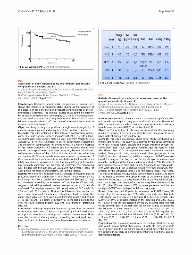
Fig. 23 (Abstract P31). See text for description
Fig. 24 (Abstract P32). See text for description
Critical Care 2017, 21(Suppl 2):139 Page 18 of 36
P32Assessment of body composition by two methods: tomographycomputed serial imaging and BMIAna Paula Noronha Barrére, Branca Freitas, Evandro Figueiredo, RonaldoBaroni, Silvia Piovacari, Diogo ToledoHIAE - Hospital Israelita Albert Einstein, São Paulo, SP, BrasilCritical Care 2017, 21(Suppl 2):P32
Introduction: Resources about body composition in cancer haveshown the influence in nutritional status during all the trajectory ofthis disease, in time of survive, in prognostic and tolerance of the an-tineoplastic treatment. The skeletal muscle mass could be quantifyby images in computerized tomography (CT). CT is a technology pre-cise and confiable to avaliate body composition. The use of CT possi-bilite a direct visualization of structures of transversal areas, musclemass and adipose tissue (1).Objective: Analyze body composition through serial tomography ofa cancer surgical patient and adequacy of the nutrition therapy.Methods: This study obtained written informed consent from partici-pant. Case study of one surgery oncology patient (P.P.), with adeno-carcinoma pancreatic cancer, male, 59 years old, from March/2016 toAugust/ 2016. He was submitted to gastro duodenopancreatectomyand surgery of complications of enteric fistula, in a general hospitalof Sao Paulo. Abdominal CT images and BMI obtained during fourmonths of hospitalization and were analyzed, by the SliceOmaticsoftware, at the level of the third lumbar vertebra (L3) on admissionand by month. Skeletal muscle mass was assessed by measures ofthe cross sectional muscle area, from which the skeletal muscle index(SMI) was obtained calculated by the formula cm2/heigh2 (consider-ing normality parameter for male sex 54 cm2/m2). The monitoringwas divided into five periods, we calculated the average intake ofeach period for calories and proteins, considering fasting.Results: According to anthropometric parameters, monitoring patientpresented significant weight loss (18.5%) being: initial weight 94 kg,final weight of 76.6 kg. Initial 28.3 kg/m2 BMI and BMI end 23.1 kg/m2. However, according to evaluation of the area by CT and SMI,suggests maintaining skeletal protein reserves in the last 3 periodsevaluated. The average values of SMI found were of 36.3 cm2/m2,37.5 cm2/m2, 36.5 cm2/m2. During this follow-up, the patient re-ceived oral nutritional therapy and enteral/parenteral, or being theinitial mean values of 19.73 kcal/kg and 0.99 g/kg protein and end of31.46 kcal/kg and 1.53 grams of protein/kg. In the last 3 periods, theoffer was 1.76 average protein, 1.25 and 1.53 grams of protein/kg(Fig. 24).Conclusions: Although nutritional diagnosis by BMI does not revealmalnutrition, assessment of body composition by CT indicated a lossof important muscle mass during hospitalization (sarcopenia). How-ever, the nutritional therapy offered, according to nutritional needs,has contributed to the maintenance of lean mass in recent weeks.
ReferenceParis M, Mourtzakis M. Assessment of skeletal muscle mass in critically ill
patients: considerations for the utility of computed tomography imagingand ultrasonography. Curr Opin Clin Nutr Metab Care 2016, 19:125–130
P33Bedside ultrasound muscle layer thickness assessment of thequadriceps in critically ill patientDiogo Toledo, Branca Freitas, Debora Carneiro, Dyaiane Santos, RogérioDib, Evandro Figueiredo, Silvia Piovacari, João Manoel Silva JrHIAE - Hospital Israelita Albert Einstein, São Paulo, SP, BrasilCritical Care 2017, 21(Suppl 2):P33
Introduction: Survivors of critical illness experience significant skel-etal muscle wasting that may predict clinical outcome. Ultrasound(US) is a noninvasive method that can measure muscle quadricepsmuscle layer thickness (QMLT) at the bedside (1,2).Objective: The objective of this study was to evaluate the measuringquadriceps muscle layer thickness using bedside ultrasound in critic-ally ill patient during 7 days follow up.Methods: This was a prospective, single-center study, conducted in atertiary care hospital. The study was approved by the ethics committeeof Hospital Israelita Albert Einstein and written informed consent ob-tained from each study participant. Patients aged 18 years or olderwho during their ICU stay required mechanical ventilation were in-cluded. Demographic data, anthropometric data, prognostic index(SAPS 3), nutrition risk screening (NRS) and image of US QMLT were col-lected for analysis. The thickness of the quadriceps musculature wasquantified with a portable B-mode ultrasound device. With the patientlying supine, knees extended and relaxed, 2 landmarks on each quadri-ceps were identified. The underlying tissues were then maximally com-pressed by the ultrasound probe, and the screen image was frozen.The muscle thickness was quantified using onscreen calipers and takenas the distance between the upper margin of the femoral bone andthe lower boundary of the deep fascia of the rectus femoral. Each land-mark was imaged and averaged across each leg. Measurements of thefirst (D1), third (D3) and seventh (D7) days were performed and the per-centage of QMLT was displayed for left and right legs.Results: It was enrolled 20 patients underwent to 40 QMLT using USin each leg. The mean age was 57.0 ± 20.2 years, 75% male, BMI 24.6± 3.0 kg/m2, SAPS 3 was 51.3 ± 17.4 and NRS 3.2 ± 1.0. Overall, 8.4%(22.0% to -8.9%) of muscle wasting in the right leg and 5.2% (22.6%to -11.0%) in the left leg assessed by the US occurred from the firstto the seventh day. In the right leg the median values evaluated bythe US were 1.16 cm (0.92 to 1.75) D1, 1.13 cm (0.92 to 1.60) D3,1.02 cm (0.81 to 1.41) D7; P = 0.005 (Fig. 25). In the left leg, the me-dian values evaluated by the US were 1.23 cm (0.93 to 1.43) D1,1.16 cm (0.92 to 1.34) D3, 1.13 cm (0.89 to 1.31) D7; P = 0.017(Fig. 26).Conclusions: The measuring quadriceps muscle layer thickness usingultrasound demonstrated that critical ill patients present musclewasting daily, and this procedure can be a great differential to iden-tify patients most likely to benefit from enhanced nutritional and re-habilitation support.
Critical Care 2017, 21(Suppl 2):139 Page 19 of 36
References1. Tillquist et al. Bedside Ultrasound Is a Practical and Reliable Measurement
Tool for Assessing Quadriceps Muscle Layer Thickness. JPEN J ParenterEnteral Nutr.2014; 38:886-890)
2. Paris M; Mourtzakis, M. Assessment of skeletal muscle mass in critically illpatients: considerations for the utility of computed tomography imagingand ultrasonography. Curr Opin Clin Nutr Metab Care 2016, 19:125–130
Fig. 25 (Abstract P33). See text for description
Fig. 26 (Abstract P33). See text for description
P34Potential benefits of testosterone administration on ICU-acquiredweakness and prolonged mechanical ventilation: a pilot studySara Cardoso da Rosa1, Milena Artifon1, Ana Carolina Peçanha Antonio1,2,Silvia Regina Rios Vieira11HCPA - Hospital de Clínicas de Porto Alegre, Porto Alegre, RS, Brasil;2HMV - Hospital Moinhos de Vento, Porto Alegre, RS, BrasilCritical Care 2017, 21(Suppl 2):P34
Introduction: Intensive care unit acquired weakness (ICU-AW) is diag-nosed in up to 67% among long-term ventilated patients and is asso-ciated with a high morbidity and mortality [1]. Testosterone hasbeen shown to ameliorate the catabolism and increase net proteinsynthesis in severe burn injury [2], chronic obstructive pulmonary dis-ease (COPD)[3] and acquired immunodeficiency syndrome (AIDS) [4].Objective: To assess the potential role of testosterone on ICU-AW re-covery and reduction in duration of mechanical ventilation (MV).Methods: A pilot open-label, randomized, controlled trial of testosteronetherapy on chronic critically ill adult patients requiring prolonged ventila-tory support, defined as endotracheal intubation and MV for more than14 days, or need for tracheostomy and MV for more than 8 days. Exclu-sion criteria: arterial or venous thromboembolism in the last 6 months;prior or current history of prostate cancer; refractory shock; severethrombocytopenia (less than 20,000 platelets per microliter); acute liver
failure or decompensate chronic liver disease; overt congestive heart fail-ure; end-of-life care; and patient or surrogate’s refusal to participate. Sub-jects are randomly assigned to receive standard nutritional support andphysical therapy, or an additional bi-weekly dose of 200 mg intramuscu-lar testosterone cypionate up to ICU discharge. We use a computer-generated, randomization scheme with varying block sizes ranging from4 to 8, stratified by gender and coexistence of COPD and/or AIDS; alloca-tion is concealed. Diaphragm ultrasound and Medical Research Council(MRC) score assessment are performed to evaluate muscular weakness.Patients are monitored through complete blood count, lipid panel, renaland hepatic function tests, blood gas analysis, serum free testosteronelevels, and caloric and protein intake. Minimum sample foreseen is 15 in-dividuals in each arm. Primary outcome is ventilator-free days.Results: The present research is in initial phase of recruitment, and itwill be finished by January 2018. Eleven individuals have been enrolledso far - 7 men and 4 women. In this sample, 7 patients were dischargedfrom the ICU and four died. Currently, only one patient has completedthree doses of testosterone, and duration of MV was 90 days. Eight par-ticipants have been randomized to intervention with anabolic steroids.Conclusions: High mortality showed by chronic critically ill popula-tion is the main obstacle for conduction of present study. Firstly, weaim to better define which type of patient most derives benefit, ifany, from anabolic steroids administration. Data obtained from thisclinical research may provide great contribution to fill the gap inknowledge of best management of prolonged MV and ICU-AW.
References1. Stanojcic M et al.: Curr Opin Crit Care. 2016 Aug; 22(4):325-312. Ferrando AA et al.: Crit Care Med 2001; 29:1936 – 19423. Pan L et al.: PLoS One. 2014 Jan 10; 9(1):e848554. Blick G et al.: Postgrad Med. 2013 Mar; 125(2):19-29
PneumologyP35Validation of the PESI (Pulmonary embolism severity index) scorefor risk stratification after acute pulmonary embolism in a Brazilianretrospective cohortLeonardo Soriano, Talita Castro, Kelvin Vilalva, Marcos Borges, AntônioPazin-Filho, Carlos MirandaFMRP/USP - Divisão de Emergências Clínicas -FMRP/USP, Ribeirão Preto,São Paulo, BrasilCritical Care 2017, 21(Suppl 2):P35
Introduction: The PESI is a score for risk stratification after acute pul-monary embolism. It was inserted in the 2014 ESC guidelines forguiding the management of these patients.Objective: The objective of this study was validated the PESI score ina Brazilian cohort.Methods: This investigation was a retrospective cohort that in-cluded patients hospitalized at the emergency department of ourhospital with the diagnosis of acute pulmonary embolism fromthe period of January 2009 until December 2015. The PESI score(original and simplified version) was applied in all patients withthe admission data. The mortality rate in 30 days was the out-come observed.Results: One hundred twenty-three (123) patients were included(57 ± 17 years, 60%female). The mortality rate according to thePESI class (original version) was: Class I (0.80%), Class II (1.62%),Class III (4.87%), Class IV (3.25%), Class V (12.20%); p < 0,0001. Di-chotomized analysis showed: Class I-II (5,8%) vs. Class III-IV-V(34,7%), relative risk (RR): 5.9; 95% confidence interval (CI): 1.88-18.51; p = 0.0002 and simplified version was 0 points (3,25%) vs.1 points (19,51%); RR: 2.38; 95%CI: 0.89-6.38; p = 0.06. The survivalanalysis through the Kaplan-Meier curve showed that the Class Iand II had similar curves (p = 0.59) and Class III-IV-V had similarcurves too (p = 0.25) and the comparison of curves of the ClassIII-IV-V vs. Class I-II showed significantly higher mortality rate atthe first group, RR: 7.63; 95%CI: 2.29-25.21; p = 0.0001(log-rank-test) justifying this dichotomized analysis (Fig. 27). The simplifiedversion higher or equal 1 points was associated with higher
Critical Care 2017, 21(Suppl 2):139 Page 20 of 36
mortality rate compared to 0 points (RR: 2.95; 95%CI: 1.02-8.51; p= 0.03). The dichotomized analysis of the original version showedhigher accuracy than simplified version (ROC-curve area: 0.70(95%CI: 0.62-0.77) vs. 0.60 (95%CI: 0.51-0.67); p = 0.05 for mortalityprediction.Conclusions: The PESI score measures adequately the prognosticafter acute pulmonary embolism in a Brazilian cohort. The dichot-omized analysis of the original version should have higher accur-acy than simplified version to predict the 30 days.
Fig. 27 (Abstract P35). Kaplan-Meier curve showing the survival at30 days in patients with acute pulmonary embolism dividedaccording the PESI score (Class I and II versus Class III, IV and V)
P36Troponin and NT-proBNP levels for prognostic stratification innormotensive patients with acute pulmonary embolism in aBrazilian cohortLeonardo Soriano, Talita Castro, Kelvin Vilalva, Marcos Borges, AntônioPazin-Filho, Carlos MirandaFMRP/USP - Divisão de Emergências Clínicas -FMRP/USP, Ribeirão Preto,São Paulo, BrasilCritical Care 2017, 21(Suppl 2):P36
Introduction: Troponin and N-terminal (NT)-pro natriuretic pep-tide of type B (BNP) can be used for prognostic stratificationafter acute pulmonary embolism (APE), mainly in haemo-dynamically stable patients. Despite this, the appropriate cut-off values of these biomarkers are not clear. Usually, the tropo-nin cut-off value used is the 99th percentile of the curve ofnormal individuals, which is the same value used for myocar-dial infarction definition. The NT-proBNP cut-off value used is600 pg/ml.Objective: The objective was to evaluate the performance of thiscut-off value for these biomarkers in patients with APE in a Braziliancohort.
Methods: This is a retrospective cohort that included patientshospitalized at the emergency department of our hospital withAPE diagnosis from the period of January 2009 until December2015. We analyzed only the haemodynamically stable patients inwhich these biomarkers was quantified through Vidas® Troponin IUltra and Vidas® NT-proBNP (bioMérieux, France). The 99th per-centile for this troponin assay is 0.01 mcg/L. The mortality rate in30 days was the outcome observed. The levels of biomarkerswere expressed in median and interquartile range (75th percent-ile -25th percentile).Results: Troponin levels were quantified in 75 patients (55 ±11 years, 40%male), this biomarker was positive in 36 patients(48%), comparing its levels between survivors vs. non-survivorswas not observed difference: 0.01(0.08) mcg/L vs. 0.19 (0.34)mcg/L; p = 0.19 and in survival analysis positive troponin was nota predictor of mortality (RR: 1.38; 95%CI: 0.42-4.54; p = 0.58). Thistroponin cut-off value showed low sensibility 55%; 95%CI: 23-83e low specificity 53%, 95%CI: 40-66 and low accuracy (curve-ROCarea: 0.53; 95%CI: 0.37-0.70) for mortality prediction. NT-proBNPlevels were quantified in 64 patients (57 ± 17 years, 34% male),this biomarker was positive in 45 patients (70%), comparing itslevels between survivors vs. non-survivors was observed a ten-dency of higher value in the second group: 1484 (3498) pg/mlvs. 3953 (8418) pg/ml; p = 0.09 and in survival analysis there wasa tendency of higher mortality at the group with values higherthan 600 pg/ml (RR: 5.12; 95%CI: 0.66-39.7); p = 0.08. This cut-offvalue of NT-proBNP levels showed adequate sensibility 92%;95%CI: 62-100, but low specificity 35%; 95%CI: 54-74 and regularaccuracy (curve-ROC area: 0.63; 95%CI: 0.52-0.73) for mortalityprediction.Conclusions: Despite the underpowered sample, the cut-offvalue usually used for troponin showed inadequate performanceand the cut-off value used for NT-proBNP showed high sensibil-ity however low specificity for mortality prediction in thissetting.
Reference1. Konstantinides SV, Torbicki A, Agnelli G, Danchin N, et. al. 2014 ESC
Guidelines on the diagnosis and management of acute pulmonaryembolism. Eur Heart J. 2014; 35(43):3033-69
P37Potentially Modifiable Factors Contributing To Outcome OfInvasively Ventilated Patients Without ARDS – The PRoVENT StudyFlavia Pfeilsticker1, Ary Serpa Neto1, Fabienne Simonis2, Marcelo GamaAbreu3, Paolo Pelosi4, Marcus Schultz2; for the PROVE NetworkInvestigators1Hiae - Hospital Israelita Albert Einstein, São Paulo, Brazil; 2AMC - UVA -University Of Amsterdam, Amsterdam, The Netherlands; 3UHCGC -University Hospital Carl Gustav Carus, Dresden, Germany; 4UNIGE - IrccsSan Martino Ist, Genoa, ItalyCritical Care 2017, 21(Suppl 2):P37
Introduction: Mechanical ventilation is increasingly recognized as po-tentially harmful intervention in patients without the acute respira-tory distress syndrome (ARDS), and tidal volume size and airwaypressures have been associated with worse outcomes in thesepatients.Objective: To improve the outcome of invasively ventilated patientswithout the acute respiratory distress syndrome (ARDS), potentiallymodifiable factors associated with mortality need to be identified.
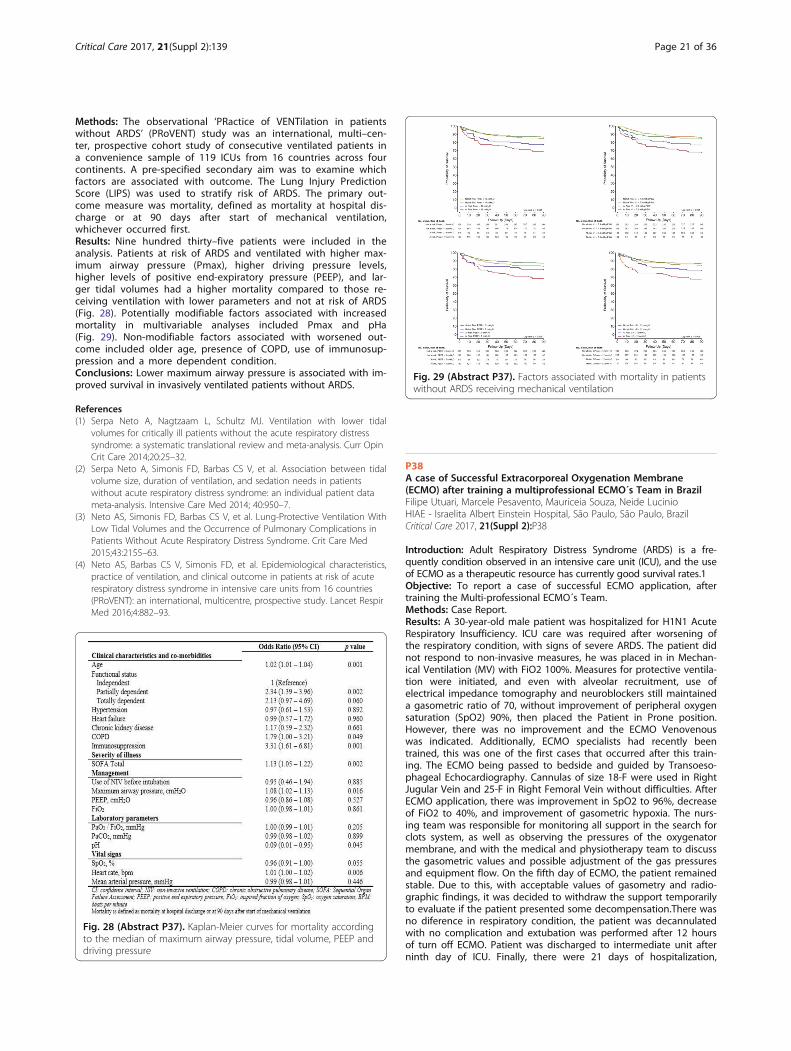
Fig. 29 (Abstract P37). Factors associated with mortality in patientswithout ARDS receiving mechanical ventilation
Critical Care 2017, 21(Suppl 2):139 Page 21 of 36
Methods: The observational ‘PRactice of VENTilation in patientswithout ARDS’ (PRoVENT) study was an international, multi–cen-ter, prospective cohort study of consecutive ventilated patients ina convenience sample of 119 ICUs from 16 countries across fourcontinents. A pre-specified secondary aim was to examine whichfactors are associated with outcome. The Lung Injury PredictionScore (LIPS) was used to stratify risk of ARDS. The primary out-come measure was mortality, defined as mortality at hospital dis-charge or at 90 days after start of mechanical ventilation,whichever occurred first.Results: Nine hundred thirty–five patients were included in theanalysis. Patients at risk of ARDS and ventilated with higher max-imum airway pressure (Pmax), higher driving pressure levels,higher levels of positive end-expiratory pressure (PEEP), and lar-ger tidal volumes had a higher mortality compared to those re-ceiving ventilation with lower parameters and not at risk of ARDS(Fig. 28). Potentially modifiable factors associated with increasedmortality in multivariable analyses included Pmax and pHa(Fig. 29). Non-modifiable factors associated with worsened out-come included older age, presence of COPD, use of immunosup-pression and a more dependent condition.Conclusions: Lower maximum airway pressure is associated with im-proved survival in invasively ventilated patients without ARDS.
References(1) Serpa Neto A, Nagtzaam L, Schultz MJ. Ventilation with lower tidal
volumes for critically ill patients without the acute respiratory distresssyndrome: a systematic translational review and meta-analysis. Curr OpinCrit Care 2014;20:25–32.
(2) Serpa Neto A, Simonis FD, Barbas CS V, et al. Association between tidalvolume size, duration of ventilation, and sedation needs in patientswithout acute respiratory distress syndrome: an individual patient datameta-analysis. Intensive Care Med 2014; 40:950–7.
(3) Neto AS, Simonis FD, Barbas CS V, et al. Lung-Protective Ventilation WithLow Tidal Volumes and the Occurrence of Pulmonary Complications inPatients Without Acute Respiratory Distress Syndrome. Crit Care Med2015;43:2155–63.
(4) Neto AS, Barbas CS V, Simonis FD, et al. Epidemiological characteristics,practice of ventilation, and clinical outcome in patients at risk of acuterespiratory distress syndrome in intensive care units from 16 countries(PRoVENT): an international, multicentre, prospective study. Lancet RespirMed 2016;4:882–93.
Fig. 28 (Abstract P37). Kaplan-Meier curves for mortality accordingto the median of maximum airway pressure, tidal volume, PEEP anddriving pressure
P38A case of Successful Extracorporeal Oxygenation Membrane(ECMO) after training a multiprofessional ECMO´s Team in BrazilFilipe Utuari, Marcele Pesavento, Mauriceia Souza, Neide LucinioHIAE - Israelita Albert Einstein Hospital, São Paulo, São Paulo, BrazilCritical Care 2017, 21(Suppl 2):P38
Introduction: Adult Respiratory Distress Syndrome (ARDS) is a fre-quently condition observed in an intensive care unit (ICU), and the useof ECMO as a therapeutic resource has currently good survival rates.1Objective: To report a case of successful ECMO application, aftertraining the Multi-professional ECMO´s Team.Methods: Case Report.Results: A 30-year-old male patient was hospitalized for H1N1 AcuteRespiratory Insufficiency. ICU care was required after worsening ofthe respiratory condition, with signs of severe ARDS. The patient didnot respond to non-invasive measures, he was placed in in Mechan-ical Ventilation (MV) with FiO2 100%. Measures for protective ventila-tion were initiated, and even with alveolar recruitment, use ofelectrical impedance tomography and neuroblockers still maintaineda gasometric ratio of 70, without improvement of peripheral oxygensaturation (SpO2) 90%, then placed the Patient in Prone position.However, there was no improvement and the ECMO Venovenouswas indicated. Additionally, ECMO specialists had recently beentrained, this was one of the first cases that occurred after this train-ing. The ECMO being passed to bedside and guided by Transoeso-phageal Echocardiography. Cannulas of size 18-F were used in RightJugular Vein and 25-F in Right Femoral Vein without difficulties. AfterECMO application, there was improvement in SpO2 to 96%, decreaseof FiO2 to 40%, and improvement of gasometric hypoxia. The nurs-ing team was responsible for monitoring all support in the search forclots system, as well as observing the pressures of the oxygenatormembrane, and with the medical and physiotherapy team to discussthe gasometric values and possible adjustment of the gas pressuresand equipment flow. On the fifth day of ECMO, the patient remainedstable. Due to this, with acceptable values of gasometry and radio-graphic findings, it was decided to withdraw the support temporarilyto evaluate if the patient presented some decompensation.There wasno diference in respiratory condition, the patient was decannulatedwith no complication and extubation was performed after 12 hoursof turn off ECMO. Patient was discharged to intermediate unit afterninth day of ICU. Finally, there were 21 days of hospitalization,
Critical Care 2017, 21(Suppl 2):139 Page 22 of 36
followed by hospital discharge with recovered lung function andgood general clinical condition for the residence.Conclusions: This case had a favourable outcome to the patient, thetraining of the multiprofessional team in ECMO is essential for goodresults. Nursing is inserted in this context by performing actionsaimed at ensuring the safety of the procedure, identifying signs sug-gestive of possible complications and acting in decision making withthe team.
Reference1. Repessé X, Aubry A, Vieillard-Baron A. On the complexity of scoring acute
respiratory distress syndrome: do not forget hemodynamics!. J ThoracDis. 2016;8(8):E758-E764.
P39Electrical impedance tomography at clinical practiceKarina T. Timenetsky, Denise Carnieli-Cazati, Carolina S.A. Azevedo,Marcelo Beraldo, Raquel A.C. EidHIAE - Critically Ill Department,Hospital Israelita Albert Einstein, SãoPaulo, SP, BrasilCritical Care 2017, 21(Suppl 2):P39
Introduction: Electrical impedance tomography (EIT) monitors bedsidelung ventilation distribution, allowing lung heterogeneity diagnosis, aslung collapse, overdistension, and ventilation asynchrony (1,2).Objective: To describe cases of EIT clinical applicability and its impact intherapeutic management of critically ill mechanically ventilated patients.Methods: Observational, descriptive case series study of electrical im-pedance tomography use to guide bedside mechanical ventilation(MV) during a six month period.Results: 7 patients had hypoxemic respiratory failure and necessityto adjust ventilator setting in order to reverse hypoxemia. BedsidePEEP titration: 4 patients were submitted to lung recruitment andPEEP titration guided by the EIT in order to choose a PEEP level withthe least lung collapse. The median PaO2/FiO2 ratio before recruit-ment was 158 (100-170). After lung recruitment and PEEP titrationPaO2/FiO2 ratio improved, with a median value of 264 (190-302). ThePEEP level varied between these patients, as two patients after lungrecruitment showed no lung collapse during PEEP titration, maintain-ing a PEEP level of 10 and 12 cmH2O. Before lung recruitment anempirical PEEP level of 10 and 18 respectively. The other two pa-tients that had PaO2/FiO2 ratio lower than 200, during PEEP titrationthere was lung collapse, after adjusting the PEEP level by EIT therewas an improvement in gas exchange (160 improving to 302, andanother patient from 143 to 240). The initial PEEP levels of these pa-tients were 10 and 12 cmH2O and after PEEP titration, 14 and 20cmH2O. EIT to minimize lung overdistension: One patient had bonch-opleural fistula after lung biopsy, with a PaO2/FiO2 ratio of 100. Ini-tially, PEEP level was empirically adjusted to 18 cmH2O, with noimprovement in gas exchange. At EIT baseline a flattening image inthe upper right lung was observed representing a gain in thebronchopleural fistula debit. After choosing PEEP by EIT there was animprovement in the upper right lung ventilation. The second patientsubmitted to unilateral lung transplantation due to lung fibrosis hadPaO2/FiO2 ratio of 225 with a PEEP level of 6 cmH2O and FiO2 of60%. This low gas exchange was due to the non-transplanted lung,and to avoid lung overdistension during MV EIT was installed. Thethird case patient with Non Hodgkin Lymphoma had an air cyst andEIT was maintained to guide patient’s MV in order to avoid baro-trauma and lung injury.
Conclusions: Electrical impedance tomography seems a very helpfultool in clinical practice of patients in mechanical ventilation.
References1. Victorino JA, Borges JB, Okamoto VN, Matos GF, Tucci MR, Caramez MP,
et al. Imbalances in regional lung ventilation: a validation study on electricalimpedance tomography. Am J Respir Crit Care Med 2004;169:791–800.
2. Costa EL V, Amato MBP. Electrical impedance tomography in critically illpatients. Clin Pulm Med 2013;20:178–86.
P40Use of a new isokinetic device oriented by software for inspiratorymuscle training in prolonged weaningBruno Leonardo Guimarães1,3, Leonardo de souza2,4, FernandoGuimaraes4, Hebe Cordeiro3, Fernanda Puga3, Cristiane Almeida3, SergioAlvim3, Thiago Regis3, Jocemir Lugon11UFF - Universidade Federal Fluminense, Niterói, RJ, Brasil; 2UNESA -Universidade Estácio de Sá, Niterói, RJ, Brasil; 3HNMD - Hospital NavalMarcílio Dias, Rio de Janeiro, RJ, Brasil; 4HI - Hospital Icaraí, Niterói, RJ, BrasilCritical Care 2017, 21(Suppl 2):P40
Introduction: Introduction: It is well established that inspiratory muscletraining (IMT) improves muscle performance in hospitalized individuals[1]. Nowadays, patients on prolonged ventilation are among the majorchallenges of intensive care [2,3]. In this regard, specific exercises for re-spiratory muscles can result in better functional performance in critic-ally ill patients during weaning process [1].Objective: To evaluate the effects of inspiratory muscle training (IMT)program on patients in prolonged weaning.Methods: Prospective randomized controlled trial. By the time partici-pants were judged as apt for ventilator weaning, they underwent IMTwith a new device called POWERbreathe K-5 (UK), Intervention group,or were managed in a conventional way with a tracheal collar, Controlgroup. The primary endpoint was successful weaning. Muscle strength,and 30 days after intervention survival in the ICU were also analysed.Inspiratory strength was measured by the timed inspiratory effort index(TIE) employing a digital vacuometer MVD300 (Globalmed, Brazil) [4].Results: Sixty-five tracheostomized patients were selected. Thirty-four pa-tients comprised the Intervention group (20 male, mean age 65± 17 years,mean APACHE II score 28.9 ± 4.8). The weaning process duration was 13.7± 8.8 days, and the initial and final TIE indexes were 0.8 ± 0.3 and 1.7 ± 0.9,respectively (P = 0.0001). Thirty patients (88%) were successfully weaned,and 11 (31%) had a fatal course. The control group encompassed 31 pa-tients (14 male, mean age 68± 16 years, mean APACHE II score 26.1 ±5.5). The weaning process duration was 21.7 ± 16.4 days, and the initialand final TIE indexes were 0.8 ± 0.6 and 1.0 ± 0.8 respectively (P = 0,053).Thirteen cases (42%) were successfully weaned, and 22 (63%) had a fatalcourse. The 30 days after intervention survival rates for the interventionand control groups were 79% and 44%, respectively (P = 0.025), Fig. 30.Conclusions: In support to our working hypothesis, the rate of wean-ing success and the survival rate 30 days after intervention werehigher for patients undergoing inspiratory muscle training.
Reference1. Mark E, Ruth D. Inspiratory muscle training facilitates weaning from
mechanical ventilation among patients in the intensive care unit: asystematic review Journal of Physiotherapy 61 (2015) 125–134. 2. Boles JM,Bion J, Connors A, Herridge M, Marsh B, Melot C, et al. Weaning frommechanical ventilation. Eur Respir J. 2007; 29:1033-56. 3.Jubran A, Grant BJB,Duffner LA, Collins EG, Lanuza DM, Hoffman LA, Tobin MJ. Effect of Pressure
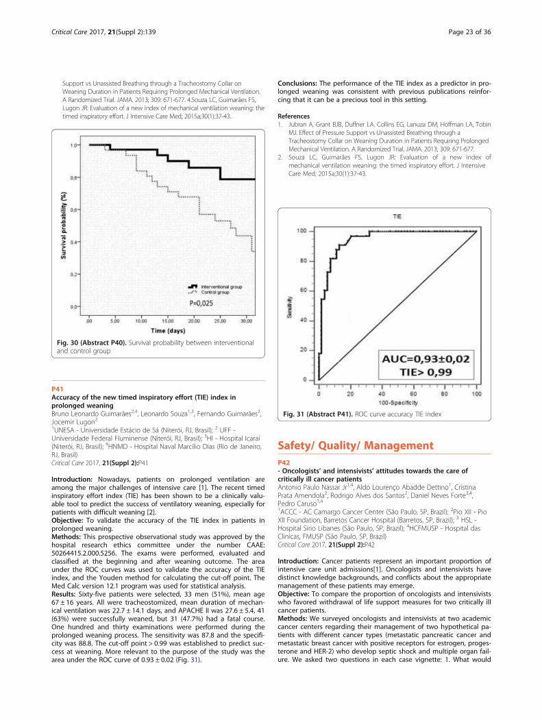
Critical Care 2017, 21(Suppl 2):139 Page 23 of 36
Support vs Unassisted Breathing through a Tracheostomy Collar onWeaning Duration in Patients Requiring Prolonged Mechanical Ventilation.A Randomized Trial. JAMA. 2013; 309: 671-677. 4.Souza LC, Guimarães FS,Lugon JR: Evaluation of a new index of mechanical ventilation weaning: thetimed inspiratory effort. J Intensive Care Med; 2015a;30(1):37-43.
Fig. 30 (Abstract P40). Survival probability between interventionaland control group
Fig. 31 (Abstract P41). ROC curve accuracy TIE index
P41Accuracy of the new timed inspiratory effort (TIE) index inprolonged weaningBruno Leonardo Guimarães2,4, Leonardo Souza1,3, Fernando Guimarães2,Jocemir Lugon21UNESA - Universidade Estácio de Sá (Niterói, RJ, Brasil); 2 UFF -Universidade Federal Fluminense (Niterói, RJ, Brasil); 3HI - Hospital Icaraí(Niterói, RJ, Brasil); 4HNMD - Hospital Naval Marcílio Dias (Rio de Janeiro,RJ, Brasil)Critical Care 2017, 21(Suppl 2):P41
Introduction: Nowadays, patients on prolonged ventilation areamong the major challenges of intensive care [1]. The recent timedinspiratory effort index (TIE) has been shown to be a clinically valu-able tool to predict the success of ventilatory weaning, especially forpatients with difficult weaning [2].Objective: To validate the accuracy of the TIE index in patients inprolonged weaning.Methods: This prospective observational study was approved by thehospital research ethics committee under the number CAAE:50264415.2.000.5256. The exams were performed, evaluated andclassified at the beginning and after weaning outcome. The areaunder the ROC curves was used to validate the accuracy of the TIEindex, and the Youden method for calculating the cut-off point. TheMed Calc version 12.1 program was used for statistical analysis.Results: Sixty-five patients were selected, 33 men (51%), mean age67 ± 16 years. All were tracheostomized, mean duration of mechan-ical ventilation was 22.7 ± 14.1 days, and APACHE II was 27.6 ± 5.4, 41(63%) were successfully weaned, but 31 (47.7%) had a fatal course.One hundred and thirty examinations were performed during theprolonged weaning process. The sensitivity was 87.8 and the specifi-city was 88.8. The cut-off point > 0.99 was established to predict suc-cess at weaning. More relevant to the purpose of the study was thearea under the ROC curve of 0.93 ± 0.02 (Fig. 31).
Conclusions: The performance of the TIE index as a predictor in pro-longed weaning was consistent with previous publications reinfor-cing that it can be a precious tool in this setting.
References1. Jubran A, Grant BJB, Duffner LA, Collins EG, Lanuza DM, Hoffman LA, Tobin
MJ. Effect of Pressure Support vs Unassisted Breathing through aTracheostomy Collar on Weaning Duration in Patients Requiring ProlongedMechanical Ventilation. A Randomized Trial. JAMA. 2013; 309: 671-677.
2. Souza LC, Guimarães FS, Lugon JR: Evaluation of a new index ofmechanical ventilation weaning: the timed inspiratory effort. J IntensiveCare Med; 2015a;30(1):37-43.
Safety/ Quality/ ManagementP42- Oncologists’ and intensivists’ attitudes towards the care ofcritically ill cancer patientsAntonio Paulo Nassar Jr1,4, Aldo Lourenço Abadde Dettino1, CristinaPrata Amendola2, Rodrigo Alves dos Santos2, Daniel Neves Forte3,4,Pedro Caruso1,41ACCC - AC Camargo Cancer Center (São Paulo, SP, Brazil); 2Pio XII - PioXII Foundation, Barretos Cancer Hospital (Barretos, SP, Brazil); 3 HSL -Hospital Sirio Libanes (São Paulo, SP, Brazil); 4HCFMUSP - Hospital dasClinicas, FMUSP (São Paulo, SP, Brazil)Critical Care 2017, 21(Suppl 2):P42
Introduction: Cancer patients represent an important proportion ofintensive care unit admissions[1]. Oncologists and intensivists havedistinct knowledge backgrounds, and conflicts about the appropriatemanagement of these patients may emerge.Objective: To compare the proportion of oncologists and intensivistswho favored withdrawal of life support measures for two critically illcancer patients.Methods: We surveyed oncologists and intensivists at two academiccancer centers regarding their management of two hypothetical pa-tients with different cancer types (metastatic pancreatic cancer andmetastatic breast cancer with positive receptors for estrogen, proges-terone and HER-2) who develop septic shock and multiple organ fail-ure. We asked two questions in each case vignette: 1. What would
Critical Care 2017, 21(Suppl 2):139 Page 24 of 36
be your choice regarding ICU admission? 2. Considering the patientswas admitted to ICU and full support was instituted, but the patient’sclinical condition continued to deteriorate after three days of fullsupport, how would you then define the patient status? a) Full codeb) Withholding of life support measures; c) Withdrawal of life supportmeasures.Results: Sixty intensivists and 46 oncologists responded to the sur-vey. More intensivists than oncologists [45/60 (75%) vs. 24/46 (52%),p = 0.02] opted for ICU admission with restriction of life support mea-sures for the pancreatic cancer patient (Fig. 32a). More intensiviststhan oncologists favored an ICU admission with restrictions of lifesupport measures [46/59 (78%) vs. 27/46 (59%), p = 0.055] (Fig. 32b).Oncologists and intensivists similarly favored withdrawal of life sup-port measures for the pancreatic cancer patient [33/46 (72%) vs. 48/60 (80%), p = 0.45] (Fig. 32c). On the other hand, intensivists favoredmore withdrawal of life support measures for the breast cancer pa-tient than oncologists [32/59 (54%) vs. 9/44 (21%), p < 0.001]. In themultinomial logistic regression, the oncology specialists were morelikely to advocate for a full code status for the breast cancer patient(OR = 5.931; CI 95%, 1.762-19.956; p = 0.004). On the other hand, novariable was associated with withdrawal of life support measures forthe pancreatic cancer patient.Conclusions: Oncologists and intensivists share different views re-garding life support measures in critically ill cancer patients. On-cologists tend to focus on the cancer characteristics, whereasintensivists focus on multiple organ failure when weighing in onthe same decisions. Regular meetings between oncologists andintensivists may reduce possible conflicts regarding the criticalcare of cancer patients.
Reference1. Soares M, Caruso P, Silva E, Teles JM, Lobo SM, Friedman G, Dal Pizzol F,
Mello PV, Bozza FA, Silva UV et al: Characteristics and outcomes ofpatients with cancer requiring admission to intensive care units: aprospective multicenter study. Crit Care Med 2010, 38(1):9-15.
Fig. 32 (Abstract P42). Answers to the survey. (a) ICU admission ofthe pancreatic cancer patient, (b) ICU admission of the breast cancerpatient, (c) Code status of the pancreatic cancer patient after threedays of ICU full-support, and (d) Code status of the breast cancerpatient after three days of ICU full-support
Fig. 33 (Abstract P43). Kaplan-Meier survival curves for patientsthat received urgent chemotherapy
P43Impact of urgent chemotherapy in critically ill patients: apropensity score matched retrospective studyMaria Cristina Oliveira1, Antonio Paulo Junior1, Juliana Ferreira1,2, AldoDettino1, Pedro Caruso1,21HACC - Hospital A.C. Camargo (São Paulo, SP, Brasil); 2InCor - HCFMUSP- InCor - Hospital das Clínicas da FMUSP (São Paulo, SP, Brasil)Critical Care 2017, 21(Suppl 2):P43
Introduction: Some critically ill patients receive urgent chemotherapythat is nowadays considered feasible [1-3]. Urgent chemotherapy hasthe potential to decrease or increase the mortality in critically ill pa-tients, but its effect on mortality was only evaluated in descriptivestudies and not in studies comparing similar patients that receivedor not urgent chemotherapy.Objective: The primary objective of the present study was to com-pare the ICU mortality, hospital mortality and the long-term survivalbetween critically ill patients that received or not urgent chemother-apy during the ICU stay. The secondary objective was to comparethe effect of urgent chemotherapy on mortality and survival of pa-tients with hematological malignancies or solid tumors.Methods: We designed a retrospective observational study includingadult patients that received at least one day of urgent intravenouschemotherapy for a cancer-related life-threatening complication. Usingthe propensity score method and adjusting for ten different variables,we elected a population of critically ill patients that not received urgentchemotherapy but was similar to the one that received. We comparedthe intensive care unit mortality, hospital mortality and long-term sur-vival between patients that received nor not urgent chemotherapy. Asa subgroup analysis, we compared the mortality of patients with solidtumors or hematological malignancies.Results: We included 47 patients that received urgent chemotherapyand 94 matched patients that did not receive it. At intensive care unitadmission, the groups were similar except that patients that receivedurgent chemotherapy less frequently had received chemotherapy be-fore the intensive care unit. The intensive care unit (48.9% vs 23.4%; P< 0.01) and hospital (76.6% vs 46.8%; P < 0.01) mortality of the patientsthat received urgent chemotherapy was higher than the patients thatdid not. Likewise, the long-term survival of patients that received ur-gent chemotherapy was lower (P < 0.01). The subgroup analysisshowed that the higher mortality was limited to the patients with solidtumors, while the urgent chemotherapy did not change the mortalityof the patients with hematological malignancies (Fig. 33).Conclusions: The use of urgent chemotherapy in unselected criticallyill patients increases the ICU mortality, hospital mortality and de-crease the long-term mortality in patients with solid tumors but notin patients with hematological malignancies.
References1. Azoulay E. A new standard of care for critically ill patients with cancer.
Chest 2014; 146: 241-244.2. Benoit DD, Depuydt PO, Vandewoude KH et al. Outcome in severely ill
patients with hematological malignancies who received intravenouschemotherapy in the intensive care unit. Intensive Care Med 2006; 32:93-99.
3. Darmon M, Thiery G, Ciroldi M et al. Intensive care in patients with newlydiagnosed malignancies and a need for cancer chemotherapy. Crit CareMed 2005; 33: 2488-2493.
Critical Care 2017, 21(Suppl 2):139 Page 25 of 36
P44Correlation between the time of nursing care and care qualityindicators in Intensive Care UnitsPaulo Garcia1, Daisy Tronchin2, Fernanda Fugulin21HU USP - Hospital Universitário da Universidade de São Paulo (SãoPaulo, SP, Brasil); 2EEUSP - Escola de Enfermagem da Universidade deSão Paulo (São Paulo, SP, Brasil)Critical Care 2017, 21(Suppl 2):P44
Introduction: Introduction: The evidence of association betweennursing care time spent with patients and healthcare quality indica-tors may be an important management tool, supporting thedecision-making process on issues related to quantitative and quali-tative adequacy of nursing professionals when handling the carequality and safety standards needed by health services.Objective: Objective: To investigate the correlation between averagetimes of nursing care spent with adult patients in intensive care units(ICU) in São Paulo and care quality indicators: Non Planned Incidenceof Loss of Oro / Nasogastric tube (NGT) for Nutritional Intake; NonPlanned Extubating (NPE) incidence of Endotracheal Cannula; Inci-dence of Loss of Central Venous Catheter (CVC).Methods: Method: this is a quantitative observational, correlationalstudy carried out in 11 adult ICU patients in two public and one pri-vate hospital, located in the city of São Paulo. The population under-stood quantity and quality records of nursing professionals, thenumber of patients with at least one of therapeutic devices (NGT,NPE, CVC) and occurrences relating to device losses. Hospitals werenominated by HA, HB and HC initials. Data collection in HA and HB,occurred in the period from 17 July to 17 November 2015. In HC datawere collected from August 12 to December 12, 2015. The analysiswas based on descriptive and inferential statistics, with 5%significance.Results: Results: 2,569 patients were assisted in the four months ofthe study. The average time of nursing care spent with patientsaccounted for 18, 86 hours at HA, HB and 21 hours at HB and 19, 50at the HC. The average percentage of time dedicated by the nurseswas 37.75% in HA, 35.00% in HB and 41.36% in HC. The indicatorNon Planned Loss of NGT for Nutritional Intake averaged 2.19 / 100patient-days (SD = 10.93). The average indicator incidence of NPE ofEndotracheal Cannula corresponded to 0.42 / 100 patient-days (SD =4.51) and the average indicator Incidence loss of CVC was 0.22 / 100patient-days (SD = 2, 04). There was no statistically significant correl-ation between the average time of nursing care spent by the team,average time spent by professional category and the mentionedquality indicators.Conclusions: Conclusions: Though the study hypothesis was dis-proved, this research moves towards elucidating other variables thatmay affect the correlation between adverse events related to the an-alyzed therapeutic artifacts and the nursing professionals. The resultsof this study may support methodological decisions for the verticali-zation of the technical / scientific knowledge in nursing and themanagement of future researches aiming to demonstrate the impactof nursing human resources in the quality and safety of patients,health professionals and institutions.
P45Analysis of adverse events during intra-hospital transportation ofcritically ill patientsFrancielli Gimenez1, Tomás Moreira1, Jeisibel Maroco1, Isadora Grion2,Josiane Festti1, Claudia Carrilho1, Cintia Grion11UEL - Universidade Estadual de Londrina (Londrina/Paraná/Brasil);2UNICESUMAR - Centro Universitário de Maringá (Maringá/Paraná/Brasil)Critical Care 2017, 21(Suppl 2):P45
Introduction: Intra-hospital transport is defined as the temporary ordefinitive referral of patients within the hospital environment andmay have a diagnostic and/or therapeutic purpose. It is a complexactivity and must ensure the preservation of clinical conditions tothose who are transported, throughout the course of the procedure.
Objective: To describe the adverse events occurring during the intra-hospital transportation of adult patients hospitalized in an IntensiveCare Unit (ICU) and to evaluate the association with morbidity andmortality.Methods: A prospective cohort study conducted from July 2014 toJuly 2015. Data collection comprised demographic data and clinicaldata, such as medical diagnosis, prognostic scores of the SequentialOrgan Failure Assessment (SOFA) and Simplified Acute PhysiologyScore (SAPS 3), the presence of comorbidities, length of stay, andoutcome at discharge from the ICU and hospital. Data was also col-lected on transport and adverse events. Adverse events were classi-fied according to the World Health Organization following thedegree of damage into: None, Mild, Moderate, Severe, and Death.The level of significance was set at 5%.Results: A total of 293 patients were analyzed during the studyperiod, with follow-up of 143 patient transportations and records of86 adverse events. Of these events, 44.1% were related to physio-logical alterations, 23.5% occurred due to equipment failure, 19.7%due to team failure, and 12.7% due to delays. Half of the events wereclassified as moderate degree. The mean time spent in the ICU ofthe group that presented adverse events during transportation washigher when compared to patients transported without the occur-rence of adverse events (21.7 versus 9.2 days respectively, p < 0.001),as was the mean time of hospital stay (31.4 versus 16.6 days, respect-ively, p < 0.001). No difference in mortality was found between thesetwo groups of patients.Conclusions: Adverse events were frequent during intra-hospitaltransportation of critically ill patients. Physiological alterationswere the most frequently encountered events, followed by equip-ment and team failures. The degree of damage associated withthe adverse events was classified as moderate in most cases andassociated with an increase in the length of ICU and hospitalstay.
ReferencesBlakeman TC, Branson RD. Inter- and intra-hospital transport of the critically
ill. RespirCare. 2013 Jun;58(6):1008-23.Jia L, Wang H, Gao Y, Liu H, Yu K.High incidence of adverse events during
intra-hospital transport of critically ill patients and new related riskfactors: a prospective, multicenter study in China. Crit Care. 2016 Jan18;20:12.
P46Customer satisfaction: reducing complaints in the SAC in real timeMaira Lima, Adriana Marcos, Debora Schettini, Neide LucinioHIAE - Department of Severe Patients Morumbi (São Paulo, SP, Brasil)Critical Care 2017, 21(Suppl 2):P46
Introduction: Customer satisfaction is an essential issue for compan-ies to stay on the market. In the hospital environment, the dissatis-fied customer spreads negative information and thus the image ofthe organization is impaired, therefore, customer satisfaction is animportant marketing tool that can be used to retain customer loyalty,avoid complaints and make the company more competitive in theMarketplace.Objective: The objective of this study is to evaluate in real time thesatisfaction of the client regarding the assistance offered during thehospitalization in the Intensive and Semi-Intensive Therapy and con-sequently reduce the number of complaints in the SAC.Methods: Used the Ishikawa diagram, brainstorming and 5W2H tools.The Care Team defines as action strategy for the year, chooses customersatisfaction as a value proposition, bother with high number of com-plaints, performs mapping before implementation of the customer satis-faction index project: Client / family / medical team, performs first draft ofthe project and presents for nursing leadership, defines vision of the unit- "to be a reference in patient satisfaction, Coordination of the area meetswith team - strengthens the project, Establishment of the first channelCommunication with client - Visit the seniors in the first 48 hours ofhospitalization (routines / glossary items / team presentation), Defines
Critical Care 2017, 21(Suppl 2):139 Page 26 of 36
metrics: no complaints X no exits, Elaborates and implants satisfaction flag- "Lighthouse of satisfaction", Elaborates an active search Complaint: "Nearcomplaint", Identifies patients at risk for complaint and visits the bedside(active unstructured search), Project structure - triggering StakeholdersResults: There were 97 patient visits to the ICU during the data col-lection period, with 64% visits by senior nurses to guide the filling ofthe satisfaction lighthouse, of these 52% were in the first 48 hours ofhospitalization, 8% reported dissatisfaction. 2% related to the cour-tesy of the multiprofessional team, 2% hygiene of the apartment, 3%furniture, 1% nursing practice. All non-formalized complaints were re-solved in a timely manner, even during ICU patient admission,thereby avoiding 8 formal complaints to the critical care sector.Conclusions: Future actions will be necessary to disseminate and ex-pand this method of work for every institution. As well as creating adynamic framework to assist the quantification of data and to spreadnew collaborators with the Patient Satisfaction Week
ReferencesAasland, O. G., Olff, M., Falkum, E., Schweder. T. & Ursin, H. (1997). Health
complaints and job stress in norwegian physicians: the use of an overlappingquestionnaire design. Social Science and Medicine, 45 (11), 1615-1629.
Begley, T. M. & Czajka, J. M. (1993). Panel analysis of the moderating effects ofcommitment on job satisfaction, intent to quit, and health followingorganizational change. Journal of Applied Psychology, 78 (4), 552-556.
P47Implementation of a managed sepsis protocol based on the IHI-quality improvement model in a city hospital in São Paulo, Brazil:report of the experienceLeidy Katerine Calvo Nates1,2, Ariano Jose Pereira1,2, Antonio CaponeNeto1,2, Eliezer Silva1,21HMVSC - Hospital Municipal da Vila Santa Catarina (São Paulo,SP,Brasil);2HIAE - Hospital Israelita Abert Einstein (São Paulo,SP,Brasil)Critical Care 2017, 21(Suppl 2):P47
Introduction: Although 13 years have passed since the first publica-tion of the international treatment guidelines, sepsis continues to bethe leading cause of death in Brazilian Intensive Care Units (ICU),with lethality rates of 50% [1].Objective: The present study reports our experience at implementinga Managed Sepsis Protocol (MSP) in a public hospital. This action wasbased on the Quality Improvement Model (QIM), a methodology pro-posed by the Institute for Healthcare Improvement (IHI –USA).Methods: The study is a longitudinal, prospective, non-randomized one, lasting 18 months (Jan-16 to Jun-17), and tookplace in a high complexity municipal hospital of the city of SãoPaulo. Applying the IHI methodology required establishing Multi-disciplinary Work Teams (MWT), in 3 phases (ICU, ward andemergency department - ED). Area-specific driver diagrams weredeveloped as well as "Plan-Do-Study-Act”-PDSA cycles in orderto test the proposed changes and improvements. Semi-structured interviews were performed with members of MWTand hospital staff.Results: The main challenges identified by the research team were:need of adaptation the new local MSP, poor clinical staff engage-ment, problems with case notifications and data collection, lack ofalarm systems to identify, absence of a reference multidisciplinaryteam, inadequate settings of priorities, unclear patient flows andteam member roles. The interviewed MWT members (8 in total)agreed to the need of improving the procedures regarding the carefor septic patients and that the use of the QIM in MSP implementa-tion improved their work performance by making it more synchro-nized, faster, on time and more standardized. Furthermore, they didnot perceive workload increases and reported satisfaction with the
used methodology, as they became participants in the decision mak-ing processes. In addition, specific management changes were intro-duced and sustainable long lasting improvements are expected toimprove recognition, diagnosis and treatment of sepsis, and reducelethality.Conclusions: The IHI quality-improvement model seems to be a suit-able tool to implement sepsis management protocols and should befurther tested. Full results of this study will be available by the endof 2017, as quantitative analysis about its impacts on outcomes (le-thality rates), processes (compliance 3-hour care bundles) and staffopinions about workload.
Reference1. Instituto Latino-Americano para Estudos da Sepse. Sepse: Um problema
de saúde pública. Brasília (DF): Conselho Federal de Medicina (BRA); 2015.Disponível em: http://www.ilas.org.br/materiais-adulto.php.
P48Patients with oncological terminal stages and its relations: studentmedical school perspectiveEnzo Santiago Rezende1, Ana Lídia Ferreira de Oliveira1, Luiz Henriquede Almeida Ventura11FASA - Faculdades Santo Agostinho (Vitória da Conquista, BA, Brasil);2FASA - Faculdades Santo Agostinho (Vitória da Conquista, BA, Brasil);3FASA - Faculdades Santo Agostinho (Vitória da Conquista, BA, Brasil)Critical Care 2017, 21(Suppl 2):P48
Introduction: Cancer is the third major cause of death in Brazil. Thestudents of medical schools should know how to deal with patientsdiagnosed with oncological final stages very carefully. The care ofthe patient in these oncological final stages must be holistic andconsider his biosocial environment. Nowadays, procedements andtherapeutic diagnosis try to improve the patient´s quality of life. Italso gives to the patient palliative cares. In this bibliographic review,we found articles with strong evidences about the professional’s diffi-culties on how to deal with oncological final stages patients. Thosehealthcare professionals try to preserve life and see the death of apatient as a loss.Objective: This study intends to verify how the formation of the newmedical professionals is in front of the terminal oncologic phase;Analyze how the contact with the patient in this period affects theprofessional future; As well as discussing the importance of the mul-tiprofessional team against these patients.Methods: For the construction of the article, the bibliographic reviewwas also used, where references were made available in the Scieloand PubMed database. We selected articles published between 2006and 2016, the research being carried out through four basic descrip-tors, the terminal patient, Cancer institutes, Humanization of careand Oncology. Thus articles were used as inclusion criteria in Portu-guese, and exclusion of re-readings of works and articles with an ex-clusive view of nursing.Results: Humanized care has increasingly been pointed out as the idealway of taking care of health, considering an integrated vision and anadoption of an ethics of care, leading to a quality of care, but many pa-tients are still not followed up in this way. It is indisputable that the in-stitutions that involve the medical areas need to focus on this pillar ofthe training of future professionals always showing the importance ofthis concept in the improvement of the patient, since these profes-sionals are finding difficulties in dealing with this type of situation.Conclusions: It is important that the health professional understandsthe death process since graduation, in order to prepare it that can re-duce stress and anxiety. In this way, he will be able to maintain aninterpersonal relationship of help, which is the essence of the act ofcaring humanized.

Critical Care 2017, 21(Suppl 2):139 Page 27 of 36
References1. Govidan, Ramaswamy; Arquette, Matthew. Washington Manual de
Oncologia, Rio de Janeiro – RJ: Guanabara Koogan, 2002, páginas 203-236
2. FARES, Abdo. Et al. Tratado de Oncologia, Volume 02, São Paulo – Sp;Atheneu, 1°edição, 2013, páginas 1625-1647, 1731-1780
3. Figueiredo, Euridice; Monteiro, Mauro, Ferreira, Alexandre. Tratado deOncologia, volume 1, Rio de Janeiro – RJ; Revinter 2013, páginas 795-859
Fig. 34 (Abstract P50). Distribution semiannual number PAVdensity Year 2012 - 2016
P49Analysis of the knowledge of adequate hygiene and profile ofultrasound users in an intensive care environmentRicardo Minuzzi, Juliana FernandesHed - Hospital Ernesto Dorneles (Porto Alegre, Rgs, Brasil)Critical Care 2017, 21(Suppl 2):P49
Introduction: Ultrasonography (US) has been increasingly used in in-tensive care units due to its easy access, low cost and also ability toguide the diagnosis of many clinical conditions, assist in proceduresand punctures and also evaluate the response to specific therapies.However, ultrasound transducers and associated materials have beenan increasingly important source of cross-infection and nosocomialinfections.Objective: To evaluate the knowledge about adequate hygiene andthe profile of the users of ultrasound devices and probes in theemergency and intensive care units (ICU) of the Ernesto DornelesHospital during the 12-month period.Methods: A prospective, cross-sectional study at a single medicalcenter (Ernesto Dornelles Hospital), with a questionnaire analyzingthe profile and knowledge about the ultrasonography of the physi-cians working in the emergency and ICU sections of the hospital. Weinterviewed with structured questionnaire all physicians contractedactive of the respective sectors in the period of February 3, 2016until February 3, 2017. For analysis were described categorical vari-ables by frequencies and percentagesResults: The responses of 52 physicians (n = 52) were analyzed. Itwas seen that 31 (60%) of the participants were female. The predom-inant age group of the studied population was 20 to 40 years old(88%). 49 (95%) had already performed some type of specific trainingwith the ultrasound device. The main reason for using the devicewas to guide procedures - 51 (98%). Finally, 43 (83%) participantsconcluded that they had never received any guidance and / or train-ing on proper hygiene of the ultrasound deviceConclusions: Most physicians, with a labor relationship with theinstitution, who use ultrasound in their work routine, have neverreceived guidelines and or formal instructions on proper hygienetechniques of the device. The numbers reached by this study arein agreement with the results found in research carried out inother institutions, which reinforces the need for more invest-ments and studies in this area and the creation of a protocol tobe followed by the institution.
Reference1. Miyague André Hadyme, Mauad Fernando Marum, Martins Wellington de
Paula, Benedetti Augusto César Garcia, Ferreira Ana Elizabeth Gomes deMelo Tavares, Mauad-Filho Francisco. O exame ultrassonográfico comopotencial fonte de infecção cruzada e nosocomial: uma revisão de litera-tura. Radiol Bras, 2015 Oct [cited 2016 Sep 01] ; 48( 5 ): 319-323. 2.FlatoUri Adrian Prync, Petisco Gustavo Mascari, Santos Fernanda Bezerra dos.Punção venosa guiada por ultra-som em unidade de terapia intensiva.Rev. bras. ter. Intensiva [Internet]. 2009 June [cited 2016 Sep 01] ; 21( 2 ):190-196. 3.Nasri Carolina O, Martins Wellington P. Higienização dos trans-dutores ultrassonográficos. Experts in Ultrasound: Reviews and Perspec-tives. 2011; 3(2): 38-40. 4.Ridley EL. Ultrasound probe cleaning educationmay be lacking. Disponível em: http://www.auntminnie.com/index.aspx?-sec=ser & sub = def & pag = dis & ItemID = 85322.
P50Successful strategy to reduce ventilator-associated pneumoniahospital municipal in Sao Paulo BrasilAna Helena Andrade, Roseleine Rosa, Jane Catarina Ananias, VictorLeonardo Amorim, Vinicius Agibert De Souza, Carlos Alberto AbreuHMBOIMIRIM - Hospital Municipal Dr Moysés Deutsch Municipal Hospital(Sao Paulo)Critical Care 2017, 21(Suppl 2):P50
Introduction: Ventilator-associated pneumonia (VAP) is a major com-plication of patients admitted to intensive care and follow-up is ne-cessary to implement preventive actions, as well as analyze theresults and train as teams. VAP rates in Brazil are higher than thoserelated in Europe and USA.Objective: was to examine the effect of the Institute for HealthcareImprovement's ventilator bundle plus oral decontamination withchlorhexidine (ODC) in the incidence of VAP in an intensive care unit.Methods: The study was conducted in a 20-bed, medical-surgical ICU.Criteria for nosocomial pneumonia are those from the CDC. Strategywas to implement the IHI's ventilator bundle plus ODC. The goals werethe ICU team adhesion of 80% achieved in ninth month after bundleimplementation and 100% after one year of follow up. These measuresincluded five strategies to prevent ventilator-associated pneumonia:30°- 45° elevation of the head of the bed, adequate sedation level(RASS 1 or 2), DVT/PE prevention, peptic ulcer prophylaxis and oral de-contamination with chlorhexidine 0.12%. From February 2012 on, theICU nursing staff and ICT performed a daily checklist in order to ob-serve the five issues accomplishment. If any item was found to be inad-equate it was promptly corrected.Results: February 2012 and December 2016, adherence to the wholepackage was gradually increased up to 100%, respectively (p <0.001). VAP density was proportionately smaller to group member-ship in the same period, 20 per 1000 ventilation / day and 4.5, re-spectively. In 2015 - 2016 zero density VAP.Conclusions: Initial VAP rates were extremely high even for Brazilianbenchmarks. Although we could not implement expensive technolo-gies like continuous aspiration of subglottic secretions, ICU team andICT efforts were crucial for satisfactory results, as well the administra-tive board support, which turned this issue an institutional priority.Our goals are to reduce even more, implementing ''ventilator bun-dle—getting to zero'' program, maintaining a continuum effort tosustain these results.
ReferenceAmerican Thoracic Society. Guidelines for the management of adults with
hospital-acquired, ventilator-associated, and healthcare-associa- tedpneumonia. Am J Respir Crit Care Med, Diretrizes sobre Pneumoniaassociada a Ventilação mecânica (PAV)
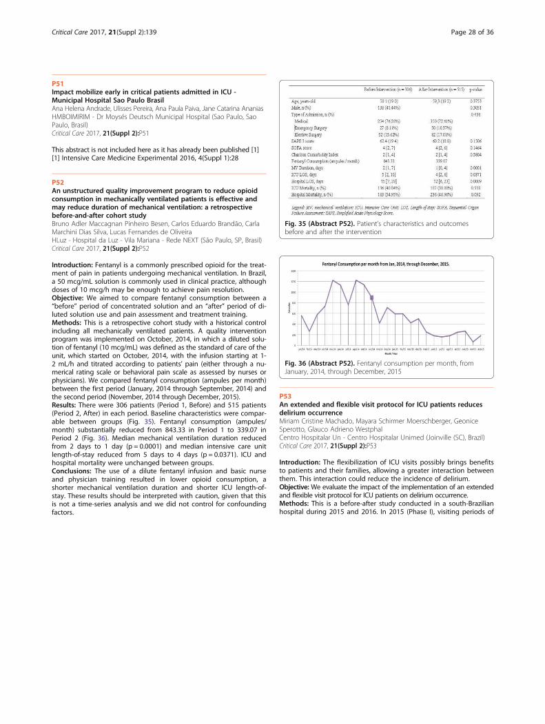
Fig. 35 (Abstract P52). Patient’s characteristics and outcomesbefore and after the intervention
Fig. 36 (Abstract P52). Fentanyl consumption per month, fromJanuary, 2014, through December, 2015
Critical Care 2017, 21(Suppl 2):139 Page 28 of 36
P51Impact mobilize early in critical patients admitted in ICU -Municipal Hospital Sao Paulo BrasilAna Helena Andrade, Ulisses Pereira, Ana Paula Paiva, Jane Catarina AnaniasHMBOIMIRIM - Dr Moysés Deutsch Municipal Hospital (Sao Paulo, SaoPaulo, Brasil)Critical Care 2017, 21(Suppl 2):P51
This abstract is not included here as it has already been published [1][1] Intensive Care Medicine Experimental 2016, 4(Suppl 1):28
P52An unstructured quality improvement program to reduce opioidconsumption in mechanically ventilated patients is effective andmay reduce duration of mechanical ventilation: a retrospectivebefore-and-after cohort studyBruno Adler Maccagnan Pinheiro Besen, Carlos Eduardo Brandão, CarlaMarchini Dias Silva, Lucas Fernandes de OliveiraHLuz - Hospital da Luz - Vila Mariana - Rede NEXT (São Paulo, SP, Brasil)Critical Care 2017, 21(Suppl 2):P52
Introduction: Fentanyl is a commonly prescribed opioid for the treat-ment of pain in patients undergoing mechanical ventilation. In Brazil,a 50 mcg/mL solution is commonly used in clinical practice, althoughdoses of 10 mcg/h may be enough to achieve pain resolution.Objective: We aimed to compare fentanyl consumption between a“before” period of concentrated solution and an “after” period of di-luted solution use and pain assessment and treatment training.Methods: This is a retrospective cohort study with a historical controlincluding all mechanically ventilated patients. A quality interventionprogram was implemented on October, 2014, in which a diluted solu-tion of fentanyl (10 mcg/mL) was defined as the standard of care of theunit, which started on October, 2014, with the infusion starting at 1-2 mL/h and titrated according to patients’ pain (either through a nu-merical rating scale or behavioral pain scale as assessed by nurses orphysicians). We compared fentanyl consumption (ampules per month)between the first period (January, 2014 through September, 2014) andthe second period (November, 2014 through December, 2015).Results: There were 306 patients (Period 1, Before) and 515 patients(Period 2, After) in each period. Baseline characteristics were compar-able between groups (Fig. 35). Fentanyl consumption (ampules/month) substantially reduced from 843.33 in Period 1 to 339.07 inPeriod 2 (Fig. 36). Median mechanical ventilation duration reducedfrom 2 days to 1 day (p = 0.0001) and median intensive care unitlength-of-stay reduced from 5 days to 4 days (p = 0.0371). ICU andhospital mortality were unchanged between groups.Conclusions: The use of a dilute fentanyl infusion and basic nurseand physician training resulted in lower opioid consumption, ashorter mechanical ventilation duration and shorter ICU length-of-stay. These results should be interpreted with caution, given that thisis not a time-series analysis and we did not control for confoundingfactors.
P53An extended and flexible visit protocol for ICU patients reducesdelirium occurrenceMiriam Cristine Machado, Mayara Schirmer Moerschberger, GeoniceSperotto, Glauco Adrieno WestphalCentro Hospitalar Un - Centro Hospitalar Unimed (Joinville (SC), Brazil)Critical Care 2017, 21(Suppl 2):P53
Introduction: The flexibilization of ICU visits possibly brings benefitsto patients and their families, allowing a greater interaction betweenthem. This interaction could reduce the incidence of delirium.Objective: We evaluate the impact of the implementation of an extendedand flexible visit protocol for ICU patients on delirium occurrence.Methods: This is a before-after study conducted in a south-Brazilianhospital during 2015 and 2016. In 2015 (Phase I), visiting periods of
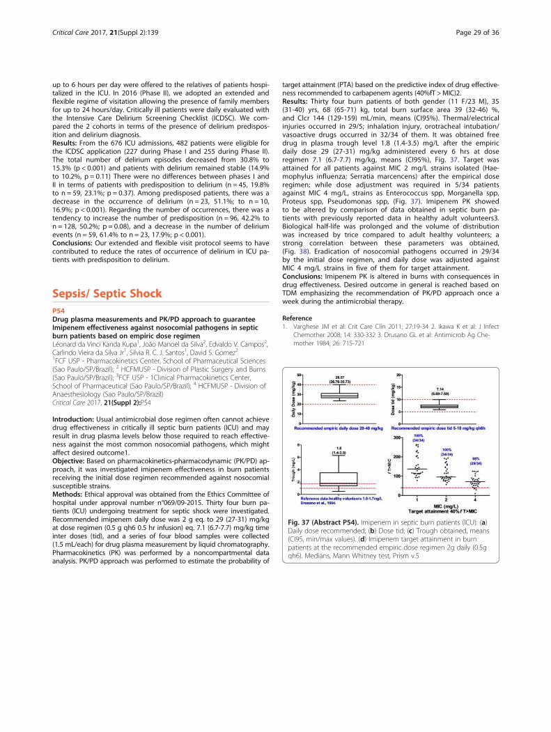
Critical Care 2017, 21(Suppl 2):139 Page 29 of 36
up to 6 hours per day were offered to the relatives of patients hospi-talized in the ICU. In 2016 (Phase II), we adopted an extended andflexible regime of visitation allowing the presence of family membersfor up to 24 hours/day. Critically ill patients were daily evaluated withthe Intensive Care Delirium Screening Checklist (ICDSC). We com-pared the 2 cohorts in terms of the presence of delirium predispos-ition and delirium diagnosis.Results: From the 676 ICU admissions, 482 patients were eligible forthe ICDSC application (227 during Phase I and 255 during Phase II).The total number of delirium episodes decreased from 30.8% to15.3% (p < 0.001) and patients with delirium remained stable (14.9%to 10.2%, p = 0.11) There were no differences between phases I andII in terms of patients with predisposition to delirium (n = 45, 19.8%to n = 59, 23.1%; p = 0.37). Among predisposed patients, there was adecrease in the occurrence of delirium (n = 23, 51.1%; to n = 10,16.9%; p < 0.001). Regarding the number of occurrences, there was atendency to increase the number of predisposition (n = 96, 42.2% ton = 128, 50.2%; p = 0.08), and a decrease in the number of deliriumevents (n = 59, 61.4% to n = 23, 17.9%; p < 0.001).Conclusions: Our extended and flexible visit protocol seems to havecontributed to reduce the rates of occurrence of delirium in ICU pa-tients with predisposition to delirium.
Fig. 37 (Abstract P54). Imipenem in septic burn patients (ICU): (a)Daily dose recommended; (b) Dose tid; (c) Trough obtained, means(CI95, min/max values). (d) Imipenem target attainment in burnpatients at the recommended empiric dose regimen 2g daily (0.5gqh6). Medians, Mann Whitney test, Prism v.5
Sepsis/ Septic ShockP54Drug plasma measurements and PK/PD approach to guaranteeImipenem effectiveness against nosocomial pathogens in septicburn patients based on empiric dose regimenLéonard da Vinci Kanda Kupa1, João Manoel da Silva2, Edvaldo V. Campos2,Carlindo Vieira da Silva Jr1, Silvia R. C. J. Santos1, David S. Gomez21FCF USP - Pharmacokinetics Center, School of Pharmaceutical Sciences(Sao Paulo/SP/Brazil); 2 HCFMUSP - Division of Plastic Surgery and Burns(Sao Paulo/SP/Brazil); 3FCF USP - 1Clinical Pharmacokinetics Center,School of Pharmaceutical (Sao Paulo/SP/Brazil); 4 HCFMUSP - Division ofAnaesthesiology (Sao Paulo/SP/Brazil)Critical Care 2017, 21(Suppl 2):P54
Introduction: Usual antimicrobial dose regimen often cannot achievedrug effectiveness in critically ill septic burn patients (ICU) and mayresult in drug plasma levels below those required to reach effective-ness against the most common nosocomial pathogens, which mightaffect desired outcome1.Objective: Based on pharmacokinetics-pharmacodynamic (PK/PD) ap-proach, it was investigated imipenem effectiveness in burn patientsreceiving the initial dose regimen recommended against nosocomialsusceptible strains.Methods: Ethical approval was obtained from the Ethics Committee ofhospital under approval number n°069/09-2015. Thirty four burn pa-tients (ICU) undergoing treatment for septic shock were investigated.Recommended imipenem daily dose was 2 g eq. to 29 (27-31) mg/kgat dose regimen (0.5 g qh6 0.5 hr infusion) eq. 7.1 (6.7-7.7) mg/kg timeinter doses (tid), and a series of four blood samples were collected(1.5 mL/each) for drug plasma measurement by liquid chromatography.Pharmacokinetics (PK) was performed by a noncompartmental dataanalysis. PK/PD approach was performed to estimate the probability of
target attainment (PTA) based on the predictive index of drug effective-ness recommended to carbapenem agents (40%fT >MIC)2.Results: Thirty four burn patients of both gender (11 F/23 M), 35(31-40) yrs, 68 (65-71) kg, total burn surface area 39 (32-46) %,and Clcr 144 (129-159) mL/min, means (CI95%). Thermal/electricalinjuries occurred in 29/5; inhalation injury, orotracheal intubation/vasoactive drugs occurred in 32/34 of them. It was obtained freedrug in plasma trough level 1.8 (1.4-3.5) mg/L after the empiricdaily dose 29 (27-31) mg/kg administered every 6 hrs at doseregimen 7.1 (6.7-7.7) mg/kg, means (CI95%), Fig. 37. Target wasattained for all patients against MIC 2 mg/L strains isolated (Hae-mophylus influenza; Serratia marcencens) after the empirical doseregimen; while dose adjustment was required in 5/34 patientsagainst MIC 4 mg/L, strains as Enterococcus spp, Morganella spp,Proteus spp, Pseudomonas spp, (Fig. 37). Imipenem PK showedto be altered by comparison of data obtained in septic burn pa-tients with previously reported data in healthy adult volunteers3.Biological half-life was prolonged and the volume of distributionwas increased by trice compared to adult healthy volunteers; astrong correlation between these parameters was obtained,(Fig. 38). Eradication of nosocomial pathogens occurred in 29/34by the initial dose regimen, and daily dose was adjusted againstMIC 4 mg/L strains in five of them for target attainment.Conclusions: Imipenem PK is altered in burns with consequences indrug effectiveness. Desired outcome in general is reached based onTDM emphasizing the recommendation of PK/PD approach once aweek during the antimicrobial therapy.
Reference1. Varghese JM et al: Crit Care Clin 2011; 27:19-34 2. Ikawa K et al: J Infect
Chemother 2008; 14: 330-332 3. Drusano GL et al: Antimicrob Ag Che-mother 1984; 26: 715-721
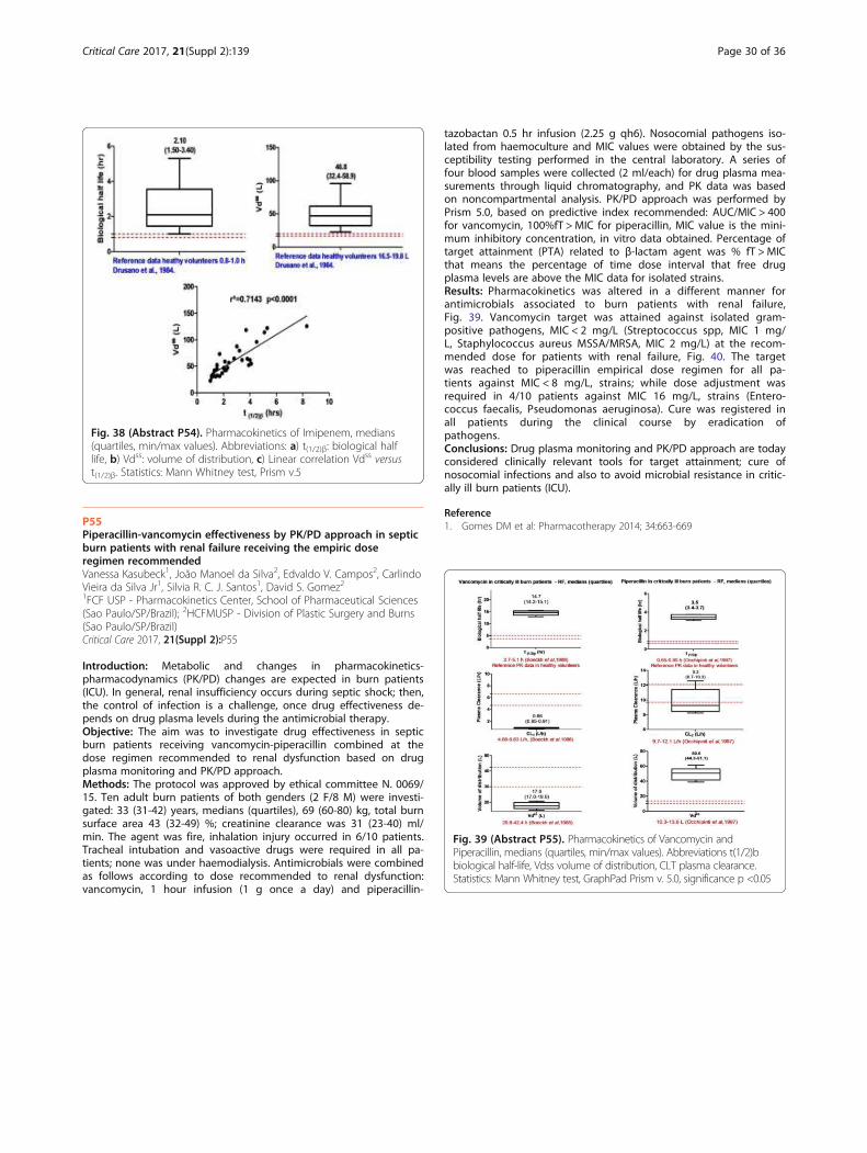
Fig. 39 (Abstract P55). Pharmacokinetics of Vancomycin andPiperacillin, medians (quartiles, min/max values). Abbreviations t(1/2)bbiological half-life, Vdss volume of distribution, CLT plasma clearance.Statistics: Mann Whitney test, GraphPad Prism v. 5.0, significance p <0.05
Fig. 38 (Abstract P54). Pharmacokinetics of Imipenem, medians(quartiles, min/max values). Abbreviations: a) t(1/2)β: biological halflife, b) Vdss: volume of distribution, c) Linear correlation Vdss versust(1/2)β. Statistics: Mann Whitney test, Prism v.5
Critical Care 2017, 21(Suppl 2):139 Page 30 of 36
P55Piperacillin-vancomycin effectiveness by PK/PD approach in septicburn patients with renal failure receiving the empiric doseregimen recommendedVanessa Kasubeck1, João Manoel da Silva2, Edvaldo V. Campos2, CarlindoVieira da Silva Jr1, Silvia R. C. J. Santos1, David S. Gomez21FCF USP - Pharmacokinetics Center, School of Pharmaceutical Sciences(Sao Paulo/SP/Brazil); 2HCFMUSP - Division of Plastic Surgery and Burns(Sao Paulo/SP/Brazil)Critical Care 2017, 21(Suppl 2):P55
Introduction: Metabolic and changes in pharmacokinetics-pharmacodynamics (PK/PD) changes are expected in burn patients(ICU). In general, renal insufficiency occurs during septic shock; then,the control of infection is a challenge, once drug effectiveness de-pends on drug plasma levels during the antimicrobial therapy.Objective: The aim was to investigate drug effectiveness in septicburn patients receiving vancomycin-piperacillin combined at thedose regimen recommended to renal dysfunction based on drugplasma monitoring and PK/PD approach.Methods: The protocol was approved by ethical committee N. 0069/15. Ten adult burn patients of both genders (2 F/8 M) were investi-gated: 33 (31-42) years, medians (quartiles), 69 (60-80) kg, total burnsurface area 43 (32-49) %; creatinine clearance was 31 (23-40) ml/min. The agent was fire, inhalation injury occurred in 6/10 patients.Tracheal intubation and vasoactive drugs were required in all pa-tients; none was under haemodialysis. Antimicrobials were combinedas follows according to dose recommended to renal dysfunction:vancomycin, 1 hour infusion (1 g once a day) and piperacillin-
tazobactan 0.5 hr infusion (2.25 g qh6). Nosocomial pathogens iso-lated from haemoculture and MIC values were obtained by the sus-ceptibility testing performed in the central laboratory. A series offour blood samples were collected (2 ml/each) for drug plasma mea-surements through liquid chromatography, and PK data was basedon noncompartmental analysis. PK/PD approach was performed byPrism 5.0, based on predictive index recommended: AUC/MIC > 400for vancomycin, 100%fT >MIC for piperacillin, MIC value is the mini-mum inhibitory concentration, in vitro data obtained. Percentage oftarget attainment (PTA) related to β-lactam agent was % fT >MICthat means the percentage of time dose interval that free drugplasma levels are above the MIC data for isolated strains.Results: Pharmacokinetics was altered in a different manner forantimicrobials associated to burn patients with renal failure,Fig. 39. Vancomycin target was attained against isolated gram-positive pathogens, MIC < 2 mg/L (Streptococcus spp, MIC 1 mg/L, Staphylococcus aureus MSSA/MRSA, MIC 2 mg/L) at the recom-mended dose for patients with renal failure, Fig. 40. The targetwas reached to piperacillin empirical dose regimen for all pa-tients against MIC < 8 mg/L, strains; while dose adjustment wasrequired in 4/10 patients against MIC 16 mg/L, strains (Entero-coccus faecalis, Pseudomonas aeruginosa). Cure was registered inall patients during the clinical course by eradication ofpathogens.Conclusions: Drug plasma monitoring and PK/PD approach are todayconsidered clinically relevant tools for target attainment; cure ofnosocomial infections and also to avoid microbial resistance in critic-ally ill burn patients (ICU).
Reference1. Gomes DM et al: Pharmacotherapy 2014; 34:663-669
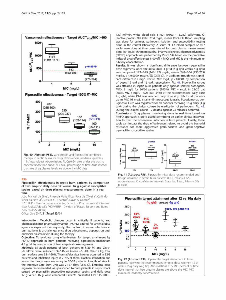
Fig. 41 (Abstract P56). Piperacillin initial dose recommended andtrough obtained in septic burn patients (ICU), means (CI95).Abbreviations: CI confidence intervals. Statistics: T test, Prism v. 5.0,p <0.05
Fig. 40 (Abstract P55). Vancomycin and Piperacillin combinedtherapy in septic burns for drug effectiveness, medians (quartiles,min/max values). Abbreviations AUCss0-24: area under the plasmaconcentration time curve; fT > MIC: percentage of time dose intervalthat free drug plasma levels are above the MIC data
Fig. 42 (Abstract P56). Piperacillin target attainment in burnpatients receiving the recommended empiric dose regimen 12 gversus 16 g daily (4 g tid). Abbreviations: fT > MIC: percent of timedose interval that free drug in plasma are above the MIC, MIC:minimum inhibitory concentration
Critical Care 2017, 21(Suppl 2):139 Page 31 of 36
P56Piperacillin effectiveness in septic burn patients by comparisonof two empiric daily dose 12 versus 16 g against susceptiblestrains based on drug plasma measurements done in a realtimeJoão Manoel da Silva2, Amanda Maria Ribas Rosa de Oliveira2, CarlindoVieira da Silva Jr1, Silvia R. C. J. Santos1, David S. Gomez21FCF USP - Pharmacokinetics Center, School of Pharmaceutical Sciences(Sao Paulo/SP/Brazil); 2HCFMUSP - Division of Plastic Surgery and Burns(Sao Paulo/SP/Brazil)Critical Care 2017, 21(Suppl 2):P56
Introduction: Metabolic changes occur in critically ill patients, andpharmacokinetics-pharmacodynamics (PK/PD) altered for antimicrobialagents is expected. Consequently, the control of severe infections inburn patients is a challenge, once drug effectiveness depends on anti-microbial plasma levels during the therapy.Objective: To evaluate drug effectiveness for target attainment byPK/PD approach in burn patients receiving piperacillin-tazobactam4.5 g tid by comparison of two empirical dose regimens.Methods: 35 adult patients of both genders (6 F/29 M) and Clcr >50 ml/min were included: 39+/-16 yrs (mean +/- SD), 70+/-12 kg, totalburn surface area 33+/-20%. Thermal/electrical injuries occurred in 32/3patients and inhalation injury in 21/35 of them. Tracheal intubation andvasoactive drugs were necessary in 30/35 patients. Length of stay inthe Intensive Care Burn Unit was 21-37 days (95% CI). Empirical doseregimen recommended was prescribed to burn patients in septic shockcaused by piperacillin susceptible nosocomial strains and daily dose12 g versus 16 g were compared. Patients presented Clcr 115 (100 -
130) ml/min, white blood cells 11,601 (9,923 - 13,280) cells/mm3, C-reactive protein 202 (187- 255) mg/L, means (95% CI). Blood samplingwas done for cultures, pathogens isolation and susceptibility testingdone in the central laboratory. A series of 3-4 blood samples (2 mL/each) were done at time dose interval for drug plasma measurementdone by liquid chromatography. Pharmacokinetics-pharmacodynamics(PK/PD) approach was performed by Prism 5.0, based on the predictiveindex of drug effectiveness (100%fT >MIC), and MIC is the minimum in-hibitory concentration.Results: It was shown a significant difference between piperacillindose regimens, once the initial dose 4 g tid (4 g qh8 versus 4 g qh6)was compared: 173+/-29 (163-182) mg/kg versus 248+/-54 (230-265)mg/kg, p < 0.0009, means/SD (95% CI). In addition, trough was signifi-cant different 8.7 mg/L versus 20.2 mg/L, p < 0.0001 by comparisonof doses 12 g/d and 16 g/d, respectively, Fig. 41. Piperacillin targetwas attained in septic burn patients only against isolated pathogens,MIC < 2 mg/L for 26/26 patients (100%), MIC 4 mg/L in 23/26 pat(88%), MIC 8 mg/L 14/26 pat (54%) at the recommended daily dose4 g qh8; while PTA was reached daily dose 4 g qh6 for all patientsup to MIC 16 mg/L, strains (Enterococcus faecalis, Pseudomonas aer-uginosa). Cure was registered for all patients receiving 16 g daily (4 gqh6) during the clinical course by eradication of pathogens, Fig. 42.During the clinical course 12 deaths against 23 releases occurred.Conclusions: Drug plasma monitoring done in real time based onPK/PD approach is quite useful permitting an earlier clinical interven-tion to treat the nosocomial infection in burn patients. Finally, thesetools can impact the drug effectiveness related to avoid the bacterialresistance for more aggressive gram-positive and gram-negativepiperacillin susceptible strains.
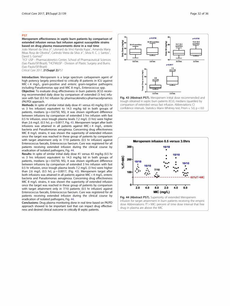
Fig. 43 (Abstract P57). Meropenem initial dose recommended andtrough obtained in septic burn patients (ICU), medians (quartiles) bycomparison of extended versus fast infusion. Abbreviations: CIconfidence intervals. Statistics: Mann Whitney test, Prism v. 5.0, p < 0.0
Fig. 44 (Abstract P57). Superiority of extended Meropeneminfusion for target attainment in burn patients receiving the empiricdose Abbreviations: fT > MIC: percent of time dose interval that freedrug in plasma are above the MIC
Critical Care 2017, 21(Suppl 2):139 Page 32 of 36
P57Meropenem effectiveness in septic burn patients by comparison ofextended infusion versus fast infusion against susceptible strainsbased on drug plasma measurements done in a real timeJoão Manoel da Silva Jr2, Léonard da Vinci Kanda Kupa1, Amanda MariaRibas Rosa de Oliveira2, Carlindo Vieira da Silva Jr1, Silvia R. C. J. Santos1,David S. Gomez21FCF USP - Pharmacokinetics Center, School of Pharmaceutical Sciences(Sao Paulo/SP/Brazil); 2HCFMUSP - Division of Plastic Surgery and Burns(Sao Paulo/SP/Brazil)Critical Care 2017, 21(Suppl 2):P57
Introduction: Meropenem is a large spectrum carbapenem agent ofhigh potency largely prescribed to critically ill patients in ICU againstMIC < 4 mg/L, gram-positive and enteric gram-negative pathogensincluding Pseudomonas spp and MIC 8 mg/L, Enterococcus spp.Objective: To evaluate drug effectiveness in burn patients (ICU) receiv-ing recommended daily dose by comparison of extended (3 hrs) infu-sion with fast (0.5 hr) infusion by pharmacokinetics-pharmacodynamics(PK/PD) approach.Methods: In spite of similar initial daily dose 41 versus 43 mg/kg (0.5 hrvs 3 hrs infusion) equivalent to 14.3 mg/kg tid in both groups ofpatients, medians (p = 0.6750, NS), it was shown significant differencebetween infusions by comparison of extended 3 hrs infusion with fast0.5 hr infusion, once trough plasma levels 7.2 mg/L (3 hrs) were higherthan 2.6 mg/L (0.5 hr), p = 0.0017, Fig. 43. Meropenem target after bothinfusions was attained in all patients against MIC < 4 mg/L, entericbacteria and Pseudomonas aeruginosa. Concerning drug effectivenessMIC 8 mg/L strains, it was shown the superiority of extended infusiononce the target was reached in these group of patients by comparisonwith target attainment only in 7/10 patients (0.5 hr infusion) againstEnterococcus faecalis, Enterococcus faecium. Cure was registered for allpatients receiving extended infusion during the clinical course byeradication of isolated pathogens, Fig. 44.Results: In spite of similar initial daily dose 41 versus 43 mg/kg (0.5 hrvs 3 hrs infusion) equivalent to 14.3 mg/kg tid in both groups ofpatients, medians (p = 0.6750, NS), it was shown significant differencebetween infusions by comparison of extended 3 hrs infusion with fast0.5 hr infusion, once trough plasma levels 7.2 mg/L (3 hrs) were higherthan 2.6 mg/L (0.5 hr), p = 0.0017, (Fig. 43). Meropenem target afterboth infusions was attained in all patients against MIC < 4 mg/L, entericbacteria and Pseudomonas aeruginosa. Concerning drug effectivenessMIC 8 mg/L strains, it was shown the superiority of extended infusiononce the target was reached in these group of patients by comparisonwith target attainment only in 7/10 patients (0.5 hr infusion) againstEnterococcus faecalis, Enterococcus faecium. Cure was registered for allpatients receiving extended infusion during the clinical course byeradication of isolated pathogens, Fig. 44.Conclusions: Drug plasma monitoring done in real time based on PK/PDapproach showed to be important tool that can impact drug effective-ness and desired clinical outcome in critically ill septic patients.
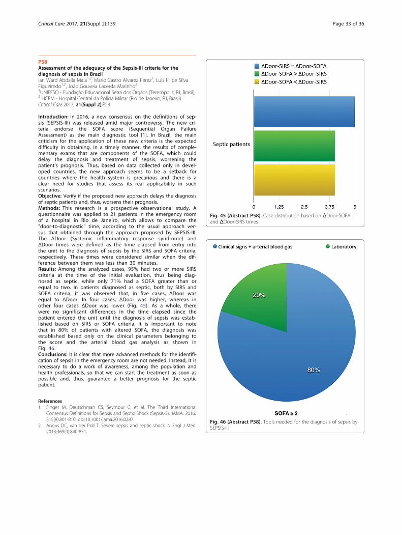
Fig. 45 (Abstract P58). Case distribution based on ΔDoor-SOFAand ΔDoor-SIRS times
Fig. 46 (Abstract P58). Tools needed for the diagnosis of sepsis bySEPSIS-III
Critical Care 2017, 21(Suppl 2):139 Page 33 of 36
P58Assessment of the adequacy of the Sepsis-III criteria for thediagnosis of sepsis in BrazilIan Ward Abdalla Maia1,2, Mario Castro Alvarez Perez1, Luís Filipe SilvaFigueiredo1,2, João Gouveia Lacerda Marinho21UNIFESO - Fundação Educacional Serra dos Órgãos (Teresópolis, RJ, Brasil);2 HCPM - Hospital Central da Polícia Militar (Rio de Janeiro, RJ, Brasil)Critical Care 2017, 21(Suppl 2):P58
Introduction: In 2016, a new consensus on the definitions of sep-sis (SEPSIS-III) was released amid major controversy. The new cri-teria endorse the SOFA score (Sequential Organ FailureAssessment) as the main diagnostic tool [1]. In Brazil, the maincriticism for the application of these new criteria is the expecteddifficulty in obtaining, in a timely manner, the results of comple-mentary exams that are components of the SOFA, which coulddelay the diagnosis and treatment of sepsis, worsening thepatient's prognosis. Thus, based on data collected only in devel-oped countries, the new approach seems to be a setback forcountries where the health system is precarious and there is aclear need for studies that assess its real applicability in suchscenarios.Objective: Verify if the proposed new approach delays the diagnosisof septic patients and, thus, worsens their prognosis.Methods: This research is a prospective observational study. Aquestionnaire was applied to 21 patients in the emergency roomof a hospital in Rio de Janeiro, which allows to compare the"door-to-diagnostic" time, according to the usual approach ver-sus that obtained through the approach proposed by SEPSIS-III.The ΔDoor (Systemic inflammatory response syndrome) andΔDoor times were defined as the time elapsed from entry intothe unit to the diagnosis of sepsis by the SIRS and SOFA criteria,respectively. These times were considered similar when the dif-ference between them was less than 30 minutes.Results: Among the analyzed cases, 95% had two or more SIRScriteria at the time of the initial evaluation, thus being diag-nosed as septic, while only 71% had a SOFA greater than orequal to two. In patients diagnosed as septic, both by SIRS andSOFA criteria, it was observed that, in five cases, ΔDoor wasequal to ΔDoor. In four cases, ΔDoor was higher, whereas inother four cases ΔDoor was lower (Fig. 45). As a whole, therewere no significant differences in the time elapsed since thepatient entered the unit until the diagnosis of sepsis was estab-lished based on SIRS or SOFA criteria. It is important to notethat in 80% of patients with altered SOFA, the diagnosis wasestablished based only on the clinical parameters belonging tothe score and the arterial blood gas analysis as shown inFig. 46.Conclusions: It is clear that more advanced methods for the identifi-cation of sepsis in the emergency room are not needed. Instead, it isnecessary to do a work of awareness, among the population andhealth professionals, so that we can start the treatment as soon aspossible and, thus, guarantee a better prognosis for the septicpatient.
References1. Singer M, Deutschman CS, Seymour C, et al. The Third International
Consensus Definitions for Sepsis and Septic Shock (Sepsis-3). JAMA. 2016;315(8):801-810. doi:10.1001/jama.2016.0287
2. Angus DC, van der Poll T. Severe sepsis and septic shock. N Engl J Med.2013;369(9):840-851.
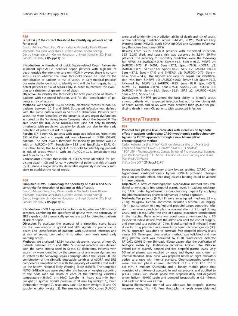
Critical Care 2017, 21(Suppl 2):139 Page 34 of 36
P59Is qSOFA ≥ 2 the correct threshold for identifying patients at riskfor sepsis?Glauco Adriano Westphal, Míriam Cristine Machado, Flávia RibeiroMachado, Maurício Gonçalves, Lucimeri Albino, Álvaro KoenigCentro Hospitalar Un - Centro Hospitalar Unimed (Joinville (SC), Brazil)Critical Care 2017, 21(Suppl 2):P59
Introduction: A threshold of quick Sepsis-related Organ Failure As-sessment (qSOFA) ≥ 2 identifies septic patients with high-risk ofdeath outside the intensive care unit (ICU). However, there is no con-sensus as to whether the same threshold should be used for theidentification of patients at risk of sepsis. In daily medical practice,our main challenge is not to identify who will die from sepsis, but todetect patients at risk of sepsis early, in order to interrupt the evolu-tion to a situation of greater risk of death.Objective: To identify the thresholds for both prediction of death inpatients with suspected infection, and for the identification of pa-tients at risk of sepsis.Methods: We analyzed 18,724 hospital electronic records of non-ICUpatients between 2013 and 2016. Suspected infection was definedwith the same criteria used in Sepsis-3.0 definitions. Patients withsepsis risk were identified by the presence of any organ dysfunction,as stated by the Surviving Sepsis Campaign about the Sepsis-3.0. Thearea under the ROC curve (AUROC) was used not only to identifythresholds with predictive capacity for death, but also for the earlydetection of patients at risk of sepsis.Results: 5,715 non-ICU patients with suspected infection. From them,353 (6.2%) died, and sepsis risk was observed in 3,394 (59.4%)patients. The best qSOFA threshold for mortality prediction was ≥ 2,with an AUROC = 0.71, Sensitivity = 53.8 and Specificity = 83.7). Onthe other hand, the best qSOFA threshold for identifying patientsat risk of sepsis was ≥ 1, with an AUROC = 0.76, Sensitivity = 86.1and Specificity = 52.5.Conclusions: Distinct thresholds of qSOFA were identified for pre-dicting death ( ≥2) and for early detection of patients at risk of sepsis( ≥1). Hence, a single clinically detectable organic dysfunction is suffi-cient to establish the risk of sepsis.
P60Simplified NEWS - Combining the specificity of qSOFA and SIRSsensitivity for detection of patients at risk of sepsisGlauco Adrieno Westphal, Míriam Cristine Machado, Flávia RibeiroMachado, Maurício Gonçalves, Lucimeri Albino, Álvaro KoenigCentro Hospitalar Un - Centro Hospitalar Unimed (Joinville (SC), Brazil)Critical Care 2017, 21(Suppl 2):P60
Introduction: qSOFA appears to be too specific, whereas SIRS is quitesensitive. Combining the specificity of qSOFA with the sensitivity ofSIRS signals could theoretically generate a tool for detecting patientsat risk of sepsis.Objective: To evaluate the accuracy of a simplified score basedon the combination of qSOFA and SIRS signals for prediction ofdeath and identification of patients with suspected infection andat risk of sepsis, comparing it to other commonly used earlywarning scores.Methods: We analyzed 18,724 hospital electronic records of non-ICUpatients between 2013 and 2016. Suspected infection was definedwith the same criteria used in Sepsis-3.0 definitions. Patients withsepsis risk were identified by the presence of any organ dysfunction,as stated by the Surviving Sepsis Campaign about the Sepsis-3.0. Thecombination of the clinically detectable variables of qSOFA and SIRScomposed a simplified score with the majority of variables that makeup the known National Early Warning Score (NEWS). The simplifiedNEWS (S-NEWS) was generated after attribution of weights accordingto the odds ratio for death of each of the following variables:temperature > 38.5oC or < 36oC (weight 1), heart rate > 90 bpm(weight 1), systolic arterial pressure ≤100 mm Hg (weight 1), braindysfunction (weight 2), respiratory rate ≥22 mpm (weight 2) and O2supplementation (weight 2). The area under the ROC curves (AUROC)
were used to identify the prediction ability of death and risk of sepsisof the following prediction scores: S-NEWS, NEWS, Modified EarlyWarning Score (MEWS), quick SOFA (qSOFA) and Systemic Inflamma-tory Response Syndrome (SIRS).Results: From 5,715 non-ICU patients with suspected infection,353 (6.2%) died, and sepsis risk was observed in 3,394 (59.4%)patients. The accuracy for mortality prediction were the followingfor NEWS ≥8 (AUROC = 0.78; Sens = 64.8, Spec = 76.9), MEWS ≥4(AUROC = 0.73, P < 0.001; Sens = 57.2; Spec = 78.5), qSOFA ≥2:(AUROC = 0.71; Sens = 53.8; Spec = 83.7), SIRS ≥2: (AUROC = 0.71;Sens = 87.2, Spec = 17.1) and S-NEWS ≥3: (AUROC = 0.70; Sens =63.4; Spec = 64.2). The highest accuracy for sepsis risk identifica-tion was from S-NEWS ≥2: (AUROC = 0.87; Sens = 81.5; Spec = 79.0),followed by: NEWS ≥5 (AUROC = 0.83; Sens = 82.8, Spec = 65.7),MEWS ≥2 (AUROC = 0.78; Sens = 75.4; Spec = 70.0), qSOFA ≥1:(AUROC = 0.76; Sens = 86.1; Spec = 52.5), SIRS ≥2: (AUROC = 0.69;Sens = 77.7, Spec = 55.4).Conclusions: S-NEWS presented the best ability to identify sepsisamong patients with suspected infection but not for identifying riskof death. MEWS and NEWS were more accurate than qSOFA for pre-dicting death in non-ICU patients with suspected infection.
Surgery/TraumaP61Propofol free plasma level correlates with increases on hypnoticeffect in patients undergoing CABG-hypothermic cardiopulmonarybypass by PK/PD approach through a new bioanalyticalchromatographic methodCarlos Roberto da Silva Filho1, Carlindo Vieira da Silva Jr1, Maria JoséCarvalho Carmona2, David S. Gomez2, Silvia R. C. J. Santos11 FCF USP - Pharmacokinetics Center, School of Pharmaceutical Sciences(Sao Paulo/SP/Brazil), 2HCFMUSP - Division of Plastic Surgery and Burns(Sao Paulo/SP/Brazil)Critical Care 2017, 21(Suppl 2):P61
Introduction: During coronary artery bypass grafting (CABG) underhypothermic cardiopulmonary bypass (CPB-H) profound changesoccur on propofol effect, once drug plasma binding could be alteredin these patients.Objective: A new chromatographic bioanalytical method was vali-dated to investigate free propofol plasma levels in patients undergo-ing CABG under hypothermic cardiopulmonary bypass by applyingthe pharmacokinetics-pharmacodynamics (PK/PD) approach.Methods: Ten patients (9 M/1 F) were investigated, medians: 62 yrs;75 kg; 28 kg/m2. General anesthesia included sufentanil (500 mg.kg-1.h-1), pancuronium (0.1 mg/kg) and propofol target controlled infu-sion to achieve a predicted plasma concentration of 2.0 mg/l duringCABG and 1.0 mg/l after the end of surgical procedure standardizedin the hospital. Brain activity was continuously monitored by a BIS(bispectral index) device from the admission until the end of surgery,and extended to a period of 12 hrs. Blood sampling (3 ml/each) weredone for drug plasma measurements by liquid chromatography (LC).PK/PD approach was done to correlate free propofol plasma levelsversus BIS. Developed bioanalytical method was validated and freedrug plasma level was measured by LC10 fluorescence detectorRF10AXL (276/310 nm) Shimazdu (Kyoto, Japan) after the purification ofbiological matrix by ultrafiltration technique Amicon Ultra MilliporeIreland Ltd to quantify bonded and free propofol plasma levels. Only0.5 ml of plasma was required by assay and thymol was chosen asinternal standard. Daily curve was prepared based on eight calibratorsadded to a tube with internal standard. Chromatographic conditionswere reversed phase column ShimPack CLC – ODS C18 150 x6.0 mm, 5 microns Shimazdu and a binary mobile phase thatconsisted of a mixture of acetonitrile and water-acetic acid acidified topH 4.6 (60:40, v/v). Mobile phase was prepared daily and degassedunder helium (99.9%) strain and pumped isocratically at 0.8 ml/min,and each run time was 25 min.Results: Bioanalytical method was adequate for propofol plasmameasurements, (Fig. 47). Free drug plasma levels were obtained
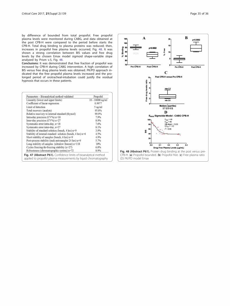
Critical Care 2017, 21(Suppl 2):139 Page 35 of 36
by difference of bounded from total propofol. Free propofolplasma levels were monitored during CABG, and data obtained atthe post CPB-H were compared to the period before starts theCPB-H. Total drug binding to plasma proteins was reduced; then,increases in propofol free plasma levels occurred, Fig. 48. It wasshown a strong correlation between BIS values and free druglevels by the chosen Emax model sigmoid shape-variable slopeanalyzed by Prism v.5, Fig. 48.Conclusions: It was demonstrated that free fraction of propofol wasincreased by CPB-H during CABG intervention. A high correlation ofBIS versus free drug plasma levels was obtained. PK/PD approach in-dicated that the free propofol plasma levels increased and the pro-longed period of orotracheal-intubation could justify the residualhypnosis that occurs in these patients.
Fig. 47 (Abstract P61). Confidence limits of bioanalytical methodapplied to propofol plasma measurements by liquid chromatography
Fig. 48 (Abstract P61). Protein drug binding at the post versus pre-CPB-H. (a) Propofol bounded. (b) Propofol free. (c) Free plasma ratio(D) PK/PD model Emax
Critical Care 2017, 21(Suppl 2):139 Page 36 of 36
P62Trauma patients’ functional outcomes at dischargeRaquel A.C. Eid, José A.D.S Junior, Blenda N.G.M. Caldeira, MiltonSteinman, Karina T. TimenetskyHIAE - Critically Ill Department,Hospital Israelita Albert Einstein (SãoPaulo, SP, Brazil)Critical Care 2017, 21(Suppl 2):P62
Introduction: Trauma patients due to its severity tends to decreasefunctionality during hospital stay. Physiotherapy during hospital staymay minimize this impact. But how these patients return home afterhospital discharge regards to functionality?Objective: To evaluate trauma patient’s functionality at hospitaldischarge.Methods: A retrospective study, through medical chart and a specificprospective database of trauma patients admitted to a private hospital.All patients admitted with trauma diagnosis in the hospital, and thathad information regards to functionality were included. Demographicdata, trauma diagnosis, patients’ severity (through SAPS), mechanicalventilation time, length of hospital stay and functionality were col-lected. We classified functionality in 5 categories: totally independent,modified independence (patient is independent but needs help with aspecific device as walking stick, walker, etc), 25% functionality depend-ence, 50% functionality dependence, and total dependence.Results: 26 trauma patients were admitted to the hospital in 2015and 2016. Of these patients 42% were polytrauma patients, followed
by 27% with polytrauma and brain injury associated, 11.5% firearminjury, 11.5% blunt trauma, 3.8% stab wound, 3.8% brain injury. Mostpatients were male (77%), with median age of 42 years old (range of13-64), mean SAPS of 26 (±4,2), median mechanical ventilation timeof 15 (range of 4-26), median length of hospital stay of 4 days (rangeof 1-48), most patients were sent home after hospital discharge(96%). At hospital admission, 11.5% of patients had independentfunctionality, 30% had 25% functionality dependence, 50% of pa-tients had 50% functionality dependence, and 7.7% had total de-pendence. At hospital discharge most patients had modifiedindependent functionality (34%), 30% were totally independent,19.2% had 25% functionality dependence, 15.4% had 50% functional-ity dependence, and no patients were discharge from the hospitaltotally dependent, showing an improvement of patients’ functional-ity. There was no correlation of functionality with patients’ severity orhospital length of stay.Conclusions: At hospital discharge there was an improvement intrauma patients’ functionality, most patients with modified independ-ent functionality. But some patients are still dependent in some degree,they would probably benefit from post hospital rehabilitation program.Download to read this full article text
Publisher’s NoteSpringer Nature remains neutral with regard to jurisdictional claimsin published maps and institutional affiliations.
• We accept pre-submission inquiries
• Our selector tool helps you to find the most relevant journal
• We provide round the clock customer support
• Convenient online submission
• Thorough peer review
• Inclusion in PubMed and all major indexing services
• Maximum visibility for your research
Submit your manuscript atwww.biomedcentral.com/submit
Submit your next manuscript to BioMed Central and we will help you at every step:
Related Documents